Embed Size (px)
Citation preview

The nature of working memory deficits in aphasia
Jamie MayerIndiana University
Northern Illinois University
Laura MurrayIndiana University
Lyn TurkstraUniversity of Wisconsin-Madison
Bonnie LorenzenIndiana University
ASHA- 11/18/06Miami, FL

The nature of working memory deficits in aphasia
• Aphasia and cognition• WM definition• WM neuroanatomy• WM models• Two hypotheses• One study: WM in aphasia• Clinical implications

Aphasia and cognition
• It has been observed that the language and communication problems in aphasia go beyond simply an impaired linguistic system and involve a complex mixture of cognitive deficits
• Negatively impact: – Functional communication– Social, academic, vocational outcomes– Profit from treatment

Aphasia and cognition
• Twofold goal:– Formulating a more accurate and useful model of
aphasia– Seeking best possible treatment outcomes for our
patients
• “This is an area ripe for investigation as we rightfully move away from the conceptualization of language as being separate from cognition and accept that language is one aspect of cognition” (Helm-Estabrooks, 2002, p. 184)

Co-morbidity of aphasia and higher-level cognitive deficits
• Short-term memory• Attention• Executive function• Working memory
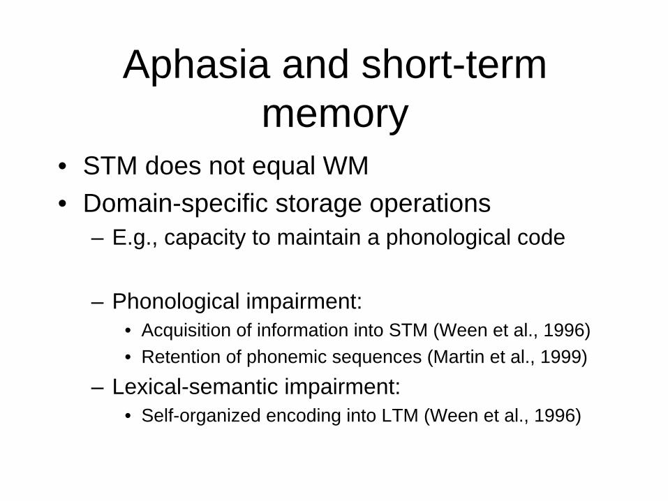
Aphasia and short-term memory
• STM does not equal WM• Domain-specific storage operations
– E.g., capacity to maintain a phonological code
– Phonological impairment:• Acquisition of information into STM (Ween et al., 1996)• Retention of phonemic sequences (Martin et al., 1999)
– Lexical-semantic impairment:• Self-organized encoding into LTM (Ween et al., 1996)

Aphasia and attention
• Definition: – Resource allocation (Kahneman, 1973)
– Aphasia is associated with limited attentional resources, misallocation of attentional resources, or both.
• Sustained attention (Erickson et al., 1996; Laures et al., 2003)
• Divided attention (Tseng et al., 1993; Murray, 1999)
– Misallocation– Failure to appropriately evaluate task demands

Aphasia and executive function
• Covert verbalization– “Although these patients use words as labels,
these words do not function to control their behavior effectively” (Helmquist, 1989, p. 253)
• Non-verbal problem solving– RCPM scores– Relationship to severity of language impairment
• Planning– SAS: necessary for “satisfactory performance of
non-routine tasks” (Shallice, 1982)
• Cognitive flexibility– WCST, TOH (Dunbar & Sussman, 1995; Purdy, 2002)

Aphasia and working memory
• Two approaches:– Domain-specific
• Individuals with aphasia have WM problems to the extent that they suffer from WM impairments specific to language
– Domain-general• Individuals with aphasia have WM problems to the extent that
they suffer from domain-general, executive-processing impairments that affect multiple aspects of cognitive processing, including WM

Aphasia and working memory
• Domain-specific approach– WM for components of interpretation
process (e.g., syntax) (Caplan & Waters, 1999)
– Phonological loop deficits (Beeson et al., 1993; Caspari et al., 1998)
Aphasia and working memory
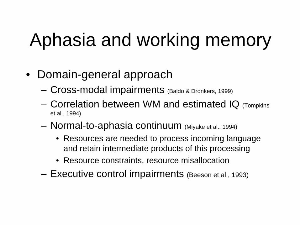
• Domain-general approach– Cross-modal impairments (Baldo & Dronkers, 1999)
– Correlation between WM and estimated IQ (Tompkins et al., 1994)
– Normal-to-aphasia continuum (Miyake et al., 1994)
• Resources are needed to process incoming language and retain intermediate products of this processing
• Resource constraints, resource misallocation
– Executive control impairments (Beeson et al., 1993)

The construct of working memory
“It is quite unlikely that immediate memory evolved for the purpose of allowing an organism to store or rehearse information (such as a phone number) while doing nothing else. Instead, an adaptive immediate memory system would allow the organism to keep task-relevant information active and accessible during the execution of complex cognitive and behavioral tasks. The ‘work’ of immediate memory is to serve an organism’s goals for action” (Engle & Kane, 2004, p. 147).

Neuroanatomy of working memory
• “Standard Model” (Postle, 2006)
– Explicit connections between PFC areas mediating WM and projections from posterior areas
– Exact organizational scheme is not agreed upon

Neuroanatomy of working memory
• WM is neuroanatomically distributed• Involves, at a minimum:
– Pre-frontal cortex– Anterior cingulate– Hippocampal cortex– Posterior sensorimotor cortices

Neuroanatomy of working memory
• PFC activation may be affected by:
– Bottom-up processes: sensory input
– Top-down processes: learning, past experience

Neuroanatomy of working memory
• Neurotransmitter involved = dopamine– DA circuits between the PFC and midbrain
areas may allow PFC to increase activity in excitatory or inhibitory loops to maintain or block information, respectively, as needed
• E.g., signal-to-noise modulator

Neuroanatomy of working memory
• Callicott et al. (1999):– Capacity-constrained response:
• Bilateral DLPFC• “failure to activate one or more key regions during a working
memory challenge” (p. 20)• Functional implications downstream: parietal cortex, premotor
cortex, thalamus– Capacity-unconstrained response:
• Anterior cingulate• Consistent with previous studies implicating this area for
increased effort, attention, or compensation for prefrontal limitations.

Neuroanatomy of working memory
• Domain-specific storage and processing components of WM– Closely linked to neural systems
specialized for perception and action (Postle, 2006; Ranganath, 2006; Smith & Jonides, 1999)

The construct of working memory: Four models
• Multi-component model– Baddeley, 1986
• Resource-sharing view– Daneman & Carpenter, 1980, 1983
• General capacity approach– Engle et al., 1999
• Emergent view– MacDonald & Christiansen, 2002– Goldman-Rakic, 1987, 1993

Multi-component model
• Working memory is storage plus domain-specific processing (Baddeley, 1986)
Central Executive
Phonological loop
Visuospatial sketchpad

Resource-sharing
• Working memory is a unitary system (e.g., Just & Carpenter, 1992)
• Capacity = “The maximum amount of activation available in working memory to support either of the two functions”
Central Executive = Storage + Processing

General capacity approach
• Working memory is executive attention(e.g., Engle et al., 1999)
Executive attention
component processes

Emergent view
• Working memory is tied to domain- specific representations (e.g., MacDonald & Christiansen, 2002)
phonology
Phon WM
Sent comp
Sent comp WM
Spatial ability
Spatial WM
semantics
Semantic WM

Summary: Current status of WM models
• Who’s right?– Four views differ primarily in their
conceptualizations of the source(s) of known individual differences in WM capacity.
– Domain-specific view– Domain-general view

Aphasia and working memory
• Two approaches:– Domain-specific
• Individuals with aphasia have WM problems to the extent that they suffer from WM impairments specific to language
– Domain-general• Individuals with aphasia have WM problems to the extent that
they suffer from domain-general, executive-processing impairments that affect multiple aspects of cognitive processing, including WM

Aphasia and working memory
• Two approaches:– Resource-sharing view, Emergent view
• Individuals with aphasia have WM problems to the extent that they have WM impairments specific to language
– Multi-component model, General capacity view• Individuals with aphasia have WM problems to the extent that
they have impaired domain-general, executive-processing impairments that affect multiple aspects of cognitive processing, including WM

Review and summary
Relationship between aphasia and higher-level cognitive deficits

Domain-specific hypothesis• Primary language difficulties caused by left-
hemisphere damage have a direct impact on other cognitive skills (Buckingham, 1985; De Renzi & Faglioni, 1965)
• Previously used measures of WMC in adults with aphasia have been heavily influenced by the linguistic nature of the WM tasks, OR by covert verbal encoding during “nonlinguistic” task performance (Nystrom et al., 2000; Tompkins et al., 1994)
• Are “domain-general” WM deficits in patients with aphasia an artifact or manifestation of the primary, linguistic deficit?
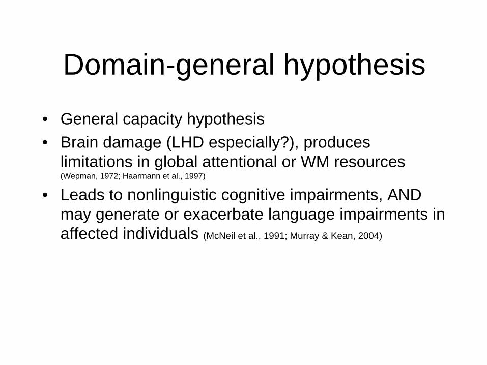
Domain-general hypothesis
• General capacity hypothesis• Brain damage (LHD especially?), produces
limitations in global attentional or WM resources (Wepman, 1972; Haarmann et al., 1997)
• Leads to nonlinguistic cognitive impairments, AND may generate or exacerbate language impairments in affected individuals (McNeil et al., 1991; Murray & Kean, 2004)

Nature of WM in aphasia
• How can we better differentiate between a linguistically-mediated WM deficit and a more general loss of WM capacity in adults with aphasia? – Systematically vary linguistic complexity– Include stimuli which minimize verbal encoding
during WM tasks• Further specification of the proposed
underlying WM deficit in aphasia will considerably strengthen its power as an explanatory factor in aphasia symptomology.

Nature of WM in aphasia
• SO: Given a parametric WM task, will adults with aphasia demonstrate greater sensitivity to systematic variation of: (1) linguistic complexity, or (2) WM load, compared to healthy controls?

Nature of WM in aphasia
• Which WM task?
• How to define linguistic complexity?
• How to define nonlinguistic stimuli?

Behavioral measures of WM
• Span tasks– Verbal span– Operation span– Rotation span

Example Span task:
• Birds can fly. • Babies drive cars.• The sky is blue.

Example Span task:
• What were the last words of each of those sentences?

Problems with span tasks
• Verbal load• Dual task load

Behavioral measures of WM
• Other WM tasks:– SOPT– N-back (parametric WM task)

Nature of WM in aphasia
• Which WM task?
• How to define linguistic complexity?
• How to define nonlinguistic stimuli?
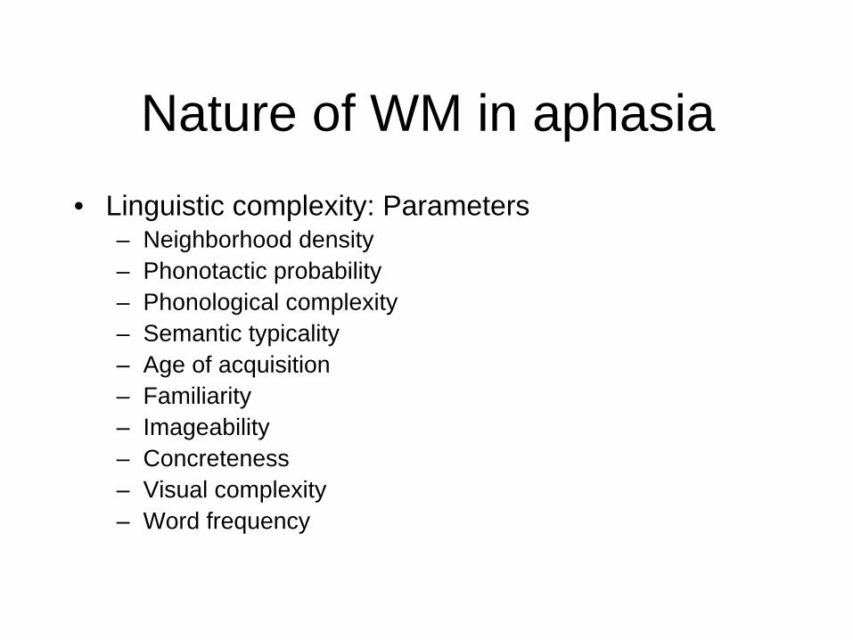
Nature of WM in aphasia• Linguistic complexity: Parameters
– Neighborhood density– Phonotactic probability– Phonological complexity– Semantic typicality– Age of acquisition– Familiarity– Imageability– Concreteness– Visual complexity– Word frequency
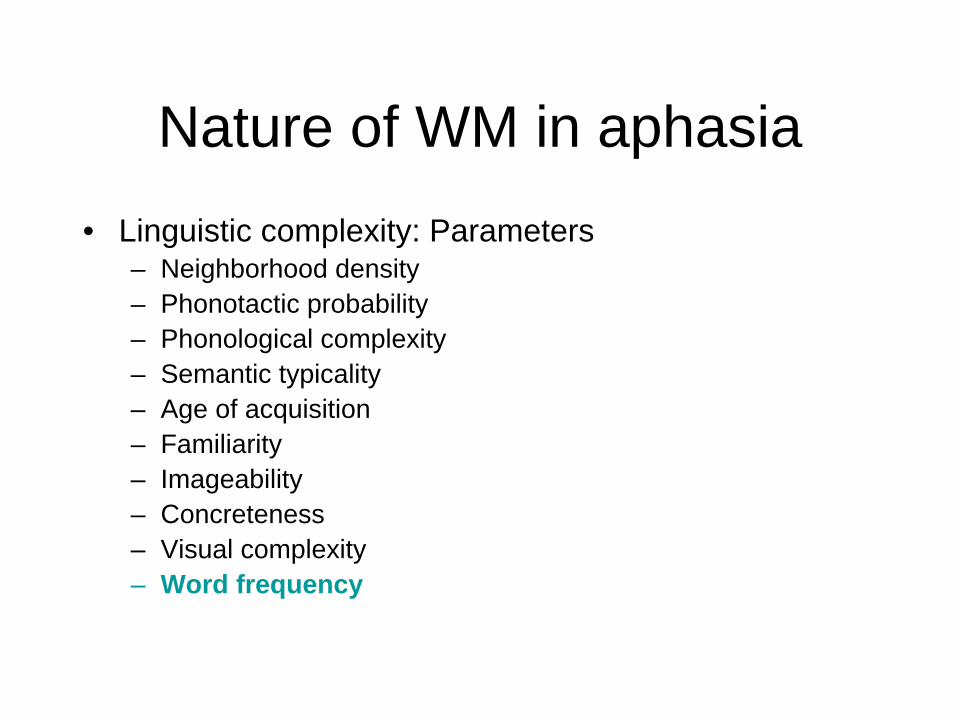
Nature of WM in aphasia• Linguistic complexity: Parameters
– Neighborhood density– Phonotactic probability– Phonological complexity– Semantic typicality– Age of acquisition– Familiarity– Imageability– Concreteness– Visual complexity– Word frequency

Word frequency: Stimuli
QuickTime™ and aTIFF (Uncompressed) decompressor
are needed to see this picture.
QuickTime™ and aTIFF (Uncompressed) decompressor
are needed to see this picture.
(Evans et al., in prep)

Nature of WM in aphasia
• Which WM task?
• How to define linguistic complexity?
• How to define nonlinguistic stimuli?

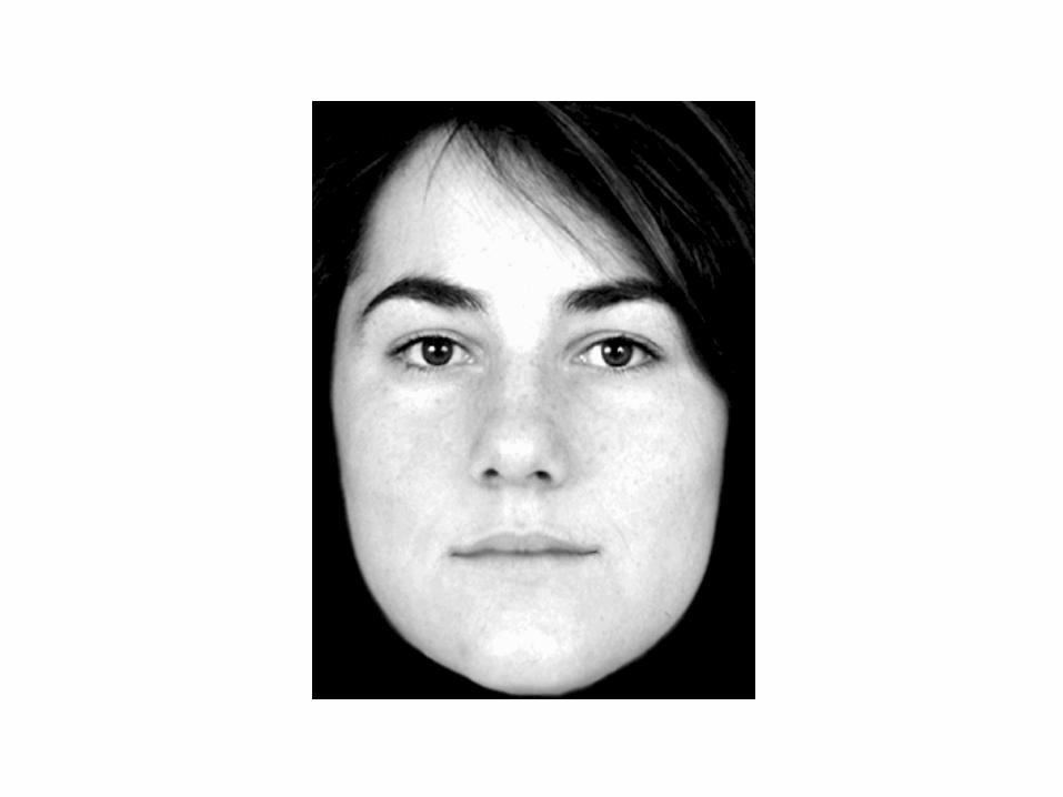
Nature of WM in aphasia
• Non-linguistic stimuli:– Faces– Neutral expression– No identifying features

Nature of WM in aphasia
• Participants– 15 adults with aphasia (LHD)
• Mild-moderate• Fluent-nonfluent
– 10 healthy control subjects (NBD)• Age- and education-matched to LHD group

Nature of WM in aphasia
• Tasks– WAB, RCPM– Picture naming– N-back tasks
• 3 levels of linguistic complexity• 3 levels of WM load
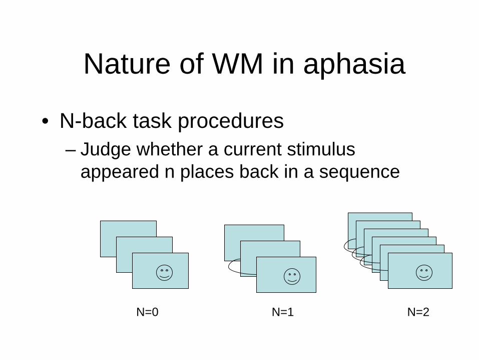
Nature of WM in aphasia
• N-back task procedures– Judge whether a current stimulus
appeared n places back in a sequence
N=0 N=1 N=2

Demo n-back task
See how well you can do!


Nature of WM in aphasia
• Selected results– Picture naming task– N-back task
• Group effects• Language effects• Working memory load effects• Vigilance analysis• Reliability

Picture naming
• NBD: Ceiling• LHD: Frequency
effect– High frequency >>
low frequency
40
45
50
55
60
65
70
High Low
LHDNBD

Group effects
0
0.5
1
1.5
2
2.5
3
PR0
PR1
PR2
LHDNBD
• Signal detection statistic = PR– Hits, misses, false
alarms• LHD << NBD• WM Load
interaction: such that…
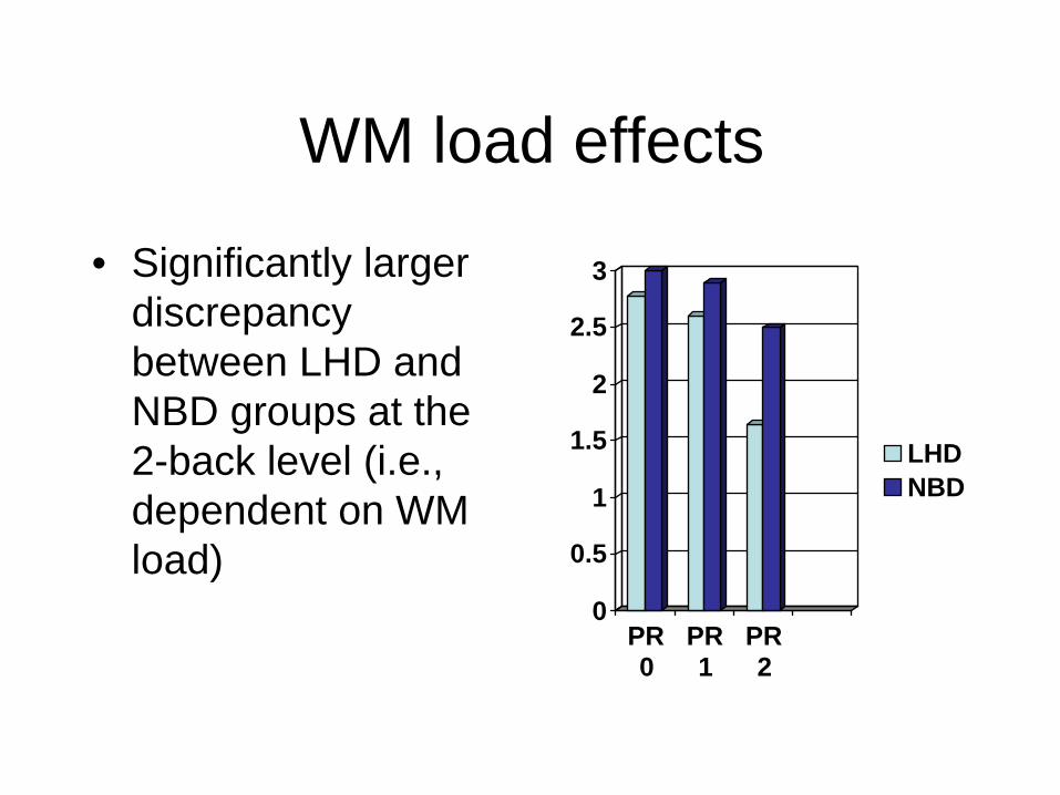
WM load effects
0
0.5
1
1.5
2
2.5
3
PR0
PR1
PR2
LHDNBD
• Significantly larger discrepancy between LHD and NBD groups at the 2-back level (i.e., dependent on WM load)

Language effects
• Effects of language load collapsed across WM condition
• Parallel and flat frequency effect across groups
• Faces << objects 0
0.5
1
1.5
2
2.5
3
High Low Faces
LHDNBD

Vigilance analysis
• LHD group: – Significant task decrement within tasks
• Especially at 2-back level (across language load conditions)
– 0-back: basic sustained attention problems ruled out

Reliability of the n-back task for adults with aphasia
• 25% participants re-tested a minimum of 4 weeks following completion of study protocol
• Test- Retest reliability (PR) = .93• RT retest reliability = .91
Nature of WM in aphasia
Discussion

Summary of results• Expected effects of word frequency elicited in
picture-naming task for LHD adults• Use of same stimuli in visual WM task did not
yield the same effect• LHD<< NBD• LHD: Significant performance decrement
relative to increased WM load ONLY• Both groups: objects >> faces• LHD: Sustained attention effect at highest
WM level• LHD: Reliable performance across repeat
administrations

Objects versus faces
• Recognizable, common objects– Associated representations in long-term
phonological and semantic memory– Subvocal rehearsal
That the LHD group experienced this linguistic advantage to a similar degree as NBD group demonstrates that despite their aphasia, they were able to take advantage of an impaired lexical- semantic network to support WM processes during the n-back tasks.

Low versus high-frequency object names
• Task demands– Lemma versus lexeme access? (e.g., Bock &
Levelt, 1994; Levelt et al., 1991; Levelt, 1999)
• Lemma = concept + syntactic frame• Lexeme = phonological encoding • Item recognition versus confrontation naming
• But what about subvocal rehearsal effects (objects versus faces)?

Vigilance
• Sustained attention problems– Consistent with previous reports
• LaPointe & Erickson, 1991• Laures et al., 2003, 2005
– But: no significant differences between LHD and NBD during 0-back tasks
– Sustained attention (fatigue) played a role only when the task grew more complex (2-back)

Working memory in aphasia
A domain-general phenomenon

A domain-general phenomenon
• Resource view of aphasia– Navon (2004): An operationally defined,
attention-dependent disorder should be “manifested mainly in specific conditions conventionally thought to constrain attention (e.g., high load)” (p. 840)

A domain-general phenomenon
• Broaden our perspective of resource-based disorders in aphasia
• Similar cognitive disorders have been identified in virtually every type of brain damage– RHD (Glosser & Goodglass, 1990)– TBI (Kimberg et al., 1997)– Schizophrenia (Honey & Fletcher, 2006)– Dementia (Baddeley, 2002)– Aging (Salthouse et al., 2003)
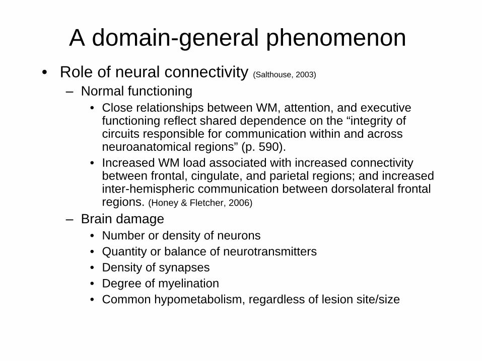
A domain-general phenomenon• Role of neural connectivity (Salthouse, 2003)
– Normal functioning• Close relationships between WM, attention, and executive
functioning reflect shared dependence on the “integrity of circuits responsible for communication within and across neuroanatomical regions” (p. 590).
• Increased WM load associated with increased connectivity between frontal, cingulate, and parietal regions; and increased inter-hemispheric communication between dorsolateral frontal regions. (Honey & Fletcher, 2006)
– Brain damage• Number or density of neurons• Quantity or balance of neurotransmitters• Density of synapses• Degree of myelination• Common hypometabolism, regardless of lesion site/size

Nature of WM in aphasia
Another piece of evidence towards the growing realization that aphasia symptomology cannot be explained on a purely linguistic basis

Nature of WM in aphasia• Clinical implications:
– So now what do we do?
• WM function does not seem to depend purely on linguistic impairment
• But it is problematic for many patients with aphasia• May be part of a larger phenomenon affecting a wide range of
cognitive processing activities
• Assess/treat/monitor separately from (in addition to) language
• Realize functional implications– e.g., expectations for generalization of treated
skills to more complex settings

Nature of WM in aphasia
• Remaining questions:– Effects of manipulating other linguistic
parameters?– Linguistic instantiation of subvocal
rehearsal?– Vigilance versus working memory?– Treatment options?

Selected references• Baddeley (1986). Working memory. New York: Oxford University Press.• Beeson et al. (1993). Brain and Language, 45, 253-275.• Callicott et al. (1999). Cerebral Cortex, 9, 20-26• Caplan & Waters (1999). Behavioral and Brain Sciences, 22, 77-126.• Engle (2002). Current Directions in Psychological Science, 11, 19-23.• Just & Carpenter (1992). Psychological Review, 99, 122-149.• Laures et al. (2003). Aphasiology, 17, 1133-1152.• Levelt et al. (1999). Behavioral and Brain Sciences, 22, 1-75.• MacDonald & Christiansen (2002). Psychological Review, 109, 35-54.• Miyake et al. (1994). Cognitive Neuropsychology, 11, 671-717.• Navon (2004). Aphasiology, 18, 840-843.• Postle (2006). Neuroscience, 139, 23-38.• Salthouse et al. (2003). Journal of Experimental Psychology: General,
132, 566-594.• Tompkins et al. (1994). Journal of Speech and Hearing Research, 37,
896-912.• Ween et al. (1996). Neurology, 47, 795-801.

AcknowledgementsThis research was supported, in part, by:
American Speech-Language-Hearing Foundation (ASHF) New Century Doctoral Scholarship
Indiana University College of Arts and Sciences Dissertation Year Research Fellowship & Bernice Eastwood Covalt Memorial Scholarship
National Institute on Deafness and Other Communication Disorders, Grant RO1-DC03886