Embed Size (px)
Citation preview

(
c
'.' .. 2DOCUMENT RESUME
.
- 2 .ED 213 965 CE 011 7711'
'TITLE. The Mukuldskeletal System [and] Instructor's Guide:The Musculoskeletal System. Health Occupations.
^ Education Module: Instructional Materials in Atttomy.
. and Physiology for Pennsylvania Health Occupations
.. Programs.
INSTITUTION 'National Evaluation Systems, Inc., Amherst, Mass.SPONS AGENCY-, Pennsylvania State Dept. of Education, Harrisburg.
..
Bureau of Vocational and Technical Education,.PUB DATE Jun 79 .
. .
NOTE ". 64p.; for related documents see listing in note of CE031 758. . .
0
EDRS PRICE MF01/PC03 Plus Postage.'DESCRIPTORS *Allied Health OccupatiOns Education; *Anatomy;". Behavioral Objectives; *Individualized Instruction;
*Learning Activities; Learning Modules; Medical-Vocabulary; '*Physiology; Postsecondary Education;
, Pretests Posttests; Programed -Instructional,Materials; Secondary Education; Self Evaluation
- (Individuals).; Teaching MethodsIDENTIFIERS): *Muscular System; Pennsylvania; *Skeletal Systems
4. ,
ABSTRACT4
4
-
04 This Module on the musculoskeletal system is one ofmodules designed tor individualized instruction-in-health
occUpations education progrqts at' both the secondary andpostsecondary levels. It is.,part of an eight-unii miniseries onanatOmy and phySiology within the Apaies of 17 Moddles. Following apreface which explains to the student how to use the module; the unitconsistsof a pretest with afiswer, seven'sections (informationsheets)' with their goals (e.g., classify different -types of bone),optional activities (e.g., on diagrams of the skeleton, draw the:major muscles of the body), and posttests, and a glossary of. terms.Tojics covered .in the unitare introddctiol to the ikeletal systemfaxial skeleton, appendicuta skeleton, introduction tothe muscularsystem, major muscles of the body, Upporti!'tg structures of themRsculoskeletal system, and movements. An accompanying instructor's
Ale contains suggestions for.using,the modttle and. answers. to the,sttesi.KC)
***** ****************************************************** ************Reproductions suppliedbY EDRS are the-best that can be made
* froffi the original document. .
/ **********************************************t***v:**p*****/i**********
ti .4*

0
/HEALTH
OCCUPATIONS EDUCATION
MODULE
4
r
US DEPARTMENT OF EDUCATIONNATIONAL INSTIThlt Of f PUS AVON
PERMISSION TO REPRODUCE THISMATERIAL HAS BEEN GRANTED BY
TO THE EDUCATIONAL RESOURCESINT'ORMATIGN CENTER IERICI
E MUSCULOSKELETAL SYSTEM
..
.
,
0 .
r i
\,
Instructional Materials in Anatomy and Physiologyfor PennsylvaniiHealth Occupations Programs
..
. ,,.
THE MUS'CULOSKELETAL SYSTEM1
d
Prepared for;
1
Research Coordinating Unit for Vocationallucation:..' Pennsylvania Department of Educatio
. Box 911Harriiburg, Pennsylvania 17126
IP
I
. Prepared by:
National Evalu Systems, Inc.30 ehouse Road
Amher Massachusetts 01002Bs
0 ,
A
,
.7.
%
. .
June, 1979 .
, g
Y ,... .e. .
3
4
,

-
PREFACE
V
An understanding of bapic human.anatomy and physiology is essential to anyperson preparing t o e nter a health occupation. This instructional unit is designed (to introduce you to thb-- structures and functions of \the human 'musculoskeletalsystemand the interrelationships of\ the twoand to familiarize you with someof the terms arid concepts necessary for an understanding of the musculoskeletalsystem.
. , 9.This unit consists of a retest- seven modules with their optional activities
and post-tests; and a glossary o terms. "Begin this modular unit by taking the brief pretest at the feint of the,
booklet. The pretest is for your use only__to give you an ideaOf what is includedin this unit, and to give you an indication of the areas within the unit to whichyou should pdy special attention (perhaps by working 'on the optional activities).When you have completed the pretest, tura to the answers in the back (page 52)to check your own score. You will not be graded on the pretest.
Next, read through each of- the modu- les (Introduction to the SkeletalSystem, Aiial Skeleton, Appendicular Skeleton, Introduction to the MurularSystem, Major Muscles of the Body* Supportirig Structures of the MusculoskeletalSystem, and Movements) and investigate any of, the optional activities that maybe helpful, or interesting to you. The optional activities will, help yoi.1 leafs moreabout some of the material presented.,-;
At the end of this unit (page 49) is a glossary which providei-you with brief-definitioras of many' of the terms used in the modules.
Upon completion of each module, you should be able. to *demonstrate' anunderstanding of the material presented, by your`performance on the post-test.When you have finished a module and feel that you understand theUrtforrnation,inthat module, take the post-test that follows it. Write down your answers on, ONEpiece of paper and pass it in to your instructor, w4o will give you your grade:
0
r

#+I
. %
, PRETEST
\
I. Which of the following is a part of the. skeletal system?
e 'A. bloodB. nervesC. muscles'D. bones
S.
0 ,
i.
.1*
i
2 One of the major functions of the skeletal system ie, to:. ,
\ °
A. transport oxygen.B. protect body organs.C. process food.D. regulate body temperature.
ae
!..
3
.
J
3. Another name for the breastbone is the
.
4... The foreiire contains which of the following.pairs of bones?
A. iclavicie and sternum .
B. tibia and fibula .
C. *us and ulnaD. ilium and ischium Apo.
1
...m/.1010
I 1
5. ,All of the movements of the body are produced by what type of tissue? )..
.,
tgs
to,
1c
t'
\
s
t
I
1
4k
$
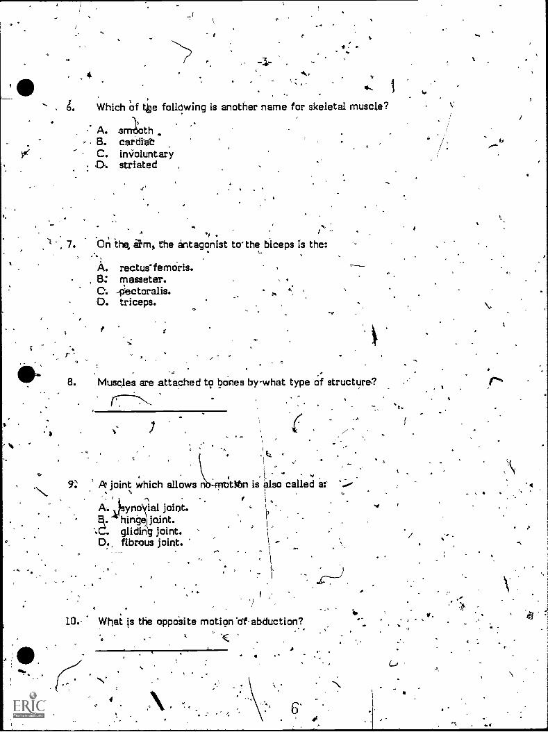
,Which of tbe following is another name for skeletal muscle?
.
dk,
1
*
A. smoothB. cardiabC. in 'VoluntaryDti striated
. ,
7. On the, elm, the antagonist to-the biceps is the:
A
I.
A. rectuefemoris.B: masseter.C. -pactoralis.0. triceps.
As.
8. Muscles are attached to bones bywhat type of structure.?
. Itz .
i ,-
, .. ...
5': A joint which allows tihn is also called a:'
)3 jf
1 ,A. ynoral joint.I?. hinge joint. k).
:C. glidin)g joint.D. fibrous joint.
AV,
.VI- What is the oppoSite motion 'of abduction?
$
.
r'
/
r. .13
4
. /
4
INTRODUCTION TO THE SKI:V:6AL SYSTEM
,
Upon completion of thi, module, you should be able to:'.
1. Identify the major components'and functions of the skeletal system.
2. classify different types Of bone.$. . t
r
THE FUNCTION OF, H SKELETPN .
4
...
.,
. . . e-.The adult human skeleton Is composed of, 206 bones, each serving its own.
distinct function. As 'a whole, the skeleton is like the fremewoik of a building; itsupports and gives shape to the structures around and within it. Individual partsof the skeleton providethe support that maintains 4* fur3ctranil shape of -certainorgans. 'For instance, the thin tissue' of,the lungs would' collapse an itself andlose the Shape necessary for adequate respiration were it not for the attithmentof the lungs to the.bonf ribcage that holds them open.
, . 1 .,
a
- \The structure of bones anti of the skeletal system helps the .skeleton to- .provide protection for many of the body's organs. For example, the hard skullhouses the vulnerable brain; the vertebral column protects the sniffs' cord; andthe ribcage.shields the heart and lungs from injury.
, . -
* Body movements also depend on the 'framewor o oheuse the bones as suppo'rts and levers to produce movement. Its rigid nature, and ,
the arrangement of bon makes the skeletal system's function as a structuralsupport, rather obvious. Less obvious are its -other functions, such as theproduction of erythrocytes (red blood cells) and the storage of minerals. Mostbones have, a diving tare cal d bone marrow, which is classified as either "red" or .
"yellow." Ascii bone marrow I found giboracic (ribs and sternum) and pelvicbones, and in the ends of Ion bones; it is the site of-red blood .cell formation.-yellow bone *marrow, found.1 the shafts of long bones and elsewhere, can in
. emergeociesconveitt.to.red blo d producing\marrow. 'Bones also provide' thebody with ts.minerals. $ubstarices such as magnesium, icalcium, and phosphorus,which are cart of the boeve meter: , are stored until the body needs them.-N.
t,
4E1

CLASSIFICATION OF BONES
-5-
N. \
1
\, \
The many bones of the skeleton range in size and shape from, the tiny bone;atiithe middle ear-to the large heavy-bones of the leg. pifferent kinds of bones
. are classified by ,their shapes' as isig, short, irregular, or flat. Long bones arelonger than they are wide; examples of long bones include, the femur of the thigh, .the .bones of the arm,' and even the phalanges of the fingers. 'The long part of thebone Lathe shaft or diaphysis; the ends of the shaft, called epiphyses, are broaderthan the shaft. Figure shows the structure of 1Png bores:.
ti
,
4,
,
f.
Figure 1. ..The'Structure of l_ong'fi*,161
14Y w'r).
(C.
S.
4-
1
c
-6-4
li Short bones are unifOrmly as broad as they are .wide; an example of a shortbane is oneof the carpals' of the wrist. Irregularbones, such as the vertebrae, ,-have- odd on complex shapes. Flat bones, such as those fqund in theAkull, arebroad plates of bone. I
ARRANGEMENT OF THE, SKELETON y.
t
I
The human- skeleton is a ccnfiguiation or arrangeme9t of these differenttypes of bones, held together by ligaments, tendons, muscles, and cartilage; andjoined together' in junctions called articulations, of joints.
., . I ,
. .
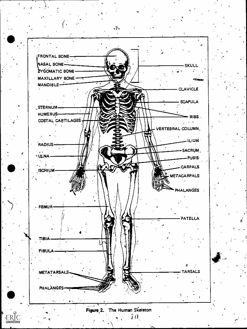
Two different systems make up the entire skeletal: the axial skeleton andthe appendiCPlar skeleton. The axial skeleton consists of the skull, vertebralcolumn, and tibcage; and the appendicular skeleton is composed of the bones ofr the appendages, or extremities (arms and legs).
.Figure. 2 shows a frontal view Of the human skeleton.
.\\. .
a.
-.r4
p
. I .
lo
1,
a,
-7-
FRONTAL BONE_
ASAL BONE
GOMATIC BONE
MAXILLARY BONE ---t
MANDIBLE
STERNUM
HUMERUS
COSTAL CAI3TILAGE
1t
_ .
Ituswilleal
AII I I niX\
,,,..,.-
.,)._
,,,....f.......,_
.--?o7......i, SCAPULA
It:-. 41 Rik.. RIBS
SKULL
CLAVICLE
.1
.RADIUS
VERTEBRAL COLUMN.
ILIUM
SACRUM.
PUBIS-
CARPALS
METACARPALS
PHALANGES
FEMUR
TIBIA
'FIBULA
4
METATARSALS
PHALANGES
.1
PATELLA
TARSALS
_ ,
Figure 2. The Human Skeleton
q.
e,../
-8-
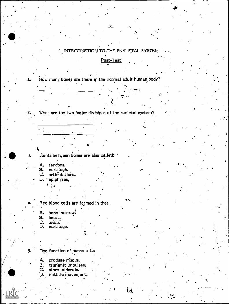
INTRODUCTION TO THE SKELETAL SYSTEM
Post-Test
k
L him many bones are there ip the normal adult human, body?
K.
2. What are the two major divisipns of the skeletal system?
3. Joints between bones are also called:
A. tendons.B. cartilage.C. articulations.D. epiphyses.,
t.
4. Red blood cells are formed in the: ,
A. bone marrow:B. heart.C. brhin:D. cartilage.
5. One function of bones is to:
A. prodtioe Mucus.B. transmit impulses:C. ,store mirierala.ep. initiate movement.
e
,s4
4
p
EIF
/ itt
6
All of the organs .and structures of tfie body are supported.by the:
A. nerves.B. skeleton.C.. -appendages.,D. ligaments.
.. . .
.. 7. Which of the following is classified as a shirt bone?
-I A. carpal
1 '
B. phalanxC. sternumD. humeru.4
8. The vertebrae Ste clas sified as which type of bone?
A., longB. flat'C. shortD. irregular..
4.
ao
The femur is clatsified as what type of bone?
1
10. The diaphysie is what part of along bone?
A. the end° - -
.,B. a projedtion- C. the shaft,-
D. a hole
'2
_
ti
'4
9.

p
Goals
-10-
AXIAL SKELETON
0 ,
0
Upon'completion of-this module, you should be able to:
1. Identify the components of the axial skeleton.
.
O
2. Identify and describe the structure and functions of the xial skeleton.
The axial keleton is the -framework of the trunk. of the body: the head, .
neck, chest, and *per abdomen. This part of the skeletal system serves mainly aprotective functi n, since itenclosed most oaf the vital organs. It also houses theorgans of sight, ell, taste, andhearing, and is responsible for the flexibility of
ovement that allows these organs the fullest "range of exposure to thevironment.
THETHE SKULL
At the top of the axial. skeleton is the skull, which is' made up of the bonesof the head. With one exception, the 29 bones of the skull, articulate in (or areconnected ,by) immovable joints. The joint of the lower-jaw is the only one thatmoves; It allows for the movements necessary for speaking and chewing. In 'the-
, skull, the immovable joints provide strength and rigidity for the protection of kher fragile sensory organs (eyes, ears, mouth, and nose), as well as for the brain.
The skull is divided into two areas. In the front portion are 14 facial boneswhich Ile beneath the flesh of the face. Theie bones forth the inner structures of
,-the mouth and nose. +They also define the 'shape of the facie by forming. the/cheekbones, the arch of the nose, the chiri, and the jaw. The forehead is formedby pait of the larger portion of the skull, the crate.. The eight bones of thecranium, or cranial bones, form the top, sides, and back of the head.
. -
13
t.
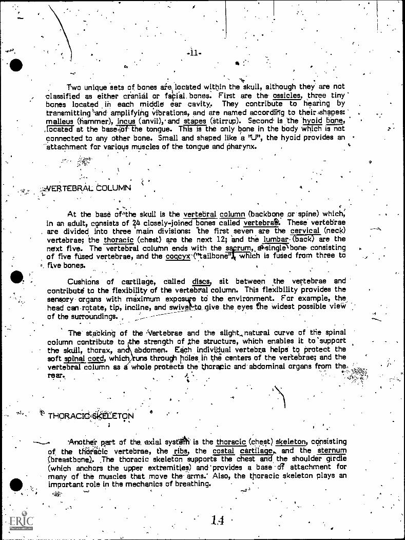
Two unique sets of bones are, located within the skull, although they are notclassified as either cranial or faOial. bones. First are the ossicles, three tinybones located in each middle ear cavity. They contribute to hearing by .
transmitting nand amplifying vibrations, and are named according to their Ashapes:'malleus (hammer), incus (anvi), and stapes (stirrup). Second- is the hyoid bone,RFERFd at the baseTo7The tongue. This is the only bone in the body which is notconnected to any other bone. Small and shaped like a "U", the hyoid provides an
-attachment for various muscles of the tongue and pharynx.
..-WERTEBRAL COLUMN 4
At the base oft he skull is the vertebral column (backbone ,or spine) which;in an adult, consists of 24 closely-joined bones called vertebrae. These vertebraeare divided into three main divisions: he first seven are the cervical (neck)vertebrae; the thoracic (chest) are the next 12; and the lumbar7gicir are thenext five. The vertebral column ends with the saprumolvairiaTbone- consistingof five fUsed vertebrae, and the coccyx-("tailbone"), which is fuded from three tofive bones.
Cushions of cartilage, called discs, sit between the vertebrae andcontribute to the flexibility of the vertebral column. This fleidbility provides thesensory organs with maximum exposure to the environment. For example, thehead can .rotate, tip, incline, and swivetto_give_the eyes the widest possible viewof the surroundings.
The' stacking of the 'Vertebrae and the slight,natural curve of the spinalcolumn contribute to the strength of Pie structure, which enables it to 'supportthe skull, thorax, an abdomen. Each individual vertebra helps to protect thesoft spinal cord, which uns through holes in the centers of the vertebrae; and thevertebral column as whole protects the thoracic and abdominal organs from the,re -
THORACIC-s istLETONe
Another part of the, axial systA is the thoracic (chest) skeleton, consistingof the thiirikiC vertebrae, the ribs, the costal cartilage and the sternumbreastbone). .The thoracic skeleton supports the chest and, shoulder girdle
(which anchors the upper extremities) and 'provides a base dr attachment formany of the muscles that move the arms.' Also, the thoracic skeleton plays animportant role in the mechanics of breathing.
1.4
.
o
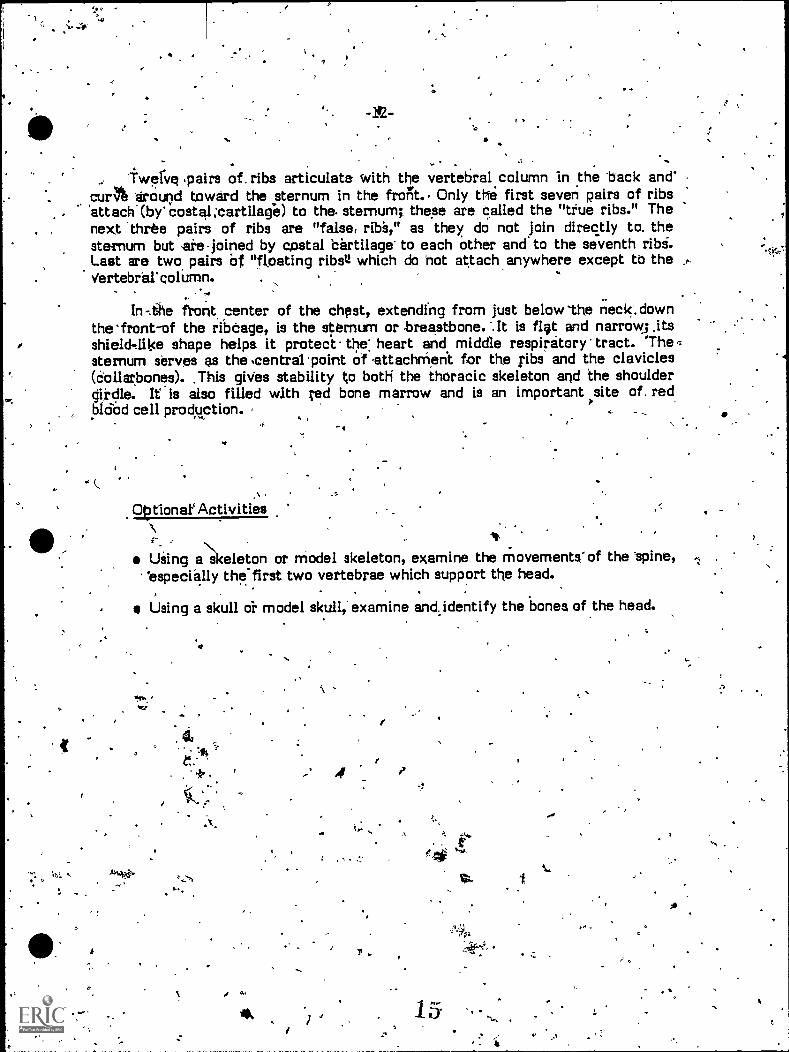
`twelve! .pairs of. ribs articulate with the vertebral column in the 'back andcur 'around toward the sternum in the frorst. Only the first seven pairs of ribs'attach(by'costal;cartilage) to the. sternum; these are called the "true ribs." Thenext 'three pairs of ribs are "false, rib's," as they do not join directly to. thesternum but ere .joined by costal cartilage' to each other and to the seventh ribi.Last are two pairs of "floating ribsQ which do not attach anywhere except to theVertebrecolUmn. ,
In-.the front center of the chest, extending from just below -the neck'. downthe'front-of the ribcage, is the sternum or -breastbone. -.It is flit and narrow; itsshielthlike shape helps it protect the: heart and middle respiratory tract. 'The.sternum serves as the ,central 'point of -attachrhent for the ribs and the clavicles(collarbones). .This gives stability to both the thoracic skeleton and the shouldergirdle. It is also filled with red bone marrow and is an important site of. redbldod cell production.
OtAlonar Activities
fa.
Using akeleton or model skeleton, examine the m ovements-of the Spine,'especially the first two vertebrae which support the head.
Using a skull dr model skull,' examine and identify the bones of the head.
+ ' .41
ar
1I
Cy w
1
1.
,
ti
O
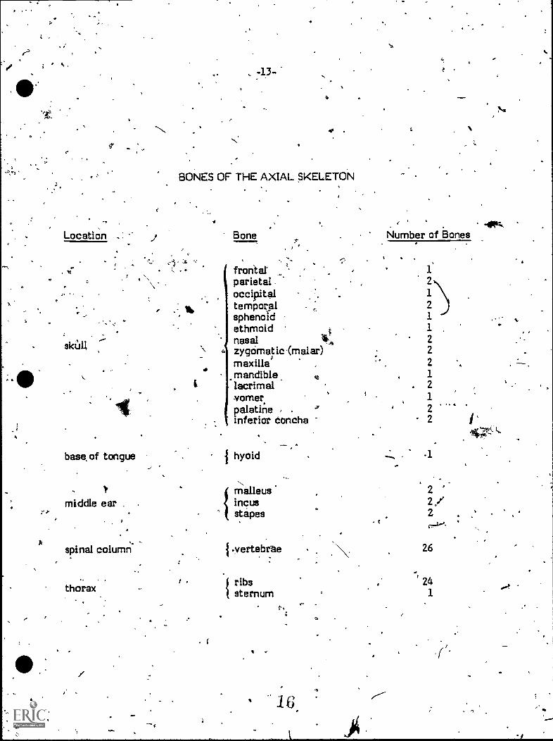
Location
skull
:13-1
BONES OF THE AXIAL SKELETON
.1
Bone
frontal'parietaloccipitaltemporalsphenoidethmoidnasalzygomatic(malar)maxillamandible, sa.
lacrimalvomer,palatineinferior Concha
{
base. of tongue 1 hyoid
middle ear
spinal column,
thorax.
malleusincusstapes
I .vertebrae. _
J ribssternum
16,
Number of Bones
t
1
2
1
21
1
22
2
12
1
22
14Z.
-1
22/2
26
241
4

V
.,1. . Which of the following is part of the axial skeleton?
A. pha'anges'B. skull t r
C. tibiaD. humerus
6- 0
AXIAL SKELETON
Post-Testi
a
2. The only bone in the body wh ch does not articulate with another bone)-
is the
3. Name the five divisions of the eptebral column,
4. The tin ones of the middle ear are called the:
A. 'hyoid.B. ossicles.
_ ,C. vertebrae.- D. clavicles.
17
r
I
1.15-
\.::N 4
.4
. : , ... 5. What are the two major divisions of the skull? , ,.
t . i, rj
li
__ ,_. , ': r -.,;',(Ak
,`-\\,K I ' 't. ,-:,6. Orie of the nrjor function0 fAthe viitebral column is to:
1 -,, 1 , , 4
..%, . .A.. protect the spinal Cord. .. F.1
.8. articulate with the arms. ./ '
*P, ' Co aamplify vibrationi.: ',,,"/ - . .1 i',. D. support the lungs. $ .
. .
.7. What are the 12 peirs of
Ibones that protect the chest cavity? .
8. What is the primary function of the skull?.
A. 'movementB. thoughtC. support-D. ,protection
4
9. Which of the following is, tin important site of red blood cell production?
A. hyoidB. costal cartilageC:' sternumD.' stapes
O

Goals
-16-
APPENDICULAR SKELETON
car
Upon completion of this module; -you should be able to:
(
1. 'Identify the components of the appendicular skeleton.
2. Identify and describe theKstrueture and functions of the appendicularskeleton.
Providin the- body with structures for movement is the appendicularskeleton, whi is made .up -of the bones of the appendaces-(,eirrhs and legs) andthe rings of b ne tot, which they are attached: the, shoulder girdle and;the pelvic,girdle.
. PECTORAL GIRDLE I
. 'The pectoril (shoulder) girdle- serves as the bony 'foundation for theattachment of the arms to the body., It rides over the 'thorax (chest), anchoredonly_by two' bohe-to-bone articulations and the muscle' of the thOrax. This looseattachment does ,not prbvide foLgreat statttlity, or strength, but it does giveflexibility of movement and a great range of. motion to, the upper extremities.Anterior (front) components of the sholilder, girdle are the two -clavicles(collarbones), small long bones that curve slightly from the shoulder to the topedge of the sternum. The-Clavicles hold the shoulder joints out to the sides ofthe body. The sternum is the. only 'bony connection betvieen the axial skeletonand the shoulder girdle., The tip of each clavicle joins a scapula' or ; shoulderblade, which farms' the sooket where the upper `arm attaches. The scapulae do-not articulate at all with the ribcage. The scapt4lae are broad flat bones,
) triangular shape, pointing dokynward over the true ribs about two inches fromthe spinal column. They slide over the posterior (back) of the thorax, buried andbustiioned in large masses of muscle. '
. :
4 .

, -17-
UPPER EXTREMITIES - N
s. ;. , , . . ,4
. Each arm is composed of 30 bones. The humerus, the upper arm bcine, isthe bone that joins he scapula at the shoulder. In the forearm are two bones;the radius and the ulna. The ulna is on the little-finger side Of the Vrearni, and
,-
connects with the humerus to form the elbow. On the thUmb side is. the radius,which,rotates with the ulna to 'turn the palm of the hand up and down. Eightsmall bones called carpals form the wrist. They are arranged in two rows, and ,their placement allows for a wide variety
themovements. Five metacarpals form .
the hand. These bbnes can be felt along the back of the hand and thumb wherethey join the bones of the, fingers or phalanges. Each finger is made up of threephalanges (except the thumb,which has two) that articulate at the.knuckles.
5
LOWER EXTREMITIES ,
,lower extremities attach to the pelvis, -or pelvic girdle; a healvy set .ofbones that forms the lower wall of the abdomen. Iq the peliis are three areas ofbone called the ilium, the ischium, and the pubis. Firsts the ilium; which formsthe wing-like structures that are, commonly termed the, "hipbones." It connectsto the flasacrum in back, in immovable joints that add the sturdin'eas of thepelvisi,to Me sides, the ilium- forms part of the.socket orEbe hip. Second is,tthe
which .curves under to form two arch-like projections that bear thewet t Cf the body,owhen sitting, The ischium,eheets\ehe to foam the SecoridRent' of the hip socket. Third, the remaining part of the socket is formed by thepUbis, the front portion of the
'This bony ticket, formed by the connection of the ilium, ittchiuMI, andpubis, le the -point of articulation with the leg. It is 'comparatively deep, a'h?d the
bones:Composing it are thick and heavy; this adds to the arability of.theleg-pelvis joint, which 'beard the weight of the body kthe same time. th.itallows forfreedom of.movement.
.
'Connected to the pelvis the, femur of the upper leg, the longest bone inthe body. Like other long bones (anci especially like. the hutherus), the femur hasa long shaft .broadening out at both ends. One end of the femur forms a heavyball, which fits very - closely into the sgaket of the pelvis; the other end widens toform the knee joint.
)r
The patella or kneecap is a short bone which rides over,and ptotects theknee joint. It articulates with the femur but is fairly loose; like the scapula, the
ipatella is buried in muscle, which gives it the freedom of Movement necessary toglide over the knee joint regardless of the' position of the joint.
1
f
l
1 The knee is also formed by,the' tibia, or shinbone. /Along with the fibula,the tibia forrls the'lower ieg,'and both bones join to form the anklte:'
.... .
1(\ There they articulate with the -talus, one of the seven short bones of thefoot called tarbals. The tarsals correspond to the carpals of the wrist. The restof the tarsal bones help distribute the weight of the body throughout_ the.foot;the tarsals form .the heel, which bears some of the Weight, and `the arches of the -t.,
foot, which distribute 'weight forward to the metatarsals. These are sislilar tothe metacarpals of the hand in that they provide the framewprk of the foot; 'this
4 is, . however; a weight-bearing instead of a manipulatim0 'fraDlOork. Themetatarsals are therefore heavier,' especially fhe first metatarsal (joining. with -.
the 'great toe), which:.bears much of the body's forward weight. The closearticulation of tarsals and-metatarsals helps distribute the weight of the body as
,equally as, possible to ail parts of the feet:, to the phalanges of the hared(except that they- are shatter and, broader) phalanges of.the foot; which
. form'the toes. -. . .
c
k.
I
The -upper and lower extremities in general are very 'similar in ,terms ofnumbers and- arrangements (and even-shapes) of .bones;' however,' the actual
,structUres of these appendages 'differ according to their functions: :The bones ofthe 'arms are relatively loosely attached to the body and otherwiseiltructured fox',the greatest possible freedoreof 'movement. "This serves the, arm &manipulative'
k.function. The bones pf the _leg articulate closely with the heavy pelvis, and are
',Themselves quite heavy; they enjoy a' wide range of motion but not- so varied asthat of the arm. This- suits the-weight-bearing and locomotive ,functions of the
- legs. . ./A I F
_I
a
,S
Optional Activity.
-
r
*
. .
Identify the bones of the appendages on a skeleton or model skeleton.t.
-It
21. .04-541,1
sat
4
a a .
tsr
/ . . :01.
c -
A
a
.7.
BONES OF THE APPENIDICULAR SKELETON-,
Upper Extremities
'Location Bone
'shoulder girdle
upper arm
forearm-.-
1.
arkals
A
claviclescapula
1humerus
radius k'1 ulna
lunatetriangular (triquetrum)
-hamatecapitate,pisiformscaphoid (navicular)trapezium
trarzoid," ......
hand , 1 metacarpals
11fingers/thumbs
'1
1
1
1 phalanges.
.r42
9
ti'
o 4`1"
Nutnber of Bones
+A
a
22
2 '""
o
''28
440
r
a
LoCaticint
hip.
upper leg
i.
kneecap.
tarsals,
foot
toes.
0
4.-20- as
BONES OF THE APPENDICULAR'SKELETON
I phalanges
4
taivrer.ExEremitiei
I
Bone
;.pelvic bone
femur
patella ,
tibiafibula
taluscalcaneuscuboidnavicularcuneiform I
, cuneiform IIcuneiform 111
Number of Bones
2
2
22-2
222
-s.-.4 metatarsal, .: 10
.. ) ., 1
28
J.
9
42 3
J.

A
5
-21-. %
APRENDICULAR SKELETON
Post-Test =
47P
7 /'' r
1. The appendicular skeleton includes which of the following?
A. vertebraeB. femur' 4
C. ribsD. cranium,
2. The shoulder girdle is also called the
THe bone of the Upper arm is the:.
A. humerus.B. sternum.C. femur..0. tibia.
L
4 The lower 'extremities attach to-the:,
A. sternurriTB. vertebral column.
f. 1 C. scapula. \
D. pelvis.
'
®.
2.4
s.
1,
AI/I
*e,
5. The eight small bones othe wrist are called:
B. carpals.C. vertebrae.D. tarsals. .
-22-
-\
6.0 The kneecapis also called the
f
'7. The foot is designed primarilx to:-
A. move upward toward the knee:B: protect vital orgart
IF !C. articulate with the pelvis.'D. bear the weight of the 1dy.
x '
The strOct Ures of the appendicular s eleton allow the body to:
O
fr
A. ,.breathe..B. .see.C. move.D. heat. *
1
46
'9. The primary function of the patella is to:
, A. protect the kriee.
f B. initiate movement./ C. 'support muscles./ 0 produce blood cells.
IMO
25
is-
I
a
-23--,
41( "o-
10. Which of the following articulates with the femur?
(
A. scapula.B: vertebra.C. pelvic,girdle.D. .
/
V
A
; t. , ; . . 4. . '',. . .
,
'7
.4
-e
0
4
4
ti
-24-
INTRODUCTION TO THE MUSCULAR SYSTEM
Goals
Upon completion of this module, you `should be able to:
1. 'identify the major components and functions of the ltiscular system.
2. ClaSsify different types of muscle,.
.
quo
Without muscles, the respiratory system could not take in air, the digestivesystem could not process food,' and the circulatory system could not pufnp blood.Without muscle, the skeletal system would-be like the poles in a tent._ However, _the Cody is packed with musclealmost one-half of total body 3eveight is made upof muscle.
FUNCTIONS OF MUSCLE2,
16, .
IslusCle Tissue is aspecial,type of tissue made up of layers of very king, thincells. These cells have the ability to contract, or become 'shorter, causing theentire tissue .to become shorter. If the muscle is attached to bones, thecontraction causes the bimes to move; if the muscle surrounds an -organ, thecontraction may move the contents of the organ. MOvement is the primaryfunction of muscle, whether jt is movement of the body (as in walking or dancing)o movement of 'materials within the body (for instance, food thiough thedigestive system or blood through the Pirculatory system).
,'Contraction cif muscles requires energy. Not all of ihis energy is used toproduce movement,hovever; about three-fourths of the muscular energy goes toproducing heat-7a very important function of muscle. Hdw muscular actioncontributes to heat production is' probably apparent to 'anyone ekho has exercised'heavily on a warm day, or to anyone who has stamped feet or paced around tctkeep warm while waiting out in the cold. Even .shivering is: an attempt by thebody to 'eroduce more heat- by raising the. level df muscular activity throughoutthe bodyll' Muscular activity-is the body's major inechanisni for heat production./ ,
r.
Irv:tering of structure, the skeletal system would contribute very little tothe shape and support of the body if it were not for muscle. To return to thetent 'pole comparison, the poles will.. not support, the tent-it something (usuallyrope)Ns not holding them in the necessary 'pOsitions of support. The same is truefor the bones in the l?oclyt. Muscles exert the necessary pull to keep' the bones ingooclAgnment, pep of support. The alignment of the body is'-cilleickistute.:Main:tainingposture is another impor tant function of muscle.
.01
TPES 'OF MUSCLE
Muscular functions are performed by three types of muscle: 'smoothmuscle, cardiac muscle, and skeletallUi#14. Smooth muscle is the simplest typeof muscle tissue. It is made up df eldrigiitted cells,arranged in sheets, which 'formthe walls of many of the' Internal organs or viScera.. (For this reason, smoothmuscle is often called viscerip, muscle.),'SmoothffitiaCle is found in the holloworgans of the digestive system (esophagus,- stomach, intestine) as well as in thewalls of blood vesselsthe bladder, the uterus, in glands, and in the skin.
. Smooth muscle contraction 'is involuntary. That is, it is not consciouslycontrolled; the complexity of the functions performed by smOOMmuscle wouldoccupy the mind tomPletely if the actions of the'MUscle had-to be 'controlledconsciously. Smooth muscle causes the movement of, materials within thesmooth-muscled organs. Thus it is responsible for the movement of food throughthe digestive system, elimination of waste from the body, and,so on. .
.. Cardiac (heart) muscle Is also, involuntary muscle, although it has a very
different function. Composed of elongated muscle cells or fibers which branchapart and rejoin thrdughout the tissue, cardiac muscle (also called myocardium)is the tissue- responsible for the beating action of the heart. When the musclecontracts, the heart itself contracts, pausing a, heartbeat. Circulation of blood .
would not be possible without this type of muscle..
.Unlike cardiac and smooth muscle, skeletal muscle is voluntary; that is,' Itsaction can be controlled consciously. This is the muscle attached to, andresponsible for, the movement of the skeletal system. Skeletal muscle is madeup of long, cylindrical cells Jined up in strands or fibers. These fibers, whenviewed through a microscope, are cross-banded with light` and dark stripes, orstriations. (For this reason, skeletal muscle is also called, striated 'muscle.) Thestrands of muscle tissue are arranged in bundles that)fotiri the skeletal muscled.
0
° 7
d
.11Z'
MUSCLE ACTION
-26:
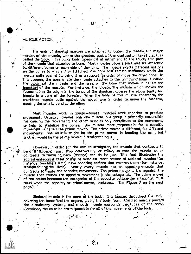
The ends of skeletal, muscles are attached. to bones; the middle and major',portion.of 'the muscle, where the greatest part .of the contraction takes place, iscalled the body. This bulky body tapers off at either end to the tough, thin partof the muscle that attaches to bone: Most muscles cross a joint and are attachedto. different bones on each side of the' joint. The muscle exerts different actionsone the bones. to which it is attached; one bone will remain stationary while themuscle pulls against it, using it as a support, n order to move the 'other bone. Inthis process, the area. where the muscle attac es to the unmoving" bone is calledthe origin of the muscle and the area on he bone that moves is called theInsertion of the muscle. For instance, the 'cep's, 'the muscle which moves theforearm, has its origin in the bones of the oulder, crosses, the elbow joint, andInserts-in a bone of the forearm. When the ody of this muscle contracts,.,theshortened muscle pulls againit the upper arm in order to, move the forearm,causing the arm to bend at the elbow.
Most "muscles work in groupsseveral muscleS work *together to producemovement. Usually, however, only e muscle in a group is primarily responsiblefor causing the movement; the of muscles only contribute to the movement,or help to stabilize the, bones. he muscle most responsible for a specificmovement is called the prime mov r. The prime mover is different for differentmovements: one muscle might b the prime mover in bending'the arm, but.)'another would be the prime mover'i straightening it.
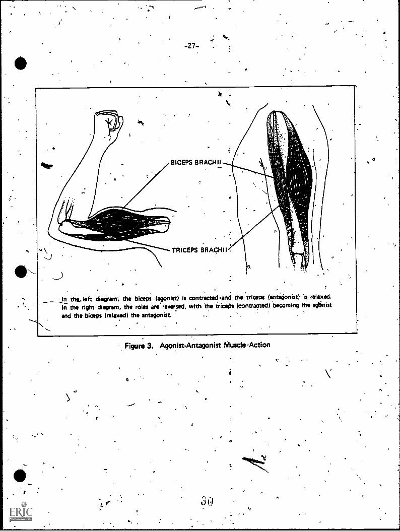
However; in order for the arm to straighten, the muscle that contracts to, 50- bend '1t' (-biceps), must stop contracting, or relax, so that the ,muscle which
contracts to 'move its deck (triceps) can do its job. This fact 'illustrates theagonist-antagonist rerationship of muscles: most actions of skeletal muscles (for,instance, bending a limb) have .oppositig actions that reverse, them (for instance,straightenin%gthe limb). Nearly every muscle has an opposing muscle thatcontracts to Sause the opposite movement. The, prime mover is the agonist; themuscle that causes the opposite movement is, the antagonist. The prime moverof one action becomes the antagonist of the opposite action the antagonist mustrelax when the agonist, or prime; mover, contracts. (See Figure 3 on the nextpage.)
Skeletal muscle is the meat of the body. It is lOcated throughout The body,covering the bones Sand the organs, gRting' the' body form. Cardiac muscle powersthe circulatory system, and smooth muscle surrounds they,tubes of the body.Combined, the muscles are- responsible for all of the movements of the body.
O_
-27-
rt
BICEPS BRACHII
TRICEPS BRACHII
In the...left diagram) the biceps (monist) is contractediand the triceps ( antagonist) is relaxed.
In the right diagram, the roles are'reversed, with .the triceps (contracted) becoming the agtinist
and the biceps (relaxed) the antagonist. ,
At
Figure 3. Agonist-Antagonist Muscle-Action
v.
a

Optional Activities
-28-
Find muscle masses on yourself. .01 tench fists,' point toes, raise limbs, andbend joints. Feel the areas where the muscles are contracted.
Try to end agonist-antagonist muscle- groups. Perform an action, asingle movement, and then figure and perform the opposing action. Canyou isolate the groups of muscle by feeling them?
4110
4
,/
. ,
0
31:

4,
INTRODUCTION TO.,THE MUSCULAR SYSTEM.,
. Post-Test
1. Muscle cell's have the ability to:
\ A. produce hormones.B. ingest other cells.C. produce borke cells,D. contract. \
t
2. One of the major functiths of the muscular system is to:
A. transport nutrients.'B. produte heat.'C. protect the brain.D. support the intestine.
\3. The muscle most responsible for aparticular action is called the:
4.
A. antagonist.B. -prime, mover.C. stabilizer.D. goductIr.
tt
. IThe ares', where the two ends of a muscle attach are the
A: agonist and antagonist.B. fl*xor and extensor.
* C. origin and insertion.D. tendon and ligament.
32ra
,1^
f
a
"Ik
-30-.10
5. The major portion - of a muscle, where most of the contraction occurs, iscalled the.
6. muscle is also called
I
7, Visceral muscle is also known as
41.
ti
..8. Which of the following types of muscle is usually consciously controlled?
A: skeletal.B. smoothC. visceralD. cardidc
t
t
c3
it

. . .
4
Goals
MAJOR 'MUSCLES OF THE BODY
e .
Upon completion of this module, you should be able to:
1. Identify the major muscles and muscle groups of the body.
2. Identify_ add describe the action's of the major muscles of the body.
),
Jr
M9st of the movements of the _body produced by the muscles are eitherintended t; ir locomotion (walking, running, cfrnbing), or manipulation (liftingtwisting, carrying). Other movements serve neither function; bbt are .responsibfor important activities' nonethelessaiding in the , mechanics of respiration, orhelping to communicate. Muscles are structured in groups that act on sets ofbones and joints; they are named by their action, location, .origin or insertion,
. shape, or by any combination of these characteristics. ru
FACIAL MUSCLES
Facial muscles are among the few muscles in the body that attach to skinas well as to bane. They are responsiljle for the expressions of the face; thedeeper muscles are necessary to speech - and chewing. Some of the mostimportant of the facial musclei are gientioned here. For example, the'orbicularis ocull Is the muscle that surrounds the eye and serves to close it. Thezygomat:cuir s up on the corners df ther mouth when it contracts, causing a
-smile. To make an unhappy expression, the triangularis does the opposite, pullingdown the corners of the -mouth. In the forehead is the corrugator, so namedbecause it "Corrugates" the forehead with frown lines. Contractions of thetemporallsond masseter muscles close the mouth with a 'great clenching force.The lateral to oid an internal muscle, pulls the jaw down to open the mouth.The tongue is also a muscle, contracting into different shapes to create thedifferent sounds that contribute to speech.
o
. .
1.
-32-
MUSCLESOF THE EXTREMITIES .
. , 44Most or the other Muscles"of the body are not attached to the skin as the
facial muscles ate; many of the muscles of the extremities attach to the trunk ofhe body in order to obtain the anchOrage necessary to exert an effective pull on
the appendiculaf bones. even those muscles of the shoulder girdle that do notdirectly 'attach to the arm affect its actions. An example of this kind of muscleis the trapezios of the back; Ihich connects to the, base of the skull and thevertebral column down to the middle of the back; it moves the scapula, liftingand lowering it, and pulling the shoulder's back.. This is the,.muscle that movesyour, shoulders when you shrug. Also. on the back is the latissimus dorsi, a broad;fan-like Muscle which pulls the arm back. In the front, the pectoralis majororiginates on 'the clavicle and sternum, covering the chest to the seventh rib, andinserts on the upper arm; it raises' and rotates the arm. The deltoid covers the
shoulder and inserts on the humeriis to raise the arm Straight out-hke a wing.
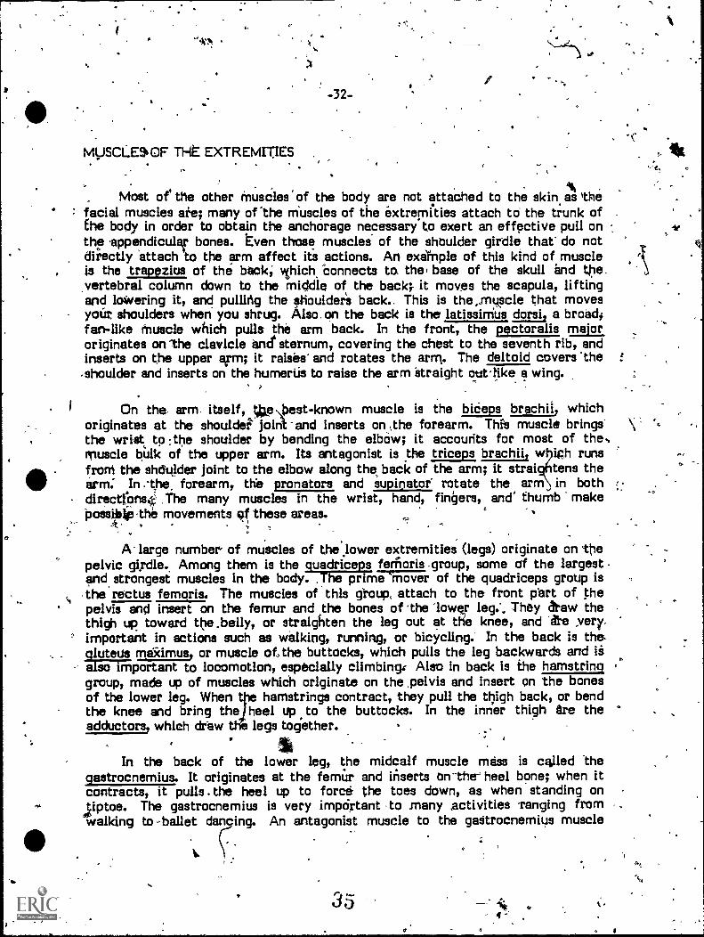
On the arm. itself, 904:est-known muscle is the bideps brachil, whichoriginates at the shoulder"' joint-and inserts on .the forearm. Thi's muscle brings'the wrist to :the shoulder by bending the elbow; it accounts for most of the,muscle bulk of the upper arm. Its antagonist is the triceps, brachii, w iph runsfroM the shdulder joint to the elbow along the. back of the arm; it strai tens thearm. In the, forearm, the pronators and supmator rotate the arm in bothciirectIonsi: The many muscles in the wrist, hand, fingers, and' thumb makePossibip the movements these areas.
,k
A- large number, of muscles of the lower extremities (legs) originate on thepelvic gi.rdle. Among them is the quadriceps fenioris.group, some cif the largestand strongest muscles in the body. ,The prime 'mover of the quadriceps group Isthe rectos femoris. The muscles of this gtoup, attach to the front part of thepelvis and insert on the femur and the bones of the 'lower leg.'.. They &raw thethigh up toward the .belly, or straighten the leg out at tKe knee, and 'fie ,very.important in actions such as walking, running, or bicycling. In the back is thegluteus mex-imus, or muscle of, the buttocks, which pulls the leg backwards and is
importantmportant to locomotion, especially climbing Also in back is the hamstringgroup, made up of muscles whiCh originate on the .pelvis and insert on the bonesof the lower leg. When hamstrings contract, they pull the thigh back, or bendthe knee and bring the heel up to the buttocks. In the inner thigh are theadductors, which draw t legs together.
In the back of the lower leg, the midcalf muscle mass is called theqastrocnemius. It originates at the femur and inserts on-the= heel bone; when itcontracts, it pulls. the heel up to force the toes down, as when standing on4iptoe. The gastrocnemius is very impOrtant to many activities 'ranging fromwalking to -ballet dancing. An antagonist muscle to the gastrocnemius muscle
35
°
.0
is the tibialis ant rtibialls anterior onpull the toes up towand toes that contr
/
33-
or which runs down Op lensith of the tibia or shinbone. Thenates near the knee sand inserts on the toes; it contracts tod the knee. There are also many.rtludies.in the ankle, foot,
ute to lower leg and foot movements. f-c.
c1
MUSCLES OF THE TRUNK
Muscles of the trunk Control-the, position of the spine and ere impoitant tobody posture. The erector spinae of the back are muscles that etto'hs to the baseof the skull and the pelvis; when contracted, they hold straight andupright. The antagonist. muscle is the rectus abdominus which originates at tliepelvis and inserts on ts ribs. Running down the mmid e of the belly, this musclecontracts to pull the head down toward the knees,. .as in :doing a sit-up. Thetransverse abdominal muscle, a deep Muscle, contracts to flatten the abdomen
seful for any 'abdominal pushing, such as the movement that occurs in awo an' body when she is giving girth. In the chest, the internal muscles includethe inte costal muscles of the ribcage, which lift, and lower the ribs in breathing.The diaphragm is the major 'internal muscle of breathing. It has no antagonistgroup; its relaxation produces the movement opposite to that produced bitscontraction. 4
. f
MUSCLE CONTRACTION
In all of these major muscles, the function .of contraction depends, on t'strutti3fe of the muscle Usage. Basically, the long strands or''fibers of muscletissue are composed of overlapping bands of,cells. When stimulated bje electricalimpulses, the overlapping bands of muscle are drawn together and slide over onanother, which' shortens each individual muscle fiber.' A shortening of all the'fibers' at once causes a shortening (or contraction) of the entire muscle.Depending on the orientation of the fibers, the contraction is along the length Qfthe muscle "(as in skeletal muscle), or in a ririg around the organ walls (assmooth-muscled organs). Different types of muscles thus accomplish theirdifferent functions by contracting to produce movements. Sorhe of theserriuscles contract quickly end strongly t6' produce movements such as running.Others contract slowly -and steadily, such' as the muscles: which maintain thebody's posture.
O
, Figure 4, on the next page, shows a frontal view of Lhe major muscles ofthe body. _
..
0
36 4 ,

S
O
-34- -
TEMPORAL1S
MASSETER
STERNOCLEIDOMASTOID
TRAPEZIUS
DELTOID
FRONTAL'S
ORBICULARIS OCULI
ORBICULAR'S MS,
BRACHIORADIALIS
FLEXOR CARPIRADIAL'S
F,LEXOR CARPIIS
PALiGIA
LONGUSPECTORAL'S MAJOR
SERRATIJS ANTERIOR'
LAT1SSIMUS DORSI `.
-) BRACH AL1S
EXTENSOR CARPIRADIAL'S LONGUS
EXTENSOR DIGITORUMCOMMUNIS
TENSOR FASCIA AA
VASTUS LATERAL'S
RECTUS FEMORIS
VASTUS MEDIALIS
BICEPS BRACHIl
TRICEPS BRACH
EXTERNAL OBLIQUE
RECTUS ABDOMINUS
ILIOPSOTSe .
AD0U OR LONGUS
GRAM LIS
ADDUCTOR MAG,NUS ,
SARTOR IUS
VASTUS MEDIALIS
PERONEUS LONGUS
EXTENSOR DIGITDRUMLONGUS\,,
TIBIALIS ANTERIOR
11,
1GASTROCNEMIUS
ir
SOLEUS
. Figure 4. The Muscular System
`
fie
1
Optional Activities
--35-
Clench your jaw tightly, feeling different areas of your face -as you doso. Cn you identify the muscles that are producing the tension?
On diagrams of the skeleton, draw the major muscles ofthe body.
A
.
(%.
33
Cr

: .
°
-36-
MAJOR MUSCLES OF THE BODY
Post-Test
1. Which of the following is a muscle of the face?
A. tibialis anteriorB. pectoralis
obicularis oculiD. trapezius
a
2. Which group of musclet is located on the fronted th thigh?
3. The hamstring .muscles are located:
A. in the chest..B. on the back of the thigh.C. in the lower back.
above and behind the jaw.
4. The rntiscles which raise the arms up froni the side ara,,the:
A. deltoids..B.. intercostals.C. late' simus dorsi.D. gluteals.
39
'
ink
0
o.
4
. Which of the following is a pri
A. intercostalB. diaphragm
. C. tibialis anteriorD.. rectus abdonlinus
737-
mover in the action of doir a sit-up?
.' 6. The pgime mover ih straightening the lower leg is the:
A. ,gastrocnerrius.1B. rectus ferar is.C. trapezius.'D. pectoralis,
.7. Which of the following muscles move the ribcage?
A. adductorsB. bic6psC. intercostalsD. hamstrings
A
40
1=,
7 -

e
Ado
-38-
SUPPORTING STRUCTURES OF THE MUSCIJLOSKELETAL SYSTEM
Goals
Upon completion of this module, you should be able to:
Oro
:
1. Identify the supporting structures of the musculoskeletal system..
2. Describe the functions of the supporting Structures of the. muscploskeletal system. ,
In order for the muscular system and the skeletal system to. functiOntogether smoothly, there must. be several supporting structures to link the two,systems. For instance,.therp are tough connective materials to attach" muscle tobone, and even tougher connective material tog attach. bone to bone in. thehard-working joints. The jointsA.theinselves, which-- -are structured for specific,functions; are crucial to the functioning of the musculoskeletatsystem.
-TENDONS
Muscle-to-bone connections are made by tendons. tendons are ,very strong,thick Strands of fibrous (and therefore tough) connective tissue that connectmuscles to bone. They can be vet), long, if necessary, to extend the effectivereach of a muscle' to a length that the muscle alone might not be able toachieve. this it important for bones that are fairly far Apart (as in mysclet of Athe thigh, .for 'example, which originate on the upper edge of .the.pelyis and insertbelow the knee). Because they are muCht.thinner.than muscle tissue, tendons can
oo. . attach in places where bulky muscle tissUg would not fit. tendons,.beingc° tough, can, withstand far. more of the wear and tear caused-by:joint action than
° can the; softer fleshiof muscle.
$
es
O
1
S.J 4
to-

O.
-39-
LIGAMENTS
4..
Ligaments surround or cross joints to connect bone to bone. In manyarticulations, thick layers of lig&nents are wrapped around the bories, and thushelp to form the joint structure; they also pass from, bone to ,bone along' thelengths of parallel limb bones, as in the forearm and lower leg. Made of verytough fibrous connective tissue, ligaments give stability and support 'to the jointsand the skeletal system as a whole.
ARTICULATIONS r
Joints, or articulatiOni,. are classified into three types according 'to 'structure; their Structures relater to- the functions they serve within themusculoskeletal system. Articulations are classified as non-movable, partlymovable, or freely movable, depending on the range of motion they allow,between theiponnecting bones.
Non-movable articulations are also calledefibrous joints. because the bonesale tightly bound by fibrous sheets of tissue which no bone motion. Anexample of a non-movable joint is the connection of the tibia and the fibulaabove the ankle. Sometimes, as in the skull sutures, the joint is almostcompletely- bone-to-bone, with very little . tissue needed because the Nines fittogether so closely. Ron-movable joints serve a protective function (as in theskull) or a strengthening and supportive function as in the articulation betweenthe pelvis and the sacrum).
Partly 'movable joInts are also called cartilaginous joints because a cushionof cartilage connects the bones. Because 'cartilage is not very flexible, little
ak movement is allowed in each individual cartilagiridus jbint. However, many1r, partly movable joints in a stgieeteach with a slight degree. of motion, can permit
a large range of motionp This explains' the great flexibility of the spinal, column,which, articfates in ,cartilaginous, joints. Examples-of cartilaginous joints where
rovery little movement is permitted include the sternum-rib articulations and theconnection of the two pubis bones of the, pelvis. Movement in these joints is notdesirable because they serve mainly protective functions. ,
Movements associated with locomotion and rhanipulation are perinitted bythe. freely :movable articulations --the ithird category of joints. Freely movablearticulations are. also' celled synovial-joints becaude of a reembrane, the synovialmembrane, which lines the' joint area and secretes a special lubricating flitid
.
42
0
(
-40-
(synovial fluid) to help :reduce friction, as the moving bones slide over 'oneanother. Ligaments around the joint forrit the capsule that contains this fluid,and pads of cartilage coat, each articulating surface for improved smoothness ofmoverrient. The cartilage pads also absorb the shock associated with the functionof many of these bones (for instance,,landing'on your feet after you jump causesa shock to the knee joints).
-iviost of the freely movable joints are in the skeletal areas of the body f atare involved in active movementthat is, the appendicular skeleton. There areseveral kinds. of freely movable joints, each permitting a different type ofmotion. A hinge joint allows the type of motion involved in bending andstraightening the elbow, the fingers, or the knee. A pivot joint allOws for therotation seen between the ulna and the radius of the forearm. A- saddle jointallows the relatively free motion of the 'thumb. Ball- and - socket joints, such as
-those found in the hip and shoulder, permit the great freedom o movementallowed by those joints. .
, . . ..Exactly how muscles and bones work together and produce movement, and
what kinds of movement they can produce, is the subject of the next modble.
Optional ,,ativities
On a skeleton or model skeleton, exargine_the different types of jointsand their movements.
Find out abbut the structure of tie knee joints and why po many athleteshave problems with it.
The next time yoLi%at chicken at hone, examine the tendons, ligaments,and joints of the chicken. Are they similar to those found in the human
.bbdy?$
o Examine 'clays of various types of joints. Can you guess the function ofthe joint from the structure?- What movement does it,permit? 4
s Discuss what would happen to, the joints, and to the muscblaskeletalsystem as a whble,. if there were no ligAmenis.
,
,43

a,
-41-
SUPPORTING STRUCTURES OF THE MUSCOL.*ELETAL SYSTEM° .16
Post-Test
- . .
1. The, most important structures in the condection, ofrbone to bone are called:-, .
: 'A. tendons.B. Iliornents.
$
C. synovial capsules.D. articular cartilage.
a
2. The sutures of the skull are examples of what Eype of joint?
3. 3 Cartilaginous joints are:
A. -freely movable.B. non-movable. .5
C. partly movable.D. capable of only one movement.
--
;\
The variety of movements required for .tqcdmotion and mripulation areprovided mainly by what type joint? s ,
,-
44
. -42-
MOVEMENTS
/`Goals
Upon completion of this module, you should be able to:O
1. Identify and describe different human movements.
Movements, like muscle groups, generally occur in pairs. For mostmovements that .take a part of the body in one direction/ there is anothermovement that takes it in the oppotiite direction. Not surprifingly, most Df theopposing,movements are produced by agonist-antagonist muscle groups.
r
There are four major types of. movement possible inthe musculoskeletalsystem, as well as "several other types of movement. When these differentmovements are combined, they allow for the amazing range and variety ofmotions that characterize the actions of the musculoskeletal system.
t FLEXION-EX SION .,
4
Two -movements necessary to most actions of the body are' flexion andextension. Flexion involves bending a joint to` tiring areas closer together, asw lrie elbow is bent to bring the forearm to the shoulder. In a betterdefinition,. flexion Is the movement that makes the angle,of a joint smaller. Thebppoeite. motion is extension? which straightens joints to straighten or stretch thebody or the extremities. At the elbow, flexion is accomplished by the bicepsbrachli. When this muscle contracts, it pulls on the forearm bones to bend theelbow and raise the wrist toward the shoulder. Its antagonist muscle, -the tricepsbrachii, is the extensor of the arm; when it contracts, it shottens over the backof the - arm and elbow to pull the forearm back into 'a straight poSitibn:(Remember that for .any .one.. muscle .gts2Cip tottaccomplish its task,. while itcontracts -its antagonist muscle group Miitit.4*elax. With a few exceptions, atleast' two muscle groups are involved in'eveey movement.)
k,
45
I
J.
-43-
/.
In the leg, the hamstring muscles In back contract to-bend the knee and \flex the leg; this action brings the heel toward the: buttocks. The leg isstraightened out at the knee by the quadriceps farnoris muscles, which run overthe knee and which, when contracted, pull the lower leg to a straight position:Flexion arid extension are the two movements alloWed by the hinge structure ofthe elbow and knee joints.
The mrtebrat column has less freedom of motion, but also flexes andextends like any other bone-joint combination. In flexion of the vertebral .
column, the body. bends forward (as when perfoOrng sit=ups); this movement isaccomplished by strong contraction of the rectos abdoMinus muscle of the belly.This Muscle attaches to the bottom edges, of the'ribs and pelvis and, whencontracted, pulls these areas together; this makes the spinal column bend andcurve forward. The vertebral column is etraightened, out, or. extended, by thelong muscles- of posture, which a4tach to the styli and pelvis. When thesemuscles contract, they pull the Tback of the head ,,,tdcvard the buttOcks,straightening and lifting the spinal column.
ABDUCTION-ADDUCTION
Abduction and adduction are two other paired movements. Abduction isthe movement of a body part away from the knidline of the body t adduction is themovement of a pa1t toward the midline of the body. Again, these movements areaccomplished by agonist-antagonist muscle groups. In lbduction of the arm, theprime mover, is the deltoid muscle of the shoulder. This muscle attaches to the.clavicle and the upper humerus; when it contracts, it shbrtens over the shoulder
\ joint, pulling ,the arm directly up over the side} Adduction of the .arm isaccomplished by the latissimus- dorsi, which attaches across the back to the, ,stlinal column and humerus, and the pectoral's of the chest. They contract topull the arm in toward the side of the body: The action of "flapping wings" istactually a rapid abduction and adduction of the arms. ,.0
Abduction of the leg, or lifting it out to the side; is done by the deepOluteal muscles and a muscle:1'0st attaches to the outside of the pelvis and theoutside surface of the femur. 'Adduction is mollshed by, the adductor.tnuscles Oft the inside of the thigh, which 'ea) the pubis bone and to the.Upper femur. These muscles do not bend t hen they contract becausethe knee joint is not structured to bend-si ways. . , ,,
1 ..
42,))IS
46
II
A
a
-44-
ROTATION-CIRCUMDUCTION
Rotation and circumduction, two other kinds of movements, are bothbasically circular motions.. Rogation is an actual, basic movement;circumduction is a cbmbination of basic movements (flexion, abduction;extension, and adduction). Rotation (e.g., pointing the knee or elbow outward or
-in = d) is the rotation of a body . part. The humerus, femur, neck, spine, andscapul= capable of rotation. Tracing an imaginary circle with the fingep (armextended) is = example of cirdamcluction. .
..
k. - -
SPECIAL MOVEMENTS
5/
Se,veral, other special movements are made possible by muscular andskeletal interaction. Supination and pronation are two forms of rotation of theforearm made possible by the RiCiiit joint which ?ROWS the radius to rotate aroundthe ulna. Supination is the adt of turning the Palm from facing the floor tofacing the ceiling when the arm is extended. Pronation is the oppiisite "movement, turning the palm towards the floor.. These movements -contributegreatly to the manipulative abilities of the arms and hands. a
,110'
.Muscles of the lower lag and -foot contract to turn the *foot inward(inversion) and the antagonist muse es act to pull it outward ,(evereion). Twooth{77mc7vements Of the foot are dors flexion and plantar-flexion. orsiflexion is .
a for'rn of flexion, or bending of the of at the ankle. This movement pulls thefoot u; so that the toes point toward the knees. Plantar-flexion. is a form ofextension, or straightening out of the foot at the ankle (the foot, howev is
normally`. at right angles to the leg, so that plantar-flexion is not the f t's'stomary state of rest but a definite movement). In plantar-flexion, muscle at
back of the leg pull the heel up;, which forces the toes down. Obviously,se two movements are very important in walking and in other forms of
on.
Muscles, bones, and joints must, act togethei to produce .these movements.The combinations of all these .individual ynOvements of the musculoskeletatsystem make possible the incredible varieti of motion .of which the human body =is capable. Each movement and combination of movements has its own specialfunctionsfunctions made possible by the structures of the musculoskeletalsystem working together.
47.
D.
0
O
Optional, ActivitiesS
s Take a simple ser =s o ions such as walking" across the room andsitting down, and akit into its component movements. What musclegroups are being us ? Joints? Bones?
Use a model skeleton to study the movements of different parts of the. body. For example, note why abductors-adductors do not bend the knee,
how the radius rotates. around the ulna, and how the head moves in '-different directions.
.. -
\\-
Try .to complete' the, word maze on the following page. It uses some of,the terms that you learned in this unit. . . ,
4
v./
O
411".
48
0

r 1
, , C.t
,
4 .:
,7 0
-46-
WO
RD
MA
ZE
S K S U P IT6-14AR
R 0 W L F
CSYNSDEEFRL
LT'APL
AGN
IC
TS
XNOA
SUP.ME
P X\OD,kRICA'DG
T IEP
X
1411V
SP
AD'POHO
E-
B E A
I.CI
SUCN
I TCNNE
L F 0
N CA D 'L
P H I G.
H I D D 0 N
D 0 L B A L I0
N Y S NGM
A T
'X C J R C g T t44
B E T .G
L R I
A YO'AE,AL
X A I.N
AR C B 0
Y H I VF
AL
LE
TA
P X A A N
N-XNEPIPHYSI
S I S T,R
OJTR
HIGAI1QR
OUT'R
0
GI(
AWJQU.I
S A V IN'R
E R
A C R ANIUMCWANG144-P
B T E R E G W O L ODEO
Y
r
Find
the
fgktowing
terms
in this
maze
by circling
the
words.
They.,
may
appear
frontwards
or
backwards,
vertically,
horizontally,
or
diagonally.44:.
-pgonist
.
flexion
'
carpal.'H
incus
1
coccyx
marrow
`
cranium
patella
epiphysis
scapola
%
.1/
sup
inat
fon
synoviai
joint
tendonvertebra
1'

-47-
MOVEMENTS
Post -Test
1. Bringing the palm of the hand from the hip to the shoulder is an example ofwhich of, the following movements?
A. extensionB. circuinductionC. eversionD. flexion
r' .oe ..-.2. ,Going from .a iquatiirig position to a st
following inovementaRf the lower leg?
A. adductionB. extensionC. flexionD. rotation
ing position requires vihigh of the
'<t:
3. Which of the following are special movements of the for"egirry?
A. inversion - eversionB. dorsiflexibn - plaritar-flexionC. supination pronationD. rotation - circumduction
0
I.
'Pointing the knee,,ouiwar. d requires what basic movement of the upper leg?
50
I

ti
I
5. Which of the following movement.Sfdescribes dorsif14xion?
A: pointing the toes inwardB. bringing the palm to the shoulderC. pullrhg the toes'toward the kneeQ: pointing,the palm upward
I-
5 10
rJ
V
,
-
, a
-49-
aLOSSARY\-----
,abduction: lateral movement away from the midline of, the backL, . .
/ a duction: lateral rhovemenetoward the midline of the body.. goniiit: a muscle primarily responsible for a movement; a prime mover. ,
antagonist: a muscle with a movement opposite to that of an agonist.anterior: pertaining to the.front.appendicular skeleton: the bones of the pelvis, shoulder, and extremities:articulation: a connection between bones; a joint.axial skeleton: `the bones of the skull, vertebral column, and ribcage. %
.* 2
ball-and-socket joint: the shoulder and hip faints.2& (of a muscle): the main portion of a muscle; the area where most of the
contraction-takes place..
cardiac muscle: the muscle of thejeart; myocardium.°carpals: the eight short bones of the wrist.- cervicarvertebrae: the first seven vertebrae.
oircumduction: the movement of a body part in a circular motion.clavicle: the collarbone; a part of the shoulder girdle.coccyx: the tailbone; the last portion of the vertebral column.contraction: .$he shortening of muscle fibers.costal cartilage:, the cartilage that attaches the ribs to the sternum,craniu7,the portion of the skull that encloses the brain; consists of eight bones.
diaphysis: thhaft of. along bone.
0
epiphysis: one of the two ends of a long bone.eversion: the,outward movement of the foot.extension: a moVement.which straightens a joint; the opposite o $lexion.
flat bone: a flat sheet-like type of bone, as found in the skull.Wei( lord7 a movement which closes a joint; the opposite of extension.
gluteal: muscles of the buttoCks.
52,
-4
-50-
hamstrir)tse-'thr-musale group of the back of 'the thigh which flexes the lower leg.hin joint: a joint with two-we motion, such as the elbow and knee.hyoid bone: a small bone at the base of thd tongue.
incus: a small anvil-shaped bone of t middle ear.insertion: one of the areas where a m scle ItCaches.intercostals: muscles of the ribcage.inversion: an inward movement of the foot.irregular bone: bones with uniguti structures.
ligaments: connective tissues which support and attach structures, especiallybones.
long bones 'he mostcommon type of bone, found in the arms arid'legs.lumbar vertebrae: the vertebrae of the lower back.
n2alleus; a 'small hammer-shaped/bone of the middle earmarrow: a substance found inside bone; red or yellow.muscle: body tissues which contract to produce movements.
origin: one of the areas where a muscle attaches.ossicles: the small bones of the middle 'ear; the incus, malleus, and stapes. \
patella: the kneecap.ectoral girdle: the shoulder,girdle, formed by the clavicle, scapula, and
umerus.pelvic" girdle: the bones which form the hip joint...phalanges: the bones which form the fingers and toes.pivot joint: a joint which allows' rotation, such 4 between the radius and ulna.lantar-fle t e movement which points the toes and foot downward.
pertaining to the back ofrearprime m ver: the muscle primarily, responsible for a given moyerneLt; agonist.ronation the, movement of the forearm which turns the palm upw* d.
quadriceps: the muscle group of the front qt the thigh which extends the lower- leg. .
rotation: a movement ausing a body part to rotate.
\r'
53
-51-
sacrum: five fused vertebrae which form a part of the pelvis.scapula: the shoulder blade.,skeletal muscle: muscles which produce movements of the skeleton; striated
muscle.skull: the bones of the head; comprises the facial bones and the cranium.smooth muscle: Involuntary muscle; visceral muscle..stapes: a small stirrup-shaped bone of the middle ear.6triated muscle: skeletal muscle.
*suTh3M7--ilat on: the movement of the forearm which turns the palm downward.sutures: the immdVable joints of the skull.synovial fluid: the lubricating fluid prodOced by synovial membranes.synovial joint: a freely movable joint.synovial membrane: the membrane which surrounds a synovial joint.
....'.
'tarsals: short bones of the foot4>tendon: a strip of strong connective tissue that connects muscle to -bone.
Fic vertebrae: ,the 12 vertebrae of the thorax.
O
vertebral column: the spine or backbone.visceral muscle: muscle of the internal organs; smooth1muscle.
st,
(
l 4
P.
4
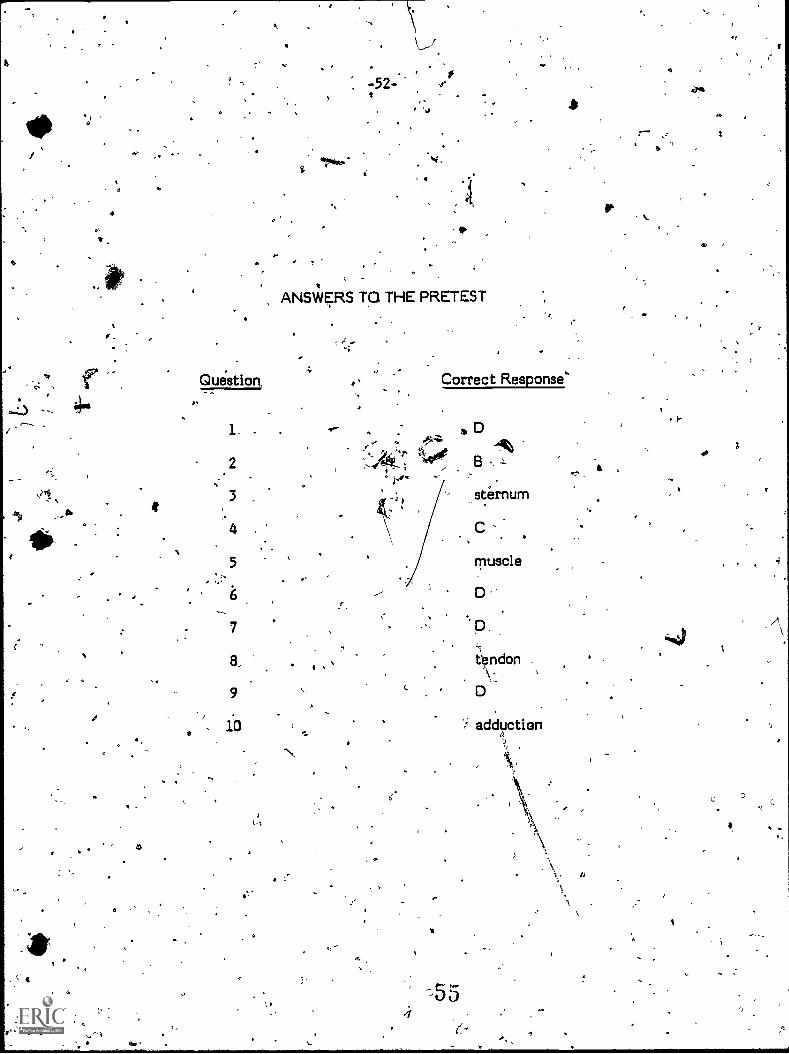
-52-
Question,
1
5
, 6
7
9
10
ANSWERS TO THE PRETEST
01"
Correct Response'
5
sternum
C
muscle
D
D.
tendon
D
.
I
00
C


a
A
4
O
.11
.,Instructional Materials in Anatomy and Physiology
for Pennsylitanid Health Occupations Progranie
INSTRUCTOR'S GUIDE:
THE 1viUSCUC.OSKELETAL SYSTEM
I
f 1 . ,.,Prepared for:_ .
Research Coordifiating Unit for Vocational Educatiori
"re - ' Pennsylvania Department of,Education- Box:911'
Harrttburg, Pennsyl4ania 17126/
...
. Prepared by:
National Evaluation Systems, Inc.,30 Gatehouse Road
Amherst, Messachusette, 01002
4
June, 1979
57
\\
s
'0INTRODUCTION
a 0
These' instructional modular units have been developed for the PennsylvaniaDepartment of Education for use in vocational education programs. They ,weredesigned on the assumption that a basic understanding of human anatomy andphysiology is essential to any person preparing to enter a.health care occupationsuch as - practical nursing, nursing assAtant, medical assistant, emergencymedical technician, or dental assistant. - Each of these modular units will covertie most important aspedts of one of the major systems of the human body. Intf% first four units the following systems will be covered: circulatory system,respiratory system, musculoskeletal system, and digestive system.
4
This Instructor's Guide is designed to provide suggestions to you on how touse a modular unit most effectivelj,,in your instruction. These recommendations,
\ however, do not represent the only way to use theseunits: you may be able todevise more beneficial uses for the materials.
THE MODULAR UNITS
Each modular unit is made up of several components: a pretest, fourseven instructional-modules with corresponding post-tests, optional activities fothe studeOts; end a,.glossary of terms used in the unit. Each of these componentshas a specific purpose and is organized, in. a specific way, as will be explained inthe following sections. \ 9
Pretest
After reading the prefab:), which ks simply an introduction to theseinstructional units, a student working through a modular unit should first take thepretest. As its name implies, this test is designed to be taken by the studentbefore beginning work on the materials contained in the unit. Its purpose istviofoldt (1) to stimulate interest the modular unit by giving, the student apre.vieviof the topics covered, and (2) to Provide a means of self-diagnosis so thestudent Tay identify, based on performance on' the pretest, those areas of the

0
-2-
modular unit which may require special attention and extra effort on the part ofthe student. After selecting answer to each of the pretest question's' thestudent should turn to the back of the modular unit and check the correctanswers. If the student answers incorrectly on a number of questions dealing,with a particular subject, 'then the student should, pay closer attention to themodule on that subject.
_ .
Instructional Modules116
This modular: Unit is composed of seven separate but closely related
modules, including: Introduction to the 'Skeletal System, Axial Skeleton,'Appendidular Skeleton, Introduction to the ,Muscular System, Major Muscles ofthe Body, and Movements. After taking the' pretest and checking the answers,the student shOuld read through and 'study each of the instructional modules. Forthe student's benefit, each module begihs with a statement- of the goals, orobjectives, that a student sh Id have mastered upon completion of thatparticular Module. The leyel df achievement of these' goals, is measured by thestudent's performance on the rresponding post - test. The language leYel andcontent of each 'module is aimed toward' the student seeking an Introduction tothe components, structures, and functions and the basic.terminology required foran undehtanding of the musculoskeletal system. ' '
Optional Activities
Fallowing many modules are optional activities intended to` provide the'student vitthtrp opportunity to pursue the content of the module at a morein-depth- level. Many of these .activities may 'require teacher participation, atleast in obtaining and preparing additional materials for the student to utilize.
In addition to the optional activities available to the students, you maychoose to provide further information to the studebts by teaching a brief unit onthe common disorders of the musculoskeletal syitem. Discussion of- thesedisorders- has not been in-the texts because 'a basic knowledge of the
proper structure and function of the human body ina healthy individual seemsmore appropriate for the purposes of an introductory program,. If you do chooseto discuss common disorders, the most effective approach may be one in which
you use. disorders to illustrate what can go wrong In ti*Ircitlyt) as a means ofclarifying the students' understanding of how the body works when functioningproperly.
You may also wish to provide students with the names of books or articlesas suggested readings to further their understanding °of a particUlar area.
59
Gldesary I
-31
'After the last thethe modulesitetthe unit is a glossary. This is not intendedto he a comprehensive glossary to be used by the student. as a dictionary.Rath r, irincludes the basic terms used in the unit which are necessary to an
standind,of the system covered. Those words which appear in the, modulesand have been defined in the text are not always defined in the 'glossary. Some-of these' particular, terms have been used in the module because they areessential but difficult terms needed to explain th4 cobtent taught in the unit.The student should use the glossary to review the vocabulary essential to the unitbefore taking the post-tests.
Post-Tests "1.
'
The post-tests are the final assessment of a student's understanding of theMaterial piesented in each module. They Consist of multiple-choice andopen-ended questions designed tp measure a student's mastery. of the goals(objectives) stated at the beginning'of each module: Each 'of the-questions hasbeen written to measure an aspect of the skills and/or knowledge that a studentmay be expected to acquire as a result of working through .a particular module.When a student has finished studying a module, hes pursued any cho'sen optionalactivities, and has reviewed the vocabulary in the glossary; the student shouldtake the post-test that follbwe the module.
F
SCORING THE POST-M-TS
As previously _mentioned,_ the purpose of the post-tests is to measurewhether or hot a student has mattered the objectives -(goeld) stated at-- thebeginning of each module. Due to the differing lengths of the post-tests, thevariety of ways in which teachers may choose to utilize these modules, anddiscrepancies among students' previous exposure to the subject matter, it is notpractiCal to set a standard cut-off Boors on 'each of the tests that =would indicatemastery of the objectives. Rather, teachers are asked to use their professionaljudgment in individual cases td determine if a student's performance on apost-tast indicates that he or she has mastered the objectives stated for that,module; in making this determination, you should -consider at least -all of thefollowing factors:
60

1
-4-
(1) How lo'ng is each post -teat?
(2) Him much information is included in each module and how Complex isthe information, relative to other modules?
(3) Has the' student been exposed to the kind of curricular material.before? That Is, has the student been taught the basics of this systemof the body before?
O
(4) Should.the entire class be required to achieve a certain score in orderto pass, or shoul,d each student be considered individually? (Thisdepends on how and with whom ytiu use thi module as instructiortalmaterial.)
(5) Should the student be graded pass/fail on each post - test- -i.e., onmastery of each module or on the unit es a whole?
v.
.,.). ,
To facilitate the scoring of post-tests, each student will record his or her .answers to all the post-tests' on one separate sheet of paper.. You should markeach answer correct or incorrect, then give .the student Ernpass" or "fail" on eachmodule, or on the unit as a whole. ,
Because of the subject matter, responses to opeh-ended questions may varyslightly. from those listed below, but these responses, may also be acceptable.
gain, in these cases instructors are asked to use their professional judgmenttodetermine if a response is correct.r
Use -the following list of answers to questions on the post-tests to gradeyour ardentie papers.
s`
I
-4
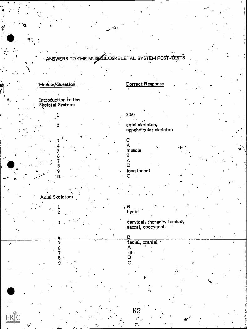
\ ANSWERS TO THE MU LOSKELETAL SYSTEM POST--451i
i'Module/Question Correct Responser r
01
o
Introduction to theSkeletal System:
; .
1
o 24 '
3456789
10-
,
Axial Skeleton:
206-
axial skeleton,appehdicular skeleton
AmuscleBADlong (bone)C
1 ,'B2 hyoid
3 . Cervical, thoracic, lumbar,
456789
sacral, coccygeal
B
ar-
facial, cranialA.ribs 9
1
62 e

SI
-6-
ft
Module/Question Correct Response
Appendicular Skeleton:
12,
3-,,
Bpectoral girdleA
4 D5"
r6B.patella
7 D8 C9 ,
.0A
10
Introduction to theMuscular System:
1_2;
345.
67
Major Muscles ofL theaody: ;
4
2 DB.BC-; ( .body <
myocardiumsmooth (involuntary) muscleA
t
1
2 quadriceps34 A5 D67 C
63
.-
Module /Question
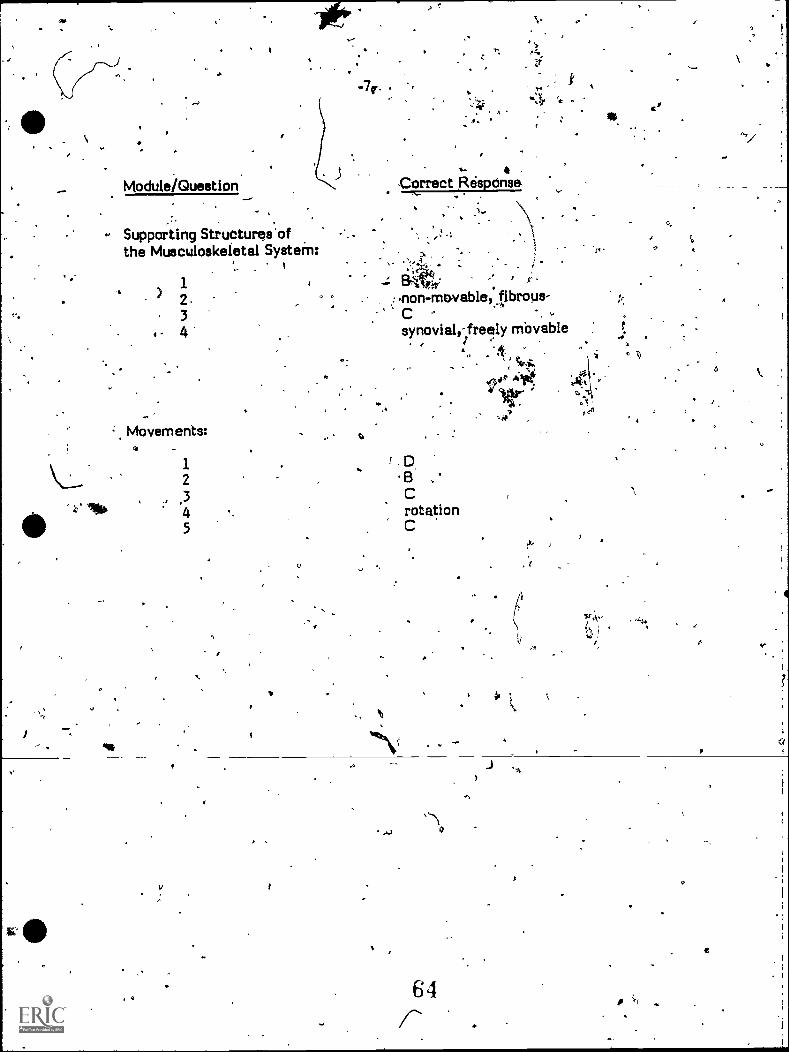
-7r .
Supporting Structures ofthe Musculoskeletal SysteM:
1
34'
Movements:
12
345
0
Correct Response.
;. ,.I .,
,t,-,..it,.
non-movable) fibrous-.
synovial,-freely Movable.. 7 .
. . : C.s.
ruir."1°
,0"
.
B ,
CrotationC
64