Embed Size (px)
Citation preview
1997 Oxford University Press 247–258Human Molecular Genetics, 1997, Vol. 6, No. 2
The murine homologue of HIRA, a DiGeorgesyndrome candidate gene, is expressed in embryonicstructures affected in human CATCH22 patientsLaurens G. Wilming 1, C. A. Sylvia Snoeren 1, Angelique van R ijswijk 1,2, Frank Gro sveld 2 and Carel M eijers 1,2,*
1Institute of Pediatric Surgery and 2Department of Cell Biology and Genetics, Erasmus University, PO Box 1738,3000 DR Rotterdam, the Netherlands
Received August 21, 1996; Revised and Accepted November 1, 1996
A wide spectrum of birth defects is caused by deletionsof the DiGeorge syndrome chromosomal region at22q11. Characteristic features include cranio-facial,cardiac and thymic malformations, which are thought toarise from disturbances in the interactions betweenhindbrain neural crest cells and the endoderm of thepharyngeal pouches. Several genes have beenidentified in the shortest region of deletion overlap at22q11, but nothing is known about the expression ofthese genes in mammalian embryos. We report here theisolation of several murine embryonic cDNAs of theDiGeorge syndrome candidate gene HIRA. Weidentified several alternatively spliced transcripts.Sequence analysis reveals that Hira bears homology tothe p60 subunit of the human Chromatin AssemblyFactor I and yeast Hir1p and Hir2p, suggesting that Hiramight have some role in chromatin assembly and/orhistone regulation. Whole mount in situ hybridization ofmouse embryos at various stages of development showthat Hira is ubiquitously expressed. However, higherlevels of transcripts are detected in the cranial neuralfolds, frontonasal mass, first two pharyngeal arches,circumpharyngeal neural crest and the limb buds. Sincemany of the structures affected in DiGeorge syndromederive from these Hira expressing cell populations wepropose that haploinsufficiency of HIRA contributes toat least some of the features of the DiGeorgephenotype.
INTRODUCTION
Hemizygosity of a region of chromosome 22q11 has beenassociated with a wide variety of congenital malformations thatreceive several diagnostic labels, the main features being coveredby the acronym CATCH22 [Cardiac defect, Abnormal facies,Thymic hypoplasia, Cleft palate, Hypocalcemia, (interstitial)22q11 deletions] (1). The congenital malformation syndromes
associated with these deletions include DiGeorge syndrome(DGS), velo-cardio-facial syndrome (VCFS), conotruncalanomaly face syndrome and Opitz syndrome (2–8). For the lattersyndrome Robin et al. recently reported linkage with loci onchromosome 22q11 and Xp22 (9).
Several well characterized DNA probes can detect CATCH22deletions (3,4,10–15). The interstitial deletions are large,spanning 2–3 Mb of DNA. A limited number of CATCH22patients have been reported with an interstitial deletion smallerthan the commonly deleted region (16–18). The current shortestregion of deletion overlap (SRDO1) is defined between thecentromeric breakpoint in DGS patient G (16) and the unbalancedtranslocation breakpoint in cell line GM00980 [from a VCFSpatient with a t(11;22)(q25;q11) translocation (19)]. Recently,Kurahashi et al. provided evidence for the existence of a secondcritical region (SRDO2) located in the distal part of thecommonly deleted region (17). The two SRDOs are mutuallyexclusive.
At present it is not clear whether the CATCH22 phenotype isthe result of haploinsufficiency of one or more genes. Thebalanced t(2;22)(q14.1;q11.1) translocation breakpoint in DGSpatient ADU (20) was the major target of positional cloning invarious laboratories. These attempts led to the isolation ofDGCR3 and DGCR5. DGCR3 is an open reading frame (ORF) of260 amino acids (aa) that is interrupted by the translocationbreakpoint (21). However, no cDNAs were isolated andexpression studies were only informative under low stringencyconditions. This prompted Sutherland et al. (22) to screen foradjacent coding sequences in the region. They identified a seriesof alternatively spliced transcripts from the DGCR5 gene. TheDGCR3 sequence is embedded in an intron of DGCR5 and in thesame transcriptional orientation. The DGCR5 transcripts do nothave an obvious protein encoding potential. They may befunctional RNAs. The recent refinement of the SRDO places theADU breakpoint ∼100 kb centromeric to the proximal deletionboundary of SRDO1 (16), so the relevance of DGCR3 andDGCR5 for CATCH22 etiology is unclear. It could very well bethat the CATCH22 phenotype results from haploinsufficiency ofseveral relevant genes in SRDO1 and that the ADU breakpointexerts a position effect on such genes.
*To whom correspondence should be addressed
Human Molecular Genetics, 1997, Vol. 6, No. 2248
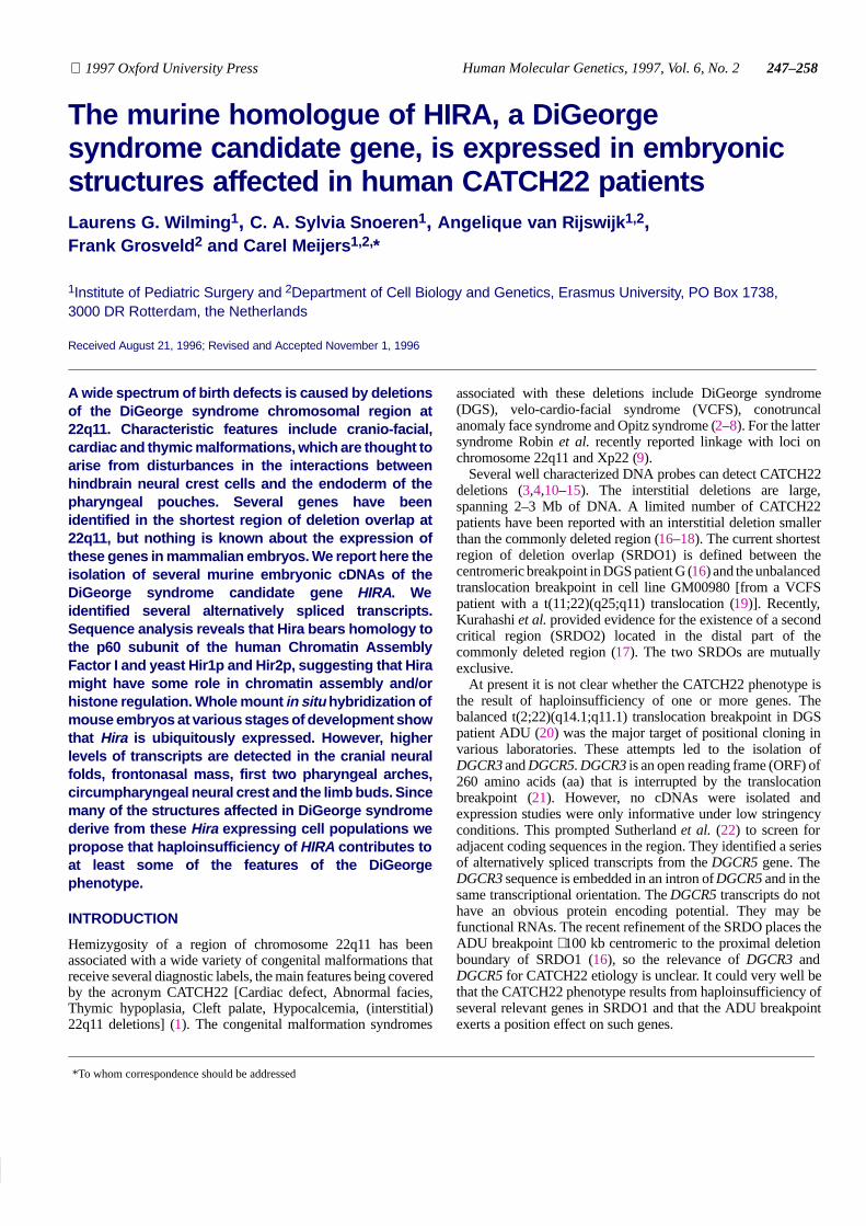
Figure 1. (A) A scale schematic drawing of the assembled mouse Hira cDNA and the individual cDNA clones isolated from an E11.5 mouse embryo cDNA library(12F, 12B, 12E, 6A, 12D, 12C and 6B). As a reference the human and murine Tuple1 C5 and MF2 cDNAs and human HIRA are shown. Nucleotide positions areindicated on top. Open boxes depict open reading frames; dark gray boxes depict WD40 domain coding regions; black oval indicates the penta-Q stretch coding region;closed arrowheads mark exon–exon boundaries determined from mouse genomic sequences; open arrowheads mark human exon–exon boundaries (exon numbersare between the arrowheads); asterixes above arrowheads show the positions of additional exons 2A, 3A and 11A; black bars indicate the additional exons. (B) Drawingof a putative mouse Hira transcript with additional exons and the positions of the various primers and probes used. Probes preceded by m are mouse probes, the onespreceded by h are human probes. Restriction sites on mouse or human HIRA are given as a reference.
The saturation cloning and sequencing of the 22q11 region areat an advanced stage (23,24). Two transcription maps of SRDO1have recently been reported (24,25). Currently at least sixtranscription units have been identified: HIRA (DGCR1/TUPLE1) (26,27), CLH-22 (clathrin heavy chain like,CLTCL/ES3/DGS-K) (24,25,28,29), CTP (mitochondrial citratetransporter protein, DGS-J) (24,30), DGS-I/ES2 (24,25), DGS-H(24) and DGS-G (a serine/threonine kinase) (24). Sequenceshomologous to the 3′ UTR of the human Dishevelled 1 gene weremapped just centromeric to the HIRA gene (31). It is unclearwhether these sequences belong to a gene. Little is known aboutthe expression of the CATCH22 candidate genes in adult andembryonic tissues. It will be important to establish the embryonicexpression patterns and functions of the candidate genes.
As an initial step to determine the expression pattern of genesfrom the CATCH22 SRDO1 we chose to analyze the expressionpattern of HIRA. The HIRA gene (DGCR1) was originallyisolated as TUPLE1 by Halford et al. (26). Lamour et al. isolatedhuman cDNAs containing the complete TUPLE1 sequence with621 additional nucleotides in the ORF (27). They renamed the
gene HIRA because the most significant peptide homology foundat the time was with Hir1p and Hir2p, two histone gene repressorproteins from the yeast Saccharomyces cerevisiae (32). Thepredicted HIRA protein is characterized by the presence of sevenN-terminal WD40 repeats, two bipartite nuclear localizationsignals and a penta-Q stretch (frequently seen in transcriptionfactors). The gene product has been implicated in transcriptionalregulation based on its homology to gene regulators like yeastHir1p, Hir2p and Tup1p (32,33).
We report here the identification of cDNA clones of the murinehomologue of HIRA and studies on the expression of Hira duringmouse embryogenesis. The sequence of the predicted protein ishomologous with the p60 subunit of human Chromatin AssemblyFactor 1 (CAF1A) (34). On the basis of sequence homologieswith Tup1p, CAF1A, Hir1p and Hir2p, we propose that HIRA isinvolved in the assembly of chromatin, either by interacting withhistones or by regulating their genes. Our results demonstrate thatHira mRNA is ubiquitously present from early developmentalstages through adulthood. Raised levels of mRNA are detected inthe neural folds, pharyngeal arches, circumpharyngeal neural
249
Nucleic Acids Research, 1994, Vol. 22, No. 1Human Molecular Genetics, 1997, Vol. 6, No. 2249
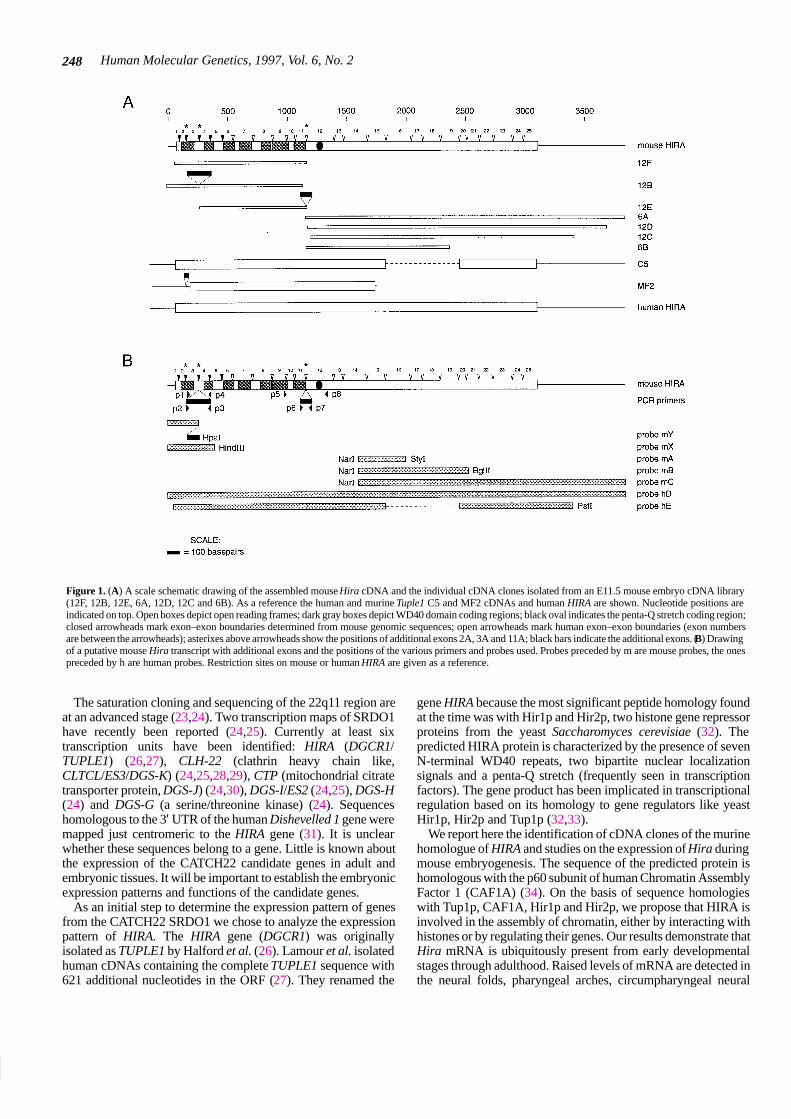
Figure 2. Conservation of the HIRA gene. A human HIRA probe, hD, detectsconserved sequences in EcoRI digests of genomic DNA from human, monkey,rat, mouse, dog, cow and yeast.
crest and limb buds. We think that HIRA is normally involved inseveral aspects of neurogenesis, pharyngeal arch developmentand limb development by regulating genes that control theseprocesses.
RESULTS
Murine Hira cDNA clones coding for a protein withhomology with human Chromatin Assembly Factor 1A
We examined the evolutionary conservation of HIRA by hybrid-ization of radiolabeled probe hD (from the 4 kb insert of one ofour human HIRA cDNA clones) (Fig. 1B) to a blot containingEcoRI digested genomic DNA from various species. The probehybridizes to homologous sequences in the DNA of all mentionedorganisms (Fig. 2). This high level of conservation from man toyeast indicates an essential function for HIRA.
As an initial step to study whether haploinsufficiency of theHIRA gene contributes to the CATCH22 phenotype, we hybrid-ized probe hE (Fig. 1B) to an E11.5 mouse embryo cDNA libraryunder stringent conditions. This screening yielded seven positivecDNA clones of the mouse homologue of the human HIRA gene(Fig. 1A). Complete sequencing and assembly of selected clonesallowed us to reconstitute a cDNA of 3852 bp (accession numberEMBL X99712), containing an ORF of 3045 bp. The 1015 aapredicted protein has seven WD40 repeats (at aa positions 10–44,68–98, 129–159, 172–202, 234–259, 266–313 and 326–356),two bipartite nuclear localization signals (at aa 267–286 and626–643), a penta-Q stretch (aa 408–412) and a molecular massof 111.72 kDa.
Sequence comparison between the human and mouse proteinsreveals an overall identity of 95.8% and a similarity of 97.2%.The N-terminal third of the protein, containing the WD40 repeats,is 99.4% conserved (identity and similarity). The difference is inone amino acid in WD40 repeat 3 and one in repeat 6. TheC-terminal two-thirds have a relatively higher divergence(identity 93.7%; similarity 96%). Compared to the 1017 aahuman HIRA translated sequence, the Hira sequence lacks amino
acids 510 and 607 or 608. Our Hira sequence is 99% identical tothe recently reported mouse Hira sequence (35) and 100%identical to the mouse Tuple1 cDNA clone MF2 (26).
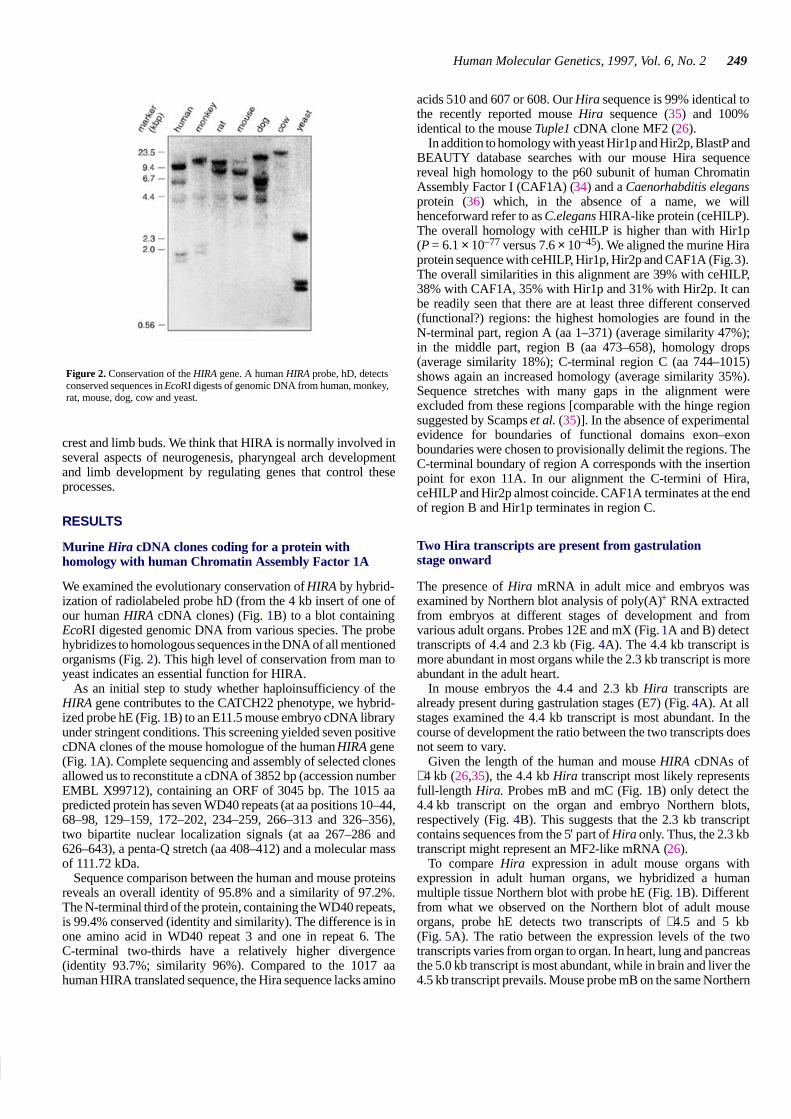
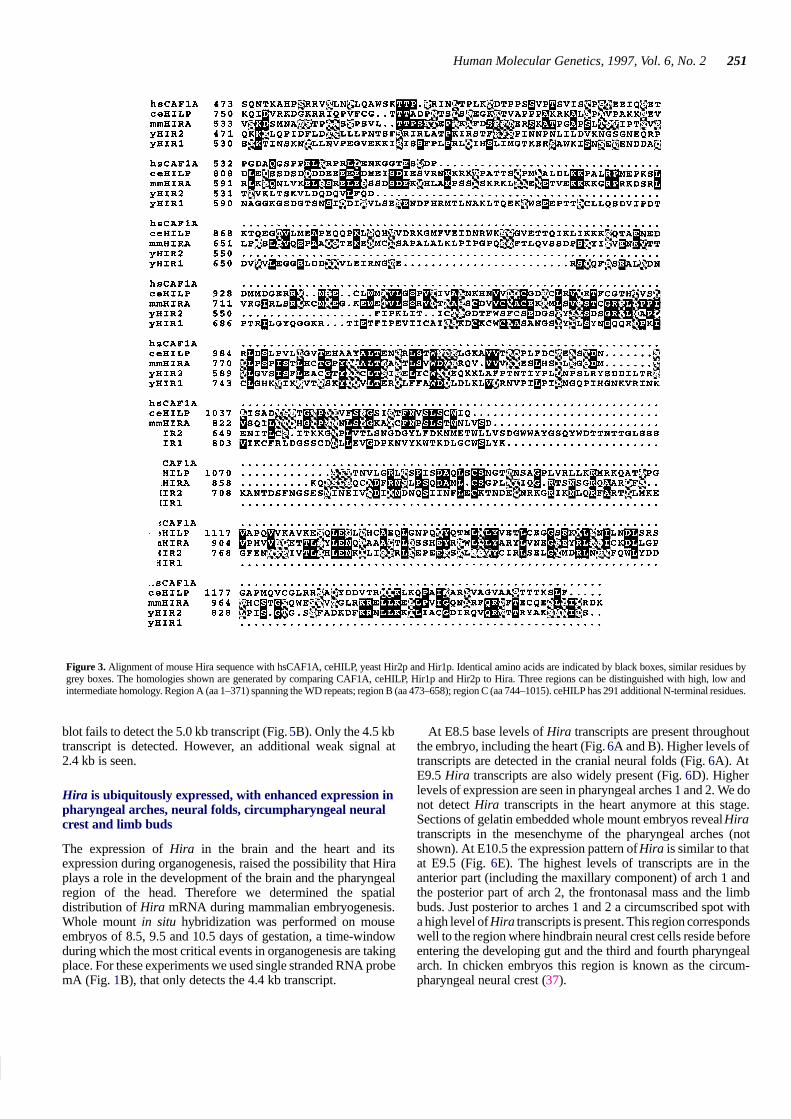
In addition to homology with yeast Hir1p and Hir2p, BlastP andBEAUTY database searches with our mouse Hira sequencereveal high homology to the p60 subunit of human ChromatinAssembly Factor I (CAF1A) (34) and a Caenorhabditis elegansprotein (36) which, in the absence of a name, we willhenceforward refer to as C.elegans HIRA-like protein (ceHILP).The overall homology with ceHILP is higher than with Hir1p(P = 6.1 × 10–77 versus 7.6 × 10–45). We aligned the murine Hiraprotein sequence with ceHILP, Hir1p, Hir2p and CAF1A (Fig. 3).The overall similarities in this alignment are 39% with ceHILP,38% with CAF1A, 35% with Hir1p and 31% with Hir2p. It canbe readily seen that there are at least three different conserved(functional?) regions: the highest homologies are found in theN-terminal part, region A (aa 1–371) (average similarity 47%);in the middle part, region B (aa 473–658), homology drops(average similarity 18%); C-terminal region C (aa 744–1015)shows again an increased homology (average similarity 35%).Sequence stretches with many gaps in the alignment wereexcluded from these regions [comparable with the hinge regionsuggested by Scamps et al. (35)]. In the absence of experimentalevidence for boundaries of functional domains exon–exonboundaries were chosen to provisionally delimit the regions. TheC-terminal boundary of region A corresponds with the insertionpoint for exon 11A. In our alignment the C-termini of Hira,ceHILP and Hir2p almost coincide. CAF1A terminates at the endof region B and Hir1p terminates in region C.
Two Hira transcripts are present from gastrulationstage onward
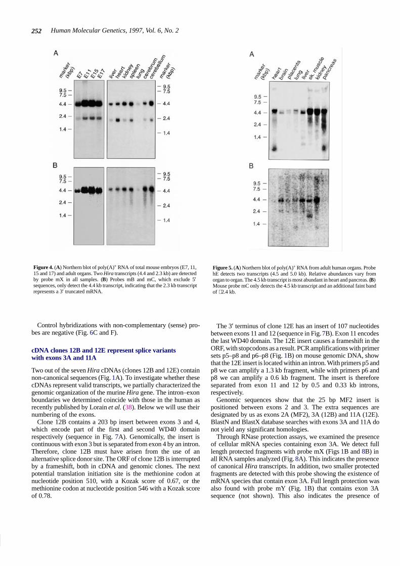
The presence of Hira mRNA in adult mice and embryos wasexamined by Northern blot analysis of poly(A)+ RNA extractedfrom embryos at different stages of development and fromvarious adult organs. Probes 12E and mX (Fig. 1A and B) detecttranscripts of 4.4 and 2.3 kb (Fig. 4A). The 4.4 kb transcript ismore abundant in most organs while the 2.3 kb transcript is moreabundant in the adult heart.
In mouse embryos the 4.4 and 2.3 kb Hira transcripts arealready present during gastrulation stages (E7) (Fig. 4A). At allstages examined the 4.4 kb transcript is most abundant. In thecourse of development the ratio between the two transcripts doesnot seem to vary.
Given the length of the human and mouse HIRA cDNAs of∼4 kb (26,35), the 4.4 kb Hira transcript most likely representsfull-length Hira. Probes mB and mC (Fig. 1B) only detect the4.4 kb transcript on the organ and embryo Northern blots,respectively (Fig. 4B). This suggests that the 2.3 kb transcriptcontains sequences from the 5′ part of Hira only. Thus, the 2.3 kbtranscript might represent an MF2-like mRNA (26).
To compare Hira expression in adult mouse organs withexpression in adult human organs, we hybridized a humanmultiple tissue Northern blot with probe hE (Fig. 1B). Differentfrom what we observed on the Northern blot of adult mouseorgans, probe hE detects two transcripts of ∼4.5 and 5 kb(Fig. 5A). The ratio between the expression levels of the twotranscripts varies from organ to organ. In heart, lung and pancreasthe 5.0 kb transcript is most abundant, while in brain and liver the4.5 kb transcript prevails. Mouse probe mB on the same Northern
Human Molecular Genetics, 1997, Vol. 6, No. 2250
251
Nucleic Acids Research, 1994, Vol. 22, No. 1Human Molecular Genetics, 1997, Vol. 6, No. 2251
Figure 3. Alignment of mouse Hira sequence with hsCAF1A, ceHILP, yeast Hir2p and Hir1p. Identical amino acids are indicated by black boxes, similar residues bygrey boxes. The homologies shown are generated by comparing CAF1A, ceHILP, Hir1p and Hir2p to Hira. Three regions can be distinguished with high, low andintermediate homology. Region A (aa 1–371) spanning the WD repeats; region B (aa 473–658); region C (aa 744–1015). ceHILP has 291 additional N-terminal residues.
blot fails to detect the 5.0 kb transcript (Fig. 5B). Only the 4.5 kbtranscript is detected. However, an additional weak signal at2.4 kb is seen.
Hira is ubiquitously expressed, with enhanced expression inpharyngeal arches, neural folds, circumpharyngeal neuralcrest and limb buds
The expression of Hira in the brain and the heart and itsexpression during organogenesis, raised the possibility that Hiraplays a role in the development of the brain and the pharyngealregion of the head. Therefore we determined the spatialdistribution of Hira mRNA during mammalian embryogenesis.Whole mount in situ hybridization was performed on mouseembryos of 8.5, 9.5 and 10.5 days of gestation, a time-windowduring which the most critical events in organogenesis are takingplace. For these experiments we used single stranded RNA probemA (Fig. 1B), that only detects the 4.4 kb transcript.
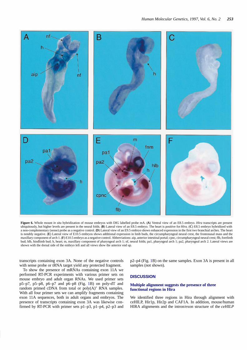
At E8.5 base levels of Hira transcripts are present throughoutthe embryo, including the heart (Fig. 6A and B). Higher levels oftranscripts are detected in the cranial neural folds (Fig. 6A). AtE9.5 Hira transcripts are also widely present (Fig. 6D). Higherlevels of expression are seen in pharyngeal arches 1 and 2. We donot detect Hira transcripts in the heart anymore at this stage.Sections of gelatin embedded whole mount embryos reveal Hiratranscripts in the mesenchyme of the pharyngeal arches (notshown). At E10.5 the expression pattern of Hira is similar to thatat E9.5 (Fig. 6E). The highest levels of transcripts are in theanterior part (including the maxillary component) of arch 1 andthe posterior part of arch 2, the frontonasal mass and the limbbuds. Just posterior to arches 1 and 2 a circumscribed spot witha high level of Hira transcripts is present. This region correspondswell to the region where hindbrain neural crest cells reside beforeentering the developing gut and the third and fourth pharyngealarch. In chicken embryos this region is known as the circum-pharyngeal neural crest (37).
Human Molecular Genetics, 1997, Vol. 6, No. 2252
Figure 4. (A) Northern blot of poly(A)+ RNA of total mouse embryos (E7, 11,15 and 17) and adult organs. Two Hira transcripts (4.4 and 2.3 kb) are detectedby probe mX in all samples. (B) Probes mB and mC, which exclude 5′sequences, only detect the 4.4 kb transcript, indicating that the 2.3 kb transcriptrepresents a 3′ truncated mRNA.
Control hybridizations with non-complementary (sense) pro-bes are negative (Fig. 6C and F).
cDNA clones 12B and 12E represent splice variantswith exons 3A and 11A
Two out of the seven Hira cDNAs (clones 12B and 12E) containnon-canonical sequences (Fig. 1A). To investigate whether thesecDNAs represent valid transcripts, we partially characterized thegenomic organization of the murine Hira gene. The intron–exonboundaries we determined coincide with those in the human asrecently published by Lorain et al. (38). Below we will use theirnumbering of the exons.
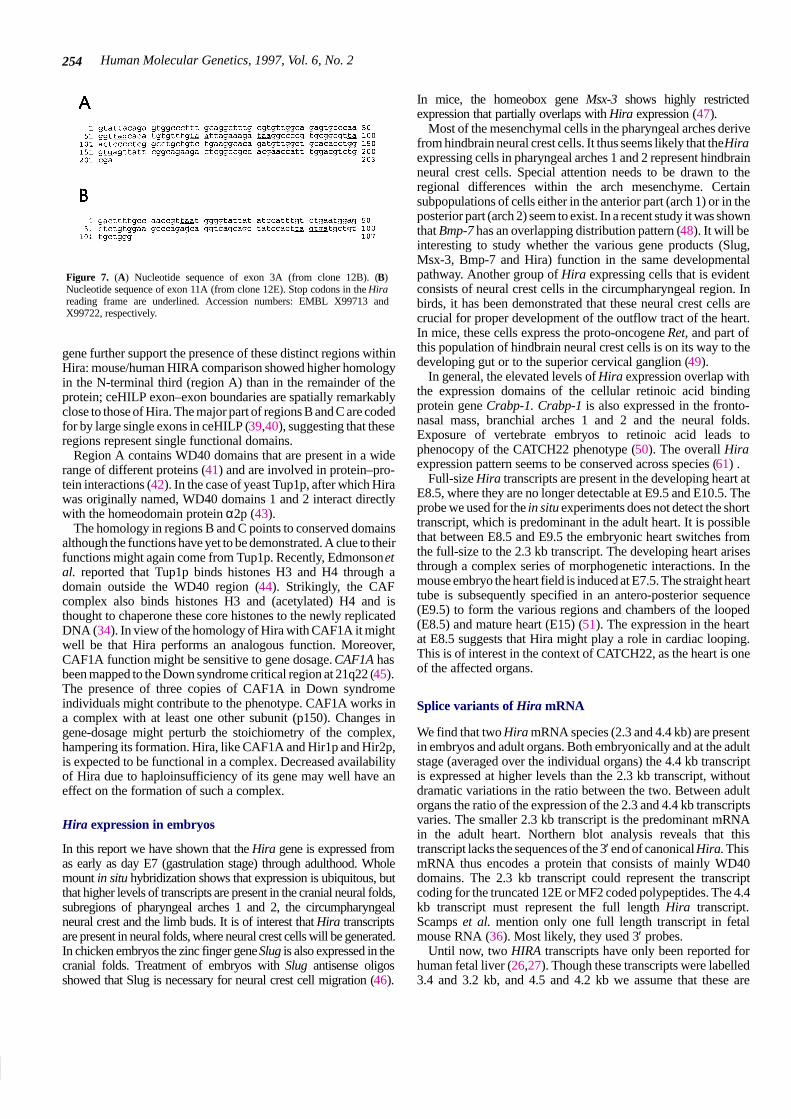
Clone 12B contains a 203 bp insert between exons 3 and 4,which encode part of the first and second WD40 domainrespectively (sequence in Fig. 7A). Genomically, the insert iscontinuous with exon 3 but is separated from exon 4 by an intron.Therefore, clone 12B must have arisen from the use of analternative splice donor site. The ORF of clone 12B is interruptedby a frameshift, both in cDNA and genomic clones. The nextpotential translation initiation site is the methionine codon atnucleotide position 510, with a Kozak score of 0.67, or themethionine codon at nucleotide position 546 with a Kozak scoreof 0.78.
Figure 5. (A) Northern blot of poly(A)+ RNA from adult human organs. ProbehE detects two transcripts (4.5 and 5.0 kb). Relative abundances vary fromorgan to organ. The 4.5 kb transcript is most abundant in heart and pancreas. (B)Mouse probe mC only detects the 4.5 kb transcript and an additional faint bandof ∼2.4 kb.
The 3′ terminus of clone 12E has an insert of 107 nucleotidesbetween exons 11 and 12 (sequence in Fig. 7B). Exon 11 encodesthe last WD40 domain. The 12E insert causes a frameshift in theORF, with stopcodons as a result. PCR amplifications with primersets p5–p8 and p6–p8 (Fig. 1B) on mouse genomic DNA, showthat the 12E insert is located within an intron. With primers p5 andp8 we can amplify a 1.3 kb fragment, while with primers p6 andp8 we can amplify a 0.6 kb fragment. The insert is thereforeseparated from exon 11 and 12 by 0.5 and 0.33 kb introns,respectively.
Genomic sequences show that the 25 bp MF2 insert ispositioned between exons 2 and 3. The extra sequences aredesignated by us as exons 2A (MF2), 3A (12B) and 11A (12E).BlastN and BlastX database searches with exons 3A and 11A donot yield any significant homologies.
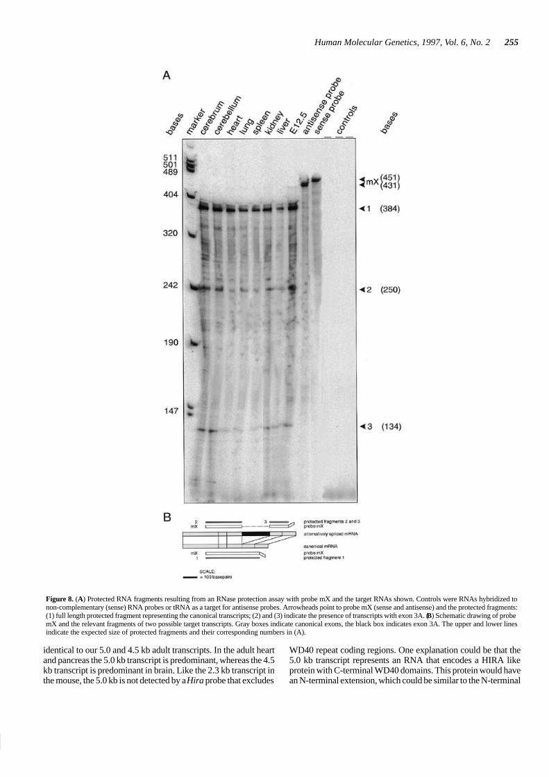
Through RNase protection assays, we examined the presenceof cellular mRNA species containing exon 3A. We detect fulllength protected fragments with probe mX (Figs 1B and 8B) inall RNA samples analyzed (Fig. 8A). This indicates the presenceof canonical Hira transcripts. In addition, two smaller protectedfragments are detected with this probe showing the existence ofmRNA species that contain exon 3A. Full length protection wasalso found with probe mY (Fig. 1B) that contains exon 3Asequence (not shown). This also indicates the presence of
253
Nucleic Acids Research, 1994, Vol. 22, No. 1Human Molecular Genetics, 1997, Vol. 6, No. 2253
Figure 6. Whole mount in situ hybridization of mouse embryos with DIG labelled probe mA. (A) Ventral view of an E8.5 embryo. Hira transcripts are presentubiquitously, but higher levels are present in the neural folds. (B) Lateral view of an E8.5 embryo. The heart is positive for Hira. (C) E8.5 embryo hybridized witha non-complementary (sense) probe as a negative control. (D) Lateral view of an E9.5 embryo shows enhanced expression in the first two branchial arches. The heartis notably negative. (E) Lateral view of E10.5 embryos shows additional expression in limb buds, the circumpharyngeal neural crest, the frontonasal mass and themaxillary component of arch 1. (F) E10.5 embryo as a negative control. Abbreviations: aip, anterior intestinal portal; cpnc, circumpharyngeal neural crest; flb, forelimbbud; hlb, hindlimb bud; h, heart; m, maxillary component of pharyngeal arch 1; nf, neural folds; pa1, pharyngeal arch 1; pa2, pharyngeal arch 2. Lateral views areshown with the dorsal side of the embryo left and all views show the anterior end up.
transcripts containing exon 3A. None of the negative controlswith sense probe or tRNA target yield any protected fragment.
To show the presence of mRNAs containing exon 11A weperformed RT-PCR experiments with various primer sets onmouse embryo and adult organ RNAs. We used primer setsp5–p7, p5–p8, p6–p7 and p6–p8 (Fig. 1B) on poly-dT andrandom primed cDNA from total or poly(A)+ RNA samples.With all four primer sets we can amplify fragments containingexon 11A sequences, both in adult organs and embryos. Thepresence of transcripts containing exon 3A was likewise con-firmed by RT-PCR with primer sets p1–p3, p1–p4, p2–p3 and
p2–p4 (Fig. 1B) on the same samples. Exon 3A is present in allsamples (not shown).
DISCUSSION
Multiple alignment suggests the presence of threefunctional regions in Hira
We identified three regions in Hira through alignment withceHILP, Hir1p, Hir2p and CAF1A. In addition, mouse/humanHIRA alignments and the intron/exon structure of the ceHILP
Human Molecular Genetics, 1997, Vol. 6, No. 2254
Figure 7. (A) Nucleotide sequence of exon 3A (from clone 12B). (B)Nucleotide sequence of exon 11A (from clone 12E). Stop codons in the Hirareading frame are underlined. Accession numbers: EMBL X99713 andX99722, respectively.
gene further support the presence of these distinct regions withinHira: mouse/human HIRA comparison showed higher homologyin the N-terminal third (region A) than in the remainder of theprotein; ceHILP exon–exon boundaries are spatially remarkablyclose to those of Hira. The major part of regions B and C are codedfor by large single exons in ceHILP (39,40), suggesting that theseregions represent single functional domains.
Region A contains WD40 domains that are present in a widerange of different proteins (41) and are involved in protein–pro-tein interactions (42). In the case of yeast Tup1p, after which Hirawas originally named, WD40 domains 1 and 2 interact directlywith the homeodomain protein α2p (43).
The homology in regions B and C points to conserved domainsalthough the functions have yet to be demonstrated. A clue to theirfunctions might again come from Tup1p. Recently, Edmonson etal. reported that Tup1p binds histones H3 and H4 through adomain outside the WD40 region (44). Strikingly, the CAFcomplex also binds histones H3 and (acetylated) H4 and isthought to chaperone these core histones to the newly replicatedDNA (34). In view of the homology of Hira with CAF1A it mightwell be that Hira performs an analogous function. Moreover,CAF1A function might be sensitive to gene dosage. CAF1A hasbeen mapped to the Down syndrome critical region at 21q22 (45).The presence of three copies of CAF1A in Down syndromeindividuals might contribute to the phenotype. CAF1A works ina complex with at least one other subunit (p150). Changes ingene-dosage might perturb the stoichiometry of the complex,hampering its formation. Hira, like CAF1A and Hir1p and Hir2p,is expected to be functional in a complex. Decreased availabilityof Hira due to haploinsufficiency of its gene may well have aneffect on the formation of such a complex.
Hira expression in embryos
In this report we have shown that the Hira gene is expressed fromas early as day E7 (gastrulation stage) through adulthood. Wholemount in situ hybridization shows that expression is ubiquitous, butthat higher levels of transcripts are present in the cranial neural folds,subregions of pharyngeal arches 1 and 2, the circumpharyngealneural crest and the limb buds. It is of interest that Hira transcriptsare present in neural folds, where neural crest cells will be generated.In chicken embryos the zinc finger gene Slug is also expressed in thecranial folds. Treatment of embryos with Slug antisense oligosshowed that Slug is necessary for neural crest cell migration (46).
In mice, the homeobox gene Msx-3 shows highly restrictedexpression that partially overlaps with Hira expression (47).
Most of the mesenchymal cells in the pharyngeal arches derivefrom hindbrain neural crest cells. It thus seems likely that the Hiraexpressing cells in pharyngeal arches 1 and 2 represent hindbrainneural crest cells. Special attention needs to be drawn to theregional differences within the arch mesenchyme. Certainsubpopulations of cells either in the anterior part (arch 1) or in theposterior part (arch 2) seem to exist. In a recent study it was shownthat Bmp-7 has an overlapping distribution pattern (48). It will beinteresting to study whether the various gene products (Slug,Msx-3, Bmp-7 and Hira) function in the same developmentalpathway. Another group of Hira expressing cells that is evidentconsists of neural crest cells in the circumpharyngeal region. Inbirds, it has been demonstrated that these neural crest cells arecrucial for proper development of the outflow tract of the heart.In mice, these cells express the proto-oncogene Ret, and part ofthis population of hindbrain neural crest cells is on its way to thedeveloping gut or to the superior cervical ganglion (49).
In general, the elevated levels of Hira expression overlap withthe expression domains of the cellular retinoic acid bindingprotein gene Crabp-1. Crabp-1 is also expressed in the fronto-nasal mass, branchial arches 1 and 2 and the neural folds.Exposure of vertebrate embryos to retinoic acid leads tophenocopy of the CATCH22 phenotype (50). The overall Hiraexpression pattern seems to be conserved across species (61) .
Full-size Hira transcripts are present in the developing heart atE8.5, where they are no longer detectable at E9.5 and E10.5. Theprobe we used for the in situ experiments does not detect the shorttranscript, which is predominant in the adult heart. It is possiblethat between E8.5 and E9.5 the embryonic heart switches fromthe full-size to the 2.3 kb transcript. The developing heart arisesthrough a complex series of morphogenetic interactions. In themouse embryo the heart field is induced at E7.5. The straight hearttube is subsequently specified in an antero-posterior sequence(E9.5) to form the various regions and chambers of the looped(E8.5) and mature heart (E15) (51). The expression in the heartat E8.5 suggests that Hira might play a role in cardiac looping.This is of interest in the context of CATCH22, as the heart is oneof the affected organs.
Splice variants of Hira mRNA
We find that two Hira mRNA species (2.3 and 4.4 kb) are presentin embryos and adult organs. Both embryonically and at the adultstage (averaged over the individual organs) the 4.4 kb transcriptis expressed at higher levels than the 2.3 kb transcript, withoutdramatic variations in the ratio between the two. Between adultorgans the ratio of the expression of the 2.3 and 4.4 kb transcriptsvaries. The smaller 2.3 kb transcript is the predominant mRNAin the adult heart. Northern blot analysis reveals that thistranscript lacks the sequences of the 3′ end of canonical Hira. ThismRNA thus encodes a protein that consists of mainly WD40domains. The 2.3 kb transcript could represent the transcriptcoding for the truncated 12E or MF2 coded polypeptides. The 4.4kb transcript must represent the full length Hira transcript.Scamps et al. mention only one full length transcript in fetalmouse RNA (36). Most likely, they used 3′ probes.
Until now, two HIRA transcripts have only been reported forhuman fetal liver (26,27). Though these transcripts were labelled3.4 and 3.2 kb, and 4.5 and 4.2 kb we assume that these are
255
Nucleic Acids Research, 1994, Vol. 22, No. 1Human Molecular Genetics, 1997, Vol. 6, No. 2255
Figure 8. (A) Protected RNA fragments resulting from an RNase protection assay with probe mX and the target RNAs shown. Controls were RNAs hybridized tonon-complementary (sense) RNA probes or tRNA as a target for antisense probes. Arrowheads point to probe mX (sense and antisense) and the protected fragments:(1) full length protected fragment representing the canonical transcripts; (2) and (3) indicate the presence of transcripts with exon 3A. (B) Schematic drawing of probemX and the relevant fragments of two possible target transcripts. Gray boxes indicate canonical exons, the black box indicates exon 3A. The upper and lower linesindicate the expected size of protected fragments and their corresponding numbers in (A).
identical to our 5.0 and 4.5 kb adult transcripts. In the adult heartand pancreas the 5.0 kb transcript is predominant, whereas the 4.5kb transcript is predominant in brain. Like the 2.3 kb transcript inthe mouse, the 5.0 kb is not detected by a Hira probe that excludes
WD40 repeat coding regions. One explanation could be that the5.0 kb transcript represents an RNA that encodes a HIRA likeprotein with C-terminal WD40 domains. This protein would havean N-terminal extension, which could be similar to the N-terminal
Human Molecular Genetics, 1997, Vol. 6, No. 2256
extension of ceHILP. This part of ceHILP, which has noequivalent in HIRA, CAF1A, Hir1p or Hir2p, bears very highhomology to other C.elegans proteins that are similar toSchizosaccharomyces pombe SDS22 and L.monocytogenesInternalin (52,53). The yeast SDS22 positively modulates proteinphosphatase 1 (54). Internalin is required for the internalizationof L.monocytogenes by eukaryotic cells (55). Alternatively, the5.0 kb transcript encodes a protein with N-terminal WD40 repeatsfollowed by hitherto unknown sequences not present in canonicalHIRA.
The insertion of exon 3A into the Hira mRNA disrupts thecanonical Hira open reading frame. An N-terminally truncatedprotein starting from either of the next two in-frame translationstart sites lacks the first WD40 domain. This is comparable withthe protein coded for by the MF2 Tuple1 (Hira) cDNA, whichlacks half of the first WD40 domain due to a 25 bp insert in themRNA. In Tup1p the array of WD40 domains appears toinfluence the binding specificity of the individual domains. In thecontext of other WD40 domains, a single domain has differentbinding specificities than in isolation (43). The lack of a WD40domain is therefore very likely to alter the affinity of Hira forother proteins. The insertion exon 11A introduces stop codons,yielding a polypeptide consisting only of WD40 domains. Thistruncated polypeptide could compete with full length Hira forbinding to other proteins. This putative truncated form of Hiraresembles CAF1A which also consists mainly of WD40 repeats.
In conclusion, at least four different splice forms of HIRA canbe identified in humans and mice. This complex expressionpattern of the mammalian HIRA gene is regulated in an organ- anddevelopmental-specific manner and arises from alternativesplicing and exon skipping.
HIRA is being analyzed for point mutations and microdeletionsby several groups that are looking for a cause for the CATCH22phenotype in apparently non-deleted CATCH22 patients. Thenew exons 2A, 3A and 11A should be included in these screens.Further analyses of the human 5.0 kb transcript should help inidentifying more HIRA sequences to check for mutations.
Molecular genetics of CATCH22
In the absence of direct evidence that the CATCH22 phenotypeis the result of a single gene defect, it is most likely that thephenotype is the result of haploinsufficiency of several genes withmajor effect. The spatio-temporal expression pattern of HIRArenders the HIRA gene one of the genes of which haploinsuffi-ciency could lead to part of the CATCH22 phenotype. Sincedeletion screens in CATCH22 patients have so far not reduced theSRDO to less than 300 kb, it will be necessary to analyze thefunction of genes with major effect in mice. Therefore, we areanalyzing the effect on development of a null mutation of themurine Hira gene.
One has to bear in mind, that it will also be important to identifygenes with major effect in SRDO2. Furthermore, position effectscannot be excluded. In campomelic dysplasia and aniridia, SOX9and PAX6, respectively, can be influenced by translocationbreakpoints up to 250 kb distant from the disease causing gene(56,57). A similar effect could play a role in CATCH22. It is alsopossible that as a result of the CATCH22 deletion a heterochro-matic region (e.g. close to the centromere) can exert its influenceonto former distal regions, thereby silencing the genes in theseregions. In this model it is even possible that the genes of major
effect are located outside the commonly deleted region. Sincethere appear to be at least two SRDOs, one cannot exclude thatboth position effects and gene deletions can lead to the CATCH22phenotype. Considering the fact that there are many phenocopiesof the CATCH22 phenotype, deletions or silencing of differentgenes on 22q11 could lead to similar phenotypes.
MATERIALS AND METHODS
Embryos
Embryos were derived from crosses between FVB/N mice. Themorning of vaginal plug was considered 0.5 days post coitum(dpc). The embryos were stages according to the number ofdevelopmental day (E8.5–E12.5). The E8.5 embryos were stagedunder the dissecting microscope by counting somites.
cDNA cloning, sequencing and database searches
A mouse embryo 11.5 dpc cDNA library was purchased fromClontech. Plaques were lifted onto nitrocellulose membranes(Millipore HATF filters) and hybridized as recommended by themanufacturer. The cDNA clones were sequenced by primerwalking using the Pharmacia T7 sequencing kit. Sequences wereassembled, aligned and compared using the GCG package at theCAOS/CAMM server in Nijmegen. Database searches wereperformed using Blast programs (58). Multiple sequence align-ments were edited manually with the GeneDoc program andprinted with BoxShade.
Whole mount in situ hybridization
Complementary (antisense) or non-complementary (sense) RNAprobes were synthesized from linearized DNA templates usingthe DIG-UTP labelling kit (Boehringer Mannheim). In situhybridization on whole mouse embryos (E8.5–E11.5) wasperformed according to the protocol of Wilkinson et al. (59), withposthybridization washes modified as follows: 50 and 70 min insolution 1 at 70�C; 20 min in solution 1 and 2 (1:1) at roomtemperature; 2× 20 min in solution 2 at room temperature; 2× 30min in solution 2 with RNase A at 37�C; 10 min in solution 2 atroom temperature; 2× 30 min in solution 3 at 65�C; 2× 30 min inTBST at room temperature. Adsorption of anti-digoxigeninantibodies to embryo powder was omitted. Embryos were eithercaptured on film and subsequently scanned, or captured electroni-cally with an electronic camera. Images were processed usingAdobe Photoshop software.
RNA isolation
Adult organs or embryos were homogenized in GIT or LiCl ureawith a tissue homogenizer. Total RNA was purified by ultracentrifugation on a cesium-chloride cushion or by LiCl precipita-tion and organic extractions. Poly(A)+ RNA was isolated fromtotal RNA with the PolyATtract mRNA isolation system III(Promega).
Southern and Northern analyses
A mouse embryo and a human multiple tissue Northern blot,together with a Zoo blot were purchased from Clontech (PaloAlto, CA). Blots were hybridized as per manufacturer’s protocol.The mouse multiple tissue Northern blot was produced byrunning 5 µg poly(A)+ RNA per lane on a denaturing formalde-
257
Nucleic Acids Research, 1994, Vol. 22, No. 1Human Molecular Genetics, 1997, Vol. 6, No. 2257
hyde agarose gel, blotting in 20× SSC onto Qiabrene+ nylonmembrane (Qiagen), and UV cross-linking in a Stratalinker(Stratagene).
RNase protection assay
Complementary (antisense) or non-complementary (sense)[α-32P]UTP labelled RNA probes were synthesized from linearizedHira cDNA templates, gel-eluted, hybridized to total RNA at 50�Cand subjected to RNase protection assay according to ref. 60.
RT-PCR detection of alternative Hira mRNA
RT-PCR was performed on 5 µg total RNA or 1 µg poly(A)+ RNA(from E8.5, E9.5, E10.5 and E14.5 mouse embryos and adultmouse brain, spleen, liver and lung) with a mix of 300 ng each ofoligo-dT and random hexamer primers according to establishedprotocols. The RT reaction product was used for the followingPCR amplification: one cycle of 2 min at 94�C, 2 min at 50�C,2 min at 72�C; 30 cycles of 1 min at 94�C, 1 min at 50�C, 1.5 minat 72�C and one cycle of 10 min at 72�C. Primer sequences: p1,ctgggaaggttgtgatctgg; p2, ggtattacagagtggccctt; p3, tcgcagacgtc-caaatggtt; p4, ccaatgtacgtagcccgctt; p5, atactgctgctgtgctgttg; p6,tttgccaaccgttaatgggg; p7, cccagcaacagcatcactaa; p8, atgct-gagcttgtctcccta. Positive controls were 1 pg of cDNAs 12B, E andF. Negative controls were omission of cDNA and a sample froman RT reaction without RNA.
ACKNOWLEDGEMENTS
We thank Drs Frans Lohman, Vincent Lui and Maarten Mulderfor comments on the manuscript. We thank Peter Scambler for hisgift of the human C5 cDNA and sharing unpublished results. Prof.Jan C. Molenaar is acknowledged for his continuous support ofour work. This study was financially supported by the ErasmusUniversity, the Sophia Foundation for Medical Research, theEuropean Community and the Dutch Heart Foundation.
ABBREVIATIONS
E, embryonic age in days post coitum; SRDO, smallest region ofdeletion overlap.
REFERENCES
1. Wilson,D.I., Burn,J., Scambler,P. and Goodship,J. (1993) DiGeorge syn-drome: part of CATCH22. J. Med. Genet., 30, 852–856.
2. de la Chapelle,A., Herva,R., Koivisto,M. and Aula,P. (1981) A deletion inchromosome 22 can cause DiGeorge syndrome. Hum. Genet., 57, 253–256.
3. Carey,A.H., Roach,S., Williamson,R., Dumanski,J.P., Nordenskjold,M.,Collins,V.P., Rouleau,G., Blin,N., Jalbert,P. and Scambler,P.J. (1990) Localiz-ation of 27 DNA markers to the region of human chromosome 22q11–pterdeleted in patients with the DiGeorge syndrome and duplicated in the der22syndrome. Genomics, 7, 299–306.
4. Driscoll,D.A., Budarf,M.L. and Emanuel,B.S. (1992) A genetic etiology forDiGeorge syndrome: consistent deletions and microdeletions of 22q11. Am.J. Hum. Genet., 50, 924–933.
5. Scambler,P.J., Kelly,D., Lindsay,E., Williamson,R., Goldberg,R.,Shprintzen,R., Wilson,D.I., Goodship,J.A., Cross,I.E. and Burn,J. (1992)Velo-cardio-facial syndrome associated with chromosome 22 deletionsencompassing the DiGeorge locus. Lancet, 339, 1138–1139.
6. Driscoll,D.A., Spinner,N.B., Budarf,M.L., McDonald-McGinn,D.M., Zack-ai,E.H., Goldberg,R.B., Shprintzen,R.J., Saal,H.M., Zonana,J., Jones,M.C.,Mascarello,J.T. and Emanuel,B.S. (1992) Deletions and microdeletions of22q11.2 in velo-cardio-facial syndrome. Am. J. Med. Genet., 44, 261–268.
7. Burn,J., Takao,A., Wilson,D., Cross,I., Momma,K., Wadey,R., Scambler,P.and Goodship,J. (1993) Conotruncal anomaly face syndrome is associatedwith a deletion within chromosome 22q11. J. Med. Genet., 30, 822–824.
8. McDonald-McGinn,D.M., Driscoll,D.A., Bason,L., Christensen,K.,Lynch,D., Sullivan,K., Canning,D., Zavod,W., Quinn,N., Rome,J., Paris,Y.,Weinberg,P., Clark III,B.J., Emanuel,B.S. and Zackai,E.H. (1995) Autosomaldominant ‘Opitz’ GBBB syndrome due to a 22q11.2 deletion. Am. J. Med.Genet., 59, 103–113.
9. Robin,N.H., Feldman,G.J., Aronson,A.L., Mitchell,H.F., Weksberg,R., Leo-nard,C.O., Burton,B.K., Josephson,K.D., Laxova,R., Aleck,K.A., Allan-son,J.E., Guionalmeida,M.L., Martin,R.A., Leichtman,L.G., Price,R.A.,Opitz,J.M. and Muenke.M. (1995) Opitz syndrome is genetically heteroge-nous with one locus on Xp22, and a second locus on 22q11.2. Nature Genet.,11, 459–461.
10. Fibison,W.J., Budarf,M.L., McDermid,H., Greenberg,F. and Emanuel,B.S.(1990) Molecular studies of DiGeorge syndrome. Am. J. Hum. Genet., 46,888–895.
11. Lindsay,E.A., Halford,S., Wadey,R., Scambler,P.J. and Baldini,A. (1993)Molecular cytogenetic characterization of the DiGeorge syndrome regionusing fluorescence in situ hybridization. Genomics, 17, 403–407.
12. Aubry,M., Demczuk,S., Desmaze,C., Aikem,M., Aurias,A., Julien,J.P. andRouleau,G. (1993) Isolation of a zinc finger gene consistently deleted inDiGeorge syndrome. Hum. Mol. Genet., 2, 1583–1587.
13. Kurahashi,H., Akagi,K., Karakawa,K., Nakamura,T., Dumanski,J.P.,Sano,T., Okada,S., Takai,S.I. and Nishisho,I. (1994) Isolation and mapping ofcosmid markers on human chromosome 22, including one within thesubmicroscopically deleted region of DiGeorge syndrome. Hum. Genet., 93,248–254.
14. Mulder,M.P., Wilke,M., Langeveld,A., Wilming,L.G., Hagemeijer,A., vanDrunen,E., Zwarthoff,E.C., Riegman,P.J., Deelen,W.H., van den Ouwe-land,A.M.W., Halley,D.J.J. and Meijers,C. (1995) Positional mapping of lociin the DiGeorge critical region at chromosome 22q11 using a new marker(D22S183). Hum. Genet., 96, 133–141.
15. Morrow,B., Goldberg,R., Carlson,C., Gupta,R.D., Sirotkin,H., Collins,J.,Dunham,I., O.Donnell,H., Scambler,P., Shprintzen,R. and Kucherlapati,R.(1995) Molecular definition of the 22q11 deletions in velo-cardio-facialsyndrome. Am. J. Hum. Genet., 56, 1391–1403.
16. Levy,A., Demczuk,S., Aurias,A., Depetris,D., Mattei,M.G. and Philip,N.(1995) Interstitial 22q11 microdeletion excluding the ADU breakpoint in apatient with the DiGeorge syndrome. Hum. Mol. Genet., 4, 2417–2419.
17. Kurahashi,H., Nakayama,T., Osugi,Y., Tsuda,E., Masuno,M., Imaizumi,K.,Kamiya,T., Sano,T., Okada,S. and Nishisho,I. (1996) Deletion mapping of22q11 in CATCH22 syndrome: identification of a second critical region. Am.J. Hum. Genet., 58, 1377–1381.
18. Dallapiccola,B., Pizzuti,A. and Novelli,G. (1996) How many breaks do weneed to CATCH on 22q11? Am. J. Hum. Genet., 59, 7–11.
19. Fu,W.N., Borgaonkar,D.S., Ledewig,P.P., Weaver,J. and Pomerance,H.H.(1976) Structural aberrations of the long arm of chromosome no. 22. Clin.Genet., 10, 329–336.
20. Augusseau,S., Jouk,S., Jalbert,P. and Prieur,M. (1986) DiGeorge syndromeand 22q11 rearrangements. Hum. Genet., 74, 206.
21. Budarf,M.L., Collins,J., Gong,W., Roe,B., Wang,Z., Bailey,L.C., Sel-linger,B., Michaud,D., Driscoll,D.A. and Emanuel,B.S. (1995) Cloning abalanced translocation associated with DiGeorge syndrome and identifica-tion of a disrupted candidate gene. Nature Genet., 10, 269–277.
22. Sutherland,H.F., Wadey,R., McKie,J.M., Taylor,C., Atif,U., Johnstone,K.A.,Halford,S., Kim,U.J., Goodship,J., Baldini,A. and Scambler,J. (1996)Identification of a novel transcript disrupted by a balanced translocationassociated with DiGeorge syndrome. Am. J. Hum. Genet., 59, 23–31.
23. Collins,J.E., Cole,C.G., Smink,L.J., Garett,C.L., Leversha,M.A., Soder-lund,C.A., Maslen,G.L., Everett,L.A., Rice,K.M., Coffey,A.J., Gregory,S.G.,William,R., Dunham,A., Davies,A.F., Hassock,S., Todd,C.M., Lehrach,H.,Hulsebos,T.J.M., Weissenbach,J., Morrow,B., Kucherlapati,R.S., Wadey,R.,Scambler,P.J., Kim,U.J., Simon,M.I., Peyrard,M., Xie,Y.G., Carter,N.P., Dur-bin,R., Dumanski,J.P., Bentley,D.R. and Dunham,I. (1995) A high-density YACcontig map of human chromosome 22. Nature, 377 Suppl., 367–379.
24. Gong,W., Emanuel,B.S., Collins,J., Kim,D.H., Wang,Z., Chen,F., Zhang,G.,Roe,B. and Budarf,M.L. (1996) A transcriptional map of the DiGeorge andvelo-cardio-facial syndrome minimal critical region on 22q11. Hum. Mol.Genet., 5, 789–800.
25. Lindsay,E.A., Rizzu,P., Antonacci,R., Juecic,V., Delmas-Mata,J., Lee,C.C.,Kim,U.J., Scambler,P.S. and Baldini,A. (1996) A transcription map in theCATCH22 critical region: identification, mapping, and ordering of four noveltranscripts expressed in heart. Genomics, 32, 104–112.
Human Molecular Genetics, 1997, Vol. 6, No. 2258
26. Halford,S., Wadey,R., Roberts,C., Daw,S.C.M., Whiting,J.A., O’Donnell,H.,Dunham,I., Bentley,D., Lindsay,E., Baldini,A., Francis,F., Lehrach,H., Wil-liamson,R., Wilson,D.I., Goodship,J., Cross,I., Burn,J. and Scambler,P.J.(1993) Isolation of a putative transcriptional regulator from the region of22q11 deleted in DiGeorge syndrome, Sphrintzen syndrome and familialcongenital heart disease. Hum. Mol. Genet., 2, 2099–2107
27. Lamour,V., Lécluse,Y., Desmaze,C., Spector,M., Bodescot,M., Aurias,A.,Osley,M.A. and Lipinski,M. (1995) A human homolog of the S. cerevisiaeHIR1 and HIR2 transcriptional repressors cloned from the DiGeorgesyndrome critical region. Hum. Mol. Genet., 4, 791–799.
28. Sirotkin,H., Morrow,B., Dasgupta,R., Goldberg,R., Patanjali,S.R., Shi,G.P.,Cannizzaro,L., Shprintzen,R., Weissman,S.M. and Kucherlapati,R. (1996)Isolation of a new clathrin heavy chain gene with muscle-specific expressionfrom the region commonly deleted in velo-cardio-facial syndrome. Hum.Mol. Genet., 5, 617–624.
29. Kedra,D., Peyrard,M., Fransson,I., Collins,J.E., Dunham,I., Roe,B.A. andDumanski,P.J. (1996) Characterization of a second human clathrin heavychain polypeptide gene (CLH-22) from chromosome 22q11. Hum. Mol.Genet., 5, 625–631.
30. Heisterkamp,N., Mulder,M.P., Langeveld,A., Ten Hoeve,J., Wang,Z.,Roe,B.A. and Groffen,J. (1995) Localization of the human mitochondrialcitrate transporter protein gene to chromosome 22q11 in the DiGeorge criticalregion. Genomics, 29, 451–456.
31. Pizzuti,A., Novelli,G., Mari,A., Ratti,A., Colosimo,A., Amati,F., Penso,D.,Sangiuolo,F., Calabrese,G., Palka,G., Salani,V., Gennarelli,M., Mingarel-li,R., Scarlatto,G., Scambler,P. and Dallapiccola,B. (1996) Human homo-logue sequences to the Drosophila dishevelled segment polarity gene aredeleted in the DiGeorge syndrome. Am. J. Hum. Genet., 58, 722–729.
32. Sherwood,P.W., Tang,S.V. and Osley,M.A. (1993) Characterization of HIR1and HIR2, two genes required for regulation of histone gene transcription inSaccharomyces cerevisiae. Mol. Cell Biol., 13, 28–38.
33. Promisel Cooper,J., Roth,S. and Simpson,R.T. (1994) The global transcrip-tional regulators SSN6 and TUP1, play distinct roles in the establishment of arepressive chromatin structure. Genes Dev., 8, 1400–1410.
34. Kaufman,P.D., Kobayashi,R., Kessler,N. and Stillman,B. (1995) The p150and p60 subunits of chromatin assembly factor I: a molecular link betweennewly synthesized histones and DNA replication. Cell, 81, 1105–1114.
35. Scamps,C., Lorain,S., Lamour,V. and Lipinski,M. (1996) The HIR proteinfamily: isolation and characterization of a complete murine cDNA. Biochim.Biophys. Acta, 1306, 5–8.
36. Miller,N. and Waterston,R. (PID g687860)37. Kuratani,S.C. and Kirby,M.L. (1991) Initial migration and distribution of the
cardiac neural crest in the avian embryo: an introduction to the concept of thecircumpharyngeal crest. Am. J. Anat., 191, 215–227.
38. Lorain,S., Demczuk,S., Lamour,V., Toth,S., Aurias,A., Roe,B.A. and Lipin-ski,M. (1996) Structural organization of the WD40 repeat protein-encodinggene HIRA in the DiGeorge syndrome critical region of human chromosome22. Genome Res., 6, 43–50.
39. Miller,N. and Waterston,R. (U21322).40. Wilson,R., Ainscough,R., Anderson,K., Baynes,C., Berks,M., Bonfield,J.,
Burton,J., Connell,M., Copsey,T., Cooper,J., Coulson,A., Craxton,M.,Dear,S., Du,Z., Durbin,R., Favello,A., Fraser,A., Fulton,L., Gardner,A.,Green,P., Hawkins,T., Hillier,L., Jier,M., Johnston,L., Jones,M., Kershaw,J.,Kirsten,J., Laisster,N., Latreille,P., Lightning,J., Lloyd,C., Mortimore,B.,O’Callaghan,M., Parson,J., Percy,C., Rifken,L., Roopra,A., Saunders,D.,Shownkeen,R., Sims,M., Smaldon,N., Smith,A., Smith,M., Sonnhammer,E.,Staden,R., Sulston,J., Thierry-Mieg,J., Thomas,K., Vaudin,M., Vaughan,K.,Waterston,R., Watson,A., Weinstock,L., Wilkinson-Sproat,J. and Wohld-man,P. (1994) 2.2 Mb of contiguous nucleotide sequence from chromosomeIII of C. elegans. Nature, 368, 32–38.
41. Neer,E.J., Schmidt,C.J., Nambudripad,R. and Smith,T.F. (1994) The ancientregulatory-protein family of WD-repeat proteins. Nature, 371, 297–300.
42. Sondek,J., Bohm,A., Lambright,D.G., Hamm,H.E. and Sigler,P.B. (1996)Crystal structure of a GA protein ßγ dimer at 2.1 Å resolution. Nature, 379,369–374.
43. Komachi,K., Redd,M.J. and Johnson,A.D. (1994) The WD repeats of Tup1interact with the homeo domain protein α2. Genes Dev., 8, 2857–2867.
44. Edmondson,D.G., Smith,M.M. and Roth,S.Y. (1996) Repression domain ofthe yeast global repressor Tup1 interacts directly with histones H3 and H4.Genes Dev., 10, 1247–1259.
45. Blouin,J.L., Duriaux-Sail,G., Chen,H., Gos,A., Morris,M.A., Rossier,C. andAntonorakis,S.E. (1996) Mapping of the gene for the p60 subunit of thehuman chromatin assembly factor (CAF1A) to the Down syndrome region ofchromosome 21. Genomics, 33, 309–312.
46. Nieto,M.A., Sargent,M.G., Wilkinson,D.G. and Cooke,J. (1994) Control ofcell behavior during vertebrate development by Slug, a zinc finger gene.Science, 264, 835–839.
47. Shimeld,S.M., McKay,I.J. and Sharpe,P.T. (1996) The murine homeoboxgene Msx-3 shows highly restricted expression in the developing neural tube.Mech. Dev., 55, 201–210.
48. Wall,N.A. and Hogan,B.L.M. (1995) Expression of bone morphogeneticprotein-4 (BMP-4), bone morphogenetic protein-7 (BMP-7), fibroblastgrowth factor-8 (FGF-8) and sonic hedgehog (SHH) during branchial archdevelopment in the chick. Mech. Dev., 53, 383–392.
49. Durbec,P.L., Larsson-Blomberg,L.B., Schuchard,A., Constantini,F. andPachnis,V. (1996) Common origin and developmental dependance on c-ret ofsubsets of enteric and sympathetic neuroblasts. Development, 122, 349–358.
50. Vaessen,M.J., Meijers,J.H.C., Bootsma,D. and Geurts van Kessel,A. (1990)The cellular retinoic acid-binding protein is expressed in tissues associatedwith retinoic-acid-induced malformations. Development, 110, 371–378.
51. Kaufman,M.H. (1992) The atlas of mouse development, Academic Press,London
52. Leimbach,D. and Waterston,R. (Genbank U39849)53. Lightning,J. (P45969)54. Ohkura,H. and Yanagida,M. (1991) S. pombe gene SDS22+ essential for a
midmitotic transition encodes a leucine-rich repeat protein that positivelymodulates protein phosphatase-1. Cell, 64, 149–157.
55. Mengaud,J., Ohayon,H., Gounon,P., Mege,R.M. and Cossart,P. (1996)E-cadherin is the receptor for internalin, a surface protein required for entry ofL. monocytogenes into epithelial cells. Cell, 84, 923–932.
56. Wirth,J., Wagner,T., Meyer,J., Pfeiffer,R.A., Tietze,H.U., Schempp,W. andScherer,G. (1996) Translocation breakpoints in three patients with campo-melic dysplasia and autosomal sex reversal map more than 130 kb from Sox9.Hum. Genet., 97, 186–193.
57. Fantes,J., Redeker,B., Breen,M., Boyle,S., Brown,J., Fletcher,J., Jones,S.,Bickmore,W., Fukushima,Y., Mannens,M., Danes,S., van Heyningen,V. andHanson,I. (1995) Aniridia-associated cytogenetic rearrangements suggestthat a position effect may cause the mutant phenotype. Hum. Mol. Genet., 4,415–422.
58. Altschul,S.F., Gish,W., Miller,W., Meyers,E.W. and Lipman,D.J. (1990)Basic local alignment search tool. J. Mol. Biol., 215, 403–410.
59. Wilkinson,D.G. (1992) in Wilkinson,D.G. (ed.), In Situ Hybridization: APractical Approach. IRL Press, Oxford, pp. 75–83.
60. Gilman,M. (1987) in Ausubel,F.M., Brent,R., Kingston,R.E., Moore,D.D.,Seidman,J.G., Smith,J.A. and Struhl,K. (eds), Current Protocols in Molecu-lar Biology. Wiley Interscience, New York, Vol. I, pp.4.7.1.
61. Roberts, C., Daw, S.C.M., Halford, S. and Scambler, P.J. (1997) Cloning anddevelopmental expression analysis of chick Hira (Chira), a candidate genefor DiGeorge syndrome. Hum. Mol. Genet. 6, 235–243.
NOTE ADDED IN PROOF
One of the reviewers pointed to the fact that patients and theirfamilies often find the use of the term CATCH22 offensive. Wepropose to use the acronym in scientific communications only.For clinical purposes the various syndromes could be referred tocollectively as the ‘chromosome 22q11 deletion syndrome’.