Embed Size (px)
Citation preview

The Most Versatile AFM System for Materials Research
swiss qualitywww.nanosurf.com
Flex-Axiom � Measurement capabilities in air and liquid
� Versatility in applications and modes
� Compatibility with inverted microscopes
� High precision scanning and data acquisition
Nanosurf®
FlexAFM Axiom
Stiffness (kPa)
Perc
enta
ge o
bser
ved
0 200 400 600 800 10000
10
20
30
40E
Displacement (nm)
Forc
e (n
N)
0 50 100 150 200 2500.0
0.2
0.4
0.6
0.8
1.0F
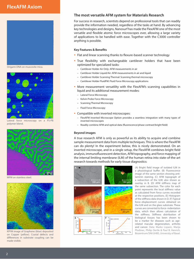
The most versatile AFM system for Materials ResearchFor success in research, scientists depend on professional tools that can readily provide the information needed, regardless of the tasks at hand. By advancing key technologies and designs, Nanosurf has made the FlexAFM one of the most versatile and flexible atomic force microscopes ever, allowing a large variety of applications to be handled with ease. Together with the C3000 controller anything is possible.
Key Features & Benefits
� Flat and linear scanning thanks to flexure-based scanner technology
� True flexibility with exchangeable cantilever holders that have been optimized for specialized tasks:– Cantilever Holder Air Only: AFM measurements in air
– Cantilever Holder Liquid/Air: AFM measurements in air and liquid
– Cantilever Holder Scanning Thermal: Scanning thermal microscopy
– Cantilever Holder FluidFM: Fluid Force Microscopy applications
� More measurement versatility with the FlexAFM’s scanning capabilities in liquid and its additional measurement modes:– Lateral Force Microscopy
– Kelvin Probe Force Microscopy
– Scanning Thermal Microscopy
– Fluid Force Microscopy
� Compatible with inverted microscopes:– FlexAFM Inverted Microscope Option provides a seamless integration with many types of
inverted microscopes
– Readily combine AFM and optical data (fluorescence/phase contrast/bright field)
Beyond images
A true research AFM is only as powerful as its ability to acquire and combine sample measurement data from multiple techniques. This is where the FlexAFM can do plenty! In the experiment below, this is nicely demonstrated. On an inverted microscope, and in a single setup, the FlexAFM combines bright field analysis, immunofluorescent detection, AFM topography, and force mapping of the internal limiting membrane (ILM) of the human retina into state-of-the-art research towards methods for early tissue diagnostics:
(A) Bright field image of isolated ILM in a physiological buffer. (B) Fluorescence image of the same section showing anti-laminin staining. (C) AFM topograph of a subsection of the ILM; also shown as overlay in B. (D) AFM stiffness map of the same subsection. The color for each point represents the local stiffness value as calculated from force curves recorded at the respective positions. (E) Histogram of the stiffness data shown in D. (F) Typical force–displacement curves obtained on the ILM and on the glass substrate. These curves are converted to force–indentation data, which then allows calculation of the stiffness. Stiffness distribution of biological tissues has been shown to be a marker for diseases such as age-related macular degeneration, arthritis and cancer. Data: Marko Loparic, Marija Plodinec, Philip Oertle & Paul B. Henrich, Biozentrum/SNI/UHBS, University of Basel.
180 µm
BA
180 µm
20 µm
C D
20 µm
ILMglass substrateILM
membrane edge
glass substrate
ILM
membrane edge
Lateral force microscopy on a PS-PB polymer blend.
MFM on stainless steel.
Origami DNA on muscovite mica.
200 nm
KPFM image of Graphene (blue) deposited on Copper (yellow). Crystal defects and differences in substrate coupling can be made visible.
2
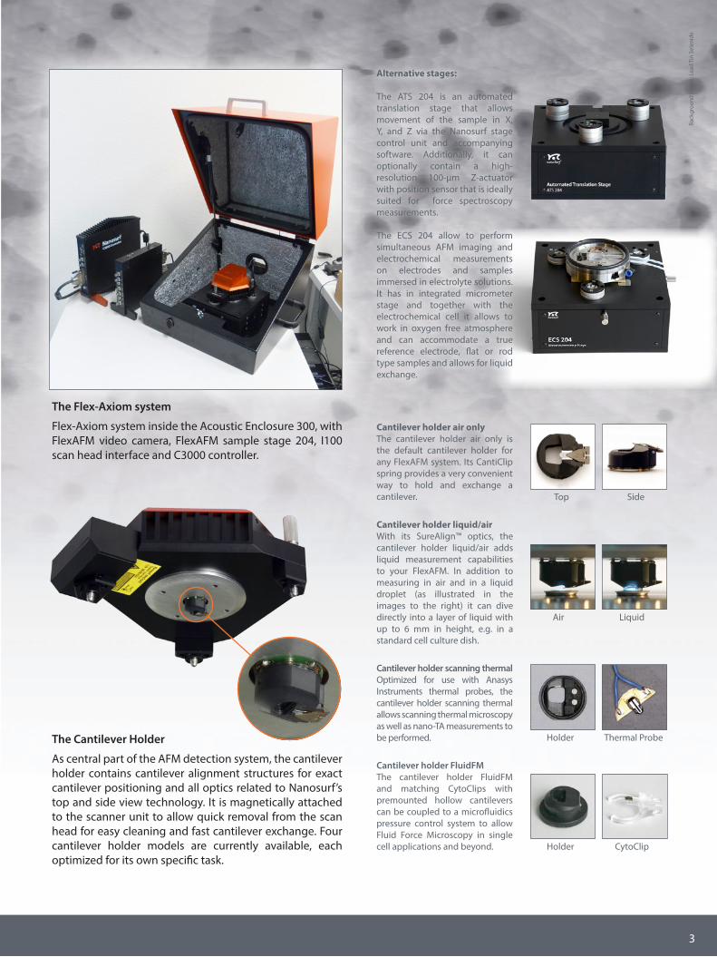
The Cantilever Holder
As central part of the AFM detection system, the cantilever holder contains cantilever alignment structures for exact cantilever positioning and all optics related to Nanosurf’s top and side view technology. It is magnetically attached to the scanner unit to allow quick removal from the scan head for easy cleaning and fast cantilever exchange. Four cantilever holder models are currently available, each optimized for its own specific task.
The Flex-Axiom system
Flex-Axiom system inside the Acoustic Enclosure 300, with FlexAFM video camera, FlexAFM sample stage 204, I100 scan head interface and C3000 controller.
Cantilever holder air onlyThe cantilever holder air only is the default cantilever holder for any FlexAFM system. Its CantiClip spring provides a very convenient way to hold and exchange a cantilever. Top Side
Air Liquid
Cantilever holder liquid/airWith its SureAlign™ optics, the cantilever holder liquid/air adds liquid measurement capabilities to your FlexAFM. In addition to measuring in air and in a liquid droplet (as illustrated in the images to the right) it can dive directly into a layer of liquid with up to 6 mm in height, e.g. in a standard cell culture dish.
Holder Thermal Probe
Cantilever holder scanning thermalOptimized for use with Anasys Instruments thermal probes, the cantilever holder scanning thermal allows scanning thermal microscopy as well as nano-TA measurements to be performed.
Holder CytoClip
Cantilever holder FluidFMThe cantilever holder FluidFM and matching CytoClips with premounted hollow cantilevers can be coupled to a microfluidics pressure control system to allow Fluid Force Microscopy in single cell applications and beyond.
Alternative stages:
The ATS 204 is an automated translation stage that allows movement of the sample in X, Y, and Z via the Nanosurf stage control unit and accompanying software. Additionally, it can optionally contain a high-resolution 100-µm Z-actuator with position sensor that is ideally suited for force spectroscopy measurements.
The ECS 204 allow to perform simultaneous AFM imaging and electrochemical measurements on electrodes and samples immersed in electrolyte solutions. It has in integrated micrometer stage and together with the electrochemical cell it allows to work in oxygen free atmosphere and can accommodate a true reference electrode, flat or rod type samples and allows for liquid exchange.
Back
grou
nd im
age:
Lea
d Ti
n Se
leni
de
3

C3000 controllerHigh-end AFM controller for more performance and precisionThe versatility and performance of the FlexAFM scan head is brought to its full potential by the C3000 controller. With this AFM controller’s fully digital internal data processing, 24-bit ADC/DAC conversion depth, and programmable FPGA CPU, it is a huge step up from the standard Easyscan 2 controller. It allows high-speed data acquisition, dynamic filtering and analysis, and real-time signal monitoring directly from within the C3000 control software.
Through soft- and firmware changes, the C3000 controller can be updated and upgraded to support new options and features at any time!
Main features
� All digital data processing in FPGA
� 24-bit DACs for accurate scanning with widely varying scan ranges
� 24-bit ADCs and adaptive filters for high-resolution and low-noise data
� Fast and sensitive digital Z-feedback and spectroscopy
� Fully equiped with integrated thermal tuning, data monitoring, user I/O and signal access, advanced operating modes
Additional options
Available C3000 controller options/packages include: advanced spectroscopy, signal modulation, advanced lithography, scripting interface, external synchronization.
DNA on mica. Scan size: 300 nm.
Elastic modulus. Elasticity map of a PS-PB blend (top) and corresponding histogram (bottom).
Lateral force microscopy on mica. Using a 10-µm high-resolution FlexAFM 5 scan head and the C3000 controller, the atomic grid of mica was imaged in LFM mode.
4
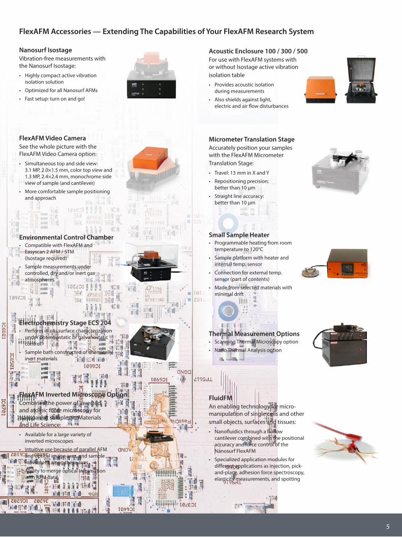
Thermal Measurement Options• Scanning Thermal Microscopy option
• Nano Thermal Analysis option
FlexAFM Inverted Microscope OptionCombines the power of inverted and atomic force microscopy for transparent samples in Materials and Life Science:
• Available for a large variety of inverted microscopes
• Intuitive use because of parallel AFM and optical image axes, and sample movement along these axes
• Ability to merge optical information with AFM data
FlexAFM Video CameraSee the whole picture with the FlexAFM Video Camera option:
• Simultaneous top and side view: 3.1 MP, 2.0×1.5 mm, color top view and 1.3 MP, 2.4×2.4 mm, monochrome side view of sample (and cantilever)
• More comfortable sample positioning and approach
Acoustic Enclosure 100 / 300 / 500For use with FlexAFM systems with or without Isostage active vibration isolation table
• Provides acoustic isolation during measurements
• Also shields against light, electric and air flow disturbances
Nanosurf IsostageVibration-free measurements with the Nanosurf Isostage:
• Highly compact active vibration isolation solution
• Optimized for all Nanosurf AFMs
• Fast setup: turn on and go!
Environmental Control Chamber• Compatible with FlexAFM and
Easyscan 2 AFM / STM (Isostage required)
• Sample measurements under controlled, dry and/or inert gas atmospheres
Small Sample Heater• Programmable heating from room
temperature to 120°C
• Sample platform with heater and internal temp. sensor
• Connection for external temp. sensor (part of contents)
• Made from selected materials with minimal drift
Micrometer Translation StageAccurately position your samples with the FlexAFM Micrometer Translation Stage:
• Travel: 13 mm in X and Y
• Repositioning precision: better than 10 µm
• Straight line accuracy: better than 10 µm
FluidFMAn enabling technology for micro-manipulation of single cells and other small objects, surfaces and tissues:
• Nanofluidics through a hollow cantilever combined with the positional accuracy and force control of the Nanosurf FlexAFM
• Specialized application modules for different applications as injection, pick-and-place, adhesion force spectroscopy, elasticity measurements, and spotting
FlexAFM Accessories — Extending The Capabilities of Your FlexAFM Research System
Electrochemistry Stage ECS 204• Perform in situ surface characterization
under potentiostatic or galvanostatic control
• Sample bath constructed of chemically inert materials
5
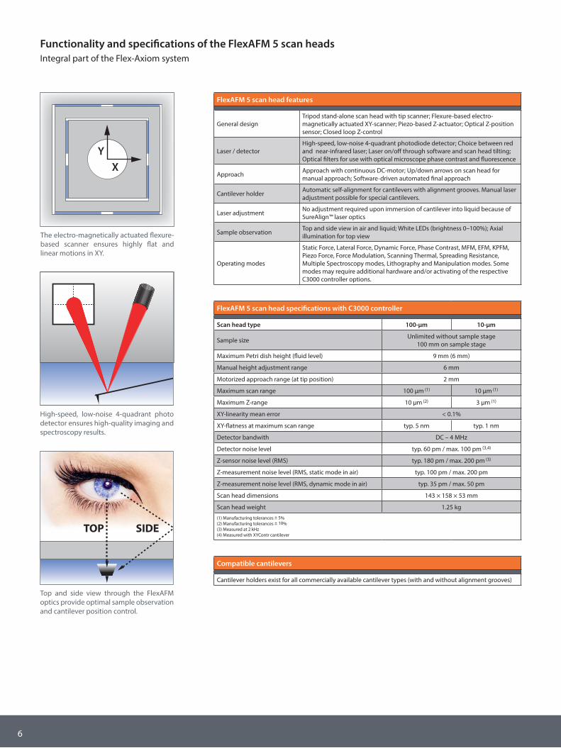
Functionality and specifications of the FlexAFM 5 scan headsIntegral part of the Flex-Axiom system
Compatible cantilevers
Cantilever holders exist for all commercially available cantilever types (with and without alignment grooves)
FlexAFM 5 scan head specifications with C3000 controller
Scan head type 100-µm 10-µm
Sample size Unlimited without sample stage100 mm on sample stage
Maximum Petri dish height (fluid level) 9 mm (6 mm)
Manual height adjustment range 6 mm
Motorized approach range (at tip position) 2 mm
Maximum scan range 100 µm (1) 10 µm (1)
Maximum Z-range 10 µm (2) 3 µm (1)
XY-linearity mean error < 0.1%
XY-flatness at maximum scan range typ. 5 nm typ. 1 nm
Detector bandwith DC – 4 MHz
Detector noise level typ. 60 pm / max. 100 pm (3,4)
Z-sensor noise level (RMS) typ. 180 pm / max. 200 pm (3)
Z-measurement noise level (RMS, static mode in air) typ. 100 pm / max. 200 pm
Z-measurement noise level (RMS, dynamic mode in air) typ. 35 pm / max. 50 pm
Scan head dimensions 143 × 158 × 53 mm
Scan head weight 1.25 kg
(1) Manufacturing tolerances ± 5%(2) Manufacturing tolerances ± 10%(3) Measured at 2 kHz(4) Measured with XYContr cantilever
FlexAFM 5 scan head features
General designTripod stand-alone scan head with tip scanner; Flexure-based electro-magnetically actuated XY-scanner; Piezo-based Z-actuator; Optical Z-position sensor; Closed loop Z-control
Laser / detectorHigh-speed, low-noise 4-quadrant photodiode detector; Choice between red and near-infrared laser; Laser on/off through software and scan head tilting; Optical filters for use with optical microscope phase contrast and fluorescence
Approach Approach with continuous DC-motor; Up/down arrows on scan head for manual approach; Software-driven automated final approach
Cantilever holder Automatic self-alignment for cantilevers with alignment grooves. Manual laser adjustment possible for special cantilevers.
Laser adjustment No adjustment required upon immersion of cantilever into liquid because of SureAlign™ laser optics
Sample observation Top and side view in air and liquid; White LEDs (brightness 0–100%); Axial illumination for top view
Operating modes
Static Force, Lateral Force, Dynamic Force, Phase Contrast, MFM, EFM, KPFM, Piezo Force, Force Modulation, Scanning Thermal, Spreading Resistance, Multiple Spectroscopy modes, Lithography and Manipulation modes. Some modes may require additional hardware and/or activating of the respective C3000 controller options.
TOP SIDE
Top and side view through the FlexAFM optics provide optimal sample observation and cantilever position control.
High-speed, low-noise 4-quadrant photo detector ensures high-quality imaging and spectroscopy results.
XY
The electro-magnetically actuated flexure-based scanner ensures highly flat and linear motions in XY.
6
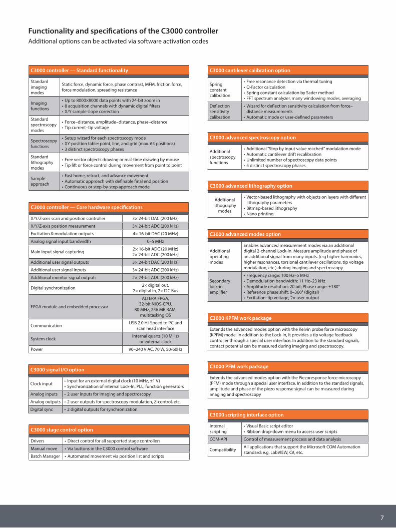
Functionality and specifications of the C3000 controllerAdditional options can be activated via software activation codes
C3000 advanced modes option
Additional operating modes
Enables advanced measurement modes via an additional digital 2-channel Lock-In. Measure amplitude and phase of an additional signal from many inputs. (e.g higher harmonics, higher resonances, torsional cantilever oscillations, tip voltage modulation, etc.) during imaging and spectroscopy
Secondary lock-in amplifier
• Frequency range: 100 Hz–5 MHz• Demodulation bandwidth: 11 Hz–23 kHz• Amplitude resolution: 20 bit; Phase range: ±180°• Reference phase shift: 0–360° (digital)• Excitation: tip voltage, 2× user output
C3000 scripting interface option
Internal scripting
• Visual Basic script editor• Ribbon drop-down menu to access user scripts
COM-API Control of measurement process and data analysis
Compatibility All applications that support the Microsoft COM Automation standard: e.g. LabVIEW, C#, etc.
C3000 signal I/O option
Clock input • Input for an external digital clock (10 MHz, ±1 V)• Synchronization of internal Lock-In, PLL, function generators
Analog inputs • 2 user inputs for imaging and spectroscopy
Analog outputs • 2 user outputs for spectroscopy modulation, Z-control, etc.
Digital sync • 2 digital outputs for synchronization
C3000 controller — Standard functionality
Standard imaging modes
Static force, dynamic force, phase contrast, MFM, friction force, force modulation, spreading resistance
Imaging functions
• Up to 8000×8000 data points with 24-bit zoom in • 8 acquisition channels with dynamic digital filters • X/Y sample slope correction
Standard spectroscopy modes
• Force–distance, amplitude–distance, phase–distance • Tip current–tip voltage
Spectroscopy functions
• Setup wizard for each spectroscopy mode• XY-position table: point, line, and grid (max. 64 positions)• 3 distinct spectroscopy phases
Standard lithography modes
• Free vector objects drawing or real-time drawing by mouse • Tip lift or force control during movement from point to point
Sample approach
• Fast home, retract, and advance movement • Automatic approach with definable final end position • Continuous or step-by-step approach mode
C3000 controller — Core hardware specifications
X/Y/Z-axis scan and position controller 3× 24-bit DAC (200 kHz)
X/Y/Z-axis position measurement 3× 24-bit ADC (200 kHz)
Excitation & modulation outputs 4× 16-bit DAC (20 MHz)
Analog signal input bandwidth 0–5 MHz
Main input signal capturing 2× 16-bit ADC (20 MHz)2× 24-bit ADC (200 kHz)
Additional user signal outputs 3× 24-bit DAC (200 kHz)
Additional user signal inputs 3× 24-bit ADC (200 kHz)
Additional monitor signal outputs 2× 24-bit ADC (200 kHz)
Digital synchronization 2× digital out, 2× digital in, 2× I2C Bus
FPGA module and embedded processor
ALTERA FPGA, 32-bit NIOS-CPU,
80 MHz, 256 MB RAM, multitasking OS
Communication USB 2.0 Hi-Speed to PC and scan head interface
System clock Internal quarts (10 MHz)or external clock
Power 90–240 V AC, 70 W, 50/60Hz
C3000 cantilever calibration option
Spring constant calibration
• Free resonance detection via thermal tuning• Q-Factor calculation• Spring constant calculation by Sader method • FFT spectrum analyzer, many windowing modes, averaging
Deflection sensitivity calibration
• Wizard for deflection sensitivity calculation from force–distance measurements
• Automatic mode or user-defined parameters
C3000 stage control option
Drivers • Direct control for all supported stage controllers
Manual move • Via buttons in the C3000 control software
Batch Manager • Automated movement via position list and scripts
C3000 advanced spectroscopy option
Additional spectroscopy functions
• Additional “Stop by input value reached” modulation mode• Automatic cantilever drift recalibration• Unlimited number of spectroscopy data points• 5 distinct spectroscopy phases
C3000 advanced lithography option
Additional lithography
modes
• Vector-based lithography with objects on layers with different lithography parameters
• Bitmap-based lithography• Nano printing
C3000 PFM work package
Extends the advanced modes option with the Piezoresponse force microscopy (PFM) mode through a special user interface. In addition to the standard signals, amplitude and phase of the piezo response signal can be measured during imaging and spectroscopy
C3000 KPFM work package
Extends the advanced modes option with the Kelvin probe force microscopy (KPFM) mode. In addition to the Lock-In, it provides a tip voltage feedback controller through a special user interface. In addition to the standard signals, contact potential can be measured during imaging and spectroscopy.
7

Nanosurf AGGräubernstrasse 12–144410 LiestalSwitzerland+41 61 927 47 47 (phone)+41 61 927 47 00 (fax)www.nanosurf.com [email protected]
Nanosurf Inc.300 Trade Center, Suite 5450Woburn, MA 01801United States of America781 549 7361 (phone)781 549 7366 (fax)www.nanosurf.com [email protected]
Nanosurf GmbHRheinstrasse 563225 LangenGermany+49 6103 202 7163 (phone)+49 6103 202 7182 (fax)www.nanosurf.de [email protected]
Nanosurf 中国中心 Nanosurf China, Shanghai上海市天宝路578号 (200086)飘鹰世纪大厦703室, 中国+86 18621896399 (电话)+86 21 5512 7698 (传真)[email protected]
Flex-Axiom
Nanosurf and the Nanosurf Logo are trademarks of Nanosurf AG Copyright © 2014 Nanosurf AG, Switzerland


![Versatile Embankment Slide Plastic Open Slide [EM-PO] Contentsdownloads.playdale.co.uk/DATA - Versatile Embankment Slide - Plasti… · frame height and slide length Materials Stainless](https://img.pdfslide.us/doc/110x75/5f6eb887abf9b020de4bab88/versatile-embankment-slide-plastic-open-slide-em-po-versatile-embankment-slide.jpg)