Embed Size (px)
Citation preview

THE MINERALOGICAL MAGAZINE A ~ D
J O U R N A L OF
T H E M I N E R A L O G I C A L SOCIETY
No. 237 June, 1957 Vol. XXXI
Accurate determination of the cell dimensions of magnetite.
By E. Z. BASTA, B.Sc., M.Sc., Ph.D., F.G.S.
Department of Geology, Cairo University, Giza, Egypt.
[Read 24 January 1957.]
Summary. An analysed magnetite from Bisperg, S~tter, Dalecarlia, Sweden, gave F 2+ M 2+ F 3+ 0 the formula el.024 g0.010 el.gs4 4000, which corresponds closely to the ideal
composition. A precise determination of the cell dimensions gave a 8-3963:t: 0.0005 ~. at 18 ~ C. Gentle heating of the powdered magnetite in evacuated sifica glass tubes, before X-ray examination, resulted in sharply defined lines with high Bragg angles ; an improvement which enabled precision data to he obtained. Four other new analyses of magnetites are also given and their lattice parameters are determined in the same way and are found to range from 8.3960 ~_. to 8.3970 -~. The Bisperg magnetite being the purest sample examined, the value of its cell edge is taken as represer~tative of pure magnetite.
The effect of the different ionic substitutions on the cell dimensions of natural magnetites is discussed. An attempt is also nmde to explain the great variations among the published values of cell dimensions of artificial preparations ; one main reason being the presence of defect structures with varying oxygen contents in excess of the formula requirements.
M A N Y e lemen t s m a y s u b s t i t u t e e i the r t h e d i v a l e n t or the t r i v a l e n t
a t o m s in Fe3Od, a n d as a r e su l t pu re m a g n e t i t e is t he excep t ion
a n d n o t t he rule. A knowledge of t he exac t va lue of t h e cell d imens ions
of pu re n a t u r a l m a g n e t i t e is i m p o r t a n t for a n y i n v e s t i g a t i o n of these
d i f ferent s u b s t i t u t i o n s a n d t h e i r effect on t he size of t he u n i t cell.
D u r i n g a r e c e n t s t u d y of t he s y s t e m F e O - F % O 3 - T i O 2 a n d t h e
m a g n e t i t e solid solut ions , t h e a u t h o r could n o t f ind in t he l i t e r a t u r e a n y
re l iable d a t a for t h e uni t -ce l l d imens ions of m a g n e t i t e (Bas ta , 1953).
Holgersson (1927) gave m e a s u r e m e n t s l of two n a t u r a l m a g n e t i t e s f rom
Throughout this paper, measurements are given in ~ngstrom units, converted
]3 4937 O g
432 E . Z . BASTA ON
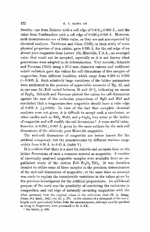
Sweden: one from Dalarne with a cell edge of 8-416• ~., and the other from Nordmarken with a cell edge of 8.429• A. However, such measurements are of little value, as they are not accompanied by chemical analyses. Newhouse and Glass (1936), in their study of some physical properties of iron oxides, gave 8.395 ~. for the cell edge of an almost pure magnetite from Lovers' Pit, Mineville, U.S.A. ; an averaged value that could not be accepted, especially as it is not known what precautions were adopted in its determination. Very recently, Schmidt and Vermaas (1955) using a 57-3 mm.-diameter camera and unfiltered cobalt radiation gave the values for cell dimensions of five samples of magnetites, from different localities, which range from 8.386 to 8.398 (~_0.001) ~_. Such relatively large variations of the lattice parameters were attributed to the presence of appreciable amounts of Mg, A1, and in one case ~i ; FeO varied between 25 and 28 ~ indicating an excess of Fe20 3. Schmidt and Vermaas plotted the values for cell dimensions against the sum of the molecular proportions of MgO and NiO and concluded that a magnesium-free magnetite should have a cube edge of 8.402 ~. (• In view of the fact that complete chemical analyses were not given, it is difficult to accept such a conclusion, as other oxides such as Ti02, MnO, and ~,-Fe~O 3 may enter in the lattice of magnetite and will modify the cell dimensions. 1 A more useful value, however, is 8"392• ~. given by the same authors for the unit-ceil dimensions of the relatively pure Mineville magnetite.
The unit-cell dimensions of magnetite are better known for the artificial compound, but the measurements by different workers range widely from 8"36 _~. to 8.45 ~. (table V).
I t is evident that there is a need for reliable and accurate data on the lattice dimensions of such a common mineral as magnetite. A number of chemically analysed magnetite samples were available from an un- published study of the system FeO-F%O3-TiO e. It was therefore decided to utilize some of these samples in the precision determination of the unit-cell dimensions of magnetite ; at the same time an attempt was made to explain the considerable variations in the values given by the previous investigators for the artificial preparations. An additional purpose of the work was the possibility of correlating the variations in composition and cell edge of naturally occurring magnetites with the when necessary from the original values in the references cited (W. L. Bragg, Journ. Sci. Instr. , 1947, vol. 24, p. 27). In the absence of a s t a tement of the wave- length used, particularly before 1943, the measurements , a l though usual ly specified as being in /~ngst roms, were presumed to be in kX.
1 See below, p. 436.

CELL DIMENSIONS OF MAGNETITE 433
changes in thei r physical propert ies , and thus t ry ing to account for
some of the con t rad ic to ry s t a t ements as to the appearance of magne t i t e
in polished sections. This pa r t of the work has, however , no t progressed
sufficiently, and is therefore lef t for a la ter publicat ion.
Technique and results of analyses.
Small f ragments of selected mater ia l were crushed into a fine powder
of about 0.1 mm. part ic le diameter . To avoid i ron con tamina t ion t h a t
TABLE I. Chemical analyses of magnetites.
1. 2. 3. 4. 5. 6.
FeO 31.43 30.58 31.05 30-76 3 0 - 6 5 31.03 MgO 0.18 0.28 0.46 0.19 0.4~ - - MnO 0.05 0-27 0.03 0-29 0.27 - - CaO 0.01 tr. 0.00 tr. tr. - - Fe20 a 67.55 66.92 66.42 65.08 68.59 68.97 A120 a 0"04 tr. 0.37 1"54 tr. - - V203 0'01 n.d. 0-27 n.d. n.d. - - Cr~O a 0.02 0-00 0'00 0.00 0'00 - - TiO~ 0'05 0.28 0-27 0'36 tr. - - SiO 2 0.73 1.08 1.22 1.20 0-00 - -
100-07 99.41 100.09 99.42 100.00 100.00
1 t .
Fe 2+ 1.024) Mg 2+ 0-010~3.018 Fe a+ 1.984) O 4.000 4.000
1. Magnetite, Bisperg, S~ter, Dalecarlia, Sweden. Analyst, I-I. B. Milner. 2. Magnetite, Barras Nose, Tintagel, Cornwall. Analyst, W. H. Herdsman. 3. 5'[agnetite, Arendal, Norway. Analyst, H. B. Milner. 4. Magnetite, Hall, Ottawa, Canada. Analyst, W. H. Herdsman. 5. Magnetite, recalculated after deduction of 2.02% SiO 2 as quartz, Binnenthal,
Switzerland. Analyst, W. H. Herdsman. 6. Theoretical composition of magnetite, Fe30 a. 1'. Atomic ratios calculated from analysis 1 on basis of 4 0 .
migh t resul t i f hardened steel mor tars were used, the pre l iminary
crushing was carr ied out be tween two horizontal disks of non-magnet ic
Cu-Be al loy under a pressure of 3 to 4 tons per sq. in. The powder was
subjec ted to magnet ic and heavy- l iqu id separat ions in order to r emove
impur i t ies (see Basra, 1953, for details). A check for homogene i ty was
made under the microscope and by X- ray powder photographs .
A specimen of magne t i t e (Bristol No. 636) f rom Bisperg, S/~ter, Dale-
carlia, Sweden, was found by chemical and spectrochemical analysis
(table I, col. 1) to be exceedingly close to the ideal s toichiometr ic
composi t ion Fe304. A por t ion of the analysed sample was s tudied
by X - r a y diffraction methods using iron-fi l tered cobal t radia t ion in
a 114.59 mm.-d iamete r powder camera of the type described by

434 E . Z . BASTA ON
W. Parrish and E. Cisney. 1 The change of temperature of the camera during the period of exposure (1~-2 hours) did not exceed 2 ~ C. under operating conditions. The wave-lengths of the X-rays from the cobalt target were taken as Ku 1 = 1.78890, K% = 1.79279, Ka = 1.79020A., and the measurements were made for the specimen at 18 ~ C.
Although the low 6 values were indicated by sharp lines, the more important back-reflection lines were diffuse, possibly as a result of lattice distortion or strain of the crystal structure due to the grinding process. Although such a phenomenon is frequently encountered in metals, it has only rarely been reported in minerals, for example by B. Wasserstein 2 in his work on galena; guided by his experience, the powdered magnetite sample was heated to about 400 ~ C. for a few minutes; to safeguard against oxidation, the heating was carried out in evacuated silica, glass tubes. X-ray powder photographs of the an- nealed samples showed a remarkable improvement in definition, which could be attributed to recrystallization. That such recrystallization takes place at the temperature of annealing is supported by the observa- tion made by Gheith (1952), in his work on the differential thermal analysis of iron oxides, that for natural magnetite there is a small exothermie peal{ at, 380-395 ~ C., which he considered to be due to ~he recrystallization of the finer particles.
Measurements on the films were carried out by means of a Thornton Heath precision instrument equipped with a travelling microscope. Duplicate measurements showed a precision of 0"02 ram. The diffraction lines were measured on either side of the entrance and exit holes of the X-ray beam, and as the Straumanis method of film mounting ~ was used, it was possible to apply accurately the 'shrinkage correction'.
The lattice parameters based on measurements of eight lines from 1-0922 to 0.9386 _~. (see table II) were extrapolated according to the method of Nelson and Riley 4 ; the cell edge determined by this extra- polation was 8"3963 ,~. (fig. 1).
Another photograph of the same specimen taken with nickel-filtered copper radiation (Cu-Ka, - 1'5405, Cu-Ka 2 = 1"5443, Cu-Ka = 1"5418 ~.), although slightly fogged owing to fluorescent radiation, gave the result of 8"3965 A. for the cell edge, which, despite a small error, shows with what precision results can be duplicated. I t was also
1 Philips Techn. Review, 1948, vol. 10, p. 157 [M.A. 11-278]. Amer. Min., 1951, vol. 36, p. 102 [M.A. 11-316]. M. E. Straumanis, Journ. Appl. Physics, 1949, vol. 20, p. 726.
4 j. B. Nelson and D. P. Riley, Proc. Phys. Soc., London, 1945, vol. 57, p. 160.

435
L%
possible to duplicate the results when different samples from the same locality were similarly examined ; a sample of Bisperg magnetite (British Museum, No. 89240) showed very little variation in results from those of the original chemically analysed sample. The complete measurements
39~ A
Q
8,39?
8.396
8.395
8.394
8,393
8 .Z92
8.~91
8.~90
8.389
8.388
8.38'f
CELL DIMENSIONS OF MAGNETITE
o o'.1o o :20 o ~o '
FIG. l. Extrapolation for the accurate determination of the lattice parameter of Bisperg *nagnetite at 18 ~ C.
of the X-ray powder pattern of the Bisperg magnetite are given in table I I ; the spacings recorded show fairly good agreement with the calculated data.
Errors in temperature are considered to be less than one degree, which in terms of the value of cell dimension amounts to an error of less than 0.0001 ~. The positive correction for refraction for magnetite is 0.0001 ~.,

436 E . Z . BASTA ON
calculated according to the method of Lipson and Wilson I (1941), and has been neglected. Taking into account factors of X-ray absorption, thermal expansion, sharpness of the lines, elimination of strain, and the like, the l imit of error can be safely taken as 0.0005 ~_.
Four other samples of pract ical ly pure magneti te from different localities, which have also been chemically analysed (table I, cols. 2
TABLE II. X-ray powder pattern of magnetite, Bisperg, S~ter, Sweden; Co-Ka radiation. Camera diameter 114-59 mm. Intensities estimated visually, a 8.3963 • 0.0005 A. Spacings are compared with those from the A.S.T.M. index card d 1-1120, recommended by the Joint Committee as the best data for magnetite. These data are given by J. D. Hanawalt, H. W. Rinn, and L. K. Frevel, Ind. Eng. Chem.,
I . d(obs.) hkl.
mw 4.847 111 s 2.966 220 vvs 2.530 311 v v w 2.419 222 s 2-096 400 ms 1.712 422 vs 1.614 333, 511 vs 1"483 440 vw 1"327 620 w 1"279 533 v v w 1"264 622 vw 1.2112 444 w 1.1214 642
* Measurements of these radiation.
1938, vol.
d(calc.) 1-1 ld20 1-1~20
4"847 4"85 1"09 2"968 2"97 1"05 2-532 2"53 2"423 2'42 2'098 2-10 0"97 1"714 1"71 0'97 1"616 1"61 0'94 1"484 1 "48 1"328 1"33 0"88 1'281 1-28 0-86 1"266 - - 0"85 1"2120 1-21 0'81 1-1220 1.12 0"81
five lines were obtained from a photograph taken with Cu-K~
10, p. 457.
I . d(obs.) hkl. d(calc.)
ms 1-0922% 553, 731 1-0930 mw 1"0489% 800 1-0495 v v w 0-9890% 660, 822 0.9895 v v w 0"9890% 660, 822 0.9895 mw 0.9692% 555, 751 0-9695 w 0-9692a.~ 555, 751 0.9695 w 0-9386% 840 0"9387 vw 0-9386% 840 0"9387 mw* 0-8794% 931 0.8800 m* 0"8565~1 844 0"8589 w* 0"8565% 844 0"8589 row* 0.8113a1 951 0-8115 vw* 0.8113% 951 0.8115
to 5), were examined in the same way and the values of their lat t ice parameters were found to range from 8.3960 to 8.3970 A. In view of the fact tha t the Bispcrg magneti te was the purest sample examined, i t is proposed to take its value, 8-3963• ~., as representat ive of pure magneti te until such t ime as a purer specimen is measured.
Discussion.
Analyses of natural magnetites show tha t they frequently contain other cations, which may substi tute either the divalent or the t r ivalent iron and may modify the cell dimensions. Table I I I shows tha t among the divalent elements tha t may enter the crystal structure of mag- netite, only Mn 2+ has an ionic radius greater than tha t of Fe 2+, while all the t r ivalent elements have an ionic radius less than tha t of Fe 3+. From the table i t is clear tha t the cell dimensions of magnet i te are increased only when replacement by Mn ~+ or Ti 4+ takes place ; the cell- edge values of the spinels MnF%04 and F%TiO 4 thus formed are 8.51
1 ]-I. L i p s o n a n d A. J . C. W i l s o n , J o u r n . Sci. I n s t r . , 1941, vo l . 18, p. 144.

CELL DIMENSIONS OF MAGNETITE 437
a n d 8"50 ~ . r e s p e c t i v e l y ( B ~ n a r d a n d C h a u d r o n , 1937 ; P o n i l l a r d , 1949).
H o w e v e r , m a n g a n e s e is r a r e l y p r e s e n t in a p p r e c i a b l e a m o u n t a n d n a t u r a l
m a g n e t i t e ores u s u a l l y c o n t a i n less t h a n 3 % M n O c o r r e s p o n d i n g to 10 %
M n F % 0 4 . U l v S s p i n e l , F % T i 0 ~ , on t h e o t h e r h a n d , h a s b e e n s h o w n
TABLE III . Ionic radii of the elements tha t may enter the crystal structure of magnetite, and the cell dimensions (in /~) of the spinels thus formed (various
authorities).
Ionic A13+ Cr a+ Fe 3+ Ti 4+ Metal. radius. 0.57. 0.64. 0.67. 0.64. Ni 2+ 0.78 a 8-05 a 8.30 a 8.36 - - Mg 2+ 0-78 8-07 8.31 8.37 - - Co s+ 0.82 8'08 8-32 8"38 - - Zn ~+ 0.83 8-07 8'30 8"40 - - Fe 2+ 0.83 8.12 8.34 8"40* a 8.50 Mn ~+ 0.91 8.26 8.49 8.51 - -
* The value of cell edge for magnetite as determined three significant figures.
TABLE IV. Chemical analyses of some substituted dimensions.
in this paper, simplified to
magnetites and their cell
1. Titanomagnetite separated from basalt, Giant 's Causeway, Ireland. Anal. W. H. Herdsman. a = 8.4697 • ~-.
2. Titanomagnetite from Magnet Cove, Arkansas, U.S.A. Anal. H. B. Milner. a = 8.3960~0-001/~.
3. Magnetitc-maghemite separated from basalt, Co. Antrim, Ireland. A n a l I-I. B. Milner. a = 8,369•
1', 2', 3' are the percentages of the end-members calculated from the analyses 1, 2, and 3.
t o b e m o r e c o m m o n t h a n h a d p r e v i o u s l y b e e n s u p p o s e d (Bas ra , 1953 ;
R a m d o h r , 1953 ; V i n c e n t a n d P h i l l i p s , 1954). T h e p r e s e n t a u t h o r , w o r k i n g
o n ca r e fu l l y s e p a r a t e d h o m o g e n e o u s t i t a n o m a g n e t i t e s f r o m v o l c a n i c
rocks , h a s f o u n d u p to 32.5 ~ F % T i 0 4 ( t ab le IV , col. 1), a s u b s t i t u t i o n
t h a t r e su l t s in a c o n t i n u o u s i n c r e a s e in t h e cell d i m e n s i o n s .
R e p l a c e m e n t s o f F e e+ or F e a+ in m a g n e t i t e b y o t h e r d i v a l e n t o r
1. 2. 3. 1'. 2'. 3'. FeO 42.33 21.83 12-75 FeF%04 33.18 39.9 35-1 MgO 2.07 7.18 0-91 MgF%04 4.67 28'8 4.6 MnO 0"72 1.82 0.18 MnF%04 2.31 6-0 - - CaO - - 0.94 0-76 CaF%O4 - - 3.7 2.4 F%O a 26.88 57.11 80.63 ~-F%O a - - - - 53.0 AlcOa 0.40 3.62 0.30 MgAI~O4 - - 5.1 - - V20a - - 0-10 0.12 Cr~O a - - 0.01 0.01 F%TiO 4 32-48 10.3 - - Ti02 24.86 6.98 2.54 ~-FeTiO a 27.36 6.2 4.9 SiO 2 2.64 0.38 0-84
100.00 100-0 100.0 99.90 99.97 99.04

438 E . Z . :BASTA ON
trivalent ions, e.g. Mg 2+, Co s+, Ni 2+, A13+, and Cr ~+, also occur, but the effect in these cases is a decrease in the cell dimensions of magnetite. Exceptionally up to 8 % MgO (table IV, col. 2) and up to 7 ~ A120 a have been recorded in the analyses of magnetites, but such values are uncommon. Contents of ZnO, NiO, CoO, and Cr20 a are usually too low to influence the cell-edge value of natural magnetite significantly. Zn reported by Vincent and Phillips (1954) in their analyses of magnet- ites from the Skaergaard intrusion, East Greenland, did not exceed 0-2 %; Schmidt and Vermaas (1955) determined 1.76 % )~iO in one of their specimens of magnetite from Witbank, S. Africa; Cr203 is usually less than 0.5 %. V~O 3 rarely exceeds 2 %, corresponding to a replacement of less than 3 ~o of the Fe 3+ ions by V 3+, but its effect on the cell dimensions is not known. Another possibility leading to a decrease in the size of the unit cell of natural magnetites is partial oxidation to maghemite (a = 8-32 ~.). In an unpublished study (Basta, 1953), the author was able to prove, by chemical and X-ray analyses, the existence in nature of minerals intermediate in composition between FeaO 4 and y-F%O 3 ; one of the analyses is given in table IV, col. 3.
Since several substitutions are possible, some increasing and some decreasing the cell dimensions, it is impossible to deduce the chemical composition of natural magnetite from the cell dimensions alone ; even if the unit cell has the size of that of pure Fe304 there may be substantial substitutions, as in the Magnet Cove titanomagnetite (table IV, col. 2).
Variations in the chemical composition and cell edge of naturally occurring magnetite may account for the considerable differences in its physical properties reported in theliterature ; thus figures for the specific gravity vary between 4.96 and 5-3. From the present X-ray data on the Bisperg magnetite, its specific gravity was calculated as 5.192.
Literature data for the cell dimensions of artificial magnetite show marked variations (table V).
The values given by earlier workers up to 1940 may not be quite as precise as indicated, for at the time when these measurements were made the nature of the systematic errors was not fully known, the wave- length values assumed for the various radiations differ appreciably from the present values, and the cited lattice parameters were usually averages of the values calculated from all the lines of both low and high angle 0. Thus the measurement of Clarke, Ally, and Badger (1931), which is that given in the 7th edition of Dana's System of Mineralogy ~
1 C. Palache, H. Berman, and C. Frondel, Dana's System of Mineralogy, 7th edn, New York, 1944, vol. 1, p. 698.

CELL D I M E N S I O N S OF M A G N E T I T E 439
and in Wyckoff's Crystal Structures 1, requires revision. In their original paper, the value of 8-374-4-0.003 ~. was obtained from measurements with a scale reading directly in lattice spacings. The two values listed in table V were obtained by conversion, firstly to ~ngstrom units from the wave-length 0.712 [kX] quoted for the Mo radiation used, secondly from the wave-length 0"70783 kX then known for Mo-K, by which the scale may have been calibrated. The exceptionally high value obtained by Van der Marel (1951) is most probably owing, as the author himself indicated, to the presence of impurities, possibly Na, in his
TABLE V. Cell edge of FeaO 4 (A.).
]-Iolgersson (1927) Clarke, Ally, and Badger (1931) Hagg {1935) Gaglioti and D'Agostino (1936) B~nard (1939) Frank-Kamenecky (1939) Pouillard (1949) Var~ der Marel (1951) Tombs and Rooksby ( 1951) Abrahams and Calhoun (1953) Basta, on Bisperg magnetite, this paper
8.434 8.361 ~0.003 or 8-389• 8.397 8-427 8.390• 8.437 8-413 8.441 • 8-3940 • 0.0005 8.3940 ~_ 0-0005 8.3963 •
samples, which were prepared by precipitation. Michel (1937, p. 37) has found that up to 7 ~ of the Fe 2+ ions of synthetic magnetites may be replaced by Na + and Fe ~+, a replacement that causes the cell dimensions to rise from 8"41 to 8-43 A.
An important possibility that may account for the distinct range of 8.36 to 8.44 ~. in values of the cell edge obtained by different workers on artificial preparations is the presence of oxygen in excess of the stoichiometric formula requirements, as has already been men- tioned in the case of natural magnetite. H~gg (1935) found that mag- netite, when oxidized at up to 300 ~ C., may vary continuously in composition from FeaO 4 to Fes/304, i.e. to ~-Fe2Oa, the cell edge de- creasing uniformly from 8.40 to 8.32 ~. Furthermore, from the work of Greig and his co-workers (1935), magnetite is capable of containing about 30 ~o Fe~03 at 1452 ~ C. ; the product gives an X-ray powder pattern of the spinel type but with a value of the cell edge slightly lower than that of pure Fen04. The artificial preparations of Fe30 ~ on which the cell dimensions quoted in table V were measured were made either fro.m slags, the so-called hammer-scale (Holgersson, 1927) ; or by heating at up to 1300 ~ C. preparations containing Fe 2+ and Fe 8+ in the atomic
1 R. W. G. Wyckoff, Crystal Structures, 1952. vol. 2, New York.

440 E . Z . BASTA ON
ratio 1:2 (Clark, Ally, and Badger, 1931); or by reducing hematite at high temperatures, up to 1400 ~ C., with hydrogen. In none of these preparations was a check made on the Fe2+:Fe 3+ ratio after heating. An increase of oxygen, i.e. a decrease in iron beyond the formula require- ment, may well have occurred, and may be the reason for the low values of the cell edge as observed in some of the preparations. Starke (1939) sought to explain the relation of the structure of synthetic magnetite to its stability by assuming that the unit cell of precipitated magnetite contains: 16 Fe 3+, 51 Fe 2+, 26~02-, 5~OH-, and not 16Fe ~+, 8Fe ~+, 3202-, as in the case of the high-temperature magnetite.
Recalculation of the chemical analysis of the Bisperg magnetite (table I, col. 1') gave the formula ~ 2 + M,2+ F,~+ r~ �9 the ratio ~1.024~'~0.010 ~1.984v4.000 of total cations to oxygen was 3.018:4-000, which shows that such a magnetite is free from vacant cation positions. Similar calculations on the four other new analyses in table I led to the same conclusion.
Another factor that may influence the cell dimensions of magnetite is the possibility of cation rearrangement. The equality of the scattering power of Fe e+ and Fe 3+ makes it impossible to decide from the X-ray data whether magnetite is a normal spinel, X2+Y~+O~, with divalent atoms in the eightfold and trivalent in the sixteenfold positions, or an inversed spinel, Y3+(X2+Y3+)O~, with trivalent atoms in the eight- fold positions and the sixteenfold positions divided between di- and trivalent atoms.
Verwey and De Boer (1936) assigned magnetite to the inversed group on account of its high electrical conductivity, and Verwey and HeiL mann (1947) arrived at the same conclusion from a consideration of the cell dimensions of a large number of spinels. 1
I t is possible that at high temperatures there is a more random distribution of the cations, or that a partial change to the normal spinel structure takes place. I f this is so, small variations in the cell dimensions of pure FeaO 4 may occur after heating, varying with the temperature attained and the rate of cooling; further work is needed to test this suggestion. A change in the cation arrangement at high temperatures has been found for some other members of the spinel group (A1F%04, Verwey, Hayman, and Raneijn, 1947; MnFe20 a, McAndrew, 1952).
Verwey and Hei lmann found tha t normal ferrites generally have a uni t cell about 0.12/~., larger than t ha t of the corresponding chromitcs, while for inversed ferrites the increase is only about 0.05 -~. ; for the pair FeCr~O~-F%04 the increase is 0.06/~ (see table III).

C E L L D I M E N S I O N S O F M A G I ~ E T I T E 441
Conclusions.
The great variation among the reported cell dimensions of artificially prepared magnetites is mainly owing to the formation of defect structures
with varying oxygen contents in excess of the formula requirements. Another possible cause of variation is change in the cation arrangement in the spinel structure of magnetite at high temperatures, depending
on the method of preparation. The Bisperg magnetite, which has the formula
~1"024~v~0"010 x' ~1"984~4 '000 '
is substantially free from defect structures, and from accurate X-ray
determinations carried out on annealed specimens free of crystal distor- tion the following result was obtained for its cell edge and is suggested to be the best known value for pure natural magnetite: a at 18 ~ C. = 8"39630-0-0005 ~_.
An appreciable increase in the cell dimensions of natural magnetite above this value probably indicates the presence of F%Ti04 or MnFeeO ~ in solid solution, while an appreciable lowering of the above value may indicate partial oxidation to y-F%0 3. Other substitutions of Fe 2+ or Fe a+ in the magnetite lattice by Mg 2+, Ni 2+, Co 2+, Zn ~+,
Cr a+, A1 a+, or V a+ occur but they are of minor importance, either because the cell dimensions of the resultant spinel are not much different from those of pure magnetite or because the extent of substitution is
very limited in natural ores. In all cases it is impossible to predict the exact composition from cell dimensions alone.
Acknowledgements. I wish to express my thanks to Prof. W. F. Whittard for the laboratory facilities of the Geology Department, University of Bristol, and for pro- viding most of the materials used in this work ; to Dr. I. S. Loupekine for reading the manuscript and for his valuable suggestions. I am also indebted to the Egyptian Education Authorities for a grant which defrayed the cost of the chemical analyses.
A~RA~AMS (S. C.) and CAL~OU~ (B. A.), 1953. Acta Cryst., vol. 6, p. 105. [M.A. 12-318.]
BASTA (E. Z.), 1953. Mineralogical aspects of the system FeO-Fe203-TiO2. Ph.D. thesis, Bristol.
B~A~D (J.), 1939. D6composition du protoxyde de fer. Th~se, Paris. - - and C~AV])~ON (G.), 1937. Compt. Rend. Acad. Sci. Paris, vol. 204, p. 766. CLARKE (G. L.), ALLY (A.), and BADGER (A. E.), 1931. Amer. Journ. Sei., ser. 5,
vol. 22, p. 539. FRA~K-KAME~nCKY (V. A.), 1939. Compt. Rend. (Doklady) Acad. Sci. URSS,
vol. 23, p. 561. GAGLIOTI (V.) and D'AGosTINO (O.), 1936. Gazz. Chim: Ita]., vol. 66, p. 547.

442 E. Z. BASTA ON CELL DIMENSIONS OF MAGNETITE
GHEITH (M. A.), 1952. Amer. Journ. Sei., vol. 250, p. 677. [M.A. 12-175.] GREta (J. W.), POSNJAK (E.), MERWI~ (H. E.), and SOSMAN (R. B.), 1935. Amer.
Journ. Sci., ser. 5, vol. 30, p. 239. HXcc (G.), 1935. Zeits. physikal. Chem., ser. B, vol. 29, p. 95. HOLGERSSON (Sven), 1927. Lunds Univ. Arsskrift, N. F., Avd. 2, vol. 23, no. 9;
Kungl. Fysiogr. Si~llsk. Handl., N. F., vol. 38, no. 9. MCANDREW (J.), 1952. Amer. Min., vol. 37, p. 453. [M.A. 12~3.] MICHEL (A.), 1937. Propridtds magn6tiques de quelques solutions solides. Th~se,
Paris, p. 37. NEW~OVSE {W. H.) and GLASS (J. P.), 1936. Econ. Geol., vol. 31, p. 699. POUILLARD (E.), 1949. Sur le comportement de l'alumine et de l'oxyde de titane
vis-s des oxydes de fer. Th~se, Lille. RAMDO~R (P.), 1953. Econ. Geol., vol. 48, p. 667. [M.A. 12-388.] SCHMIDT (E. 1~.) and V~aMAAS (F. H. S.), 1955. Amer. Min., vol. 40, p. 422. STARKE (K.), 1939. Zeits. physikal. Chem., set. B, voh 42, p. 159. TOMBS (N. C.) and I~OOKSEY (H. P.), 1951. Aeta Cryst., vol. 4, p. 474. VAN ~)EX MAREL (H. W.), 1951. Journ. Sedim. Petr., vol. 21, p. 12. V~.RWEY (E. J. W.) and DE BOE~ (J. H.), 1936. Ree. tray. chim. Pays-Bas, vol. 55,
p. 531. - - and HEILMAI~N (E. L.), 1947. Journ. Chemical Physics, vol. 15, p. 174.
, I~AYMAN (P. W.), and Ro~IJN (F. C.), 1947. Ibid., p. 181. VInCEnT (E. A.) and PHmL~PS (R.), 1954. Geochemiea Aeta, vol. 6, p. 1. [M.A. i2 -
293, 499.J