Embed Size (px)
Citation preview

The management of B3 lesions with
emphasis on lobular neoplasia
Abeer Shaaban
Queen Elizabeth Hospital Birmingham

NHSBSP core biopsy categories
B1 - Normal
B2 - Benign
B3 – Uncertain malignant potential
B4 – Suspicious of Malignancy
B5 – Malignant

B3 category
•Proportion: 5-9% of core biopsy diagnoses
• Includes lesions of variable significance.
•Diagnostically challenging! • Includes lesions with and without atypia.
•Requires further sampling: traditionally by surgery.
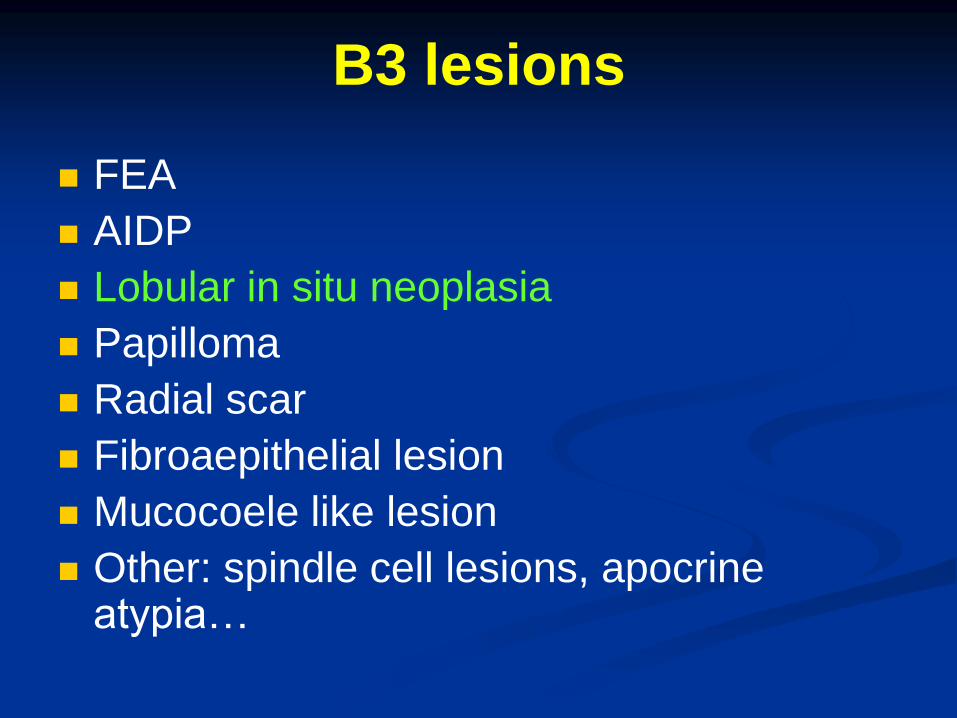
B3 lesions
FEA
AIDP
Lobular in situ neoplasia
Papilloma
Radial scar
Fibroaepithelial lesion
Mucocoele like lesion
Other: spindle cell lesions, apocrine atypia…

Factors affecting frequency &
upgrade rate
Patient population: screening vs
symptomatic
Method of biopsy: NCB vs VAB (gauge of
needle)
Type and combination of B3 lesion
Size of lesion
Presence/absence of atypia

Papillary lesions
Without atypia: 3.8% Noyak et al 2013
5% Youk et al 2011 (age, size)
With atypia 33% (vs 3%) McGhan et al 2013
Radial scar
Without atypia (4-9%)
With atypia (24-44%) Histopathology
2008;52:650, Brenner et al 2002
Mucocoele - like lesion
Without atypia 4%
With atypia 21% Rakha et al, Histopathology 2013

UPGRADE RATES: FEA + AIDP
Study
FEA FEA +
AIDP
AIDP
Lee et al
(2010)
14% 29% 37%
Rakha et al
(2011a, b)
21%
14%
29%
50.4%
40%
Rajan et al
(2011)
19% 29% 53%
Total
18%
29% 44%

Why is there need for B3
management standardisation?

1. Management is inconsistent across
screening units.
2. Increasing use of VAB
3. Over-treatment is a recognised
issue
4. Local guidelines: Leeds, London
QARC
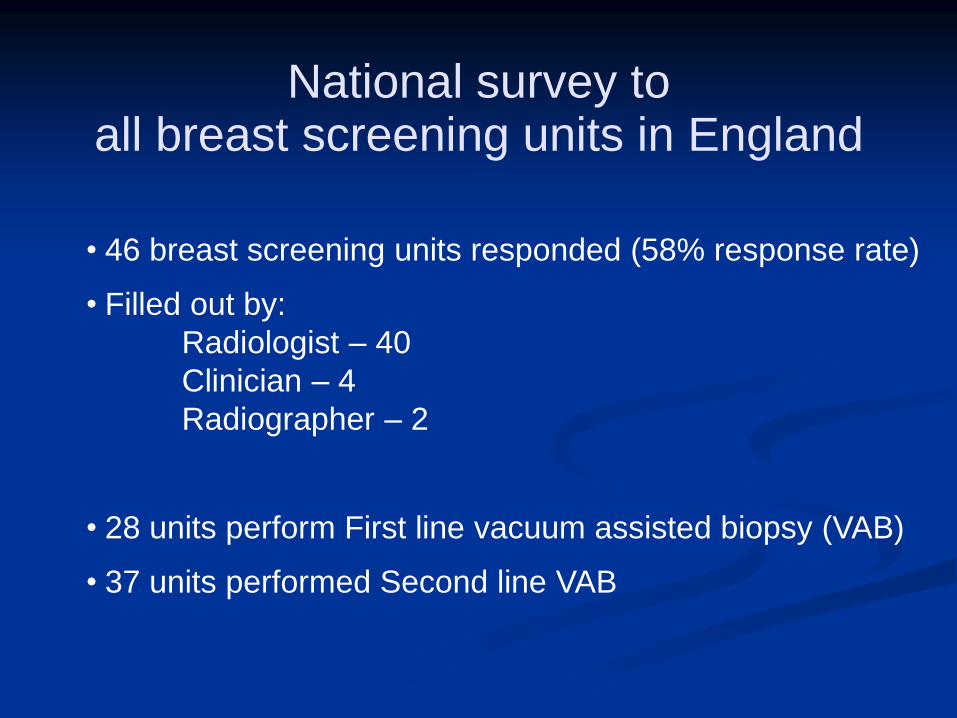
National survey to all breast screening units in England
• 46 breast screening units responded (58% response rate)
• Filled out by:
Radiologist – 40
Clinician – 4
Radiographer – 2
• 28 units perform First line vacuum assisted biopsy (VAB)
• 37 units performed Second line VAB

B3 lesions with no atypia –
radial scar/papilloma with no atypia
• If conventional 14G core biopsy shows B3 lesion with no atypia
– 34 units responded – 74% will offer second line VAB – 26% will offer Surgical Diagnostic Biopsy
• If 1st line VAB shows B3 lesion with no atypia
– 27 units – 41% will offer second line VAB – 44% will offer Surgical biopsy – 15% will discharge due to
adequate sampling

Management of B3 lesion with atypia
on 14G core biopsy
2nd line
VAB
Surgical
biopsy
Discharge EC
FEA 66 34 0 0
AIDP 53 47 0 0
AIDP + FEA 56 44 0 0
ALH 57 34 3 6
LCIS 51 40 0 9
Radial scar + atypia 37 63 0 0
Papilloma + atypia 40 60 0 0
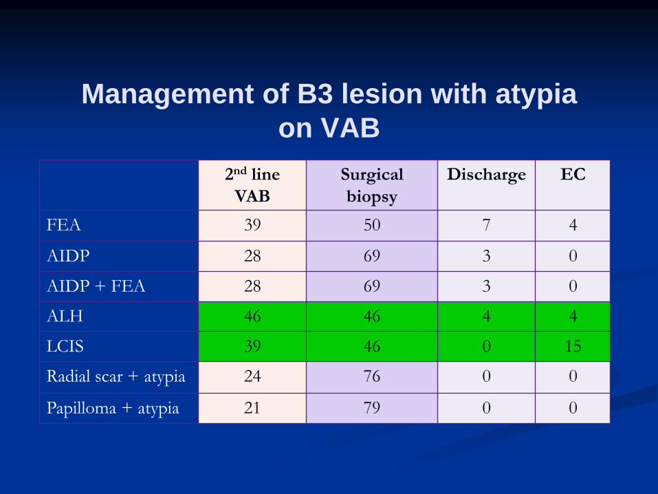
Management of B3 lesion with atypia
on VAB
2nd line
VAB
Surgical
biopsy
Discharge EC
FEA 39 50 7 4
AIDP 28 69 3 0
AIDP + FEA 28 69 3 0
ALH 46 46 4 4
LCIS 39 46 0 15
Radial scar + atypia 24 76 0 0
Papilloma + atypia 21 79 0 0

Benign open biopsies
UK benign open biopsy rates
1.73 per 1,000 women screened (Prevalent)
0.48 per 1,000 women screened (Incident)
0.0
1.0
2.0
3.0
4.0
5.0
6.0
7.0
Ben
ign
op
en
bio
psy
rate
- p
reva
len
t (f
irst
) sc
reen
s
(p
er
1,000 w
om
en
scre
en
ed
)
UK average: 1.74
Target: 1.0
Minimum std: 1.5
Leeds prevalent open biopsy rate 0.9
Increasing use of VAB

Core samples
1st line VAB 2nd line VAB
With thanks to Dr Nisha Sharma

Vacuum-assisted core biopsy of
breast lesions of uncertain
malignant potential (B3) –
an alternative to surgery in selected
cases
Tennant et al. Breast 2008;17:456
Papillary lesions and radial scars without
atypia

Over-treatment is a recognised issue

B3 Positive predictive value
21% Dillon et al 2007 (Ireland)
20% El-Sayed et al 2008
25% Rakha et al 2011 1025 (NCB)
21.2% Bianchi et al 2011 3107 cases (VACNB
Study group, Italy)
20% Strachan et al 2015, under review, 398
cases

The majority of patients (75-80%) with B3
diagnosis will have a benign histology on
further sampling.
Is surgical excision necessary?

Local guidelines such as London
QARC and Leeds pathway have been
developed

Local guidelines

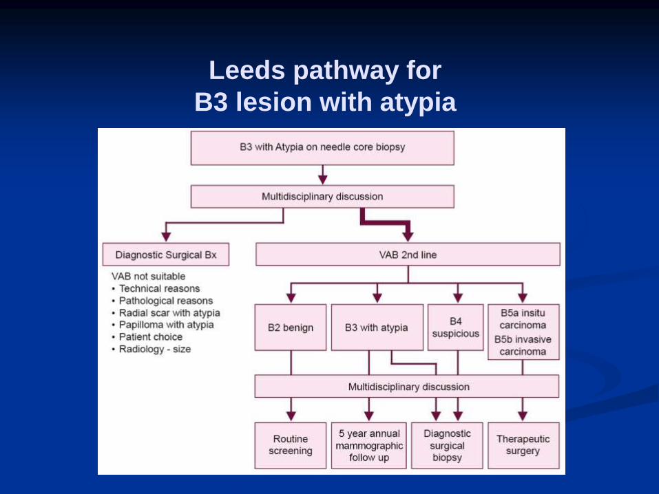
Leeds pathway for
B3 lesion with atypia

Aim to adequately sample by VAB to rule
out coexistent cancer
Follow up

Lobular neoplasia
Encompasses atypical lobular
hyperplasia (ALH) and lobular in situ
carcinoma (LCIS).
LCIS: classical and variants

Classical LCIS
Disease of premenopausal women (90%)
Most LCIS diagnosed between 40-50 yrs.
Often multifocal (60-80%) and bilateral (up
to 35%)
True incidence is difficult to assess
(reported between 0.5-3.8% (Haagenson 1978,
Page 1991)

Classical LCIS
Presents on mammographic
screening but can also be incidental.
Risk of malignancy is x3 likely to be
unilateral than bilateral (Page et al
2003)

LCIS
Marker of breast cancer risk: RR 8-10
Direct precursor:
LOH: in LCIS and the associated lobular ca:
17q, 17p, 11q. Lakhani 1995, Nayae 1997
Mutation of the e-cadherin gene: in LCIS and
invasive lobular ca, Berx et al., 1996

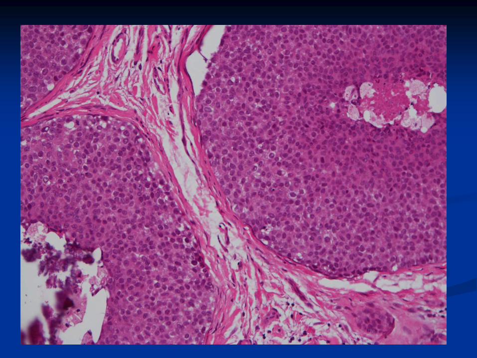
Histologically
A monomorphic proliferation within TDLU
of dyscohesive cells with uniform round
nuclei, indistinct nucleoli and scant
cytoplasm.
Intracytoplasmic lumina are often present
Pagetoid spread can be seen.
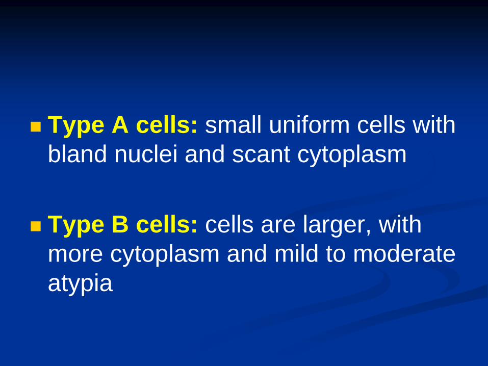
Type A cells: small uniform cells with
bland nuclei and scant cytoplasm
Type B cells: cells are larger, with
more cytoplasm and mild to moderate
atypia

Type A

Type B
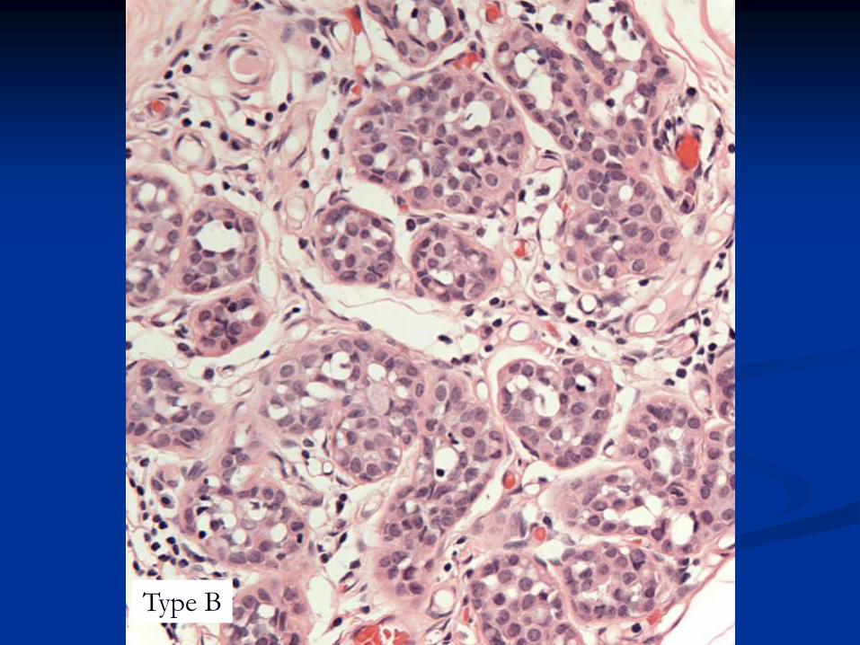
Type B


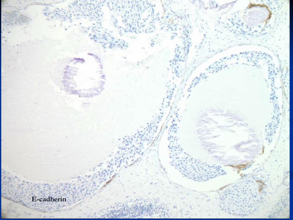
E-cadherin

ALH vs LCIS
Depends on extent of lesion
LCIS: more than half of the acini are filled,
distended and distorted by the
dyscohesive lobular cells.
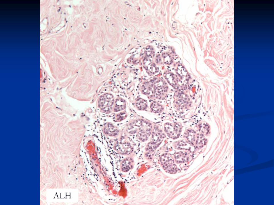
ALH

ALH

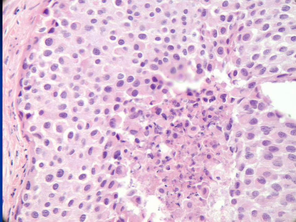
DD: LCIS vs DCIS
Cellular cohesion
Look for architectural pattern of DCIS
E-cadherin

E-cadherin

Variants of LCIS
Pleomorphic LCIS (PLCIS)

A more recently recognized variant of
Lobular Carcinoma In Situ (LCIS)
May calcify hence present through
breast screening
Biology and natural history uncertain
Histologically: mimics high grade
DCIS
PLCIS


E-cadherin

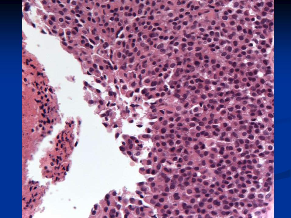
Rare variant of LCIS
LCIS with comedo necrosis
A study of 18 cases reported a strong
association with invasive ca (67% of cases)
Fadare et al Am J Surg Pathol 2006 30:1445–1453

LCIS upgrade rate
Ranged from 0-60%, (majority 2-25%), Buckley et
al 2014 systematic review, Murray et al 2013
Upgrade rate 3% in concordant and 38% if
imaging-pathological discordant, Murray et al
2013.
ALH upgrade rate (27%) not significantly
different from LCIS (33%), Ibrahim et al 2012

Reasons for variation
Screening vs symptomatic
Radiological correlation
Amount of tissue : core vs VAB
LCIS in structured lesions
Family history vs sporadic
Inclusion of PLCIS
Co-existing other lesions as ADH

Upgrade rate of PLCIS
41%, range 30-60%, Hussain and Cunnick
2011, Carder et al 2011
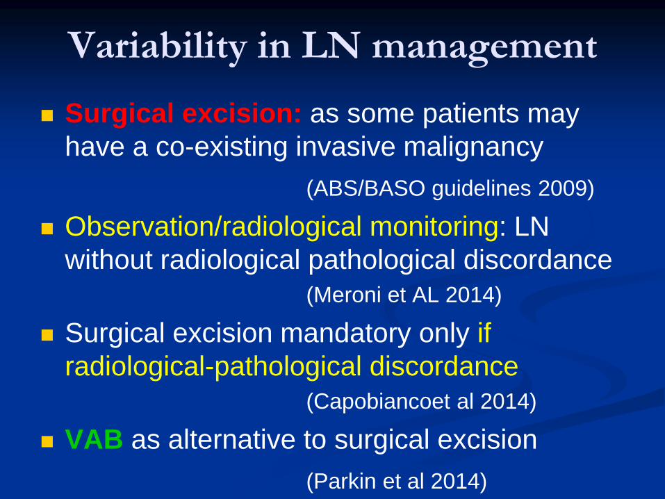
Variability in LN management
Surgical excision: as some patients may
have a co-existing invasive malignancy
(ABS/BASO guidelines 2009)
Observation/radiological monitoring: LN
without radiological pathological discordance
(Meroni et AL 2014)
Surgical excision mandatory only if
radiological-pathological discordance
(Capobiancoet al 2014)
VAB as alternative to surgical excision
(Parkin et al 2014)

B3 guidelines group
Commissioned by NHSBSP
Invited members representing imaging,
surgery and pathology
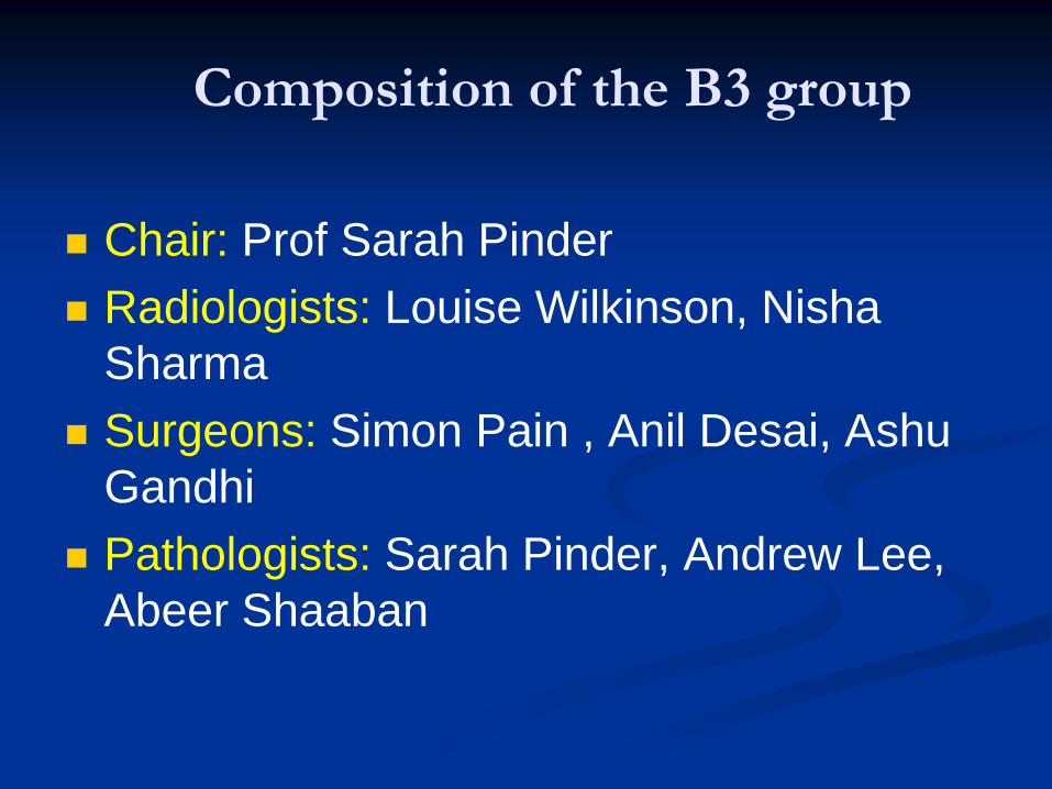
Composition of the B3 group
Chair: Prof Sarah Pinder
Radiologists: Louise Wilkinson, Nisha
Sharma
Surgeons: Simon Pain , Anil Desai, Ashu
Gandhi
Pathologists: Sarah Pinder, Andrew Lee,
Abeer Shaaban
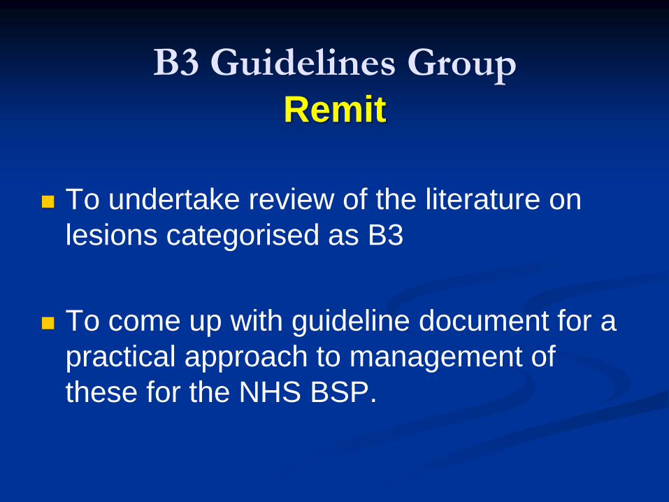
B3 Guidelines Group Remit
To undertake review of the literature on
lesions categorised as B3
To come up with guideline document for a
practical approach to management of
these for the NHS BSP.

Progress
First meeting: October 2014
Management recommendations lesion by
lesion.
Use of diagrams/flow charts
General principles:
radiological/pathological concordance
Draft for discussion/consultation

Recommendations
2nd line VAB as method of choice for
further sampling of B3 lesions, following
either conventional core or 1st line VAB B3
diagnosis.
All cases should be discussed at MDT
meeting
Centres should plan to acquire 2nd line
VAB or refer to a centre that can do it
Diagnostic excision for fibroepithelial
lesions, spindle cell lesions, papilloma with
atypia
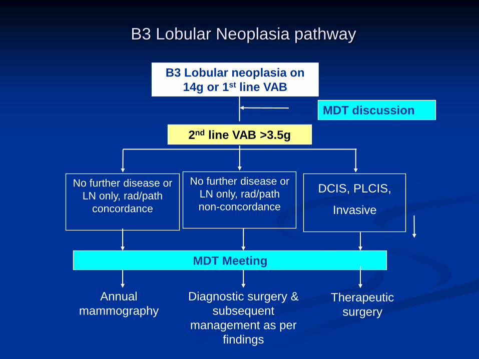
B3 Lobular Neoplasia pathway
2nd line VAB >3.5g
Annual
mammography Therapeutic
surgery
MDT discussion
No further disease or
LN only, rad/path
concordance
DCIS, PLCIS,
Invasive
Diagnostic surgery &
subsequent
management as per
findings
MDT Meeting
B3 Lobular neoplasia on
14g or 1st line VAB
No further disease or
LN only, rad/path
non-concordance
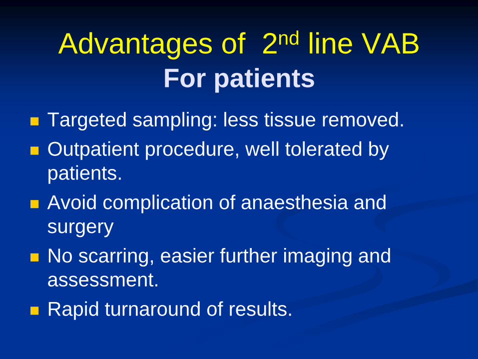
Advantages of 2nd line VAB
For patients
Targeted sampling: less tissue removed.
Outpatient procedure, well tolerated by
patients.
Avoid complication of anaesthesia and
surgery
No scarring, easier further imaging and
assessment.
Rapid turnaround of results.
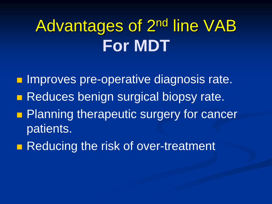
Improves pre-operative diagnosis rate.
Reduces benign surgical biopsy rate.
Planning therapeutic surgery for cancer
patients.
Reducing the risk of over-treatment
Advantages of 2nd line VAB
For MDT
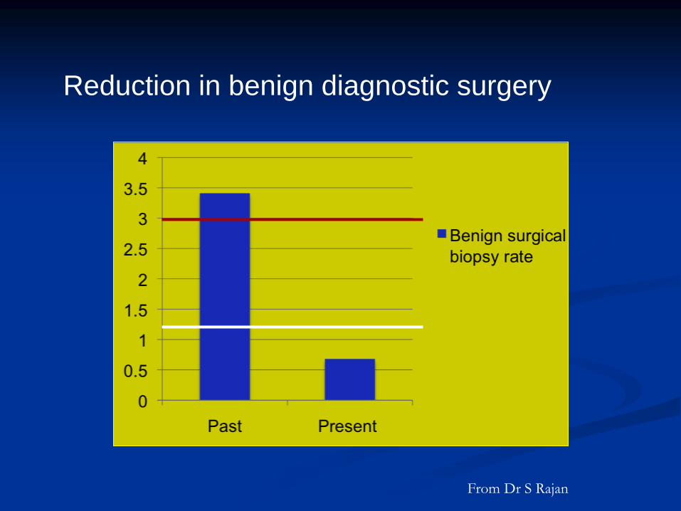
Reduction in benign diagnostic surgery
From Dr S Rajan

Lobular neoplasia on core biopsy
ALH/Classical LCIS: code as B3 and
recommend further tissue examination by
VAB
PLCIS: code as B5a and manage as DCIS
LCIS with necrosis: rare, best coded as
B4, recommend surgical excision.

B3 management FAQ
How to deal with extensive
calcification?
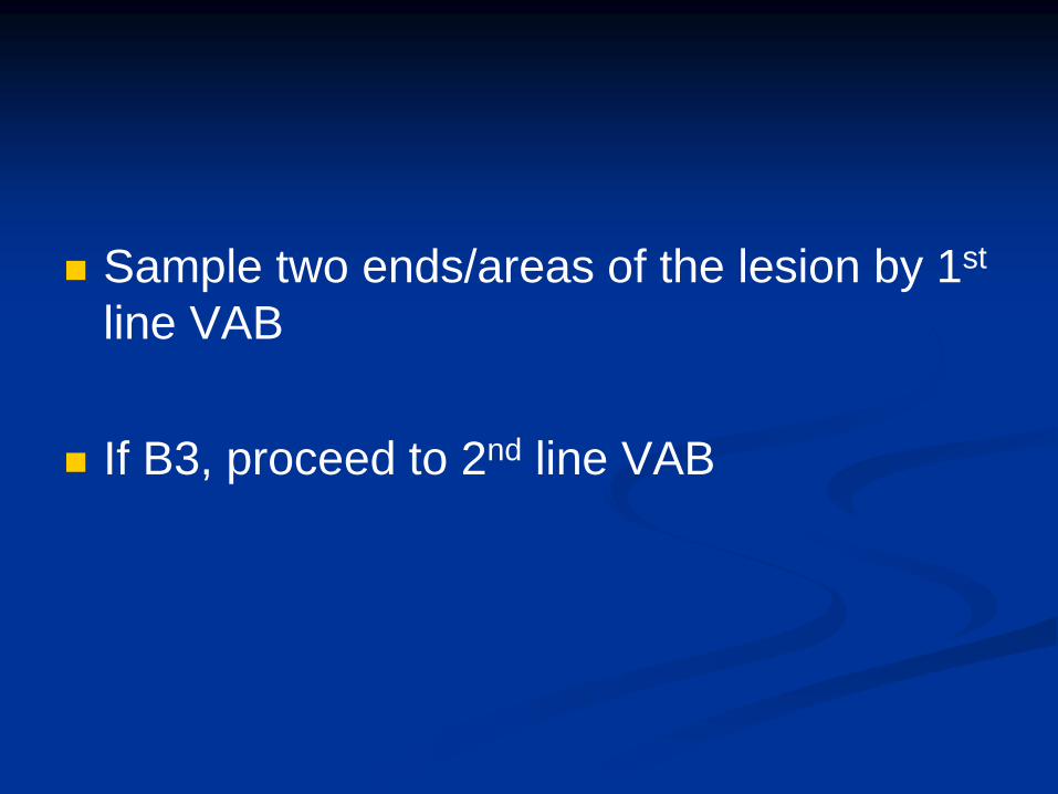
Sample two ends/areas of the lesion by 1st
line VAB
If B3, proceed to 2nd line VAB
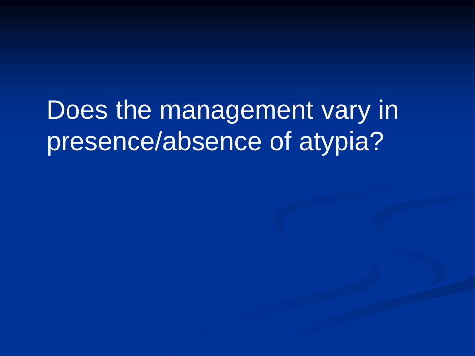
Does the management vary in
presence/absence of atypia?
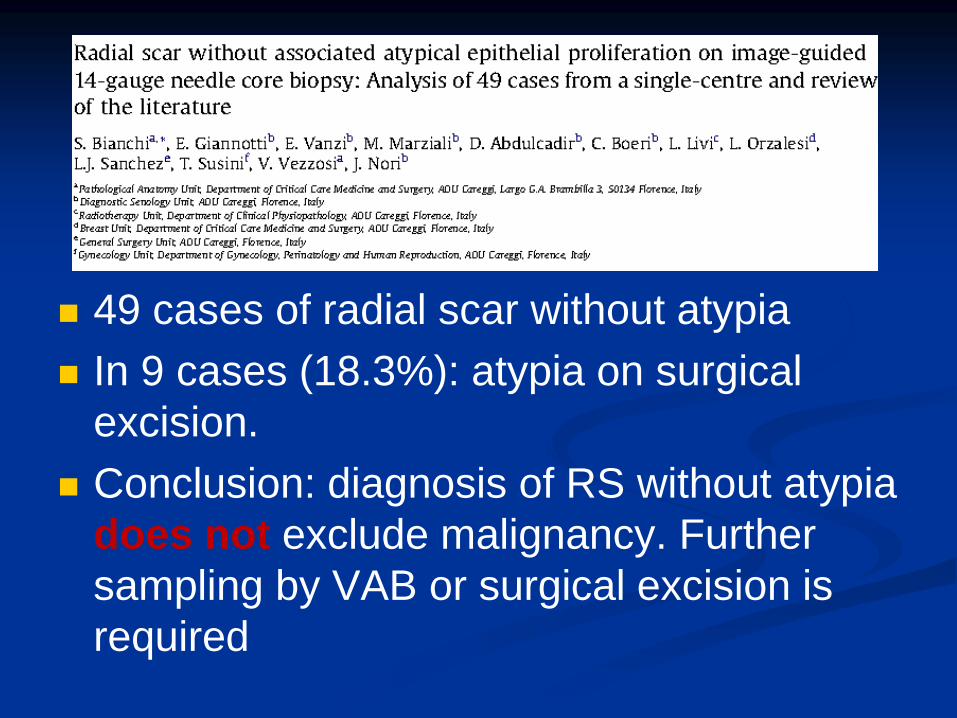
49 cases of radial scar without atypia
In 9 cases (18.3%): atypia on surgical
excision.
Conclusion: diagnosis of RS without atypia
does not exclude malignancy. Further
sampling by VAB or surgical excision is
required
Rakha et al 2014

Does the management vary if B3 lesion shows atypia vs no atypia?
Overall : No
Further sampling is required for
papillomas/radial scars without atypia
(unless lesion completely removed
radiologically)
Guidelines recommend excision of
papilloma with atypia to assess size (for
DCIS size cut off)

Does the pathway differ if first sample
is by conventional core or VAB?

No
The purpose of the first biopsy (14 g
core or VAB) is to obtain a small
sample to make a diagnosis
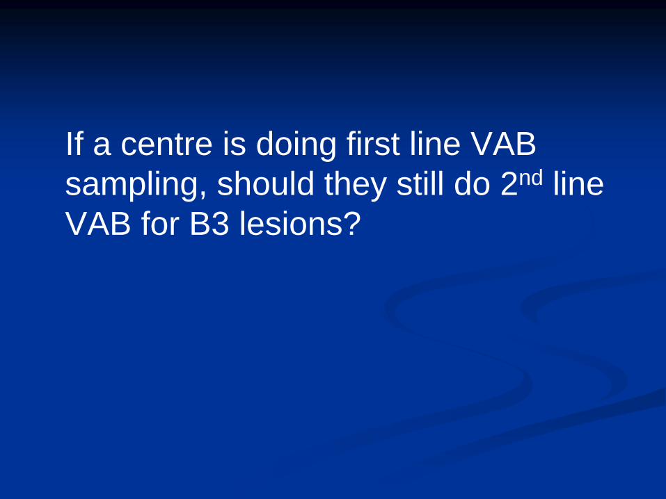
If a centre is doing first line VAB
sampling, should they still do 2nd line
VAB for B3 lesions?
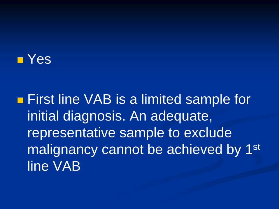
Yes
First line VAB is a limited sample for
initial diagnosis. An adequate,
representative sample to exclude
malignancy cannot be achieved by 1st
line VAB

22 Italian centres
3107 B3 VAB diagnoses
1644 (54.2%) underwent surgical excision
Overall PPV: 21.2 %

Lesion PPV (%)
Pure ADH 27.3
FEA 12.7
ALH 24.2
LIN 22
RS 10.6
All B3 21.2
VAB upgrade
What if a unit cannot implement 2nd
line VAB?

Units should try to implement 2nd line VAB
Otherwise, they should refer to another
centre that provides the service.
The group felt they should recommend
what is best for patients.

Majority of UK units have 1st line VAB
It is hoped that the guidelines will be a
catalyst to enable units to justify a
business case and implement the pathway
Should 2nd line VAB aim to excise
the whole lesion?
Not necessarily, depending on size
The aim is to obtain further tissue and a
representative sample to exclude co-
existent malignancy
The group will provide guidance on what
represents adequate sampling

Should we therefore aim to always
extensively sample on 1st line
VAB/sample all calcification?

No. 1st line VAB/cores are meant to
provide a small sample for diagnosis
and not to remove the whole
abnormality
It may be feasible to fully sample a
small area of calcification
However, it would not be justified to
excessively sample all patients by 1st
line VAB
How much tissue should be taken by
second line VAB?

Standard is: 12-20 cores, 7 or 8g
needle, or equivalent to 3.5 gm
2nd line VAB is targeted sampling.
While tissue taken is less than a
diagnostic excision, it is likely to be
more representative
Should incidental lesions such as
incidental ALH, LCIS, ADH…be
managed using the same pathway?

Yes
Evidence show that those lesions are
associated with upgrade to
malignancy on further tissue sampling

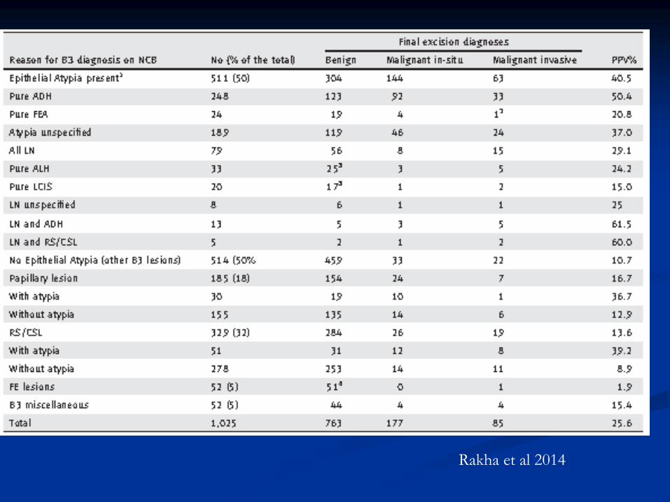
1025 core biopsies 1999- 2006.
Two UK regions: West Midlands and
South Central region
Final histology : 25% malignant
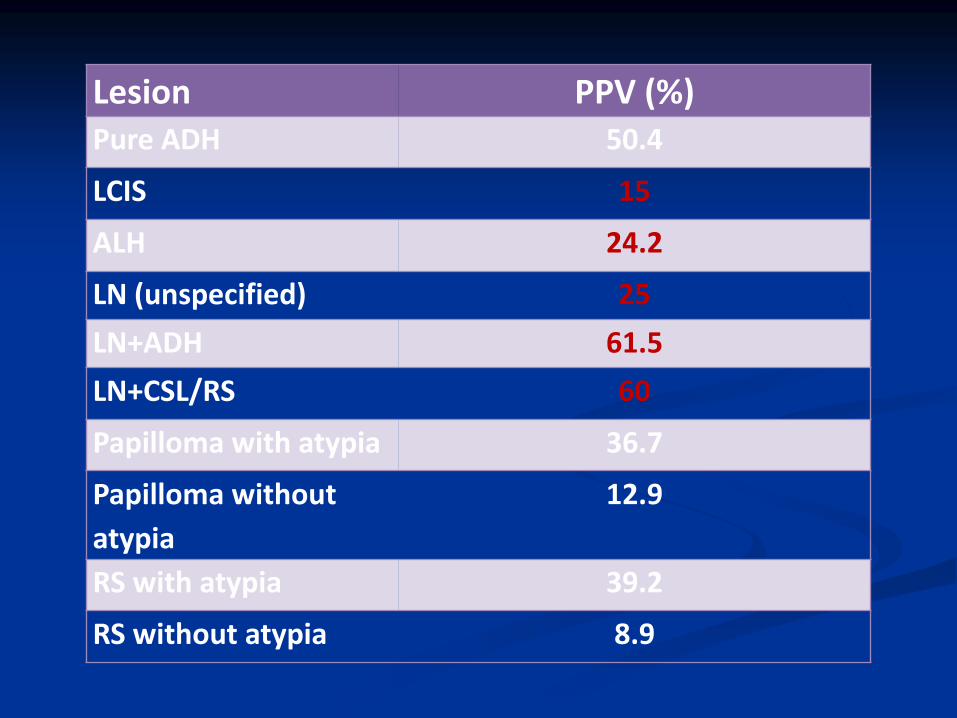
Lesion PPV (%) Pure ADH 50.4
LCIS 15
ALH 24.2
LN (unspecified) 25
LN+ADH 61.5
LN+CSL/RS 60
Papilloma with atypia 36.7
Papilloma without
atypia
12.9
RS with atypia 39.2
RS without atypia 8.9

How should small incidental papilloma
and/or radial scar without atypia be
managed?

If no atypia and the lesion is small and
fully excised on core/VAB, categorise as
B2.
If not sure is completely excised, code as
B3 and discuss at MDT meeting. If
confirmed wholly excised, no further action
is needed.
If not wholly excised, follow the
management pathway by 2nd line VAB

Summary
Current management of B3 lesions is not
uniform and likely to represent over-
treatment.
The majority of lesions are benign on
excision.
MDT discussion and radiological-
pathological correlation are essential for
planning management.
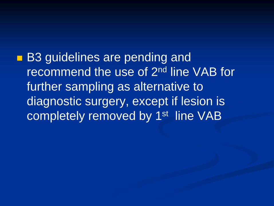
B3 guidelines are pending and
recommend the use of 2nd line VAB for
further sampling as alternative to
diagnostic surgery, except if lesion is
completely removed by 1st line VAB

THANK YOU