Embed Size (px)
Citation preview

LUND UNIVERSITY
PO Box 117221 00 Lund+46 46-222 00 00
Prognostic Factors and Metastatic Routes in Lobular Breast Cancer. Same Same butDifferent.
Narbe, Ulrik
2020
Document Version:Publisher's PDF, also known as Version of record
Link to publication
Citation for published version (APA):Narbe, U. (2020). Prognostic Factors and Metastatic Routes in Lobular Breast Cancer. Same Same butDifferent. Lund University, Faculty of Medicine.
Total number of authors:1
General rightsUnless other specific re-use rights are stated the following general rights apply:Copyright and moral rights for the publications made accessible in the public portal are retained by the authorsand/or other copyright owners and it is a condition of accessing publications that users recognise and abide by thelegal requirements associated with these rights. • Users may download and print one copy of any publication from the public portal for the purpose of private studyor research. • You may not further distribute the material or use it for any profit-making activity or commercial gain • You may freely distribute the URL identifying the publication in the public portal
Read more about Creative commons licenses: https://creativecommons.org/licenses/Take down policyIf you believe that this document breaches copyright please contact us providing details, and we will removeaccess to the work immediately and investigate your claim.

Prognostic Factors and Metastatic Routes in Lobular Breast CancerSame Same but DifferentULRIK NARBE
DEPARTMENT OF CLINICAL SCIENCES | FACULTY OF MEDICINE | LUND UNIVERSITY

Department of Clinical SciencesDivision of Oncology and Pathology
Lund University, Faculty of Medicine Doctoral Dissertation Series 2020:122
ISBN 978-91-7619-985-5ISSN 1652-8220 9
789176
199855
Ulrik Narbe

Prognostic Factors and Metastatic Routes in Lobular Breast Cancer


Prognostic Factors and Metastatic Routes in Lobular Breast Cancer
Same Same but Different
Ulrik Narbe
DOCTORAL DISSERTATION by due permission of the Faculty of Medicine, Lund University, Sweden.
To be defended at Segerfalkssalen, BMC Biomedical Centre, Sölvegatan 17, Lund, Friday December 4, 2020 at 9:00 a.m.
Faculty opponent Professor Bjørn Naume, MD, PhD
Department of Oncology, Division of Cancer Medicine, Oslo University Hospital, Oslo, Norway

Organization LUND UNIVERSITY
Document name: DOCTORAL DISSERTATION
Faculty of Medicine, Department of Clinical Sciences, Division of Oncology, Lund
Date of issue: December 4, 2020
Author: Ulrik Narbe Sponsoring organization
Title and subtitle: Prognostic factors and metastatic routes in lobular breast cancer – same same but different
Abstract Invasive lobular carcinoma (ILC) is the second most common histopathological type of breast cancer (~10%) after invasive ductal carcinoma of no special type (NST) (~80%). Compared with NST, ILC has distinguishing clinicopathological and genomic features, and its responsiveness to systemic treatment differs. Despite such differences, current diagnostic work-up and clinical management are similar. The overall aim of this thesis was to contribute to the general understanding of the clinical value of different prognostic factors and the characteristics of metastatic dissemination in ILC and to compare the results with those seen in NST. Paper I: In a retrospective cohort of patients with primary ILC (n=192, median follow-up [FU] time: 21 years), the long-term prognostic effect of the proliferation marker Ki67 and Nottingham histological grade (NHG) were analyzed alone and in combination with estrogen receptor (ER), tumor size (T), and axillary nodal status (N)— known as the prognostic index (KiGE-TN). Ki67 and NHG were prognostic factors significantly associated with breast cancer mortality (BCM). Further, KiGE-TN was able to identify a low-risk group of patients (37%) with an excellent long-term prognosis. Paper II: This study is based on an extended version of the cohort in paper I, including patients with exclusively ER positive/HER2 negative ILC (n=224, median FU time: 26 years). The putative prognostic biomarkers, amplified in breast cancer 1 (AIB1), androgen receptor (AR), and G protein-coupled estrogen receptor (GPER), all of which are related to endocrine signaling pathways, were assessed. Validation gene expression (GEX) analysis of these biomarkers was also performed using 3 independent publicly available ILC datasets. AIB1 was an independent prognostic factor for BCM, a result that was strenghtened through GEX analysis. Paper III: This was a large, Swedish registry study in which patients with primary ILC (n=2921) and NST (n=16,711) were included. Compared with patients with NST, patients with ILC were diagnosed with a higher metastatic nodal burden and more often with a luminal A-like subtype. Among patients fulfilling the St. Gallen 2019 criteria for omission of completion axillary lymph node dissection (cALND) and compared with NST cases, patients with ILC had an independently higher risk of non-sentinel lymph node metastases (SLNMs) and ≥ 4 axillary lymph node metastases (ALNMs). Further, compared with patients with NST, luminal A-like subtype and ≥ 4 ALNMs were overrepresented in patients with ILC and 1–2 SNLMs (odds ratio 6.35, 95% confidence interval [CI] 4.18–9.65). Omission cALND in this subset of patients warrants future attention, as it might affect important clinical information for the guidance of adjuvant treatment. Paper IV: In this study, patients with metastatic ILC (n=28) and NST (n=111) were included in an observational trial. Distributional and prognostic characteristics of circulating tumor cells (CTCs) were explored. CTC count (number/7.5 mL blood) was evaluated with serial sampling (CellSearch). At baseline, CTC counts were higher in patients with ILC (median 70) than in NST (median 2) (P<0.001). The evidence for ≥ 5 CTCs as a prognostic factor for progression-free survival in ILC was weak (hazard ratio [HR] 1.5, 95% CI 0.55–4.0) but strong with higher cut-offs (CTC ≥ 20: HR 3.0, 95% CI 1.3–6.8; CTC ≥ 80: HR 3.6, 95% CI 1.5–8.8). Among patients with NST, the prognostic effect of the CTC count was strong for all cut-offs (≥5, ≥20 and ≥80). A decline in the CTC count from baseline to 3 months was associated with improved prognosis in patienst with ILC and NST. Further, the number of CTCs was higher in patients with metastatic ILC than in patients with NST, implying that a higher CTC cut-off could be considered for ILC when applying the CellSearch technique. In conclusion, the results from the studies included in this thesis confirm that lobular breast cancer is a histopathological type associated with a variety of unique clinicopathological features and that such features need to be considered during the multidisciplinary discussion of diagnostic and treatment decision-making. Key words: lobular breast cancer, invasive lobular carcinoma, invasive ductal carcinoma of no special type, prognostic factors, circulating tumor cells, axillary lymph node metastasis Classification system and/or index terms (if any)
Supplementary bibliographical information Language English
ISSN 1652-8220 ISBN 978-91-7619-985-5
Recipient’s notes Number of pages 104 Price
Security classification
I, the undersigned, being the copyright owner of the abstract of the above-mentioned dissertation, hereby grant to all reference sources permission to publish and disseminate the abstract of the above-mentioned dissertation.
Signature Date 2020-10-29

Prognostic Factors and Metastatic Routes in Lobular Breast Cancer
Same Same but Different
Ulrik Narbe, MD

Author: Ulrik Narbe, MD
Principal supervisor: Professor Lisa Rydén, MD, PhD
Co-supervisors: Associate Professor Pär-Ola Bendahl, PhD Professor Mårten Fernö, PhD Professor Christian Ingvar, MD, PhD
Coverphoto by Kristina Lövgren
Copyright Ulrik Narbe Paper 1 © Springer Paper 2 © Springer Paper 3 © by the Authors (Manuscript unpublished) Paper 4 © by the Authors
Lund University, Faculty of Medicine Doctoral Dissertation Series 2020:122 Department of Clinical Sciences, Division of Oncology, Lund University, Lund, Sweden
ISBN 978-91-7619-985-5 ISSN 1652-8220
Printed in Sweden by Media-Tryck, Lund University Lund 2020

To the best two-thirds of the super trio
“Om man inte vet vart man ska så kan man lika gärna ta det lugnt, för man vet ju ändå inte när man kommit fram” –Nalle Puh

Contents
Populärvetenskaplig sammanfattning .......................................................... 11 Abbreviations ............................................................................................... 15 Studies included in the thesis ....................................................................... 18 Thesis at a glance ......................................................................................... 19
Introduction .......................................................................................................... 21 Breast cancer ................................................................................................ 21
General background ............................................................................. 21 Lobular breast cancer ................................................................................... 26
General background ............................................................................. 26 Epidemiology and risk factors ............................................................. 29 Histopathological variants ................................................................... 30 Classic type .......................................................................................... 30 Pleomorphic lobular breast cancer ...................................................... 34 Lobular carcinoma in situ .................................................................... 34 Pleomorphic lobular carcinoma in situ ................................................ 35 Diagnostic imaging and tissue biopsies ............................................... 35 Locoregional treatment ........................................................................ 36 Systemic treatment .............................................................................. 38 Metastatic lobular breast cancer .......................................................... 41
Putative prognostic and treatment predictive factors ................................... 42 Androgen receptor ............................................................................... 42 Amplified in breast cancer 1 ................................................................ 42 G protein-coupled estrogen receptor ................................................... 43 Tumor infiltrating lymphocytes ........................................................... 43 Programmed death-ligand 1 and programmed cell death-1 ................. 43 Phosphoinositide 3-kinase ................................................................... 44 Disseminated tumor cells .................................................................... 44
Liquid biopsies ............................................................................................. 44 Circulating tumor cells ........................................................................ 44 Circulating tumor cell clusters ............................................................. 45 Circulating tumor DNA ....................................................................... 45 Cancer antigen 15-3 ............................................................................. 45

Aims ....................................................................................................................... 47 Summary of materials and methods ................................................................... 49
Paper I .......................................................................................................... 49 Paper II ......................................................................................................... 51 Paper III ........................................................................................................ 54 Paper IV ....................................................................................................... 56
Summary of results and discussion ..................................................................... 59 Paper I .......................................................................................................... 59 Paper II ......................................................................................................... 62 Paper III ........................................................................................................ 64 Paper IV ....................................................................................................... 66
General discussion ................................................................................................ 73 Conclusions ........................................................................................................... 77 Future perspectives .............................................................................................. 79 Acknowledgements ............................................................................................... 83 References ............................................................................................................. 87


11
Populärvetenskaplig sammanfattning Bröstcancer (BC) är den i särklass vanligaste cancerformen hos kvinnor. Varje år drabbas över hela världen drygt 2 miljoner kvinnor (och ett fåtal män) av sjukdomen, varav ungefär 8000 är från Sverige. Trots betydande behandlingsframsteg under de senaste årtiondena, så är antalet BC relaterade dödsfall globalt fortfarande drygt 600,000 per år och i Sverige drygt 1400. Aktuell överlevnadsstatistik visar att av alla i västvärlden som drabbats av BC, lever omkring 90% 5 år efter diagnosen och efter 10 år drygt 80%.
BC indelas vanligen utifrån tumörcellens ursprung (även kallat histopatologisk typ). Den helt dominerande typen är den duktala, som uppstår i bröstets gångsystem (duktus = gång) och står för omkring 80% av all BC. Den näst vanligaste, men betydligt mer sällsynta typen är den lobulära, som uppstår i bröstets mjölkproducerande lober (lobulus = lob) och står för omkring 10%. Övriga histopatologiska typer utgörs av ett flertal mer ovanliga varianter innefattande bland annat: medullär-, tubulär-, papillär- och mucinös BC.
Även om lobulär BC är relativt ovanlig jämfört med duktal BC så är det fortfarande en vanlig cancerform som drabbar ett stort antal kvinnor. Enligt aktuell statistik från American Cancer Society så är lobulär BC den 6:e vanligaste typen av cancer i USA med nästan 40,000 nya fall per år, vilket är fler än både antalet insjuknande i lymfom respektive malignt melanom. Omkring 450,000 nu levande kvinnor i USA beräknas ha eller ha haft en lobulär BC.
För varje patient med nydiagnostiserad BC görs en bedömning av olika tumörfaktorer med betydelse för prognos. Dessa prognostiska faktorer är histologisk grad (förenklat en bedömning i mikroskopet av hur avvikande cancercellerna ser ut och hur avvikande cancern växer i förhållande till normala celler av samma ursprung), ER (östrogenreceptor), PR (progesteronreceptor), Ki67 (delningshastighet), HER2 (tillväxtfaktorreceptor), tumörstorlek (T) och lymfkörtelspridning till armhålan (N). Dessutom ger ER och HER2 även viktig information för val av behandling (behandlingsprediktiva).
Tester där man delar in BC i olika så kallade molekylära subtyper baserat på tumörcellernas genuttrycksprofil, blir mer och mer vanliga i kliniken och kan i utvalda fall ge viktig prognostisk och behandlingsprediktiv tilläggsinformation.
Lobulär BC har många särskiljande drag jämfört med duktal BC. Det mest karaktäristiska är en genmutation som gör att vidhäftningsproteinet E-cadherin antingen inte fungerar eller helt saknas hos cancercellerna. Motsvarande defekt ses nästan aldrig hos duktal BC. Förlusten av E-cadherin bidrar till ett speciellt lobulärt växtsätt och atypiskt spridningsmönster. Istället för att bilda avgränsbara tumörer så växer lobulär BC ofta diffust med tumörcellerna arrangerade i enkla rader (så kallade ”single-files”) som gömmer sig i normal vävnad utan att invadera och

12
förstöra den. Detta medför att tumören ofta inte går att känna i bröstet och den kan också vara svår att upptäcka med mammografi och/eller ultraljudsundersökning. När lobulär BC sprider sig till andra organ via lymfsystemet eller blodbanan (fjärrmetastasering) är skelettet den i särklass vanligaste lokalen. Dessutom förkommer spridning till mer ovanliga lokaler i en betydligt högre omfattning än vad man ser hos duktal BC. Exempel på dessa är bukhinna, magsäck, tunntarm, hjärnhinna, äggstock, och mer sällsynt även till urinblåsa, ögonhåla, binjure och hypofys.
Andra typiska drag hos lobulär BC är att den oftast är ER och PR positiv och histologiskt grad 2, har lågt Ki67 och saknar HER2.
Prognosen för lobulär jämfört med duktal BC verkar totalt sett vara likvärdig, men studier visar på en trend där prognosen är något bättre för patienter med lobulär BC de 5 första åren efter diagnos, för att sedan gradvis försämras, och efter 10 år har överlevnadskurvorna skurit varandra och prognosen ser sämre ut för lobulär BC, på grund av en överrepresentation av sena återfall.
När det gäller behandling visar studier att lobulär BC generellt sett har en god effekt av endokrin behandling (antihormonbehandling vid ER positiv BC) medan effekten av cytostatika anses tveksam i de flesta fall.
De studier som ligger till grund för dagens behandlingsrekommendationer för BC bygger nästan uteslutande på patientmaterial där blandade histopatologiska bröstcancertyper inkluderats (och där duktal bröstcancer på grund av sin vanlighet kraftigt dominerar). Således är behandlingsprincipernas giltighet för lobulär BC sämre underbyggda och mindre utvärderade jämfört med duktal BC.
Lobulär BC är en understuderad och lite bortglömd typ i förhållande till duktal BC. Tillgängliga studier som specifikt inriktar sig på lobulär BC är relativt få och storleksmässigt ofta små.
Samtliga delar i den här avhandlingen handlar om lobulär BC, antingen som ensamt studieobjekt eller i direkt jämförelse med duktal BC. Det övergripande syftet har varit att ta reda på hur olika prognostiska faktorer (både etablerade och experimentella) fungerar i lobulär bröstcancer och hur dessa kan användas på ett optimalt sätt i kliniken. Vidare har olika spridningsvägar kartlagts, med fokus på lymfkörtelspridning till armhålan och förekomst av cirkulerande tumörceller i blodbanan och jämförelse av dessa spridningssätt har gjorts mellan lobulär respektive duktal BC.
Studie I: Resultaten visar att både bedömning av Ki67 och histologisk grad ger värdefull prognostisk information både på kort och lång sikt hos patienter med lobulär BC. Studien visade också att en kombination av olika prognostiska faktorer (Ki67, histologisk grad, ER, T och N) har en starkare prognostisk effekt tillsammans än var och en för sig, och kan skilja ut en grupp lågriskpatienter med extremt god

13
prognos (37% av alla patienter) där tilläggsbehandling efter operation med cytostatika kan undvikas och endokrin behandling troligen begränsas.
Studie II: Här studerades de experimentella biomarkörerna: amplified in breast cancer 1 (AIB1), androgen receptor (AR) och G protein-coupled estrogen receptor (GPER). Dessa hormonellt kopplade biomarkörer har uppvisat lovande prognostisk effekt i några tidigare bröstcancerstudier men deras specifika prognostiska värde i lobulär BC är oklart. Resultaten visade att AIB1 har ett prognostiskt värde i lobulär BC, där högt AIB1 är sammankopplat med en dålig prognos. Övriga faktorer uppvisade inget tydligt prognostiskt värde i lobulär BC.
Studie III: Spridning av cancer till lymfkörtlar (lymfkörtelmetastaser) i armhålan är en av de starkaste prognostiska faktorerna i BC. Hos patienter med BC där övriga prognostiska faktorer är gynnsamma, vilket ofta är fallet vid lobulär BC, är information om förekomst av lymfkörtelspridning en avgörande faktor för beslut om tilläggsbehandling efter operation, och enligt dagens behandlingsriktlinjer rekommenderas cytostatikabehandling till patienter med 4 eller fler lymfkörtelmetastaser. Idag görs i samband med bröstoperationen också ett mindre kirurgiskt ingrepp i armhålan, en undersökning av de lymfkörtlar dit cancern bedöms kunna sprida sig först. Dessa så kallade portvaktslymfkörtlar (sentinel nodes) identifieras och plockas ut för vidare undersökning. Om det finns 1 till 2 metastaser i de undersökta portvaktskörtlarna behövs enligt aktuella behandlingsriktlinjer i de allra flesta fall ingen ytterligare operation (så kallad lymfkörtelutrymning). Studier har nämligen visat att det inte finns någon skillnad i återfallsrisk eller överlevnad mellan patienter som genomgått lymfkörtelutrymning jämfört med de som inte opererades. Vinsten med att inte operera är att risken för nedsatt funktion och svullnad i armen minskar. Antalet patienter med lobulär BC i dessa studier var dock mycket begränsat.
I denna studie undersöktes skillnader i risk att det skall finnas ytterligare metastaser i övriga kvarvarande lymfkörtlar, mellan lobulär och duktal BC med 1 till 2 metastaser i portvaktskörtlarna. Dessutom undersöktes skillnader i förekomst av totalt 4 eller fler lymfkörtelmetastaser i armhålan men för övrigt gynnsamma prognosfaktorer hos patienter med lobulär och duktal BC. Resultaten visade att risken för ytterligare metastaser i kvarvarande lymfkörtlar är tydligt högre i lobulär jämfört med duktal BC. Andelen patienter med gynnsamma prognostiska faktorer och 4 eller fler lymfkörtelmetastaser var också överrepresenterade i lobulär BC jämfört med duktal BC, vilket i praktiken innebär att i ungefär 1 av 6 (17%) lobulära och i 1 av 25 (4%) duktala BC riskerar man att på grund av bristande information om det totala antalet lymfkörtelmetastaser i armhålan missa att rekommendera tilläggsbehandling med cytostatika, vilket skulle kunna ha en negativ inverkan på prognosen hos dessa patienter.
Studie IV: Med en speciell teknik (CellSearch) kan man analysera förekomst av cirkulerande tumörceller (CTC) i blodet vid metastaserad BC (BC som spridit sig

14
från bröstet till andra organ). Förekomst av CTC i metastaserad BC har visat sig vara sammankopplat med en sämre prognos, särskilt om antalet CTC är 5 eller fler mätt i 7.5 ml blod. Förekomsten av CTC och dess prognostiska betydelse i metastaserad lobulär BC är mycket sparsamt undersökt. Denna studie visar att förkomsten av CTC i metastaserad lobulär BC (medianvärde 70) är mycket högre än i metastaserad duktal BC (medianvärde 2). Det prognostiska värdet av den normalt rekommenderade brytpunkten ≥5 CTC var svagt i metastaserad lobulär BC, men om en högre brytpunkt på ≥20 eller ≥80 användes istället så stärktes den prognostiska betydelsen avsevärt, talande för att en högre CTC brytpunkt är mer optimal i metastaserad lobulär BC när man använder CellSearch tekniken.
Sammanfattningsvis bekräftar resultaten i denna avhandling att lobulär BC på ett flertal punkter skiljer sig åt från den duktala, och även andra delvis nya skillnader avseende lymfkörtelspridning och cirkulerande tumörceller har påvisats. Dagens behandlingsriktlinjer gör trots dessa skillnader, och på relativt lösa grunder, mycket liten skillnad mellan lobulär och duktal BC. Fortsatta och fler större studier behövs för att förbättra diagnostik, behandling och prognos för patienter med lobulär BC.

15
Abbreviations AIB1 amplified in breast cancer 1
ALN axillary lymph node
ALND axillary lymph node dissection
ALNM axillary lymph node metastasis
AR androgen receptor
Bcl2 B-cell lymphoma 2
BCM breast cancer mortality
BCT breast conserving therapy
BL base line
cALND completion axillary lymph node dissection
CA15-3 cancer antigen 15-3
CI confidence interval
CNB core needle biopsy
CTC circulating tumor cell
ctDNA circulating tumor DNA
CT chemotherapy
DFS disease-free survival
DTC disseminated tumor cell
ECE extracapsular extension
ER estrogen receptor
ET endocrine therapy
FNA fine needle aspiration
FU follow-up
GPER G protein-coupled estrogen receptor
HER2 human epidermal growth factor receptor 2
HRT hormone replacement treatment
HR hazard ratio
IDC invasive ductal carcinoma

16
IHC immunohistochemistry
ILC invasive lobular carcinoma
IQR interquartile range
ITC isolated tumor cells
Ki67 proliferation marker
KiGE Prognostic index including Ki67, NHG and ER
KiGE-TN Prognostic index including Ki67, NHG, ER, T and N
KVAST Swedish Quality Document for Pathology
LN lymph node
MBC metastatic breast cancer
MRI magnetic resonance imaging
NHG Nottingham histological grade
NKBC National Quality Breast Cancer Register
NST invasive ductal carcinoma of no special type (also referred to as IDC)
N nodal status
OS overall survival
pCR pathological complete remission
PD-L1 programmed death-ligand 1
PD-1 programmed cell death-1
PFS progression-free survival
PI3K Phosphoinositide 3-kinase
PLC pleomorphic invasive lobular carcinoma
PR progesterone receptor
RECIST response evaluation criteria in solid tumors
REMARK reporting recommendations for tumor marker prognostic studies
RCT randomized controlled trial
RS recurrence score
RT radiotherapy
SLN sentinel lymph node

17
SLNM sentinel lymph node metastasis
STROBE strengthening the reporting of observational studies in Epidemiology
TDLU terminal duct lobular unit
TILs tumor infiltrating lymphocytes
TMA tissue microarray
T tumor size

18
Studies included in the thesis This thesis is based on the following papers, which will be referred to in the text by their corresponding Roman numerals:
I. Narbe U, Bendahl PO, Grabau D, Rydén L, Ingvar C, Fernö M.Invasive lobular carcinoma of the breast: long-term prognostic value ofKi67 and histological grade, alone and in combination with estrogenreceptor. Springerplus. 2014, 3:70.
II. Narbe U, Sjöström M, Forsare C, Bendahl PO, Alkner S, Leeb-LundbergLMF, Lövgren K, Rydén L, Ingvar C, Fernö M. The estrogen receptorcoactivator AIB1 is a new putative prognostic biomarker in ER-positive/HER2-negative invasive lobular carcinoma of the breast. BreastCancer Res Treat. 2019, 175(2): 305-316.
III. Narbe U, Bendahl PO, Fernö M, Ingvar C, Dihge L, Rydén L. Lobularbreast cancer and axillary lymph node management according to the St.Gallen 2019 Guidelines – a population-based study of 20,139 patients.Submitted manuscript. Under review.
IV. Narbe U, Bendahl PO, Aaltonen K, Fernö M, Forsare C, Jørgensen L TC, Larsson AM, Rydén L. The distribution of circulating tumor cells isdifferent in metastatic lobular compared to ductal carcinoma of the breast– long-term prognostic significance. Cells. 2020, 9(7): 1718.

19
Thesis at a glance Study Question Patients and Methods Figure Results
I
Are Ki67 and Nottingham histological Grade (NHG) long-term prognostic factors in invasive lobular carcinoma (ILC), and does the addition of Estrogen receptor (ER) in a prognostic index (KiGE), together with Tumor size and Nodal status, identify high vs. low risk patients?
Biomarkers were analyzed with immunohistochemistry (IHC) in tumors from 192 patients with ILC. The median follow-up (FU) time was 21 years, and the primary endpoint was breast cancer mortality (BCM).
KiGE-TN
Breast cancer mortality by KiGE-TN
Ki67 and NHG have long-term prognostic value in ILC and patients low-risk KiGE score and ≤20mm node negative ILC had an excellent long-term prognosis.
II
Do endocrine related biomarkers (amplified in breast cancer 1 [AIB1], androgen receptor [AR] and G protein-coupled estrogen receptor [GPER]) have prognostic value in luminal-like (ER+/HER2-) ILC?
Putative prognostic biomarkers were analyzed with IHC in tumors from 224 patients with ILC. The median FU time was 26 years, and the primary endpoint was BCM. Validation analysis for these biomarkers was performed using 3 publicly available gene expression (GEX) datasets.
Breast cancer mortality by AIB1
AIB1 is a putative prognostic biomarker in patients with luminal-like ILC, whereas no evident prognostic effect was seen for AR and GPER. The above results were strengthened through GEX analysis.
III
Is the metastatic nodal burden different in patients with ILC compared with invasive ductal carcinoma of no special type (NST), and does nodal staging information from completion axillary lymph node dissection (cALND) have an impact on adjuvant treatment decision-making?
In this large registry study, patients with ILC and NST were included. Those with 1 to 2 sentinel lymph node metastases (SLNMs) fulfilling the St. Gallen 2019 criteria for omission of cALND were further analyzed regarding surrogate molecular subtype and prevalence of non-SLNMs and axillary lymph node metastases (ALNM).
Flow chart of the study cohort
The risk of non-SLNMs and ≥4 ALNMs was higher in ILC than in NST. Patients with a luminal A-like subtype and ≥4 ALNMs were overrepresented in ILC compared with those in NST patients (17% vs. 3%). Thus, omission of cALND in this subgroup warrants futute attention as it may affect information which is important for the guidance of adjuvant treatment.
IV
Is there a difference in distributions and prognostic utility of circulating tumor cells (CTCs) in metastatic ILC and NST?
The CTC count was evaluated with serial sampling (CellSearch) in 28 patients with metastatic ILC and 111 patients with NST. The primary endpoint was progression-free survival (PFS).
PFS by CTC count
CTC counts were higher in ILC than in NST cases (median 70 vs. 2). The evidence for ≥ 5 CTCs/7.5ml blood as a prognostic factor was weak in ILC, but strong with higher cut-offs (≥ 20 and ≥ 80), implying that a higher cut-off could be considered for patients with ILC.


21
Introduction
Breast cancer
General background Breast cancer (BC) is the most common of all malignancies in women. In 2018, 2.1 million new cases were diagnosed worldwide 1,2 and the corresponding figure in Sweden was just over 7,800 3. BC represents ~30% of all female cancers and about 1 in 8 (~12%) of all women will develop BC over the course of their lifetime. The BC incidence has increased over the last decades, and this trend is likely to continue 2, but due to earlier detection and better treatment options, the survival rates have also improved. In developed countries, the current 5- and 10-year survival rates are approximately 90% and 80%, respectively 4,5. Nonetheless, at certain time 20%–30% of all BCs present as metastatic breast cancer (MBC) with dissemination to distant organs. MBC is a chronic and often fatal disease, resulting in a global BC-related death rate exceeding 600,000 among women annually 2,6 (in Sweden ~1,400†).
Historically, BC classification is commonly based on the histological appearance of the tumor. The vast majority of BCs arise in the same segment of the breast; the terminal duct lobular unit (TDLU), where the tumor cells originate either from the milk ducts or the milk producing lobules.
Invasive ductal breast cancer of no special type (NST) is the dominant histopathological type, comprising 80-85% of all BC in the Western world 7. The second most common type, although a minor one in relation to NST, is invasive lobular carcinoma (ILC), comprising 10-15% of all BC 7. Other less common histopathological types include: medullary-, tubular-, papillary- and mucinous breast cancer 7.
Different prognostic factors in BC are well-established. The most important clinically validated are: estrogen receptor (ER) and progesterone receptor (PR), human epidermal growth factor receptor 2 (HER2), proliferation marker Ki67, Nottingham histological grade (NHG), tumor size (T), axillary nodal status (N), and age. The combination of these factors into prognostic indices has been shown to result in a stronger prognostic assessment than separate analyses of each factor 8-12. In addition, ER and HER2 each provide treatment predictive information.

22
At the present time, molecular (also referred to as intrinsic) subtyping, based on gene sequencing is another type of BC classification that is increasingly used in the clinic for prognostication and treatment prediction. These tests have become available in many Western countries, but the cost is still high 13. A complementary much less expensive surrogate definition of the molecular subtypes, based on immunohistochemical (IHC) analyses of tumor markers (e.g. ER, PR, HER2, proliferation marker Ki67) and Nottingham histological grade (NHG) has been developed 14. Surrogate molecular subtyping together with tumor size, axillary lymph node status and age has become widely utilized for prognostication and treatment prediction in the clinic.
Furthermore, several freely available decision-making tools (e.g. PREDICT breast cancer, Nottingham Prognostic Index [NPI] and CancerMath) exist to help predict recurrence risk and potential benefit from systemic treatments 12,15,16
Figure 1. Breast Cancer Overview. Reprinted with permission from Springer Nature: Nature Reviews Disease Primers, Harbeck et al. 2019. aESR1 mutations induced by aromatase inhibitor targeted therapy. bArtefact; expression of normal breast components due to low tumor cellularity.

23
In recent decades, modern BC care has considerably progressed, but unmet needs still remain. Research focusing on new prognostic and monitoring factors, as well as improving those used today, and a better understanding of the complex mechanisms behind the metastatic process and routes of metastasis, is important to further optimize clinical strategies and treatments.


25
”In a nutshell, the lobular breast cancer is freaky, it is sneaky, it breaks a lot of common rules, and it is not on people´s radar” –Anonymous lobular breast
cancer survivor at the LBCA tweet #lobmob

26
Lobular breast cancer
General background The first known illustrations of invasive lobular cancer, based on its microscopic appearance, were done in 1908 by Victor Cornil, and at that time this type of breast cancer was described as “acinar or scirrhous spheroidal cell carcinoma” 17,18. The term “lobular” was first coined by Foote and Stewart, who defined the non-invasive variant as “lobular carcinoma in situ” (LCIS) in 1941 19 and subsequently its invasive counterpart as “invasive lobular carcinoma” (ILC) in 1946 20. Since then, further subclassification into different histopathological ILC subtypes has been developed 7,21-31.
Compared to NST, ILC has unique clinicopathological and genomic features, and it responds differentially to systemic treatment 27,30,32-36 (Figure 2 A-B).
Among all these features, the most quintessential, and often referred to as the hallmark of ILC, is the loss of the adhesion molecule E-cadherin.
In several studies that compared patients with ILC and NST, the long-term overall prognosis seemed to be the same, although time dependent prognostic trend with a better 5-year survival rate and a tendency towards higher incidence of late recurrences (>10 years of follow-up [FU]) has been seen in ILC 33,34,37. However, the results were not unanimous; in a study by Colleoni et al. who compared survival outcomes between luminal-like (ER+/HER2-) ILC and NST, patients with ILC had a significantly worse survival outcome with 5 years of FU 38.
Although ILC is a relatively rare form of BC, compared to NST, it is still a very common condition, affecting a large population of women. According to the American Cancer Society, ILC is the 6th most common histopathological type of female cancer in the United States (U.S.), with a higher incidence than lymphoma and melanoma (Figure 3). Nearly 40,000 new cases of ILC are diagnosed annually, and approximately 450,000 women are living with a previous or current ILC diagnosis 39.

27
Figure 2. Microscopic pictures of (A) classic invasive lobular carcinoma (ILC) and (B) invasive ductal carcinoma of no special type (NST), displaying the distinct histopathological differences between the two types.
B
A

28
Figure 3. Incidences of female cancers in U.S. 2018. Reprinted with permission from Fred Hutch News Service.
In the BC research community as a whole, ILC specific research is a neglected area, and patients with ILC belong to an understudied subgroup. Most BC studies are conducted on patient populations with mixed histopathological BC types, where the vast majority of cases have NST. Studies exclusively exploring ILC and/or comparing ILC vs. NST (and other types) are sparse and the sample sizes are usually small. Moreover, results from subgroup analyses of ILC in randomized controlled trials (RCTs) of mixed BC are seldom reported.
Additionally, in the era of molecular subtyping of BC, the potential significance of histopathological type is overlooked to a large extent.
In 2016, the Lobular Breast Cancer Alliance (LBCA) 40 was founded in Pittsburgh, PA, by patient advocates who attended the First International Lobular Breast Cancer Symposium (Figure 4A). The purpose is to increase awareness of ILC and to promote ILC specific research. LBCA is advised by an international Scientific Advisory Board of researchers and clinicians who focus on ILC. This is probably the first subtype of an organ specific cancer type, that has its own international scientific symposium as well as a patient advocate organization.

29
In 2018, the European Lobular Breast Cancer Consortium (ELBCC) 41 was founded (Figure 4B). This is a collaboration consisting of leading ILC researchers and their mission is to establish a European research platform for ILC specific research in order to achieve a common goal of improved understanding, diagnosis and treatment for ILC. Furthermore, in collaboration with ELBCC, a European patient advocacy organization, “the ELBCC Patient Advocates” has been formed.
Despite there is a lower number of patients diagnosed with ILC in Sweden than in the U.S., a similar advocacy initiative on a national level would be strongly encouraged.
Figure 4. (A) The Lobular Breast Cancer Allience (LBCA) logotype. Reprinted with permission from LBCA. (B) The European Lobular Breast Cancer Consortium (ELBCC) logotype. Reprinted with permission from ELBCC.
Epidemiology and risk factors Compared to NST, the incidence of ILC has increased disproportionately between 1975 and 2000 26,42,43 and this finding seems to be strongly related to the frequent use of hormone replacement treatment (HRT) in menopausal women, during this time period. An association between HRT and increased risk of BC, has been shown repeatedly, and this effect seems to be more pronounced in ILC than in other histopathological types, probably due to its extraordinarily high endocrine sensitivity 44. Around 2002, a seminal publication from the Women´s Health Initiative 45 addressing this issue, had an almost instant impact on decreasing HRT prescriptions, and a subsequent decline in ILC incidence was seen. Despite a continuous restrained policy about HRT, the incidence of ILC has started to rise again in recent years, and the reason for this is not clear 46.
Other known promoting risk factors that have a slightly stronger impact on ILC than NST incidence, are: early menarche, older age at first parity, late menopause and alcohol consumption 26,47.

30
Overall, germline mutations are less common in patients with ILC than in those with NST, although a relatively higher proportion of ILC has been identified in BCs associated with BRCA2 mutations (8.8%) than in those with BRCA1 mutations (2.2%) 47. Moreover, women with germline CDH1 mutations have an increased risk of developing an ILC 47.
Moreover, the ILC incidence is significantly lower (~5%) in Asia/Africa/Middle East than in Western world 26. Genetic factors might play a role since it has been shown that Asian Americans living in the U.S., still have the same lower ILC incidence 48, but most likely both endo- and exogenous risk factors are contributing to the development of this disease.
Histopathological variants ILC can be divided into two histopathological subtypes: (1) based on different tumor tissue architecture and growth patterns including classic, solid, alveolar and trabecular; and (2) based on cytological features including pleomorphic, apocrine, histiocytoid and signet-ring cell 7,21-31.
According to the WHO Classification of Tumors of the Breast 7, BCs composed of ≥90% lobular tumor component are referred to as pure ILC. Mixed lobular types, where >50% and <90% of the tumor tissue consists of ILC, also exist. The most common is the mixed ductal/lobular type (ductolobular BC) 49,50 and there is also a less common tubular/lobular type (tubulolobular BC) 51.
Except for the prognostically unfavorable subtypes such as pleomorphic ILC (PLC) 52,53 and signet ring cell ILC 31, no significant prognostic differences between classic type ILC and other ILC subtypes has been shown 28.
Classic type The majority of lobular BCs are the classic ILC. These tumors are characterized by small round discohesive tumor cells with a scant cytoplasm, monomorphic nuclei and a relatively harmless appearance 27,32,33,54. They have a characteristic growth pattern with single-files of tumor cells diffusely infiltrating benign breast tissue often without destroying normal anatomical structures 30.
Clinical characteristics The clinical characteristics of ILC differ from those of NST 33,34,37,55-59. ILC patients are generally diagnosed at an older age and a postmenopausal status is more common. Due to the characteristic lobular growth pattern, both primary tumors and axillary lymph node metastases (ALNMs) tend to be nonpalpable. The tumor size at diagnosis is somewhat larger (higher T-stage, with a two-times higher occurrence

31
of T3 tumors [>5cm]) 37,60. Compared to NST, patients with ILC who are node-positive, tend to have a larger proportion of >4 ALNMs and a higher number of non-sentinel lymph node metastases (non-SLNMs) 55-59, whereas no clear difference in the distribution of node-negative vs. node-positive cases are seen 37,61. Multifocality (≥2 synchronous invasive tumors located in the same quadrant in the same breast), multicentricity (≥2 synchronous invasive tumors located in different quadrants in the same breast) and bilaterality (≥1 synchronous invasive tumor in both breasts) are also more common in ILC 33,34,37,55-59. The risk for metachronous contralateral BC is not consistently higher in ILC 37.
A better 5-year survival, but also an overrepresentation of late (>10 years past diagnosis) recurrences has been seen in ILC compared to NST, and interestingly, this finding was independent of ER-status indicating an effect related to other factors associated with histopathology 34,37.
Pathological characteristics Compared to NST, ILC has a higher frequency (>90%) of hormone receptor positivity with immunohistochemically highly ER positive (ER+) and PR positive (PR+) status, while the frequency of HER2 positivity (HER2+) as assessed using IHC and/or gene amplification (-ISH) test, is clearly lower (~5%) 26-28.
Nottingham histological grade (NHG) is generally lower in ILC than in NST. NHG is divided into three categories, based on the composition of three variables; tubule formation, nuclear pleomorphism and mitotic score. Each variable is scored from 1 to 3 and the scores are added together to a total score where 3 to 5 = NHG1, 6 to 7 = NHG2 and 8 to 9 = NHG3 62. This grading system is mainly based on characteristics typically seen in NST. Its applicability in ILC has historically been a matter of controversy since ILC normally do not form tubules and hence almost always score 3, and the mitotic score is normally low, 1 or 2 63. Given this, the NHG in ILC depends to a large extent on the variability of nuclear pleomorphism and the majority of cases are classified as NHG2 (~75%) 34,64.
The proliferating index in ILC is generally lower than that in NST (measured as percentage of proliferating tumor cells using a proliferation marker Ki67) 65.
The St. Gallen 2019 surrogate molecular subtypes 14 is based on pathological tumor characteristics (e.g. ER/PR status, HER2 status, proliferation index [e.g. Ki67] and NHG). Tumors are divided into; luminal A-like (HER2-), luminal B-like (HER2-), HER2 positive (luminal-like), HER positive (nonluminal-like) and triple-negative.
The luminal A-like subtype is predominant (~65%) in ILC and more common than in NST (~50%) 66-68
The loss of cell-cell adhesion transmembrane protein E-cadherin, coded by the CDH1 gene located on chromosome 16q22, is very common (~90%), and considered as one of the cardinal features in ILC. The intracellular domain of E-

32
cadherin interacts with α-, β-, γ- and p120 catenins to form a cadherin-catenin complex with important functions in cell-cell adhesion 28,69. Lack of E-cadherin also results in a simultaneous loss of α-, β-, γ-catenins, while p120-catenin relocates from the cell membrane and shows increased cytoplasmic expression 65,70,71 (Figure 5A-B).
Figure 5. (A) Schematic picture of cell-cell adhesion (Source: Wikipedia, created by Mariana Ruiz, 2006). (B) The cadherin-catenin complex. Reprinted with permission from Elsevier, Seminars in Oncology, Thomas et al. 2019.
A
B

33
Molecular characteristics Molecular subtyping of BC, based on gene expression analysis was performed in a seminal study by Perou and Sørlie et al., where BCs were classified into different subgroups based on their genomic profiles: luminal A (50%), luminal B (20%), HER2-enriched (15%) and basal-like (15%) 13,72,73. Molecular subtyping has been shown to provide useful prognostic and treatment predictive information and hence, has become increasingly used in the clinic.
Based on this pioneering research, in recent years, several BC gene signatures (also referred to as gene expression assays) estimating recurrence risk and benefit of chemotherapy (CT) in women with early stage BC, have become clinically available (e.g. Oncotype DX, Prosigna-PAM50, Mammaprint and EndoPredict) 74-79.
Oncotype Dx Breast Cancer Assay is a 21-gene assay used in ER+/HER2- BC to predict benefit of CT in addition to endocrine therapy (ET) and the risk of distant recurrence (low, intermediate and high) based on a recurrence score (RS).
The Prosigna-PAM50 algorithm defines the molecular subtype (luminal A, luminal B, HER2-enriched or basal-like) and calculates a risk of RS (using 1-100 scale) that correlates with the probability of distant recurrence in ER+/HER2- BC.
Mammaprint is a 70-gene assay estimating the risk of distant recurrence (low vs. high) in ER+ and ER-/HER2- BC.
EndoPredict is a 12-gene assay for assessment of distant recurrence risk in ER+/HER2- BC.
Overall, these tests seem to have an equal prognostic value in ILC and NST, although in studies investigating OncotypeDX and Prosigna-PAM50, the recurrence scores are generally lower in ILC. In Oncotype DX, a low RS indicates no benefit, an intermediate RS uncertain benefit and a high RS a benefit of CT. The majority of patients with ILC have a low (21% to 63%) or intermediate (35% to 71%) RS, whereas only a minority (1.5% to 8 %) have a high RS 80-86.
Thus, only a small fraction of patients with ILC are predicted as “chemo gainers”; moreover, a study has indicated that the actual benefit of CT was insignificant in the high RS ILC subgroup 86.
Pleomorphic ILC is an exception, displaying higher recurrence scores, comparable with those seen in NST 81.
A comprehensive analysis of mutated and amplified genes in ILC and NST has been performed to decipher distinct genomic profiles in ILC compared to those in NST 35,65,87. Somatic mutations in CDH1 (54% to 63% vs. 2%), PIK3CA (42% to 48% vs. 33%), RUNX1 (10% vs. 3%), TBX3 (9% vs. 2%) and FOXA1 (7% vs. 2%), were more frequent in ILC and mutations in TP53 and GATA3 were more frequent in NST. An amplification of ERBB2 (~5% vs. ~15%) was also more frequent in NST.

34
In a subgroup analysis of luminal A-like ILC vs. luminal A-like NST, a higher frequency of CDH1, TBX3 and FOXA1 mutations were still found, but the difference in PIK3CA mutations was no longer significant. GATA3 mutational rate was still significantly higher in NST; in addition, that of PTEN was higher in luminal A-like ILC (14% vs. 3%) 35,65,87.
Pleomorphic lobular breast cancer PLC is an uncommon, but clinically relevant ILC subtype, and accounts for ~1% of all BC, and up to 15% of all ILC cases. It was first described by Page and Anderson in 1987 23. Compared to classic ILC, PLC has characteristic histopathologic features. Morphologically, there is a typical nuclear pleomorphism with medium to large sized nuclei and irregular cell shape. Loss of E-cadherin is also very common in PLC, and the lobular single-file growth pattern persists.
Compared to classic ILC, PLC has a less favorable prognostic profile, with a higher proportion of ER- and PR-negativity, HER2-positivity, triple-negativity (ER-/PR-/HER2-), NHG3 and non-luminal A subtype (~75%) 23,88. Furthermore, PLC has a higher mammographic detection rate compared to classic ILC 89, and one study showed a higher frequency (40%) of BRCA2 mutations in patients with PLC 90.
Studies comparing PLC and classic ILC have suggested a slightly more aggressive clinical behavior and a worse outcome in PLC 23.
Lobular carcinoma in situ Lobular carcinoma in situ (LCIS) is a noninvasive abnormality with proliferative cells originating in the milk producing lobules of the breast, predominantly affecting premenopausal women 91. It is not considered pre-cancerous, but the presence of LCIS in the breast increases the risk of invasive breast cancer later on in life 91. LCIS is an asymptomatic nonpalpable condition, normally undetectable on mammography 92,93. It is incidentally found in microscopic assessment of a core needle biopsy or a specimen from a surgical excision originally targeting another lesion 92,93. In classic LCIS, multicentricity and bilaterality is commonly seen 91. The abnormal cells are monomorphic and discohesive due to their loss of E-cadherin. LCIS is clinically managed as a benign lesion and neither a radical excision nor pathological evaluation of excision margins are required, and postoperative radiotherapy (RT) is not recommended 91. Foci of LCIS are often present synchronously with ILC (≥50%) 94. In the 8th edition of the TNM staging by the American Joint Committee on Cancer (AJCC), LCIS is no longer staged as T in situ 95.

35
Pleomorphic lobular carcinoma in situ Pleomorphic LCIS (P-LCIS) was first described by Frost et al in 1996 96 as a subtype with different features compared to classic LCIS. In P-LCIS a central necrosis and calcifications are commonly seen, and these distortions are often detected mammographically 91. Patients with P-LCIS are significantly older than those with classic LCIS 91. Due to its nuclear pleomorphism, necrosis and calcifications, P-LCIS resembles ductal carcinoma in situ (DCIS) 91,97. However, immunohistochemically, the cells of P-LCIS, compared to DCIS cells, show loss of E-cadherin and cytoplasmic localization of p120 98. The current clinical management of P-LCIS is similar to that of DCIS. Areas of P-LCIS should be surgically excised with clear margins and in some cases adjuvant RT should be given 98.
Diagnostic imaging and tissue biopsies Due to its characteristic growth pattern, ILS is considered more difficult to detect using diagnostic imaging, fine needle aspiration (FNA) and core needle biopsy (CNB) than NST 28,99-102. The detection rate of ILC is lower than that of NST on screening mammography, and hence, interval cancers (a breast cancer diagnosed in the time between a regular screening mammography that appears normal and the next scheduled examination) are overrepresented among ILC cases 26.
Typical mammographic findings such as a well-defined mass and calcifications are less often seen in ILC than in NST 99,103. Studies have shown that the sensitivity of mammography for detection of mixed invasive BC (where a majority of cases is NST) ranged from 63% to 98%, whereas the sensitivity of detecting ILC was lower (34% to 81%). Furthermore, in a study by Berg et al., the differences in sensitivity of mammographic detection of ILC compared to NST in patients with dense breast tissue was even more pronounced (11% vs. 60%) 28,99,104.
Studies exploring the sensitivity of ultrasound for detection of ILC has shown rates from 68% to 98% 28,99,104. Furthermore, Butler et al. showed that 73% (11/15) of the mammographically invisible ILC could be visualized using ultrasound 105. Ultrasound is considered a valuable complementary diagnostic tool for detecting ILC, when combined with a concurrent normal mammography, especially in patients with clinical symptoms and/or physical examination suspicious of invasive BC.
Studies evaluating preoperative axillary ultrasound for detecting lymph node metastases showed that the sensitivity was lower in patients with ILC than in those with NST (32% to 59% vs. 50% to 76%) and the same trends were seen for axillary ultrasound-guided FNA (54% to 55% vs. 76% to 98%) and CNB (33% to 86% vs. 79% to 86%) 99-102,106.

36
Studies have shown that the sensitivity of magnetic resonance imaging (MRI) was high and ranged from 93% to 96% in ILC, and the corresponding rates in mixed invasive BC were similar (~90%) 28,99. However, there is also a lower specificity associated with MRI. MRI is considered a valuable tool for detection of multiple ipsilateral and contralateral BC, a feature especially useful in ILC where these characteristics are common. A preoperative MRI in patients with BC has been shown to reduce the number of mastectomies and re-excisions due to positive surgical margins after BCT in ILC, and paradoxically the opposite was true for NST 107.
New imaging techniques are emerging. Tomosynthesis is a three-dimensional digital mammography technique based on x-ray computed tomography 99. Tomosynthesis has a unique strength in detecting architectural distortion, a typically and subtle mammographic manifestation of malignancy, and a feature commonly seen in ILC. In one study where tomosynthesis was added to digital mammography, the detection rate for ILC increased from 0.27 to 0.55 per 1000 cases, indicating that tomosynthesis may be particularly useful for the identification of ILC 108.
A study exploring the utility of 18F-FDG positron emission tomography in different histopathological BC types showed that primary tumors and metastases of ILC were less detectable than those of NST, and generally they demonstrated lower 18F-FDG uptake values 109.
Locoregional treatment
Surgical treatment
Breast surgery For decades, breast conserving therapy (BCT) was considered as a relative contraindication for patients with ILC due to the specific clinicopathological features accompanying this histopathological type.
In current treatment guidelines for BC 14,110-114, recommendations for surgery are the same for all types of BC. BCT is normally the preferred type of surgery, and secondly a mastectomy is chosen for those patients where BCT is not applicable.
Fodor et al. investigated the long-term (15-year FU) outcomes in ILC patients treated with BCT vs. mastectomy and found no significant differences in recurrence-free and breast cancer specific survival 115.
Nonetheless, mastectomy is more often required in patients with ILC than in those with NST due to a generally larger tumor size at diagnosis, a more frequent preoperative clinical understaging and higher frequency in multifocality/centricity. Compared to NST, ILC is also associated with a higher rate of positive resection margins after BCT and a secondary surgical procedure (either re-excision or mastectomy) is more often required 26,28.

37
Available long-term FU studies comparing risk of metachronous contralateral breast cancer in ILC vs. NST, showed no significant differences, with a few exceptions. Hence, there is no clear indication for prophylactic mastectomy of the contralateral breast in women diagnosed with ILC 33,37,116,117.
Axillary surgery Until recently, the standard surgical procedure for axillary lymph node (ALN) staging in clinically node negative (cN0) patients has been a sentinel lymph node biopsy (SLNB), followed by completion axillary lymph node dissection (cALND) in patients with confirmed sentinel lymph node metastases (SLNM). Several RCTs on ALN management, where The American College of Surgical Oncology Group (ACOSOG) Z0011 trial was the most influential, have shown that omitting cALND in patients with clinically ≤5 cm (T1-2) node negative (N0) BC and 1-2 SLNMs, did not affect the recurrence and survival rates during the first 10 years of FU 118-120. The findings from these trials have led to a change in practice of the axillary management in all histopathological types of BC, although a limited number of patients included in these RCTs had ILC (8%, 334/4192). Furthermore, a larger proportion of patients with ILC compared to those with NST, tend to have >4 ALNMs and non-sentinel lymph node metastases (SLNMs) 55-59.
In the updated clinical guidelines from the St. Gallen 2019 international consensus meeting, the expert panel included all histopathological types in the extended indication for omission of cALND. They recommend that cALND can be omitted in clinically >5 cm (T3) node negative BC with 1-2 SLNMs, undergoing either BCT or mastectomy, provided that adjuvant systemic treatment and regional nodal irradiation will be delivered 14,121.
Radiotherapy In current treatment guidelines for BC 14,110-114, recommendations for postoperative RT are the same for all histopathological types.
According to the Swedish Treatment Guidelines 111, patients with node-negative BC undergoing BCT are recommended local RT (also referred to as whole breast irradiation). Patients with node-positive BC (with ≥1 ALN macrometastasis) undergoing BCT are recommended locoregional RT, including also regional lymph node stations (also referred to as regional nodal irradiation). Patients with node-negative BC undergoing mastectomy are recommended local RT to the chest wall if the tumor size is >5cm (T3) and locoregional RT if the tumor is inflammatory or has grown into the chest wall and/or skin (T4). Patients with node-positive BC (with ≥1 ALN macro metastasis) undergoing mastectomy are recommended locoregional RT.
Results from large meta-analyses trials by the Early Breast Cancer Trialist Collaborative Group (EBCTCG) 122,123 showed lower recurrence rates (both loco-

38
regional and distant) and a survival advantage with postoperative RT. The effect was dependent on the underlying risk of recurrence which in turn was dependent on the following: tumor size, surrogate molecular/molecular type, axillary lymph node status, age and type of surgery (BCT and mastectomy).
Interestingly, there are studies indicating that there could be a higher radiosensitivity in ILC than in NST 124.
Systemic treatment
Adjuvant therapy
Chemotherapy Adjuvant CT is delivered postoperatively to eradicate potential remaining micrometastatic locoregional disease, or breast cancer cells that have spread beyond the breast and regional lymph nodes, either by hematogenous or lymphatic dissemination, but have yet not established an identifiable metastasis.
The current St. Gallen 2019 Treatment Guidelines 14 recommend that adjuvant CT can be safely avoided in patients with ER+/HER2- <1cm node negative BC, whereas it should be offered to patients presenting with ER+/HER2- BC (independent of nodal status) without a history of neoadjuvant CT, whose tumors are classified, using gene expression assays or IHC surrogate molecular subtyping, as luminal B (-like), and to those presenting with ≥4 ALNMs and a prognostically more favorable tumor type, including: luminal A, ILC and NHG1. Furthermore, practically all patients with a HER2+ or triple-negative (TN) BC are recommended CT (the only exception is TN ≤5mm node negative BC, where treatment consideration should be decided in a case-by-case manner).
The current standard of care agents in modern adjuvant CT include sequential use of both anthracycline and taxane.
For every BC patient, a multidisciplinary discussion, based on current BC treatment guidelines, is a crucial step in the personalized treatment decision-making. Moreover, before the start of treatment, a discussion between the medical oncologist and the patient about pros and cons of CT considering comorbidity and personal preferences is strongly encouraged.
In two studies investigating the effect of adjuvant endocrine therapy (ET) alone vs. ET+ CT on overall survival (OS) in patients with ER+/HER- ILC and NST, there was no survival benefit associated with the addition of CT in ILC, whereas a significant better OS was seen in NST 125,126. Unfortunately, neither of these studies, reported whether these differences persisted also after adjustment for luminal A-like vs. luminal B-like subtype.

39
Anti-HER2 therapy With very few exceptions, patients with HER2-positive BC are offered treatment with trastuzumab, a HER2 targeted monoclonal antibody, in combination with CT. Patients with HER2+ ≤2cm node negative BC are recommended adjuvant anti-HER2 therapy and all others are basically recommended neoadjuvant therapy. HER2-positivity is a rare feature (~5%) in ILC but the survival and recurrence reducing benefit of anti-HER2 therapy seems to be the same as in NST 127.
Endocrine therapy The vast majority (>90%) of ILCs are hormone receptor positive, with a quantitatively high ER/PR expression, and thus they are considered responsive to ET.
According to St. Gallen 2019 Treatment Guidelines 14, adjuvant ET is the standard of care for women with ER+ BC. In postmenopausal women treatment options include aromatase inhibitors (AI) (e.g. anastrozole, letrozole and exemestane) and tamoxifen (with an AI as preferred initial therapy). In premenopausal women tamoxifen is the standard treatment option for patients with ER+ node negative BC and tamoxifen or an AI combined with ovarian suppression (gonadotropin releasing hormone-agonist) for those presenting with prognostically unfavorable features (e.g. node positive, large tumor size [>5cm], young age [<35 years], NHG3 and adverse gene expression signature). The recommended duration of ET is 5 years in node-negative BC and 5-10 years in node-positive BC 14.
A study by Rakha et al., comparing patients with hormone receptor positive ILC and NST, showed that among those receiving ET, patients with ILC had a more pronounced improvement in breast cancer specific survival and distant metastasis-free survival than the matched NST patients did 34.
A retrospective study by Metzger-Filho et al., based on a subpopulation of exclusively ER+/HER- ILC and NST patients from the BIG 1-98 trial, showed a stronger treatment benefit with a more pronounced positive effect on survival (disease-free survival [DFS]) of ET with letrozole than tamoxifen, in ILC than in NST 67.
Neoadjuvant therapy
Chemotherapy Neoadjuvant (also referred to as preoperative) CT is the standard of care in locally advanced BC and in recent years it has become increasingly used also in earlier BC stages, especially for those patients with unfavorable molecular/surrogate molecular subtypes (e.g. HER2 positive, triple-negative) 14,111.
No significant benefit in OS and DFS has been shown, but the downstaging of the tumor and lymph node metastases was often achieved, increasing the rates of BCT.

40
Furthermore, neoadjuvant treatment gives a unique opportunity to evaluate and monitor the chemosensitivity of the tumor and the treatment effect in every patient 27,28, and this is in contrast with the adjuvant treatment approach, which is, concerning individual treatment effect, merely a “blind procedure”.
Multiple studies, investigating the effect of neoadjuvant CT, have consistently shown a lower chemosensitivity in ILC compared to NST 26,128-134. Most likely this finding is related to the higher frequency in luminal A/luminal A-like (luminal A[-like]) tumors, with strong ER-positivity and generally lower NHG and proliferation rate seen in ILC. The degree of tumor shrinkage and the rate of pathological complete remission (pCR) after neoadjuvant CT is lower in ILC than in NST (pCR rate: ILC 0% to 11%; NST 9% to 25%). Accordingly, a lower proportion of patients with locally advanced ILC have BCT after neoadjuvant CT.
Anti-HER2 therapy The standard neoadjuvant systemic treatment in HER2+ BC includes a dual HER2 blockage by targeting HER2 using the combination of trastuzumab and pertuzumab together with CT. In patients with non-pCR (residual invasive cancer identified in the breast specimen at postoperative pathological assessment) the addition of adjuvant trastuzumab emtansine is recommended instead of maintenance trastuzumab 14,110.
Endocrine therapy For patients with ER+/HER2- locally advanced BC, neoadjuvant ET is an option 135. Compared to CT, ET is associated with less toxicity, which potentially enablesthis treatment also for those with older age, comorbidity or other relativecontraindications for CT.
A study evaluating the effect of neoadjuvant ET in postmenopausal women with ER+/HER2- locally advanced BC of mixed histopathological types showed that neoadjuvant ET with anastrozole or tamoxifen could downstage ER+/HER2- tumors and thus increase the rate of BCT in patients where mastectomy originally was the only surgical treatment option 28,136
Considering the known clinicopathological features associated with ILC (e.g. poor responsiveness to CT, high endocrine sensitivity, predominantly postmenopausal and luminal A[-like] subtype), neoadjuvant ET appears particularly attractive in this histopathological type of BC.
A study by Dixon et al. including postmenopausal women with large primary nonoperable or locally advanced ILC treated with neoadjuvant letrozole showed a ≥50% clinically reduction in tumor volume after 3 months of FU. At this time-point 38% of the downstaged tumors were operable, whereof a majority with BCT. At the end of FU, 65% of the tumors were downstaged and operable, and the median duration of neoadjuvant ET was 9 months 137.

41
The optimal duration of neoadjuvant ET still needs to be further elucidated. Current studies indicate an overall response rate of ~40% 28 in ER+/HER2- BC, but the response seems to develop slower than in neoadjuvant CT. Due to this, compared to CT, neoadjuvant ET most likely requires a longer treatment duration (≥6 months) to achieve its full clinical impact 28,110.
Metastatic lobular breast cancer The metastatic pattern of ILC has similarities with but also clinically important differences from NST. Patients with primary ILC tend to have a higher incidence of late distant recurrences (>10 years past primary diagnosis) than those with NST. In addition, de novo metastatic breast cancer (MBC) (also referred to as stage IV BC) is slightly more common in patients with ILC than than in those with NST 26. Metastatic ILC typically infiltrates the normal tissue of the metastatic site in a diffusive manner rather than forming distinct masses, thereby resembling the growth pattern seen in primary lobular breast tumors 138.
The most common distant metastatic site in BC, including all histopathological types, is bone, followed by lung, liver, distant lymph nodes and brain 139.
Prognostic differences in MBC related to the number of metastatic sites have been shown. MBC patients with a solitary metastatic site, especially those with bone only MBC, have a more indolent course of the disease and a longer expected survival time than the patients with multiple metastatic sites140-142.
Compared to metastatic NST, metastatic ILC has an equal or slightly higher frequency of bone metastasis 138, whereas the cases with bone only metastases are more common 143,144. The frequency of lung metastases is lower in ILC than in NST, and the frequency of liver metastases is equal 138.
Dissemination to unusual distant sites is more common in metastatic ILC than in NST 22,33,138,144-148. Atypical metastatic spread to the gastrointestinal (GI) tract, peritoneum/retroperitoneum, genitourinary tract and leptomeninges is overrepresented in ILC. Furthermore, metastatic spread to ultrarare sites (e.g. orbitae, pituitary and adrenal glands) is also seen in metastatic ILC.
In one study including patients with metastatic ILC, 32% (n=57) had GI involvement 149. In three studies on MBC with mixed histopathological types and gastric metastases, the proportion of patients with ILC was 64% (n=53) 150, 74% (n=27) 151 and 97% (n=35) 152, respectively. The most commonly affected GI sites were the stomach and small intestines 138.
Ovarian metastasis is a rare condition. In one study of patients with different organ specific primary cancers and known ovarian metastasis (n=29), 41% had metastatic ILC 153.

42
Metastatic spread to the bladder is extremely rare in BC. In a case report review of BC with known bladder metastasis (n=19), 33% had metastatic ILC 154.
The characteristics of metastases to the central nervous system (CNS) differ between ILC and NST. In metastatic ILC with CNS involvement, a spread to leptomeninges is common, whereas formation of distinct metastatic masses in the brain parenchyma is more rare, and the opposite is true for metastatic NST 138. In one study investigating metastatic dissemination in BC, 90% of the patients with ILC and known CNS metastases had leptomeningeal involvement compared to 6% in patients with NST 155.
Raap et al. investigated (1) orbital metastases in a total of 14 patients with metastatic cancer of different origin in a single institution series, and (2) orbital metastases in 72 metastatic cancer patients from a case report review. In the single institution series, they found that 8/14 cases had MBC, whereof 7 (50%) had metastatic ILC and 1 (7%) NST; and furthermore in the reviewed case reports, 21/72 cases had MBC, whereof 11 (15%) had metastatic ILC, 2 (3%) NST and 8 (11%) BC with unknown histopathological type 156.
Putative prognostic and treatment predictive factors
Androgen receptor The androgen receptor (AR) belongs to the steroid nuclear receptor family and is frequently expressed in BC, especially in ER+/HER2 ILC (>85%) 34,157,158. The prognostic role of AR in BC is still unclear with some studies showing that AR positivity is associated with better prognosis 159-161 and others showing non-prognostic results 162,163. The prognostic impact of AR in ILC is sparsely studied.
Amplified in breast cancer 1 Amplified in breast cancer 1 (AIB1) is a member of the steroid receptor coactivator family and interacts with ER. AIB1 is often expressed in BC and high expression level of AIB1 is suggested to be a negative prognostic factor and at the same time a predictive factor for response to endocrine therapy, although the findings are not unanimous 164-170. The prognostic and treatment predictive effects of AIB1 in ILC is hitherto unknown.

43
G protein-coupled estrogen receptor G protein-coupled estrogen receptor (GPER), formerly also referred to as GPR30, is distinct from ER and mediates nongenomic estrogenic responses. The reported prognostic value of GPER expression in BC is inconsistent 171-175. Furthermore, lack of GPER in the plasma membrane (PM GPER negativity) has been identified as a good prognostic feature in ER-positive BC 175. The prognostic effect of GPER in ILC has not been reported previously.
Tumor infiltrating lymphocytes Infiltration of lymphocytes within the tumor and in the surrounding tumor stroma is commonly seen in BC 176. The levels of these tumor infiltrating lymphocytes (TILs) are usually higher in triple-negative and HER2-positive BC than in luminal (ER+/HER2-) BC. In these first two subtypes the presence of high levels of TILs is associated with a better prognosis, whereas no significant prognostic effect of TILs in ER+/HER2- BC has been shown 177,178. In a study by Desmedt et al, the distribution and prognostic value of TILs in ILC and NST were investigated 179. They found that the levels of TILs were generally lower in ILC, and that high levels of TILs in ILC might indicate a worse prognosis in this histopathological type.
Programmed death-ligand 1 and programmed cell death-1 Programmed death-ligand 1 (PD-L1) is an immune checkpoint protein normally expressed on the cell surface of immunological cells (e.g. macrophages and dendritic cells). It binds to the programmed cell death-1 (PD-1) receptor expressed on activated T-cells, and thereby inhibits the T-cells, and regulates the immunological response. Tumor cells often exhibit an overexpression of PD-L1 on the cell surface, and, thus can bind to PD-1 and suppress T-cells that are immunologically activated to attack the cancer itself. In recent years, immunotherapy, with monoclonal antibodies targeting PD-L1 and PD-1, has become clinically available.
In BC, positive effect with prolonged survival, has been seen for immunotherapy (PD-L1 antibodies) in combination with CT (nab-paclitaxel) for the treatment of PD-L1 overexpressing (PD-L1 ≥1%) metastatic triple-negative BC 180. To date, no survival benefit has been shown clinically in luminal (ER+/HER2-) BC and in current studies on immunotherapy in BC, no subgroup analyses of ILC have been reported.

44
Phosphoinositide 3-kinase Phosphoinositide 3-kinase (PI3K) belongs to a family of proteins involved in many crucial cellular processes. The gene PIK3CA, that encodes this protein is mutated in a variety of cancers, including BC, where the highest frequency is found in ER+/HER2- ILC 35,87,181. Studies have shown that PIK3CA-mutated ER+/HER2- MBC are less sensitive to systemic therapy (e.g. ET and CT) and associated with a poor outcome 182,183. New treatment options involving PI3K inhibitors have shown promising results in patients with this specific type of MBC 184.
Disseminated tumor cells Disseminated tumor cells (DTCs) are defined as single or a small group of tumor cells (micrometastasis) that have escaped from the primary tumor and spread via the blood or lymphatic system to distant sites, typically found in a bone marrow aspiration 185. In one study by Gainer et al., evaluating distributions of DTCs have shown that presence of DTCs was more common in primary (stage I-III) ILC than in NST (23/53, 43% vs. 81/298, 29%) 185. Furthermore, DTC-positivity was a negative prognostic factor in primary BC, independently associated with impaired survival 186,187. Furthermore, promising results have been shown for DTCs as a monitoring tool for treatment response 188,189.
Liquid biopsies
Circulating tumor cells Circulating tumor cells (CTCs) have been extensively studied and have repeatedly been shown to carry prognostic and monitoring information in MBC. A CTC count of ≥5 cells per 7.5 mL blood is a validated cut-off in MBC for the CellSearch technique 190-193. CTC detection using the CellSearch system is based on the use of epithelial cell adhesion molecule (EpCAM) for the capture and isolation of CTCs, and, is currently the only United States Food and Drug Administration (FDA) approved system for enumeration of CTCs in the clinic. A strong correlation between CTC and diagnostic imaging for predicting progressive disease has been found, and some studies suggest that CTCs can detect disease progression before diagnostic imaging and could thus be a valid monitoring tool 194-196.

45
Circulating tumor cell clusters CTC clusters are defined as a group of two or more tumor cells with strong cell–cell adhesion properties, held together through E-cadherin and catenin-dependent intercellular adhesion, where high levels of plakoglobin (γ-catenin) has been identified as one of the most important factors for CTC cluster formation 197. Studies suggested that the presence of CTC clusters was a negative prognostic factor in MBC, and could be of potential prognostic significance in addition to single CTCs 198,199. Findings in a preclinical study based on mouse models also indicated that the metastatic capacity of CTC clusters might be up to 50-fold higher compared to that of single CTCs 197.
Circulating tumor DNA Cell-free circulating tumor DNA (ctDNA), are the fragments of DNA from the remnants of dying tumor cells released to the bloodstream, that can be detected using special techniques. The presence and amount of ctDNA can be used for prognostication, treatment guidance, evaluation of treatment effect and disease monitoring 200-202. Circulating tumor DNA is a developing technology, commonly used in research studies. To date, no ctDNA test has been FDA cleared for clinical use.
Cancer antigen 15-3 The serum tumor marker cancer antigen 15-3 (CA 15-3) is a BC-associated tumor marker with putative monitoring potential and might also harbor prognostic information. However, its clinical usefulness and reliability have not been fully validated, and no clear cut-off value has been established 203,204.
Figure 6. A Circulating tumor cell (CTC) cluster from one of the patients included in the study in paper III. The CTC cluster was captured and isolated from the blood using the CellSearch system (Menarini Silicon Biosystems, Florence, Italy).

47
Aims
The overall aim of this thesis was to provide further understanding of the clinical value of different prognostic factors and the characteristics of metastatic routes in patients with ILC – a rare and understudied breast cancer type compared to NST, although still a very common type of female cancer.
The specific aims were:
1. To investigate the long-term prognostic effect of well-established prognostic factors in BC, in a subset of ILC, with a special focus on Ki67 and NHG alone and together with ER, Tumor size and Nodal status, combined into a prognostic index (KiGE-TN).
2. To investigate the prognostic value of new putative prognostic biomarkers AIB1, AR and GPER, related to endocrine signaling pathways, in ER-positive/HER2-negative ILC.
3. To compare the distribution of cases with (1) ≥4 axillary lymph node metastases and (2) non-sentinel lymph node metastases.
Furthermore, we aimed to assess the proportion of luminal A-like tumors with ≥4 axillary lymph node metastases, in patients with ILC and NST meeting the criteria for omission of completion axillary lymph node dissection according to the St. Gallen 2019 and the Z0011 criteria.
4. To evaluate the distribution and long-term prognostic significance of liquid biopsies with circulating tumor cells and cancer antigen 15-3 in metastatic ILC compared to NST.


49
Summary of materials and methods
Paper I The study cohort was a long-term follow-up (FU) retrospective series of well-characterized patients with histopathologically reevaluated ILC. All female patients diagnosed with primary breast cancer and classified as ILC at the Department of Pathology, Lund University Hospital, between 1980-1991 were identified (n=264). After excluding 72 patients, 192 were finally included in the study (Figure 7). Most patients were diagnosed before the start of public screening mammography. All breast and axillary surgeries were performed before the introduction of the sentinel lymph node biopsy technique, and adjuvant treatment (endocrine-, chemo-, and radiotherapy) was administered in accordance with Swedish treatment guidelines valid at that time. The median FU time was 21 years for those who were still alive at the end of the study. Patient and tumor characteristics were retrieved from clinical records and pathology reports, as were FU-data. The median age was 62 years and 138 (74%) of the patients were postmenopausal at diagnosis. One hundred and fifty-two (80%) underwent mastectomy and 181 (94%) axillary lymph node dissection (ALND). Ninety-three (48%) of the patients received radio-, 78 (41%) endocrine- and 5 (3%) CT.
For all tumors, NHG, ER, PR, HER2 and Ki67 were reassessed using IHC from whole tissue sections. NHG was evaluated according to Elston and Ellis 62. ER and PR positivity were defined as >10% of stained nuclei. Ki67 was categorized into three groups depending on the percentage of stained nuclei: low (0-10%), intermediate (11-30%) and high (>30%). HER2 was categorized into four different groups depending on the cell membrane staining intensity: 0, 1+, 2+, 3+. A value of IHC 3+ was considered as HER2 positive 205. A HER2 gene amplification test (HER2 in situ hybridization [ISH] test) was not performed. All cut-off values were decided according to a predefined protocol before linking expression to survival data.
By combining Ki67, NHG and ER, into a prognostic index (KiGE) 9, the tumors could be categorized into a KiGE low-risk (low-KiGE) and a KiGE high-risk (high-KiGE) group (Figure 8).

50
Study endpoint for paper I was cumulative breast cancer mortality (BCM). For each patient, the FU time was counted from the date of surgery to death with or without breast cancer, and for the survivors until November 2009.
Statistical analyses: Pearson correlation was used for assessing dependencies between Ki67, NHG and other prognostic factors. The log-rank test was used to compare BCM in different strata, the trend alternative for variables with three ordered categories, and the Cox proportional hazards model for estimation of hazard ratios (HR). Proportional hazard assumptions were checked graphically. All tests were two-sided and P values <0.05 were considered statistically significant. In the survival analyses, NHG, Ki67 and the number of axillary lymph node metastases (ALNMs) were analyzed as factor variables on three levels (2 degrees of freedom) with the category with the highest prevalence as reference, age as a continuous variable, and all other factors as dichotomous covariates. The prognostic value of age, tumor size (T), axillary nodal status (N), NHG, ER, PR, Ki67 and KiGE were evaluated in univariable analysis. Independent prognostic effects of these factors were also analyzed in multivariable models.
Based on the results from multivariable analysis the three strongest factors were combined into a simplified classifier (KiGE-TN) defined as presence of at least one of the three risk factors: tumor size >20 mm, ≥1 positive lymph node and high-KiGE. Patients with ILCs who did not display any of these unfavorable risk factors (n=60, 37%) were considered to have a good prognosis in a subsequent exploratory analysis.
The study was ethically approved (LU 240-01), and whenever applicable the REMARK (REporting recommendations for tumor MARKer prognostic studies) recommendations were followed 206.
Figure 7. Flow-chart of the study cohort (paper I).

51
Figure 8. KiGE definition.
Paper II The main study cohort (n=224) was based on the same cohort as in paper I, with the addition of reevaluated ILC cases from the Department of Pathology, Helsingborg Hospital, diagnosed during the same period. Exclusively ER+/HER2- cases were included (Figure 9). The FU time was longer (median 26 years). Moreover, three independent publicly available cohorts with gene expression data from ILC 207-209 were used to validate the findings in the main cohort. The Metzger-Filho dataset 64 consisted of 117 ILC tumors of which 100 were ER+/HER2-, and the primary endpoint was distant disease-free survival (DDFS). The Michaut et al. dataset 208 consisted of 137 ILC tumors of which 108 were ER-positive/HER2- negative, and primary endpoint was recurrence-free survival (RFS). The METABRIC dataset 207 consisted of 141 ILC tumors of which 123 were ER-positive/HER2-negative, and primary endpoint was OS.
Putative prognostic biomarkers, related to endocrine signaling pathways (e.g. amplified in breast cancer 1 (AIB1), androgen receptor (AR) and G protein-coupled estrogen receptor [GPER]), and well-established prognostic factors (e.g. ER, PR, HER2 and Ki67) were analyzed immunohistochemically on tissue microarray (TMA). NHG was reevaluated on whole tissue sections. All cut-off values were decided according to a predefined protocol before linking protein expression to survival data. Patient and tumor characteristics were retrieved from clinical records and pathology reports, as were FU data.

52
AIB1 was assessed in line with previous publications 210-212. Each sample was semi-quantitatively scored from 0 to 3 for percentage of stained nuclei and staining intensity. Proportion score 0 represented no stained nuclei, 1: 1 to 10%, 2: 11 to 50%, and 3: 51 to 100%. Staining intensity 0 represented negative staining, 1 weak, 2 moderate and 3 intense staining. Proportion and intensity scores were added to a total score ranging from 0 to 6. In line with results from the ER-positive/HER2-negative subgroup in a previous study from our group, the total scores were categorized into two groups: high-AIB1 (score 6) and low-AIB1 (score <6) 164. AR positivity was defined as >10% of stained nuclei 159. Total GPER staining was scored, according to a previous study from our group, as intensity at 5 levels (0 negative, 1 very weak, 2 weak, 3 moderate, and 4 strong) 175. In addition, the prognostic value of gene expression data for AIB1, AR and GPER was analyzed in the gene expression ILC datasets. The cut-off values were set at levels to mimic the fractions of the concurrent IHC analyses in this study.
ER and PR positivity were defined as ≥1% stained nuclei. PR expression was also analyzed with a 20% cut-off value for the luminal-like classification 11,213.
Ki67 proliferation index was considered high if ≥24% cells were stained. The cut-off value was set at this level to mimic the fraction of high Ki67 tumors (7.8%) in our previous whole tissue section analyses of ILC (paper I) 214.
HER2 was categorized into four different IHC groups depending on the cell membrane staining intensity: 0, 1+, 2+, 3+. A value of IHC 3+ was considered as HER2 positive. A HER2 gene amplification test (HER2 ISH test) was not performed.
Based on the 2017 St. Gallen luminal-like definitions 11, the tumors were divided into: luminal A-like: grade 1 + 2, low Ki67, and PR > 20% and luminal B-like: at least one of the three criteria fulfilled: grade 3, high Ki67 or PR ≤20%.
The primary endpoint was cumulative BCM. For each patient, the FU time was counted from the date of surgery until death with or without breast cancer or for the survivors, until June 2015. For the gene expression datasets, we used the same endpoints as originally published.
Statistical analyses: associations between the expression of AIB1, AR, GPER and other prognostic factors were assessed using Pearson’s chi-squared test. A trend version of this test was used if one or both variables in a pair was ordinal with more than two categories. The log-rank test was used to compare BCM, or other endpoints, in different strata (for variables with three or more ordered categories, a log-rank test for trend was used). Cause-specific Cox proportional hazards regression was used to estimate HR. Proportional hazard assumptions were checked graphically for each biomarker, and were found to be violated for, e.g., high vs. low AIB1. Hence, estimated HRs depended on FU time. Our pragmatic solution to this problem was to restrict the FU to the first 10 years. Complementary analyses with 25 years of FU were also performed to show how the estimated effects on BCM for the biomarkers level

53
off with increased FU. These long-term effects should be cautiously interpreted as time averages. All tests were two-sided and the corresponding unadjusted P values should be regarded as level of evidence against the null hypotheses tested. In the survival analyses, NHG and nodal status were analyzed as factor variables on three levels, age as a continuous variable, and all other factors as dichotomous covariates.
The study was ethically approved (LU 240-01 and LU 2015/102) and the REMARK recommendations for reporting of tumor biomarker studies were followed when applicable 206.
Figure 9. Flow-chart of the study cohort (paper II).
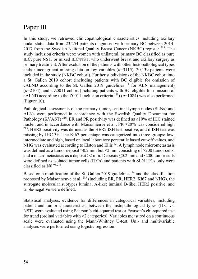
54
Paper III In this study, we retrieved clinicopathological characteristics including axillary nodal status data from 23,254 patients diagnosed with primary BC between 2014-2017 from the Swedish National Quality Breast Cancer (NKBC) register 215. The study inclusion criteria were: women with unilateral, primary BC classified as pure ILC, pure NST, or mixed ILC/NST, who underwent breast and axillary surgery as primary treatment. After exclusion of the patients with other histopathological types and/or incongruent missing data on key variables (n=3115), 20,139 patients were included in the study (NKBC cohort). Further subdivisions of the NKBC cohort into a St. Gallen 2019 cohort (including patients with BC eligible for omission of cALND according to the St. Gallen 2019 guidelines 14 for ALN management) (n=2104), and a Z0011 cohort (including patients with BC eligible for omission of cALND according to the Z0011 inclusion criteria 118) (n=1084) was also performed (Figure 10).
Pathological assessments of the primary tumor, sentinel lymph nodes (SLNs) and ALNs were performed in accordance with the Swedish Quality Document for Pathology (KVAST) 216. ER and PR positivity was defined as ≥10% of IHC stained nuclei, and in accordance with Maisonneuve et al., PR ≥20% was considered high 213. HER2 positivity was defined as the HER2 ISH test positive, and if ISH test wasmissing by IHC 3+. The Ki67 percentage was categorized into three groups: low,intermediate and high, based on local laboratory percentile based cut-off values, andNHG was evaluated according to Elston and Ellis 62. A lymph node micrometastasiswas defined as a tumor deposit >0.2 mm but ≤2 mm consisting of ≥200 tumor cells,and a macrometastasis as a deposit >2 mm. Deposits ≤0.2 mm and <200 tumor cellswere defined as isolated tumor cells (ITCs) and patients with SLN ITCs only wereclassified as N0 95,216.
Based on a modification of the St. Gallen 2019 guidelines 14 and the classification proposed by Maisonneuve et al. 213 (including ER, PR, HER2, Ki67 and NHG), the surrogate molecular subtypes luminal A-like; luminal B-like; HER2 positive; and triple-negative were defined.
Statistical analyses: evidence for differences in categorical variables, including patient and tumor characteristics, between the histopathological types (ILC vs. NST) were evaluated using Pearson’s chi-squared test or Pearson’s chi-squared test for trend (ordinal variables with >2 categories). Variables measured on a continuous scale were evaluated using the Mann-Whitney U-test. Uni- and multivariable analyses were performed using logistic regression.

55
The study was approved by the Ethics Committee (2019–02139) and adheres to the Strengthening The Reporting of Observational studies in Epidemiology (STROBE) guidelines for observational studies 217.
aInclusion criteria: women who underwent breast and axillary surgery for unilateral primary breast cancer (2014 – 2017) as primary therapy identified in the Swedish National Quality Breast Cancer (NKBC) Register. Exclusion criteria: neoadjuvant treatment, locally- and/or regional recurrent breast cancer, distant metastases at the time of surgery. bNodal data incongruent or missing (N = 1237), tumor size missing (N = 235), no invasive tumor (N =33), histopathological type missing (N = 54), histopathological type other than ILC, NST or mixed ILC/NST (N = 1865). Note that some patients were excluded for more than one reason cWomen with histopathological type: pure ILC, pure NST or mixed ILC/NST. dCohort including patients eligible for omission of completion ALND according to St. Gallen 2019 International Consensus Guidelines. Inclusion criterias: T1-3 (any size), clinically N0 with 1-2 SLN metastases, BCT + whole breast irradiation or mastectomi + regional node irradiation (for T3 undergoing BCT = regional node irradiation was also required), SLNB and completion ALND. eCohort including patients eligible for omission of completion ALND according to ACOSOG Z0011 criteria. Inclusion criteria: T1-2, clinically N0 with 1-2 SLN metastases, BCT + whole breast irratiation. Abbreviations: NST = invasive ductal carcinoma of no special type, ILC = invasive lobular carcinoma, SLNB = sentinel lymph node biopsy, ALND = axillary lymph node dissection.
Figure 10. Flow-chart of the study cohort (paper III).

56
Paper IV The study was based on a previously reported prospective observational MBC cohort comprising mixed histopathological types (n=156) 199,218, including a subpopulation of 28 patients with pure ILC and 111 patients with pure NST (Figure 11). Patients diagnosed with MBC and scheduled for first-line systemic treatment at Skåne University Hospital and Halmstad County Hospital, Sweden, between April 2011 and June 2016 were enrolled in a trial (ClinicalTrials.gov NCT01322893) conducted by the Department of Oncology and Pathology at Lund University, Sweden. Inclusion criteria were: age older than 18 years, Eastern Cooperative Oncology Group (ECOG) performance status score 0–2, predicted life expectancy longer than two months, and available data on histopathological subtype. Exclusion criteria were: prior systemic therapy for metastatic breast cancer, inability to provide informed consent and any diagnosis of malignancy within 5 years before inclusion 199. Patient and tumor characteristics and FU data were retrieved from case report forms (CRFs) and clinical records. FU data were updated as of 23 April 2019, after which the database was locked. The median FU time was 49 (range, 27–93) months.
CTCs were isolated and enumerated using the Food and Drug Administration-approved CellSearch system (Menarini Silicon Biosystems, Florence, Italy), as described in detail previously 193,219. CTC count and number of CTC clusters per 7.5 mL blood were evaluated at baseline (BL) and during treatment at 1, 3 and 6 months.
A CTC count of ≥5 cells was a previously validated cut-off in MBC for the CellSearch technique 190-193. A CTC count of ≥5 was considered high, and additional exploratory cut-off values of ≥20 and ≥80, based on prior studies by Botteri et al. 220 and Peeters et al. 221, were also evaluated. CTC clusters were defined as groups consisting of ≥2 CTCs clustered together, with non-overlapping nuclei. A blood sample was considered positive for CTC clusters if ≥1 CTC cluster was detected.
The serum marker CA 15-3 was analyzed with an accredited method used in clinical practice (CA 15-3 on Cobas, NPU01449, Roche, Basel, Switzerland). CA 15-3 values ≥30 U/mL were considered high 222. Additional experimental cut-offs of ≥100, ≥200, and ≥400 U/mL (predefined) were also analyzed.
The patients underwent structured evaluation via clinical examination and diagnostic imaging (standard monitoring methods) at least every 3 months according to a prespecified study protocol during FU, which was continued after the serial blood sampling. Progression vs. non-progression was defined according to clinical practice based on clinical examination and diagnostic imaging, using modified response evaluation criteria in solid tumors (RECIST) 1.1 criteria 223 (progression: progressive disease vs. non-progression: stable disease, partial response or complete response). Aiming at detection of early progression during 0–12 months by CTCs and CA15-3 in relation to routinely diagnosed progression,

57
CTC progression was defined as either (1) an increase in CTC count from <5 to ≥5 or, for those patients with ≥5 CTCs at BL, (2) an increase ≥25% (predefined) in the number of detected CTCs, between two adjacent time-points (BL and 1 month, 1 and 3 months or 3 and 6 months). Similarly, an increase in CA 15-3 levels of ≥25% between two time-points was classified as CA 15-3 progression 222. The change in levels of CTCs and CA 15-3 during 0–6 months of FU were related to progression confirmed by standard monitoring methods, which were restricted to 0–12 months of FU.
Based upon the 50 gene expression signatures detailed by Parker et al. 74, PAM50 breast cancer intrinsic subtyping analysis was completed at NanoString Technologies, Seattle, WA, USA, and samples were classified into luminal A, luminal B, HER2-enriched, and basal-like subtypes.
Statistical analyses: evidence for differences in categorical variables, including patient and tumor characteristics, and CTC and CA 15-3 levels between the histopathological types (ILC vs. NST) were evaluated using Pearson’s chi-squared test or, if expected counts under the null hypothesis were lower than 5 in one or more of the cells of a contingency table, using Fisher’s exact test. Ordinal data were evaluated using Pearson’s chi-squared test for trends, also known as the linear-by-linear association test, whereas variables measured on a continuous scale were evaluated using the Mann–Whitney U test.
The primary endpoint was progression-free survival (PFS) and the secondary endpoint was OS in relation to CTC counts. For each patient, FU time was calculated from the date of the first blood draw to progression or death from any cause. Patients without disease progression or those who were still alive at the last FU date were censored for PFS and OS, respectively. Kaplan–Meier plots and the log-rank test were used to illustrate and compare survival between subgroups. Cox proportional hazards regression was used for the estimation of HRs, and proportional hazards assumptions were checked graphically. Landmark analysis was used to study change in CTC status from baseline to 3 months in relation to PFS and OS from 3 months and onwards.
The REMARK recommendations for reporting of tumor marker studies were followed whenever applicable 224. The study was approved by the Lund University Ethics Committee (LU 2010/135).
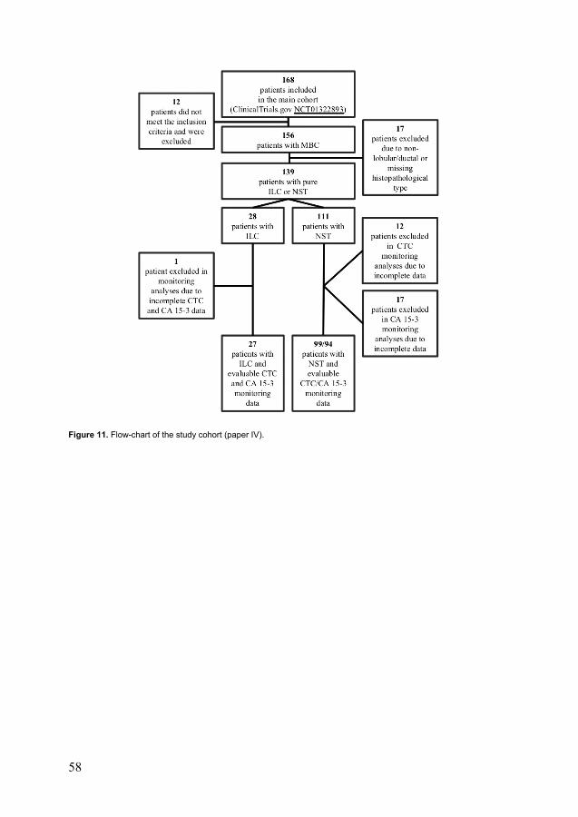
58
Figure 11. Flow-chart of the study cohort (paper IV).

59
Summary of results and discussion
Paper I Overall, 67 (35%) of the included patients with ILC had a distant recurrence and 60 (31%) ultimately died from breast cancer. A majority of the tumors were HER2- (188/192, 98%), ER+ (169/192, 88%), NHG2 (133/175 ,76%) and Ki67 low (115/192, 60%).
Age, tumor size, axillary lymph node status (nodal status), NHG, Ki67 and KiGE were significant prognostic factors BCM in univariable analysis (Figures 12-13).
Figure 12. Breast cancer mortality by Ki67.

60
Figure 13. Breast cancer mortality by histological grade.
In a multivariable model, adjusted for adjuvant treatment, age and PR, the strongest prognostic factors for BCM were: nodal status (HR = 2.9, 95% confidence interval [CI]: 1.4–6.1), KiGE (HR = 2.0, 95% CI: 1.1–3.6), and tumor size (HR = 1.9, 95% CI: 0.98-3.8). By combining these three factors, 37% of the ILC cases could be further divided into a low-risk group, consisting of small (≤ 20 mm) node negative low-KiGE tumors, with a BCM of 5% (95% CI: 1–13%) at 10 years and 12% (95% CI: 5–22%) at 20 years follow-up (Figure 14). None of these patients were administered CT and only two underwent endocrine treatment with tamoxifen.
Surgery, RT and adjuvant systemic treatment were performed in accordance with treatment guidelines valid at the time of diagnosis. Mastectomy and rates in this cohort were high (79% and 94% respectively) and only 44% of the hormone receptor positive patients were treated with endocrine therapy (none of them received aromatase inhibitors), and despite the fact that 41% were node positive only 3% were administered CT.
According to current treatment guidelines in breast cancer, the patients in our cohort had a substantial “local over-treatment” and “systemic under-treatment”. Possible consequences were a higher degree of surgery related sequelae and higher frequency of both early and late distant recurrences. Concurrently, we saw a low frequency of local recurrences (11%) and given the fact that so few of the patients underwent

61
endocrine and/or CT gives us an exclusive opportunity to study systemically untreated ILC, close to the natural history of ILC, in a way not possible today.
Distant recurrences resulting in BC death were common in this cohort. Many of the recurrences occurred during 10–20 years of FU and some of them over 20 years past diagnosis, which is also seen in other studies 34,37, mirroring the chronic history of the disease and suggesting that early detection is not directly linked to good long-term prognosis for the individual patient. Interestingly, patients with ILC in the extremely low-risk group (low-KiGE, pT1, pN0) had a slightly higher BCM year 10–20 (7%), compared to the first 10 years (5%) of FU indicating that, even in this group of patients with a very good overall prognosis, improvements in the adjuvant management is still needed in order to prevent late distant recurrences and breast cancer death.
The main purpose of this study was to evaluate the long-term impact of different prognostic factors in ILC, with a primary focus on Ki67 and histological grade alone, and in combination with ER. At the time of the study, the prognostic value of Ki67 was sparsely investigated and the role of NHG was disputed, in ILC. Our results suggest that Ki67 and NHG are important long-term prognostic factors (Figures 6 and 7), and furthermore that the combination of Ki67, NHG and ER into KiGE, together with tumor size and nodal status (KiGE-TN) makes it possible to identify a low-risk group of ILC patients (37%) with an excellent long-term prognosis, in whom CT can be safely avoided and exclusion of ET considered (Figure 14).
Figure 14. Breast cancer mortality by KiGE-TN.

62
One of the strengths of the present study is the reevaluation of histopathological type by clinical pathologists specialized in breast pathology. Another one is the long FU time (median 21 years) since the lobular subtype are associated with an increased risk of late recurrences. Limitations were the retrospective study design, the absence of a ductal control group and a risk of uncontrolled bias. Furthermore, the study sample size was small, resulting in a low analytic power.
Paper II In this well-characterized case series of patients with ER-positive/HER2-negative ILC, 7% of the tumors was found to be high-AIB1 (14/208) and 93% were AR positive (183/196). For total GPER, most tumors were negatively stained (level 0; 28%) or showed very weak (level 1; 42%) and weak (level 2; 29%) staining intensity, whereas only three tumors had a moderate staining (level 3) and no tumor showed a strong staining (level 4) intensity.
In univariable analyses, AIB1 (high vs. low) was associated with BCM with 10-year FU (HR 3.2, 95% CI 1.4–7.8, P = 0.008), but the effect and the evidence was weaker when analyzed with 25-year FU (HR 2.0, 95% CI 0.87–4.8, P = 0.10). AR (positive vs. negative) showed a trend for a prognostic difference in BCM with 10-year FU (HR 0.56, 95% CI 0.17–1.8), but the evidence was very weak (P = 0.33), and the effect was lost when analyzed with 25-year FU (HR 0.93, 95% CI 0.29–3.0, P = 0.90). Total GPER (log-rank test for trend over the three observed categories) was not associated with BCM neither with 10-year (P = 0.33) nor with 25-year FU (P = 0.55). Ki67 (high vs. low) and NHG (3 vs. 1 + 2) were prognostic for BCM with 10- and 25-year FU, whereas PR (positive vs. negative) was not.
Furthermore, a positive association between AIB1 and Ki67 (P = 0.002) was found.
In a multivariable analysis adjusted for age, tumor size, nodal status, NHG, Ki67, luminal-like classification and adjuvant systemic therapy (endocrine +/- chemo), AIB1 was associated with BCM with 10-year FU (HR 6.8, 95% CI 2.3–20, P = 0.001). However, with longer follow-up, the independent AIB1 effect was found to level off (25-year FU: HR 3.0, 95% CI 1.1–7.8, P = 0.03).
In analyses of gene expression data, high AIB1 expression was associated with worse outcome (HR>>1.00) in two out of the three datasets (METABRIC (HR 3.1, 95% CI 1.3–7.4, P = 0.01), and Metzger Filho et al. (HR 3.6, 95% CI 0.78–16, P = 0.10)). High AR expression was associated with better outcome (HR<<1.00) in two out of three datasets (Metzger Filho et al. (HR 0.24, 95% CI 0.07–0.87, P = 0.03) and Michaut et al. (HR 0.35, 95% CI 0.08–1.6, P = 0.18). GPER was not associated with survival in any of the datasets.

63
In the small subgroup of 14 patients (7%) with high expression of AIB1, 5 died from breast cancer within approximately 5 years, translating to a high cumulative 5-year mortality in this subgroup compared to that in the large subgroup of patients with lower, or no, expression of AIB1. However, no late breast cancer deaths were registered in this group with six patients surviving for more than 10 years and three more than 25 years (Figure 15). Hence, the estimated mortality ratio for AIB1 (high vs. low) was strongly dependent on FU time. In univariable analysis, it was estimated to 3.2 and 2.0 with FU of 10 and 25 years, respectively. Furthermore, the uncertainty in the estimated cumulative BCM for high-AIB1 was large for this group compared to that in low-AIB1 group, as reflected by the shaded 95% point-wise confidence bands in Figure 15. Nevertheless, with 10-year FU, AIB1 was found to be an independent prognostic factor for BCM after adjustment for age, tumor size, nodal status, NHG, Ki67, luminal-like classification and adjuvant systemic therapy.
Figure 15. Breast cancer mortality by amplified in breast cancer 1 (AIB1) with 10- and 25-year follow-up.
The association between high-AIB1 and poor prognosis was in agreement with previous BC studies from our group (including NST) 164-166, but the percentage of tumors with high-AIB1 was found to be lower in this ILC cohort.
In addition to AIB1, we studied the prognostic importance of AR and GPER, but without finding any significant results associated with outcome for these two

64
endocrine biomarkers. The skewed distribution of both AR and GPER reduced the power to detect prognostic effects.
Results from analyses of the publicly available gene expression ILC datasets strengthened our IHC findings for AIB1. High expression of AIB1 was a negative prognostic factor in two out of three datasets (HR 3.1 and HR 3.6, respectively) but the evidence was, however, modest (P = 0.01 and P = 0.10, respectively).
One of the strengths of the present study is the reevaluation of histopathological type by clinical pathologists specialized in breast pathology. Another is the long FU time (median 26 years), since the lobular subtype, with a high proportion of luminal A-like tumors, were associated with an increased risk of late recurrences (e.g., inthis cohort, 21 out of 66 breast cancer deaths occurred ≥10 years after diagnosis).The study also had limitations. In addition to the limited number of patients and theskewed distribution of the experimental biomarkers, one might also argue thatTMAs are not optimal for IHC evaluation of biomarkers in ILC, with a scatteredgrowth pattern characterized by single file infiltrating cells. However, whencomparing the present results for ER, PR and Ki67, analyzed on TMA, with thosein the previous study (paper I) using whole tissue sections instead, essentially thesame results were obtained for prognostic considerations.
Paper III In the NKBC cohort, comparing between 2921 patients with pure ILC and 16,711 with pure NST, patients with ILC were older, had a lower detection rate with screening mammography and more often had mastectomy. Their tumors were larger, more often multifocal and the metastatic burden in ALNs higher. Additionally, the proportion of luminal A-like tumors was higher, while other subtypes were less frequent in ILC compared to NST (all P-values <0.001). The characteristics of the mixed ILC/NST group (n=507) is reported separately. Briefly, the tumors of mixed type seemed to have a biomarker profile closer to pure ILC than to pure NST.
In the St. Gallen 2019 cohort (ILC; n=355, NST; n=1687), the number of excised SLNs and LNs did not differ by histopathological subtype. However, ≥1 non-SLNMs was more common in ILC than in NST (170 [48%] vs. 577 [34%]), P <0.001; odds ratio [OR] 1.77, 95% CI 1.40–2.23) and the prevalence of non-SLNMs was higher (median, interquartile range [IQR]: 3, 1–6 vs. 2, 1–3). The same pattern was seen for ≥4 ALNMs (102 [29%] vs. 232 [4%, P <0.001; OR 2.53, 95% CI 1.93–3.31).
In the Z0011 cohort (ILC; n=111, NST; n=954), similar results were seen with ≥1 non-SLNMs (40% vs. 29%, P = 0.03; OR 1.58, 95% CI 1.05–2.37), prevalence of non-SLNMs (median, IQR: 3,1–8 vs. 2,1–3) and ≥4 ALNMs (24% vs. 10%, P <0.001; OR 2.98, 95% CI 1.84–4.83).

65
In the St. Gallen 2019 cohort, patients with luminal A-like subtype and ≥4 ALNMs were overrepresented in ILC compared to the NST cases (n=52 [17%] vs. n=46 [3%], P <0.001; OR 6.35 95% CI 4.18–9.65). Similar results were seen in the Z0011 cohort (n=15 [17%] vs. n=40 [5%], P <0.001; OR 4.01, 95% CI 2.11–7.60).
The odds of non-SLNM was higher for ILC than for NST also after adjustment for other relevant predictors including age, detection by screening mammography, tumor size, multifocality, number and size of SLNMs and surrogate molecular subtypes both in the St. Gallen 2019 (OR 1.42, 95% CI 1.07–1.88, P = 0.02) and in the Z0011 cohort (OR 1.64, 95% CI 1.01–2.65, P = 0.04). Additionally, the odds of ≥4 ALNM was higher in ILC than in NST after adjustment for the same variables as above, both in the St. Gallen 2019 (OR 2.08, 95% CI 1.47–2.93, P <0.001) and in the Z0011 cohort (OR 2.76, 95% CI 1.1.48–5.13, P = 0.001) (Table 1).
Table 1. Multivariable analysis including patients eligible for omission of completion ALND according to the St. Gallen 2019 International Consensus Guidelinesa
Variables
Non-SLN metastases yes/no ALN metastases ≥4 vs. <4 OR (95% CI) P valueb OR (95% CI) P value
Histopathological type 0.04c <0.001c ILC vs. NST 1.42 (1.07-1.88) 0.02 2.08 (1.47-2.93) <0.001 Mixed ILC/NST vs. NST 0.83 (0.44-1.56) 0.56 0.82 (0.33-2.05) 0.68 Age (per year) 1.01 (1.00-1.01) 0.18 1.01 (1.00-1.02) 0.11
Detection by screening mammography Yes vs. no 0.86 (0.70-1.06) 0.15 0.76 (0.57-1.02) 0.06 T-stage <0.001c <0.001c T2 vs. T1 1.43 (1.15-1.78) 0.001 1.81 (1.33-2.45) <0.001 T3 vs. T1 3.22 (2.06-5.02) <0.001 3.41 (2.06-5.64) <0.001 Multifocality Yes vs. no 1.44 (1.15-1.81) 0.002 1.39 (1.03-1.87) 0.03 SLN metastases 2 vs. 1 1.50 (1.19-1.89) 0.001 2.79 (2.11-3.68) <0.001 SLN macrometastasisd Yes vs. no 2.52 (1.72-3.70) <0.001 3.89 (1.87-8.09) <0.001 Surrogate molecular subtypese 0.22c 0.12c Luminal B-like vs. Luminal A-like 1.24 (0.98-1.56) 0.07 1.51 (1.10-2.08) 0.01 HER2 positive vs. Luminal A-like 1.30 (0.95-1.79) 0.10 1.90 (1.25-2.88) 0.003 Triple-negative vs. Luminal A-like 1.06 (0.69-1.64) 0.78 1.25 (0.69-2.26) 0.46
Abbreviations: CI = confidence interval, ALN = axillary lymph node, ALND = axillary lymph node dissection, BCT = breast conservation therapy, cN0 = clinically node negative, ER = estrogen receptor, HER2 = human epidermal growth factor receptor 2, SLN = sentinel lymph node, Ki67 = proliferation marker IHC = immunohistochemical aSt. Gallen 2019 International Consensus Guidelines for omission of completion ALND: T1-3, cN0, 1-2 SLN metastases, BCT or mastectomy bP-value from logistic regression cOverall test of the factor dSLN macrometastasis: >2 mm tumor deposit eBased on a modification of the St. Gallen 2019 guidelines and the classification proposed by Maisonneuve et al. the tumors were defined as: luminal A-like if (1) ER+, HER2-, NHG 1 or (2) ER+, HER2-, NHG 2, Ki67 low or (3) ER+, HER2-, NHG 2, Ki67 intermediate and PR ≥20%; luminal B-like if (1) ER+, HER2-, NHG 3 or (2) ER+, HER2-, NHG 2, Ki67 high or (3) ER+, HER2-, NHG 2, Ki67 intermediate and PR<20%; HER2 positive (all HER2+ independent of ER, NHG, Ki67 and PR status); triple-negative (ER-, PR- and HER2-)

66
This was one of the largest studies exploring ALN status in ILC vs. NST in patients meeting the criteria for omission of cALND according to St. Gallen 2019 and Z0011. All tumor deposits were assessed according to current classification for ITCs, micro- and macrometastasis 95. Studies including retrospective registry data may be considered unreliable, given the risks of incomplete and misclassified data. In the present study, however, the original dataset was complete for histopathological type in all except 54 cases and data on nodal status was found to be incongruent in 1144/23,254 patients. Moreover, there was no data on gross extracapsular extension (ECE) or vascular invasion and risk factors for ALN metastases. This large population-based Swedish registry study showed that when applying the St. Gallen 2019 guidelines for omitting cALND, the presence of non-SLN metastases and the proportion of ≥4 ALNMs were higher in ILC than in NST. Similar results were seen for the narrower but more generally accepted Z0011 criteria for abstaining cALND. Importantly, both in the St. Gallen 2019 and Z0011 cohort, ILC was an independent predictor of non-SLNM and ≥4 ALNMs after adjustment for validated predictors of nonsentinel node metastases.
BC classified as luminal, A-like with ≥4 ALNMs was overrepresented in patients with ILC compared to NST (St. Gallen 2019 cohort: 17% vs. 3%, Z0011 cohort: 17% vs. 5%, respectively). In patients with this subtype, treatment recommendation on adjuvant chemotherapy depends on ALN staging information according to the St. Gallen 2019 consensus guidelines even in the era of genomic testing. Our results suggest that when applying the St. Gallen 2019 or the Z0011 criteria for omitting cALND, approximately 1 out of 6 ILC and 1 out of 25 NST patients will not be offered adjuvant chemotherapy as a result of understaging of the axilla. Whether these findings have implications on clinical outcome needs to be investigated further. However, a preoperative identification of ILC and an accurate axillary staging is important, and a multidisciplinary discussion regarding omission of cALND is encouraged for all patients with ILC.
Paper IV At the time of database lock, 9% of the patients were progression-free and 30% were alive. The progression and mortality rates for patients with ILC compared to NST were similar over the whole FU period (PFS: HR 0.89, 95% CI 0.57–1.4, P = 0.59; OS: HR 0.99, 95% CI 0.60–1.6, P = 0.96), but during the first two years, PFS and OS were higher among those with ILC. Compared to NST, metastatic ILC cases were more often luminal A subtype (71% vs. 31%; P = 0.001) and axillary node-positive (92% vs. 64%; P = 0.007) at the time of primary diagnosis. They presented with three or more metastatic sites in 14% vs. 32% (P = 0.10), visceral metastases

67
in 29% vs. 65% (P = 0.001) and solitary bone metastases in 39% vs. 22% (P = 0.09) for metastatic ILC and NST cases, respectively. First-line systemic treatment was similar among both ILC and NST cases.
A CTC count of five or more was more common at BL in ILC than in NST cases (22/28 vs. 49/107; P = 0.003), a difference corresponding to an OR for CTC positivity of 4.3 (95% CI: 1.6–12). The evidence was strong (P <0.001) for a difference in the distribution of CTC counts between ILC (median 70, IQR: 121) and NST cases (median 2, IQR: 32) at BL (Figure 16), and the presence of CTC clusters was also more common among ILC cases (36% vs. 18%, P = 0.07).
Figure 16. Circulating tumor cell (CTC) count at baseline by histopathological type. Abbreviations: ILC, invasive lobular carcinoma; NST, invasive ductal carcinoma of no special type; CTC count, number of CTCs per 7.5 mL blood.
Higher CA 15-3 values (median, IQR) were observed in ILC than in NST cases both at BL (392, 1132 vs. 91, 230, P = 0.004) and after one month of systemic treatment (345, 650 vs. 61, 219, p = 0.007).
The evidence for CTC ≥5 as a prognostic factor for PFS (HR 1.5, 95% CI 0.55–4.0, P = 0.44) and OS (HR 2.4, 95% CI 0.71–8.3, P = 0.16) in ILC cases was weak. In contrast, there was strong evidence of prognostic effects for the established cut-off CTC ≥5 in the considerably larger subgroup of NST cases (PFS: HR 1.7, 95% CI 1.2–2.6, P = 0.007; OS: HR 2.1, 95% CI 1.3–3.3, P = 0.002). The prognostic impact of CTC count on PFS and OS in ILC cases was stronger with higher cut-off values

68
(CTC ≥20: HR 3.0, 95% CI 1.3–6.8, P = 0.01, and HR 3.1, 95% CI 1.2–8.3, P = 0.02, respectively) (CTC ≥80: HR 3.6, 95% CI 1.5–8.8, P = 0.004, and HR 5.9, 95% CI 2.0–18, P = 0.002, respectively). The prognostic effect was essentially the same among NST cases for these higher cut-off values (Figure 17).
The presence of one or more CTC clusters was a negative prognostic factor associated with impaired survival among ILC cases (PFS: HR 4.6, 95% CI 1.7–12, P = 0.003; OS: HR 4.9, 95% CI 1.7–14, P = 0.003), whereas the effect was weaker in NST cases (PFS: HR 1.2, 95% CI 0.69–2.0, P = 0.55; OS: HR 1.9, 95% CI 1.1–3.3, P = 0.02).
The presence of CTC clusters in ILC cases was highly correlated with a CTC count ≥80, leading to multicollinearity problems in the Cox models that included both variables. Hence, the support for independent prognostic value of CTC clusters was weak in the present study.
The evidence for differences in PFS and OS was weak among both ILC and NST cases with a CA 15-3 cut-off of ≥30 U/mL. With higher cut-off values (≥100, ≥200 and ≥400 U/mL), stronger evidence for negative prognostic effects was observed for OS but not for PFS in both ILC and NST cases, with the most pronounced effect evident in NST cases.
The CTC count at BL and presence of CTC clusters before the start of first-line treatment was remarkably higher among ILC cases. Despite this finding, we could for the first time show that the evidence for a prognostic value of the validated CTC cut-off (≥5) was weaker in this group (Figure 16). However, the prognostic impact was stronger in ILC with higher CTC cut-off values. The longitudinal design of the study enabled us to show a decline in CTCs and CTC clusters after one month of systemic treatment in both NST and ILC patient groups (Figure 16). Importantly, a decline in CTCs was translated into improved outcomes in both ILC and NST patients compared with no change in CTC status. This finding supports the notion that a change in CTC status reflects the effect of systemic therapy even after longer follow-up time (Figure 18).

69
Figure 17. Progression-free survival (PFS) by circulating tumor cell (CTC) count. Kaplan–Meier plots displaying PFS for the invasive lobular carcinoma (ILC) and invasive ductal carcinoma of no special type (NST) subgroups. Cut-off ≥5 CTCs (A–B). Cut-off ≥20 CTCs (C–D). Cut-off ≥80 CTCs (E–F).

70
Figure 18. Distribution of CTC counts at baseline and with serial sampling at different time points. Spaghetti plots showing number of CTCs (per 7.5 mL blood) per patient from baseline (BL) to 6 months of follow-up for invasive lobular carcinoma (ILC), left panel (A), and invasive ductal carcinoma of no special type (NST), right panel (B). The p-values correspond to pairwise tests of the null hypothesis of no change in CTC count (Wilcoxon matched-pairs signed-rank test). Note that the scale on the y-axis is logarithmic and that the y-axis has been broken to enable presentation of zeros. A small amount of random noise was added to all zeros to separate the lines. The red line connects the medians at the four time points.
Previous publications on CTCs have, for the most part, investigated their distribution and prognostic effects in BC subtypes based either on IHC or gene expression assay classification, but differences related to histopathological type have, to a large extent, been overlooked.
Numerous CTC studies based on mixed histopathological BC types have shown significant prognostic effects when classifying MBCs with <5 CTCs as a better
prognostic outcome (MBCindolent) and MBCs with ≥5 CTCs as worse prognostic outcome (MBCaggressive) according to the CellSearch technique 191,193. This cut-off originates from the median CTC count in a seminal CTC study by Cristofanilli et al. 193.
Our analyses showed a negative prognostic CTC effect in both ILC and NST cases; however, the evidence for a prognostic value of the generally accepted CTC cut-off (≥5) was weaker in the substantially smaller ILC subgroup. A higher proportion of ILC than NST cases (79% vs. 46%, respectively) was classified as MBCaggressive, indicating a potentially worse prognosis for metastatic ILC. Despite this, the survival analyses showed similar prognosis for metastatic ILC and NST patients.
There were increasing prognostic effects associated with higher CTC cut-offs (≥20 and ≥80). The negative prognostic effect, as well as the evidence thereof, was stronger in the ILC subgroup, suggesting that a higher cut-off based on the median

71
CTC count in ILC might be more suitable in ILC cases to better discriminate between the MBCindolent and MBCaggressive forms.
In this study, the presence of CTC clusters was more common in ILC than in NST cases. This finding is paradoxical, since previous studies suggested that CTC clusters had strong cell–cell contacts held together through E-cadherin and catenin-dependent intercellular adhesion, where high levels of plakoglobin (γ-catenin) were identified as one of the most important factors for CTC cluster formation 197, whereas absence or dysfunction of these proteins is one of the cardinal features of ILCs 26.
The strengths of this study include its prospective monitoring design, with a predefined protocol, and the composition of the cohort including only previously untreated patients with MBC scheduled for first-line systemic treatment. Few patients were lost to FU, and the evaluation process, with serial CTC sampling, was executed based on validated state-of-the-art techniques. Limitations include the relatively small study sample size, indicating that the study was not powered to detect clinically relevant prognostic differences among ILC cases. Another limitation is that no phenotypic characterization of CTCs and CTC clusters was performed.


73
General discussion
Breast cancer is one of the most thoroughly studied diseases in modern medicine, but in spite of this, ILC stands out as a persistently understudied subgroup. In large RCTs including patients with mixed histopathological BC types, results from subgroup analyses, where ILC were compared to NST, were rarely reported. Additionally, in the era of molecular subtyping of BC, the potential clinical significance of histopathological type is to a large extent overlooked.
ILC studies are relatively few, often retrospective and the sample sizes are small. Given these conditions, the likelihood of detecting clinically relevant prognostic impact of different patient and tumor characteristics in ILC, and furthermore, to distinguish between true effects and random variations, is reduced 225. Generally, findings in these types of ILC studies needs to be interpreted carefully, and subsequent larger studies are necessary to test the reproducibility of the results. Unfortunately, these highly requested confirmation studies are seldom conducted. Hence, the treatment recommendations in clinical guidelines for BC, are less validated for patients with ILC compared to those for NST patients.
In this thesis, different aspects of primary and metastatic ILC have been investigated, either in a cohort consisting of exclusively ILC patients (paper I, II), or in cohorts including both ILC and NST patients (paper III, IV). The overall aim was to investigate previously infrequently studied prognostic factors and features of metastatic dissemination in ILC, and to compare the results with those seen in patients with NST.
Overall, distributional differences in ILC compared to NST, among a variety of the variables were identified, and in spite of this, in those variables harboring prognostic information, similar effects were seen in either of the two histopathological types.
Cut-off values for well-established and experimental prognostic factors in this thesis (paper I, II, IV) were based on previous BC studies including mixed histopathological BC types (comprising ~80% NST). Whether these predefined cut-off values are equally prognostic for all histopathological BC types, or if adjusted ILC-specific cut-off values are more applicable, is hitherto insufficiently studied, and needs to be evaluated in further studies.
The luminal A(-like) subtype is overrepresented in ILC compared to NST 66-68. This is a commonly suggested confounding factor for explaining much of the observed

74
differences in clinical behavior, treatment response and late recurrences, between the two types, although no clear data to confirm this assumption have been reported.
In this thesis, the results confirmed that the luminal A(-like) subtype was more commonly found in ILC compared to NST. However, CTC count (paper IV) and axillary metastatic tumor burden (paper III) was still significantly higher in ILC after adjustment for luminal A(-like) subtype.
Many challenges in the diagnostic work-up and clinical management of ILC still remain.
Early detection is a key component in the diagnostic work-up of BC. The stage of disease at diagnosis has implications on the forthcoming clinical management and prognosis. The detection rate of primary tumors with lobular vs. ductal histopathology is lower and the same is true for ALNMs 26,28,99,104. Furthermore, ILCs are often clinically nonpalpable and if a lobular cancer is hiding in a dense breast, early detection can be an extraordinary diagnostic challenge.
Detection of ILCs with breast MRI has a higher sensitivity than mammography and ultrasound 28,99, although this imaging technique is time-consuming, quite expensive, and its relatively low specificity is a disadvantage. The use of MRI in the diagnostic work-up is an important complementary imaging technique, especially in patients with ILC.
Digital breast tomosynthesis is a new 3-D imaging technique. Studies indicated that tomosynthesis may be particularly useful for the identification of ILC 108, and routine use of tomosynthesis in the clinic, might be a new standard of care in the near future.
Metastatic ILC has a high rate of bone only metastases and overrepresentation of diffuse metastatic infiltration in atypical/unusual sites 143,144. Due to these features, diagnostics, monitoring, and treatment evaluation are considered more difficult in metastatic ILC compared to NST. Studies evaluating distribution of metastatic sites and total tumor burden in metastatic ILC, show that the detection rate of metastases is clearly lower with physical examination and diagnostic imaging compared to that made with autopsy findings, indicating that a proportion of patients have clinically occult metastatic manifestations and that current imaging techniques and diagnostic tools are suboptimal in detecting micrometastatic lesions 138.
Liquid biopsies (e.g. CTC 190-196, ctDNA 200-202), where clinically useful tumor information can be derived from a blood sample, are rapidly emerging techniques with great potential as diagnostic-, monitoring- and treatment-evaluation tools in the clinic.
In this thesis (paper IV) we showed that CTCs analyzed with the CellSearch technique harbored prognostic information in metastatic ILC and NST, and had a potential as a monitoring tool for treatment response. Interestingly, in an exploratory

75
analysis, a CTC increase in ILC indicated disease progression before detection with diagnostic imaging, in four out of eight cases, whereas no such trend was found in NST. These results must be carefully interpreted but could be considered hypothesis-generating for analyses in subsequent studies of CTCs as a surrogate marker for disease progression in BC.
To date, there are no specific treatment recommendations based on histopathological type. Molecular/surrogate molecular subtyping is extensively studied and considered important, but the role of histopathological type in the adjuvant treatment decision-making is largely unknown. The majority of ILCs are classified as luminal A(-like) subtype. ET is a well-established adjuvant treatment for patients with luminal A(-like) BC, whereas the addition of CT, and the potential benefit thereof is debated, especially in those with luminal A(-like) ILC 226,227. Presumably, most of these patients have little or no chemo-benefit, although a few might have, and the question is: “who is who”?
Indeed, the only indication for CT in luminal A(-like) BC, according to current treatment guidelines 14,110, is the presence of ≥4 ALNM. By using gene expression assays additional information of recurrence risk and potential chemo-benefit can be obtained. These tests are validated for clinical use based on the results from studies comprising patients with mixed histopathological BC types. The recurrence scores for ILC compared to NST was generally lower in these tests, and their applicability in patients with ILC was sparsely studied 80-86. Importantly, to date, none of these tests has been validated in BCs with ≥4 axillary lymph node metastases or, for estimation of long-term risk of distant recurrence (>10 years past diagnosis) (a clinical feature typical for ILC). According to the results presented in this thesis (paper III), another obstacle is the current surgical axillary lymph node management 14,110, where omission of cALND in patients with luminal A(-like) BC and 1-2 SLNMs might hamper clinical information important for decision-making on adjuvant CT in a disproportionally large proportion of ILC cases compared to NST (17% vs. 4%).
Moreover, in a study by Colleoni et al. the 5-year DFS and OS were poorer in patients with luminal-like ILC compared to NST 38. These patients underwent adjuvant systemic treatment in accordance with modern guidelines and a higher proportion of ILC compared to NST patients had ET alone, and the addition of CT was thus more uncommon. These results indicate that the adjuvant ET and/or CT needs to be improved and better tailored for patients with luminal-like ILC and further studies addressing this issue are warranted.


77
Conclusions
• Ki67 and NHG had a long-term prognostic impact on breast cancer mortality in patients with exclusively ILC. The combination of Ki67, NHG and ER into KiGE, together with tumor size and nodal status (KiGE-TN) identified a large group of ILC patients with such an excellent long-term prognosis for whom CT could be safely avoided, and exclusion of endocrine therapy considered.
• AIB1 was a new putative prognostic factor in ER-positive/HER2-negative
ILC, whereas AR and GPER showed no prognostic effect.
• ILC had an independently higher risk of non-SLNM and ≥4 ALNMs compared to NST. Omission of cALND in patients with luminal A-like ILC with 1-2 SLNMs warrants future attention as it might hamper clinical information important for guidance in the decision-making of adjuvant treatment.
• There were different distributional and prognostic CTC features in
metastatic ILC and NST cases. The number of CTCs and CTC clusters were higher in ILC than in NST cases before the start of first-line systemic treatment, and a higher CTC cut-off could be considered for more accurate prognostication in metastatic ILC cases. Finally, eradication of CTCs from BL to three months predicted favorable long-term outcome and indicated treatment response in both ILC and NST cases.
In summary, putting together the results derived from this thesis and the current knowledge concerning ILC, the final conclusion is that ILC compared to NST is a “same same but different disease”.
– Same diagnostic work-up, clinical management and follow-up.
– Same overall long-term prognosis and survival.
– Different clinicopathological and genomic features, treatment response and pattern of recurrence.


79
Future perspectives
Lobular breast cancer is a disease where challenging unmet needs still persist. ILC has been described by ILC researchers as “an initially indolent but slowly progressive disease” 34 and “a disease with ambivalent clinical characteristics” 26, indicating that ILC can be somewhat of a “wolf in a sheep´s clothing”.
These are my thoughts on future lobular breast cancer perspectives:
• The general awareness of ILC being a special disease must be enhanced, and ILC research needs to be promoted. The work done by patient advocate organizations and by ILC researchers, collaborating in consortiums and arranging international lobular breast cancer symposiums, is invaluable. Further similar initiatives are highly welcomed.
• Overall, an increase in the total amount of studies focusing exclusively on ILC are crucial. Further small hypothesis-generating exploratory studies are important. Clinically important findings must be followed up in larger confirmatory ILC studies in a much higher degree than what is the case today. Pooled analysis of ILC data and meta-analysis of ILC studies are encouraged. In order to get large data samples, a continuous work led by international collaborations is important. In recent years, very promising initiatives have been started (LBCA, ELBCC), and hopefully these can yield valuable results in the near future. In large RCTs, the planning of subgroup analyses of histopathological types is important to identify similarities and differences between ILC and NST.
• The use of liquid biopsies as clinical tools for prognostication and disease monitoring in ILC is exciting. The validated prognostic CTC cut-off for the CellSearch system is 5. The results in this thesis (paper IV) indicated that a higher cut-off was more prognostic for ILC, and further studies confirming this finding are warranted. Studies evaluating CTCs as a surrogate/complementary marker to diagnostic imaging for disease monitoring in metastatic ILC, and especially those with bone only metastasis, is an interesting area of investigation. The CellSearch technique uses the epithelial cell adhesion molecule (EpCAM) for the detection of CTCs and at least one study has shown that EpCAM expression was lower in ILC than in NST 228. Results from studies exploring the distributions of phenotypically heterogenous CTCs in metastatic ILC compared to NST,

80
using label-free CTC capturing techniques (e.g. microfluid and size-based capturing) would also be valuable.
• Based on the findings in this thesis (paper III), a study investigating thelong-term (>10 years of FU) prognostic impact of current axillary lymphnode management with omission cALND according to the Z0011 and St.Gallen 2019 criteria, in luminal A(-like) ILC vs. NST is stronglyencouraged.
• Hitherto, there are no available results from studies investigatingdistributional, prognostic and monitoring features of ctDNA in metastaticILC compared to NST. Future reports from these types of studies would beof great interest.
• The hallmark of ILC is the mutated/dysfunctional CDH1 gene and thefollowing lack of cell-cell adhesion molecule E-cadherin. Many of theunique features and the atypical clinical behavior of ILC are proposed to berelated to this deficiency. More studies investigating the role of differentadhesion molecules, epithelial to mesenchymal transition factors 229 andstroma related proteins involved in the tumor microenvironment 230,231,would be of great value for the deeper understanding of the underlyingbiology of ILC.
• Further studies for establishing the most optimal endocrine therapy in ILCare warranted.
In one study by Metzger-Filho et al., letrozole has shown a significanttreatment benefit over tamoxifen in ILC. Although its exploratoryretrospective nature, this study has had a significant clinical impact 67.Confirmatory studies are highly anticipated, but until now no such resultshave been presented.
Late recurrences (>10 years past diagnosis) are overrepresented in ILC.Prevention of late recurrences is a clinical challenge. Studies investigatingthe impact on late recurrences with an “ultra-extended” (>10 years) orlifelong ET in women with ILC compared to NST would be of clinicalinterest.
An interesting approach to identify ILC patients at a high risk of laterecurrence, could be to design a study where a liquid biopsy (e.g. CTC,ctDNA) is analyzed at 5 and 10 years past diagnosis and those patients witha positive liquid biopsy test are randomized between (1) a second round ofadjuvant treatment (preferably an endocrine regimen with the addition of aCDK4/6 inhibitor) and (2) no additional treatment. A previous studyanalysing DTCs in BC with a similar study design and treatment rationalehave shown interesting results188.
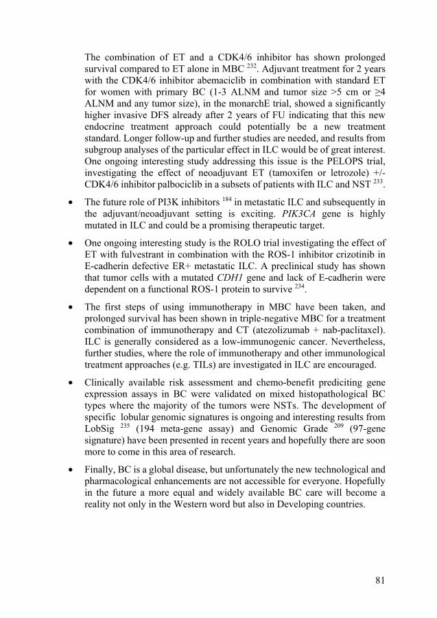
81
The combination of ET and a CDK4/6 inhibitor has shown prolonged survival compared to ET alone in MBC 232. Adjuvant treatment for 2 years with the CDK4/6 inhibitor abemaciclib in combination with standard ET for women with primary BC (1-3 ALNM and tumor size >5 cm or ≥4 ALNM and any tumor size), in the monarchE trial, showed a significantly higher invasive DFS already after 2 years of FU indicating that this new endocrine treatment approach could potentially be a new treatment standard. Longer follow-up and further studies are needed, and results from subgroup analyses of the particular effect in ILC would be of great interest. One ongoing interesting study addressing this issue is the PELOPS trial, investigating the effect of neoadjuvant ET (tamoxifen or letrozole) +/- CDK4/6 inhibitor palbociclib in a subsets of patients with ILC and NST 233.
• The future role of PI3K inhibitors 184 in metastatic ILC and subsequently in the adjuvant/neoadjuvant setting is exciting. PIK3CA gene is highly mutated in ILC and could be a promising therapeutic target.
• One ongoing interesting study is the ROLO trial investigating the effect of ET with fulvestrant in combination with the ROS-1 inhibitor crizotinib in E-cadherin defective ER+ metastatic ILC. A preclinical study has shown that tumor cells with a mutated CDH1 gene and lack of E-cadherin were dependent on a functional ROS-1 protein to survive 234.
• The first steps of using immunotherapy in MBC have been taken, and prolonged survival has been shown in triple-negative MBC for a treatment combination of immunotherapy and CT (atezolizumab + nab-paclitaxel). ILC is generally considered as a low-immunogenic cancer. Nevertheless, further studies, where the role of immunotherapy and other immunological treatment approaches (e.g. TILs) are investigated in ILC are encouraged.
• Clinically available risk assessment and chemo-benefit prediciting gene expression assays in BC were validated on mixed histopathological BC types where the majority of the tumors were NSTs. The development of specific lobular genomic signatures is ongoing and interesting results from LobSig 235 (194 meta-gene assay) and Genomic Grade 209 (97-gene signature) have been presented in recent years and hopefully there are soon more to come in this area of research.
• Finally, BC is a global disease, but unfortunately the new technological and pharmacological enhancements are not accessible for everyone. Hopefully in the future a more equal and widely available BC care will become a reality not only in the Western word but also in Developing countries.


83
Acknowledgements
Taking this thesis from the starting position to the finish line has been a long and winding road. Instead of going straight on the ductal highway, I took the first exit to the breast cancer backroads and ended up at the lobular trail, which led to unknown territories and unmapped places. When traveling down these narrow single-filed paths, you need to go nice and slow, and I did! After struggling with the second gear for almost 10 years, the low-speed maneuvering days are over, and this lobular breast cancer thesis has reached its final destination.
This was not a journey for lone riders, and fortunately, I have not been traveling alone. Without my traveling companions and all the people I met along the trails, I never would have made it.
Thank you all. Thank you very much.
To Lisa Rydén, my principal supervisor, “The Queen of structure and effectiveness”: Always one step ahead, with the ability to see around corners, you skate to where the puck is going to be, not where it has been. Your professional guidance, based on extraordinary scientific and clinical knowledge, coupled with a great personality, is unique. Lisa, thank you for your invaluable support.
To Pär-Ola Bendahl, my co-supervisor, “Mr STATA”: There is nothing that this statistician wizard cannot do with this program. I would guess that if you command it to make a cup of coffee, STATA will do it, and then ask if you want it black or with milk. PO, thank you for your fantastic supervision and for all the interesting discussions we had about all sorts of subjects and life matters (i.e., football).
To Mårten Fernö, my co-supervisor, “Mr Been there, done that”, from cytosol based methods to next generation sequencing: Your in-depth research knowledge and tons of experience combined with your extreme humility, soft charisma, and warmth is impressive. Mårten, it is always a pleasure to be in your company. You have my profound gratitude for all the support and encouraging pep-talks over the years. I would also like to thank you for the good conversations about sports and arts and life in general, especially those during our early morning coffee breaks at Malmö C and Chris Madrid´s (probably the best burger place in San Antonio). To have had you as a supervisor has been truly a privilege.

84
To Christian Ingvar, my co-supervisor, “The lobular breast cancer pioneer” and the one who introduced me to this exciting, unique breast cancer entity and inspired me to register as a PhD student: Among many areas of expertise, you have a unique ability to focus on what is important, with almost surgical precision, and at the same time, you always see the big picture. Thank you for sharing your expertise and for your continuous support and gracious advice throughout this PhD project.
To all my co-authors, Kristina Aaltonen, Sara Alkner, Looket Dihge, Carina Forsare, Dorthe Grabau, Charlotte Levin Tykjær Jørgensen, Anna-Maria Larsson, Fredrik Leeb-Lundberg, Kristina Lövgren, and Martin Sjöström: Thank you for all the good collaborations and constructive feedback.
To all members of the Fernö-Rydén Group: Thank you for the inspiring scientific environment and the interesting gatherings and journal clubs we have had through the years. Special thanks to Kristina Lövgren and Carina Forsare for helping me out with different technical issues, immunohistochemical staining, tumor tissue photos, and Looket Dihge for helping me out with the NKBC database, and Anna-Maria Larsson for assistance with the CTC MBC cohort.
To all my colleagues at the Department of Oncology, Växjö Central Hospital: Thank you for your support and for taking good care of “my patients” in the clinic while I was finishing this thesis, and most of all, for contributing to the unique and positive atmosphere at the department, making it a good workplace. My special thanks to the present and former heads of the department, Katarina Planhammar Hörberg, and Göran Carlstedt for your excellent leadership and for giving me the opportunity, and time, to finalize my research work.
To all my “multi-disciplinary friends” at the breast conferences at Växjö Central Hospital: Thank you for the fruitful collaboration.
To all my former colleagues at the Department of Oncology, Skåne University Hospital and all former heads of the Department: Thank you! My special thanks to the former heads of the department or sections within the department, Gunnar Westman, Annika Håkansson, Carsten Rose, Lennart Hallsten, Mona Ridderheim and Kristina Arnljots.
To all members of the South Swedish Breast Cancer Group (SSBCG): Thank you for the important continuous work to improve breast cancer care in southern Sweden.
To the Department of Clinical Sciences, Division of Oncology, Lund University, Lund, Sweden. Thank you for providing a stimulating research environment and providing general support during my PhD studies. Special thanks to the present and former heads of the Department, Mikael Bodelsson and Bo Baldetorp, respectively, and present and former heads of the Division, Mats Jerkeman and Lars Ekblad, respectively, and Susanne André for the excellent administrative support.

85
To Martin Söderberg, my former colleague, mentor, second father-figure, and friend: Your clinical expertise in the field of oncology is truly extraordinary. Thank you for teaching me the “oncological craftmanship” and for all the good years in your company. You are one of the most generous and finest persons I know.
To Göran Carlstedt, my former boss and current colleague at the Department of Oncology Växjö Central Hospital: Thank you for our good collaboration on the breast cancer patients in the clinic and all the good conversations about music, good food, wine, sports, et cetera.
To Lena Myrskog, my former breast cancer collaborator, and one of the hardest working ”cancer fighters” in the field of breast surgery: Thank you for the golden years in Kronoberg! Hopefully, we can work together again in the future.
To Ingrid Idvall and Poul Boiesen, Anna Ehinger: Thank you for your extensive and highly professional work on the pathological reevaluation of all the lobular breast cancers included in this thesis (papers I and II).
To Kerstin and Alexander, my wonderful little family, the dream team of my life, and the best two-thirds of the super trio: Sharing my life with you is like a fairy tale. Together with you, I love the Monday mornings, the freezing cold Tuesdays at the football pitch, the “lillelördagar”, the Pokemon-walks on rainy-day Thursdays, the “fredagsmys”, the family and friend activities on Saturdays, the lazy Sundays, and most of all, I love you! Kerstin - to have your shoulder to lean on in life, is all I need.
To Christer, my dad, thank you for all that you and mum have given me throughout life, providing me with the best possible conditions to become the person I am today, and for your invaluable support in all different aspects of life throughout my life.
To IngMarie, my late mother, for all the love, tenderness, kindness, and everything else that you have given me. I miss you so much, and I always will.
To Malin and Johanna, my dear sisters: Thank you for sharing childhood, adolescence, and grown up life with me, and for you just being who you are.
To my everlasting friends: Jan Marsal, Fredrik Resman, and Peter Svensson: Thank you for your outstanding support in all types of life matters and if needed, scientific ditto, and most of all for just being close and reliable friends.
To my English friend: Roger Paterson for your kind support with language editing (paper I).
To the rest of my friends and family: You all have my most profound gratitude for being an important part of my life.
To all the funders of this work, Växjö Central Hospital, the Swedish Cancer Society, the Swedish Research Council, the Gunnar Nilsson Cancer Foundation, the Mrs Berta Kamprad Foundation, the Anna and Edwin Bergers Foundation, the

86
Skåne University Hospital Research Foundation, the Skåne County Council´s Research and Development Foundation, the Governmental Funding of Clinical Research within the National Health Service (ALF), the Swedish Breast Cancer Association (BRO), the Cancer Foundation Kronoberg, Kronoberg County Council´s Research and Development Foundation, the Swedish Cancer Foundation, BioCARE, Crafoord Foundation, Skåne University Hospital Funds, the King Gustaf V Jubilee Fund, and the Erling Persson Family Foundation: Thank you.
To the Nationella Kvalitetsregistret för Bröstcancer (NKBC): Thank you for the administrative support and for providing the high-quality registry data included in paper III.
To all the patients who contributed to the studies. Thank you.
For all the lobular breast cancer patients and survivors: Hopefully, this thesis can contribute to further progress in the care of your disease.

87
References
1. WHO Cancer Prevention Early Diagnosis and Screening: Breast Cancer. 2020; https://www.who.int/cancer/prevention/diagnosis-screening/breast-cancer/en/. Accessed September 9, 2020.
2. Bray F, Ferlay J, Soerjomataram I, Siegel RL, Torre LA, Jemal A. Global cancer statistics 2018: GLOBOCAN estimates of incidence and mortality worldwide for 36 cancers in 185 countries. CA Cancer J Clin. 2018;68(6):394-424.
3. Socialstyrelsen: Statistik om nyupptäckta cancerfall 2018. 2020; https://www.socialstyrelsen.se/globalassets/sharepoint-dokument/artikelkatalog/statistik/2019-12-6523.pdf. Accessed September 9, 2020.
4. Socialstyrelsen: Cancer i siffror 2018. 2020; https://www.socialstyrelsen.se/globalassets/sharepoint-dokument/artikelkatalog/statistik/2018-6-10.pdf. Accessed September 17, 2020.
5. Cancer.Net: Breast Cancer Statistics. 2020; https://www.cancer.net/cancer-types/breast-cancer/statistics. Accessed September 17, 2020.
6. Fitzmaurice C, Allen C, Barber RM, et al. Global, Regional, and National Cancer Incidence, Mortality, Years of Life Lost, Years Lived With Disability, and Disability-Adjusted Life-years for 32 Cancer Groups, 1990 to 2015: A Systematic Analysis for the Global Burden of Disease Study. JAMA Oncol. 2017;3(4):524-548.
7. Lakhani SR, Ellis IO, Schnitt S, Tan PH, van de Vijver M. WHO Classification of Tumours of the Breast. 2012.
8. Klintman M, Bendahl PO, Grabau D, et al. The prognostic value of Ki67 is dependent on estrogen receptor status and histological grade in premenopausal patients with node-negative breast cancer. Mod Pathol. 2010;23(2):251-259.
9. Strand C, Bak M, Borgquist S, et al. The combination of Ki67, histological grade and estrogen receptor status identifies a low-risk group among 1,854 chemo-naive women with N0/N1 primary breast cancer. Springerplus. 2013;2(1):111.
10. Balslev I, Axelsson CK, Zedeler K, Rasmussen BB, Carstensen B, Mouridsen HT. The Nottingham Prognostic Index applied to 9,149 patients from the studies of the Danish Breast Cancer Cooperative Group (DBCG). Breast Cancer Res Treat. 1994;32(3):281-290.
11. Curigliano G, Burstein HJ, E PW, et al. De-escalating and escalating treatments for early-stage breast cancer: the St. Gallen International Expert Consensus Conference on the Primary Therapy of Early Breast Cancer 2017. Ann Oncol. 2017;28(8):1700-1712.
12. Blamey RW, Pinder SE, Ball GR, et al. Reading the prognosis of the individual with breast cancer. Eur J Cancer. 2007;43(10):1545-1547.

88
13. Sorlie T, Perou CM, Tibshirani R, et al. Gene expression patterns of breastcarcinomas distinguish tumor subclasses with clinical implications. Proc Natl AcadSci U S A. 2001;98(19):10869-10874.
14. Burstein HJ, Curigliano G, Loibl S, et al. Estimating the benefits of therapy for early-stage breast cancer: the St. Gallen International Consensus Guidelines for theprimary therapy of early breast cancer 2019. Ann Oncol. 2019;30(10):1541-1557.
15. CancerMath. 2020; http://www.lifemath.net/cancer/. Accessed October 1, 2020.16. Wishart GC, Bajdik CD, Azzato EM, et al. A population-based validation of the
prognostic model PREDICT for early breast cancer. Eur J Surg Oncol.2011;37(5):411-417.
17. Cornil V. Les tumeurs du sein. Alcan; 1908.18. Willis RJPoRoT. Epithelial tumors of the breast. 1957.19. Foote FW, Stewart FW. Lobular carcinoma in situ: A rare form of mammary cancer.
Am J Pathol. 1941;17(4):491-496 493.20. Foote FW Jr SF. A Histologic classification of carcinoma in the breast. Surgery.
1946;19:74-99.21. Talman ML, Jensen MB, Rank F. Invasive lobular breast cancer. Prognostic
significance of histological malignancy grading. Acta Oncol. 2007;46(6):803-809.22. Rakha EA, Ellis IO. Lobular breast carcinoma and its variants. Semin Diagn Pathol.
2010;27(1):49-61.23. Al-Baimani K, Bazzarelli A, Clemons M, Robertson SJ, Addison C, Arnaout A.
Invasive Pleomorphic Lobular Carcinoma of the Breast: Pathologic, Clinical, andTherapeutic Considerations. Clin Breast Cancer. 2015;15(6):421-425.
24. Vargas AC, Lakhani SR, Simpson PT. Pleomorphic lobular carcinoma of the breast:molecular pathology and clinical impact. Future Oncol. 2009;5(2):233-243.
25. Eusebi V, Magalhaes F, Azzopardi JG. Pleomorphic lobular carcinoma of the breast: anaggressive tumor showing apocrine differentiation. Hum Pathol. 1992;23(6):655-662.
26. Christgen M, Steinemann D, Kuhnle E, et al. Lobular breast cancer: Clinical, molecularand morphological characteristics. Pathol Res Pract. 2016;212(7):583-597.
27. Barroso-Sousa R, Metzger-Filho O. Differences between invasive lobular andinvasive ductal carcinoma of the breast: results and therapeutic implications. TherAdv Med Oncol. 2016;8(4):261-266.
28. Thomas M, Kelly ED, Abraham J, Kruse M. Invasive lobular breast cancer: A reviewof pathogenesis, diagnosis, management, and future directions of early stage disease.Semin Oncol. 2019;46(2):121-132.
29. Fechner RE. Histologic variants of infiltrating lobular carcinoma of the breast. HumPathol. 1975;6(3):373-378.
30. Martinez V, Azzopardi JG. Invasive lobular carcinoma of the breast: incidence andvariants. Histopathology. 1979;3(6):467-488.
31. Eltorky M, Hall JC, Osborne PT, el Zeky F. Signet-ring cell variant of invasivelobular carcinoma of the breast. A clinicopathologic study of 11 cases. Arch PatholLab Med. 1994;118(3):245-248.

89
32. Acs G, Lawton TJ, Rebbeck TR, LiVolsi VA, Zhang PJ. Differential expression of E-cadherin in lobular and ductal neoplasms of the breast and its biologic and diagnostic implications. Am J Clin Pathol. 2001;115(1):85-98.
33. Arpino G, Bardou VJ, Clark GM, Elledge RM. Infiltrating lobular carcinoma of the breast: tumor characteristics and clinical outcome. Breast Cancer Res. 2004;6(3):R149-156.
34. Rakha EA, El-Sayed ME, Powe DG, et al. Invasive lobular carcinoma of the breast: response to hormonal therapy and outcomes. Eur J Cancer. 2008;44(1):73-83.
35. Ciriello G, Gatza ML, Beck AH, et al. Comprehensive Molecular Portraits of Invasive Lobular Breast Cancer. Cell. 2015;163(2):506-519.
36. Desmedt C, Zoppoli G, Sotiriou C, Salgado R. Transcriptomic and genomic features of invasive lobular breast cancer. Semin Cancer Biol. 2017.
37. Pestalozzi BC, Zahrieh D, Mallon E, et al. Distinct clinical and prognostic features of infiltrating lobular carcinoma of the breast: combined results of 15 International Breast Cancer Study Group clinical trials. J Clin Oncol. 2008;26(18):3006-3014.
38. Colleoni M, Rotmensz N, Maisonneuve P, et al. Outcome of special types of luminal breast cancer. Ann Oncol. 2012;23(6):1428-1436.
39. American Cancer Society. 2020; https://www.cancer.org. Accessed September 11, 2020.
40. Lobular Breast Cancer Alliance. 2020; https://lobularbreastcancer.org. Accessed September 11, 2020.
41. European Lobular Breast Cancer Consortium (ELBCC). 2020; https://www.elbcc.org. Accessed October 16, 2020.
42. Li CI, Anderson BO, Porter P, Holt SK, Daling JR, Moe RE. Changing incidence rate of invasive lobular breast carcinoma among older women. Cancer. 2000;88(11):2561-2569.
43. Li CI, Anderson BO, Daling JR, Moe RE. Trends in incidence rates of invasive lobular and ductal breast carcinoma. JAMA. 2003;289(11):1421-1424.
44. Reeves GK, Beral V, Green J, Gathani T, Bull D. Hormonal therapy for menopause and breast-cancer risk by histological type: a cohort study and meta-analysis. Lancet Oncol. 2006;7(11):910-918.
45. Rossouw JE, Anderson GL, Prentice RL, et al. Risks and benefits of estrogen plus progestin in healthy postmenopausal women: principal results From the Women's Health Initiative randomized controlled trial. JAMA. 2002;288(3):321-333.
46. Wachtel MS, Yang S, Dissanaike S, Margenthaler JA. Hormone Replacement Therapy, Likely Neither Angel Nor Demon. PLoS One. 2015;10(9):e0138556.
47. Dossus L, Benusiglio PR. Lobular breast cancer: incidence and genetic and non-genetic risk factors. Breast Cancer Res. 2015;17:37.
48. Hausauer AK, Keegan TH, Chang ET, Clarke CA. Recent breast cancer trends among Asian/Pacific Islander, Hispanic, and African-American women in the US: changes by tumor subtype. Breast Cancer Res. 2007;9(6):R90.

90
49. Rakha EA, Gill MS, El-Sayed ME, et al. The biological and clinical characteristics ofbreast carcinoma with mixed ductal and lobular morphology. Breast Cancer ResTreat. 2009;114(2):243-250.
50. Metzger-Filho O, Ferreira AR, Jeselsohn R, et al. Mixed Invasive Ductal and LobularCarcinoma of the Breast: Prognosis and the Importance of Histologic Grade.Oncologist. 2019;24(7):e441-e449.
51. Wheeler DT, Tai LH, Bratthauer GL, Waldner DL, Tavassoli FA. Tubulolobularcarcinoma of the breast: an analysis of 27 cases of a tumor with a hybrid morphologyand immunoprofile. Am J Surg Pathol. 2004;28(12):1587-1593.
52. Ohashi R, Matsubara M, Watarai Y, et al. Pleomorphic lobular carcinoma of thebreast: a comparison of cytopathological features with other lobular carcinomavariants. Cytopathology. 2017;28(2):122-130.
53. Jung SP, Lee SK, Kim S, et al. Invasive pleomorphic lobular carcinoma of the breast:clinicopathologic characteristics and prognosis compared with invasive ductalcarcinoma. J Breast Cancer. 2012;15(3):313-319.
54. Rakha EA, Patel A, Powe DG, et al. Clinical and biological significance of E-cadherin protein expression in invasive lobular carcinoma of the breast. Am J SurgPathol. 2010;34(10):1472-1479.
55. Adachi Y, Sawaki M, Hattori M, et al. Comparison of sentinel lymph node biopsybetween invasive lobular carcinoma and invasive ductal carcinoma. Breast Cancer.2018.
56. Wasif N, Maggard MA, Ko CY, Giuliano AE. Invasive lobular vs. ductal breast cancer:a stage-matched comparison of outcomes. Ann Surg Oncol. 2010;17(7):1862-1869.
57. Fernandez B, Paish EC, Green AR, et al. Lymph-node metastases in invasive lobularcarcinoma are different from those in ductal carcinoma of the breast. J Clin Pathol.2011;64(11):995-1000.
58. Majid S, Ryden L, Manjer J. Determinants for non-sentinel node metastases inprimary invasive breast cancer: a population-based cohort study of 602 consecutivepatients with sentinel node metastases. BMC Cancer. 2019;19(1):626.
59. Caudle AS, Kuerer HM, Le-Petross HT, et al. Predicting the extent of nodal diseasein early-stage breast cancer. Ann Surg Oncol. 2014;21(11):3440-3447.
60. Petrausch U, Pestalozzi BC. Distinct clinical and prognostic features of invasivelobular breast cancer. Breast Dis. 2008;30:39-44.
61. Wachtel MS, Halldorsson A, Dissanaike S. Nottingham Grades of LobularCarcinoma Lack the Prognostic Implications They Bear for Ductal Carcinoma. JSurg Res. 2010.
62. Elston CW, Ellis IO. Pathological prognostic factors in breast cancer. I. The value ofhistological grade in breast cancer: experience from a large study with long-termfollow-up. Histopathology. 1991;19(5):403-410.
63. Adams AL, Chhieng DC, Bell WC, Winokur T, Hameed O. Histologic grading ofinvasive lobular carcinoma: does use of a 2-tiered nuclear grading system improveinterobserver variability? Annals of Diagnostic Pathology. 2009;13(4):223-225.

91
64. Metzger Filho O, Ignatiadis M, Sotiriou C. Genomic Grade Index: An important tool for assessing breast cancer tumor grade and prognosis. Crit Rev Oncol Hematol. 2011;77(1):20-29.
65. McCart Reed AE, Kutasovic JR, Lakhani SR, Simpson PT. Invasive lobular carcinoma of the breast: morphology, biomarkers and 'omics. Breast Cancer Res. 2015;17:12.
66. Garcia-Fernandez A, Lain JM, Chabrera C, et al. Comparative Long-term Study of a Large Series of Patients with Invasive Ductal Carcinoma and Invasive Lobular Carcinoma. Loco-Regional Recurrence, Metastasis, and Survival. Breast J. 2015;21(5):533-537.
67. Metzger Filho O, Giobbie-Hurder A, Mallon E, et al. Relative Effectiveness of Letrozole Compared With Tamoxifen for Patients With Lobular Carcinoma in the BIG 1-98 Trial. J Clin Oncol. 2015;33(25):2772-2779.
68. Narbe U, Sjostrom M, Forsare C, et al. The estrogen receptor coactivator AIB1 is a new putative prognostic biomarker in ER-positive/HER2-negative invasive lobular carcinoma of the breast. Breast Cancer Res Treat. 2019.
69. Goldstein NS. Does the level of E-cadherin expression correlate with the primary breast carcinoma infiltration pattern and type of systemic metastases? Am J Clin Pathol. 2002;118(3):425-434.
70. Dabbs DJ, Bhargava R, Chivukula M. Lobular versus ductal breast neoplasms: the diagnostic utility of p120 catenin. Am J Surg Pathol. 2007;31(3):427-437.
71. Dabbs DJ, Schnitt SJ, Geyer FC, et al. Lobular neoplasia of the breast revisited with emphasis on the role of E-cadherin immunohistochemistry. Am J Surg Pathol. 2013;37(7):e1-11.
72. Makki J. Diversity of Breast Carcinoma: Histological Subtypes and Clinical Relevance. Clin Med Insights Pathol. 2015;8:23-31.
73. Perou CM, Sorlie T, Eisen MB, et al. Molecular portraits of human breast tumours. Nature. 2000;406(6797):747-752.
74. Parker JS, Mullins M, Cheang MC, et al. Supervised risk predictor of breast cancer based on intrinsic subtypes. J Clin Oncol. 2009;27(8):1160-1167.
75. Laenkholm AV, Jensen MB, Eriksen JO, et al. PAM50 Risk of Recurrence Score Predicts 10-Year Distant Recurrence in a Comprehensive Danish Cohort of Postmenopausal Women Allocated to 5 Years of Endocrine Therapy for Hormone Receptor-Positive Early Breast Cancer. J Clin Oncol. 2018;36(8):735-740.
76. Sparano JA. A 21-Gene Expression Assay in Breast Cancer. N Engl J Med. 2016;374(14):1387.
77. Cardoso F, van't Veer LJ, Bogaerts J, et al. 70-Gene Signature as an Aid to Treatment Decisions in Early-Stage Breast Cancer. N Engl J Med. 2016;375(8):717-729.
78. Sparano JA, Gray RJ, Makower DF, et al. Adjuvant Chemotherapy Guided by a 21-Gene Expression Assay in Breast Cancer. N Engl J Med. 2018;379(2):111-121.
79. Filipits M, Rudas M, Jakesz R, et al. A new molecular predictor of distant recurrence in ER-positive, HER2-negative breast cancer adds independent information to conventional clinical risk factors. Clin Cancer Res. 2011;17(18):6012-6020.

92
80. Bomeisl PE, Thompson CL, Harris LN, Gilmore HL. Comparison of Oncotype DX Recurrence Score by Histologic Types of Breast Carcinoma. Arch Pathol Lab Med. 2015;139(12):1546-1549.
81. Conlon N, Ross DS, Howard J, Catalano JP, Dickler MN, Tan LK. Is There a Role for Oncotype Dx Testing in Invasive Lobular Carcinoma? Breast J. 2015;21(5):514-519.
82. Felts JL, Zhu J, Han B, Smith SJ, Truica CI. An Analysis of Oncotype DX Recurrence Scores and Clinicopathologic Characteristics in Invasive Lobular Breast Cancer. Breast J. 2017;23(6):677-686.
83. Tsai ML, Lillemoe TJ, Finkelstein MJ, et al. Utility of Oncotype DX Risk Assessment in Patients With Invasive Lobular Carcinoma. Clin Breast Cancer. 2016;16(1):45-50.
84. Laenkholm AV, Jensen MB, Eriksen JO, et al. Population-based Study of Prosigna-PAM50 and Outcome Among Postmenopausal Women With Estrogen Receptor-positive and HER2-negative Operable Invasive Lobular or Ductal Breast Cancer. Clin Breast Cancer. 2020;20(4):e423-e432.
85. Beumer IJ, Persoon M, Witteveen A, et al. Prognostic Value of MammaPrint((R)) in Invasive Lobular Breast Cancer. Biomark Insights. 2016;11:139-146.
86. Kizy S, Huang JL, Marmor S, Tuttle TM, Hui JYC. Impact of the 21-gene recurrence score on outcome in patients with invasive lobular carcinoma of the breast. Breast Cancer Res Treat. 2017;165(3):757-763.
87. Desmedt C, Zoppoli G, Gundem G, et al. Genomic Characterization of Primary Invasive Lobular Breast Cancer. J Clin Oncol. 2016;34(16):1872-1881.
88. Buchanan CL, Flynn LW, Murray MP, et al. Is pleomorphic lobular carcinoma really a distinct clinical entity? J Surg Oncol. 2008;98(5):314-317.
89. Jung HN, Shin JH, Han BK, Ko EY, Cho EY. Are the imaging features of the pleomorphic variant of invasive lobular carcinoma different from classic ILC of the breast? Breast. 2013;22(3):324-329.
90. Simpson PT, Reis-Filho JS, Lambros MB, et al. Molecular profiling pleomorphic lobular carcinomas of the breast: evidence for a common molecular genetic pathway with classic lobular carcinomas. J Pathol. 2008;215(3):231-244.
91. Wen HY, Brogi E. Lobular Carcinoma In Situ. Surg Pathol Clin. 2018;11(1):123-145.
92. Beute BJ, Kalisher L, Hutter RV. Lobular carcinoma in situ of the breast: clinical, pathologic, and mammographic features. AJR Am J Roentgenol. 1991;157(2):257-265.
93. Maxwell AJ, Clements K, Dodwell DJ, et al. The radiological features, diagnosis and management of screen-detected lobular neoplasia of the breast: Findings from the Sloane Project. Breast. 2016;27:109-115.
94. Wallace AS, Xiang D, Hockman L, et al. Synchronous lobular carcinoma in situ and invasive lobular cancer: marker or precursor for invasive lobular carcinoma. Eur J Surg Oncol. 2014;40(10):1245-1249.
95. Giuliano AE, Edge SB, Hortobagyi GN. Eighth Edition of the AJCC Cancer Staging Manual: Breast Cancer. Ann Surg Oncol. 2018;25(7):1783-1785.

93
96. Frost AR, Tsangaris TN, Silverberg SGJAR, Reports. Pleomorphic lobular carcinoma in situ. 1996;1(1):27-31.
97. Flanagan MR, Rendi MH, Calhoun KE, Anderson BO, Javid SH. Pleomorphic Lobular Carcinoma In Situ: Radiologic-Pathologic Features and Clinical Management. Ann Surg Oncol. 2015;22(13):4263-4269.
98. Murray L, Reintgen M, Akman K, et al. Pleomorphic lobular carcinoma in situ: treatment options for a new pathologic entity. Clin Breast Cancer. 2012;12(1):76-79.
99. Johnson K, Sarma D, Hwang ES. Lobular breast cancer series: imaging. Breast Cancer Res. 2015;17:94.
100. Morrow E, Lannigan A, Doughty J, et al. Population-based study of the sensitivity of axillary ultrasound imaging in the preoperative staging of node-positive invasive lobular carcinoma of the breast. Br J Surg. 2018;105(8):987-995.
101. Topps A, Clay V, Absar M, et al. The sensitivity of pre-operative axillary staging in breast cancer: comparison of invasive lobular and ductal carcinoma. Eur J Surg Oncol. 2014;40(7):813-817.
102. Hackney L, Williams S, Bajwa S, Morley-Davies AJ, Kirby RM, Britton I. Influence of tumor histology on preoperative staging accuracy of breast metastases to the axilla. Breast J. 2013;19(1):49-55.
103. Porter AJ, Evans EB, Foxcroft LM, Simpson PT, Lakhani SR. Mammographic and ultrasound features of invasive lobular carcinoma of the breast. J Med Imaging Radiat Oncol. 2014;58(1):1-10.
104. Berg WA, Gutierrez L, NessAiver MS, et al. Diagnostic accuracy of mammography, clinical examination, US, and MR imaging in preoperative assessment of breast cancer. Radiology. 2004;233(3):830-849.
105. Butler RS, Venta LA, Wiley EL, Ellis RL, Dempsey PJ, Rubin E. Sonographic evaluation of infiltrating lobular carcinoma. AJR Am J Roentgenol. 1999;172(2):325-330.
106. Singletary SE, Patel-Parekh L, Bland KI. Treatment trends in early-stage invasive lobular carcinoma: a report from the National Cancer Data Base. Ann Surg. 2005;242(2):281-289.
107. Lobbes MB, Vriens IJ, van Bommel AC, et al. Breast MRI increases the number of mastectomies for ductal cancers, but decreases them for lobular cancers. Breast Cancer Res Treat. 2017;162(2):353-364.
108. Friedewald SM, Rafferty EA, Rose SL, et al. Breast cancer screening using tomosynthesis in combination with digital mammography. JAMA. 2014;311(24):2499-2507.
109. Dashevsky BZ, Goldman DA, Parsons M, et al. Appearance of untreated bone metastases from breast cancer on FDG PET/CT: importance of histologic subtype. Eur J Nucl Med Mol Imaging. 2015;42(11):1666-1673.
110. Cardoso F, Kyriakides S, Ohno S, et al. Early breast cancer: ESMO Clinical Practice Guidelines for diagnosis, treatment and follow-updagger. Ann Oncol. 2019;30(8):1194-1220.

94
111. Nationellt Vårdprogram Bröstcancer. 2020; https://www.swebcg.se/vardprogram.Accessed August 7, 2020.
112. ASCO Guidelines: Breast Cancer. 2020; https://www.asco.org/research-guidelines/quality-guidelines/guidelines/breast-cancer. Accessed October 2, 2020.
113. The American Society of Breast Surgeons: Treatment Guidelines. 2020;https://www.breastsurgeons.org/resources/statements. Accessed October 7, 2020.
114. NICE Guidelines: Early and locally advanced breast cancer diagnosis andmanagement. 2018;https://www.nice.org.uk/guidance/ng101/chapter/Recommendations. AccessedOctober 2, 2020.
115. Fodor J, Major T, Toth J, Sulyok Z, Polgar C. Comparison of mastectomy withbreast-conserving surgery in invasive lobular carcinoma: 15-Year results. Rep PractOncol Radiother. 2011;16(6):227-231.
116. de Glas NA, Engels CC, Bastiaannet E, et al. Contralateral breast cancer risk inrelation to tumor morphology and age-in which patients is preoperative MRIjustified? Breast Cancer Res Treat. 2015;150(1):191-198.
117. Langlands F, White J, Kearins O, et al. Contralateral breast cancer: incidenceaccording to ductal or lobular phenotype of the primary. Clin Radiol.2016;71(2):159-163.
118. Giuliano AE, Ballman KV, McCall L, et al. Effect of Axillary Dissection vs NoAxillary Dissection on 10-Year Overall Survival Among Women With InvasiveBreast Cancer and Sentinel Node Metastasis: The ACOSOG Z0011 (Alliance)Randomized Clinical Trial. JAMA. 2017;318(10):918-926.
119. Galimberti V, Cole BF, Viale G, et al. Axillary dissection versus no axillarydissection in patients with breast cancer and sentinel-node micrometastases (IBCSG23-01): 10-year follow-up of a randomised, controlled phase 3 trial. Lancet Oncol.2018;19(10):1385-1393.
120. Donker M, van Tienhoven G, Straver ME, et al. Radiotherapy or surgery of the axillaafter a positive sentinel node in breast cancer (EORTC 10981-22023 AMAROS): arandomised, multicentre, open-label, phase 3 non-inferiority trial. Lancet Oncol.2014;15(12):1303-1310.
121. Lyman GH, Somerfield MR, Giuliano AE. Sentinel Lymph Node Biopsy for PatientsWith Early-Stage Breast Cancer: 2016 American Society of Clinical OncologyClinical Practice Guideline Update Summary. J Oncol Pract. 2017;13(3):196-198.
122. Early Breast Cancer Trialists' Collaborative G, Darby S, McGale P, et al. Effect ofradiotherapy after breast-conserving surgery on 10-year recurrence and 15-yearbreast cancer death: meta-analysis of individual patient data for 10,801 women in 17randomised trials. Lancet. 2011;378(9804):1707-1716.
123. Ebctcg, McGale P, Taylor C, et al. Effect of radiotherapy after mastectomy andaxillary surgery on 10-year recurrence and 20-year breast cancer mortality: meta-analysis of individual patient data for 8135 women in 22 randomised trials. Lancet.2014;383(9935):2127-2135.

95
124. Poortmans PM, Bollet M, Van Limbergen E. Infiltrating lobular breast cancer: truly a separate entity! Consequences for radiation therapy. Radiother Oncol. 2013;106(1):1-4.
125. Truin W, Voogd AC, Vreugdenhil G, van der Heiden-van der Loo M, Siesling S, Roumen RM. Effect of adjuvant chemotherapy in postmenopausal patients with invasive ductal versus lobular breast cancer. Ann Oncol. 2012;23(11):2859-2865.
126. Marmor S, Hui JYC, Huang JL, et al. Relative effectiveness of adjuvant chemotherapy for invasive lobular compared with invasive ductal carcinoma of the breast. Cancer. 2017;123(16):3015-3021.
127. Metzger-Filho O, Procter M, de Azambuja E, et al. Magnitude of trastuzumab benefit in patients with HER2-positive, invasive lobular breast carcinoma: results from the HERA trial. J Clin Oncol. 2013;31(16):1954-1960.
128. Delpech Y, Coutant C, Hsu L, et al. Clinical benefit from neoadjuvant chemotherapy in oestrogen receptor-positive invasive ductal and lobular carcinomas. Br J Cancer. 2013;108(2):285-291.
129. Mathieu MC, Rouzier R, Llombart-Cussac A, et al. The poor responsiveness of infiltrating lobular breast carcinomas to neoadjuvant chemotherapy can be explained by their biological profile. Eur J Cancer. 2004;40(3):342-351.
130. Tubiana-Hulin M, Stevens D, Lasry S, et al. Response to neoadjuvant chemotherapy in lobular and ductal breast carcinomas: a retrospective study on 860 patients from one institution. Ann Oncol. 2006;17(8):1228-1233.
131. Cristofanilli M, Gonzalez-Angulo A, Sneige N, et al. Invasive lobular carcinoma classic type: response to primary chemotherapy and survival outcomes. J Clin Oncol. 2005;23(1):41-48.
132. Cocquyt VF, Blondeel PN, Depypere HT, et al. Different responses to preoperative chemotherapy for invasive lobular and invasive ductal breast carcinoma. Eur J Surg Oncol. 2003;29(4):361-367.
133. Loibl S, Volz C, Mau C, et al. Response and prognosis after neoadjuvant chemotherapy in 1,051 patients with infiltrating lobular breast carcinoma. Breast Cancer Res Treat. 2014;144(1):153-162.
134. Petrelli F, Barni S. Response to neoadjuvant chemotherapy in ductal compared to lobular carcinoma of the breast: a meta-analysis of published trials including 1,764 lobular breast cancer. Breast Cancer Res Treat. 2013;142(2):227-235.
135. Barchiesi G, Mazzotta M, Krasniqi E, et al. Neoadjuvant Endocrine Therapy in Breast Cancer: Current Knowledge and Future Perspectives. Int J Mol Sci. 2020;21(10).
136. Cataliotti L, Buzdar AU, Noguchi S, et al. Comparison of anastrozole versus tamoxifen as preoperative therapy in postmenopausal women with hormone receptor-positive breast cancer: the Pre-Operative "Arimidex" Compared to Tamoxifen (PROACT) trial. Cancer. 2006;106(10):2095-2103.
137. Dixon JM, Renshaw L, Dixon J, Thomas J. Invasive lobular carcinoma: response to neoadjuvant letrozole therapy. Breast Cancer Res Treat. 2011;130(3):871-877.

96
138. He H, Gonzalez A, Robinson E, Yang WT. Distant metastatic disease manifestations in infiltrating lobular carcinoma of the breast. AJR Am J Roentgenol. 2014;202(5):1140-1148.
139. Harbeck N, Penault-Llorca F, Cortes J, et al. Breast cancer. Nat Rev Dis Primers. 2019;5(1):66.
140. Lobbezoo DJ, van Kampen RJ, Voogd AC, et al. Prognosis of metastatic breast cancer: are there differences between patients with de novo and recurrent metastatic breast cancer? Br J Cancer. 2015;112(9):1445-1451.
141. Plunkett TA, Smith P, Rubens RD. Risk of complications from bone metastases in breast cancer. implications for management. Eur J Cancer. 2000;36(4):476-482.
142. Coleman RE, Smith P, Rubens RD. Clinical course and prognostic factors following bone recurrence from breast cancer. Br J Cancer. 1998;77(2):336-340.
143. Narbe U, Bendahl PO, Aaltonen K, et al. The Distribution of Circulating Tumor Cells Is Different in Metastatic Lobular Compared to Ductal Carcinoma of the Breast-Long-Term Prognostic Significance. Cells. 2020;9(7).
144. Sastre-Garau X, Jouve M, Asselain B, et al. Infiltrating lobular carcinoma of the breast. Clinicopathologic analysis of 975 cases with reference to data on conservative therapy and metastatic patterns. Cancer. 1996;77(1):113-120.
145. El-Hage A, Ruel C, Afif W, et al. Metastatic pattern of invasive lobular carcinoma of the breast-Emphasis on gastric metastases. J Surg Oncol. 2016;114(5):543-547.
146. Ferlicot S, Vincent-Salomon A, Medioni J, et al. Wide metastatic spreading in infiltrating lobular carcinoma of the breast. Eur J Cancer. 2004;40(3):336-341.
147. Borst MJ, Ingold JA. Metastatic patterns of invasive lobular versus invasive ductal carcinoma of the breast. Surgery. 1993;114(4):637-641; discussion 641-632.
148. Lamovec J, Bracko M. Metastatic pattern of infiltrating lobular carcinoma of the breast: an autopsy study. J Surg Oncol. 1991;48(1):28-33.
149. Winston CB, Hadar O, Teitcher JB, et al. Metastatic lobular carcinoma of the breast: patterns of spread in the chest, abdomen, and pelvis on CT. AJR Am J Roentgenol. 2000;175(3):795-800.
150. McLemore EC, Pockaj BA, Reynolds C, et al. Breast cancer: presentation and intervention in women with gastrointestinal metastasis and carcinomatosis. Ann Surg Oncol. 2005;12(11):886-894.
151. Taal BG, den Hartog Jager FC, Steinmetz R, Peterse H. The spectrum of gastrointestinal metastases of breast carcinoma: II. The colon and rectum. Gastrointest Endosc. 1992;38(2):136-141.
152. Almubarak MM, Lae M, Cacheux W, et al. Gastric metastasis of breast cancer: a single centre retrospective study. Dig Liver Dis. 2011;43(10):823-827.
153. Bigorie V, Morice P, Duvillard P, et al. Ovarian metastases from breast cancer: report of 29 cases. Cancer. 2010;116(4):799-804.
154. Feldman PA, Madeb R, Naroditsky I, Halachmi S, Nativ O. Metastatic breast cancer to the bladder: a diagnostic challenge and review of the literature. Urology. 2002;59(1):138.

97
155. Harris M, Howell A, Chrissohou M, Swindell RI, Hudson M, Sellwood RA. A comparison of the metastatic pattern of infiltrating lobular carcinoma and infiltrating duct carcinoma of the breast. Br J Cancer. 1984;50(1):23-30.
156. Raap M, Antonopoulos W, Dammrich M, et al. High frequency of lobular breast cancer in distant metastases to the orbit. Cancer Med. 2015;4(1):104-111.
157. Riva C, Dainese E, Caprara G, et al. Immunohistochemical study of androgen receptors in breast carcinoma. Evidence of their frequent expression in lobular carcinoma. Virchows Arch. 2005;447(4):695-700.
158. Moinfar F, Okcu M, Tsybrovskyy O, et al. Androgen receptors frequently are expressed in breast carcinomas: potential relevance to new therapeutic strategies. Cancer. 2003;98(4):703-711.
159. Vera-Badillo FE, Templeton AJ, de Gouveia P, et al. Androgen receptor expression and outcomes in early breast cancer: a systematic review and meta-analysis. Journal of the National Cancer Institute. 2014;106(1):djt319.
160. Aleskandarany MA, Abduljabbar R, Ashankyty I, et al. Prognostic significance of androgen receptor expression in invasive breast cancer: transcriptomic and protein expression analysis. Breast Cancer Res Treat. 2016;159(2):215-227.
161. Bozovic-Spasojevic I, Zardavas D, Brohee S, et al. The Prognostic Role of Androgen Receptor in Patients with Early-Stage Breast Cancer: A Meta-analysis of Clinical and Gene Expression Data. Clin Cancer Res. 2017;23(11):2702-2712.
162. Carreno G, Del Casar JM, Corte MD, et al. Local recurrence after mastectomy for breast cancer: analysis of clinicopathological, biological and prognostic characteristics. Breast Cancer Res Treat. 2007;102(1):61-73.
163. Lakis S, Kotoula V, Eleftheraki AG, et al. The androgen receptor as a surrogate marker for molecular apocrine breast cancer subtyping. Breast. 2014;23(3):234-243.
164. Alkner S, Bendahl PO, Ehinger A, Lovgren K, Ryden L, Ferno M. Prior Adjuvant Tamoxifen Treatment in Breast Cancer Is Linked to Increased AIB1 and HER2 Expression in Metachronous Contralateral Breast Cancer. PLoS One. 2016;11(3):e0150977.
165. Alkner S, Bendahl PO, Grabau D, et al. AIB1 is a predictive factor for tamoxifen response in premenopausal women. Annals of oncology : official journal of the European Society for Medical Oncology / ESMO. 2010;21(2):238-244.
166. Alkner S, Jensen MB, Rasmussen BB, et al. Prognostic and predictive importance of the estrogen receptor coactivator AIB1 in a randomized trial comparing adjuvant letrozole and tamoxifen therapy in postmenopausal breast cancer: the Danish cohort of BIG 1-98. Breast Cancer Res Treat. 2017.
167. Chang AK, Wu H. The role of AIB1 in breast cancer. Oncol Lett. 2012;4(4):588-594. 168. Lee K, Lee A, Song BJ, Kang CS. Expression of AIB1 protein as a prognostic factor
in breast cancer. World J Surg Oncol. 2011;9:139. 169. Osborne CK, Bardou V, Hopp TA, et al. Role of the estrogen receptor coactivator
AIB1 (SRC-3) and HER-2/neu in tamoxifen resistance in breast cancer. J Natl Cancer Inst. 2003;95(5):353-361.

98
170. Weiner M, Skoog L, Fornander T, Nordenskjold B, Sgroi DC, Stal O. Oestrogen receptor co-activator AIB1 is a marker of tamoxifen benefit in postmenopausal breast cancer. Ann Oncol. 2013;24(8):1994-1999.
171. Broselid S, Cheng B, Sjostrom M, et al. G protein-coupled estrogen receptor is apoptotic and correlates with increased distant disease-free survival of estrogen receptor-positive breast cancer patients. Clin Cancer Res. 2013;19(7):1681-1692.
172. Filardo EJ, Graeber CT, Quinn JA, et al. Distribution of GPR30, a seven membrane-spanning estrogen receptor, in primary breast cancer and its association with clinicopathologic determinants of tumor progression. Clin Cancer Res. 2006;12(21):6359-6366.
173. Ignatov T, Weissenborn C, Poehlmann A, et al. GPER-1 expression decreases during breast cancer tumorigenesis. Cancer Invest. 2013;31(5):309-315.
174. Kuo WH, Chang LY, Liu DL, et al. The interactions between GPR30 and the major biomarkers in infiltrating ductal carcinoma of the breast in an Asian population. Taiwan J Obstet Gynecol. 2007;46(2):135-145.
175. Sjostrom M, Hartman L, Grabau D, et al. Lack of G protein-coupled estrogen receptor (GPER) in the plasma membrane is associated with excellent long-term prognosis in breast cancer. Breast Cancer Res Treat. 2014;145(1):61-71.
176. Luen SJ, Savas P, Fox SB, Salgado R, Loi S. Tumour-infiltrating lymphocytes and the emerging role of immunotherapy in breast cancer. Pathology. 2017;49(2):141-155.
177. Savas P, Salgado R, Denkert C, et al. Clinical relevance of host immunity in breast cancer: from TILs to the clinic. Nat Rev Clin Oncol. 2016;13(4):228-241.
178. Loi S, Sirtaine N, Piette F, et al. Prognostic and predictive value of tumor-infiltrating lymphocytes in a phase III randomized adjuvant breast cancer trial in node-positive breast cancer comparing the addition of docetaxel to doxorubicin with doxorubicin-based chemotherapy: BIG 02-98. J Clin Oncol. 2013;31(7):860-867.
179. Desmedt C, Salgado R, Fornili M, et al. Immune Infiltration in Invasive Lobular Breast Cancer. J Natl Cancer Inst. 2018.
180. Schmid P, Rugo HS, Adams S, et al. Atezolizumab plus nab-paclitaxel as first-line treatment for unresectable, locally advanced or metastatic triple-negative breast cancer (IMpassion130): updated efficacy results from a randomised, double-blind, placebo-controlled, phase 3 trial. Lancet Oncol. 2020;21(1):44-59.
181. My Cancer Genome: Genetically Informed Cancer Medicine. 2020; https://www.mycancergenome.org/content/gene/pik3ca/. Accessed October 6, 2020.
182. Mosele F, Stefanovska B, Lusque A, et al. Outcome and molecular landscape of patients with PIK3CA-mutated metastatic breast cancer. Ann Oncol. 2020;31(3):377-386.
183. Miller TW, Rexer BN, Garrett JT, Arteaga CL. Mutations in the phosphatidylinositol 3-kinase pathway: role in tumor progression and therapeutic implications in breast cancer. Breast Cancer Res. 2011;13(6):224.
184. Andre F, Mills D, Taran T. Alpelisib for PIK3CA-Mutated Advanced Breast Cancer. Reply. N Engl J Med. 2019;381(7):687.

99
185. Gainer SM, Lodhi AK, Bhattacharyya A, Krishnamurthy S, Kuerer HM, Lucci A. Invasive lobular carcinoma predicts micrometastasis in breast cancer. J Surg Res. 2012;177(1):93-96.
186. Braun S, Vogl FD, Naume B, et al. A pooled analysis of bone marrow micrometastasis in breast cancer. N Engl J Med. 2005;353(8):793-802.
187. Hartkopf AD, Taran FA, Wallwiener M, et al. Prognostic relevance of disseminated tumour cells from the bone marrow of early stage breast cancer patients - results from a large single-centre analysis. Eur J Cancer. 2014;50(15):2550-2559.
188. Naume B, Synnestvedt M, Falk RS, et al. Clinical outcome with correlation to disseminated tumor cell (DTC) status after DTC-guided secondary adjuvant treatment with docetaxel in early breast cancer. J Clin Oncol. 2014;32(34):3848-3857.
189. Synnestvedt M, Borgen E, Wist E, et al. Disseminated tumor cells as selection marker and monitoring tool for secondary adjuvant treatment in early breast cancer. Descriptive results from an intervention study. BMC Cancer. 2012;12:616.
190. Bidard FC, Peeters DJ, Fehm T, et al. Clinical validity of circulating tumour cells in patients with metastatic breast cancer: a pooled analysis of individual patient data. Lancet Oncol. 2014;15(4):406-414.
191. Cristofanilli M, Pierga JY, Reuben J, et al. The clinical use of circulating tumor cells (CTCs) enumeration for staging of metastatic breast cancer (MBC): International expert consensus paper. Crit Rev Oncol Hematol. 2019;134:39-45.
192. Yan WT, Cui X, Chen Q, et al. Circulating tumor cell status monitors the treatment responses in breast cancer patients: a meta-analysis. Sci Rep. 2017;7:43464.
193. Cristofanilli M, Budd GT, Ellis MJ, et al. Circulating tumor cells, disease progression, and survival in metastatic breast cancer. N Engl J Med. 2004;351(8):781-791.
194. Alunni-Fabbroni M, Muller V, Fehm T, Janni W, Rack B. Monitoring in metastatic breast cancer: is imaging outdated in the era of circulating tumor cells? Breast Care (Basel). 2014;9(1):16-21.
195. Liu MC, Shields PG, Warren RD, et al. Circulating tumor cells: a useful predictor of treatment efficacy in metastatic breast cancer. J Clin Oncol. 2009;27(31):5153-5159.
196. Budd GT, Cristofanilli M, Ellis MJ, et al. Circulating tumor cells versus imaging--predicting overall survival in metastatic breast cancer. Clin Cancer Res. 2006;12(21):6403-6409.
197. Aceto N, Bardia A, Miyamoto DT, et al. Circulating tumor cell clusters are oligoclonal precursors of breast cancer metastasis. Cell. 2014;158(5):1110-1122.
198. Mu Z, Wang C, Ye Z, et al. Prospective assessment of the prognostic value of circulating tumor cells and their clusters in patients with advanced-stage breast cancer. Breast Cancer Res Treat. 2015;154(3):563-571.
199. Larsson AM, Jansson S, Bendahl PO, et al. Longitudinal enumeration and cluster evaluation of circulating tumor cells improve prognostication for patients with newly diagnosed metastatic breast cancer in a prospective observational trial. Breast Cancer Res. 2018;20(1):48.

100
200. Merker JD, Oxnard GR, Compton C, et al. Circulating Tumor DNA Analysis inPatients With Cancer: American Society of Clinical Oncology and College ofAmerican Pathologists Joint Review. J Clin Oncol. 2018;36(16):1631-1641.
201. Stewart CM, Kothari PD, Mouliere F, et al. The value of cell-free DNA formolecular pathology. J Pathol. 2018;244(5):616-627.
202. Ignatiadis M, Lee M, Jeffrey SS. Circulating Tumor Cells and Circulating TumorDNA: Challenges and Opportunities on the Path to Clinical Utility. Clin Cancer Res.2015;21(21):4786-4800.
203. Van Poznak C, Somerfield MR, Bast RC, et al. Use of Biomarkers to GuideDecisions on Systemic Therapy for Women With Metastatic Breast Cancer:American Society of Clinical Oncology Clinical Practice Guideline. J Clin Oncol.2015;33(24):2695-2704.
204. Duffy MJ, Evoy D, McDermott EW. CA 15-3: uses and limitation as a biomarker forbreast cancer. Clin Chim Acta. 2010;411(23-24):1869-1874.
205. Barnes DM, Lammie GA, Millis RR, Gullick WL, Allen DS, Altman DG. Animmunohistochemical evaluation of c-erbB-2 expression in human breast carcinoma.British journal of cancer. 1988;58(4):448-452.
206. McShane LM, Altman DG, Sauerbrei W, et al. REporting recommendations fortumor MARKer prognostic studies (REMARK). Breast Cancer Res Treat.2006;100(2):229-235.
207. Curtis C, Shah SP, Chin SF, et al. The genomic and transcriptomic architecture of2,000 breast tumours reveals novel subgroups. Nature. 2012;486(7403):346-352.
208. Michaut M, Chin SF, Majewski I, et al. Integration of genomic, transcriptomic andproteomic data identifies two biologically distinct subtypes of invasive lobular breastcancer. Sci Rep. 2016;6:18517.
209. Metzger-Filho O, Michiels S, Bertucci F, et al. Genomic grade adds prognostic valuein invasive lobular carcinoma. Annals of oncology : official journal of the EuropeanSociety for Medical Oncology / ESMO. 2013;24(2):377-384.
210. Dihge L, Bendahl PO, Grabau D, et al. Epidermal growth factor receptor (EGFR) andthe estrogen receptor modulator amplified in breast cancer (AIB1) for predictingclinical outcome after adjuvant tamoxifen in breast cancer. Breast Cancer Res Treat.2008;109(2):255-262.
211. List HJ, Reiter R, Singh B, Wellstein A, Riegel AT. Expression of the nuclearcoactivator AIB1 in normal and malignant breast tissue. Breast Cancer Res Treat.2001;68(1):21-28.
212. Kirkegaard T, McGlynn LM, Campbell FM, et al. Amplified in breast cancer 1 inhuman epidermal growth factor receptor - positive tumors of tamoxifen-treated breastcancer patients. Clin Cancer Res. 2007;13(5):1405-1411.
213. Maisonneuve P, Disalvatore D, Rotmensz N, et al. Proposed new clinicopathologicalsurrogate definitions of luminal A and luminal B (HER2-negative) intrinsic breastcancer subtypes. Breast Cancer Res. 2014;16(3):R65.
214. Narbe U, Bendahl PO, Grabau D, Ryden L, Ingvar C, Ferno M. Invasive lobularcarcinoma of the breast: long-term prognostic value of Ki67 and histological grade,alone and in combination with estrogen receptor. Springerplus. 2014;3:70.

101
215. Lofgren L, Eloranta S, Krawiec K, et al. Validation of data quality in the Swedish National Register for Breast Cancer. BMC Public Health. 2019;19(1):495.
216. KVAST-dokument Bröstcancer. 2020; http://www.svfp.se/kvastdokument. Accessed August 7, 2020.
217. von Elm E, Altman DG, Egger M, et al. The Strengthening the Reporting of Observational Studies in Epidemiology (STROBE) statement: guidelines for reporting observational studies. Lancet. 2007;370(9596):1453-1457.
218. Jansson S, Bendahl PO, Larsson AM, Aaltonen KE, Ryden L. Prognostic impact of circulating tumor cell apoptosis and clusters in serial blood samples from patients with metastatic breast cancer in a prospective observational cohort. BMC Cancer. 2016;16:433.
219. Allard WJ, Matera J, Miller MC, et al. Tumor cells circulate in the peripheral blood of all major carcinomas but not in healthy subjects or patients with nonmalignant diseases. Clin Cancer Res. 2004;10(20):6897-6904.
220. Botteri E, Sandri MT, Bagnardi V, et al. Modeling the relationship between circulating tumour cells number and prognosis of metastatic breast cancer. Breast Cancer Res Treat. 2010;122(1):211-217.
221. Peeters DJ, van Dam PJ, Van den Eynden GG, et al. Detection and prognostic significance of circulating tumour cells in patients with metastatic breast cancer according to immunohistochemical subtypes. Br J Cancer. 2014;110(2):375-383.
222. Duffy MJ. Serum tumor markers in breast cancer: are they of clinical value? Clin Chem. 2006;52(3):345-351.
223. Eisenhauer EA, Therasse P, Bogaerts J, et al. New response evaluation criteria in solid tumours: revised RECIST guideline (version 1.1). Eur J Cancer. 2009;45(2):228-247.
224. McShane LM, Altman DG, Sauerbrei W, et al. REporting recommendations for tumor MARKer prognostic studies (REMARK). Nat Clin Pract Urol. 2005;2(8):416-422.
225. Hackshaw A. Small studies: strengths and limitations. Eur Respir J. 2008;32(5):1141-1143.
226. Han Y, Li Q, Xu BH, et al. Adjuvant chemotherapy may improve survival of patients with luminal A breast cancer and positive lymph nodes. Genet Mol Res. 2015;14(3):8563-8573.
227. Herr D, Wischnewsky M, Joukhadar R, et al. Does chemotherapy improve survival in patients with nodal positive luminal A breast cancer? A retrospective Multicenter Study. PLoS One. 2019;14(7):e0218434.
228. Soysal SD, Muenst S, Barbie T, et al. EpCAM expression varies significantly and is differentially associated with prognosis in the luminal B HER2(+), basal-like, and HER2 intrinsic subtypes of breast cancer. Br J Cancer. 2013;108(7):1480-1487.
229. McCart Reed AE, Kutasovic JR, Vargas AC, et al. An epithelial to mesenchymal transition programme does not usually drive the phenotype of invasive lobular carcinomas. J Pathol. 2016;238(4):489-494.

102
230. Downey CL, Simpkins SA, White J, et al. The prognostic significance of tumour-stroma ratio in oestrogen receptor-positive breast cancer. Br J Cancer. 2014;110(7):1744-1747.
231. Eiro N, Gonzalez LO, Fraile M, Cid S, Schneider J, Vizoso FJ. Breast Cancer Tumor Stroma: Cellular Components, Phenotypic Heterogeneity, Intercellular Communication, Prognostic Implications and Therapeutic Opportunities. Cancers (Basel). 2019;11(5).
232. Pernas S, Tolaney SM, Winer EP, Goel S. CDK4/6 inhibition in breast cancer: current practice and future directions. Ther Adv Med Oncol. 2018;10:1758835918786451.
233. Clinicaltrials.gov NCT02764541, Palbociclib and Endocrine Therapy for LObular Breast Cancer Preoperative Study (PELOPS) 2020; https://clinicaltrials.gov/ct2/show/NCT02764541. Accessed October 19, 2020.
234. Bajrami I, Marlow R, van de Ven M, et al. E-Cadherin/ROS1 Inhibitor Synthetic Lethality in Breast Cancer. Cancer Discov. 2018;8(4):498-515.
235. McCart Reed AE, Lal S, Kutasovic JR, et al. LobSig is a multigene predictor of outcome in invasive lobular carcinoma. NPJ Breast Cancer. 2019;5:18.

“The Dude abides” –Jeff Lebowski