Embed Size (px)
Citation preview

This stuand TecDPI2010Salud CABDOMtional Rider ProSalud CDevelop
886 S
The long-term behavior of lightweightand heavyweight meshes used to repairabdominal wall defects is determinedby the host tissue repair processprovoked by the meshGemma Pascual, PhD,a Bel�en Hern�andez-Gasc�on, PhD,b Marta Rodr�ıguez, PhD,a
Sandra Sotomayor, PhD,a Estefania Pe~na, PhD,b Bego~na Calvo, PhD,b and Juan M. Bell�on, MD, PhD,a
Madrid and Zaragoza, Spain
Background. Although heavyweight (HW) or lightweight (LW) polypropylene (PP) meshes are widelyused for hernia repair, other alternatives have recently appeared. They have the same large-pore structureyet are composed of polytetrafluoroethylene (PTFE). This study compares the long-term (3 and 6 months)behavior of meshes of different pore size (HW compared with LW) and composition (PP compared withPTFE).Methods. Partial defects were created in the lateral wall of the abdomen in New Zealand White rabbitsand then repaired by the use of a HW or LW PP mesh or a new monofilament, large-pore PTFE mesh(Infinit). At 90 and 180 days after implantation, tissue incorporation, gene and protein expression ofneocollagens (reverse transcription-polymerase chain reaction/immunofluorescence), macrophage re-sponse (immunohistochemistry), and biomechanical strength were determined. Shrinkage was measuredat 90 days.Results. All three meshes induced good host tissue ingrowth, yet the macrophage response wassignificantly greater in the PTFE implants (P < .05). Collagen 1/3 mRNA levels failed to vary at 90days yet in the longer term, the LW meshes showed the reduced genetic expression of both collagens(P < .05) accompanied by increased neocollagen deposition, indicating more efficient mRNA transla-tion. After 90–180 days of implant, tensile strengths and elastic modulus values were similar for all3 implants (P > .05).Conclusion. Host collagen deposition is mesh pore size dependent whereas the macrophage responseinduced is composition dependent with a greater response shown by PTFE. In the long term, macroporousmeshes show comparable biomechanical behavior regardless of their pore size or composition. (Surgery2012;152:886-95.)
From the Department of Surgery and Medical Specialities,a Faculty of Medicine, University of Alcal�a, Net-working Research Center on Bioengineering, Biomaterials and Nanomedicine (CIBER-BBN), Alcal�a de He-nares, Madrid, Spain; and Group of Structural Mechanics and Materials Modelling (GEMM),b Arag�onInstitute of Engineering Research (I3A), University of Zaragoza, Zaragoza, Spain
DESPITE THE APPEARANCE ofmany new prostheticmate-rials designed to repair abdominal wall defects,polypropylene (PP) continues to be used the mostwidely. This is because of the good cost/benefits
dy was supported by the Spanish Ministry of Sciencehnology through research projects DPI2008-02335/-20746-C03-01/DPI2011-27939 and the Instituto dearlos III (ISCIII) through the CIBER initiative projectESH. CIBER-BBN is an initiative funded by the VI Na-&D&i Plan 2008-2011, Iniciativa Ingenio 2010, Consol-gram, CIBER Actions and financed by the Instituto dearlos III with assistance from the European Regionalment Fund.
URGERY
and excellent biocompatibility of PP, along withan improved tolerance to infection compared withother materials.1 However, because of certain ad-verse effects produced when it is placed at the
Accepted for publication March 8, 2012.
Reprint requests: Juan M. Bell�on, MD, PhD, Department of Sur-gery, Faculty of Medicine, Alcal�a University, Ctra. Madrid-Barcelona, Km 33,600, 28871, Alcal�a de Henares, Madrid,Spain. E-mail: [email protected].
0039-6060/$ - see front matter
� 2012 Mosby, Inc. All rights reserved.
doi:10.1016/j.surg.2012.03.009
SurgeryVolume 152, Number 5
Pascual et al 887
peritoneal interface, namely adhesions to the intes-tinal loops and fistulas,2,3 PP is not recommendedfor use in contact with the visceral peritoneum. Not-withstanding, when extraperitoneal placement iscalled for in hernia repair, this material continuesto be used the most widely.4
Research and development in prosthetic mate-rials has aimed to improve the wound repairprocess elicited by prosthetic mesh implant.Thus, rather than being relegated, macroporousmeshes have been subjected to modifications totheir structure, porosity, and composition. This hasled to the construction of new large-pore meshescomposed of other polymers such as polytetra-fluoroethylene (PTFE)5 and polyvinylidenefluoride.6
One of the main goals of surgery has been toassess the effects of varying the amount of im-planted material. To this end, composite mesheshave been designed7,8 with both absorbable andnonabsorbable components. Pore size has alsobeen considered an important factor in new meshdesigns, along with other characteristics such asthe diameter and spatial distribution of fibers.
These new designs have led to the classificationof macroporous meshes9 as heavy weight (HW),medium weight (MW), or low weight (LW), respec-tively, according to whether their density values aregreater than 80 g/m2, between 50 and 80 g/m2, orless than 50 g/m2. Some authors10 have even de-fined an ultralightweight material whose densityis less than 35 g/m2.
Prosthetic mesh density is sometimes indepen-dent of pore size, and some designs, despitehaving a small pore size, are classified as LWbecause they are constructed from a loosely wovenlight monofilament conferring them an overalllow density in g/m2.
Despite this classification, in line with the con-cepts of the German authors,11 we consider poresize to be the main factor that determines whethera material is HWor LW. Thus, it is generally consid-ered that HW meshes have a small-pore design andLW meshes have large pores.12
Given that when a LW mesh is used, less mate-rial is implanted in the host, we would expect areduced foreign body reaction and a repair pro-cess that generates less fibrosis in the host tissue,with the consequence of improved tissue compli-ance.13 Although individual variation exists interms of the repair process induced by the implantof a biomaterial,14 it is clear that the sometimes ex-cessive fibrosis induced by conventional HW mate-rials could be minimized through the use of a LWimplant.
In recent work,15 we observed the excellentshort-term collagenization of LW-PP implants.This prompted the design of the present study,in which we sought to determine whether duringlonger periods (3 and 6 months) the behavior ofa LW mesh (tissue incorporation and tensilestrength) remains the same regardless of poresize or its composing polymer (PP or PTFE). A fur-ther objective was to determine the effect of theimplant material on the host tissue response pro-duced in the long term.
MATERIAL AND METHODS
Experimental animals. The experimental ani-mals were 36 male New Zealand White rabbitsweighing approximately 2,200 g caged under con-ditions of constant light and temperature accord-ing to European Union animal care guidelines(European Directive 609/86/EEC and EuropeanConvention of the Council of Europe ETS123). Allprocedures were approved by our institutions Re-view Board.
Prosthetic materials. The biomaterials usedwere (Fig 1, A–C) as follows:
� Surgipro (Covidien, Mansfield, MA): HW PP (85 g/
m2); pore size 0.26 ± 0.03 mm2;
� Optilene elastic (B/Braun, Berlin, Germany): LW PP
(48 g/m2); pore size 7.64 ± 0.32 mm2; and
� Infinit mesh (Gore and Associates, Flagstaff, AZ):
LW nonexpanded PTFE (70 g/m2); pore size 4.05
± 0.22 mm2.
Surgical technique. To minimize pain, all ani-mals were administered 0.05 mg/kg buprenor-phine (Buprecare; Divasa Farmavic, Barcelona,Spain) 1 hour before and 3 days after the surgicalprocedure. Anesthesia was induced with a mixtureof ketamine hydrochloride (Ketolar, 70 mg/kg;Parke-Davis, S.A., Spain), diazepam (Valium, 1.5mg/kg; Roche, Madrid Spain), and chlorproma-zine (Largactil, 1.5 mg/kg; Rhone-Poulenc, S.A.,Spain), administered intramuscularly.
With the use of a sterile surgical technique, 4 34-cm defects were created in the lateral wall of theabdomen comprising the planes of the externaland internal oblique muscles, sparing the trans-versalis muscle, parietal peritoneum, and skin. Thedefects were then repaired by fixing a mesh of thesame size to the edges of the defect with a running4-0 PP suture interrupted at the 4 corners (Fig 1,D–F). The skin was closed by 3-0 PP runningsuture.
Experimental design. A total of 36 animals wereimplanted with each of the materials to establish3 groups of 12 animals each. In each of these

Fig 1. Biomaterials used in this experimental study. SEM images of the pore size detail: (A) Surgipro (153); (B) Opti-lene (153); (C) Infinit mesh (153). Implanted biomaterials: (D) Surgipro; (E) Optilene; (F) Infinit mesh. (Color ver-sion of figure is available online.)
SurgeryNovember 2012
888 Pascual et al
groups, 6 animals were euthanized in a CO2 cham-ber after 90 days, and the remaining 6 were eutha-nized at 180 days post-implant.
Shrinkage. Shrinkage of the implanted mesheswas determined by image analysis. For this pur-pose, we designed a set of transparent templates ofthe same dimensions as the original meshes (4 3 4cm). At the end of the implant period, the outlinesof the meshes were traced on the templates beforetheir removal. The surface area of the templatescould them be determined by computerized imageanalysis with the MIP program incorporated in theimage analyzer (MICRON, Barcelona, Spain). Re-sults are expressed as the percentage size reduc-tion suffered by each implant. Shrinkage wasassessed at 90 days after implant when the tissuerepair process is practically complete.
Morphological analysis. Light microscopy. Forlight microscopy, specimens were collected fromthe mesh/host tissue interface. The samples werefixed in F13 solution (ethanol 60%, methanol20%, polyethylene glycol 7%, water 13%), embed-ded in paraffin, and cut into 5-mm sections. Oncecut, the sections were stained with Masson’strichrome (Goldner–Gabe) and examined underthe light microscope (Zeiss Axiophot; Carl Zeiss,Oberkochen, Germany).
Gene expression of collagens. Real-time reversetranscription polymerase chain reaction (RT-PCR).Tissue fragments 1 cm2 in size were obtainedfrom the central mesh zone, and stored at �808Cuntil use. RNA was extracted by the use
of guanidine-phenol-chloroform isothiocyanateprocedures with trizol (Invitrogen, Carlsbad, CA).The RNA was recovered from the aqueous phaseand precipitated by adding isopropanol and incu-bating overnight at �208C. Complementary DNAwas synthesized with 200 ng of the total RNA byRT with oligo dT primers (Amersham, Fairfield,CT) and the M-MLV RT enzyme (Invitrogen). RTreactions were run in the absence of M-MLV toconfirm the RNA lacked genomic DNA.
cDNA was amplified using the followingprimers: collagen one (sense 59-GAT GCG TTCCAG TTC GAG TA-39 and antisense 59-GGT CTTCCG GTG GTC TTG TA-39); collagen three (sense59-TTA TAA ACC AAC CTC TTC CT-39 and anti-sense 59-TAT TAT AGC ACC ATT GAG AC-39);GAPDH (sense 59-TCA CCA TCT TCC AGG AGCGA-39 and antisense 59-CAC AAT GCC GAA GTGGTC GT-39).
The RT-PCR mixture contained 5 mL of theinverse transcription product (cDNA) diluted1:20, 10 mL of iQ SYBR Green Supermix (Bio-Rad,Laboratories, Hercules, CA), and 1 mL (6 mM) ofeach primer in a final reaction volume of 20 mL. RT-PCR was performed in a StepOnePlus Real-TimePCR System (Applied Biosystemx, Foster City, CA).Samples were subjected to an initial stage of 10 minat 958C. The conditions for cDNA amplificationwere: 40 cycles of 958C for 15 seconds, 608C (colla-gens I and III) or 558C (GAPDH) for 30 seconds,and 728C for 1 minute. Negative controls contain-ing ultraPureTM DNase, RNase-free distilled water

SurgeryVolume 152, Number 5
Pascual et al 889
(Invitrogen) were run in each reaction. Productswere subjected to 2% agarose gel electrophoresis,stained with SYBR Green II RNA gel stain (Invitro-gen), and visualized with ultraviolet light. Geneexpression was normalized against the expressionrecorded for the constitutive gene glyceraldehyde3-phosphate-dehydrogenase.
Immunofluorescence. To detect the proteinexpression of collagens I and III, tissue fragmentswere fixed in F13 solution, embedded in paraffin,and cut into 5 mm-thick sections. Once cut, thesections were deparaffinated, hydrated, equili-brated in phosphate-buffered saline buffer andincubated with the monoclonal antibodies anti-collagen I (Sigma Chemical Co., St. Louis, MO)and anti-collagen III (Medicorp, Montreal, Can-ada). The secondary antibody used was conjugatedwith rhodamine. An immunofluorescence tech-nique was used to detect the antigen–antibodyreaction. Cell nuclei were counterstained withDAPI. Samples were examined under a confocalmicroscope Leica SP5 (Leica Microsystems, Wet-zlar, Germany) to detect fluorescence.
Macrophage response. For immunohistochem-istry, a specific monoclonal antibody to rabbitmacrophages, RAM 11 (DAKO M-633, USA), wasapplied to paraffin-embedded sections. The alka-line phosphatase-labeled avidin-biotin method wasperformed as the following steps: incubation withthe primary antibody (1:50 in Tris-buffered salineor TBS) for 30 minutes, incubation with immuno-globulin G and biotin (1:1,000 in TBS) for 45minutes, and labeling with avidin (1:200 in TBS)for 30 minutes. These steps were conducted atroom temperature. Images were developed withthe use of a chromogenic substrate containingnaphthol phosphate and fast red. Nuclei werecounterstained for 5 minutes in acid hematoxylin.RAM-11–labeled macrophages were quantified ac-cording to a method described elsewhere.16
Biomechanical strength. To determine the bio-mechanical strength andmodulus of elasticity of themeshes after implant, strips of the different bioma-terials 1 cm wide and 5 cm long, with an effectivegauge length of 3 cm, were analyzed with anINSTRON 3340 (static load 500 N; Instron Corp.,HighWycombe,UK).The cross-head speedwas 5 cmper minute and recording speed 2 cm per minute.
The strips obtained at 90 and 180 days afterimplantation included the mesh and infiltratedhost tissue. All tests were conducted immediatelyafter animal sacrifice.
Statistical analysis. Statistical analysis was per-formed by use of the Graph Pad Prism 5 package.Shrinkage percentages, collagen one and three
mRNA expression, RAM-11–positive cells, biome-chanical strength, and modulus of elasticity valueswere compared among the 3 study groups usingthe Mann-Whitney U test. The level of statistical sig-nificance was set at P < .05.
RESULTS
There were no cases of mortality or signs ofinfection and/or rejection of the implants in theanimals operated on. Seroma was detected in 2 ofthe animals with PTFE implants at 14 days afterimplantation.
Shrinkage. Shrinkage values determined at 90days after implantation were as follows: Surgipro(13.69 ± 3.52%), Optilene (10.11 ± 3.07%), andInfinit (10.42 ± 1.19%). These values failed todiffer significantly (P < .05).
Morphological analysis. Light microscopy. At 90days after implantation, the three biomaterialstested showed ingrowth by a disorganized, well-vascularized, loose connective scar tissue. Thisneoformed tissue occupied all the spaces betweenthe PP (Surgipro and Optilene) and PTFE (In-finit) filaments and was interspersed with areas inwhich the infiltration of adipose tissue could beobserved in all the implant types. After 6 months,there was a significant increase in adipose tissueingrowth. Most neoformed connective tissue wasobserved around the prosthetic filaments. Thepreserved transversalis muscle in the lower zoneof the partial defect showed no evident morpho-logical alterations at any of the follow-up times orany of the study groups (data not shown).
Gene expression of collagens. Real time RT-PCR.The three mesh types induced similar collagengene expression patterns reflected by the collagenone and three mRNA levels detected. At 90 daysafter implantation, the PP biomaterials (Surgiproand Optilene) induced the higher expression ofmRNA for collagen three (immature) and one(mature) with significant differences with PTFE(Infinit) emerging at 180 days for collagen threewhen compared with the PP-HW mesh (Surgipro;P < .05; Fig 2, A) and for collagen one, comparedwith both PP-HW (P < .01) and PP-LW (P < .05; Fig2, B). In the PTFE (Infinit) mesh group, the dropproduced in collagen one mRNA expression from90–180 days (Fig 2, B) was significant (P < .01).
Immunofluorescence. Both collagen types wereimmunodetected in the three implant groups atboth study times. Collagen fibers ran parallel tothe mesh surface in zones far from the filaments orwere arranged concentrically to these filaments inareas closer to the implant edges. For Surgipro andOptilene, collagen III protein expression was

Fig 2. Relative amounts of collagen three (A) and one (B) mRNA in the implant of Surgipro, Optilene, and Infinitmesh determined by RT-PCR. Upper panels: RT-PCR products of both genes. Lanes: 1/2 Surgipro, 3/4 Optilene,and 5/6 Infinit mesh at 90 days, and 7/8 Surgipro, 9/10 Optilene, and 11/12 Infinit mesh at 180 days. N, Negative con-trol; Mw, molecular weight markers. Results are the mean ± SEM of three experiments performed in duplicate. Geneexpression was normalized to values recorded for the GAPDH gene. *P < .05; **P < .01.
SurgeryNovember 2012
890 Pascual et al
homogeneously distributed throughout the newlyformed tissue around the prosthetic filament at 90and 180 days after implantation. In contrast, theInfinit mesh induced an intense pattern of colla-gen III expression confined to localized areasaround the filaments.
Labeling for the mature form of collagen (col-lagen I) was more extensive and intense for thehigher porosity implants (Optilene and Infinit) atboth time points, but Optilene showed the greateststaining for this type of collagen. Surgipro showedmoderate collagen I staining. In all the studygroups, collagen I staining was confined to areasof new tissue formation adjacent to the prostheticfilaments (Fig 3).
Macrophage response. In all the study groups,macrophage cells were detected in the neoformedtissue between the mesh filaments. Most inflamma-tory cells were found to concentrate around thefilaments where, besides macrophages, multinucle-ated foreign-body giant cells, typical of a woundrepair response, couldbe seen.These cells appearedmostly around the filaments of PTFE (Infinit).
At both time points, macrophage numbers weresignificantly greater for the PTFE meshes comparedto the PP implants (P < .05). The macrophage reac-tion gradually diminished from 90 to 180 days in all3 groups (Fig 4).
Biomechanics. The tensile strengths, or break-ing points, recorded for the different meshesimplanted for 90 and 180 days were comparable(P = .05; Fig 5, A).
At 90 days, the postimplant elastic modulus wassignificantly greater for PTFE (P < .05) than the PPmeshes, although by 180 days, this variable was sim-ilar across the 3 groups (Fig 5, B).
DISCUSSION
As standard permanent prosthetic materials, PPand expanded PTFE (ePTFE) have been con-stantly subjected to modifications to improveboth their host tissue incorporation and complica-tions of bowel injury and infection, in an effort toachieve the best functional repair of the abdomi-nal wall possible.
The most recent modifications made to PPmeshes have involved minimizing the materialimplanted in the host without compromising theirmechanical resistance. This approach led to thedevelopment of composite8 and large poremeshes.17
The modifications made to sheets of ePTFEprosthetics, such as introducing multiperfora-tions18 or creating a rough surface on one side,19
have not improved their biomechanical strength.The only strategy that has served to improve tissueincorporation and tensile strength has been theconstruction of a largely porous ePTFE mesh.5 Bio-assays conducted on this mesh have revealed thatrather than the chemical composition of the bio-material, it is its loosely woven structure that deter-mines its tissue behavior. Accordingly, the behaviorof classic microporous-expanded PTFE, which in-duces little tissue ingrowth and instead becomesencapsulated by host tissue, may be manipulatedto simulate that of a largely porous PP mesh.
In the present experimental study, we comparedthe postimplantation behavior of a conventionalPP-HW mesh to that of 2 LW meshes, one com-posed of PP and the other of nonexpanded PTFE.The first prosthetic materials generated by ourgroup5 were composed of an expanded PTFEmonofilament CV-4. The material examined here

Fig 3. Tissue incorporation and collagen expression around the mesh filaments of the different biomaterials. Collagenappears as red fluorescence observed by laser scanning confocal microscopy. (A–F) Collagen III (immature): (A) Sur-gipro, 90 days (2003); (B) Optilene, 90 days (2003); (C) Infinit, 90 days (2003); (D) Surgipro, 180 days (2003); (E)Optilene, 180 days (2003); (F) Infinit, 180 days (2003). (G–L) Collagen I (mature): (G) Surgipro, 90 days (2003); (H)Optilene, 90 days (2003); (I) Infinit, 90 days (2003); (J) Surgipro, 180 days (2003); (K) Optilene, 180 days (2003); (L)Infinit, 180 days (2003). F, Prosthetic filaments. (Color version of figure is available online.)
SurgeryVolume 152, Number 5
Pascual et al 891
is composed of a nonexpanded PTFE monofila-ment that is knitted to create a large pore sizesuch that it is a MW mesh.
In our study, partial defects were created in thelateral wall of the abdomen to avoid involving theperitoneum in the repair process. When the ani-mals were killed 90 days after implantation, seroma
was detected in 2 of the animals who received aPTFE implant (similar to observations after theimplant of laminar ePTFE meshes). This findingseems consistent with our immunohistochemistryresults obtained with the anti- RAM-11 macrophagemonoclonal antibody. Hence, at each time point,the PTFE implants showed a significantly

Fig 4. Immunohistochemical labeling of rabbit macrophages (arrows) using the RAM-11 monoclonal antibody. (A)Surgipro, 90 days (6403); (B) Optilene, 90 days (5003); (C) Infinit, 90 days (3603); (D) Surgipro, 180 days (2003); (E)Optilene, 180 days (2003); (F) Infinit, 180days (2003); (G)meannumbers ofRAM-11–positive cells recorded for each studygroup and follow-up time. Significant differences between Infinit vs Surgipro and Optilene were observed at 90 (*P < .05)and 180 days after implantation (**P < .05). F, Prosthetic filaments. (Color version of figure is available online.)
SurgeryNovember 2012
892 Pascual et al
augmented macrophage reaction over that shownby the PP implants. This behavior would be com-parable with that displayed by some absorbablematerials in the early postimplant course,7,20 possi-bly having clinical implications such as the presenceof reactive seroma. For the PP meshes, macrophagecounts gradually decreased during the study periodas occurred, although at a slower pace, for the PTFEimplants. The observed immune response to theseimplants requires further investigation.
With regard to shrinkage at 90 days, the lack ofsignificant differences observed between the 3meshes is in line with previous results from ourlaboratory.21 After PP mesh placement in dogs,other authors9 observed significant shrinkage ofthe implant area close to 30% at 90 days
postimplantation. In our study, shrinkage at thistime point was closer to 15% and probably attribut-able to interspecies differences.
We contemplate the phenomenon of prostheticshrinkage as a physiological factor in the contextof the wound repair process.22 Possible variationsamong implants could be related to the implantsite. Thus, some authors23 have noted less shrink-age when the implant is placed in a retromuscularcompared with a prefascial position.
In terms of host tissue incorporation, the 3implant types showed good behavior. The PTFEmeshes behaved differently than the classic micro-porous expanded PTFE, which becomes encapsu-lated by host tissue. Although collagen I (mature)could be observed both at 90 and 180 days in the
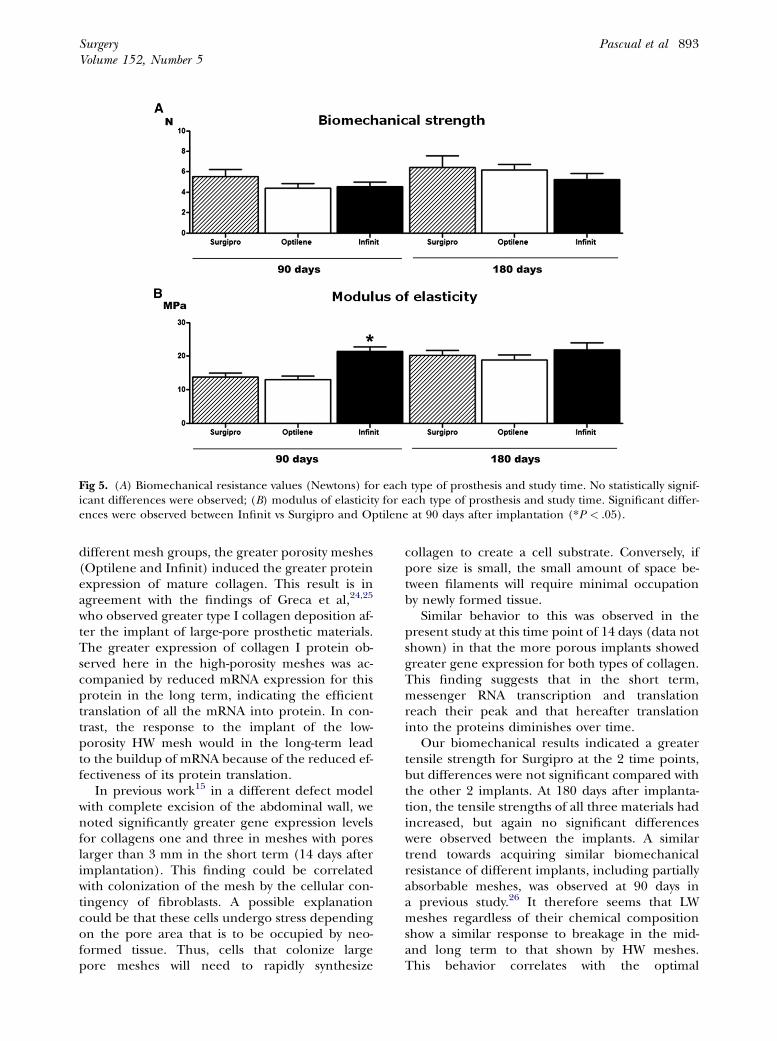
Fig 5. (A) Biomechanical resistance values (Newtons) for each type of prosthesis and study time. No statistically signif-icant differences were observed; (B) modulus of elasticity for each type of prosthesis and study time. Significant differ-ences were observed between Infinit vs Surgipro and Optilene at 90 days after implantation (*P < .05).
SurgeryVolume 152, Number 5
Pascual et al 893
different mesh groups, the greater porosity meshes(Optilene and Infinit) induced the greater proteinexpression of mature collagen. This result is inagreement with the findings of Greca et al,24,25
who observed greater type I collagen deposition af-ter the implant of large-pore prosthetic materials.The greater expression of collagen I protein ob-served here in the high-porosity meshes was ac-companied by reduced mRNA expression for thisprotein in the long term, indicating the efficienttranslation of all the mRNA into protein. In con-trast, the response to the implant of the low-porosity HW mesh would in the long-term leadto the buildup of mRNA because of the reduced ef-fectiveness of its protein translation.
In previous work15 in a different defect modelwith complete excision of the abdominal wall, wenoted significantly greater gene expression levelsfor collagens one and three in meshes with poreslarger than 3 mm in the short term (14 days afterimplantation). This finding could be correlatedwith colonization of the mesh by the cellular con-tingency of fibroblasts. A possible explanationcould be that these cells undergo stress dependingon the pore area that is to be occupied by neo-formed tissue. Thus, cells that colonize largepore meshes will need to rapidly synthesize
collagen to create a cell substrate. Conversely, ifpore size is small, the small amount of space be-tween filaments will require minimal occupationby newly formed tissue.
Similar behavior to this was observed in thepresent study at this time point of 14 days (data notshown) in that the more porous implants showedgreater gene expression for both types of collagen.This finding suggests that in the short term,messenger RNA transcription and translationreach their peak and that hereafter translationinto the proteins diminishes over time.
Our biomechanical results indicated a greatertensile strength for Surgipro at the 2 time points,but differences were not significant compared withthe other 2 implants. At 180 days after implanta-tion, the tensile strengths of all three materials hadincreased, but again no significant differenceswere observed between the implants. A similartrend towards acquiring similar biomechanicalresistance of different implants, including partiallyabsorbable meshes, was observed at 90 days ina previous study.26 It therefore seems that LWmeshes regardless of their chemical compositionshow a similar response to breakage in the mid-and long term to that shown by HW meshes.This behavior correlates with the optimal
SurgeryNovember 2012
894 Pascual et al
collagenization (collagen type I deposition) of LWimplants. The deposition of collagen in the repairtissue around the filaments of the LW meshescauses an increase in stiffness that substantiallymodifies the original properties of the mesh.Thus in long term, the growth of collagen tendsto unify the mechanical response shown by LWand HW meshes. The elastic modulus was signifi-cantly greater at 90 days (P < .05) for the PTFEmeshes compared with the other implants, al-though similar values were attained at 180 daysfor the 3 implants.
According to the biomechanical data obtained,both the resistance to breakage and elasticity of thedifferent implants was gradually modulated by thehost tissue. Thus, besides offering the advantage ofa reduced amount of implanted material, themechanical properties of LW implants seem to beimproved, especially in terms of tensile strength,by the newly formed tissue around the prostheticfilaments. In line with this observation, in a recentclinical study of incisional hernia repair,27 surgicaloutcomes at three years were similar when small orlarge pore PP meshes were used. In view of thesefindings, larger pore meshes would need to betested to determine how much further the amountof foreign material placed in the host could be re-duced without mechanically compromising theimplants.
Our study is not without its limitations. In ourexperience, although the rabbit model has pro-vided excellent results in terms of tissue repair andimmune response, its biomechanical behavior isless translatable to human clinical practice.
In conclusion, our findings indicate that thefollowing:
� Compared with PP, the use of PTFE in a macroporous
mesh induces an augmented macrophage response;
� In the long term, the collagen mRNA translation in-
duced by a high-porosity mesh is more efficient, result-
ing in increased collagen deposition in the repair
zone; and
� In the long-term postimplantation, the tensile
strengths and elastic moduli of both HW and LW ma-
terials attain comparable values.
In general terms, it therefore seems that thelong-term behavior of LW meshes used to repair anabdominal wall defect, whether composed of PP orPTFE, is conditioned by the host tissue repairprocess, with a correlation observed between col-lagen deposition and prosthetic pore size.
The authors are indebted to Gore and Associates,Flagstaff, AZ, for providing the meshes used in this study.
This company played no role in the design of this study,data collection, or analysis.
REFERENCES
1. Alaedeen DI, Lipman J, Medalie D, Rosen MJ. The single-staged approach to the surgical management of abdominalwall hernias in contaminated fields. Hernia 2007;11:41-5.
2. Chuback JA, Sigh RS, Sill C, Dick LS. Small bowel obstruc-tion resulting from mesh plug migration after open ingui-nal hernia repair. Surgery 2000;127:475-6.
3. Chew DK, Choi LH, Rogers AM. Enterocutaneous fistula 14years after prosthetic mesh repair of a ventral incisional her-nia. A life-long risk? Surgery 2000;125:109-11.
4. Bell�on JM, Contreras LA, Buj�an J, Palomares D, Carrera-SanMartin A. Tissue response to polypropylene meshes used inthe repair of abdominal wall defects. Biomaterials 1998;19:669-75.
5. Bell�on JM, Jurado F, Garc�ıa-Honduvilla N, L�opez R, Car-rera-San Mart�ın A, Buj�an J. The structure of a biomaterialrather than its chemical composition modulates the repairprocess at the peritoneal level. Am J Surg 2002;184:154-9.
6. Klinge U, Klosterhalfen B, Ottinger AP, Junge K, Schumpe-lick V. PVDF as a new polymer for the construction of surgi-cal meshes. Biomaterials 2002;23:3487-93.
7. Rosch R, Junge K, Quester R, Klinge U, Klosterhalfen B,Schumpelick V. Vypro II mesh in hernia repair: impact ofpolyglactin on long-term incorporation in rats. Eur SurgRes 2003;35:445-50.
8. Junge K, Rosch R, Krones J, Klinge U, Martens PR, Lynen P,et al. Influence of polyglecaprone 25 (Monocryl) supple-mentation on the biocompatibility of a polypropylenemesh for hernia repair. Hernia 2005;9:212-7.
9. Cobb WS, Burns JM, Peindl RD, Carbonell AM, MatthewsBD, Kercher KW, et al. Textile analysis of heavy weight,mid-weight, and light weight polypropylene mesh in a por-cine ventral hernia model. J Surg Res 2006;136:1-7.
10. Earle DB, Mark LA. Prosthetic material in inguinal herniarepair: How do I choose? Surg Clin North Am 2008;88:179-201.
11. Klinge U. Experimental comparison of monofile light andheavy polypropylene meshes: less weight does not meanless biological response. World J Surg 2007;31:867-8.
12. Deeken CR, Abdo MS, Frisella MM, Matthews BD. Physico-mechanical evaluation of polypropylene, polyester, and pol-ytetrafluoroethylene meshes for inguinal hernia repair.J Am Coll Surg 2011;212:68-79.
13. Klinge U, Klosterhalfen B, Birkenhauer V, Junge K, Conze J,Schumpelick V. Impact of polymer pore size on the interfacescar formation in a rat model. J Surg Res 2002;103:208-14.
14. Schachtrupp A, Klinge U, Junge K, Rosch R, Bhardwaj RS,Schumpelick V. Individual inflammatory response of hu-man blood monocyte to mesh biomaterials. Br J Surg2003;90:114-20.
15. Pascual G, Rodriguez M, G�omez-Gil V, Garc�ıa-Honduvilla N,Buj�an J, Bell�on JM. Early tissue incorporation and collagen de-position in lightweightpolypropylenemeshes:bioassay inanex-perimental model of ventral hernia. Surgery 2008;144:427-35.
16. Bell�on JM, Bujan J, Contreras L, Hernando A, Jurado F.Macrophage response to experimental implantation ofpolypropylene prostheses. Eur Surg Res 1994;26:46-53.
17. Klosterhalfen B, Junge K, Klinge U. The lightweight andlarge porous mesh concept for hernia repair. Espert RevMed Devices 2005;2:103-17.
18. Simmermacher RKJ, Van der Lei B, Schakenraad JM, Blei-chrodt RP. Improved tissue ingrowth and anchorage of

SurgeryVolume 152, Number 5
Pascual et al 895
expanded polytetrafluoroethylene by perforation: an exper-imental study in the rat. Biomaterials 1991;12:22-4.
19. Bell�on JM, Contreras L, Buj�an J, Carrera-San Mart�ın A. Theuse of biomaterials in the repair of abdominal wall defects:a comparative study between polypropylene meshes (Mar-lex) and a new polytetrafluoroethylene prosthesis (DualMesh). J Biomater Appl 1997;12:121-35.
20. Klinge U, Schumpelick V, Klosterhalfen B. Functional as-sessment and tissue response of short and long-term absorb-able surgical meshes. Biomaterials 2001;22:1415-24.
21. Bell�on JM, Garc�ıa-Honduvilla N, Rodriguez M, Pascual G,G�omez-Gil V, Buj�an J. Influence of the estructure of newgeneration prostheses on shrinkage alter implant in the ab-dominal wall. J Biomed Mar Res Part B: Appl Biomater2006;78B:340-6.
22. Berry DP, Harding KG, Stanton MR, Jasani B, Ehrlich P. Hu-man wound contraction: collagen organization, fibroblasts,and myofibroblasts. Plast Reconstr Surg 1998;102:124-31.
23. Garcia-Ure~na MA, Vega V, D�ıaz A, Baez JM, Mar�ın LM, Car-nero FJ, et al. Differences in polypropylene shrinkage
depending on mesh position in an experimental study.Am J Surg 2007;193:538-42.
24. Greca FH, De Paula JB, Biondo-Simoes MPL, Costa FD, DaSilva APG, Time S, et al. The influence of differing poresizes on the biocompatibility of two polypropylene meshesin the repair of abdominal defect: experimental study indogs. Hernia 2001;5:59-64.
25. Greca FH, Souza-Filho ZA, Giovanini A, Rubin MR, KuenzerRF, Reese FB, et al. The influence of porosity on the integra-tion histology of two polypropylene meshes for the treat-ment of abdominal wall defects in dogs. Hernia 2008;12:45-9.
26. Bell�on JM, Rodr�ıguez M, Garc�ıa-Honduvilla N, Pascual G,Buj�an J. Partially absorbable meshes for hernia repair offeradvantages over nonabsorbable meshes. Am J Surg 2007;194:68-74.
27. Berrevoet F, Maes L, De Baerdemaeker L, Rogiers X, TroisiR, De Hemptinne B. Comparable results with 3 year follow-up for large-pore versus small-pore meshes in open inci-sional hernia repair. Surgery 2010;148:969-75.