Embed Size (px)
Citation preview
1521-0103/351/2/359–372$25.00 http://dx.doi.org/10.1124/jpet.114.217299THE JOURNAL OF PHARMACOLOGY AND EXPERIMENTAL THERAPEUTICS J Pharmacol Exp Ther 351:359–372, November 2014U.S. Government work not protected by U.S. copyright
The Limitations of Diazepam as a Treatment for NerveAgent–Induced Seizures and Neuropathology in Rats:Comparison with UBP302
James P. Apland, Vassiliki Aroniadou-Anderjaska, Taiza H. Figueiredo, Franco Rossetti,Steven L. Miller, and Maria F. M. BragaNeurotoxicology Branch, US Army Medical Research Institute of Chemical Defense, Aberdeen Proving Ground, Maryland(J.P.A.); and Department of Anatomy, Physiology, and Genetics (V.A.-A., T.H.F., F.R., S.L.M., M.F.M.B.) and Department ofPsychiatry (V.A.-A., M.F.M.B.), F. Edward Hébert School of Medicine, Uniformed Services University of the Health Sciences,Bethesda, Maryland
Received June 10, 2014; accepted August 22, 2014
ABSTRACTExposure to nerve agents induces prolonged status epilepticus(SE), causing brain damage or death. Diazepam (DZP) is the cur-rent US Food and Drug Administration–approved drug for thecessation of nerve agent–induced SE. Here, we compared theefficacy of DZP with that of UBP302 [(S)-3-(2-carboxybenzyl)willardiine; an antagonist of the kainate receptors that contain theGluK1 subunit] against seizures, neuropathology, and behavioraldeficits induced by soman in rats. DZP, administered 1 hour or 2hours postexposure, terminated the SE, but seizures returned;thus, the total duration of SEwithin 24 hours after soman exposurewas similar to (DZP at 1 hour) or longer than (DZP at 2 hours) that inthe soman-exposed rats that did not receive the anticonvulsant.Compared with DZP, UBP302 stopped SE with a slower timecourse, but dramatically reduced the total duration of SE within 24
hours. Neuropathology and behavior were assessed in the groupsthat received anticonvulsant treatment 1 hour after exposure.UBP302, but not DZP, reduced neuronal degeneration in a numberof brain regions, as well as neuronal loss in the basolateralamygdala and the CA1 hippocampal area, and prevented in-terneuronal loss in the basolateral amygdala. Anxiety-like behaviorwas assessed in the open field and by the acoustic startle re-sponse 30 days after soman exposure. The results showed thatanxiety-like behavior was increased in the DZP-treated group andin the group that did not receive anticonvulsant treatment, but notin the UBP302-treated group. The results argue against the use ofDZP for the treatment of nerve agent–induced seizures and braindamage and suggest that targeting GluK1-containing receptors isa more effective approach.
IntroductionThe devastating effects of the sarin attack against civilians in
Syria that the world witnessed in August 2013 (Dolgin, 2013)brought again to the forefront questions regarding readinessand whether the existing medical countermeasures can savelives and protect against the long-term health consequences ofexposure. The primary action of nerve agents is the inhibitionof acetylcholinesterase (AChE). Without medical intervention,the ensuing cholinergic crisis can culminate in the collapseof the cardiorespiratory system and death (Bajgar, 2005). Inaddition to the peripheral effects, AChE inhibition in the brain
produces convulsive seizures and status epilepticus (SE), whichare initiated by the excessive stimulation of cholinergicreceptors. If immediate death is prevented by adequate controlof the peripheral symptoms, but SE is not controlled effectively,lives may still be lost from the prolonged SE, or severe braindamage can ensue with long-term behavioral consequences.The lasting behavioral deficits that follow nerve agent exposureare well known from experimental studies in animals (Filliatet al., 2007; Coubard et al., 2008; Langston et al., 2012; Prageret al., 2014) as well as from studies in the victims of the sarinattacks in Japan, who present neurologic and neuropsychiatricdisturbances years after the exposure (Ohtani et al., 2004;Yanagisawa et al., 2006; Hoffman et al., 2007).Because the initiation of seizures after nerve agent ex-
posure is due to excessive elevation of acetylcholine actingprimarily on muscarinic receptors, administration of musca-rinic receptor antagonists can halt the development of SE, butonly when administered within a short period of time afterexposure (Lallement et al., 1998; Shih and McDonough, 1999;
This work was supported by the National Institutes of Health Office of theDirector and the National Institutes of Health National Institute of NeurologicDisorders and Stroke CounterACT Program [Grant 5U01-NS058162-07]. Theviews expressed in this article are those of the authors and do not reflect theofficial policy of the Department of the Army, the Department of Defense, orthe US Government.
dx.doi.org/10.1124/jpet.114.217299.
ABBREVIATIONS: AChE, acetylcholinesterase; AMPA, a-amino-3-hydroxy-5-methyl-4-isoxazolepropionic acid; ASR, acoustic startle response;BLA, basolateral amygdala; CE, coefficient of error; dH2O, distilled water; DZP, diazepam; EEG, electroencephalography; FJC, Fluoro-Jade C; GAD-67, glutamic acid decarboxylase-67; GluK1R, kainate receptor containing the GluK1 subunit; HI-6, 1-(2-hydroxyiminomethylpyridinium)-3-(4-carbamoylpyridinium)-2-oxapropane dichloride; IQR, interquartile range; LY293558, (3S,4aR,6R,8aR)-6-[2-(1(2)H-tetrazole-5-yl)ethyl]decahydroisoquinoline-3-carboxylic acid; PBS, phosphate-buffered saline; SE, status epilepticus; UBP302, (S)-3-(2-carboxybenzyl)willardiine.
359
at ASPE
T Journals on M
ay 12, 2019jpet.aspetjournals.org
Dow
nloaded from
Skovira et al., 2010), suggesting that seizures are sustainedand reinforced by glutamatergic, rather than cholinergic,mechanisms (McDonough and Shih, 1997). One way to sup-press glutamatergic hyperactivity is by enhancing GABAergicinhibition. Benzodiazepines, which are positive allostericmodulators of GABAA receptors (Campo-Soria et al., 2006;Gielen et al., 2012), have long been the first line of treatmentfor the cessation of SE triggered by various etiologies(Shorvon, 2001). Accordingly, the benzodiazepine diazepam(DZP) is also currently the only US Food and DrugAdministration–approved injectable drug for the controlof seizures caused by nerve agents.A number of concerns, however, are associated with the use
of DZP for the cessation of nerve agent–induced SE. First, theefficacy of DZP decreases as the interval between theinitiation of SE and the administration of DZP increases(Shih et al., 1999; McDonough et al., 2010; Todorovic et al.,2012). This has also been documented in the lithium-pilocarpine model of SE (Walton and Treiman, 1988; Joneset al., 2002; Goodkin et al., 2003), which has many similaritiesto the nerve agent–induced SE, in both the mechanisms ofseizure initiation and the effects it produces (Tetz et al., 2006).The development of resistance to DZP is concerning because,in a terrorist attack with nerve agents, it may not be possiblefor medical assistance to arrive immediately; thus, when DZPis administered, the seizures may have already becomeresistant to benzodiazepines. Second, benzodiazepines areamong the anticonvulsants with the most serious side effects(Mehta et al., 2007; Kellinghaus and Stögbauer, 2012). Third,seizures often recur after termination of the initial SE bybenzodiazepines (Mayer et al., 2002; Singhi et al., 2002), andat least in the case of nerve agent–induced SE, it is unclearwhether DZP compares favorably with other anticonvulsanttreatments in the duration of its antiseizure effects. This isparticularly significant considering that the duration andintensity of seizures clearly correlate with the severity of theresulting neuropathology (Shih et al., 2003; Prager et al.,2013).We recently demonstrated that nerve agent–induced seizures
can be effectively controlled by targeting the glutamatergicsystem. LY293558 [(3S,4aR,6R,8aR)-6-[2-(1(2)H-tetrazole-5-yl)ethyl]decahydroisoquinoline-3-carboxylic acid], whichis an antagonist of both the AMPA (a-amino-3-hydroxy-5-methyl-4-isoxazolepropionic acid) receptors and the kainatereceptor subtype that contains the GluK1 subunit (GluK1R;formerly known as GluR5 kainate receptors or GLUK5
receptors; see Collingridge et al., 2009 and Jane et al., 2009)was very efficacious in stopping seizures induced by the nerveagent soman and protecting against neuronal damage(Figueiredo et al., 2011; Apland et al., 2013). In this study,we used a rat model of nerve agent exposure in which theanticonvulsant treatment was delayed to at least 1 hour, andcompared the efficacy of DZP with that of another GluK1Rantagonist UBP302 [(S)-3-(2-carboxybenzyl)willardiine], whichselectively antagonizes the GluK1Rs (More et al., 2004), againstsoman-induced seizures, as well as acute and long-term neu-ropathology. We also examined the efficacy of DZP and UBP302in preventing the development of anxiety, which is a prevalentbehavioral deficit resulting from nerve agent–induced braindamage, in both animals (Coubard et al., 2008; Langston et al.,2012; Prager et al., 2014) and humans (Ohtani et al., 2004;Hoffman et al., 2007).
Materials and MethodsAnimals. Male Sprague-Dawley rats (Charles River Laboratories,
Wilmington, MA), weighing 150–250 g (aged 6–8 weeks) at the start ofthe experiments, were individually housed in an environmentallycontrolled room (20–23°C, 12-hour light/dark cycle, lights on 6:00AM), with food and water available ad libitum. The animal care anduse programs at the US ArmyMedical Research Institute of ChemicalDefense and the Uniformed Services University of the HealthSciences are accredited by the Association for Assessment andAccreditation of Laboratory Animal Care International. All animalexperiments were conducted following the Guide for the Care and Useof Laboratory Animals by the Institute of Laboratory AnimalResources, National Research Council, and the Animal Welfare Actof 1966 (P.L. 89-544), as amended, and were approved by theInstitutional Animal Care and Use Committees of the US ArmyMedical Research Institute of Chemical Defense and the UniformedServices University of the Health Sciences.
Soman Administration and Drug Treatment. Soman (pinacolylmethylphosphonofluoridate) was obtained from the US Army EdgewoodChemical Biologic Center (Aberdeen Proving Ground, MD). The agent wasdiluted in cold saline and was administered via a single subcutaneousinjection (154 mg/kg, which is approximately 1.4� LD50; Jimmerson et al.,1989) to rats that were 7 to 8 weeks old. To increase the survival rate,rats were administered HI-6 [1-(2-hydroxyiminomethylpyridinium)-3-(4-carbamoylpyridinium)-2-oxapropane dichloride; 125 mg/kg i.p.; StarksAssociates, Buffalo, NY] 30 minutes prior to soman exposure. HI-6 isa bispyridinium oxime that reactivates inhibited AChE, primarily in theperiphery (Bajgar, 2005). Within 1 minute after soman exposure, rats alsoreceived an intramuscular injection of atropine sulfate (2 mg/kg; Sigma-Aldrich, St. Louis, MO) to minimize peripheral toxic effects. The soman-exposed rats were randomly divided into three groups: those that did notreceive any further treatment (except for the oxime pretreatment and theatropine; soman group), those that received DZP (10 mg/kg i.m.) at 1 hourafter exposure to soman (soman 1 DZP group), and those that receivedUBP302 (250 mg/kg i.p.) at 1 hour after exposure to soman (soman 1UBP302 group). Some of the soman-exposed rats had been implanted withelectrodes for electroencephalographicmonitoring (see the following sectionfor the implantation procedure), 10 days before exposure. From theimplanted rats, some were administered DZP or UBP302 (doses same asabove) at 1 hour or 2 hours after soman exposure; therefore, therewere twosoman1DZP groups and two soman1UBP302 groups for the electrode-implanted rats (for the two time points of anticonvulsant treatment;sample sizes are provided in Results). DZP and UBP302 were purchasedfrom Hospira Inc. (Lake Forest, IL) and Tocris Bioscience (Bristol, UK),respectively. Control animals receivedHI-6 and atropine, but were injectedwith saline instead of soman (control group). For the soman 1 UBP302groups, we had to decide on a dose based only on our own observationsbecause there are no previous studies in which UBP302 has been injectedsystemically. First, we tested 100 mg/kg; this concentration suppressedseizures, but with a very slow time course (it took more than 3 hours toterminate seizure activity).We concludedwith 250mg/kg after also testingthis concentration in control rats (rats not exposed to soman). Unlike DZP,which produces sedative effects even at 10 mg/kg, the 250 mg/kg UBP302administered to control rats produced only a mild reduction in overallactivity.
In the rats that were not implanted with electroencephalographicelectrodes, the occurrence and the progression of seizures weremonitored behaviorally and classified according to the Racine scale(Racine, 1972), with the following minor modifications: stage 0, nobehavioral response; stage 1, behavioral arrest; stage 2, oral/facialmovements, chewing, and head nodding; stage 3, unilateral/bilateralforelimb clonus without rearing, Straub tail, and extended bodyposture; stage 4, bilateral forelimb clonus plus rearing; stage 5,rearing and falling; and stage 6, full tonic-clonic seizures.
Electrode Implantation and ElectroencephalographicRecordings. Rats (aged 6 weeks) were anesthetized with isofluraneusing a gas anesthesia system (Kent Scientific, Torrington, CT). Five
360 Apland et al.
at ASPE
T Journals on M
ay 12, 2019jpet.aspetjournals.org
Dow
nloaded from

stainless steel, cortical screw electrodes were stereotaxically implanted,as previously described (Figueiredo et al., 2011), using the followingcoordinates (from Paxinos and Watson, 2005): two frontal electrodes,2.0 mm posterior from bregma and 2.5 mm lateral from the midline,and two parietal electrodes, 5.0 mm posterior from bregma and 2.5 mmlateral from the midline; and a cerebellar reference electrode wasimplanted 1.0 mm posterior to lambda. Each screw electrode (PlasticsOne Inc., Roanoke, VA) was placed in a plastic pedestal (Plastics OneInc.) and fixed to the skull with dental acrylic cement. For electroen-cephalographic recordings (obtained 10 days after electrode implanta-tion), rats were placed in the electroencephalography (EEG) chamberand connected to the EEG system (200 Hz sampling rate; Stellate,Montreal, QC, Canada). Video-EEG recordings were performed in thefreely moving rats, as previously described (Figueiredo et al., 2011).EEG was continuously recorded for 24 hours after soman injection;during that time, the animals had free access to food and water. Thetermination of the soman-induced SEwas defined as the disappearanceof large amplitude, repetitive discharges (.1 Hz with at least doublethe amplitude of the background activity).
Fixation and Tissue Processing. Neuropathological analysiswas performed in the amygdala, piriform cortex, entorhinal cortex,hippocampus, and a neocortical region of rats that were not implantedwith EEG electrodes (the implantation procedure might cause somedamage that could interfere with the neuropathology results). Oneday, 7 days, and 30 days after soman administration, rats were deeplyanesthetized with pentobarbital (75–100 mg/kg i.p.) and trans-cardially perfused with phosphate-buffered saline (PBS; 100 ml)followed by 4% paraformaldehyde (200 ml). The brains were removedand postfixed overnight at 4°C, then transferred to a solution of 30%sucrose in PBS for 72 hours and frozen with dry ice before storage at–80°C until sectioning. A one-in-five series of sections from the rostralextent of the amygdala to the caudal extent of the entorhinal cortexwas cut at 40 mm on a sliding microtome. One series of sections wasmounted on slides (Superfrost Plus; Daigger, Vernon Hills, IL) in PBSfor Nissl staining with cresyl violet. Two adjacent series of sectionswere mounted on slides for Fluoro-Jade C (FJC) staining or werestored at 220°C in a cryoprotectant solution for glutamic aciddecarboxylase-67 (GAD-67) immunohistochemistry. All neuropatho-logical analysis was done in a blind fashion.
FJC Staining and Analysis. FJC (Histo-Chem, Jefferson, AR)was used to identify irreversibly degenerating neurons in all of theamygdalar nuclei and the piriform cortex (22.04mm to23.36mm frombregma), a neocortical area (22.04 and 26.36 mm from bregma), theentorhinal cortex and the CA1, CA3, and hilar areas of the ventralhippocampus (25.4 and 26.36 mm from bregma; all coordinates fromPaxinos and Watson, 2005). We studied the ventral hippocampusbecause it displays significantly more severe neurodegeneration aftersoman exposure than the dorsal hippocampus (Apland et al., 2010).Mounted sections were air-dried overnight and then immersed ina solution of 1% sodium hydroxide in 80% ethanol for 5 minutes. Theslides were then rinsed for 2 minutes in 70% ethanol and 2 minutes indistilled water (dH2O), and then incubated in 0.06% potassiumpermanganate solution for 10 minutes. After a 2-minute rinse indH2O, the slides were transferred to a 0.0001% solution of FJCdissolved in 0.1% acetic acid for 10minutes. After three 1-minute rinsesin dH2O, the slides were dried on a slide warmer, cleared in xylene forat least 1 minute, and coverslipped with Distyrene Plasticizer Xylene(Sigma-Aldrich).
To assess the extent of neurodegeneration, we used a series of adjacentNissl-stained sections to trace the regions of interest. The tracings fromthe Nissl-stained sections were superimposed on the FJC-stainedsections, using the Stereo Investigator 9.0 (MicroBrightField, Williston,VT). The following rating systemwas used to score the extent of neuronaldegeneration in each structure and substructures: 0, no damage; 1,minimal damage (1–10%); 2,mild damage (11–25%); 3,moderate damage(26–45%); and 4, severe damage (.45%). We previously showed thatqualitative assessment using this scale produces results that are inagreementwith quantitative assessments (Qashu et al., 2010). The scores
for neurodegeneration present on FJC-stained sections were assessedconsidering the density of cells from Nissl-stained sections, along theanterior to posterior extent, at 600-mm intervals.
Stereological Quantification. Design-based stereology wasused to quantify the total number of neurons in Nissl-stained sectionsin the basolateral amygdala (BLA) and CA1 area. Sections wereviewed with a Zeiss Axioplan 2ie (Carl Zeiss, Oberkochen, Germany)fluorescent microscope with a motorized stage, interfaced witha computer running StereoInvestigator 9.0 (MicroBrightField). TheBLA and CA1 regions were identified on slide-mounted sections, andwere delineated for each slide of each animal, under a 2.5� objective,based on the atlas of Paxinos and Watson (2005). All sampling wasdone under a 63� oil immersion objective. Nissl-stained neurons weredistinguished from glial cells by their larger size and pale nucleisurrounded by darkly stained cytoplasm containing Nissl bodies. Thetotal number of Nissl-stained neurons was estimated using the opticalfractionator probe, and, along with the coefficient of error (CE), wascalculated using Stereo Investigator 9.0 (MicroBrightField). The CEwas calculated by the software according to Gundersen et al. (1999)(m 5 1) and Schmitz and Hof (2000) (second estimation) equations.
For Nissl-stained neurons in the BLA, a one-in-five series ofsections was analyzed (eight sections on average). The counting framewas 35 � 35 mm, the counting grid was 190 � 190 mm, and thedissector height was 12 mm. Nuclei were counted when the cell bodycame into focus within the dissector, which was placed 2 mm below thesection surface. Section thickness was measured at every countingsite, and the average mounted section thickness was 22.3 mm. Anaverage of 365 neurons per rat was counted, and the average CE was0.045 for both the Gunderson and Schmitz–Hof equations. For Nissl-stained neurons in the CA1 area, a 1-in-10 series of sections wasanalyzed (eight sections on average). The counting frame was 20 �20 mm, the counting grid was 250 � 250 mm, and the dissector heightwas 10 mm. Nuclei were counted when the cell body came into focuswithin the dissector, which was placed 2 mmbelow the section surface.Section thickness was measured at every counting site, and theaverage mounted section thickness was 18.3 mm. An average of 253neurons per rat was counted, and the CEwas 0.065 for the Gundersonequation (m 5 1) and 0.060 for the Schmitz–Hof equation (secondestimation). For GABAergic interneurons immunolabeled for GAD-67in the BLA (see the procedure below), a 1-in-10 series of sections wasanalyzed (six sections on average). The counting frame was 60 �60 mm, the counting grid was 100 � 100 mm, and the dissector heightwas 20 mm.Nuclei were counted when the top of the nucleus came intofocus within the dissector, which was placed 2 mm below the sectionsurface. Section thickness was measured at every fifth counting site,and the average mounted section thickness was 30 mm. An average of260 neurons per rat was counted, and the average CE was 0.07 for theGunderson equation and 0.065 for the Schmitz–Hof equation.
GAD-67 Immunohistochemistry. To label GAD-67 immunoreac-tive neurons, a one-in-five series of free-floating sections was collectedfrom the cryoprotectant solution, washed three times for 5 minutes eachin 0.1 M PBS, and then incubated in a blocking solution containing 10%normal goat serum (Chemicon International, Temecula, CA) and 0.5%Triton X-100 in PBS for 1 hour at room temperature. The sections werethen incubated with mouse anti–GAD-67 serum (1:1000, MAB5406;Chemicon), 5% normal goat serum, 0.3% Triton X-100, and 1% bovineserum albumin, overnight at 4°C. After rinsing three times for 10minutes each in 0.1% Triton X-100 in PBS, the sections wereincubated with Cy3-conjugated goat anti-mouse antibody (1:1000;Jackson ImmunoResearch, West Grove, PA) and 0.0001% 49,6-diamidino-2-phenylindole (Sigma-Aldrich) in PBS for 1 hour atroom temperature. After a final rinse in PBS for 10 minutes, sectionswere mounted on slides, air-dried for at least 30 minutes, andcoverslipped with ProLong Gold antifade reagent (Life Technologies,Grand Island, NY).
Behavioral Experiments. Animals from the soman, soman 1DZP, soman 1 UBP302, and control groups were tested in the openfield and the acoustic startle apparatus, 30 days after soman
Efficacy of Diazepam and UBP302 against Soman 361
at ASPE
T Journals on M
ay 12, 2019jpet.aspetjournals.org
Dow
nloaded from
administration. In the open field apparatus (40 � 40 � 30-cm clearPlexiglas arena), anxiety-like behavior was assessed as previouslydescribed (Aroniadou-Anderjaska et al., 2012; Prager et al., 2014),following the procedure used by Faraday et al. (2001). One day prior totesting (on day 29 after soman exposure), animals were acclimated tothe apparatus for 20 minutes. On the test day, the rats were placed inthe center of the open field, and activity was measured and recordedfor 20 minutes, using an Accuscan Electronics infrared photocellsystem (Accuscan Instruments Inc., Columbus, OH). Data wereautomatically collected and transmitted to a computer equipped with“Fusion” software (Accuscan Electronics). Locomotion (distancetraveled in centimeters), total movement time, and time spent inthe center of the open field were analyzed. Anxiety behavior wasmeasured as the ratio of the time spent in the center over the totalmovement time, expressed as a percentage of the total movementtime. Subjects were exposed to an acclimation session on day 29postexposure, and were tested on the next day.
Acoustic startle response (ASR) testing was conducted with the useof the Med Associates Acoustic Response Test System (MedAssociates, Georgia, VT), which consists of weight-sensitive platformsinside individual sound-attenuating chambers. A ventilating fan builtinto the chamber provides background noise. Each rat was in-dividually placed in a ventilated holding cage. The holding cages aresmall enough to restrict extensive locomotion, but large enough toallow the subject to turn around and make other small movements.Each cage was placed on a weight-sensitive platform. Subjects’movements in response to stimuli were measured as a voltage changeby a strain gauge inside each platform. All animals were acclimated tothe apparatus in two sessions, on days 28 and 29 after somanexposure. Startle stimuli consisted of 120- or 110-db sound pressurelevel noise bursts of 20-millisecond duration. Each stimulus had a2-millisecond rise and decay time, such that the onset and offset wereabrupt, which is a primary requirement for startle. Each trial type(110- or 120-db stimulus) was presented eight times. Trial types werepresented in random order to avoid effects and habituation, andintertrial intervals ranged randomly from 15 to 25 seconds. Responseswere recorded by an interfaced Pentium computer as the maximumresponse occurring during the no-stimulus periods and during thestartle period, and were assigned a value based on an arbitrary scaleused by the software of the test system.
Statistical Analysis. The Fisher exact test was used to comparethe survival rate between the groups. Initial SE duration, total SEduration, number of convulsive seizures recurring during the 24-hourperiod after termination of the initial SE, stereological estimations ofthe number of neurons and interneurons, and results from behavioraltests were compared between the soman, the soman 1 DZP, and thesoman 1 UBP302 groups using analysis of variance followed by posthoc tests as described in the figure legends. The statistical values arepresented as the mean 6 S.E.M. Neurodegeneration scores werecompared between groups for each structure separately using theKruskal–Wallis test followed by the Mann–Whitney U test forcomparisons between pairs of groups. The statistical values arepresented as the median and interquartile range (IQR; the differencebetween the 75th and the 25th percentiles). For all tests, differenceswere considered significant when P , 0.05. Sample sizes (n) refer tothe number of animals.
ResultsBehavioral SE (stage 3 seizures progressing to higher
stages) developed within 5–15 minutes after soman injection.Eleven of 124 rats that were exposed to soman did not developseizures and were not included in the study. The survival ratefor the animals that were nonimplanted with EEG electrodes(92 rats) was 63% (22 of 34) for the soman group, which didnot receive anticonvulsant treatment, 91% (21 of 23) forthe soman 1 DZP group, which received DZP at 1 hour
after soman challenge, and 96% (24 of 25) for the soman 1UBP302 group, which received UBP302 at 1 hour aftersoman exposure. The higher survival rate of the DZP- andUBP302-treated rats, versus the soman group, was statisti-cally significant (Fisher exact test, P 5 0.018 and P 5 0.003,respectively).For the rats that were implanted with EEG electrodes (n 5
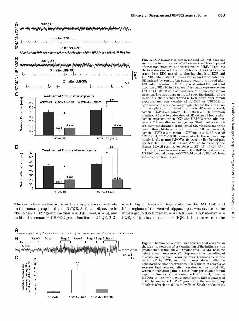
31), the survival rate was 44% for the soman group (4 of 9) and100% for the animals that were administered DZP at 1 hour(n5 6) or 2 hours (n5 4) after exposure, aswell as for the animalsadministered UBP302 at 1 hour (n 5 8) or 2 hours (n 5 4) afterexposure. In the soman 1 DZP group, the electrographic initialSE [i.e., the SE that started after soman injection and wasterminated, at least temporarily, after anticonvulsant adminis-tration (Fig. 1A)] lasted for 113.6 6 10.6 minutes (n 5 6) whenDZP was administered 1 hour after soman, and 133.7 6 10.9minutes (n 5 4) when DZP was administered at 2 hours aftersoman. In the soman1UBP302 group, the electrographic initialSE (Fig. 1B) lasted for 189.966.2minutes (n5 8) whenUBP302was administered 1 hour after soman, and 236 6 5.4 minutes(n 5 4) when UBP302 was administered 2 hours after soman.Comparedwith the duration of the SE in the somangroup (609.4637.3 minutes, n 5 4), the initial SE duration in the soman 1DZP and the soman 1 UBP302 groups was significantly lower(P , 0.001) whether the anticonvulsants were administered at1 hour after soman exposure (Fig. 1C, left set of bars) or at2 hours after soman exposure (Fig. 1D, left set of bars). Theduration of the initial SE in the soman 1 UBP302 group wassignificantly greater than in the soman1DZP group (P, 0.05).Seizures recurred in all of the rats that received DZP and in
half of the rats that received UBP302. The total duration ofelectrographic seizures within the 24-hour period after somanexposure (initial SE 1 recurring seizures) was significantlylower in rats administered UBP302 at 1 hour (203.6 6 4.4minutes, n5 8; P, 0.001; Fig. 1C, right set of bars) or 2 hours(2376 3.2 minutes, n5 4; P5 0.003; Fig. 1D, right set of bars)after exposure, compared with the soman group (655.26 36.9minutes, n 5 4). By contrast, rats treated with DZP at 1 hourafter exposure had similar total duration of SE (707.4 6 62.5minutes, n5 6) to the untreated soman rats (Fig. 1C, right setof bars), whereas in rats treated with DZP at 2 hourspostexposure, the total duration of SE (879 6 59.2 minutes,n 5 4) was significantly longer than that in the soman group(P , 0.05; Fig. 1D, right set of bars). The total duration of SEin the soman1UBP302 group was also significantly less thanin the soman 1 DZP group (P , 0.01).Using the video-EEG recording system, we also counted the
number of convulsive seizures that recurred after terminationof the initial SE and within the remaining time of the 24-hourperiod after the exposure, in the soman rats and in the ratsthat received anticonvulsant treatment at 1 hour after somanexposure. After the prolonged SE in the soman group, thenumber of recurring convulsive seizures was very low (1.5 60.86, n 5 4; Fig. 2). The number of recurring convulsiveseizures in the DZP-treated group (27.2 6 6.89, n 5 6) wassignificantly greater than in the UBP302-treated group (4.7262.10, n 5 8; P , 0.01; Fig. 2).Neuronal Loss and Degeneration—1 Day after
Soman Administration. Neuropathological analysis wasperformed in the soman group and in the groups that receivedDZP or UBP302 at 1 hour after soman exposure; neuronal losswas determined based on comparisons with the control group.
362 Apland et al.
at ASPE
T Journals on M
ay 12, 2019jpet.aspetjournals.org
Dow
nloaded from
The neurodegeneration score for the amygdala was moderatein the soman group [median 5 3 (IQR, 3–4), n 5 6], severe inthe soman 1 DZP group [median 5 4 (IQR, 3–4), n 5 6], andmild in the soman 1 UBP302 group [median 5 2 (IQR, 2–3),
n 5 6; Fig. 3]. Neuronal degeneration in the CA1, CA3, andhilar regions of the ventral hippocampus was severe in thesoman group [CA1: median 5 4 (IQR, 3–4); CA3: median 5 4(IQR, 3–4); hilus: median 5 4 (IQR, 4–4)], moderate in the
Fig. 1. DZP terminates soman-induced SE, but does notreduce the total duration of SE within the 24-hour periodafter soman exposure, as seizures return; UBP302 reducesthe total duration of SE within 24 hours. (A and B) Exampletraces from EEG recordings showing that both DZP andUBP302 (administered 1 hour after soman) terminated theSE induced by soman, but seizure activity returned afterDZP administration. (C) Duration of initial SE and totalduration of SE within 24 hours after soman exposure, whenDZP and UBP302 were administered at 1 hour after somaninjection. The three bars on the left show the duration of theinitial SE (the SE that started 5–15 minutes after somanexposure and was terminated by DZP or UBP302, orspontaneously in the soman group), whereas the three barson the right show the total duration of SE (soman: n = 4;soman + DZP: n = 6; soman + UBP302: n = 8). (D) Durationof initial SE and total duration of SE within 24 hours aftersoman exposure, when DZP and UBP302 were adminis-tered at 2 hours after soman injection. The three bars on theleft show the duration of the initial SE, whereas the threebars to the right show the total duration of SE (soman: n = 4;soman + DZP: n = 4; soman + UBP302: n = 4). *P , 0.05;**P , 0.01; ***P , 0.001, compared with the soman group(analysis of variance [ANOVA] followed by Bonferroni posthoc test for the initial SE and ANOVA followed by theGames–Howell post hoc test for total SE). *P , 0.05; **P ,0.01 for the comparisons between the DZP-treated and theUBP302-treated groups (ANOVA followed by Fisher’s leastsignificant difference test).
Fig. 2. The number of convulsive seizures that recurred inthe DZP-treated rats after termination of the initial SE wasgreater than in the UBP302-treated rats. (A) EEG baselinebefore soman exposure. (B) Representative recording ofa convulsive seizure recurring after termination of theinitial SE by DZP, and its correspondence with thebehavioral seizure observations. (C) Number of convulsiveseizures that occurred after cessation of the initial SE,within the remaining time of the 24-hour period after somanexposure (soman: n = 4; soman + DZP: n = 6; soman +UBP302: n = 8). **P , 0.01, significantly higher comparedwith the soman + UBP302 group and the soman group(analysis of variance followed by Holm–Sidak post hoc test).
Efficacy of Diazepam and UBP302 against Soman 363
at ASPE
T Journals on M
ay 12, 2019jpet.aspetjournals.org
Dow
nloaded from
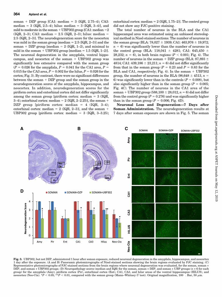
soman 1 DZP group [CA1: median 5 3 (IQR, 2.75–4); CA3:median 5 3 (IQR, 2.5–4); hilus: median 5 3 (IQR, 3–4)], andmild tomoderate in the soman1UBP302 group [CA1:median52(IQR, 2–3); CA3: median 5 2.5 (IQR, 2–3); hilus: median 52.5 (IQR, 2–3)]. The neurodegeneration score for the neocortexwas mild in the soman group [median5 2.5 (IQR, 2–3)] and thesoman 1 DZP group [median 5 2 (IQR, 1–2), and minimal tomild in the soman1UBP302 group [median5 1.5 (IQR, 1–2)].The neuronal degeneration in the amygdala, ventral hippo-campus, and neocortex of the soman 1 UBP302 group wassignificantly less extensive compared with the soman group(P 5 0.026 for the amygdala, P 5 0.041 for the CA1 area, P 50.015 for the CA3 area,P5 0.002 for the hilus,P5 0.026 for thecortex; Fig. 3). By contrast, there were no significant differencesbetween the soman 1 DZP group and the soman group in theneurodegeneration scores of the amygdala, hippocampus, andneocortex. In addition, neurodegeneration scores for thepiriform cortex and entorhinal cortex did not differ significantlyamong the soman group [piriform cortex: median 5 3 (IQR,3–4); entorhinal cortex: median5 2 (IQR, 2–2.25)], the soman1DZP group [piriform cortex: median 5 4 (IQR, 3–4);entorhinal cortex: median 5 2 (IQR, 2–3)], and the soman 1UBP302 group [piriform cortex: median 5 3 (IQR, 3–3.25);
entorhinal cortex: median5 2 (IQR, 1.75–2)]. The control groupdid not show any FJC-positive staining.The total number of neurons in the BLA and the CA1
hippocampal area was estimated using an unbiased stereolog-icalmethod inNissl-stained sections. The number of neurons inthe soman group (BLA: 78,0276 1939; CA1: 463,9766 19,972;n 5 6) was significantly lower than the number of neurons inthe control group (BLA: 118,041 6 4281; CA1: 645,450 628,232; n 5 6), in both brain regions (P , 0.001; Fig. 4). Thenumber of neurons in the soman 1 DZP group (BLA: 87,89364814; CA1: 439,1866 23,211; n5 6) did not differ significantlyfrom that in the soman group (P 5 0.23 and P 5 0.83 for theBLA and CA1, respectively; Fig. 4). In the soman 1 UBP302group, the number of neurons in the BLA (98,648 6 4513, n 56) was significantly lower than in the controls (P 5 0.008), butalso significantly higher than in the soman group (P 5 0.005;Fig. 4C). The number of neurons in the CA1 area of thesoman1UBP302 group (588,1006 28,012, n5 6) did not differfrom the control group (P5 0.276) and was significantly higherthan in the soman group (P 5 0.006; Fig. 4D).Neuronal Loss and Degeneration—7 Days after
Soman Administration. The neurodegeneration results at7 days after soman exposure are shown in Fig. 5. The soman
Fig. 3. UBP302, but not DZP, administered 1 hour after soman exposure, reduced neuronal degeneration in the amygdala, hippocampus, and neocortex1 day after the exposure. (A and B) Panoramic photomicrographs of Nissl-stained sections showing the brain regions evaluated by FJC staining. (C)Representative photomicrographs of FJC-stained sections from the brain regions where neuronal degeneration was evaluated, for the soman, soman +DZP, and soman + UBP302 groups. (D) Neuropathology scores (median and IQR) for the soman, soman + DZP, and soman + UBP groups (n = 6 for eachgroup) for the amygdala (Amy), piriform cortex (Pir), entorhinal cortex (Ent), CA1, CA3, and hilar areas of the ventral hippocampus (HILUS), andneocortex (Neo-Ctx). *P , 0.05; **P , 0.01, compared with the soman group (Mann–Whitney U test). Original magnification, 100�. Bar, 50 mm.
364 Apland et al.
at ASPE
T Journals on M
ay 12, 2019jpet.aspetjournals.org
Dow
nloaded from

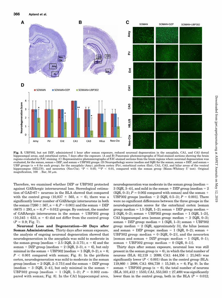
group (n 5 6) had moderate to severe neurodegeneration inthe amygdala [median 5 3 (IQR, 2.75–4)], piriform cortex[median 5 3 (IQR, 3–4)], entorhinal cortex [median 5 3 (IQR,2–3)], CA1 hippocampal area [median 5 4 (IQR, 3–4)], andCA3 hippocampal area [median5 3 (IQR, 1–3.5)], and mild tomoderate in the hilus [median5 2.5 (IQR, 0–4)] and neocortex[median 5 2.5; (IQR, 1–3)]. Similarly, in the soman 1 DZPgroup (n 5 6), neurodegeneration was moderate to severe inthe amygdala [median 5 3 (IQR, 0.75–3.25)], piriform cortex[median 5 3.5; (IQR, 1.75–4)], entorhinal cortex [median 5 3(IQR, 1.5–3)], CA1 hippocampal area [median 5 3.5; (IQR,2.5–4)], and CA3 hippocampal area [median 5 3 (IQR,0.75–3.25)], and mild to moderate in the hilus [median 52.5 (IQR, 1.75–3.25)] and neocortex [median 5 2 (IQR, 1–3)].In the soman 1 UBP302 group (n 5 6), neurodegenerationwas minimal to mild in the entorhinal cortex [median 5 1.5(IQR, 0.75–2.25)], CA3 hippocampal area [median5 0.5 (IQR,0–2)], hilus [median5 1.5 (IQR, 0–3)], and neocortex [median5 1(IQR, 0.75–2)], mild to moderate in the amygdala [median 5 2(IQR, 1.5–3)] and CA1 area [median 5 2 (IQR, 0–3)], andmoderate to severe in the piriform cortex [median5 3 (IQR,1.5–4)]. Compared with the soman group, neurodegeneration
in the soman 1 UBP302 group was significantly lower in theamygdala (P5 0.041), entorhinal cortex (P5 0.041), CA1 (P50.009), and CA3 (P 5 0.041) hippocampal areas.The total number of neurons in the BLA and the CA1
hippocampal area was estimated 7 days after soman exposure;the results are shown in Fig. 6. The number of neurons in thesoman group (BLA: 79,3946 3287; CA1: 440,6986 17,159; n56) was again significantly lower than the number of neurons inthe control group (BLA: 115,108 6 2,673; CA1: 639,701 629,350; n 5 6), in both brain regions (P , 0.001). The numberof neurons in the soman 1 DZP group (BLA: 87,729 6 6032;CA1: 463,196 6 25,729; n 5 6) did not differ significantly fromthose in the soman group (P5 0.424 and P5 0.841 for the BLAand CA1, respectively). The number of neurons in the soman1UBP302 group (BLA: 99,678 6 4,947; CA1: 568,098 6 20,386;n 5 6) was significantly higher than in the soman group (P 50.011 for the BLA and P5 0.003 for the CA1), but differed fromthe control group only in the BLA.We previously showed that the number of GABAergic
interneurons in the BLA is not altered 1 day after somanexposure, but it is significantly reduced 7 days after theexposure (Figueiredo et al., 2011; Prager et al., 2014).
Fig. 4. UBP302, but not DZP, administered 1 hour after soman exposure, reduced neuronal loss in the BLA and the CA1 hippocampal area, 1 day afterthe exposure. (A) Panoramic photomicrographs of Nissl-stained half hemispheres outlining the amygdalar nucleus and the hippocampal subfield wherestereological analysis was performed. (B) Representative photomicrographs of Nissl-stained sections showing BLA and CA1 cells from the control,soman, soman + DZP, and soman + UBP302 groups. (C and D) Group data (mean and S.E.; n = 6 for each group) of stereological estimation of the totalnumber of Nissl-stained neurons in the BLA (left) and CA1 area (right). **P , 0.01; ***P , 0.001 in comparison with control; ##P , 0.01 in comparisonwith the soman group (analysis of variance, Dunnett post hoc test). Original magnification, 630�. Bar, 50 mm.
Efficacy of Diazepam and UBP302 against Soman 365
at ASPE
T Journals on M
ay 12, 2019jpet.aspetjournals.org
Dow
nloaded from
Therefore, we examined whether DZP or UBP302 protectedagainst GABAergic interneuronal loss. Stereological estima-tion of GAD-671 neurons in the BLA showed that comparedwith the control group (10,837 6 565, n 5 6), there was asignificantly lower number of GABAergic interneurons in boththe soman (72606 367, n5 6;P5 0.001) and the soman1DZP(88756 293, n5 6; P5 0.012) groups. By contrast, the numberof GABAergic interneurons in the soman 1 UBP302 group(10,345 6 633, n 5 6) did not differ from the control group(P 5 0.9; Fig. 7).Neuronal Loss and Degeneration—30 Days after
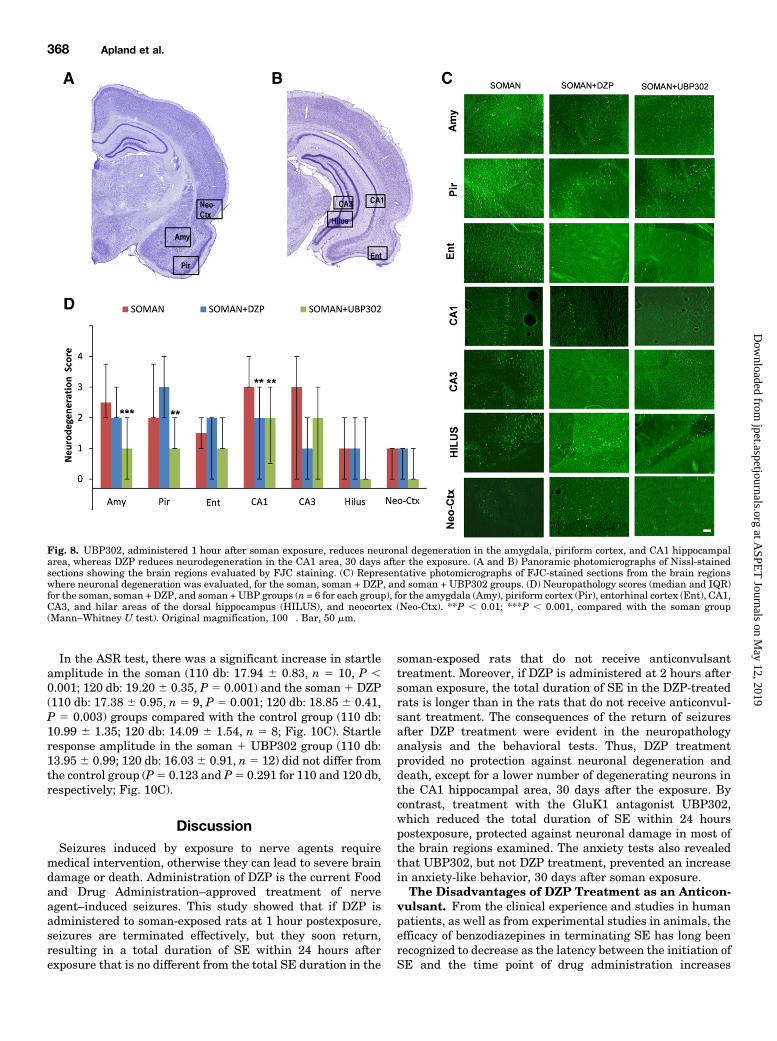
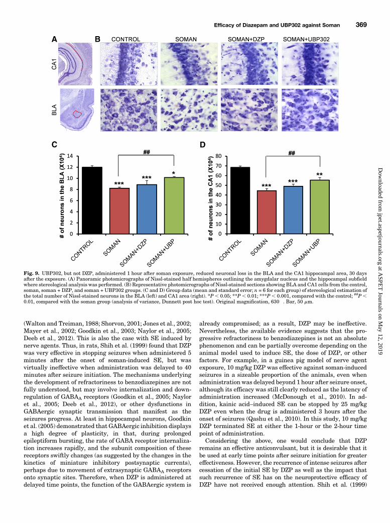
Soman Administration. Thirty days after soman exposure,the analysis of ongoing neuronal degeneration showed thatneurodegeneration in the amygdala was mild to moderate inthe soman group [median 5 2.5 (IQR, 2–3.75), n 5 6] and thesoman 1 DZP group [median 5 2 (IQR, 2–3), n 5 6], but onlyminimal in the soman1UBP302 [median5 1 (IQR, 0–2), n5 6;P , 0.001 compared with soman; Fig. 8). In the piriformcortex, neurodegeneration was mild to moderate in the somangroup [median5 2 (IQR, 2–3.75)] and the soman1DZP group[median 5 3 (IQR, 2–4)], but only minimal in the soman 1UBP302 group [median 5 1 (IQR, 1–2); P 5 0.002 com-pared with soman; Fig. 8]. In the CA1 hippocampal area,
neurodegenerationwasmoderate in the soman group [median53 (IQR, 3–4)], and mild in the soman1DZP group [median5 2(IQR, 0–3); P 5 0.002 compared with soman] and the soman1UBP302 groups [median 5 2 (IQR, 0.5–3); P 5 0.001]. Therewere no significant differences between the three groups in theneurodegeneration scores for the entorhinal cortex [somangroup: median5 1.5 (IQR, 1–2); soman1DZP group: median52 (IQR, 0–2); soman 1 UBP302 group: median 5 1 (IQR, 1–2)],CA3 hippocampal area [soman group: median 5 3 (IQR, 0–3);soman1DZP group: median5 1 (IQR, 1–3); soman1UBP302group: median 5 2 (IQR, approximately 3)], the hilus [somanand soman 1 DZP groups: median 5 1 (IQR, 0–2); soman 1UBP302 group: median 5 0 (IQR, 0–2)], and the neocortex[soman and soman 1 DZP groups: median 5 1 (IQR, 0–1);soman 1 UBP302 group: median 5 0 (IQR, 0–1)].Thirty days after soman exposure, neuronal loss was still
present in the soman group (n5 6), in which the total number ofneurons (BLA: 82,119 6 2099; CA1: 444,356 6 21,045) wassignificantly lower (P , 0.001) than in the control group (BLA:119,860 6 2898; CA1: 684,738 6 15,340; n 5 6; Fig. 9). In thesoman 1 UBP302 group (n 5 6), the total number of neurons(BLA: 101,4126 1535; CA1, 553,5836 27,489) was significantlylower than in the control group, both in the BLA (P 5 0.012;
Fig. 5. UBP302, but not DZP, administered 1 hour after soman exposure, reduced neuronal degeneration in the amygdala, CA1, and CA3 dorsalhippocampal areas, and entorhinal cortex, 7 days after the exposure. (A and B) Panoramic photomicrographs of Nissl-stained sections showing the brainregions evaluated by FJC staining. (C) Representative photomicrographs of FJC-stained sections from the brain regions where neuronal degeneration wasevaluated, for the soman, soman + DZP, and soman +UBP302 groups. (D) Neuropathology scores (median and IQR) for the soman, soman + DZP, and soman +UBP groups (n = 6 for each group), for the amygdala (Amy), piriform cortex (Pir), entorhinal cortex (Ent), CA1, CA3, and hilar areas of the ventralhippocampus (HILUS), and neocortex (Neo-Ctx). *P , 0.05; **P , 0.01, compared with the soman group (Mann–Whitney U test). Originalmagnification, 100�. Bar, 50 mm.
366 Apland et al.
at ASPE
T Journals on M
ay 12, 2019jpet.aspetjournals.org
Dow
nloaded from
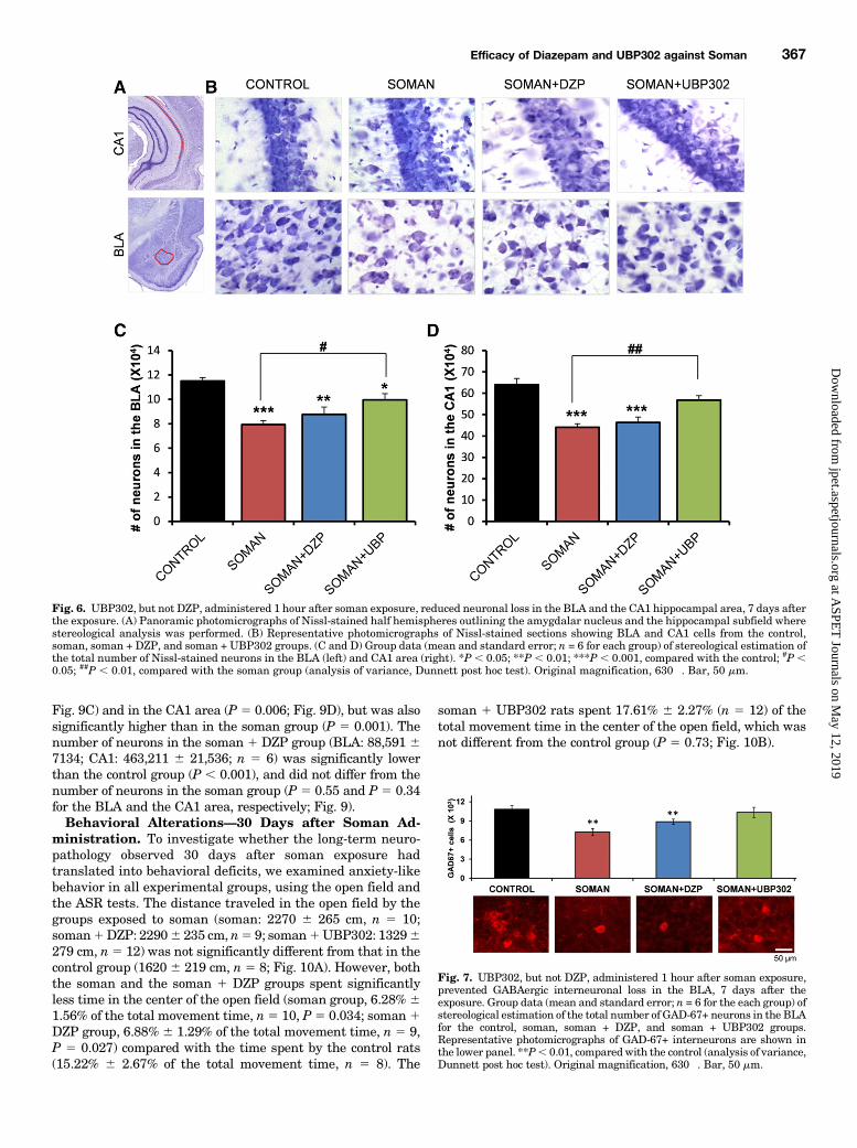
Fig. 9C) and in the CA1 area (P 5 0.006; Fig. 9D), but was alsosignificantly higher than in the soman group (P 5 0.001). Thenumber of neurons in the soman 1 DZP group (BLA: 88,591 67134; CA1: 463,211 6 21,536; n 5 6) was significantly lowerthan the control group (P , 0.001), and did not differ from thenumber of neurons in the soman group (P 5 0.55 and P 5 0.34for the BLA and the CA1 area, respectively; Fig. 9).Behavioral Alterations—30 Days after Soman Ad-
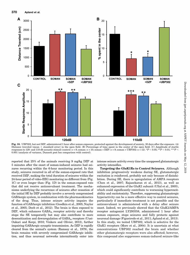
ministration. To investigate whether the long-term neuro-pathology observed 30 days after soman exposure hadtranslated into behavioral deficits, we examined anxiety-likebehavior in all experimental groups, using the open field andthe ASR tests. The distance traveled in the open field by thegroups exposed to soman (soman: 2270 6 265 cm, n 5 10;soman1DZP: 22906 235 cm, n5 9; soman1UBP302: 13296279 cm, n5 12) was not significantly different from that in thecontrol group (16206 219 cm, n 5 8; Fig. 10A). However, boththe soman and the soman 1 DZP groups spent significantlyless time in the center of the open field (soman group, 6.28% 61.56% of the total movement time, n5 10, P5 0.034; soman1DZP group, 6.88% 6 1.29% of the total movement time, n 5 9,P 5 0.027) compared with the time spent by the control rats(15.22% 6 2.67% of the total movement time, n 5 8). The
soman 1 UBP302 rats spent 17.61% 6 2.27% (n 5 12) of thetotal movement time in the center of the open field, which wasnot different from the control group (P 5 0.73; Fig. 10B).
Fig. 6. UBP302, but not DZP, administered 1 hour after soman exposure, reduced neuronal loss in the BLA and the CA1 hippocampal area, 7 days afterthe exposure. (A) Panoramic photomicrographs of Nissl-stained half hemispheres outlining the amygdalar nucleus and the hippocampal subfield wherestereological analysis was performed. (B) Representative photomicrographs of Nissl-stained sections showing BLA and CA1 cells from the control,soman, soman + DZP, and soman + UBP302 groups. (C and D) Group data (mean and standard error; n = 6 for each group) of stereological estimation ofthe total number of Nissl-stained neurons in the BLA (left) and CA1 area (right). *P , 0.05; **P , 0.01; ***P , 0.001, compared with the control; #P ,0.05; ##P , 0.01, compared with the soman group (analysis of variance, Dunnett post hoc test). Original magnification, 630�. Bar, 50 mm.
Fig. 7. UBP302, but not DZP, administered 1 hour after soman exposure,prevented GABAergic interneuronal loss in the BLA, 7 days after theexposure. Group data (mean and standard error; n = 6 for the each group) ofstereological estimation of the total number of GAD-67+ neurons in the BLAfor the control, soman, soman + DZP, and soman + UBP302 groups.Representative photomicrographs of GAD-67+ interneurons are shown inthe lower panel. **P, 0.01, comparedwith the control (analysis of variance,Dunnett post hoc test). Original magnification, 630�. Bar, 50 mm.
Efficacy of Diazepam and UBP302 against Soman 367
at ASPE
T Journals on M
ay 12, 2019jpet.aspetjournals.org
Dow
nloaded from
In the ASR test, there was a significant increase in startleamplitude in the soman (110 db: 17.94 6 0.83, n 5 10, P ,0.001; 120 db: 19.20 6 0.35, P 5 0.001) and the soman 1 DZP(110 db: 17.38 6 0.95, n 5 9, P 5 0.001; 120 db: 18.85 6 0.41,P 5 0.003) groups compared with the control group (110 db:10.99 6 1.35; 120 db: 14.09 6 1.54, n 5 8; Fig. 10C). Startleresponse amplitude in the soman 1 UBP302 group (110 db:13.956 0.99; 120 db: 16.036 0.91, n5 12) did not differ fromthe control group (P5 0.123 and P5 0.291 for 110 and 120 db,respectively; Fig. 10C).
DiscussionSeizures induced by exposure to nerve agents require
medical intervention, otherwise they can lead to severe braindamage or death. Administration of DZP is the current Foodand Drug Administration–approved treatment of nerveagent–induced seizures. This study showed that if DZP isadministered to soman-exposed rats at 1 hour postexposure,seizures are terminated effectively, but they soon return,resulting in a total duration of SE within 24 hours afterexposure that is no different from the total SE duration in the
soman-exposed rats that do not receive anticonvulsanttreatment. Moreover, if DZP is administered at 2 hours aftersoman exposure, the total duration of SE in the DZP-treatedrats is longer than in the rats that do not receive anticonvul-sant treatment. The consequences of the return of seizuresafter DZP treatment were evident in the neuropathologyanalysis and the behavioral tests. Thus, DZP treatmentprovided no protection against neuronal degeneration anddeath, except for a lower number of degenerating neurons inthe CA1 hippocampal area, 30 days after the exposure. Bycontrast, treatment with the GluK1 antagonist UBP302,which reduced the total duration of SE within 24 hourspostexposure, protected against neuronal damage in most ofthe brain regions examined. The anxiety tests also revealedthat UBP302, but not DZP treatment, prevented an increasein anxiety-like behavior, 30 days after soman exposure.The Disadvantages of DZP Treatment as an Anticon-
vulsant. From the clinical experience and studies in humanpatients, as well as from experimental studies in animals, theefficacy of benzodiazepines in terminating SE has long beenrecognized to decrease as the latency between the initiation ofSE and the time point of drug administration increases
Fig. 8. UBP302, administered 1 hour after soman exposure, reduces neuronal degeneration in the amygdala, piriform cortex, and CA1 hippocampalarea, whereas DZP reduces neurodegeneration in the CA1 area, 30 days after the exposure. (A and B) Panoramic photomicrographs of Nissl-stainedsections showing the brain regions evaluated by FJC staining. (C) Representative photomicrographs of FJC-stained sections from the brain regionswhere neuronal degeneration was evaluated, for the soman, soman + DZP, and soman + UBP302 groups. (D) Neuropathology scores (median and IQR)for the soman, soman + DZP, and soman + UBP groups (n = 6 for each group), for the amygdala (Amy), piriform cortex (Pir), entorhinal cortex (Ent), CA1,CA3, and hilar areas of the dorsal hippocampus (HILUS), and neocortex (Neo-Ctx). **P , 0.01; ***P , 0.001, compared with the soman group(Mann–Whitney U test). Original magnification, 100�. Bar, 50 mm.
368 Apland et al.
at ASPE
T Journals on M
ay 12, 2019jpet.aspetjournals.org
Dow
nloaded from
(Walton and Treiman, 1988; Shorvon, 2001; Jones et al., 2002;Mayer et al., 2002; Goodkin et al., 2003; Naylor et al., 2005;Deeb et al., 2012). This is also the case with SE induced bynerve agents. Thus, in rats, Shih et al. (1999) found that DZPwas very effective in stopping seizures when administered 5minutes after the onset of soman-induced SE, but wasvirtually ineffective when administration was delayed to 40minutes after seizure initiation. The mechanisms underlyingthe development of refractoriness to benzodiazepines are notfully understood, but may involve internalization and down-regulation of GABAA receptors (Goodkin et al., 2005; Nayloret al., 2005; Deeb et al., 2012), or other dysfunctions inGABAergic synaptic transmission that manifest as theseizures progress. At least in hippocampal neurons, Goodkinet al. (2005) demonstrated that GABAergic inhibition displaysa high degree of plasticity, in that, during prolongedepileptiform bursting, the rate of GABA receptor internaliza-tion increases rapidly, and the subunit composition of thesereceptors swiftly changes (as suggested by the changes in thekinetics of miniature inhibitory postsynaptic currents),perhaps due to movement of extrasynaptic GABAA receptorsonto synaptic sites. Therefore, when DZP is administered atdelayed time points, the function of the GABAergic system is
already compromised; as a result, DZP may be ineffective.Nevertheless, the available evidence suggests that the pro-gressive refractoriness to benzodiazepines is not an absolutephenomenon and can be partially overcome depending on theanimal model used to induce SE, the dose of DZP, or otherfactors. For example, in a guinea pig model of nerve agentexposure, 10 mg/kg DZP was effective against soman-inducedseizures in a sizeable proportion of the animals, even whenadministration was delayed beyond 1 hour after seizure onset,although its efficacy was still clearly reduced as the latency ofadministration increased (McDonough et al., 2010). In ad-dition, kainic acid–induced SE can be stopped by 25 mg/kgDZP even when the drug is administered 3 hours after theonset of seizures (Qashu et al., 2010). In this study, 10 mg/kgDZP terminated SE at either the 1-hour or the 2-hour timepoint of administration.Considering the above, one would conclude that DZP
remains an effective anticonvulsant, but it is desirable that itbe used at early time points after seizure initiation for greatereffectiveness. However, the recurrence of intense seizures aftercessation of the initial SE by DZP as well as the impact thatsuch recurrence of SE has on the neuroprotective efficacy ofDZP have not received enough attention. Shih et al. (1999)
Fig. 9. UBP302, but not DZP, administered 1 hour after soman exposure, reduced neuronal loss in the BLA and the CA1 hippocampal area, 30 daysafter the exposure. (A) Panoramic photomicrographs of Nissl-stained half hemispheres outlining the amygdalar nucleus and the hippocampal subfieldwhere stereological analysis was performed. (B) Representative photomicrographs of Nissl-stained sections showing BLA and CA1 cells from the control,soman, soman + DZP, and soman + UBP302 groups. (C and D) Group data (mean and standard error; n = 6 for each group) of stereological estimation ofthe total number of Nissl-stained neurons in the BLA (left) and CA1 area (right). *P, 0.05; **P , 0.01; ***P, 0.001, compared with the control; ##P,0.01, compared with the soman group (analysis of variance, Dunnett post hoc test). Original magnification, 630�. Bar, 50 mm.
Efficacy of Diazepam and UBP302 against Soman 369
at ASPE
T Journals on M
ay 12, 2019jpet.aspetjournals.org
Dow
nloaded from
reported that 25% of the animals receiving 9 mg/kg DZP at5 minutes after the onset of soman-induced seizures had sei-zures recurring within the 6-hour monitoring period. In thisstudy, seizures recurred in all of the soman-exposed rats thatreceived DZP, making the total duration of seizures within the24-hour period of video-EEG monitoring no different from (Fig.1C) or even longer than (Fig. 1D) in the soman-exposed ratsthat did not receive anticonvulsant treatment. The mecha-nisms underlying the recurrence of seizures after cessation ofthe initial SE by DZP probably involve a severely compromisedGABAergic system, in combination with the pharmacokineticsof the drug. Thus, intense seizure activity impairs thefunction of GABAergic inhibition (Goodkin et al., 2005; Nayloret al., 2005; Deeb et al., 2012). The brain is then exposed toDZP, which enhances GABAA receptor activity and therebystops the SE temporarily but may also contribute to moredesensitization and downregulation of GABAA receptors (Uusi-Oukari and Korpi, 2010; Vinkers and Olivier, 2012), furtherimpairing GABAergic synaptic transmission. As DZP is rapidlycleared from the animal’s system (Ramsay et al., 1979), thebrain remains with severely compromised GABAergic inhibi-tion, and thus neuronal networks intermittently enter into
intense seizure activity every time the unopposed glutamatergicactivity intensifies.Targeting the GluK1Rs to Control Seizures. Although
inhibition progressively weakens during SE, glutamatergicexcitation is reinforced, probably not only because of disinhi-bition. During SE, there is upregulation of AMPA receptors(Chen et al., 2007; Rajasekaran et al., 2013), as well asenhanced expression of the GluK1 subunit (Ullal et al., 2005),which could significantly contribute to worsening hyperexcit-ability and excitotoxicity. Therefore, suppressing glutamatergichyperactivity can be a more effective way to control seizures,particularly if immediate treatment is not possible and theanticonvulsant is administered with a delay after seizureonset. Indeed, we previously showed that the GluK1/AMPAreceptor antagonist LY293558, administered 1 hour aftersoman exposure, stops seizures and fully protects againstneuronal damage (Figueiredo et al., 2011; Apland et al., 2013).In this study, we tested UBP302, which is selective for theGluK1 receptors (More et al., 2004). It is uncertain at whatconcentrations UBP302 reached the brain and whetherother glutamatergic receptors were also affected; however,this compound also suppresses soman-induced seizure-like
Fig. 10. UBP302, but not DZP, administered 1 hour after soman exposure, protected against the development of anxiety, 30 days after the exposure. (A)Distance traveled (mean 6 standard error) in the open field. (B) Percentage of time spent in the center of the open field. (C) Amplitude of startleresponses to 120- and 110-db acoustic stimuli (control: n = 8; soman: n = 10; soman + DZP: n = 9; soman + UBP302: n = 12). *P, 0.05; **P, 0.01; ***P,0.001 (analysis of variance, Dunnett post hoc comparison with control).
370 Apland et al.
at ASPE
T Journals on M
ay 12, 2019jpet.aspetjournals.org
Dow
nloaded from
activity in vitro at relatively low concentrations that areconsidered selective for GluK1 antagonism (Apland et al.,2009). UBP302 blocked the seizures induced by soman, andprotected against neuropathology and anxiety-related be-havioral deficits.The time course of termination of the initial SE by UBP302
was slow compared with that of DZP or LY293558. Althoughthis may have to do with the type of receptors being targetedand their involvement in seizure activity, it is also possiblethat it relates to the pharmacokinetics of UBP302, which isnot known. However, despite the apparently slow time courseof action of UBP302, the total duration of SE within the 24-hour period after exposure was dramatically reduced in theUBP302-treated rats compared with either the soman-exposed rats that did not receive anticonvulsant treatmentor the rats that received DZP. The mechanisms by whicha GluK1 antagonist may suppress hyperexcitability and stopseizures were previously discussed (Figueiredo et al., 2011;Aroniadou-Anderjaska et al., 2012). GluK1Rs modulate bothGABAergic and glutamatergic synaptic transmission ina number of brain regions (Jane et al., 2009). At least in theBLA and the hippocampus, the net effect of their activation isexcitatory. This is suggested by the findings that GluK1Rantagonists block epileptiform activity in hippocampal slicesand limbic seizures in vivo (Smolders et al., 2002). In the BLA,GluK1Rs are present on postsynaptic (somatodendritic) sitesof both principal cells (Gryder and Rogawski, 2003) andinterneurons (Braga et al., 2003), as well as on the pre-synaptic terminals of both cell types, where they mediatefacilitation of glutamate release (Aroniadou-Anderjaska et al.,2012) and either facilitation or inhibition of GABA release,depending on the concentration of the agonist (Braga et al.,2003). The net effect of their activation in the BLA network isan increased excitation and excitability, as suggested bya greater enhancement of spontaneous EPSCs versus in-hibitory postsynaptic currents, the reduction of anxiety-likebehavior when these receptors are blocked by microinjectionof UBP302 selectively into the BLA, and the increased anxietyor the induction of seizures when a GluK1R agonist is injectedinto the BLA (Aroniadou-Anderjaska et al., 2012). Although itremains to be determined what role GluK1Rs play in theoverall excitation of other neuronal networks, the fact thatactivation of these receptors increases the excitation state ofthe BLA and the hippocampus, two highly seizurogenic brainregions, may explain why blockade of these receptors canterminate seizures. It should also be noted that in contrastwith the downregulation and internalization of GABAA
receptors after excessive neuronal/seizure activity (Goodkinet al., 2005; Naylor et al., 2005; Deeb et al., 2012), which canrender DZP ineffective, the upregulation of the GluK1 subunit(Ullal et al., 2005) may contribute to hyperexcitation, whichfurther explains the efficacy of GluK1R antagonists.Neuropathology and Associated Behavioral Deficits.
Depending on the extent of brain damage after prolonged SE,behavioral deficits may ensue. Increased anxiety is observed inanimals that have been exposed to nerve agents (Coubardet al., 2008; Langston et al., 2012; Prager et al., 2014).Similarly, the human victims of the sarin terrorist attack inJapan report enduring symptoms of anxiety disorders, longafter the exposure (Ohtani et al., 2004; Hoffman et al., 2007).The amygdala plays a central role in emotional behavior, anddysfunction with increased excitability of the BLA is associated
with anxiety (Gonzalez et al., 1996; Shekhar et al., 2003; Zhouet al., 2010; Pidoplichko et al., 2014). The hippocampus is alsosignificantly involved in the modulation of anxiety (Engin andTreit, 2007; Fournier and Duman, 2013). Both the amygdalaand the hippocampus are severely damaged by nerveagent–induced SE, as shown in this study and previous studies(Shih et al., 2003; Aroniadou-Anderjaska et al., 2009; Aplandet al., 2010). In the BLA, there is significant loss of GABAergicneurons by day 7 after exposure to soman (Figueiredo et al.,2011; Prager et al., 2014), and at 14 and 30 days after exposurethe ratio of GABAergic interneurons to the total number ofneurons in the BLA is significantly decreased (Prager et al.,2014). Reduction of inhibitory activity in the BLA, resultingfrom the interneuronal loss, will increase the excitability of theBLA network, which can lead to the development of anxiety.In this study, treatment with UBP302 significantly reduced
neuronal loss and degeneration in a number of brain regions,including the hippocampus and the amygdala, and preventedthe loss of GABAergic interneurons in the BLA, as assessed 30days after the exposure. The neuroprotection provided byUBP302, despite its being only partial, was sufficient toprevent the development of anxiety-like behavior, as assessedin the open field and the ASR tests. By contrast, DZP had noneuroprotective effects, except for reduced neurodegenerationin the CA1 area at 30 days after exposure, and did not preventthe development of anxiety. Since the extent of nerveagent–induced neuropathology is solely or largely determinedby the intensity and duration of seizures (Shih et al., 2003;Prager et al., 2013), the failure of DZP to protect the brain canbe attributed to the failure of this drug to reduce the totalduration of seizures. Consistent with our findings revealingthe inefficacy of DZP to reduce neuropathology and preventbehavioral deficits, previous studies in which 10 mg/kg DZPwas administered at 30minutes after soman-induced seizuresshow that this treatment did not prevent the development ofanxiety-like behavior (Langston et al., 2012) or epileptogenesis(de Araujo Furtado et al., 2010).Our data clearly argue against the use of DZP as a medical
countermeasure for the treatment of SE induced by nerveagent exposure, at least when anticonvulsant treatment isdelayed. DZP is likely to substantially increase the survivalrate of exposed individuals; however, because seizures recur,there is virtually no protection against neuropathology andthe resulting behavioral deficits. Administering DZP repeat-edly, every time intense seizures recur, not only may not befeasible, but may also be detrimental to the victims’ health.The results suggest that targeting the glutamatergic systemis a more effective approach to controlling nerve agent–inducedSE, and antagonists of the GluK1Rs appear to be both safe andefficacious in this regard.
Authorship Contributions
Participated in research design: Apland, Aroniadou-Anderjaska,Braga.
Conducted experiments: Apland, Figueiredo, Rossetti, Miller.Performed data analysis: Figueiredo, Apland, Rossetti, Aroniadou-
Anderjaska, Braga.Wrote or contributed to the writing of the manuscript: Aroniadou-
Anderjaska, Apland, Figueiredo, Miller, Braga.
References
Apland JP, Aroniadou-Anderjaska V, and Braga MF (2009) Soman induces ictogenesisin the amygdala and interictal activity in the hippocampus that are blocked by aGluR5 kainate receptor antagonist in vitro. Neuroscience 159:380–389.
Efficacy of Diazepam and UBP302 against Soman 371
at ASPE
T Journals on M
ay 12, 2019jpet.aspetjournals.org
Dow
nloaded from
Apland JP, Aroniadou-Anderjaska V, Figueiredo TH, Green CE, Swezey R, Yang C,Qashu F, and Braga MFM (2013) Efficacy of the GluK1/AMPA receptor antagonistLY293558 against seizures and neuropathology in a soman-exposure model with-out pretreatment and its pharmacokinetics after intramuscular administration. JPharmacol Exp Ther 344:133–140.
Apland JP, Figueiredo TH, Qashu F, Aroniadou-Anderjaska V, Souza AP, and BragaMF (2010) Higher susceptibility of the ventral versus the dorsal hippocampus andthe posteroventral versus anterodorsal amygdala to soman-induced neuropathol-ogy. Neurotoxicology 31:485–492.
Aroniadou-Anderjaska V, Figueiredo TH, Apland JP, Qashu F, and Braga MF (2009)Primary brain targets of nerve agents: the role of the amygdala in comparison tothe hippocampus. Neurotoxicology 30:772–776.
Aroniadou-Anderjaska V, Pidoplichko VI, Figueiredo TH, Almeida-Suhett CP, PragerEM, and Braga MF (2012) Presynaptic facilitation of glutamate release in thebasolateral amygdala: a mechanism for the anxiogenic and seizurogenic function ofGluK1 receptors. Neuroscience 221:157–169.
Bajgar J (2005) Complex view on poisoning with nerve agents and organophosphates.Acta Med (Hradec Kralove) 48:3–21.
Braga MF, Aroniadou-Anderjaska V, Xie J, and Li H (2003) Bidirectional modulationof GABA release by presynaptic glutamate receptor 5 kainate receptors in thebasolateral amygdala. J Neurosci 23:442–452.
Campo-Soria C, Chang Y, and Weiss DS (2006) Mechanism of action of benzodiaze-pines on GABAA receptors. Br J Pharmacol 148:984–990.
Chen JW, Naylor DE, and Wasterlain CG (2007) Advances in the pathophysiology ofstatus epilepticus. Acta Neurol Scand Suppl 186:7–15.
Collingridge GL, Olsen RW, Peters J, and Spedding M (2009) A nomenclature forligand-gated ion channels. Neuropharmacology 56:2–5.
Coubard S, Béracochéa D, Collombet JM, Philippin JN, Krazem A, Liscia P, LallementG, and Piérard C (2008) Long-term consequences of soman poisoning in mice: part 2.Emotional behavior. Behav Brain Res 191:95–103.
de Araujo Furtado M, Lumley LA, Robison C, Tong LC, Lichtenstein S, and YourickDL (2010) Spontaneous recurrent seizures after status epilepticus induced bysoman in Sprague-Dawley rats. Epilepsia 51:1503–1510.
Deeb TZ, Maguire J, and Moss SJ (2012) Possible alterations in GABAA receptor sig-naling that underlie benzodiazepine-resistant seizures. Epilepsia 53 (Suppl 9):79–88.
Dolgin E (2013) Syrian gas attack reinforces need for better anti-sarin drugs. NatMed 19:1194–1195.
Engin E and Treit D (2007) The role of hippocampus in anxiety: intracerebral in-fusion studies. Behav Pharmacol 18:365–374.
Faraday MM, Elliott BM, and Grunberg NE (2001) Adult vs. adolescent rats differ inbiobehavioral responses to chronic nicotine administration. Pharmacol BiochemBehav 70:475–489.
Figueiredo TH, Qashu F, Apland JP, Aroniadou-Anderjaska V, Souza AP, and BragaMF (2011) The GluK1 (GluR5) Kainate/alpha-amino-3-hydroxy-5-methyl-4-isoxazolepropionic acid receptor antagonist LY293558 reduces soman-inducedseizures and neuropathology. J Pharmacol Exp Ther 336:303–312.
Filliat P, Coubard S, Pierard C, Liscia P, Beracochea D, Four E, Baubichon D,Masqueliez C, Lallement G, and Collombet JM (2007) Long-term behavioral con-sequences of soman poisoning in mice. Neurotoxicology 28:508–519.
Fournier NM and Duman RS (2013) Illuminating hippocampal control of fearmemory and anxiety. Neuron 77:803–806.
Gielen MC, Lumb MJ, and Smart TG (2012) Benzodiazepines modulate GABAA receptorsby regulating the preactivation step after GABA binding. J Neurosci 32:5707–5715.
Gonzalez LE, Andrews N, and File SE (1996) 5-HT1A and benzodiazepine receptorsin the basolateral amygdala modulate anxiety in the social interaction test, but notin the elevated plus-maze. Brain Res 732:145–153.
Goodkin HP, Liu X, and Holmes GL (2003) Diazepam terminates brief but not pro-longed seizures in young, naïve rats. Epilepsia 44:1109–1112.
Goodkin HP, Yeh JL, and Kapur J (2005) Status epilepticus increases the in-tracellular accumulation of GABAA receptors. J Neurosci 25:5511–5520.
Gryder DS and Rogawski MA (2003) Selective antagonism of GluR5 kainate-receptor-mediated synaptic currents by topiramate in rat basolateral amygdala neurons. JNeurosci 23:7069–7074.
Gundersen HJ, Jensen EB, Kiêu K, and Nielsen J (1999) The efficiency of systematicsampling in stereology—reconsidered. J Microsc 193:199–211.
Hoffman A, Eisenkraft A, Finkelstein A, Schein O, Rotman E, and Dushnitsky T(2007) A decade after the Tokyo sarin attack: a review of neurological follow-up ofthe victims. Mil Med 172:607–610.
Jane DE, Lodge D, and Collingridge GL (2009) Kainate receptors: pharmacology,function and therapeutic potential. Neuropharmacology 56:90–113.
Jimmerson VR, Shih TM, and Mailman RB (1989) Variability in soman toxicity in therat: correlation with biochemical and behavioral measures. Toxicology 57:241–254.
Jones DM, Esmaeil N, Maren S, and Macdonald RL (2002) Characterization ofpharmacoresistance to benzodiazepines in the rat Li-pilocarpine model of statusepilepticus. Epilepsy Res 50:301–312.
Kellinghaus C and Stögbauer F (2012) Treatment of status epilepticus in a largecommunity hospital. Epilepsy Behav 23:235–240.
Lallement G, Dorandeu F, Filliat P, Carpentier P, Baille V, and Blanchet G (1998) Medicalmanagement of organophosphate-induced seizures. J Physiol Paris 92:369–373.
Langston JL, Wright LK, Connis N, and Lumley LA (2012) Characterizing the be-havioral effects of nerve agent-induced seizure activity in rats: increased startlereactivity and perseverative behavior. Pharmacol Biochem Behav 100:382–391.
Mayer SA, Claassen J, Lokin J, Mendelsohn F, Dennis LJ, and Fitzsimmons BF(2002) Refractory status epilepticus: frequency, risk factors, and impact on out-come. Arch Neurol 59:205–210.
McDonough JH, Jr and Shih TM (1997) Neuropharmacological mechanisms of nerveagent-induced seizure and neuropathology. Neurosci Biobehav Rev 21:559–579.
McDonough JH, Jr, McMonagle JD, and Shih TM (2010) Time-dependent reductionin the anticonvulsant effectiveness of diazepam against soman-induced seizures inguinea pigs. Drug Chem Toxicol 33:279–283.
Mehta V, Singhi P, and Singhi S (2007) Intravenous sodium valproate versus di-azepam infusion for the control of refractory status epilepticus in children: a ran-domized controlled trial. J Child Neurol 22:1191–1197.
More JC, Nistico R, Dolman NP, Clarke VR, Alt AJ, Ogden AM, Buelens FP, TroopHM, Kelland EE, and Pilato F, et al. (2004) Characterisation of UBP296: a novel,potent and selective kainate receptor antagonist. Neuropharmacology 47:46–64.
Naylor DE, Liu H, and Wasterlain CG (2005) Trafficking of GABA(A) receptors, lossof inhibition, and a mechanism for pharmacoresistance in status epilepticus. JNeurosci 25:7724–7733.
Ohtani T, Iwanami A, Kasai K, Yamasue H, Kato T, Sasaki T, and Kato N (2004)Post-traumatic stress disorder symptoms in victims of Tokyo subway attack: a 5-yearfollow-up study. Psychiatry Clin Neurosci 58:624–629.
Paxinos G and Watson C (2005) The Rat Brain in Stereotaxic Coordinates, 4th ed,Elsevier, New York.
Pidoplichko VI, Aroniadou-Anderjaska V, Prager EM, Figueiredo TH, Almeida-Suhett CP, Miller SL, and Braga MF (2014) ASIC1a activation enhances inhibitionin the basolateral amygdala and reduces anxiety. J Neurosci 34:3130–3141.
Prager EM, Aroniadou-Anderjaska V, Almeida-Suhett CP, Figueiredo TH, AplandJP, and Braga MF (2013) Acetylcholinesterase inhibition in the basolateralamygdala plays a key role in the induction of status epilepticus after soman ex-posure. Neurotoxicology 38:84–90.
Prager EM, Aroniadou-Anderjaska V, Almeida-Suhett CP, Figueiredo TH, AplandJP, Rossetti F, Olsen CH, and Braga MF (2014) The recovery of acetylcholines-terase activity and the progression of neuropathological and pathophysiologicalalterations in the rat basolateral amygdala after soman-induced status epilepticus:relation to anxiety-like behavior. Neuropharmacology 81:64–74.
Qashu F, Figueiredo TH, Aroniadou-Anderjaska V, Apland JP, and Braga MF (2010)Diazepam administration after prolonged status epilepticus reduces neuro-degeneration in the amygdala but not in the hippocampus during epileptogenesis.Amino Acids 38:189–197.
Racine RJ (1972) Modification of seizure activity by electrical stimulation. II. Motorseizure. Electroencephalogr Clin Neurophysiol 32:281–294.
Rajasekaran K, Joshi S, Kozhemyakin M, Todorovic MS, Kowalski S, Balint C,and Kapur J (2013) Receptor trafficking hypothesis revisited: plasticity of AMPAreceptors during established status epilepticus. Epilepsia 54 (Suppl 6):14–16.
Ramsay RE, Hammond EJ, Perchalski RJ, and Wilder BJ (1979) Brain uptake ofphenytoin, phenobarbital, and diazepam. Arch Neurol 36:535–539.
Schmitz C and Hof PR (2000) Recommendations for straightforward and rigorousmethods of counting neurons based on a computer simulation approach. J ChemNeuroanat 20:93–114.
Shekhar A, Sajdyk TJ, Gehlert DR, and Rainnie DG (2003) The amygdala, panicdisorder, and cardiovascular responses. Ann N Y Acad Sci 985:308–325.
Shih T, McDonough JH, Jr, and Koplovitz I (1999) Anticonvulsants for soman-induced seizure activity. J Biomed Sci 6:86–96.
Shih TM, Duniho SM, and McDonough JH, Jr (2003) Control of nerve agent-inducedseizures is critical for neuroprotection and survival. Toxicol Appl Pharmacol 188:69–80.
Shih TM and McDonough JH, Jr (1999) Organophosphorus nerve agents-inducedseizures and efficacy of atropine sulfate as anticonvulsant treatment. PharmacolBiochem Behav 64:147–153.
Shorvon S (2001) The management of status epilepticus. J Neurol Neurosurg Psy-chiatry 70 (Suppl 2):II22–II27.
Singhi S, Murthy A, Singhi P, and Jayashree M (2002) Continuous midazolam versusdiazepam infusion for refractory convulsive status epilepticus. J Child Neurol 17:106–110.
Skovira JW, McDonough JH, Jr, and Shih TM (2010) Protection against sarin-induced seizures in rats by direct brain microinjection of scopolamine, midazolamor MK-801. J Mol Neurosci 40:56–62.
Smolders I, Bortolotto ZA, Clarke VR, Warre R, Khan GM, O’Neill MJ, Ornstein PL,Bleakman D, Ogden A, and Weiss B, et al. (2002) Antagonists of GLU(K5)-containing kainate receptors prevent pilocarpine-induced limbic seizures. NatNeurosci 5:796–804.
Tetz LM, Rezk PE, Ratcliffe RH, Gordon RK, Steele KE, and Nambiar MP (2006)Development of a rat pilocarpine model of seizure/status epilepticus that mimicschemical warfare nerve agent exposure. Toxicol Ind Health 22:255–266.
Todorovic MS, Cowan ML, Balint CA, Sun C, and Kapur J (2012) Characterization ofstatus epilepticus induced by two organophosphates in rats. Epilepsy Res 101:268–276.
Ullal G, Fahnestock M, and Racine R (2005) Time-dependent effect of kainate-induced seizures on glutamate receptor GluR5, GluR6, and GluR7 mRNA andProtein Expression in rat hippocampus. Epilepsia 46:616–623.
Uusi-Oukari M and Korpi ER (2010) Regulation of GABA(A) receptor subunit ex-pression by pharmacological agents. Pharmacol Rev 62:97–135.
Vinkers CH and Olivier B (2012) Mechanisms underlying tolerance after long-termbenzodiazepine use: A future for subtype-selective GABA(A) receptor modulators?Adv Pharmacol Sci 2012:416864.
Walton NY and Treiman DM (1988) Response of status epilepticus induced by lith-ium and pilocarpine to treatment with diazepam. Exp Neurol 101:267–275.
Yanagisawa N, Morita H, and Nakajima T (2006) Sarin experiences in Japan: acutetoxicity and long-term effects. J Neurol Sci 249:76–85.
Zhou R, Wang S, and Zhu X (2010) Prenatal ethanol exposure attenuates GABAergicinhibition in basolateral amygdala leading to neuronal hyperexcitability andanxiety-like behavior of adult rat offspring. Neuroscience 170:749–757.
Address correspondence to: Dr. Maria F. M. Braga, Department ofAnatomy, Physiology, and Genetics, F. Edward Hébert School of Medicine,Uniformed Services University of the Health Sciences, 4301 Jones BridgeRoad, Bethesda, MD 20814. E-mail: [email protected]
372 Apland et al.
at ASPE
T Journals on M
ay 12, 2019jpet.aspetjournals.org
Dow
nloaded from