Embed Size (px)
Citation preview
Diazepam Inhibits Electrically Evoked and Tonic Dopamine Releasein the Nucleus Accumbens and Reverses the Effect of AmphetamineAlexander Gomez-A,† Amanda M. Fiorenza,† Suelen L. Boschen,†,‡ Adam H. Sugi,† Danielle Beckman,‡
Sergio T. Ferreira,‡ Kendall Lee,§ Charles D. Blaha,§ and Claudio Da Cunha*,†
†Departamento de Farmacologia, Universidade Federal do Parana, Curitiba 81.530-980, PR, Brazil‡Institute of Biophysics Carlos Chagas Filho and Institute of Medical Biochemistry Leopoldo de Meis, Federal University of Rio deJaneiro, Rio de Janeiro, Brazil§Department of Neurologic Surgery, Mayo Clinic, Rochester, Minnesota 55905, United States
*S Supporting Information
ABSTRACT: Diazepam is a benzodiazepine receptor agonist withanxiolytic and addictive properties. Although most drugs of abuseincrease the level of release of dopamine in the nucleus accumbens,here we show that diazepam not only causes the opposite effect butalso prevents amphetamine from enhancing dopamine release. Weused 20 min sampling in vivo microdialysis and subsecond fast-scancyclic voltammetry recordings at carbon-fiber microelectrodes toshow that diazepam caused a dose-dependent decrease in the levelof tonic and electrically evoked dopamine release in the nucleusaccumbens of urethane-anesthetized adult male Swiss mice. In fast-scan cyclic voltammetry assays, dopamine release was evoked byelectrical stimulation of the ventral tegmental area. We observedthat 2 and 3 mg of diazepam/kg reduced the level of electricallyevoked dopamine release, and this effect was reversed by administration of the benzodiazepine receptor antagonist flumazenil indoses of 2.5 and 5 mg/kg, respectively. No significant effects on measures of dopamine re-uptake were observed. Cyclicvoltammetry experiments further showed that amphetamine (5 mg/kg, intraperitoneally) caused a significant increase in the levelof dopamine release and in the half-life for dopamine re-uptake. Diazepam (2 mg/kg) significantly weakened the effect ofamphetamine on dopamine release without affecting dopamine re-uptake. These results suggest that the pharmacological effectsof benzodiazepines have a dopaminergic component. In addition, our findings challenge the classic view that all drugs of abusecause dopamine release in the nucleus accumbens and suggest that benzodiazepines could be useful in the treatment of addictionto other drugs that increase the level of dopamine release, such as cocaine, amphetamines, and nicotine.
KEYWORDS: Dopaminergic neurons, Electrochemistry, Ventral tegmental area, Nucleus accumbens core, GABA, Anxiolytic,Anticonvulsant
1. INTRODUCTION
Benzodiazepines (BZs) are widely used as anxiolytics, sedative-hypnotics, anticonvulsants, anesthetics, and muscle-relaxants.1
These effects of the BZs are achieved by binding to a specificsite in type A γ-aminobutyric acid (GABAA) receptors.
2 BZs arealso used for recreational purposes and can lead to addiction invulnerable people.3 It has been proposed that most addictivedrugs share the common property of increasing extracellulardopamine concentrations in the nucleus accumbens (NAc).4,5
There is solid evidence supporting this hypothesis forcocaine,6,7 amphetamines,8,9 and nicotine.10,11 However,although influential studies and review papers propose thatthis is also true for BZs,12,13 direct evidence that BZs causedopamine release is lacking. A microdialysis study by Bentue-Ferrer et al.14 reported that small doses of the BZs alprazolamand lorazepam caused a modest increase in tonic dopaminelevels in the striatum of rats. However, several other
microdialysis studies reported that systemic and intrastriataladministration of the BZs diazepam, midazolam, flunitrazepam,and imidazenil caused a decrease in tonic levels of dopamine inthe rat frontal cortex and NAc; this effect was also prevented bythe BZ receptor antagonist flumazenil.15−21 Flumazenil alsoincreased the level of tonic dopamine release in the NAc of ratschronically treated with diazepam or imidazenil.19,20 Thosestudies that showed that BZs increase levels of dopaminerelease were conducted using in vivo microdialysis, whichinherently can detect only relatively slow changes in tonic(basal) extracellular concentrations of dopamine. As such, thesestudies do not reflect the direct effect of the BZs on phasic
Special Issue: Monitoring Molecules in Neuroscience 2016
Received: October 27, 2016Accepted: December 30, 2016Published: December 30, 2016
Research Article
pubs.acs.org/chemneuro
© 2016 American Chemical Society 300 DOI: 10.1021/acschemneuro.6b00358ACS Chem. Neurosci. 2017, 8, 300−309
dopamine release, which results from burst firing activity thatoccurs on a subsecond time scale.12
Here we used subsecond sampling fast-scan cyclicvoltammetry (FSCV) recorded with carbon-fiber microelectr-odes to show that the BZ diazepam causes a dose-dependentdecrease in the level of dopamine release in the NAc evoked byelectrical stimulation of the ventral tegmental area (VTA). TheVTA and NAc are brain nuclei rich in dopaminergic neuronsand dopamine terminals, respectively.22 We also confirmed thisfinding using microdialysis and further demonstrated that BZsinteract with other drugs of abuse, such as amphetamine, toreverse their facilitator effects on dopamine release.
2. RESULTS AND DISCUSSION
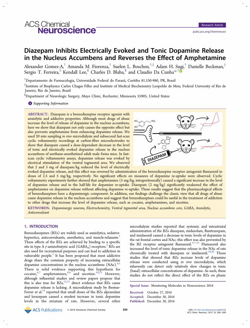
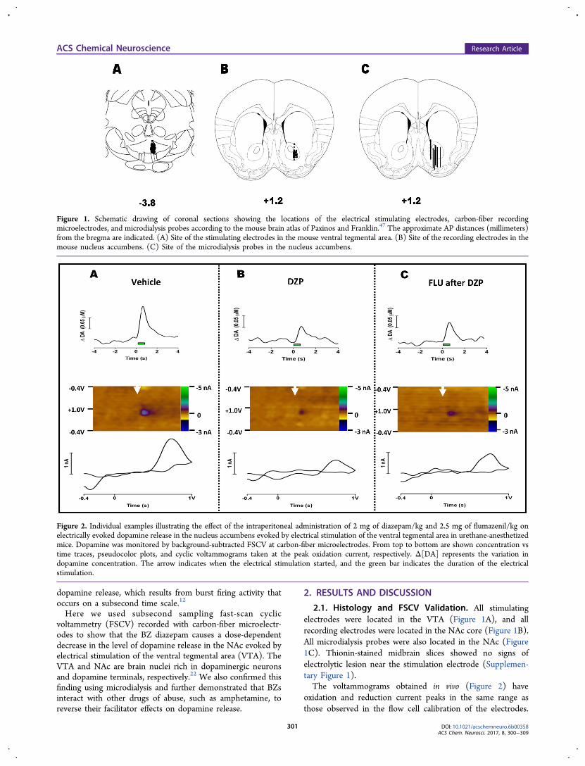
2.1. Histology and FSCV Validation. All stimulatingelectrodes were located in the VTA (Figure 1A), and allrecording electrodes were located in the NAc core (Figure 1B).All microdialysis probes were also located in the NAc (Figure1C). Thionin-stained midbrain slices showed no signs ofelectrolytic lesion near the stimulation electrode (Supplemen-tary Figure 1).The voltammograms obtained in vivo (Figure 2) have
oxidation and reduction current peaks in the same range asthose observed in the flow cell calibration of the electrodes.
Figure 1. Schematic drawing of coronal sections showing the locations of the electrical stimulating electrodes, carbon-fiber recordingmicroelectrodes, and microdialysis probes according to the mouse brain atlas of Paxinos and Franklin.47 The approximate AP distances (millimeters)from the bregma are indicated. (A) Site of the stimulating electrodes in the mouse ventral tegmental area. (B) Site of the recording electrodes in themouse nucleus accumbens. (C) Site of the microdialysis probes in the nucleus accumbens.
Figure 2. Individual examples illustrating the effect of the intraperitoneal administration of 2 mg of diazepam/kg and 2.5 mg of flumazenil/kg onelectrically evoked dopamine release in the nucleus accumbens evoked by electrical stimulation of the ventral tegmental area in urethane-anesthetizedmice. Dopamine was monitored by background-subtracted FSCV at carbon-fiber microelectrodes. From top to bottom are shown concentration vstime traces, pseudocolor plots, and cyclic voltammograms taken at the peak oxidation current, respectively. Δ[DA] represents the variation indopamine concentration. The arrow indicates when the electrical stimulation started, and the green bar indicates the duration of the electricalstimulation.
ACS Chemical Neuroscience Research Article
DOI: 10.1021/acschemneuro.6b00358ACS Chem. Neurosci. 2017, 8, 300−309
301
Cyclic voltammograms and corresponding pseudocolor plotsshow clear dopamine oxidation peaks occurring between 0.65and 0.79 V and a reduction peak between −0.20 and −0.36 V(vs the Ag/AgCl− reference electrode) with relatively lowcurrents at other potentials. On average, the oxidation of 0.5μM dopamine at an electrode with a 100 μm exposed tipcaused a current of 4.8 ± 0.4 nA. The average length of thecarbon-fiber electrodes used was 91 ± 12 μm. Backgroundnoise, defined as the variance of oxidation current measuredbetween −65 and 5 s before the electrical stimulation, was 0.04± 0.02 nA and did not vary significantly among groups[F(4,23) = 0.73; p = 0.58]. Data from 35 of 564 electricallyevoked dopamine signals were discarded because they over-lapped with obvious stimulation artifacts. The remaining datawere averaged by animal, and the composite data of all animalswere used for statistical tests.As shown in Supplementary Figure 2, intraperitoneal (ip)
administration of the dopamine transport inhibitor (DAT)blocker nomifensine (20 mg/kg) did not change the potentialsat which dopamine oxidizes or reduces (Supplementary Figure2A,C). In addition, nomifensine administration caused asignificant time-dependent increase in extracellular dopamineconcentration [calculated from the height of the oxidation peak(Supplementary Figure 2D)], an increase in the decay half-life(T1/2) (Supplementary Figure 2E), and a decrease in the decayrate constant (K) (Supplementary Figure 2F). These findingssupport the use of the FSCV oxidation current as a measure ofvariation of extracellular dopamine release and re-uptake in thisstudy.22
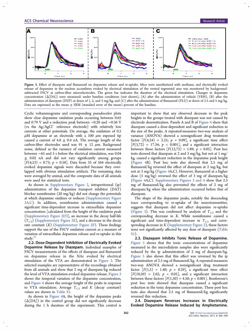
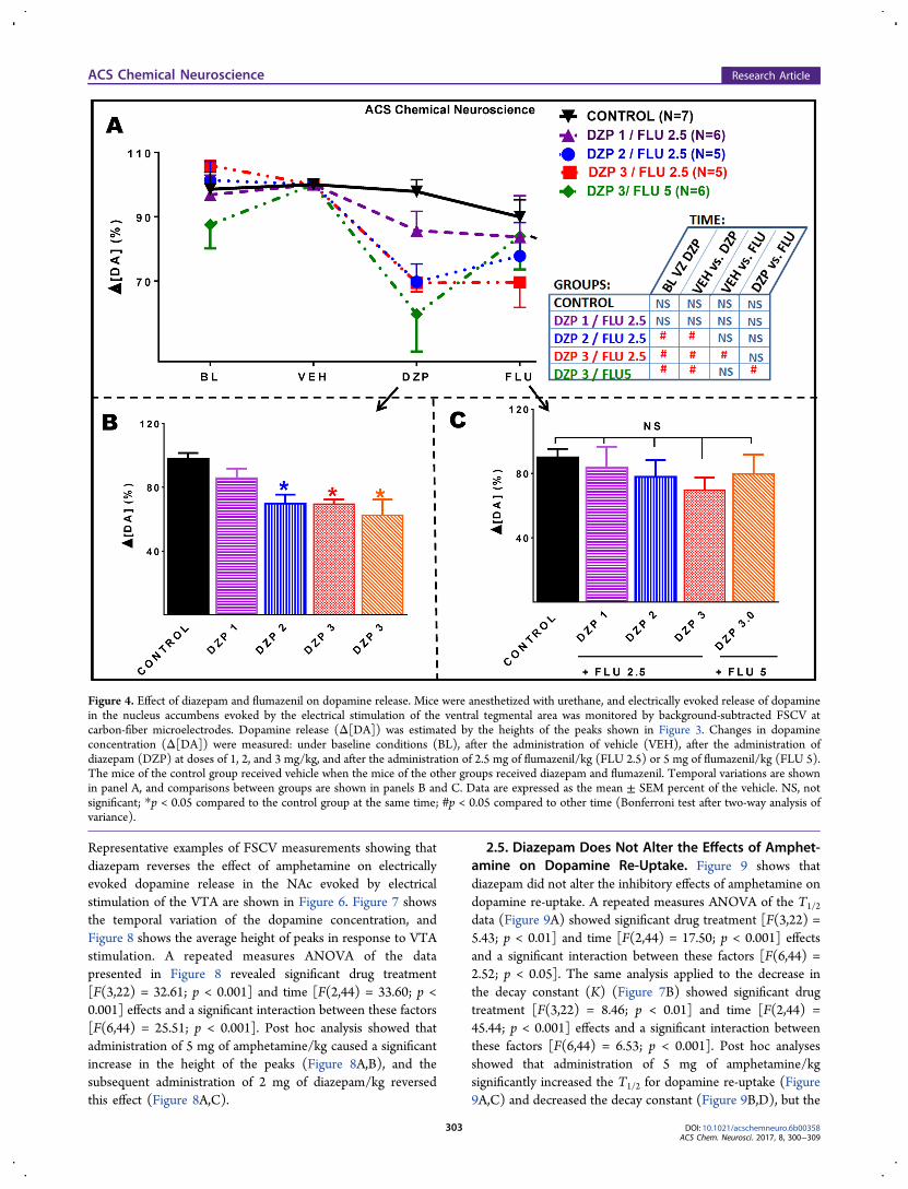
2.2. Dose-Dependent Inhibition of Electrically EvokedDopamine Release by Diazepam. Individual examples ofFSCV measurements of the effects of diazepam and flumazenilon dopamine release in the NAc evoked by electricalstimulation of the VTA are demonstrated in Figure 2. Theselected examples are representative of the recordings obtainedfrom all animals and show that 2 mg of diazepam/kg reducedthe level of VTA stimulation-evoked dopamine release. Figure 3shows the temporal variation of the dopamine concentration,and Figure 4 shows the average height of the peaks in responseto VTA stimulation. Average T1/2 and K (decay constant)values are shown in Table S1.As shown in Figure 4A, the height of the dopamine peaks
(Δ[DA]) in the control group did not significantly decreaseduring the 1 h duration of the experiment. This control is
important to show that any observed decrease in the peakheights in the groups treated with diazepam was not caused byelectrode desensitization. Panels A and B of Figure 4 show thatdiazepam caused a dose-dependent and significant reduction inthe size of the peaks. A repeated-measures two-way analysis ofvariance (ANOVA) showed a nonsignificant drug treatmentfactor [F(4,24) = 2.25; p = 0.09], a significant time effect[F(3,72) = 17.34; p < 0.001], and a significant interactionbetween these factors [F(12,72) = 1.89; p < 0.05]. Post hoctests showed that diazepam at 2 and 3 mg/kg, but not at 1 mg/kg, caused a significant reduction in the dopamine peak height(Figure 4B). Post hoc tests also showed that 2.5 mg offlumazenil/kg reversed the effect of diazepam at 2 mg/kg, butnot at 3 mg/kg (Figure 4A,C). However, flumazenil at a higherdose (5 mg/kg) reversed the effect of 3 mg of diazepam/kg(Figure 4A,C). Supplementary Figures 3 and 4 show that 2.5mg of flumazenil/kg also prevented the effects of 2 mg ofdiazepam/kg when the administration occurred before that ofdiazepam.The shape of the dopamine peaks, notably the descending
trace corresponding to re-uptake of the neurotransmitter,suggests that diazepam did not affect dopamine re-uptake(Figure 3). This was confirmed by analysis of T1/2 and thecorresponding decrease in K. While nomifensine caused asignificant and time-dependent increase in T1/2 and corre-sponding decrease in K (Supplementary Figure 2), these factorswere not significantly affected by any dose of diazepam (TableS1).
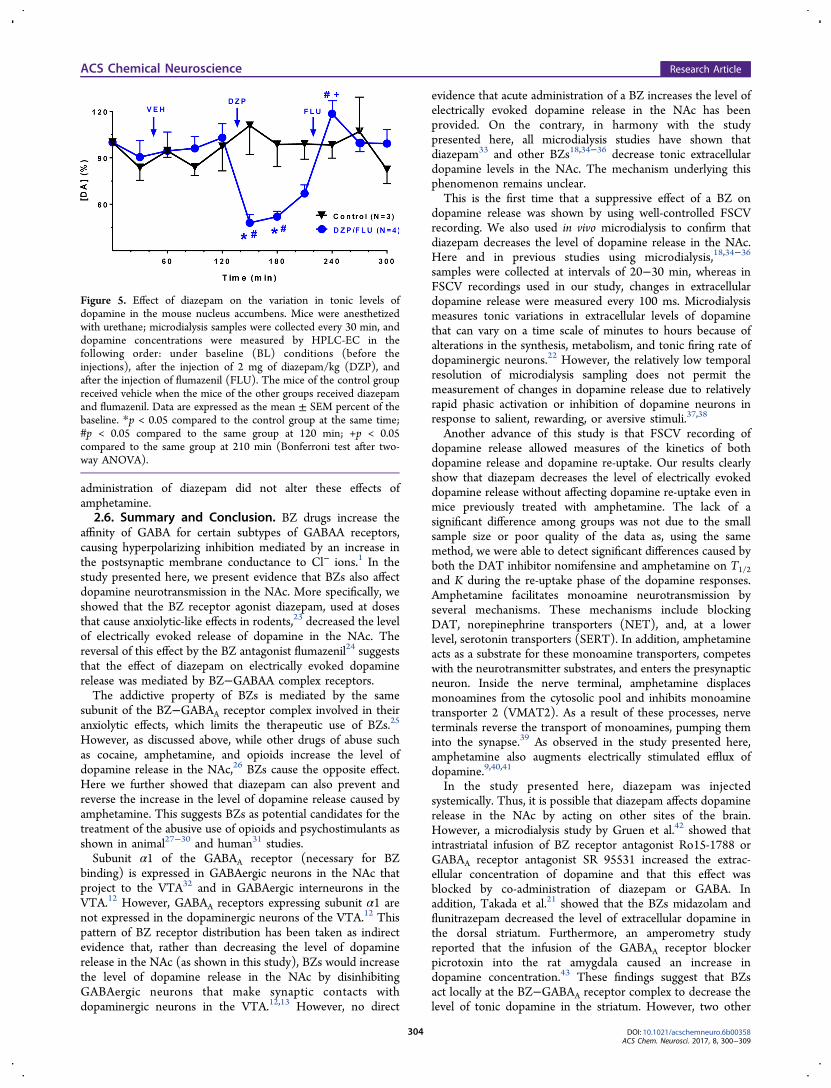
2.3. Diazepam Inhibits Tonic Release of Dopamine.Figure 5 shows that the tonic concentrations of dopaminemeasured in the microdialysis samples also were significantlyreduced by the ip administration of 2 mg of diazepam/kg.Figure 5 also shows that this effect was reversed by the ipadministration of 2.5 mg of flumazenil/kg. A repeated-measurestwo-way ANOVA showed a nonsignificant drug treatmentfactor [F(1,5) = 1.40; p = 0.29], a significant time effect[F(10,50) = 2.62; p < 0.05], and a significant interactionbetween these factors [F(1,50) = 4.50; p < 0.001]. Bonferroni’spost hoc tests showed that diazepam caused a significantreduction in the tonic dopamine concentration. These post hoctests also showed that 2.5 mg of flumazenil/kg significantlyreversed this reduction.
2.4. Diazepam Reverses Increases in ElectricallyEvoked Dopamine Release Induced by Amphetamine.
Figure 3. Effect of diazepam and flumazenil on dopamine release and re-uptake. Mice were anesthetized with urethane, and electrically evokedrelease of dopamine in the nucleus accumbens evoked by electrical stimulation of the ventral tegmental area was monitored by background-subtracted FSCV at carbon-fiber microelectrodes. The green bar indicates the duration of the electrical stimulation. Changes in dopamineconcentration (Δ[DA]) were measured: under baseline conditions (not shown), (A) after the administration of vehicle (VEH), (B) after theadministration of diazepam (DZP) at doses of 1, 2, and 3 mg/kg, and (C) after the administration of flumazenil (FLU) at doses of 2.5 and 5 mg/kg.Data are expressed as the mean ± SEM (standard error of the mean) percent of the baseline.
ACS Chemical Neuroscience Research Article
DOI: 10.1021/acschemneuro.6b00358ACS Chem. Neurosci. 2017, 8, 300−309
302
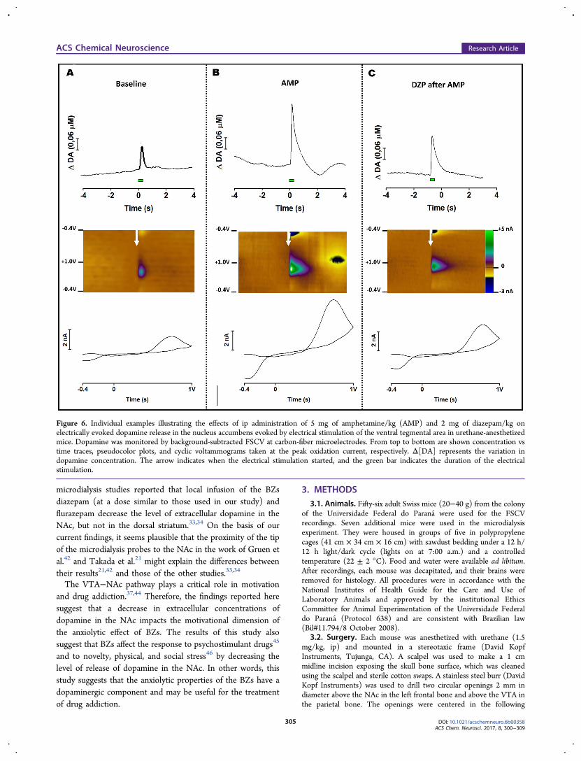
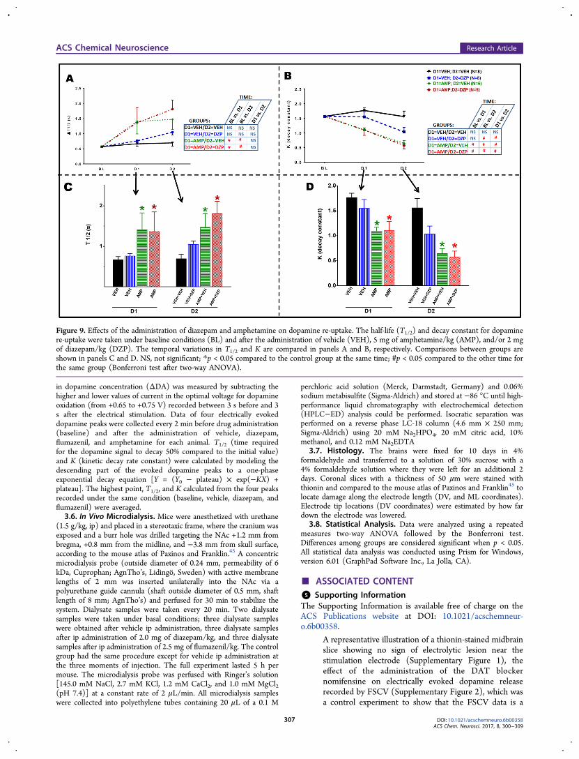
Representative examples of FSCV measurements showing thatdiazepam reverses the effect of amphetamine on electricallyevoked dopamine release in the NAc evoked by electricalstimulation of the VTA are shown in Figure 6. Figure 7 showsthe temporal variation of the dopamine concentration, andFigure 8 shows the average height of peaks in response to VTAstimulation. A repeated measures ANOVA of the datapresented in Figure 8 revealed significant drug treatment[F(3,22) = 32.61; p < 0.001] and time [F(2,44) = 33.60; p <0.001] effects and a significant interaction between these factors[F(6,44) = 25.51; p < 0.001]. Post hoc analysis showed thatadministration of 5 mg of amphetamine/kg caused a significantincrease in the height of the peaks (Figure 8A,B), and thesubsequent administration of 2 mg of diazepam/kg reversedthis effect (Figure 8A,C).
2.5. Diazepam Does Not Alter the Effects of Amphet-amine on Dopamine Re-Uptake. Figure 9 shows thatdiazepam did not alter the inhibitory effects of amphetamine ondopamine re-uptake. A repeated measures ANOVA of the T1/2
data (Figure 9A) showed significant drug treatment [F(3,22) =5.43; p < 0.01] and time [F(2,44) = 17.50; p < 0.001] effectsand a significant interaction between these factors [F(6,44) =2.52; p < 0.05]. The same analysis applied to the decrease inthe decay constant (K) (Figure 7B) showed significant drugtreatment [F(3,22) = 8.46; p < 0.01] and time [F(2,44) =45.44; p < 0.001] effects and a significant interaction betweenthese factors [F(6,44) = 6.53; p < 0.001]. Post hoc analysesshowed that administration of 5 mg of amphetamine/kgsignificantly increased the T1/2 for dopamine re-uptake (Figure9A,C) and decreased the decay constant (Figure 9B,D), but the
Figure 4. Effect of diazepam and flumazenil on dopamine release. Mice were anesthetized with urethane, and electrically evoked release of dopaminein the nucleus accumbens evoked by the electrical stimulation of the ventral tegmental area was monitored by background-subtracted FSCV atcarbon-fiber microelectrodes. Dopamine release (Δ[DA]) was estimated by the heights of the peaks shown in Figure 3. Changes in dopamineconcentration (Δ[DA]) were measured: under baseline conditions (BL), after the administration of vehicle (VEH), after the administration ofdiazepam (DZP) at doses of 1, 2, and 3 mg/kg, and after the administration of 2.5 mg of flumazenil/kg (FLU 2.5) or 5 mg of flumazenil/kg (FLU 5).The mice of the control group received vehicle when the mice of the other groups received diazepam and flumazenil. Temporal variations are shownin panel A, and comparisons between groups are shown in panels B and C. Data are expressed as the mean ± SEM percent of the vehicle. NS, notsignificant; *p < 0.05 compared to the control group at the same time; #p < 0.05 compared to other time (Bonferroni test after two-way analysis ofvariance).
ACS Chemical Neuroscience Research Article
DOI: 10.1021/acschemneuro.6b00358ACS Chem. Neurosci. 2017, 8, 300−309
303
administration of diazepam did not alter these effects ofamphetamine.2.6. Summary and Conclusion. BZ drugs increase the
affinity of GABA for certain subtypes of GABAA receptors,causing hyperpolarizing inhibition mediated by an increase inthe postsynaptic membrane conductance to Cl− ions.1 In thestudy presented here, we present evidence that BZs also affectdopamine neurotransmission in the NAc. More specifically, weshowed that the BZ receptor agonist diazepam, used at dosesthat cause anxiolytic-like effects in rodents,23 decreased the levelof electrically evoked release of dopamine in the NAc. Thereversal of this effect by the BZ antagonist flumazenil24 suggeststhat the effect of diazepam on electrically evoked dopaminerelease was mediated by BZ−GABAA complex receptors.The addictive property of BZs is mediated by the same
subunit of the BZ−GABAA receptor complex involved in theiranxiolytic effects, which limits the therapeutic use of BZs.25
However, as discussed above, while other drugs of abuse suchas cocaine, amphetamine, and opioids increase the level ofdopamine release in the NAc,26 BZs cause the opposite effect.Here we further showed that diazepam can also prevent andreverse the increase in the level of dopamine release caused byamphetamine. This suggests BZs as potential candidates for thetreatment of the abusive use of opioids and psychostimulants asshown in animal27−30 and human31 studies.Subunit α1 of the GABAA receptor (necessary for BZ
binding) is expressed in GABAergic neurons in the NAc thatproject to the VTA32 and in GABAergic interneurons in theVTA.12 However, GABAA receptors expressing subunit α1 arenot expressed in the dopaminergic neurons of the VTA.12 Thispattern of BZ receptor distribution has been taken as indirectevidence that, rather than decreasing the level of dopaminerelease in the NAc (as shown in this study), BZs would increasethe level of dopamine release in the NAc by disinhibitingGABAergic neurons that make synaptic contacts withdopaminergic neurons in the VTA.12,13 However, no direct
evidence that acute administration of a BZ increases the level ofelectrically evoked dopamine release in the NAc has beenprovided. On the contrary, in harmony with the studypresented here, all microdialysis studies have shown thatdiazepam33 and other BZs18,34−36 decrease tonic extracellulardopamine levels in the NAc. The mechanism underlying thisphenomenon remains unclear.This is the first time that a suppressive effect of a BZ on
dopamine release was shown by using well-controlled FSCVrecording. We also used in vivo microdialysis to confirm thatdiazepam decreases the level of dopamine release in the NAc.Here and in previous studies using microdialysis,18,34−36
samples were collected at intervals of 20−30 min, whereas inFSCV recordings used in our study, changes in extracellulardopamine release were measured every 100 ms. Microdialysismeasures tonic variations in extracellular levels of dopaminethat can vary on a time scale of minutes to hours because ofalterations in the synthesis, metabolism, and tonic firing rate ofdopaminergic neurons.22 However, the relatively low temporalresolution of microdialysis sampling does not permit themeasurement of changes in dopamine release due to relativelyrapid phasic activation or inhibition of dopamine neurons inresponse to salient, rewarding, or aversive stimuli.37,38
Another advance of this study is that FSCV recording ofdopamine release allowed measures of the kinetics of bothdopamine release and dopamine re-uptake. Our results clearlyshow that diazepam decreases the level of electrically evokeddopamine release without affecting dopamine re-uptake even inmice previously treated with amphetamine. The lack of asignificant difference among groups was not due to the smallsample size or poor quality of the data as, using the samemethod, we were able to detect significant differences caused byboth the DAT inhibitor nomifensine and amphetamine on T1/2and K during the re-uptake phase of the dopamine responses.Amphetamine facilitates monoamine neurotransmission byseveral mechanisms. These mechanisms include blockingDAT, norepinephrine transporters (NET), and, at a lowerlevel, serotonin transporters (SERT). In addition, amphetamineacts as a substrate for these monoamine transporters, competeswith the neurotransmitter substrates, and enters the presynapticneuron. Inside the nerve terminal, amphetamine displacesmonoamines from the cytosolic pool and inhibits monoaminetransporter 2 (VMAT2). As a result of these processes, nerveterminals reverse the transport of monoamines, pumping theminto the synapse.39 As observed in the study presented here,amphetamine also augments electrically stimulated efflux ofdopamine.9,40,41
In the study presented here, diazepam was injectedsystemically. Thus, it is possible that diazepam affects dopaminerelease in the NAc by acting on other sites of the brain.However, a microdialysis study by Gruen et al.42 showed thatintrastriatal infusion of BZ receptor antagonist Ro15-1788 orGABAA receptor antagonist SR 95531 increased the extrac-ellular concentration of dopamine and that this effect wasblocked by co-administration of diazepam or GABA. Inaddition, Takada et al.21 showed that the BZs midazolam andflunitrazepam decreased the level of extracellular dopamine inthe dorsal striatum. Furthermore, an amperometry studyreported that the infusion of the GABAA receptor blockerpicrotoxin into the rat amygdala caused an increase indopamine concentration.43 These findings suggest that BZsact locally at the BZ−GABAA receptor complex to decrease thelevel of tonic dopamine in the striatum. However, two other
Figure 5. Effect of diazepam on the variation in tonic levels ofdopamine in the mouse nucleus accumbens. Mice were anesthetizedwith urethane; microdialysis samples were collected every 30 min, anddopamine concentrations were measured by HPLC-EC in thefollowing order: under baseline (BL) conditions (before theinjections), after the injection of 2 mg of diazepam/kg (DZP), andafter the injection of flumazenil (FLU). The mice of the control groupreceived vehicle when the mice of the other groups received diazepamand flumazenil. Data are expressed as the mean ± SEM percent of thebaseline. *p < 0.05 compared to the control group at the same time;#p < 0.05 compared to the same group at 120 min; +p < 0.05compared to the same group at 210 min (Bonferroni test after two-way ANOVA).
ACS Chemical Neuroscience Research Article
DOI: 10.1021/acschemneuro.6b00358ACS Chem. Neurosci. 2017, 8, 300−309
304
microdialysis studies reported that local infusion of the BZsdiazepam (at a dose similar to those used in our study) andflurazepam decrease the level of extracellular dopamine in theNAc, but not in the dorsal striatum.33,34 On the basis of ourcurrent findings, it seems plausible that the proximity of the tipof the microdialysis probes to the NAc in the work of Gruen etal.42 and Takada et al.21 might explain the differences betweentheir results21,42 and those of the other studies.33,34
The VTA−NAc pathway plays a critical role in motivationand drug addiction.37,44 Therefore, the findings reported heresuggest that a decrease in extracellular concentrations ofdopamine in the NAc impacts the motivational dimension ofthe anxiolytic effect of BZs. The results of this study alsosuggest that BZs affect the response to psychostimulant drugs45
and to novelty, physical, and social stress46 by decreasing thelevel of release of dopamine in the NAc. In other words, thisstudy suggests that the anxiolytic properties of the BZs have adopaminergic component and may be useful for the treatmentof drug addiction.
3. METHODS3.1. Animals. Fifty-six adult Swiss mice (20−40 g) from the colony
of the Universidade Federal do Parana were used for the FSCVrecordings. Seven additional mice were used in the microdialysisexperiment. They were housed in groups of five in polypropylenecages (41 cm × 34 cm × 16 cm) with sawdust bedding under a 12 h/12 h light/dark cycle (lights on at 7:00 a.m.) and a controlledtemperature (22 ± 2 °C). Food and water were available ad libitum.After recordings, each mouse was decapitated, and their brains wereremoved for histology. All procedures were in accordance with theNational Institutes of Health Guide for the Care and Use ofLaboratory Animals and approved by the institutional EthicsCommittee for Animal Experimentation of the Universidade Federaldo Parana (Protocol 638) and are consistent with Brazilian law(Bil#11.794/8 October 2008).
3.2. Surgery. Each mouse was anesthetized with urethane (1.5mg/kg, ip) and mounted in a stereotaxic frame (David KopfInstruments, Tujunga, CA). A scalpel was used to make a 1 cmmidline incision exposing the skull bone surface, which was cleanedusing the scalpel and sterile cotton swaps. A stainless steel burr (DavidKopf Instruments) was used to drill two circular openings 2 mm indiameter above the NAc in the left frontal bone and above the VTA inthe parietal bone. The openings were centered in the following
Figure 6. Individual examples illustrating the effects of ip administration of 5 mg of amphetamine/kg (AMP) and 2 mg of diazepam/kg onelectrically evoked dopamine release in the nucleus accumbens evoked by electrical stimulation of the ventral tegmental area in urethane-anesthetizedmice. Dopamine was monitored by background-subtracted FSCV at carbon-fiber microelectrodes. From top to bottom are shown concentration vstime traces, pseudocolor plots, and cyclic voltammograms taken at the peak oxidation current, respectively. Δ[DA] represents the variation indopamine concentration. The arrow indicates when the electrical stimulation started, and the green bar indicates the duration of the electricalstimulation.
ACS Chemical Neuroscience Research Article
DOI: 10.1021/acschemneuro.6b00358ACS Chem. Neurosci. 2017, 8, 300−309
305
stereotaxic coordinates, according to the atlas of Paxinos andFranklin:47 NAc, AP +1.2 mm, ML +1.2 mm; VTA, AP −3.8 mm,ML +0.2 mm. A Ag/AgCl− wire reference electrode was inserted 0.5mm into a smaller hole drilled in the right parietal bone and fixed tothe bone with dental cement.3.3. Fast-Scan Cyclic Voltammetry Recording. The stimulating
electrode was lowered into the VTA in steps of 0.1 mm until thestrongest evoked dopamine response was recorded, over the DV rangebelow dura of 3.4−4.1 mm. This procedure was repeated to optimize
the location for the recording electrode in the NAc core (3.2−4.0mm). FSCV measurements were taken with a Wireless InstantaneousNeurotransmitter Concentration Sensor (WINCS, Mayo Clinic)system and processed with WINCSware with MINCS software(version 2.10.4.0, Mayo Clinic). Every 100 ms, a triangular waveformpotential of −0.4 V to +1.0 V to −0.4 V was applied at a rate of 300 V/s to the carbon-fiber recording electrode versus the Ag/AgCl−
reference electrode. Oxidative and reductive currents were continu-ously sampled at 100000 samples/s and 944 samples/scan. The digitaloutput was filtered with a Butterworth low-pass filter (800 Hz, threepoles) and smoothed. The triangular waveform potential was appliedto the electrode for 10 min before recording commenced to conditionthe electrode. Next, trains of 20 biphasic pulses (0.5 ms per pulse, 600μA, 60 Hz) were applied to the stimulating electrode every 180 s via aprogrammable optical isolator pulse generator (MINCS, MayoInvestigational Neuromodulation Control System, Mayo Clinic).
3.4. Drug Treatments. After the electrochemical recording signalstabilized (did not decay by more than 20%/h), four trains of electricalstimulation 3 min apart were applied in the VTA under the followingconditions: baseline (before any drug administration) and 5 min afterthe administration of vehicle, the BZ receptor agonist diazepam (1, 2,or 3 mg/kg, ip), and then the BZ receptor antagonist flumazenil (2.5or 5 mg/kg, ip). These drugs were injected ip sequentially in the sameanimals, but independent groups of mice received different doses ofdiazepam. Another group of mice was submitted to the same protocolbut received drug injections in a different order: baseline, vehicle, 2.5mg of flumazenil/kg, and 2 mg of diazepam/kg. Another group of micereceived vehicle, amphetamine (AMP, 5 mg/kg), and then DPZ (2mg/kg). A control group received three injections of vehicle at thesame time that the other groups received vehicle or the other drugs. Atthe end of this procedure, the 1 mg of diazepam/kg group alsoreceived an ip injection of the dopamine transporter (DAT) inhibitornomifensine (20 mg/kg, ip), and electrically evoked dopamine releasewas monitored 5, 8, and 11 min later.
3.5. Fast-Scan Cyclic Voltammetry Data Analysis. Back-ground-subtracted cyclic voltammograms were obtained by subtractingvoltammograms collected during stimulation from those collected upto 3 s before the stimulation. Voltammetric responses were viewed aspseudocolor plots with the abscissa as the voltage, the ordinate as theacquisition time, and the current encoded in color. Temporalresponses were determined by monitoring the current at the peakoxidation potential for dopamine in successive voltammograms.Current values were converted to concentration based on calibrationcurves obtained after the experiments with the electrodes immersed indopamine solutions (0.25, 0.5, and 1.0 μM) in a flow cell. The change
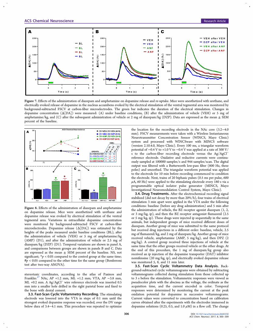
Figure 7. Effects of the administration of diazepam and amphetamine on dopamine release and re-uptake. Mice were anesthetized with urethane, andelectrically evoked release of dopamine in the nucleus accumbens evoked by the electrical stimulation of the ventral tegmental area was monitored bybackground-subtracted FSCV at carbon-fiber microelectrodes. The green bar indicates the duration of the electrical stimulation. Changes indopamine concentration (Δ[DA]) were measured: (A) under baseline conditions, (B) after the administration of vehicle (VEH) or 5 mg ofamphetamine/kg, and (C) after the subsequent administration of vehicle or 2 mg of diazepam/kg (DZP). Data are expressed as the mean ± SEMpercent of the baseline.
Figure 8. Effects of the administration of diazepam and amphetamineon dopamine release. Mice were anesthetized with urethane anddopamine release was evoked by electrical stimulation of the ventraltegmental area. Variations in extracellular dopamine concentrationwere monitored by background-subtracted FSCV at carbon-fibermicroelectrodes. Dopamine release (Δ[DA]) was estimated by theheights of the peaks measured under baseline conditions (BL), afterthe administration of vehicle (VEH) or 5 mg of amphetamine/kg(AMP) (D1), and after the administration of vehicle or 2.5 mg ofdiazepam/kg (DZP) (D1). Temporal variations are shown in panel A,and comparisons between groups are shown in panels B and C. Dataare expressed as the mean ± SEM percent of the baseline. NS, notsignificant; *p < 0.05 compared to the control group at the same time;#p < 0.05 compared to the other time for the same group (Bonferronitest after two-way ANOVA).
ACS Chemical Neuroscience Research Article
DOI: 10.1021/acschemneuro.6b00358ACS Chem. Neurosci. 2017, 8, 300−309
306
in dopamine concentration (ΔDA) was measured by subtracting thehigher and lower values of current in the optimal voltage for dopamineoxidation (from +0.65 to +0.75 V) recorded between 3 s before and 3s after the electrical stimulation. Data of four electrically evokeddopamine peaks were collected every 2 min before drug administration(baseline) and after the administration of vehicle, diazepam,flumazenil, and amphetamine for each animal. T1/2 (time requiredfor the dopamine signal to decay 50% compared to the initial value)and K (kinetic decay rate constant) were calculated by modeling thedescending part of the evoked dopamine peaks to a one-phaseexponential decay equation [Y = (Y0 − plateau) × exp(−KX) +plateau]. The highest point, T1/2, and K calculated from the four peaksrecorded under the same condition (baseline, vehicle, diazepam, andflumazenil) were averaged.3.6. In Vivo Microdialysis. Mice were anesthetized with urethane
(1.5 g/kg, ip) and placed in a stereotaxic frame, where the cranium wasexposed and a burr hole was drilled targeting the NAc +1.2 mm frombregma, +0.8 mm from the midline, and −3.8 mm from skull surface,according to the mouse atlas of Paxinos and Franklin.45 A concentricmicrodialysis probe (outside diameter of 0.24 mm, permeability of 6kDa, Cuprophan; AgnTho’s, Lidingo, Sweden) with active membranelengths of 2 mm was inserted unilaterally into the NAc via apolyurethane guide cannula (shaft outside diameter of 0.5 mm, shaftlength of 8 mm; AgnTho’s) and perfused for 30 min to stabilize thesystem. Dialysate samples were taken every 20 min. Two dialysatesamples were taken under basal conditions; three dialysate sampleswere obtained after vehicle ip administration, three dialysate samplesafter ip administration of 2.0 mg of diazepam/kg, and three dialysatesamples after ip administration of 2.5 mg of flumazenil/kg. The controlgroup had the same procedure except for vehicle ip administration atthe three moments of injection. The full experiment lasted 5 h permouse. The microdialysis probe was perfused with Ringer’s solution[145.0 mM NaCl, 2.7 mM KCl, 1.2 mM CaCl2, and 1.0 mM MgCl2(pH 7.4)] at a constant rate of 2 μL/min. All microdialysis sampleswere collected into polyethylene tubes containing 20 μL of a 0.1 M
perchloric acid solution (Merck, Darmstadt, Germany) and 0.06%sodium metabisulfite (Sigma-Aldrich) and stored at −86 °C until high-performance liquid chromatography with electrochemical detection(HPLC−ED) analysis could be performed. Isocratic separation wasperformed on a reverse phase LC-18 column (4.6 mm × 250 mm;Sigma-Aldrich) using 20 mM Na2HPO4, 20 mM citric acid, 10%methanol, and 0.12 mM Na2EDTA
3.7. Histology. The brains were fixed for 10 days in 4%formaldehyde and transferred to a solution of 30% sucrose with a4% formaldehyde solution where they were left for an additional 2days. Coronal slices with a thickness of 50 μm were stained withthionin and compared to the mouse atlas of Paxinos and Franklin45 tolocate damage along the electrode length (DV, and ML coordinates).Electrode tip locations (DV coordinates) were estimated by how fardown the electrode was lowered.
3.8. Statistical Analysis. Data were analyzed using a repeatedmeasures two-way ANOVA followed by the Bonferroni test.Differences among groups are considered significant when p < 0.05.All statistical data analysis was conducted using Prism for Windows,version 6.01 (GraphPad Software Inc., La Jolla, CA).
■ ASSOCIATED CONTENT*S Supporting InformationThe Supporting Information is available free of charge on theACS Publications website at DOI: 10.1021/acschemneur-o.6b00358.
A representative illustration of a thionin-stained midbrainslice showing no sign of electrolytic lesion near thestimulation electrode (Supplementary Figure 1), theeffect of the administration of the DAT blockernomifensine on electrically evoked dopamine releaserecorded by FSCV (Supplementary Figure 2), which wasa control experiment to show that the FSCV data is a
Figure 9. Effects of the administration of diazepam and amphetamine on dopamine re-uptake. The half-life (T1/2) and decay constant for dopaminere-uptake were taken under baseline conditions (BL) and after the administration of vehicle (VEH), 5 mg of amphetamine/kg (AMP), and/or 2 mgof diazepam/kg (DZP). The temporal variations in T1/2 and K are compared in panels A and B, respectively. Comparisons between groups areshown in panels C and D. NS, not significant; *p < 0.05 compared to the control group at the same time; #p < 0.05 compared to the other time forthe same group (Bonferroni test after two-way ANOVA).
ACS Chemical Neuroscience Research Article
DOI: 10.1021/acschemneuro.6b00358ACS Chem. Neurosci. 2017, 8, 300−309
307
reliable and selective measure of dopamine, data showingthat the administration of flumazenil before diazepamprevents the depressive effects of diazepam on dopaminerelease (Supplementary Figures 3 and 4), and data thatshow that diazepam does not affect dopamine re-uptake(Table S1) (PDF)
■ AUTHOR INFORMATIONCorresponding Author*E-mail: [email protected] Da Cunha: 0000-0001-6920-8561Author ContributionsA.G.-A and A.M.F. contributed equally to this work. A.G.-Aperformed the FSCV experiments reported in the first versionof the manuscript. A.M.F. performed the FSCV experimentsreported in the second and third versions of the manuscript.S.L.B. performed most microdialysis experiments. D.B. andS.T.F. performed the HPLC−ED analysis of the microdialysissamples. A.H.S. wrote Matlab algoritms and ran the FSCV dataanalysis. K.L. and C.D.B. provided equipment and technicalscientific support for the FSCV experiments. C.D.C. proposedthe rationale and supervised the experiments. All co-authorsmade significant contributions to the discussion of the data andwriting of the manuscript.FundingA.G.-A, A.M.F., S.L.B., A.H.S., D.B., S.T.F., and C.D.C. weresupported by CAPES, CNPq, FAPERJ, and FINEP.NotesThe authors declare no competing financial interest.
■ ACKNOWLEDGMENTSWe gratefully acknowledge the scientific consultation withDonita L. Robinson (NC), the expert contribution on FSCVregisters, data analysis and electrode preparation by Julie Esaki(UFPR), and artistic drawing of the graphical table by Ariel M.Da Cunha.
■ REFERENCES(1) Rudolph, U., and Knoflach, F. (2011) Beyond classicalbenzodiazepines: novel therapeutic potential of GABA(A) receptorsubtypes. Nat. Rev. Drug Discovery 10, 685−697.(2) Nordstrom, A. L., Farde, L., Wiesel, F. A., Forslund, K., Pauli, S.,Halldin, C., and Uppfeldt, G. (1993) Central D2-dopamine receptoroccupancy in relation to antipsychotic drug effects - a double-blind petstudy of schizophrenic-patients. Biol. Psychiatry 33, 227−235.(3) O’Brien, C. P. (2005) Benzodiazepine use, abuse, anddependence. J. Clin. Psychiatry 66, 28−33.(4) Koob, G. F., and Volkow, N. D. (2016) Neurobiology ofaddiction: A neurocircuitry analysis. Lancet Psychiat. 3, 760−773.(5) Nestler, E. J. (2005) Is there a common molecular pathway foraddiction? Nat. Neurosci. 8, 1445−1449.(6) Hernandez, L., and Hoebel, B. G. (1988) Food reward andcocaine increase extracellular dopamine in the nucleus accumbens asmeasured by microdialysis. Life Sci. 42, 1705−1712.(7) Heien, M., Khan, A. S., Ariansen, J. L., Cheer, J. F., Phillips, P. E.M., Wassum, K. M., and Wightman, R. M. (2005) Real-timemeasurement of dopamine fluctuations after cocaine in the brain ofbehaving rats. Proc. Natl. Acad. Sci. U. S. A. 102, 10023−10028.(8) Kahlig, K. M., Binda, F., Khoshbouei, H., Blakely, R. D.,McMahon, D. G., Javitch, J. A., and Galli, A. (2005) Amphetamineinduces dopamine efflux through a dopamine transporter channel.Proc. Natl. Acad. Sci. U. S. A. 102, 3495−3500.
(9) Jones, S. R., Gainetdinov, R. R., Wightman, R. M., and Caron, M.G. (1998) Mechanisms of amphetamine action revealed in micelacking the dopamine transporter. J. Neurosci. 18, 1979−1986.(10) Rice, M. E., and Cragg, S. J. (2004) Nicotine amplifies reward-related dopamine signals in striatum. Nat. Neurosci. 7, 583−584.(11) Zhang, T., Zhang, L., Liang, Y., Siapas, A. G., Zhou, F. M., andDani, J. A. (2009) Dopamine signaling differences in the nucleusaccumbens and dorsal striatum exploited by nicotine. J. Neurosci. 29,4035−4043.(12) Tan, K. R., Brown, M., Labouebe, G., Yvon, C., Creton, C.,Fritschy, J. M., Rudolph, U., and Luscher, C. (2010) Neural bases foraddictive properties of benzodiazepines. Nature 463, 769−U78.(13) Creed, M. C., Ntamati, N. R., and Tan, K. R. (2014) VTAGABA neurons modulate specific learning behaviors through thecontrol of dopamine and cholinergic systems. Front. Behav. Neurosci. 8,8.(14) Bentue-Ferrer, D., Reymann, J. M., Tribut, O., Allain, H., Vasar,E., and Bourin, M. (2001) Role of dopaminergic and serotonergicsystems on behavioral stimulatory effects of low-dose alprazolam andlorazepam. Eur. Neuropsychopharmacol. 11, 41−50.(15) Finlay, J. M., Zigmond, M. J., and Abercrombie, E. D. (1995)Increased dopamine and norepinephrine release in medial prefrontalcortex induced by acute and chronic stress - effects of diazepam.Neuroscience 64, 619−628.(16) Dazzi, L., Motzo, C., Imperato, A., Serra, M., Gessa, G. L., andBiggio, G. (1995) Modulation of basal and stress-induced release ofacetylcholine and dopamine in rat-brain by abecarnil and imidazenil, 2anxioselective gamma-aminobutyric acid(a) receptor modulators. J.Pharmacol. Exp. Ther. 273, 241−247.(17) Hegarty, A. A., and Vogel, W. H. (1995) The effect of acute andchronic diazepam treatment on stress-induced changes in corticaldopamine in the rat. Pharmacol., Biochem. Behav. 52, 771−778.(18) Murai, T., Koshikawa, N., Kanayama, T., Takada, K., Tomiyama,K., and Kobayashi, M. (1994) Local-administration of flurazepam hasdifferent effects on dopamine release in striatum and nucleusaccumbens - a microdialysis study. Eur. J. Pharmacol. 261, 65−71.(19) Motzo, C., Porceddu, M. L., Dazzi, L., Sanna, A., Serra, M., andBiggio, G. (1997) Enhancement by flumazenil of dopamine release inthe nucleus accumbens of rats repeatedly exposed to diazepam orimidazenil. Psychopharmacol. 131, 34−39.(20) Rada, P., and Hoebel, B. G. (2005) Acetylcholine in theaccumbens is decreased by diazepam and increased by benzodiazepinewithdrawal: A possible mechanism for dependency. Eur. J. Pharmacol.508, 131−138.(21) Takada, K., Murai, T., Kanayama, T., and Koshikawa, N. (1993)Effects of midazolam and flunitrazepam on the release of dopaminefrom rat striatum measured by in vivo microdialysis. Br. J. Anaesth. 70,181−185.(22) Bucher, E. S., and Wightman, R. M. (2015) Electrochemicalanalysis of neurotransmitters. Annu. Rev. Anal. Chem. 8, 239−261.(23) Leggio, G. M., Torrisi, S. A., Castorina, A., Platania, C. B. M.,Impellizzeri, A. A. R., Fidilio, A., Caraci, F., Bucolo, C., Drago, F., andSalomone, S. (2015) Dopamine D3 receptor-dependent changes inalpha6 GABAA subunit expression in striatum modulate anxiety-likebehaviour: Responsiveness and tolerance to diazepam. Eur. Neuro-psychopharmacol. 25, 1427−1436.(24) Brogden, R. N., and Goa, K. L. (1988) Flumazenil - apreliminary review of its benzodiazepine antagonist properties,intrinsic activity and therapeutic use. Drugs 35, 448−467.(25) Engin, E., Bakhurin, K. I., Smith, K. S., Hines, R. M., Reynolds,L. M., Tang, W. N., Sprengel, R., Moss, S. J., and Rudolph, U. (2014)Neural basis of benzodiazepine reward: Requirement for alpha 2containing GABA(A) receptors in the nucleus accumbens. Neuro-psychopharmacology 39, 1805−1815.(26) Koob, G. F., and Volkow, N. D. (2010) Neurocircuitry ofaddiction. Neuropsychopharmacology 35, 217−238.(27) Meririnne, E., Kankaanpaa, A., Lillsunde, P., and Seppala, T.(1999) The effects of diazepam and zolpidem on cocaine- and
ACS Chemical Neuroscience Research Article
DOI: 10.1021/acschemneuro.6b00358ACS Chem. Neurosci. 2017, 8, 300−309
308
amphetamine-induced place preference. Pharmacol., Biochem. Behav.62, 159−164.(28) Straub, C. J., Carlezon, W. A., Jr., and Rudolph, U. (2010)Diazepam and cocaine potentiate brain stimulation reward in C57BL/6J mice. Behav. Brain Res. 206, 17−20.(29) Woo, S. H., and Kim, H. S. (2001) Inhibition of diazepam onmorphine-induced hyperactivity, reverse tolerance and postsynapticdopamine receptor supersensitivity. Pharmacol. Res. 44, 467−472.(30) Suzuki, T., Tsuda, M., Funada, M., and Misawa, M. (1995)Blockade of morphine-induced place preference by diazepam in mice.Eur. J. Pharmacol. 280, 327−330.(31) Rush, C. R., Stoops, W. W., Wagner, F. P., Hays, L. R., andGlaser, P. E. A. (2004) Alprazolam attenuates the behavioral effects ofD-amphetamine in humans. J. Clin. Psychopharmacol. 24, 410−420.(32) Shibasaki, M., Masukawa, D., Ishii, K., Yamagishi, Y., Mori, T.,and Suzuki, T. (2013) Involvement of the K+ -Cl- co-transporterKCC2 in the sensitization to morphine-induced hyperlocomotionunder chronic treatment with zolpidem in the mesolimbic system. J.Neurochem. 125, 747−755.(33) Invernizzi, R., Pozzi, L., and Samanin, R. (1991) Release ofdopamine is reduced by diazepam more in the nucleus-accumbensthan in the caudate-nucleus of conscious rats. Neuropharmacology 30,575−578.(34) Zetterstrom, T., and Fillenz, M. (1990) Local-administration offlurazepam has different effects on dopamine release in striatum andnucleus accumbens - a microdialysis study. Neuropharmacology 29,129−134.(35) Finlay, J. M., Damsma, G., and Fibiger, H. C. (1992)Benzodiazepine-induced decreases in extracellular concentrations ofdopamine in the nucleus-accumbens after acute and repeatedadministration. Psychopharmacol. 106, 202−208.(36) Yoshida, Y., Koide, S., Hirose, N., Takada, K., Saigusa, T., andKoshikawa, N. (1999) In vivo microdialysis evidence that midazolamfacilitates propofol-induced reduction in rat accumbal dopaminerelease. Neurosci. Res. Commun. 25, 121−127.(37) Da Cunha, C., Gomez-A, A., and Blaha, C. D. (2012) The roleof the basal ganglia in motivated behavior. Rev. Neurosci. 23, 747−767.(38) Da Cunha, C., Boschen, S. L., Gomez-A, A., Ross, E. K., Gibson,W. S. J., Min, H. K., Lee, K. H., and Blaha, C. D. (2015) Towardsophisticated basal ganglia neuromodulation: Review on basal gangliadeep brain stimulation. Neurosci. Biobehav. Rev. 58, 186−210.(39) Robertson, S. D., Matthies, H. J. G., and Galli, A. (2009) ACloser Look at Amphetamine-Induced Reverse Transport andTrafficking of the Dopamine and Norepinephrine Transporters. Mol.Neurobiol. 39, 73−80.(40) Easton, N., Steward, C., Marshall, F., Fone, K., and Marsden, C.(2007) Effects of amphetamine isomers, methylphenidate andatomoxetine on synaptosomal and synaptic vesicle accumulation andrelease of dopamine and noradrenaline in vitro in the rat brain.Neuropharmacology 52, 405−414.(41) Fordahl, S. C., Locke, J. L., and Jones, S. R. (2016) High fat dietaugments amphetamine sensitization in mice: Role of feeding pattern,obesity, and dopamine terminal changes. Neuropharmacology 109,170−182.(42) Gruen, R. J., Friedhoff, A. J., Coale, A., and Moghaddam, B.(1992) Tonic inhibition of striatal dopamine transmission - effects ofbenzodiazepine and GABA-A receptor antagonists on extracellulardopamine levels. Brain Res. 599, 51−56.(43) Chu, T. F., and Lin, M. T. (1996) Picrotoxin induces bothhypertension and dopamine release in the rat amygdala. Neurosci. Lett.218, 169−172.(44) Ikemoto, S., Yang, C., and Tan, A. (2015) Basal ganglia circuitloops, dopamine and motivation: A review and enquiry. Behav. BrainRes. 290, 17−31.(45) Yang, X. M., Gorman, A. L., Dunn, A. J., and Goeders, N. E.(1992) Anxiogenic effects of acute and chronic cocaine administration- neurochemical and behavioral-studies. Pharmacol., Biochem. Behav.41, 643−650.
(46) Schneier, F. R., Liebowitz, M. R., Abi-Dargham, A., Zea-Ponce,Y., Lin, S. H., and Laruelle, M. (2000) Low dopamine D-2 receptorbinding potential in social phobia. Am. J. Psychiatry 157, 457−459.(47) Paxinos, G., and Franklin, K. B. J. (2005) The Mouse Brain inStereotaxic Coordinates, Compact 2nd ed., Academic Press, San Diego.
ACS Chemical Neuroscience Research Article
DOI: 10.1021/acschemneuro.6b00358ACS Chem. Neurosci. 2017, 8, 300−309
309