Embed Size (px)
Citation preview
J. exp. Biol. (1975), 62, 43-53 4 3^Lth 4 figures
rrinted in Great Britain
THE LENGTH-TENSION RELATIONSHIP OF THE DORSALLONGITUDINAL MUSCLE OF A LEECH
BY JENNIFER B. MILLER
School of Biological Sciences, University of East Anglia,Norwich NOR 88C
(Received 17 May 1974)
SUMMARY
The length-tension relationship of a preparation of the dorsal body wallof the leech Haemopis sanguisuga was determined.
Passive tension is low except at very long lengths of the preparation, whenit rises steeply. It is due mainly to the epidermis present in the preparation.
The active tension curve is very flat, with tension being reduced only atvery short and very long lengths. This shape is explained in the context of themyofilament arrangement of the muscle fibres. It may be that thin filamentscan form cross-bridges with different thick filaments at different lengths ofthe preparation.
INTRODUCTION
The somatic muscles of annelids are of the oblique-striated or helical smooth type.There has been very little work on the mechanical properties of leech muscle, althoughit has been used for many years as a biological assay for acetylcholine solutions (see,for example, Macintosh & Perry, 1950). There have been more experiments on themechanical properties of earthworm muscle, but there is only one description of thelength-tension relationship (Hidaka, Kuriyama & Yamamoto, 1969). The followingaccount forms part of an investigation into the mechanical properties of the dorsallongitudinal muscle of the horse leech Haemopis sanguisuga (L.).
METHODS
Specimens of Haemopis sanguisuga were kept in aerated tanks of dechlorinated tap-water at 10-12 °C. The dorsal longitudinal muscle was prepared for experimentationby making an incision along the whole length of the animal on each side. Then, withthe animal pinned out dorsal side uppermost, the dorsal wall was separated from thegut and connective tissue to provide a strip of the dorsal wall approximately 5 cmlong by 0-5 cm wide. The strip was tied with cotton at each end and a small fish-hook,attached to a length of fine chain, was passed through the tissue and behind the cottonat each end of the preparation. The chains were used to mount the preparation on avertical Perspex electrode assembly, with the lower end fixed and the upper endattached to a Devices Type 2STO2 strain gauge. The output from the strain gauge,which was proportional to tension, was amplified and displayed on a Devices M2hot-wire pen-recorder.
44 JENNIFER B. MILLER
The muscle was stimulated through an array of alternately positive and negati^silver wires with 4 mm spacing, using a Grass S4 stimulator. The output from thestimulator was fed through an emitter-follower circuit so that Ringer solution oflow resistance did not reduce the stimulus voltage. Between stimuli the muscle wasimmersed in Ringer solution of the following composition (mM): NaCl 113-0; KC14-3; CaCl2 2-0; NaHC03i-6; Na2HPO4i-o; the pH being adjusted to 7-4 with 10%HC1 (modified from Pantin, 1946). The experiments were carried out at room tempera-ture (20-24 °C).
It has been shown that the leech dorsal longitudinal muscle responds with differenttypes of contraction to different types of electrical stimulation, namely direct currentand pulsed stimuli (Miller & Aidley, 1973). The problem of fatigue of this muscletends to be somewhat greater with d.c. stimulation, and a full experiment to investi-gate the length-tension relationship involved a long period of experimentation and alarge number of stimuli. Therefore, only pulsed stimuli were used in this investiga-tion. A preliminary experiment had indicated that there were no parameters in theresponses to pulsed and d.c. stimuli, such as rates of contraction and relaxation, whichwere affected differently by changes in length of the preparation. The pulsed stimuliconsisted of a train of 5 msec square-wave pulses at a frequency of 50/sec for \ sec,which is just sufficient for maximum tension to be reached. In all the experiments acheck was made on possible fatigue by performing the readings in a cycle, enablingthe first few points to be repeated at the end of the experiment. No significant fatigueoccurred in any of the experiments.
For most preparations the total length change was between 2-5 and 3-5 cm. Thelength change between each set of readings was generally 4 mm. The starting lengthfor most experiments was in the middle region of the range, but some experimentswere started at very short or very long lengths. Measurements of the passive and activetensions were made at different lengths, having both increased and decreased thelength of the preparation, so that the readings were made in a complete cycle, some-times increasing the length first, and sometimes beginning by decreasing the length.Three contractions were made at each length.
The change in length of the muscle preparation was made at a steady rate (about1 mm/sec) with the muscle immersed in Ringer solution to reduce any friction againstthe electrode. The immediate change in tension on altering the muscle length wasalways greater than the final tension change. Therefore after the initial tension increasecaused by stretching the muscle, tension fell gradually to the new steady level. Thisphenomenon is known as stress relaxation. Conversely, on shortening the muscle thetension fell suddenly, but some tension was redeveloped extremely slowly, up to thefinal passive level characteristic of the muscle length.
THE STRUCTURE OF THE PREPARATION AND THELONGITUDINAL MUSCLE
Any interpretation of the shape of the length-tension diagram must take intoaccount the structure of the preparation itself, and the arrangement of the myo-filaments in the longitudinal muscle which forms the principal constituent of thepreparation. Therefore brief descriptions of both are included here.
Dorsal longitudinal muscle of a leech 45
The preparation
The preparation used in these experiments has already been described in detail(Miller & Aidley, 1973). It consists of part of the dorsal body wall of the leech, andtherefore contains tissues other than the longitudinal muscles being investigated.The epidermis, the connective tissue and the spongy inner layer of botryoidal tissueare all passive tissues, which make no contribution to the active development oftension, though they will certainly affect the passive tension. There are three layersof muscle cells, namely circular, longitudinal and oblique muscle. The circularmuscle, being perpendicular to the direction of tension measurement, will not affectthe tension developed by the longitudinal muscles, but the small number of obliquelyorientated muscle fibres will do so. It has been estimated (Miller & Aidley, 1973) thattheir contribution does not exceed 10% of the maximum tension developed by thelongitudinal muscle fibres, and will generally be much less than this. However theircontribution, though small, will depend on the length of the preparation. The anglethey make with the longitudinal axis of the preparation (i.e. the direction of tensionmeasurement) will decrease with increasing length of the preparation. Thus theircontribution, being equal to the cosine of this angle, will increase. Attempts weremade to measure accurately the angle of the oblique fibres by phase contrast andpolarized light microscopy, but this was found to be almost impossible because of thepresence of the botryoidal tissue, which was very difficult to remove cleanly andcompletely. It was quite impractical to attempt this for each of the preparations usedin the length-tension experiments. The angle is estimated to be of the order of10-250, depending on the length of the preparation.
The longitudinal muscle
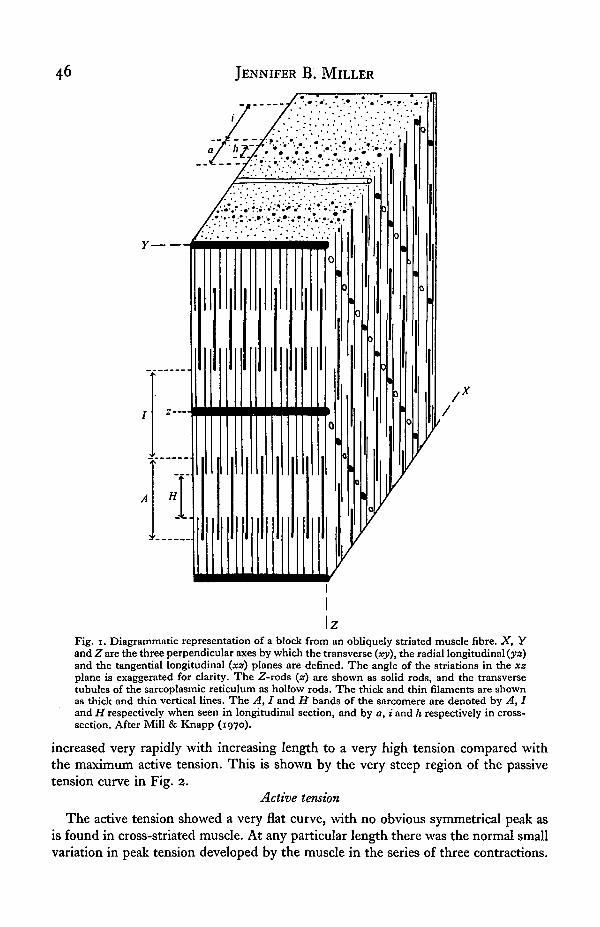
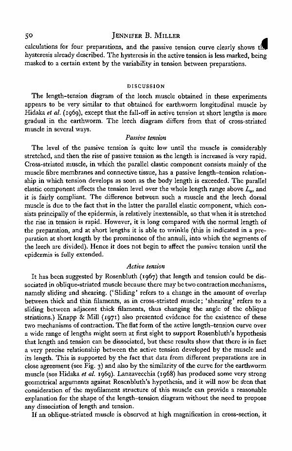
As already explained, the muscles of the leech are of the oblique-striated type. Themuscle cell has the form of a long tapering cylinder, with the contractile materialforming a peripheral cortex. Mill & Knapp (1970) have described the myofilamentarrangement in earthworm muscle, and the picture appears to be very similar forleech muscle. Fig. 1 shows a very simplified diagram of the myofilament arrangementin a block of oblique-striated muscle, based on their description. A short electron-microscope study of the longitudinal muscle cells of Haemopis sanguisuga (see Miller,1974) has indicated that the arrangement in the horse leech is basically the same as itis in earthworm muscles and in the muscles of other leeches (Rohlich, 1962; Pucci& Afzelius, 1962).
RESULTS
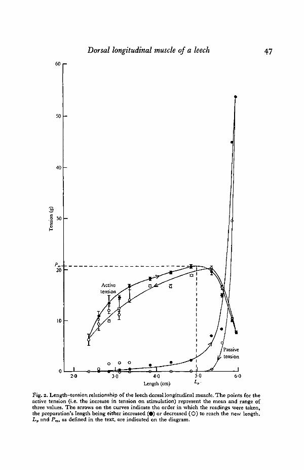
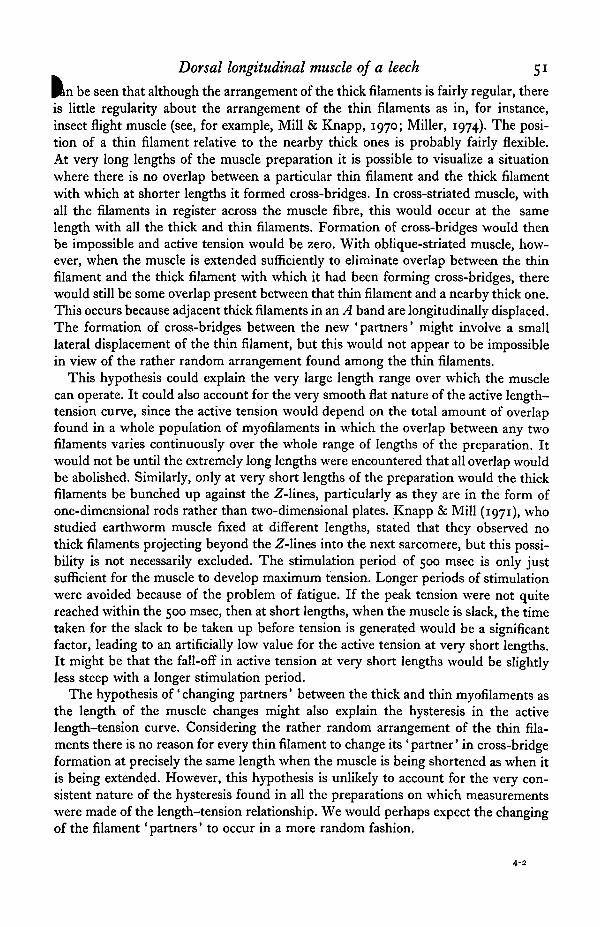
All the preparations showed the same type of length-tension relationship, andFig. 2 shows the results of a typical experiment.
Passive tension
Over most of the length range there was a very low level of passive tension, andthis was affected little by the length of the preparation, increasing only slightly as the
.length was increased. However, at long lengths of the preparation the passive tension
JENNIFER B. MILLER
Fig. i. Diagrammatic representation of a block from an obliquely striated muscle fibre. X, Yand Z are the three perpendicular axes by which the transverse (xy), the radial longitudinal (ya)and the tangential longitudinal (xz) planes are defined. The angle of the striations in the xzplane is exaggerated for clarity. The Z-rods (z) are shown as solid rods, and the transversetubules of the sarcoplasmic reticulum as hollow rods. The thick and thin filaments are shownas thick and thin vertical lines. The A, I and H bands of the sarcomere are denoted by A, Iand H respectively when seen in longitudinal section, and by a, i and h respectively in cross-section. After Mill & Knapp (1970).
increased very rapidly with increasing length to a very high tension compared withthe maximum active tension. This is shown by the very steep region of the passivetension curve in Fig. 2.
Active tension
The active tension showed a very flat curve, with no obvious symmetrical peak asis found in cross-striated muscle. At any particular length there was the normal smallvariation in peak tension developed by the muscle in the series of three contractions.
Dorsal longitudinal muscle of a leech 4760 i—
20 40Length (cm)
Fig. 2. Length-tension relationship of the leech dorsal longitudinal muscle. The points for theactive tension (i.e. the increase in tension on stimulation) represent the mean and range ofthree values. The arrows on the curves indicate the order in which the readings were taken,the preparation's length being either increased (•) or decreased (O) to reach the new length.Lv and Pm, as defined in the text, are indicated on the diagram.
48 JENNIFER B. MILLER
This is indicated in Fig. 2 by a vertical bar showing the range of values covered fl|the three points. Over the range of lengths in which the passive tension was small andfairly constant, the active tension increased very gradually with increasing length inan almost linear fashion. The peak of the curve was at approximately that length atwhich the steep passive tension increase started to occur, and then the fall-off inactive tension was very marked as the length of the preparation was increased. Atthis stage the muscle was very severely stretched, being probably 2-2 | times thelength at which the minimum readings were taken. There was also a rapid declinein active tension at very short lengths. This decline occurred at such short lengthsthat the muscle was quite slack.
Hysteresis
Hysteresis occurred in both the passive and active tension curves. If at any stageduring stretching the length of the preparation was reduced, the passive tensiondropped far more than would be predicted from the curve of ascending readings.This hysteresis could not be explained by any damage to the muscle caused by thesevere stretch, since if the cycle were repeated the ascending readings followed theupper curve and the descending readings the lower one. Because of the very highpassive tensions encountered at long muscle lengths, each preparation was carefullyexamined at the end of the experiment to see whether there was any damage tothe tissue at the point of insertion of the fish-hook. None was found.
For the active tension, the ascending readings were higher than the descendingreadings over the range of lengths covered by the more or less linear region of thecurve. At longer lengths, where the fall-off in active tension occurred, the greateractive tension was developed when the muscle was shortened prior to the readings.There was, therefore, a cross-over in the two curves at the length at which the peakactive tension occurred. The curve for the descending readings resembled that for theascending readings, but displaced to the right.
Comparison between different preparations
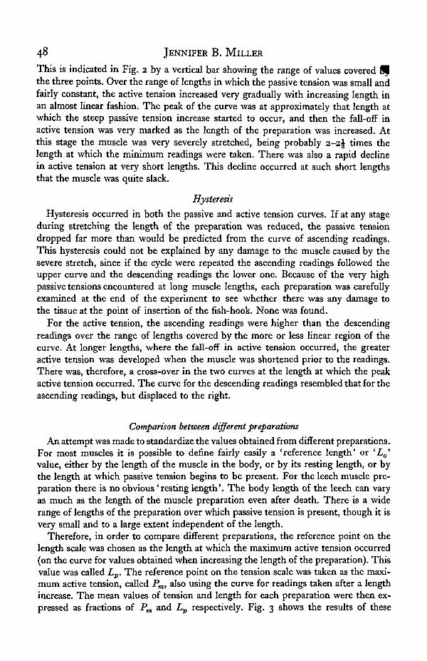
An attempt was made to standardize the values obtained from different preparations.For most muscles it is possible to define fairly easily a 'reference length* or 'Lo'value, either by the length of the muscle in the body, or by its resting length, or bythe length at which passive tension begins to be present. For the leech muscle pre-paration there is no obvious 'resting length'. The body length of the leech can varyas much as the length of the muscle preparation even after death. There is a widerange of lengths of the preparation over which passive tension is present, though it isvery small and to a large extent independent of the length.
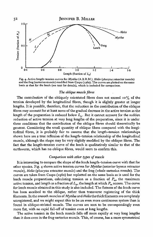
Therefore, in order to compare different preparations, the reference point on thelength scale was chosen as the length at which the maximum active tension occurred(on the curve for values obtained when increasing the length of the preparation). Thisvalue was called Lp. The reference point on the tension scale was taken as the maxi-mum active tension, called Pm, also using the curve for readings taken after a lengthincrease. The mean values of tension and length for each preparation were then ex-pressed as fractions of Pm and Lp respectively. Fig. 3 shows the results of these
Dorsal longitudinal muscle of a leech 493-5 r-
30
2-5
a? 20
1-5
10
OS
AAO A
• • •
o-°-lA Q.** .° "l,0-4 0-6 0-8 10
Length (fraction of Lp)
1-2 1-4
Fig. 3. Length-tension relationship of the leech dorsal longitudinal muscle, using data fromfour preparations. The tension is expressed as a fraction of Pm, the maximum active isometrictension for each preparation, and the length as a fraction of £„, the length at which Pm occurs.Open symbols indicate that the length was reduced prior to the reading, and filled symbolsthat the length was increased. Each point for the active tension (triangles) represents the meanof three values. Circles represent the passive tension.
4 EXB 62
So JENNIFER B. M I L L E R
calculations for four preparations, and the passive tension curve clearly shows tillhysteresis already described. The hysteresis in the active tension is less marked, beingmasked to a certain extent by the variability in tension between preparations.
DISCUSSION
The length-tension diagram of the leech muscle obtained in these experimentsappears to be very similar to that obtained for earthworm longitudinal muscle byHidaka et al. (1969), except that the fall-off in active tension at short lengths is moregradual in the earthworm. The leech diagram differs from that of cross-striatedmuscle in several ways.
Passive tension
The level of the passive tension is quite low until the muscle is considerablystretched, and then the rise of passive tension as the length is increased is very rapid.Cross-striated muscle, in which the parallel elastic component consists mainly of themuscle fibre membranes and connective tissue, has a passive length-tension relation-ship in which tension develops as soon as the body length is exceeded. The parallelelastic component affects the tension level over the whole length range above Lo, andit is fairly compliant. The difference between such a muscle and the leech dorsalmuscle is due to the fact that in the latter the parallel elastic component, which con-sists principally of the epidermis, is relatively inextensible, so that when it is stretchedthe rise in tension is rapid. However, it is long compared with the normal length ofthe preparation, and at short lengths it is able to wrinkle (this is indicated in a pre-paration at short length by the prominence of the annuli, into which the segments ofthe leech are divided). Hence it does not begin to affect the passive tension until theepidermis is fully extended.
Active tension
It has been suggested by Rosenbluth (1967) that length and tension could be dis-sociated in oblique-striated muscle because there may be two contraction mechanisms,namely sliding and shearing. ('Sliding' refers to a change in the amount of overlapbetween thick and thin filaments, as in cross-striated muscle; 'shearing' refers to asliding between adjacent thick filaments, thus changing the angle of the obliquestriations.) Knapp & Mill (1971) also presented evidence for the existence of thesetwo mechanisms of contraction. The fiat form of the active length-tension curve overa wide range of lengths might seem at first sight to support Rosenbluth's hypothesisthat length and tension can be dissociated, but these results show that there is in facta very precise relationship between the active tension developed by the muscle andits length. This is supported by the fact that data from different preparations are inclose agreement (see Fig. 3) and also by the similarity of the curve for the earthwormmuscle (see Hidaka et al. 1969). Lanzavecchia (1968) has produced some very stronggeometrical arguments against Rosenbluth's hypothesis, and it will now be s*een thatconsideration of the myofilament structure of this muscle can provide a reasonableexplanation for the shape of the length-tension diagram without the need to proposeany dissociation of length and tension.
If an oblique-striated muscle is observed at high magnification in cross-section, it
Dorsal longitudinal muscle of a leech 51
Pin be seen that although the arrangement of the thick filaments is fairly regular, thereis little regularity about the arrangement of the thin filaments as in, for instance,insect flight muscle (see, for example, Mill & Knapp, 1970; Miller, 1974). The posi-tion of a thin filament relative to the nearby thick ones is probably fairly flexible.At very long lengths of the muscle preparation it is possible to visualize a situationwhere there is no overlap between a particular thin filament and the thick filamentwith which at shorter lengths it formed cross-bridges. In cross-striated muscle, withall the filaments in register across the muscle fibre, this would occur at the samelength with all the thick and thin filaments. Formation of cross-bridges would thenbe impossible and active tension would be zero. With oblique-striated muscle, how-ever, when the muscle is extended sufficiently to eliminate overlap between the thinfilament and the thick filament with which it had been forming cross-bridges, therewould still be some overlap present between that thin filament and a nearby thick one.This occurs because adjacent thick filaments in an A band are longitudinally displaced.The formation of cross-bridges between the new 'partners' might involve a smalllateral displacement of the thin filament, but this would not appear to be impossiblein view of the rather random arrangement found among the thin filaments.
This hypothesis could explain the very large length range over which the musclecan operate. It could also account for the very smooth flat nature of the active length-tension curve, since the active tension would depend on the total amount of overlapfound in a whole population of myofilaments in which the overlap between any twofilaments varies continuously over the whole range of lengths of the preparation. Itwould not be until the extremely long lengths were encountered that all overlap wouldbe abolished. Similarly, only at very short lengths of the preparation would the thickfilaments be bunched up against the Z-lines, particularly as they are in the form ofone-dimensional rods rather than two-dimensional plates. Knapp & Mill (1971), whostudied earthworm muscle fixed at different lengths, stated that they observed nothick filaments projecting beyond the Z-lines into the next sarcomere, but this possi-bility is not necessarily excluded. The stimulation period of 500 msec is only justsufficient for the muscle to develop maximum tension. Longer periods of stimulationwere avoided because of the problem of fatigue. If the peak tension were not quitereached within the 500 msec, then at short lengths, when the muscle is slack, the timetaken for the slack to be taken up before tension is generated would be a significantfactor, leading to an artificially low value for the active tension at very short lengths.It might be that the fall-off in active tension at very short lengths would be slightlyless steep with a longer stimulation period.
The hypothesis of' changing partners' between the thick and thin myofilaments asthe length of the muscle changes might also explain the hysteresis in the activelength-tension curve. Considering the rather random arrangement of the thin fila-ments there is no reason for every thin filament to change its ' partner' in cross-bridgeformation at precisely the same length when the muscle is being shortened as when itis being extended. However, this hypothesis is unlikely to account for the very con-sistent nature of the hysteresis found in all the preparations on which measurementswere made of the length-tension relationship. We would perhaps expect the changingof the filament 'partners' to occur in a more random fashion.
4-2
JENNIFER B. MILLER
10 r
oeo
0-5
o
•"•Helix
Mylilus
04 0-6 0-8 10 1-2Length (fraction of Lp)
14 1-6
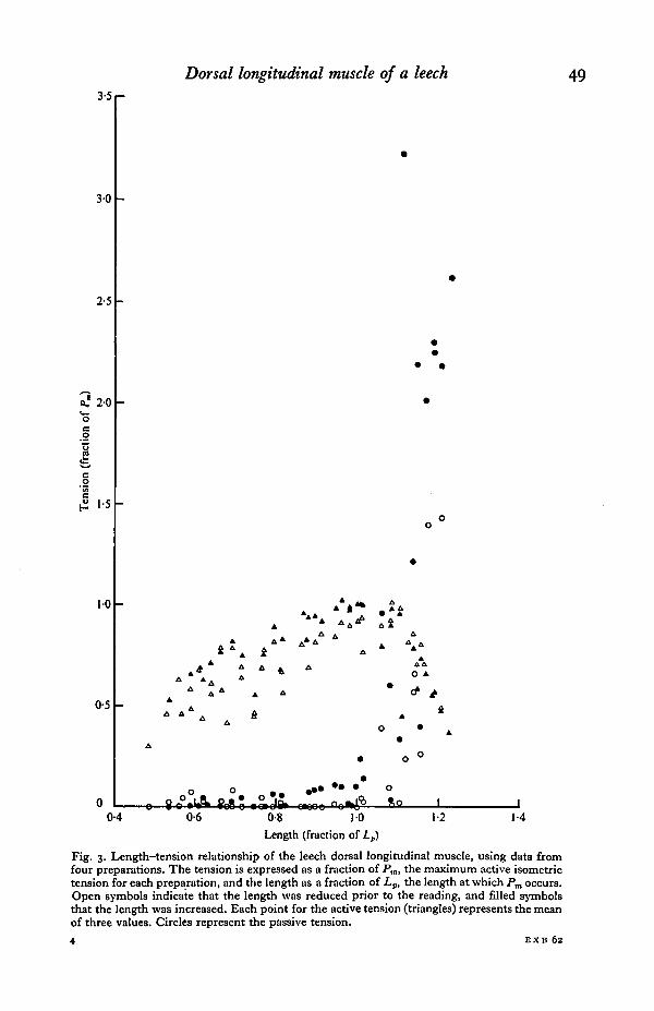
Fig. 4. Active length-tension curves for Mytilus (A.B.R.M.), Helix (pharynx retractor muscle)and the frog (sartorius muscle) modified from Csapo (i960). The curves are plotted on the samebasis as that for the leech (see text for details), which is included for comparison.
The oblique muscle fibres
The contribution of the obliquely orientated fibres does not exceed 10% of thetension developed by the longitudinal fibres, though it is slightly greater at longerlengths. It is possible, therefore, that the reduction in the contribution of the obliquefibres may account for at least some of the gradual decrease in the active tension as thelength of the preparation is reduced below Lp. But it cannot account for the suddenreduction of active tension at very long lengths of the preparation, since it is underthese conditions that the contribution of the oblique fibres should theoretically begreatest. Considering the small quantity of oblique fibres compared with the longi-tudinal fibres, it is probably fair to assume that the length-tension relationshipsshown here are a true reflexion of the length-tension relationship of the longitudinalmuscle, although the shape may be very slightly modified by the oblique fibres. Thefact that the length-tension curve of the leech is qualitatively similar to that of theearthworm, which has no oblique fibres, would seem to confirm this.
Comparison with other types of muscle
It is interesting to compare the shape of the leech length-tension curve with that forother species. Fig. 4 shows active tension curves for Mytilus (anterior byssus retractormuscle), Helix (pharynx retractor muscle) and the frog (whole sartorius muscle). Thecurves are taken from Csapo (i960) but replotted on the same basis as is used for theleech muscle preparations, calculating tension as a fraction of Pm, the maximumactive tension, and length as a fraction of Lp, the length at which Pm occurs. The curvefor leech muscle obtained in this study is also included. The flatness of the leech curvehas been ascribed to the oblique, rather than transverse registering of the thickfilaments. In the smooth muscles of Mytilus and Helix the thick filaments are completelyunregistered, and we might expect this to be an even more continuous system than isfound in oblique-striated muscle. The curves are seen to be correspondingly evenmore flat, with no rapid fall-off of tension even at long lengths.
The active tension in the leech muscle falls off more rapidly at very long lengthsthan it does even in the frog sartorius muscle. This, of course, has a more symmetrical

Dorsal longitudinal muscle of a leech 53
^ and a more prominent peak than the leech. Theoretically the fall-off of tensionat long lengths occurs when no overlap remains between the thick and thin filaments.It is difficult to see how this could occur more rapidly in a population of oblique-striated muscle fibres than in a population of cross-striated muscle fibres, in which thethick filaments are fully in register.
A satisfactory explanation of the basic features of both the active and passive length-tension curves of the leech muscle can thus be given by considering the structureof the preparation and of the longitudinal muscle itself. It is not necessary to postulatethat there are two contraction mechanisms, or that length and tension can be dis-sociated. Rather, we are dealing with a muscle in which tension can be developed overa wide range of lengths because of the particular structure of the muscle fibres.
I wish to thank my supervisor, Dr D. J. Aidley, for his advice and encouragement,and Dr G. Shelton for his constructive criticism of the manuscript. During the courseof this work I was in receipt of a research studentship from the Science ResearchCouncil.
REFERENCES
CSAPO, A. (i960). In Structure and Function of Muscle, vol. i (ed. G. Bourne). New York: AcademicPress.
HlDAKA, T., KURIYAMA, H. & YAMAMOTO, T. (1969). The mechanical properties of the longitudinalmuscle in the earthworm. J. exp. Biol. 50, 431-43.
KNAPP, M. F. & MILL, P. J. (1971). The contractile mechanism in obliquely striated body wall muscleof the earthworm, Lumbricus terrestris. J. Cell Set. 8, 413-25.
LANZAVECCHIA, G. (1968). Studi sulla muscolatura elicoidale e paramiosinica. II. Meccanismo di contra-zione dei muscoli elicoidali. Atti Accad. naz. Lincei Re. 44, 575-83.
MACINTOSH, F. C. & PERRY, W. L. M. (1950). Biological estimation of acetylcholine. In Methods inMedical Research, 3, 78-92 (ed. R. W. Gerard). Chicago: Yearbook Publishers Inc.
MILL, P. J. & KNAPP, M. F. (1970). The fine structure of obliquely striated body wall muscles in theearthworm, Lumbricus terrestris Linn. J. Cell Sci. 7, 233-61.
MILLER, J. B. (1974). Mechanical Properties of a Leech Muscle. Ph.D. Thesis, University of East Anglia.MILLER, J. B. & AIDLEY, D. J. (1973). Two rates of relaxation in the dorsal longitudinal muscle of a
leech. J. exp. Biol. 58, 91-103.PANTIN, C. F. A. (1946). Notes on Microscopical Technique for Zoologists. Cambridge University Press.Pucci, I. & AFZELIUS, B. A. (1962). An electron microscope study of sarcotubules and related structures
in the leech muscle. J. Ultrastruct. Res. 7, 210-24.R6HLICH,P. (1962). The fine structure of the muscle fiber of the leech Hirudo medicinalis. J. Ultrastruct.
Res. 7, 399-408.ROSENBLUTH, J. (1967). Obliquely striated muscle. III. Contraction mechanism of Ascaris body muscle.
J. Cell Biol. 34, 15-33.


![[45 ] THE QUANTITATIVE NUTRITIONAL …jeb.biologists.org/content/jexbio/33/1/45.full.pdf · Quantitative nutritional requirements of Drosophila melanogaster 47 spores, and the fluctuations](https://img.pdfslide.us/doc/110x75/5ac1f6ec7f8b9a4e7c8db233/45-the-quantitative-nutritional-jeb-nutritional-requirements-of-drosophila.jpg)