Embed Size (px)
Citation preview
2535
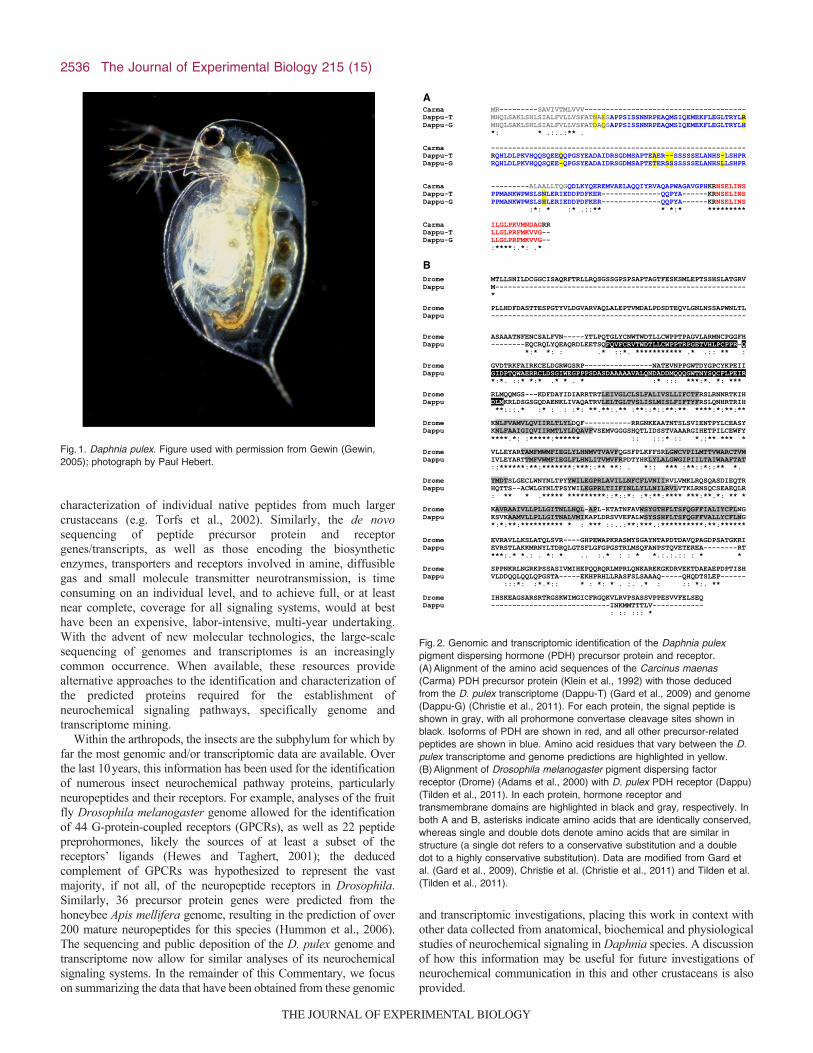
IntroductionOver its geographic range, the cladoceran crustacean Daphniapulex (Fig.1) serves as a keystone species for many freshwaterecosystems, being both the primary consumer of phytoplankton andthe primary forage species for larger invertebrates and fish (Dodsonet al., 2010). This crustacean is highly sensitive to changes in itsenvironment, and possesses the ability to rapidly adaptmorphologically, physiologically and behaviorally to a wide rangeof environmental and anthropogenic challenges (Dodson et al.,2010). Daphnia pulex’s remarkable functional flexibility, incombination with its parthenogenetic mode of reproduction andease of laboratory culture, has resulted in it being used as one ofthe primary models for the field of environmental toxicology forover half a century (Dodson et al., 2010).
Recently, D. pulex has been the subject of extensive molecularinvestigations, including both genome and transcriptome sequencing(Colbourne et al., 2011). In fact, this species is currently the onlycrustacean for which a fully sequenced genome is publicly available;D. pulex is also one of but a few crustaceans for which a large numberof transcriptome sequences have been publicly deposited. Thesemolecular resources, in combination with the biological featuresdescribed above, have resulted in the emergence of D. pulex as amodel for the field of toxicogenomics. In addition, its phylogeneticposition, presumed to be close to the point of divergence of theinsects from the crustaceans (e.g. Meusemann et al., 2010), makes ita strategic target for genomic investigations of arthropod evolution.
A key factor that led to the emergence of D. pulex as a modelorganism for many biological fields is its ability to rapidly copewith environmental challenge via changes in its morphology,physiology and/or behavior. As in all multicellular organisms, therelative levels and/or complements of locally released paracrinesand circulating hormones are undoubtedly key players in mediatingthis biological flexibility. Although many tissues undoubtedlyparticipate in this chemical communication, the nervous system isa particularly rich source of both local and hormonal signalingagents. Interestingly, few studies have focused on theneurochemistry of D. pulex at any level. In fact, prior toapproximately 2005, essentially nothing was known aboutneurochemical signaling in this species. One factor that has surelycontributed to this lack of neurochemical knowledge is its size;adults are typically just a few millimeters in overall length. This issurely true for the identification of its neuropeptides, as the standardmethod for peptide discovery until ~2005 was to isolate individualpeptides chromatographically and/or biochemically from largepools of starting tissue and, upon their purification, assess theirstructures using a combination of proteolytic cleavage, Edmananalysis and mass spectrometry (Christie et al., 2010). Given itsminute size and the need to manually dissect a sufficient amountof starting material, the identification of even a single Daphnianeuropeptide via this strategy would surely have been daunting, ashundreds, and in some cases thousands, of central nervous systems(CNSs) were needed for the biochemical isolation and
SummaryThe cladoceran crustacean Daphnia pulex has served as a standard organism for aquatic toxicity testing for decades. The modelorganism status of D. pulex rests largely on its remarkable ability to rapidly adapt morphologically, physiologically andbehaviorally to a wide range of environmental challenges, as well as on its parthenogenetic reproduction and ease of laboratoryculture. As in all multicellular organisms, neurochemical control systems are undoubtedly major contributors to the functionalflexibility of Daphnia. Surprisingly, little work has focused on understanding its neurochemistry at any level. Recently, D. pulexhas been the subject of extensive genome and transcriptome sequencing, and it is currently the only crustacean with a fullysequenced, publicly accessible genome. Although the molecular work was initiated for gene-based investigations ofecotoxicology and toxicogenomics, the data generated have allowed for investigations into numerous aspects of Daphnia biology,including its neurochemical signaling. This Commentary summarizes our knowledge of D. pulex neurochemistry obtained fromrecent genomic and transcriptomic studies, and places these data in context with other anatomical, biochemical and physiologicalexperiments using D. pulex and its sister species Daphnia magna. Suggestions as to how the Daphnia molecular data may beuseful for future investigations of crustacean neurochemical signaling are also provided.
Key words: Daphnia pulex, Daphnia magna, Cladocera, genome, transcriptome, neurohormone, neurotransmitter, peptide, amine, small moleculetransmitter, gas transmitter.
Received 15 February 2012; Accepted 29 March 2012
The Journal of Experimental Biology 215, 2535-2544© 2012. Published by The Company of Biologists Ltddoi:10.1242/jeb.070565
COMMENTARY
From genes to behavior: investigations of neurochemical signaling come of age forthe model crustacean Daphnia pulex
Andrew E. Christie1,* and Matthew D. McCoole2
1Békésy Laboratory of Neurobiology, Pacific Biosciences Research Center, University of Hawaii at Manoa, 1993 East-West Road,Honolulu, HI 96822, USA and 2College of Pharmacy, Department of Toxicology, University of Louisiana at Monroe,
700 University Avenue, Monroe, LA 71209, USA*Author for correspondence ([email protected])
THE JOURNAL OF EXPERIMENTAL BIOLOGY
2536
characterization of individual native peptides from much largercrustaceans (e.g. Torfs et al., 2002). Similarly, the de novosequencing of peptide precursor protein and receptorgenes/transcripts, as well as those encoding the biosyntheticenzymes, transporters and receptors involved in amine, diffusiblegas and small molecule transmitter neurotransmission, is timeconsuming on an individual level, and to achieve full, or at leastnear complete, coverage for all signaling systems, would at besthave been an expensive, labor-intensive, multi-year undertaking.With the advent of new molecular technologies, the large-scalesequencing of genomes and transcriptomes is an increasinglycommon occurrence. When available, these resources providealternative approaches to the identification and characterization ofthe predicted proteins required for the establishment ofneurochemical signaling pathways, specifically genome andtranscriptome mining.
Within the arthropods, the insects are the subphylum for which byfar the most genomic and/or transcriptomic data are available. Overthe last 10years, this information has been used for the identificationof numerous insect neurochemical pathway proteins, particularlyneuropeptides and their receptors. For example, analyses of the fruitfly Drosophila melanogaster genome allowed for the identificationof 44 G-protein-coupled receptors (GPCRs), as well as 22 peptidepreprohormones, likely the sources of at least a subset of thereceptors’ ligands (Hewes and Taghert, 2001); the deducedcomplement of GPCRs was hypothesized to represent the vastmajority, if not all, of the neuropeptide receptors in Drosophila.Similarly, 36 precursor protein genes were predicted from thehoneybee Apis mellifera genome, resulting in the prediction of over200 mature neuropeptides for this species (Hummon et al., 2006).The sequencing and public deposition of the D. pulex genome andtranscriptome now allow for similar analyses of its neurochemicalsignaling systems. In the remainder of this Commentary, we focuson summarizing the data that have been obtained from these genomic
and transcriptomic investigations, placing this work in context withother data collected from anatomical, biochemical and physiologicalstudies of neurochemical signaling in Daphnia species. A discussionof how this information may be useful for future investigations ofneurochemical communication in this and other crustaceans is alsoprovided.
The Journal of Experimental Biology 215 (15)
Fig.1. Daphnia pulex. Figure used with permission from Gewin (Gewin,2005); photograph by Paul Hebert.
A
Carma MR---------SAVIVTMLVVV-------------------------------------- Dappu-T MHQLSAKLSHLSIALFVLLVSFATNAESAPPSISSNNRPEAQMSIQEMEKFLEGLTRYLR Dappu-G MHQLSAKLSHLSIALFVLLVSFATDAQSAPPSISSNNRPEAQMSIQEMEKFLEGLTRYLH *: * .:..:** . Carma ------------------------------------------------------------ Dappu-T RQHLDLPKVHQQSQEEQQPGSYEADAIDRSGDMSAPTEAER--SSSSSELANHS-LSHPR Dappu-G RQHLDLPKVHQQSQEE-QPGSYEADAIDRSGDMSAPTETERSSSSSSSELANHSLLSHPR Carma ---------ALAALLTQGQDLKYQEREMVAELAQQIYRVAQAPWAGAVGPHKRNSELINS Dappu-T PPMANKWPWSLSNLERIEDDPDFKER--------------QQPYA------KRNSELINS Dappu-G PPMANKWPWSLSHLERIEDDPDFKER--------------QQPYA------KRNSELINS :*: * :* .::** * *:* ********* Carma ILGLPKVMNDAGRR Dappu-T LLGLPRFMKVVG-- Dappu-G LLGLPRFMKVVG-- :****:.*: .*
B Drome MTLLSNILDCGGCISAQRFTRLLRQSGSSGPSPSAPTAGTFESKSMLEPTSSHSLATGRV Dappu M----------------------------------------------------------- * Drome PLLHDFDASTTESPGTYVLDGVARVAQLALEPTVMDALPDSDTEQVLGNLNSSAPWNLTL Dappu ------------------------------------------------------------ Drome ASAAATNFENCSALFVN-----YTLPQTGLYCNWTWDTLLCWPPTPAGVLARMNCPGGFH Dappu --------EQCRQLYQEAQRDLEETSQFQVFCRVTWDTLLCWPPTRPGETVHLPCPPR-Q *:* *: : .* ::*. *********** .* .:: ** : Drome GVDTRKFAIRKCELDGRWGSRP----------------NATEVNPPGWTDYGPCYKPEII Dappu GIDPTQWAERRCLDSGIWEGPPPSDASDAAAAAVALQNDADDMQQQGWTNYSQCFLPEIR *:*. ::* *:* .* * . * :* ::: ***:*. *: *** Drome RLMQQMGS---KDFDAYIDIARRTRTLEIVGLCLSLFALIVSLLIFCTFRSLRNNRTKIH Dappu DLMKRLDSGSGQDAENKLIVAQATRVLELTGLTVSLISLMISLFIFTYFRSLQNHRTRIH **:::.* :* : : :*: **.**:.** :**::*::**:** ****:*:**:** Drome KNLFVAMVLQVIIRLTLYLDQF-----------RRGNKEAATNTSLSVIENTPYLCEASY Dappu KNLFAAIGIQVIIRMTLYLDQAVFVSEMVGGGSHQTLIDSSTVAAARGIHETPILCEWFY ****.*: :*****:****** :: :::* :: *.:** *** * Drome VLLEYARTAMFMWMFIEGLYLHNMVTVAVFQGSFPLKFFSRLGWCVPILMTTVWARCTVM Dappu IVLEYARTTMFVWMFIEGLFLHNLITVMVFRPDTYHKLYLALGWGIPIILTAIWAAFTAT ::******:**:*******:***::** **: . *:: *** :**::*::** *. Drome YMDTSLGECLWNYNLTPYYWILEGPRLAVILLNFCFLVNIIRVLVMKLRQSQASDIEQTR Dappu HQTTS--ACWLGYNLTPSYWILEGPRLTIIFINLLYLLNILRVLVTKLRNSQCSEAEQLR : ** * .***** *********::*::*: :*:**:**** ***:**.*: ** * Drome KAVRAAIVLLPLLGITNLLHQL-APL-KTATNFAVWSYGTHFLTSFQGFFIALIYCFLNG Dappu KSVKAAMVLLPLLGITNALVMIKAPLDRSVVEFALWSYSSHFLTSFQGFFVALLYCFLNG *:*:**:********** * : *** ::..:**:***.:**********:**:****** Drome EVRAVLLKSLATQLSVR----GHPEWAPKRASMYSGAYNTAPDTDAVQPAGDPSATGKRI Dappu EVRSTLAKKMRNYLTDRQLGTSFLGFGPGSTRLMSQFANPSTQVETEREA--------RT ***:.* *.: . *: * .. :.* : : * *.:.:.:: : * * Drome SPPNKRLNGRKPSSASIVMIHEPQQRQRLMPRLQNKAREKGKDRVEKTDAEAEPDPTISH Dappu VLDDQQLQQLQPGSTA-----EKHPRHLLRASFSLSAAAQ-----QHQDTSLEP------ :::*: :*.*:: * : *: * . :. .* : :: *:. ** Drome IHSKEAGSARSRTRGSKWIMGICFRGQKVLRVPSASSVPPESVVFELSEQ Dappu ----------------------------INKMMTTTLV------------ : :: ::: *
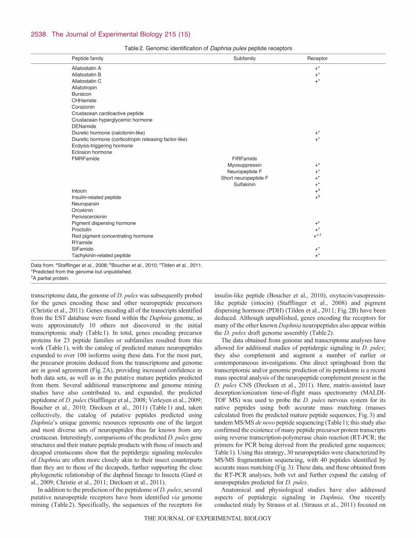
Fig.2. Genomic and transcriptomic identification of the Daphnia pulexpigment dispersing hormone (PDH) precursor protein and receptor.(A)Alignment of the amino acid sequences of the Carcinus maenas(Carma) PDH precursor protein (Klein et al., 1992) with those deducedfrom the D. pulex transcriptome (Dappu-T) (Gard et al., 2009) and genome(Dappu-G) (Christie et al., 2011). For each protein, the signal peptide isshown in gray, with all prohormone convertase cleavage sites shown inblack. Isoforms of PDH are shown in red, and all other precursor-relatedpeptides are shown in blue. Amino acid residues that vary between the D.pulex transcriptome and genome predictions are highlighted in yellow.(B)Alignment of Drosophila melanogaster pigment dispersing factorreceptor (Drome) (Adams et al., 2000) with D. pulex PDH receptor (Dappu)(Tilden et al., 2011). In each protein, hormone receptor andtransmembrane domains are highlighted in black and gray, respectively. Inboth A and B, asterisks indicate amino acids that are identically conserved,whereas single and double dots denote amino acids that are similar instructure (a single dot refers to a conservative substitution and a doubledot to a highly conservative substitution). Data are modified from Gard etal. (Gard et al., 2009), Christie et al. (Christie et al., 2011) and Tilden et al.(Tilden et al., 2011).
THE JOURNAL OF EXPERIMENTAL BIOLOGY

2537Daphnia pulex neurochemistry
Daphnia genomic resourcesThe genome of D. pulex is a unique resource for crustaceanbiology. The current draft genome for D. pulex (Colbourne et al.,2011) was assembled from over 1.5 million high-quality readsobtained from DNA collected from the naturally inbred clonalline ‘The Chosen One’. The size of this genome assembly isroughly 200megabases, which is relatively small in comparisonto the genomes thus far sequenced for other species. Surprisingly,this ‘small’ genome is predicted to contain an unusually largenumber of genes, approximately 31,000, which exceeds thenumber found in most other species, including humans. Severalfactors that contribute to the ability of D. pulex to balance a smallgenome size with a large number of genes are a reduction in thelength of its introns and an elevated rate of gene duplication andmaintenance. Of the ~31,000 D. pulex genes, nearly one-thirdhave no known homologs, and are proposed as specific to theDaphnia lineage. It is hypothesized that these ‘unique’ genes haveevolved in response to D. pulex’s organism–environmentinteractions, and thus are direct contributors to this species’remarkable functional flexibility.
In addition to the sequencing of its genome, D. pulex has alsobeen the subject of extensive transcriptome analysis; at presentnearly 150,000 expressed sequence tags (ESTs) have been publiclydeposited for this species (Colbourne et al., 2011). These ESTswere developed from 37 different cDNA libraries, which representgenes expressed under a number of distinct ecological conditions,including many commonly encountered environmental variables(e.g. differing food levels and predator kairomones) and a numberof anthropogenic stressors (e.g. heavy metals and nanoparticles).
These functional genomic data have validated over a third of the31,000 predicted Daphnia genes, and in combination with othervetting methods (e.g. tiling microarrays), nearly 90% of the genespredicted from the D. pulex genome now have at least some levelof additional support.
Peptidergic systemsPrior to the public deposition of its genome and transcriptome, littlewas known about the neurochemical signaling systems of D. pulex.In fact, little was known about the molecular machinery necessaryfor the establishment and proper functioning of neurochemicalpathways in any crustacean. Although the molecules used forchemical communication are diverse in all multicellular organisms,peptides are by far the largest class used by nervous systems(Christie et al., 2010; Christie, 2011), and it is this group ofcompounds that was the first to be investigated via genome and/ortranscriptome mining in Daphnia.
The first large-scale investigation of peptidergic signaling in D.pulex focused on the characterization of its peptidome usingtranscriptome mining (Gard et al., 2009). Here, the sequences ofknown insect and crustacean neuropeptide precursor proteins wereused to query the Daphnia EST database for putative homologs; thestructures of the mature neuropeptides were subsequently predictedfrom the deduced proteins via both online software programs andhomology to known insect and crustacean peptide isoforms(Fig.2A). Using this strategy, 63 peptide-encoding ESTs wereidentified; these encompassed 14 distinct peptide families orsubfamilies (Table1). Over 70 putative mature peptides werepredicted from the proteins deduced from these ESTs. Using the
Table1. Peptide families and subfamilies identified in Daphnia pulex via genomic, transcriptomic and mass spectral analyses
Mode of identification
Peptide family Subfamily Genome Transcriptome Mass spectrometry
Allatostatin A +a,b +e +b
Allatostatin B +a,b +e +b
Allatostatin C +a,b +e
Allatotropin +a,b +b
Bursicon +a,b +e
CCHamide +b
Corazonin +a,b +b
Crustacean cardioactive peptide +a,b +e
Crustacean hyperglycemic hormone +a,b +e
DENamide +b
Diuretic hormone (calcitonin-like) +a,b +e +b
Diuretic hormone (CRF-like) +b
Ecdysis-triggering hormone +a,b +e
Eclosion hormone +a,b
FMRFamide FIRFamide +b +b
Myosuppressin +b +b
Neuropeptide F +a,b +e +b
Short neuropeptide F +a,b +e +b
Sulfakinin +a,b +b
Intocin +c
Insulin-related peptide +a,b,d +b
Neuroparsin +b
Orcokinin +a,b +e +b
Periviscerokinin +b +b
Pigment dispersing hormone +a,b +e +b
Proctolin +a,b +b
Red pigment concentrating hormone +a,b
RYamide +b +b
SIFamide +a,b +f +b
Tachykinin-related peptide +a,b +b
Data from: aChristie et al., 2011; bDircksen et al., 2011; cStafflinger et al., 2008; dBoucher et al., 2010; eGard et al., 2009; fVerleyen et al., 2009.
THE JOURNAL OF EXPERIMENTAL BIOLOGY
2538
transcriptome data, the genome of D. pulex was subsequently probedfor the genes encoding these and other neuropeptide precursors(Christie et al., 2011). Genes encoding all of the transcripts identifiedfrom the EST database were found within the Daphnia genome, aswere approximately 10 others not discovered in the initialtranscriptomic study (Table1). In total, genes encoding precursorproteins for 23 peptide families or subfamilies resulted from thiswork (Table1), with the catalog of predicted mature neuropeptidesexpanded to over 100 isoforms using these data. For the most part,the precursor proteins deduced from the transcriptome and genomeare in good agreement (Fig.2A), providing increased confidence inboth data sets, as well as in the putative mature peptides predictedfrom them. Several additional transcriptome and genome miningstudies have also contributed to, and expanded, the predictedpeptidome of D. pulex (Stafflinger et al., 2008; Verleyen et al., 2009;Boucher et al., 2010; Dircksen et al., 2011) (Table1) and, takencollectively, the catalog of putative peptides predicted usingDaphnia’s unique genomic resources represents one of the largestand most diverse sets of neuropeptides thus far known from anycrustacean. Interestingly, comparisons of the predicted D. pulex genestructures and their mature peptide products with those of insects anddecapod crustaceans show that the peptidergic signaling moleculesof Daphnia are often more closely akin to their insect counterpartsthan they are to those of the decapods, further supporting the closephylogenetic relationship of the daphnid lineage to Insecta (Gard etal., 2009; Christie et al., 2011; Dircksen et al., 2011).
In addition to the prediction of the peptidome of D. pulex, severalputative neuropeptide receptors have been identified via genomemining (Table2). Specifically, the sequences of the receptors for
insulin-like peptide (Boucher et al., 2010), oxytocin/vasopressin-like peptide (intocin) (Stafflinger et al., 2008) and pigmentdispersing hormone (PDH) (Tilden et al., 2011; Fig.2B) have beendeduced. Although unpublished, genes encoding the receptors formany of the other known Daphnia neuropeptides also appear withinthe D. pulex draft genome assembly (Table2).
The data obtained from genome and transcriptome analyses haveallowed for additional studies of peptidergic signaling in D. pulex;they also complement and augment a number of earlier orcontemporaneous investigations. One direct springboard from thetranscriptomic and/or genomic prediction of its peptidome is a recentmass spectral analysis of the neuropeptide complement present in theD. pulex CNS (Dircksen et al., 2011). Here, matrix-assisted laserdesorption/ionization time-of-flight mass spectrometry (MALDI-TOF MS) was used to probe the D. pulex nervous system for itsnative peptides using both accurate mass matching (massescalculated from the predicted mature peptide sequences; Fig.3) andtandem MS/MS de novo peptide sequencing (Table1); this study alsoconfirmed the existence of many peptide precursor protein transcriptsusing reverse transcription-polymerase chain reaction (RT-PCR; theprimers for PCR being derived from the predicted gene sequences;Table1). Using this strategy, 30 neuropeptides were characterized byMS/MS fragmentation sequencing, with 40 peptides identified byaccurate mass matching (Fig.3). These data, and those obtained fromthe RT-PCR analyses, both vet and further expand the catalog ofneuropeptides predicted for D. pulex.
Anatomical and physiological studies have also addressedaspects of peptidergic signaling in Daphnia. One recentlyconducted study by Strauss et al. (Strauss et al., 2011) focused on
The Journal of Experimental Biology 215 (15)
Table2. Genomic identification of Daphnia pulex peptide receptors
Peptide family Subfamily Receptor
Allatostatin A +*Allatostatin B +*Allatostatin C +*AllatotropinBursiconCHHamideCorazoninCrustacean cardioactive peptideCrustacean hyperglycemic hormoneDENamideDiuretic hormone (calcitonin-like) +*Diuretic hormone (corticotropin releasing factor-like) +*Ecdysis-triggering hormoneEclosion hormoneFMRFamide FIRFamide
Myosuppressin +*Neuropeptide F +*
Short neuropeptide F +*Sulfakinin +*
Intocin +a
Insulin-related peptide +b
NeuroparsinOrcokininPeriviscerokininPigment dispersing hormone +c
Proctolin +*Red pigment concentrating hormone +*,†
RYamideSIFamide +*Tachykinin-related peptide +*
Data from: aStafflinger et al., 2008; bBoucher et al., 2010; cTilden et al., 2011.*Predicted from the genome but unpublished.†A partial protein.
THE JOURNAL OF EXPERIMENTAL BIOLOGY

2539Daphnia pulex neurochemistry
assessing the role played by the neurons expressing PDH, a peptideinitially discovered in D. pulex via transcriptome and genomeanalyses (Gard et al., 2009; Christie et al., 2011), and laterconfirmed via mass spectrometry (Dircksen et al., 2011). In thisinvestigation, an antibody generated against PDH was used to mapthe distribution of this peptide in the Daphnia brain. Approximately40 neurons were labeled by the PDH antibody in D. pulex. Basedon their locations and projection patterns, 13 distinct neuron typeswere identified and reconstructed, including two types in the visualganglion that resemble the medial lateral neurons of insects,neurons that produce pigment dispersing factor (the insect homologof crustacean PDH) and that are key players in establishingcircadian rhythmicity. The PDH antibody stains an identical set ofneurons in Daphnia magna, where experiments conducted under
12h:12h light:dark cycles and under constant conditions (eitherconstant light or constant dark) showed significant changes inlabeling intensity of specific PDH immunopositive cells over thecourse of a 24h period (Strauss et al., 2011). In addition, rhythmicchanges in the activity pattern of one PDH-positive medial lateralneuron type, which showed a peak in activity in the relative‘evening’, were seen in this species. Collectively, these datastrongly suggest that PDH, and the medial lateral neurons thatproduce it, are key components of the circadian signaling systemof Daphnia. Interestingly, the Daphnia circadian system itself hasbeen a recent subject of genome mining (Tilden et al., 2011). Here,D. pulex homologs of all the major components necessary for theestablishment of a Drosophila-like clock, e.g. period, timeless,clock and cycle proteins, as well as ones absent in Drosophila, but
Fig.3. Matrix-assisted laser desorption/ionizationtime of flight (MALDI-TOF) identification ofneuropeptides in the Daphnia pulex brain.(A)Forty-one peptides were identified by directtissue profiling of a D. pulex brain via accuratemass matches; the inset shows an enlargementof the peak corresponding to the FIRFamide 2peptide. (B–D) Enlargements of the boxed areasof the mass spectrum shown in A. ASTA, A-typeallatostatin; ASTB, B-type allatostatin; AT,allatotropin; CPON, carboxyterminal peptide ofNPY; DH31, diuretic hormone 31; IRP-2, insulin-related peptide 2; MS, myosuppressin; NPF,neuropeptide F; RYa, RYamide; SIFa, SIFamide;SK, sulfakinin; sNPF, short neuropeptide F; OK,orcokinin; TKRP, tachykinin-related peptide.Figure is reprinted with permission from Dircksenet al. (Dircksen et al., 2011).
THE JOURNAL OF EXPERIMENTAL BIOLOGY

2540
present in other more ancestral insect circadian systems, e.g.cryptochrome 2, were identified.
Aminergic systemsA second class of commonly used neuronal signaling molecules isamines. In crustaceans, these include dopamine, histamine,octopamine and serotonin (Christie, 2011). As is the case for thepeptides, multiple proteins involved in each aminergic signalingpathway have been mined from the D. pulex genome using knownDrosophila proteins as references (McCoole et al., 2011; McCooleet al., 2012a). Specifically, genes encoding the biosyntheticenzymes for each amine were identified, as were genes encodingputative receptors for each of these molecules (Table3). In addition,genes encoding dopamine and serotonin transporters were found(Table3). Taken collectively, this suite of data represents the largestnumber of aminergic genes and/or proteins thus far identified fromany crustacean.
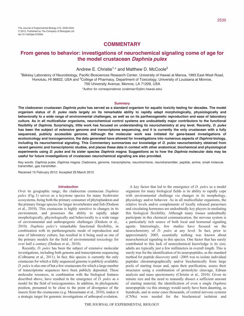
Included among the amine pathway proteins identified from theD. pulex genome were histidine decarboxylase, the rate-limitingbiosynthetic enzyme for histamine, and two histamine-gatedchloride channels, hclA and hclB (McCoole et al., 2011). Thesedata strongly support the production of histamine in this species,and were the impetus for both immunohistochemical andbiochemical analyses of the roles played by this amine in Daphnia,particularly its participation in their phototactic response toultraviolet (UV) light (McCoole et al., 2011). Usingimmunohistochemistry, an extensive network of histaminergicsomata, axons and neuropil was identified within the DaphniaCNS, including labeling of photoreceptor cells in the compoundeye and projections from them to the brain, data supporting a rolefor histamine in the visual system (Fig.4). To assess its actions onphototaxis directly, a behavioral assay was developed in whichanimals (here D. magna, whose complement of histaminergicneurons is identical to that of D. pulex; Fig.4) were exposed to
The Journal of Experimental Biology 215 (15)
Fig.4. Confocal micrographs showinghistamine-like immunoreactivity in thecentral nervous systems of Daphnia pulexand Daphnia magna. (A)Histamine-likelabeling in the D. pulex compound eye.(B)Histamine-like labeling in the D. magnasupraoesophageal ganglion (brain).(C)Histamine-like labeling in the D. magnathoracic nervous system. Scale bars,50m. VNS, ventral nervous system.Figure is reprinted from McCoole et al.(McCoole et al., 2011).
THE JOURNAL OF EXPERIMENTAL BIOLOGY
2541Daphnia pulex neurochemistry
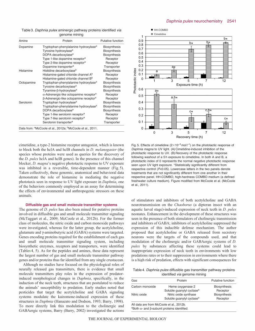
cimetidine, a type-2 histamine receptor antagonist, which is knownto block both the hclA and hclB channels in D. melanogaster (thespecies whose proteins were used as queries for the discovery ofthe D. pulex hclA and hclB genes). In the presence of this channelblocker, D. magna’s negative phototactic response to UV exposurewas inhibited in a reversible, time-dependent manner (Fig.5).Taken collectively, these genomic, anatomical and behavioral datademonstrate the role of histamine in mediating the negativephototaxis seen in response to UV light exposure in Daphnia, oneof the behaviors commonly employed as an assay for determiningthe effects of environmental and anthropogenic stressors on theseanimals.
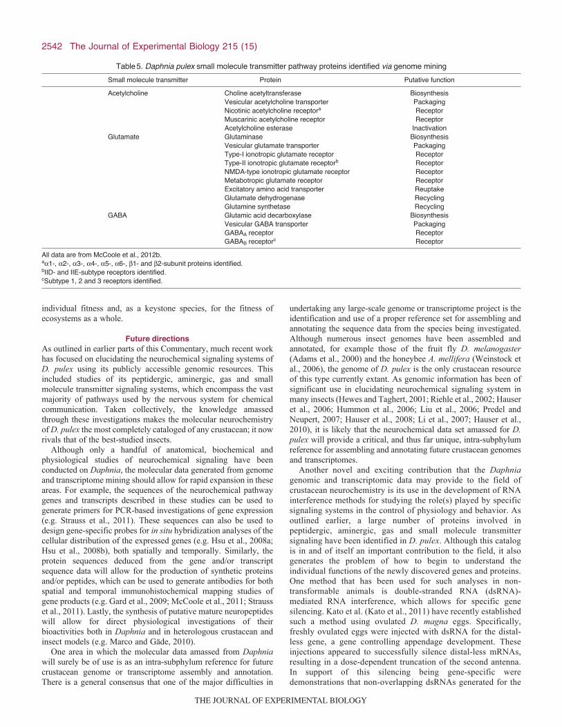
Diffusible gas and small molecule transmitter systemsThe genome of D. pulex has also been mined for putative proteinsinvolved in diffusible gas and small molecule transmitter signaling(McTaggart et al., 2009; McCoole et al., 2012b). For the formerclass of molecules, the nitric oxide and carbon monoxide pathwayswere investigated, whereas for the latter group, the acetylcholine,glutamate and -aminobutyric acid (GABA) systems were targeted.Genes encoding proteins required for the establishment of each gasand small molecule transmitter signaling system, includingbiosynthetic enzymes, receptors and transporters, were identified(Tables4, 5). As for the peptides and amines, these data representthe largest number of gas and small molecule transmitter pathwaygenes and/or proteins thus far identified from any single crustacean.
Although no studies have focused on the physiological roles ofneurally released gas transmitters, there is evidence that smallmolecule transmitters play roles in the expression of predator-induced morphological changes in Daphnia, specifically, in theinduction of the neck teeth, structures that are postulated to reducethe animals’ susceptibility to predation. Early studies noted thatpesticides that target the acetylcholine and GABA signalingsystems modulate the kairomone-induced expression of thesestructures in Daphnia (Hanazato and Dodson, 1993; Barry, 1998).To more directly link this modulation to the cholinergic andGABAergic systems, Barry (Barry, 2002) investigated the actions
of stimulators and inhibitors of both acetylcholine and GABAneurotransmission on the Chaoborus (a dipteran insect with anaquatic larval stage)-induced expression of neck teeth in D. pulexneonates. Enhancement in the development of these structures wasseen in the presence of both stimulators of cholinergic transmissionand inhibitors of GABA; inhibitors of acetylcholine suppressed theexpression of this inducible defense mechanism. The authorproposed that acetylcholine or GABA released from secretoryneurons were the targets of the compounds used, and thatmodulation of the cholinergic and/or GABAergic systems of D.pulex by substances affecting these systems could lead toinappropriate expression of neck teeth in environments with lowpredations rates or to their suppression in environments where thereis a high risk of predation, effects with significant consequences for
Table3. Daphnia pulex aminergic pathway proteins identified viagenome mining
Amine Protein Putative function
Dopamine Tryptophan-phenylalanine hydroxylasea BiosynthesisTyrosine hydroxylasea BiosynthesisDOPA decarboxylasea BiosynthesisType 1-like dopamine receptora ReceptorType 2-like dopamine receptora ReceptorDopamine transportera Transporter
Histamine Histidine decarboxylaseb BiosynthesisHistamine-gated chloride channel Ab ReceptorHistamine-gated chloride channel Bb Receptor
Octopamine Tryptophan-phenylalanine hydroxylasea BiosynthesisTyrosine decarboxylasea BiosynthesisTyramine--hydroxylasea Biosynthesis-Adrenergic-like octopamine receptora Receptor-Adrenergic-like octopamine receptora Receptor
Serotonin Tryptophan hydroxylasea BiosynthesisTryptophan-phenylalanine hydroxylasea BiosynthesisDOPA decarboxylasea BiosynthesisType 1-like serotonin receptora ReceptorType 7-like serotonin receptora ReceptorSerotonin transportera Transporter
Data from: aMcCoole et al., 2012a; bMcCoole et al., 2011.
Table4. Daphnia pulex diffusible gas transmitter pathway proteinsidentified via genome mining
Gas Protein Putative function
Carbon monoxide Heme oxygenase 2 BiosynthesisSoluble guanylyl cyclasea Receptor
Nitric oxide Nitric oxide synthase BiosynthesisSoluble guanylyl cyclasea Receptor
All data are from McCoole et al., 2012b.aBoth - and -subunit proteins identified.
Recovery time (h)
Exposure time (h)
10.90.80.70.60.50.40.30.20.1
0
0.6
0.5
0.4
0.3
0.2
0.1
00
HH-COMBO
Cimetidine
3 5 7 12
1 3 5 7 12
c*
c*
a*
aa*
b* b*b*
c,b*
a,b*
Pho
tota
ctic
inde
x
Fig.5. Effects of cimetidine (2�10–3moll–1) on the phototactic response ofDaphnia magna to UV light. (A)Cimetidine-induced inhibition of thephototactic response to UV. (B)Recovery of the phototactic responsefollowing washout of a 5h exposure to cimetidine. In both A and B, aphototactic index of 0 represents the normal negative phototactic responseseen upon UV light exposure. *Statistically significantly different fromrespective control (P<0.05). Lowercase letters in the two panels denotetreatments that are not significantly different from one another in theirrespective panel. HH-COMBO, high-hardness COMBO medium (a definedfreshwater culture medium). Figure modified from McCoole et al. (McCooleet al., 2011).
THE JOURNAL OF EXPERIMENTAL BIOLOGY
2542
individual fitness and, as a keystone species, for the fitness ofecosystems as a whole.
Future directionsAs outlined in earlier parts of this Commentary, much recent workhas focused on elucidating the neurochemical signaling systems ofD. pulex using its publicly accessible genomic resources. Thisincluded studies of its peptidergic, aminergic, gas and smallmolecule transmitter signaling systems, which encompass the vastmajority of pathways used by the nervous system for chemicalcommunication. Taken collectively, the knowledge amassedthrough these investigations makes the molecular neurochemistryof D. pulex the most completely cataloged of any crustacean; it nowrivals that of the best-studied insects.
Although only a handful of anatomical, biochemical andphysiological studies of neurochemical signaling have beenconducted on Daphnia, the molecular data generated from genomeand transcriptome mining should allow for rapid expansion in theseareas. For example, the sequences of the neurochemical pathwaygenes and transcripts described in these studies can be used togenerate primers for PCR-based investigations of gene expression(e.g. Strauss et al., 2011). These sequences can also be used todesign gene-specific probes for in situ hybridization analyses of thecellular distribution of the expressed genes (e.g. Hsu et al., 2008a;Hsu et al., 2008b), both spatially and temporally. Similarly, theprotein sequences deduced from the gene and/or transcriptsequence data will allow for the production of synthetic proteinsand/or peptides, which can be used to generate antibodies for bothspatial and temporal immunohistochemical mapping studies ofgene products (e.g. Gard et al., 2009; McCoole et al., 2011; Strausset al., 2011). Lastly, the synthesis of putative mature neuropeptideswill allow for direct physiological investigations of theirbioactivities both in Daphnia and in heterologous crustacean andinsect models (e.g. Marco and Gäde, 2010).
One area in which the molecular data amassed from Daphniawill surely be of use is as an intra-subphylum reference for futurecrustacean genome or transcriptome assembly and annotation.There is a general consensus that one of the major difficulties in
undertaking any large-scale genome or transcriptome project is theidentification and use of a proper reference set for assembling andannotating the sequence data from the species being investigated.Although numerous insect genomes have been assembled andannotated, for example those of the fruit fly D. melanogaster(Adams et al., 2000) and the honeybee A. mellifera (Weinstock etal., 2006), the genome of D. pulex is the only crustacean resourceof this type currently extant. As genomic information has been ofsignificant use in elucidating neurochemical signaling system inmany insects (Hewes and Taghert, 2001; Riehle et al., 2002; Hauseret al., 2006; Hummon et al., 2006; Liu et al., 2006; Predel andNeupert, 2007; Hauser et al., 2008; Li et al., 2007; Hauser et al.,2010), it is likely that the neurochemical data set amassed for D.pulex will provide a critical, and thus far unique, intra-subphylumreference for assembling and annotating future crustacean genomesand transcriptomes.
Another novel and exciting contribution that the Daphniagenomic and transcriptomic data may provide to the field ofcrustacean neurochemistry is its use in the development of RNAinterference methods for studying the role(s) played by specificsignaling systems in the control of physiology and behavior. Asoutlined earlier, a large number of proteins involved inpeptidergic, aminergic, gas and small molecule transmittersignaling have been identified in D. pulex. Although this catalogis in and of itself an important contribution to the field, it alsogenerates the problem of how to begin to understand theindividual functions of the newly discovered genes and proteins.One method that has been used for such analyses in non-transformable animals is double-stranded RNA (dsRNA)-mediated RNA interference, which allows for specific genesilencing. Kato et al. (Kato et al., 2011) have recently establishedsuch a method using ovulated D. magna eggs. Specifically,freshly ovulated eggs were injected with dsRNA for the distal-less gene, a gene controlling appendage development. Theseinjections appeared to successfully silence distal-less mRNAs,resulting in a dose-dependent truncation of the second antenna.In support of this silencing being gene-specific weredemonstrations that non-overlapping dsRNAs generated for the
The Journal of Experimental Biology 215 (15)
Table5. Daphnia pulex small molecule transmitter pathway proteins identified via genome mining
Small molecule transmitter Protein Putative function
Acetylcholine Choline acetyltransferase BiosynthesisVesicular acetylcholine transporter PackagingNicotinic acetylcholine receptora ReceptorMuscarinic acetylcholine receptor ReceptorAcetylcholine esterase Inactivation
Glutamate Glutaminase BiosynthesisVesicular glutamate transporter PackagingType-I ionotropic glutamate receptor ReceptorType-II ionotropic glutamate receptorb ReceptorNMDA-type ionotropic glutamate receptor ReceptorMetabotropic glutamate receptor ReceptorExcitatory amino acid transporter ReuptakeGlutamate dehydrogenase RecyclingGlutamine synthetase Recycling
GABA Glutamic acid decarboxylase BiosynthesisVesicular GABA transporter PackagingGABAA receptor ReceptorGABAB receptorc Receptor
All data are from McCoole et al., 2012b.a1-, 2-, 3-, 4-, 5-, 6-, 1- and 2-subunit proteins identified.bIID- and IIE-subtype receptors identified.cSubtype 1, 2 and 3 receptors identified.
THE JOURNAL OF EXPERIMENTAL BIOLOGY

2543Daphnia pulex neurochemistry
distal-less gene produced the same alteration in phenotype, andthat injections of unrelated dsRNA induced no changes inmorphology. Given the extensive characterization of Daphnia’sbehavioral repertoire, similar studies targeting neurochemicalpathway genes are likely to provide major insights into thefunctional roles served by their encoded proteins in these animalsand, using D. pulex as a proxy, in crustaceans more broadly.
Concluding remarksPrior to 2009, little was known about neurochemical signaling inD. pulex on any level, a surprising fact given the model organismstatus of Daphnia for many biological fields and its long history ofbehavioral study, the modulation of which is undoubtedly rootedin its neurochemistry. The sequencing and public deposition of theD. pulex genome and transcriptome have dramatically changed thissituation over the past several years, with genome andtranscriptome mining allowing for the identification of largenumbers of peptidergic, aminergic, gas and small moleculetransmitter pathway proteins from this species. Taken collectively,these data now make D. pulex perhaps the best characterizedcrustacean in terms of its molecular neurochemistry, and ourunderstanding of the putative proteins involved in itsneurochemical signaling systems now rivals or surpasses those ofmost insects. The molecular data that have been obtained forDaphnia, in combination with its well-documented behavioralrepertoire, now positions the field of crustacean neurochemistry tomake significant strides in understanding how specificneurochemical pathway proteins manifest themselves in the overtexpression of a myriad of behavioral and physiological outputs.Clearly this information will be of great benefit to ourunderstanding of Daphnia biology (e.g. phenotypic plasticity,circadian rhythmicity, escape behavior, etc.) and, for that matter,of neurochemical signaling in a general sense.
GlossaryEcotoxicology
The study of the effects of toxic compounds on organisms, typically at thelevel of populations and/or ecosystems.
GenomeThe full complement of genetic material for an individual, generallyencoded in DNA.
HormoneA signaling molecule whose target cells, tissues, etc. are located at adistance from the release site of the molecule in question, typicallytransported to a target via the circulatory system.
ParacrineA signaling molecule whose target cells, tissues, etc. are located in closeproximity to the release site of the molecule in question.
PeptidomeThe full complement of peptides present in a given cell, tissue, organismor population of organisms.
ToxicogenomicsThe study of the structure and function of genomes in response to adversexenobiotic exposure.
TranscriptomeThe full complement of RNA transcripts produced by a given cell, tissue,organism or population of organisms at the particular moment in time atwhich the sample in question was collected.
AcknowledgementsDr Patsy Dickinson and Mr Brandon DʼAndrea are thanked for reading andcommenting on earlier versions of this Commentary.
FundingA.E.C. gratefully acknowledges financial support from the Cades Foundation ofHonolulu, Hawaii.
ReferencesAdams, M. D., Celniker, S. E., Holt, R. A., Evans, C. A., Gocayne, J. D.,
Amanatides, P. G., Scherer, S. E., Li, P. W., Hoskins, R. A., Galle, R. F. et al.(2000). The genome sequence of Drosophila melanogaster. Science 287, 2185-2195.
Barry, M. J. (1998). Endosulfan-enhanced crest induction in Daphnia longicephala:evidence for cholinergic innervation of kairomone receptors. J. Plankton Res. 20,1219-1231.
Barry, M. J. (2002). Progress toward understanding the neurophysiological basis ofpredator-induced morphology in Daphnia pulex. Physiol. Biochem. Zool. 75, 179-186.
Boucher, P., Ditlecadet, D., Dubé, C. and Dufresne, F. (2010). Unusual duplicationof the insulin-like receptor in the crustacean Daphnia pulex. BMC Evol. Biol. 10, 305.
Christie, A. E. (2011). Crustacean neuroendocrine systems and their signaling agents.Cell Tissue Res. 345, 41-67.
Christie, A. E., Stemmler, E. A. and Dickinson, P. S. (2010). Crustaceanneuropeptides. Cell. Mol. Life Sci. 67, 4135-4169.
Christie, A. E., McCoole, M. D., Harmon, S. M., Baer, K. N. and Lenz, P. H. (2011).Genomic analyses of the Daphnia pulex peptidome. Gen. Comp. Endocrinol. 171,131-150.
Colbourne, J. K., Pfrender, M. E., Gilbert, D., Thomas, W. K., Tucker, A., Oakley,T. H., Tokishita, S., Aerts, A., Arnold, G. J., Basu, M. K. et al. (2011). Theecoresponsive genome of Daphnia pulex. Science 331, 555-561.
Dircksen, H., Neupert, S., Predel, R., Verleyen, P., Huybrechts, J., Strauss, J.,Hauser, F., Stafflinger, E., Schneider, M., Pauwels, K. et al. (2011). Genomics,transcriptomics, and peptidomics of Daphnia pulex neuropeptides and proteinhormones. J. Proteome Res. 10, 4478-4504.
Dodson, S. L., Cáceres, C. E. and Rogers, D. C. (2010). Cladocera and otherBrachiopoda. In Ecology and Classification of North American FreshwaterInvertebrates, 3rd edn (ed. J. H. Thorp and A. P. Covich), pp. 774-828. San Diego,CA: Academic Press.
Gard, A. L., Lenz, P. H., Shaw, J. R. and Christie, A. E. (2009). Identification ofputative peptide paracrines/hormones in the water flea Daphnia pulex (Crustacea;Branchiopoda; Cladocera) using transcriptomics and immunohistochemistry. Gen.Comp. Endocrinol. 160, 271-287.
Gewin, V. (2005). Functional genomics thickens the biological plot. PLoS Biol. 3, e219.Hanazato, T. and Dodson, S. I. (1993). Morphological responses of four species of
cyclomorphic Daphnia to a short-term exposure to the insecticide carbaryl. J.Plankton Res. 15, 1743-1755.
Hauser, F., Cazzamali, G., Williamson, M., Blenau, W. and Grimmelikhuijzen, C. J.(2006). A review of neurohormone GPCRs present in the fruitfly Drosophilamelanogaster and the honeybee Apis mellifera. Prog. Neurobiol. 80, 1-19.
Hauser, F., Cazzamali, G., Williamson, M., Park, Y., Li, B., Tanaka, Y., Predel, R.,Neupert, S., Schachtner, J., Verleyen, P. et al. (2008). A genome-wide inventoryof neurohormone GPCRs in the red flour beetle Tribolium castaneum. Front.Neuroendocrinol. 29, 142-165.
Hauser, F., Neupert, S., Williamson, M., Predel, R., Tanaka, Y. andGrimmelikhuijzen, C. J. (2010). Genomics and peptidomics of neuropeptides andprotein hormones present in the parasitic wasp Nasonia vitripennis. J. ProteomeRes. 9, 5296-5310.
Hewes, R. S. and Taghert, P. H. (2001). Neuropeptides and neuropeptide receptorsin the Drosophila melanogaster genome. Genome Res. 11, 1126-1142.
Hsu, Y. W., Stemmler, E. A., Messinger, D. I., Dickinson, P. S., Christie, A. E. andde la Iglesia, H. O. (2008a). Cloning and differential expression of two -pigment-dispersing hormone (-PDH) isoforms in the crab Cancer productus: evidence forauthentic -PDH as a local neurotransmitter and -PDH II as a humoral factor. J.Comp. Neurol. 508, 197-211.
Hsu, Y. W., Weller, J. R., Christie, A. E. and de la Iglesia, H. O. (2008b). Molecularcloning of four cDNAs encoding prepro-crustacean hyperglycemic hormone (CHH)from the eyestalk of the red rock crab Cancer productus: identification of twogenetically encoded CHH isoforms and two putative post-translationally derived CHHvariants. Gen. Comp. Endocrinol. 155, 517-525.
Hummon, A. B., Richmond, T. A., Verleyen, P., Baggerman, G., Huybrechts, J.,Ewing, M. A., Vierstraete, E., Rodriguez-Zas, S. L., Schoofs, L., Robinson, G. E.et al. (2006). From the genome to the proteome: uncovering peptides in the Apisbrain. Science 314, 647-649.
Kato, Y., Shiga, Y., Kobayashi, K., Tokishita, S., Yamagata, H., Iguchi, T. andWatanabe, H. (2011). Development of an RNA interference method in thecladoceran crustacean Daphnia magna. Dev. Genes Evol. 220, 337-345.
Klein, J. M., de Kleijn, D. P., Keller, R. and Weidemann, W. M. (1992). Molecularcloning of crustacean pigment dispersing hormone precursor. Biochem. Biophys.Res. Commun. 189, 1509-1514.
Li, B., Predel, R., Neupert, S., Hauser, F., Tanaka, Y., Cazzamali, G., Williamson,M., Arakane, Y., Verleyen, P., Schoofs, L. et al. (2007). Genomics,transcriptomics, and peptidomics of neuropeptides and protein hormones in the redflour beetle Tribolium castaneum. Genome Res. 18, 113-122.
Liu, F., Baggerman, G., DʼHertog, W., Verleyen, P., Schoofs, L. and Wets, G.(2006). In silico identification of new secretory peptide genes in Drosophilamelanogaster. Mol. Cell. Proteomics 5, 510-522.
Marco, H. G. and Gäde, G. (2010). Biological activity of the predicted red pigment-concentrating hormone of Daphnia pulex in a crustacean and an insect. Gen. Comp.Endocrinol. 166, 104-110.
McCoole, M. D., Baer, K. N. and Christie, A. E. (2011). Histaminergic signaling in thecentral nervous system of Daphnia and a role for it in the control of phototacticbehavior. J. Exp. Biol. 214, 1773-1782.
THE JOURNAL OF EXPERIMENTAL BIOLOGY

2544
McCoole, M. D., Atkinson, N. J., Graham, D. I., Grasser, E. B., Joselow, A. E.,McCall, N. M., Welker, A. M., Wilsterman, E. J., Baer, K. N., Tilden, A. R. et al.(2012a). Genomic analyses of aminergic signaling systems (dopamine, octopamineand serotonin) in Daphnia pulex. Comp. Biochem. Physiol. 7D, 35-58.
McCoole, M. D., DʼAndrea, B. T., Baer, K. N. and Christie, A. E. (2012b). Genomicanalyses of gas (nitric oxide and carbon monoxide) and small molecule transmitter(acetylcholine, glutamate and GABA) signaling systems in Daphnia pulex. Comp.Biochem. Physiol. 7D, 124-160.
McTaggart, S. J., Conlon, C., Colbourne, J. K., Blaxter, M. L. and Little, T. J.(2009). The components of the Daphnia pulex immune system as revealed bycomplete genome sequencing. BMC Genomics 10, 175.
Meusemann, K., von Reumont, B. M., Simon, S., Roeding, F., Strauss, S., Kück,P., Ebersberger, I., Walzl, M., Pass, G., Breuers, S. et al. (2010). A phylogenomicapproach to resolve the arthropod tree of life. Mol. Biol. Evol. 27, 2451-2464.
Predel, R. and Neupert, S. (2007). Social behavior and the evolution of neuropeptidegenes: lessons from the honeybee genome. Bioessays 29, 416-421.
Riehle, M. A., Garczynski, S. F., Crim, J. W., Hill, C. A. and Brown, M. R. (2002).Neuropeptides and peptide hormones in Anopheles gambiae. Science 298, 172-175.
Stafflinger, E., Hansen, K. K., Hauser, F., Schneider, M., Cazzamali, C.,Williamson, M. and Grimmelikhuijzen, C. J. (2008). Cloning and identification of
an oxytocin/vasopressin-like receptor and its ligand from insects. Proc. Natl. Acad.Sci. USA 105, 3262-3267.
Strauss, J., Zhang, Q., Verleyen, P., Huybrechts, J., Neupert, S., Predel, R.,Pauwels, K. and Dircksen, H. (2011). Pigment-dispersing hormone in Daphniainterneurons, one type homologous to insect clock neurons displaying circadianrhythmicity. Cell. Mol. Life Sci. 68, 3403-3423.
Tilden, A. R., McCoole, M. D., Harmon, S. M., Baer, K. N. and Christie, A. E.(2011). Genomic identification of a putative circadian system in the cladocerancrustacean Daphnia pulex. Comp. Biochem. Physiol. 6D, 282-309.
Torfs, P., Baggerman, G., Meeusen, T., Nieto, J., Nachman, R. J., Calderon, J., DeLoof, A. and Schoofs, L. (2002). Isolation, identification and synthesis of adisulfated sulfakinin from the central nervous system of an arthropods the whiteshrimp Litopenaeus vannamei. Biochem. Biophys. Res. Commun. 299, 312-320.
Verleyen, P., Huybrechts, J. and Schoofs, L. (2009). SIFamide illustrates the rapidevolution in arthropod neuropeptide research. Gen. Comp. Endocrinol. 162, 27-35.
Weinstock, G. M., Robinson, G. E., Gibbs, R. A., Weinstock, G. M., Robinson, G.E., Worley, K. C., Evans, J. D., Maleszka, R., Robertson, H. M., Weaver, D. B. etal. (2006). Insights into social insects from the genome of the honeybee Apismellifera. Nature 443, 931-949.
The Journal of Experimental Biology 215 (15)
THE JOURNAL OF EXPERIMENTAL BIOLOGY