Embed Size (px)
Citation preview
© 2015, Svetlana Masgutova Educational Institute® for Neuro-Sensory-Motor and Reflex Integration, SMEI (USA) » 41
Po R t a l t o n e u R o d e v e l o P m e n t a n d l e a R n i n g
The Leg Cross Flexion-Extension Reflex:Biomechanics, Neurophysiology, MNRI® Assessment, and Repatterning
t h e o R y a n d h i s t o R y o f m n R i ® R e f l e x i n t e g R a t i o n
Introduction wo separate reflexes, Phillipson’s Withdrawal and Leg Cross Flexion-Extension, are eas-ily confused because they have similar motor patterns and are elicited by stimuli that can
appear to be alike and usually manifest at the same time. The authors’ purpose is to distinguish clearly between these two reflexes and to present detailed information on the one they refer to as the Leg Cross Flexion-Extension Reflex. The other reflex, often con-fused with Leg Cross Flexion-Extension, goes by sev-eral names: Phillipson’s Withdrawal, Phillipson’s Leg Flexion, Crossed Extensor, and Leg Withdrawal Reflex, among others. For clarity in this paper, the other reflex will be referred to as Phillipson’s Withdrawal. On the neurophysiological level, these two reflex patterns present the work of two different nerve tracts – tactile and proprioceptive, activated and processed by different receptors.
The Leg Cross Flexion-Extension Reflex is extremely important for overall sensory-motor integration, mo-tor programing and control. Cross-lateral or reciprocal activities such as crawling, walking, running, climbing, swimming, and jumping depend on maturation and integration of this reflex pattern. It also plays a crucial role in the ‘training’ of proprioceptive nerve pathways (vs. sensory-tactile) and activating the spinal-cortical tract. It is an invaluable resource for supporting neurosensorimotor-proprioceptive integration in physical rehabilita-tion of children and adults with developmental deficits and/or injuries affecting motor coordination and abili-ties. However, neither the reflex, its developmental and rehabilitative importance, nor its biomechanics and neurophysiological ascending and descending pathways is clearly or adequately defined in current literature. This study presents a new understanding of this complicated reflex and describes its biomechanics and the neurophysiological tracts of its ascending and descending pathways. The authors will also interpret MNRI® techniques for practical application that enhance precision and accuracy in the treatment of dysfunctional Leg Cross Flexion-Extension and affect other lower limb motor patterns.
Apart from the work of Masgutova and her associates, other researchers, to the knowledge of the authors, have not studied Leg Cross Flexion-Extension in children or persons with neurodeficits. In contrast, a simple search yields a wealth of information on Phillipson’s Withdrawal. Two examples are 1) “The flexors in the with-
Elvin Akhmatov, MA, Ph.D. Student, Orlando, FL, USA; Jakub Buraczewski, PT, MNRI® Core Specialist; Denis Masgutov, Director of SMEI , Poland
TElvin Akhmatov Denis MasgutovJakub Buraczewski
R e f l e x e s o f t h e B R a i n
42 « © 2015, Svetlana Masgutova Educational Institute® for Neuro-Sensory-Motor and Reflex Integration, SMEI (USA)
drawing limb contract and the extensors relax, while in the other limb, the opposite occurs” (Rod, Trent, Tate, 1992; http://en.wikipedia.org/wiki/Withdrawal_reflex), and 2) “when a person steps on a nail, the leg that is stepping on the nail pulls away, while the other leg takes the weight of the whole body” (Ellrich, Steffens, Schomburg, 2000; Solomon, Schmidt, Adragna, 1990; http://en.wikipedia.org/wiki/Crossed_extensor_reflex). According to the authors of this article, important de-velopmental implications of this contralateral motor pattern are missing from these descriptions and thus will be described in detail here.
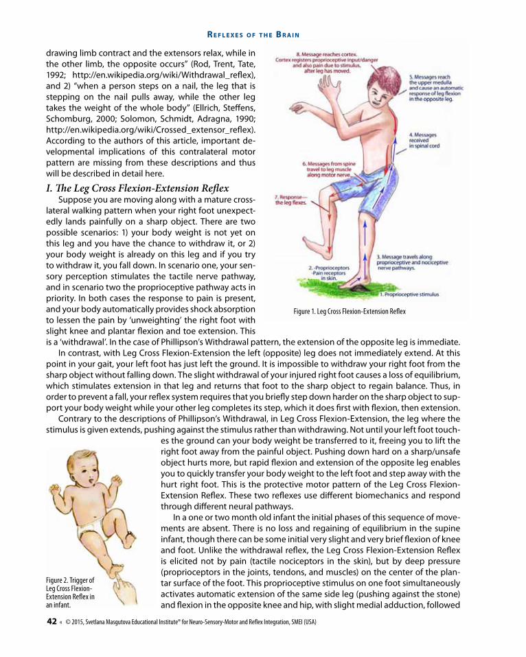
I. The Leg Cross Flexion-Extension Reflex Suppose you are moving along with a mature cross-
lateral walking pattern when your right foot unexpect-edly lands painfully on a sharp object. There are two possible scenarios: 1) your body weight is not yet on this leg and you have the chance to withdraw it, or 2) your body weight is already on this leg and if you try to withdraw it, you fall down. In scenario one, your sen-sory perception stimulates the tactile nerve pathway, and in scenario two the proprioceptive pathway acts in priority. In both cases the response to pain is present, and your body automatically provides shock absorption to lessen the pain by ‘unweighting’ the right foot with slight knee and plantar flexion and toe extension. This is a ‘withdrawal’. In the case of Phillipson’s Withdrawal pattern, the extension of the opposite leg is immediate.
In contrast, with Leg Cross Flexion-Extension the left (opposite) leg does not immediately extend. At this point in your gait, your left foot has just left the ground. It is impossible to withdraw your right foot from the sharp object without falling down. The slight withdrawal of your injured right foot causes a loss of equilibrium, which stimulates extension in that leg and returns that foot to the sharp object to regain balance. Thus, in order to prevent a fall, your reflex system requires that you briefly step down harder on the sharp object to sup-port your body weight while your other leg completes its step, which it does first with flexion, then extension.
Contrary to the descriptions of Phillipson’s Withdrawal, in Leg Cross Flexion-Extension, the leg where the stimulus is given extends, pushing against the stimulus rather than withdrawing. Not until your left foot touch-
es the ground can your body weight be transferred to it, freeing you to lift the right foot away from the painful object. Pushing down hard on a sharp/unsafe object hurts more, but rapid flexion and extension of the opposite leg enables you to quickly transfer your body weight to the left foot and step away with the hurt right foot. This is the protective motor pattern of the Leg Cross Flexion-Extension Reflex. These two reflexes use different biomechanics and respond through different neural pathways.
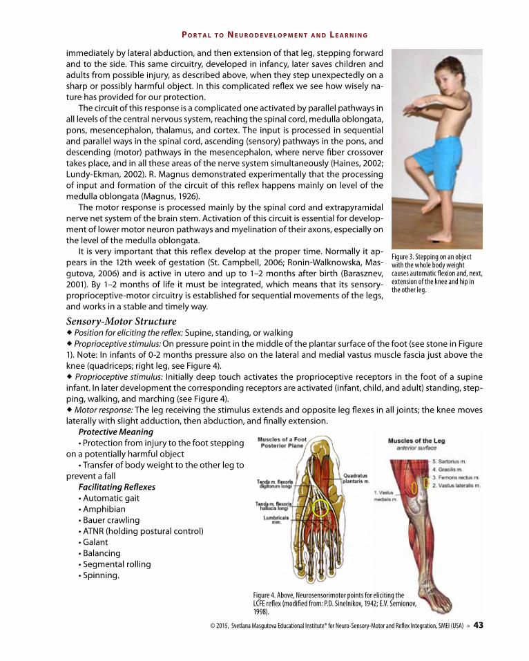
In a one or two month old infant the initial phases of this sequence of move-ments are absent. There is no loss and regaining of equilibrium in the supine infant, though there can be some initial very slight and very brief flexion of knee and foot. Unlike the withdrawal reflex, the Leg Cross Flexion-Extension Reflex is elicited not by pain (tactile nociceptors in the skin), but by deep pressure (proprioceptors in the joints, tendons, and muscles) on the center of the plan-tar surface of the foot. This proprioceptive stimulus on one foot simultaneously activates automatic extension of the same side leg (pushing against the stone) and flexion in the opposite knee and hip, with slight medial adduction, followed
Figure 2. Trigger of Leg Cross Flexion-Extension Reflex in an infant.
Figure 1. Leg Cross Flexion-Extension Reflex
© 2015, Svetlana Masgutova Educational Institute® for Neuro-Sensory-Motor and Reflex Integration, SMEI (USA) » 43
Po R t a l t o n e u R o d e v e l o P m e n t a n d l e a R n i n g
immediately by lateral abduction, and then extension of that leg, stepping forward and to the side. This same circuitry, developed in infancy, later saves children and adults from possible injury, as described above, when they step unexpectedly on a sharp or possibly harmful object. In this complicated reflex we see how wisely na-ture has provided for our protection.
The circuit of this response is a complicated one activated by parallel pathways in all levels of the central nervous system, reaching the spinal cord, medulla oblongata, pons, mesencephalon, thalamus, and cortex. The input is processed in sequential and parallel ways in the spinal cord, ascending (sensory) pathways in the pons, and descending (motor) pathways in the mesencephalon, where nerve fiber crossover takes place, and in all these areas of the nerve system simultaneously (Haines, 2002; Lundy-Ekman, 2002). R. Magnus demonstrated experimentally that the processing of input and formation of the circuit of this reflex happens mainly on level of the medulla oblongata (Magnus, 1926).
The motor response is processed mainly by the spinal cord and extrapyramidal nerve net system of the brain stem. Activation of this circuit is essential for develop-ment of lower motor neuron pathways and myelination of their axons, especially on the level of the medulla oblongata.
It is very important that this reflex develop at the proper time. Normally it ap-pears in the 12th week of gestation (St. Campbell, 2006; Ronin-Walknowska, Mas-gutova, 2006) and is active in utero and up to 1–2 months after birth (Barasznev, 2001). By 1–2 months of life it must be integrated, which means that its sensory-proprioceptive-motor circuitry is established for sequential movements of the legs, and works in a stable and timely way.
Sensory-Motor Structure ® Position for eliciting the reflex: Supine, standing, or walking® Proprioceptive stimulus: On pressure point in the middle of the plantar surface of the foot (see stone in Figure 1). Note: In infants of 0-2 months pressure also on the lateral and medial vastus muscle fascia just above the knee (quadriceps; right leg, see Figure 4).® Proprioceptive stimulus: Initially deep touch activates the proprioceptive receptors in the foot of a supine infant. In later development the corresponding receptors are activated (infant, child, and adult) standing, step-ping, walking, and marching (see Figure 4).® Motor response: The leg receiving the stimulus extends and opposite leg flexes in all joints; the knee moves laterally with slight adduction, then abduction, and finally extension.
Protective Meaning• Protection from injury to the foot stepping
on a potentially harmful object• Transfer of body weight to the other leg to
prevent a fallFacilitating Reflexes• Automatic gait• Amphibian• Bauer crawling• ATNR (holding postural control)• Galant• Balancing• Segmental rolling• Spinning.
Figure 3. Stepping on an object with the whole body weight causes automatic flexion and, next, extension of the knee and hip in the other leg.
Figure 4. Above, Neurosensorimotor points for eliciting the LCFE reflex (modified from: P.D. Sinelnikov, 1942; E.V. Semionov, 1998).
R e f l e x e s o f t h e B R a i n
44 « © 2015, Svetlana Masgutova Educational Institute® for Neuro-Sensory-Motor and Reflex Integration, SMEI (USA)
Developmental Meaning• Differentiation and coordination of legs • Precursor to walking and running• Walking, marching, running, swimming,
skipping, jumping• Cognitive development • Speed of perception and thinking.Opposing Reflexes • Trunk extension (including lower limbs)• STNR • Moro• Spinal Perez• Landau• Fear Paralysis.
Effects of Non-IntegrationIn children with neuro-motor problems, mus-
cular hypo/hypertension, cerebral palsy and other deficits, the Leg Cross Flexion-Extension Reflex is usually delayed and/or pathological. It is also poorly developed in children with autism and selective mutism. This is why skills requiring leg differentiation, shifting body weight from one leg to another for cross-lateral movements and balance control are all challenging for them. Additional effects of non-integration include:
• Poor neural maturation of lower motor neu-rons
• Hyper- (ADHD) or hypo-activity • Dysfunctions of the Automatic Gait and
Crawling Reflexes • Postural problems: chronic inclining of the body to one side or another while standing, or instability due
to poor antigravity skills• Poor ability to cross the midline and inefficient cross-motor patterns • Excessive muscle tension causing fatigue • Delay in skipping, walking, swimming, jumping, and climbing skills • Slow transition from perception to action• Poor cognitive development: comparison, classification, prioritization, calculation, and analysis • Incorrect overlapping of tactile and proprioceptive input and chaotic processing due to homologous
crawling and walking patterns.
II. Biomechanics of the Leg Cross Flexion-Extension ReflexActivation and maturation of this reflex motor pattern in infancy orients the brain-body system to its later
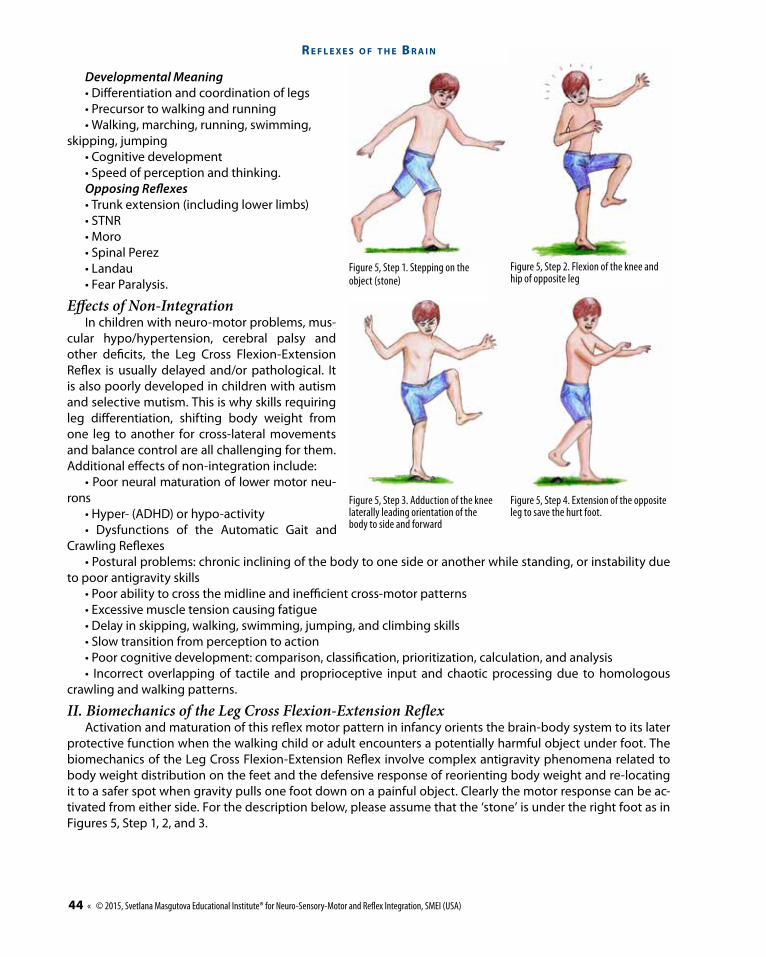
protective function when the walking child or adult encounters a potentially harmful object under foot. The biomechanics of the Leg Cross Flexion-Extension Reflex involve complex antigravity phenomena related to body weight distribution on the feet and the defensive response of reorienting body weight and re-locating it to a safer spot when gravity pulls one foot down on a painful object. Clearly the motor response can be ac-tivated from either side. For the description below, please assume that the ‘stone’ is under the right foot as in Figures 5, Step 1, 2, and 3.
Figure 5, Step 1. Stepping on the object (stone)
Figure 5, Step 3. Adduction of the knee laterally leading orientation of the body to side and forward
Figure 5, Step 2. Flexion of the knee and hip of opposite leg
Figure 5, Step 4. Extension of the opposite leg to save the hurt foot.
© 2015, Svetlana Masgutova Educational Institute® for Neuro-Sensory-Motor and Reflex Integration, SMEI (USA) » 45
Po R t a l t o n e u R o d e v e l o P m e n t a n d l e a R n i n g
Sequence of Movements in the Leg Cross Flexion-Extension Motor Pattern
A Leg Cross Flexion-Extension Re ex
Muscle Groups
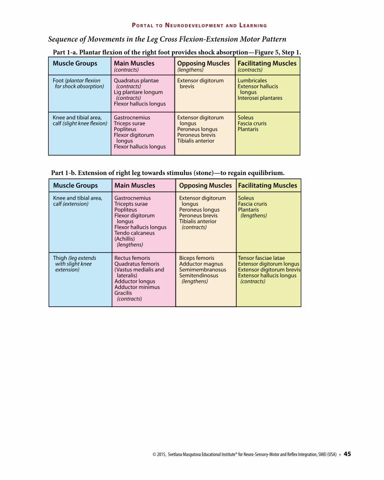
Foot (plantar �exion for shock absorption)
Knee and tibial area,calf (slight knee �exion)
Extensor digitorum longusPeroneus longusPeroneus brevisTibialis anterior
SoleusFascia crurisPlantaris
GastrocnemiusTriceps suraePopliteusFlexor digitorum longusFlexor hallucis longus
LumbricalesExtensor hallucis longusInterosei plantares
Extensor digitorum brevis
Quadratus plantae (contracts)Lig plantare longum (contracts)Flexor hallucis longus
(contracts) (contracts)(lengthens)Opposing Muscles Facilitating MusclesMain Muscles
Part 1-a. Plantar �exion of the right foot provides shock absorption—Figure 5, Step 1.
A Leg Cross Flexion-Extension Re ex
Muscle Groups
Knee and tibial area,calf (extension)
Thigh (leg extends with slight knee extension)
Biceps femorisAdductor magnusSemimembranosusSemitendinosus (lengthens)
Tensor fasciae lataeExtensor digitorum longusExtensor digitorum brevisExtensor hallucis longus (contracts)
Rectus femorisQuadratus femoris(Vastus medialis and lateralis)Adductor longusAdductor minimusGracilis (contracts)
SoleusFascia crurisPlantaris (lengthens)
Extensor digitorum longusPeroneus longusPeroneus brevisTibialis anterior (contracts)
GastrocnemiusTricepts suraePopliteusFlexor digitorum longusFlexor hallucis longusTendo calcaneus(Achillis) (lengthens)
Opposing Muscles Facilitating MusclesMain Muscles
Part 1-b. Extension of right leg towards stimulus (stone)—to regain equilibrium.
R e f l e x e s o f t h e B R a i n
46 « © 2015, Svetlana Masgutova Educational Institute® for Neuro-Sensory-Motor and Reflex Integration, SMEI (USA)
A Leg Cross Flexion-Extension Re ex
Muscle Groups
Hip
Thigh
Calf GastrocnemiusSoleusFlexor hallucis longusFlexor digitorum longusTibialis posterioriPeroneus longusPeroneus brevis
GastrocnemiusPopliteus
Tibialis anteriorPeroneus tertiusExtensor digitorum longusExtensor hallucis longus
Tensor fasciae lataeIliopsoasObturatorius internus
Gluteus maximusPiriformisLig. sacrospinaleLig. sacrotuberale
Psoas majorIliacusGluteus mediusGluteus minimusGracilis
Adductor brevisAdductor longusTensor fasciae latae
SemimembranosusSemitendinosusGracilisBiceps femorisAdductor magnus
Quadricieps femorisFemoris rectusAdductor brevisSartorius
Opposing Muscles Facilitating MusclesMain Muscles
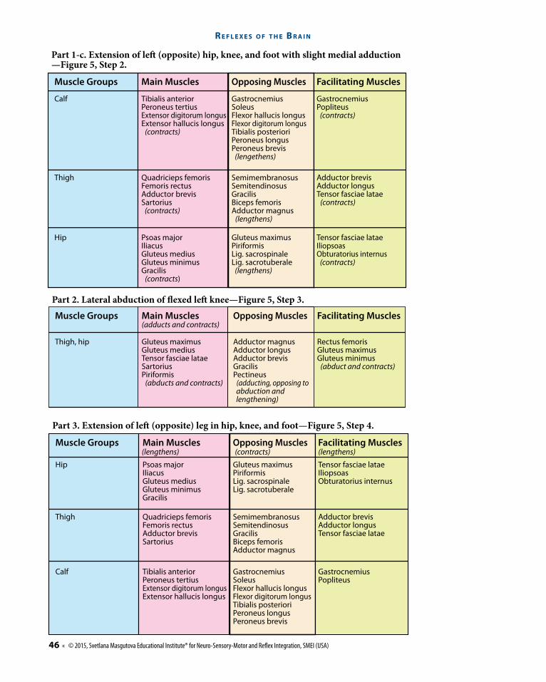
Part 3. Extension of le� (opposite) leg in hip, knee, and foot—Figure 5, Step 4.
(lengthens) (contracts) (lengthens)
A Leg Cross Flexion-Extension Re ex
Muscle Groups
Rectus femorisGluteus maximusGluteus minimus (abduct and contracts)
Adductor magnusAdductor longusAdductor brevisGracilisPectineus (adducting, opposing to abduction and lengthening)
Gluteus maximusGluteus mediusTensor fasciae lataeSartoriusPiriformis (abducts and contracts)
Thigh, hip
Opposing Muscles Facilitating MusclesMain Muscles
Part 2. Lateral abduction of exed le� knee—Figure 5, Step 3.
(adducts and contracts)
A Leg Cross Flexion-Extension Re ex
Muscle Groups
Calf
Thigh
Hip Gluteus maximusPiriformisLig. sacrospinaleLig. sacrotuberale (lengthens)
Tensor fasciae lataeIliopsoasObturatorius internus (contracts)
Psoas majorIliacusGluteus mediusGluteus minimusGracilis (contracts)
GastrocnemiusPopliteus (contracts)
GastrocnemiusSoleusFlexor hallucis longusFlexor digitorum longusTibialis posterioriPeroneus longusPeroneus brevis (lengethens)
Tibialis anteriorPeroneus tertiusExtensor digitorum longusExtensor hallucis longus (contracts)
Adductor brevisAdductor longusTensor fasciae latae (contracts)
SemimembranosusSemitendinosusGracilisBiceps femorisAdductor magnus (lengthens)
Quadricieps femorisFemoris rectusAdductor brevisSartorius (contracts)
Opposing Muscles Facilitating MusclesMain Muscles
Part 1-c. Extension of le (opposite) hip, knee, and foot with slight medial adduction—Figure 5, Step 2.
© 2015, Svetlana Masgutova Educational Institute® for Neuro-Sensory-Motor and Reflex Integration, SMEI (USA) » 47
Po R t a l t o n e u R o d e v e l o P m e n t a n d l e a R n i n g
III. The Neuro-Sensory-Motor Pathways of the Leg Cross Flexion-Extension Reflex Afferent nerve pathway
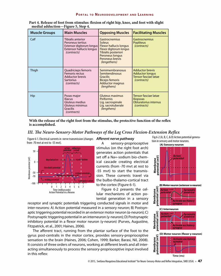
A sensory-proprioceptive stimulus (on the right foot arch) generates action potentials that set off a Na+-sodium bio-chem-ical cascade creating electrical currents (from -70 mvt at rest to -55 mvt) to start the transmis-sion. These currents travel via the bulbo-thalamo-cortical tract to the cortex (Figure 6-1).
Figure 6-2 presents the cel-lular mechanisms of action po-tential generation in a sensory
receptor and synaptic potentials triggering conducted signals in motor and inter-neurons: A) Action potential measured in a sensory neuron; B) Postsyn-aptic triggering potential recorded in an extensor motor neuron (α-neuron); C) Postsynaptic triggering potential in an interneuron (γ-neuron); D) Postsynaptic inhibitory potential in a flexor motor neuron (γ-neuron) (Purves, Augustine, Fitzpatrick, et al., 2001; Haines, 2006).
The afferent tract, running from the plantar surface of the foot to the gyrus post-centralis in the motor cortex, provides sensory-proprioceptive sensation to the brain (Haines, 2006; Cohen, 1999; Barker, Barasi, Nil, 2008). It consists of three orders of neurons, working at different levels and all inter-acting simultaneously to process the sensory-proprioceptive input involved in this reflex:
Muscle Groups
Calf
Thigh
Hip Gluteus maximusPiriformisLig. sacrospinaleLig. sacrotuberale (lengthens)
Tensor fasciae lataeIliopsoasObturatorius internus (contracts)
Psoas majorIliacusGluteus mediusGluteus minimusGracilis (contracts)
GastrocnemiusPopliteus (contracts)
GastrocnemiusSoleusFlexor hallucis longusFlexor digitorum longusTibialis posterioriPeroneus longusPeroneus brevis (lengethens)
Tibialis anteriorPeroneus tertiusExtensor digitorum longusExtensor hallucis longus (contracts)
Adductor brevisAdductor longusTensor fasciae latae (contracts)
SemimembranosusSemitendinosusGracilisBiceps femorisAdductor magnus (lengthens)
Quadricieps femorisFemoris rectusAdductor brevisSartorius (contracts)
Opposing Muscles Facilitating MusclesMain Muscles
A Leg Cross Flexion-Extension Re ex
Part 4. Release of foot from stimulus: �exion of right hip, knee, and foot with slight medial adduction—Figure 5, Step 4.
With the release of the right foot from the stimulus, the protective function of the re�exis accomplished.
Mem
bran
e po
tent
ial (
mV
)
(A) Sensory neuron
(B) Motor neuron (extensor α-neuron)
(C) Interneuron
(D) Motor neuron (�exor γ-neuron)
Time (ms)
Mem
bran
e po
tent
ial (
mV
)M
embr
ane
pote
ntia
l (m
V)
Mem
bran
e po
tent
ial (
mV
)Action potential
Synapatic potential
Synapatic potential
Action potential
Action potential
Activate excitatory synapse
Activate excitatory synapse
Activate excitatory synapse
Fig 6-2 (A, B, C, & D) Action potential genera-tion in sensory and motor neurons.
resting potential
action potential
refractoryperiod
threshold potential
depolarization
hyperpolarizationresting potential
50
0
-50-70
-1000 1 2 3 4 5 6 7
Time (milliseconds)Action Potential in a Neuron
Mem
bran
e pot
entia
l (m
V)
Membrane potential (mV)
repolarization
Figure 6-1. Electrical currents in nerve transmission (changes from -70 mvt at rest to -55 mvt).
R e f l e x e s o f t h e B R a i n
48 « © 2015, Svetlana Masgutova Educational Institute® for Neuro-Sensory-Motor and Reflex Integration, SMEI (USA)
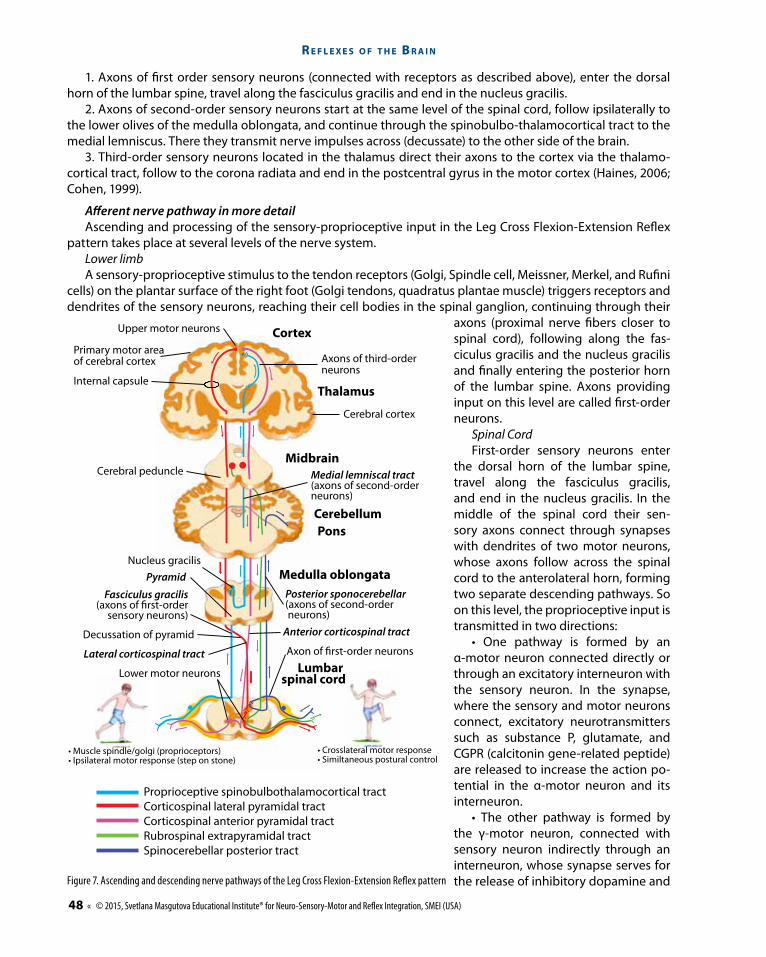
1. Axons of first order sensory neurons (connected with receptors as described above), enter the dorsal horn of the lumbar spine, travel along the fasciculus gracilis and end in the nucleus gracilis.
2. Axons of second-order sensory neurons start at the same level of the spinal cord, follow ipsilaterally to the lower olives of the medulla oblongata, and continue through the spinobulbo-thalamocortical tract to the medial lemniscus. There they transmit nerve impulses across (decussate) to the other side of the brain.
3. Third-order sensory neurons located in the thalamus direct their axons to the cortex via the thalamo-cortical tract, follow to the corona radiata and end in the postcentral gyrus in the motor cortex (Haines, 2006; Cohen, 1999).
Afferent nerve pathway in more detailAscending and processing of the sensory-proprioceptive input in the Leg Cross Flexion-Extension Reflex
pattern takes place at several levels of the nerve system. Lower limbA sensory-proprioceptive stimulus to the tendon receptors (Golgi, Spindle cell, Meissner, Merkel, and Rufini
cells) on the plantar surface of the right foot (Golgi tendons, quadratus plantae muscle) triggers receptors and dendrites of the sensory neurons, reaching their cell bodies in the spinal ganglion, continuing through their
axons (proximal nerve fibers closer to spinal cord), following along the fas-ciculus gracilis and the nucleus gracilis and finally entering the posterior horn of the lumbar spine. Axons providing input on this level are called first-order neurons.
Spinal CordFirst-order sensory neurons enter
the dorsal horn of the lumbar spine, travel along the fasciculus gracilis, and end in the nucleus gracilis. In the middle of the spinal cord their sen-sory axons connect through synapses with dendrites of two motor neurons, whose axons follow across the spinal cord to the anterolateral horn, forming two separate descending pathways. So on this level, the proprioceptive input is transmitted in two directions:
• One pathway is formed by an α-motor neuron connected directly or through an excitatory interneuron with the sensory neuron. In the synapse, where the sensory and motor neurons connect, excitatory neurotransmitters such as substance P, glutamate, and CGPR (calcitonin gene-related peptide) are released to increase the action po-tential in the α-motor neuron and its interneuron.
• The other pathway is formed by the γ-motor neuron, connected with sensory neuron indirectly through an interneuron, whose synapse serves for the release of inhibitory dopamine and
Proprioceptive spinobulbothalamocortical tractCorticospinal lateral pyramidal tractCorticospinal anterior pyramidal tractRubrospinal extrapyramidal tractSpinocerebellar posterior tract
Thalamus
Cortex
Axons of third-orderneurons
Cerebral cortex
Upper motor neurons
Primary motor areaof cerebral cortex
Internal capsule
Cerebral peduncle
Decussation of pyramid
PyramidNucleus gracilis
• Muscle spindle/golgi (proprioceptors)• Ipsilateral motor response (step on stone)
• Crosslateral motor response• Similtaneous postural control
Medial lemniscal tract(axons of second-order neurons)
Axon of �rst-order neurons
Lower motor neurons
Posterior sponocerebellar(axons of second-order neurons)
Anterior corticospinal tract
Fasciculus gracilis(axons of first-order
sensory neurons)
Lateral corticospinal tract
Midbrain
CerebellumPons
Medulla oblongata
Lumbar spinal cord
Figure 7. Ascending and descending nerve pathways of the Leg Cross Flexion-Extension Reflex pattern
© 2015, Svetlana Masgutova Educational Institute® for Neuro-Sensory-Motor and Reflex Integration, SMEI (USA) » 49
Po R t a l t o n e u R o d e v e l o P m e n t a n d l e a R n i n g
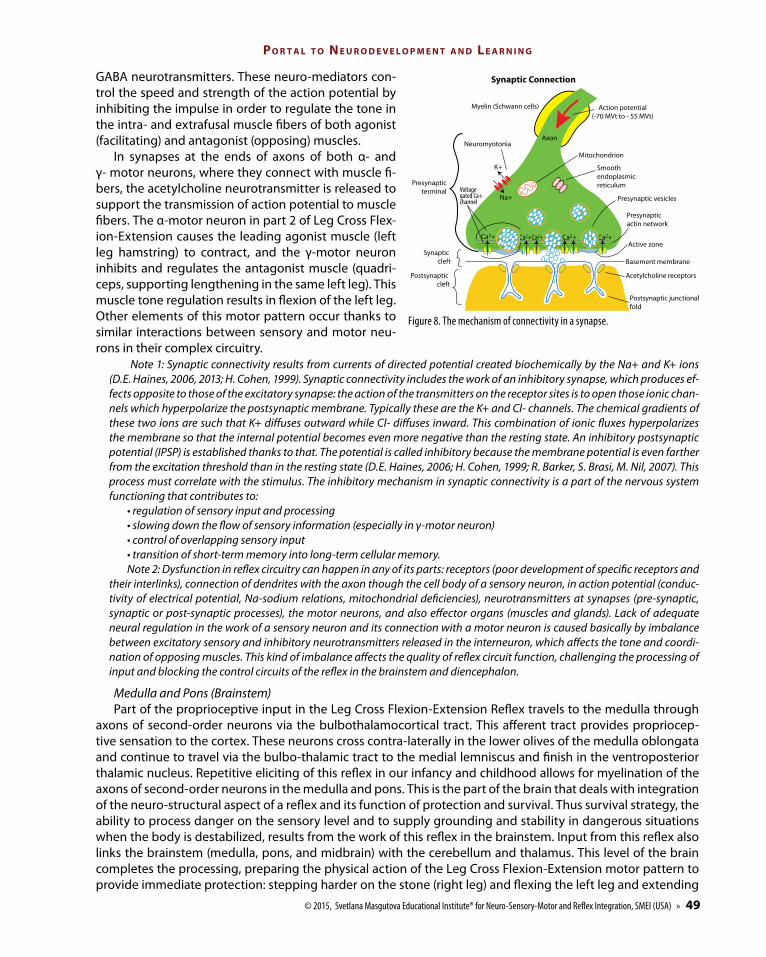
GABA neurotransmitters. These neuro-mediators con-trol the speed and strength of the action potential by inhibiting the impulse in order to regulate the tone in the intra- and extrafusal muscle fibers of both agonist (facilitating) and antagonist (opposing) muscles.
In synapses at the ends of axons of both α- and γ- motor neurons, where they connect with muscle fi-bers, the acetylcholine neurotransmitter is released to support the transmission of action potential to muscle fibers. The α-motor neuron in part 2 of Leg Cross Flex-ion-Extension causes the leading agonist muscle (left leg hamstring) to contract, and the γ-motor neuron inhibits and regulates the antagonist muscle (quadri-ceps, supporting lengthening in the same left leg). This muscle tone regulation results in flexion of the left leg. Other elements of this motor pattern occur thanks to similar interactions between sensory and motor neu-rons in their complex circuitry.
Note 1: Synaptic connectivity results from currents of directed potential created biochemically by the Na+ and K+ ions (D.E. Haines, 2006, 2013; H. Cohen, 1999). Synaptic connectivity includes the work of an inhibitory synapse, which produces ef-fects opposite to those of the excitatory synapse: the action of the transmitters on the receptor sites is to open those ionic chan-nels which hyperpolarize the postsynaptic membrane. Typically these are the K+ and Cl- channels. The chemical gradients of these two ions are such that K+ diffuses outward while Cl- diffuses inward. This combination of ionic fluxes hyperpolarizes the membrane so that the internal potential becomes even more negative than the resting state. An inhibitory postsynaptic potential (IPSP) is established thanks to that. The potential is called inhibitory because the membrane potential is even farther from the excitation threshold than in the resting state (D.E. Haines, 2006; H. Cohen, 1999; R. Barker, S. Brasi, M. Nil, 2007). This process must correlate with the stimulus. The inhibitory mechanism in synaptic connectivity is a part of the nervous system functioning that contributes to:
• regulation of sensory input and processing• slowing down the flow of sensory information (especially in γ-motor neuron) • control of overlapping sensory input • transition of short-term memory into long-term cellular memory.Note 2: Dysfunction in reflex circuitry can happen in any of its parts: receptors (poor development of specific receptors and
their interlinks), connection of dendrites with the axon though the cell body of a sensory neuron, in action potential (conduc-tivity of electrical potential, Na-sodium relations, mitochondrial deficiencies), neurotransmitters at synapses (pre-synaptic, synaptic or post-synaptic processes), the motor neurons, and also effector organs (muscles and glands). Lack of adequate neural regulation in the work of a sensory neuron and its connection with a motor neuron is caused basically by imbalance between excitatory sensory and inhibitory neurotransmitters released in the interneuron, which affects the tone and coordi-nation of opposing muscles. This kind of imbalance affects the quality of reflex circuit function, challenging the processing of input and blocking the control circuits of the reflex in the brainstem and diencephalon.
Medulla and Pons (Brainstem)Part of the proprioceptive input in the Leg Cross Flexion-Extension Reflex travels to the medulla through
axons of second-order neurons via the bulbothalamocortical tract. This afferent tract provides propriocep-tive sensation to the cortex. These neurons cross contra-laterally in the lower olives of the medulla oblongata and continue to travel via the bulbo-thalamic tract to the medial lemniscus and finish in the ventroposterior thalamic nucleus. Repetitive eliciting of this reflex in our infancy and childhood allows for myelination of the axons of second-order neurons in the medulla and pons. This is the part of the brain that deals with integration of the neuro-structural aspect of a reflex and its function of protection and survival. Thus survival strategy, the ability to process danger on the sensory level and to supply grounding and stability in dangerous situations when the body is destabilized, results from the work of this reflex in the brainstem. Input from this reflex also links the brainstem (medulla, pons, and midbrain) with the cerebellum and thalamus. This level of the brain completes the processing, preparing the physical action of the Leg Cross Flexion-Extension motor pattern to provide immediate protection: stepping harder on the stone (right leg) and flexing the left leg and extending
Neuromyotonia
Mitochondrion
Smooth endoplasmic reticulum
Presynaptic vesicles
Presynaptic actin network
Active zone
Basement membrane
Acetylcholine receptors
Postsynaptic junctionalfold
Myelin (Schwann cells) Action potential(-70 MVt to - 55 MVt)
K+
Axon
Na+Voltagegated Ca+channel
Presynapticterminal
Postsynaptic cleft
Synaptic cleft
Synaptic Connection
Figure 8. The mechanism of connectivity in a synapse.
R e f l e x e s o f t h e B R a i n
50 « © 2015, Svetlana Masgutova Educational Institute® for Neuro-Sensory-Motor and Reflex Integration, SMEI (USA)
it to regain stability on safer ground. This most important part of processing Leg Cross Flexion-Extension input is automatic and entirely unconscious.
ThalamusThird-order neurons are located in thalamus and direct their axons to the cortex along the thalamo-cortical
tract. Input from the Leg Cross Flexion-Extension Reflex that reaches this level is processed here and coordi-nated with sympathetic nervous system responses to activate the stress axis (alarming the organism about danger and activating an increase of stress hormones). Third-order neurons form one third of the posterior limb of the internal capsule, next they follow to the corona radiata and finish in the post central gyrus, where fourth order neurons are located. The thalamus controls the circuit of the Leg Cross Flexion-Extension Reflex performed at the level of the medulla and pons. It determines the relative danger or safety of a stimulus and then regulates sympathetic activation, muscle tone and motor activity/reactivity accordingly.
Sensory CortexThird-order neurons from the thalamus follow along the same thalamo-cortical tract via the corona radiata
and finish in the post central gyrus, where they reach the fourth order neurons. This final level of the brain controls the voluntary work of all the main muscles in the body and, according to I. Sechenov (1961), serves as the physiological basis for the psychological process of creating an ‘image’ of the stimulus: “Is it a stone? Is it a piece of wood, a pen?” Here the brain uses its executive functions to analyze sensory input. The whole cortex becomes involved in decoding and analyzing the input, organizing subsequent output to the muscles and making any movement more precise, goal oriented, and meaningful.
Efferent/descending nerve pathwayThere are two principal groups of descending tracts: one serves for postural/gross motor coordination and
control, and the other for voluntary control of fine motor activity. Descending tracts classified as postural/gross motor tracts in the Leg Cross Flexion-Extension Reflex control automatic responses of the skeletal muscles to extend one leg (right foot on the stone), and to flex, abduct, and extend the opposite leg (left).
The first group of tracts involves the purely automatic motor response and is generated from the lumbar spine, where descending innervation takes place in its different segments. The excitation of motor neurons activates the anterior leg muscles (quadriceps) for movement and depolarizes the contralateral motor-neu-rons of the hamstring muscles (biceps, semitendinosus, and semimemranosus), with simultaneous inhibition of motor neurons innervating antagonistic muscles creating the possibility for one leg to extend (right; led mainly by the quadriceps), and other leg to flex, then abduct and extend (left; led mainly by the biceps).
The second group of descending tracts delivers motor information from the high brain to lower motor neurons of the brainstem or spinal cord and is more involved with voluntary control. These descending tracts are formed by upper motor neurons that: a) arise in the cerebral cortex and descend to the brainstem; b) synapse with lower mo-tor neurons and/or interneurons in the brainstem and spinal cord. They would be involved in conscious direction of the left leg forward, backward or to the side, depending where the safest place to land might be.
Control circuits of the Leg Cross Flexion-Extension Reflex operate from the basal ganglia, cortex, and cer-ebellum, which is responsible for planning automatic responses and adjusting to external circumstances on different levels of the nerve system.
Descending motor pathways through different levels of the nerve systemMotor CortexDescending motor pathways in Leg Cross Flexion-Extension present sequential and simultaneous work of
the whole pyramidal and extrapyramidal nerve systems. The pyramidal system involves the corticospinal tract (lateral and anterior-pyramidal tracts), which governs rapid voluntary movements at the distal ends of limbs. They originate in the primary (Broadmann’s area 4 – motor cortex) and secondary (area 6 – premotor cortex) motor cortex and the parietal lobe (areas 1, 2, 3 – somatosensory cortex). This tract decussates in the pypra-mids and descends as the lateral corticospinal tract to its destination: an inter- or α-motor neuron. Branches of this tract are present in: the cerebral cortex, basal nuclei, red and olivary nuclei, and the reticular formation.
This part of the descending pathway involves the pyramidal upper motor neurons, called first order motor neurons because they are unable to leave the central nervous system. Multi-synaptic, they connect with lower motor neurons to send messages to the muscles. Upper motor neuron lesions can lead to dysfunctional Leg Cross Flexion-Extension responses, as happens in individuals with CP or brain damage.
© 2015, Svetlana Masgutova Educational Institute® for Neuro-Sensory-Motor and Reflex Integration, SMEI (USA) » 51
Po R t a l t o n e u R o d e v e l o P m e n t a n d l e a R n i n g
Note: 80% of the pyramidal cells are located on the precentral gyrus in the frontal lobe (motor strip), 20% of the pyramidal tract fibers originate in the postcentral gyrus of the parietal lobe (areas 1, 2, and 3). Pyramidal tract fibers begin their descent from the cortex (corona radiata) before forming the internal capsule. They are direct and monosynaptic; their axons do not synapse with other cells until they reach their final destination in the brainstem or spinal cord. Because they connect directly from the cortex to the lower motor neurons, messages can be transmitted to the periphery very rapidly.
Subcortical and Extrapyramidal tracts (diencephalon and brainstem)The efferent output from the cortex travels via the frontopontine tract and next along the rubrospinal tract.
These extrapyramidal tracks include: • tectospinal tracts• vestibulospinal tracts• rubrospinal tracts• anterior, medial, and lateral reticulospinal tractsThe tectospinal tract is involved in the control of neck muscles. It originates in the midbrain (in the brain-
stem) and ends in the spinal nerves, thus governing the lowered head position in the Leg Cross Flexion-Exten-sion Reflex pattern.
The vestibulospinal tract runs from the vestibular nuclei (lower pons and medulla) to the spinal nerves. It is involved in balance. (Note that all of these tracts receive input from the cerebellum). In Leg Cross Flexion-Extension this tract serves to keep the body in equilibrium until the legs react contra-laterally.
The rubrospinal tract passes through the red nucleus, carrying messages from the cerebellum to the spinal nerves. Information in this part of the tract flows from the superior cerebellar peduncle to the red nucleus and to the spinal nerves, allowing for somatic motor or skeletal muscle control and regulation of muscle tone for posture and voluntary movement. In Leg Cross Flexion-Extension this track will control the direction and tone of opposing muscles to make possible this extremely complex motor response.
The reticulospinal tract runs from the reticular nuclei of the pons and medulla to the spinal nerves. It is involved in somatic (voluntary) motor control, like the rubrospinal tract, and also plays an important role in the control of autonomic (involuntary) functions in the Leg Cross Flexion-Extension Reflex such as the level of stress hormones released by the sympathetic/parasympathetic system, and regulation of breathing, heart rhythm, blood pressure).
ThalamusThe efferent fibers from the cortex and subcortical centers (including control circuits of the Leg Cross Flex-
ion-Extension Reflex) of extrapyramidal system follow along the frontopontine, vestibulospinal, and rubrospi-nal tracts. After their cross-over (Foreli decussation) in the red nucleus they reach γ-motor neurons.
Control circuits of the Leg Cross Flexion-Extension Reflex reach the cortex, basal ganglia, and cerebellum which:
• regulate the excitation or inhibition of the lower motor neurons• partially determine intensity of muscle contraction• adjust the motor activity according to sensory-proprioceptive input.MidbrainOur study leads us to suppose that in the midbrain (tegmentum, pre-tectum, tectum) the descending path-
way of the Leg Cross Flexion-Extension Reflex provides automatic ‘assessment’ of relative danger/safety and adjusts cortical programing to find a safer spot on the ground, causing abduction in the hip joint in part 3 of the pattern to move the knee and foot forward and away from the source of harm or danger.
Pons Superior to the medulla, the pons consists of white matter with four cranial nerves attached to it. This part
of the CNS includes tracts that conduct signals from the cerebrum down to the cerebellum and medulla, and tracts that carry sensory signals up into the thalamus, thus connecting the cerebellum to the pons and mid-brain. Some nuclei in the pons relay signals from the forebrain to the cerebellum; others deal primarily with respiration, bladder control, hearing, equilibrium, eye movement, facial expressions, and posture. Through these connections the pons participates in control of equilibrium (vestibular system), coordination of motor, visual and auditory systems, breathing, and possible bladder activation when the Leg Cross Flexion-Extension response is triggered.
R e f l e x e s o f t h e B R a i n
52 « © 2015, Svetlana Masgutova Educational Institute® for Neuro-Sensory-Motor and Reflex Integration, SMEI (USA)
Medulla oblongataThis level of the brain is crucial for development of the Leg Cross Flexion-Extension Reflex and the timely
emergence of crawling, walking, running, jumping, marching, and swimming in children, thus we describe it in more detail. The medulla oblongata is the inferior part of the brainstem between the spinal cord and the pons. It consists of: olives, pyramids, the roots of four cranial nerves (CN XII, IX, X, XI), the inferior cerebellar peduncle and the fourth ventricle.
The anterior of the medulla has two vertical bulges called pyramids. They contain axons of the corticospinal tract projecting from the cerebral cortex to the spinal cord. In the lower part of the medulla some of these fi-bers cross each other at the anterior median fissure known as the decussation of the pyramids (also called low motor extrapyramidal cross-over). Other fibers, the external arcuate fibers, originate from the anterior median fissure above the decussation of the pyramids and run laterally across the surface of the pons.
Beside the pyramids are two small oval lumps called olives formed by nerve fibers that connect them to the pons and the cerebellum. They have a role in motor learning.
The fourth ventricle, containing cerebrospinal fluid, forms the dorsal surface of the superior part of the me-dulla, where the four cranial nerves are rooted, and extends within the inferior part.
The task of the medulla oblongata in Leg Cross Flexion-Extension is control over the very complex physi-ological circuits and bio-dynamics of cross-lateral responses (crawling, walking, running, marching, jumping, climbing, swimming), also muscle tone, posture, respiration, heartbeat, and blood pressure. Patients with brain damage can still have functioning bodies, as long as the medulla oblongata is working. The Leg Cross Flexion-Extension test can show the level of medulla oblongata function in both healthy and persistent immobile states.
Spinal CordAt the level of the anterior horn of the spinal cord the output follows via the frontopontine, vestibulospinal,
and rubrospinal tracts, crossing over in the area of the red nucleus (Foreli) to reach the α- and γ-motor neurons of flexor and extensor muscles of contralateral limbs.
Muscular and Motor ResponseJoints are controlled by two opposing sets of muscles, extensors and flexors. Thus, when a muscle spindle
is stretched and the stretch response is activated, the opposing muscle group must be inhibited to prevent it from working against the resulting contraction of the homonymous muscle. This is accomplished by an inhibi-tory interneuron in the spinal cord.
At this level the motor neurons carry acetylcholine neurotransmitters: α-motor neurons release excitatory acetylcholine to cause a tonic contraction of protagonist muscle fibers; at the same time γ-motor neurons re-lease inhibitory acetylcholine to cause lengthening of the antagonist muscle fibers.
In Leg Cross Flexion-Extension, one branch of the descending pathway innervates the α-motor neuron causing the homonymous muscle (same side quadriceps) to contract, producing the leg extension movement in the reflex. The other branch innervates the inhibitory interneuron, which in turn innervates the synapses connecting to the opposing muscle (same side hamstrings). Because the interneuron is inhibitory, it prevents the opposing α-motor neuron from firing, thereby protecting the opposing muscle against over-contraction and damage. The reciprocal excitation-inhibition innervates two opposing sets of muscles in synchrony: flex-ors and extensors causing the needed motor responses in the legs.
Note: the reticular activating system (RAS), a group of neurons coming from the thalamus and following along the brain-stem, regulates arousal and inhibitory mechanisms and provides survival/protection in stress (stone under foot). It is com-posed of several neuronal circuits connecting the brainstem to the cortex. It plays the role of a filter for defining a stressor and its components and regulates thalamo-cortical activity and corresponding behavioral states. When the sensory input is ‘not safe’ the RAS system activates automatic survival responses, regulates the flow of sensory input, posture, equilibrium, motor coordination, head movements, orientation to the vertical plane, and postural adjustments needed for defensive responses. Disorders of the RAS can lead to too much arousal and hyperactive Leg Cross Flexion-Extension, causing constant running and jumping in children and adults. Known for regulation of attention, in the Leg Cross Flexion-Extension response it allows the choice of a safe spot for the opposite foot. There are links between RAS circuits and physiological pain pathways, so when the Leg Cross Flexion-Extension and Phillipson’s Withdrawal are mixed, the Leg Cross Flexion-Extension can recruit nocicep-tion (pain receptor activation) instead of proprioception and compromise the whole Leg Cross Flexion-Extension circuit, caus-ing poor cross motor coordination for walking and running (hyperactive and hypoactive, immobility), poor balance, frequent falls, injuries, as well as emotional and behavioral challenges.
© 2015, Svetlana Masgutova Educational Institute® for Neuro-Sensory-Motor and Reflex Integration, SMEI (USA) » 53
Po R t a l t o n e u R o d e v e l o P m e n t a n d l e a R n i n g
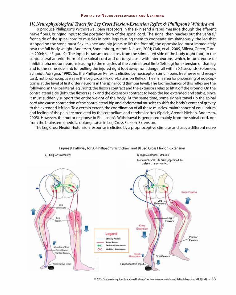
IV. Neurophysiological Tracts for Leg Cross Flexion-Extension Reflex & Phillipson’s Withdrawal To produce Phillipson’s Withdrawal, pain receptors in the skin send a rapid message though the afferent
nerve fibers, bringing input to the posterior horn of the spinal cord. The signal then reaches out the ventral/front side of the spinal cord to muscles in both legs causing them to cooperate simultaneously: the leg that stepped on the stone must flex its knee and hip joints to lift the foot off; the opposite leg must immediately bear the full body weight (Andersen, Sonnenborg, Arendt-Nielsen, 2001; Clair, et al., 2009, Mileva, Green, Turn-er, 2004; see Figure 9). The input is transmitted across from the stimulated side of the body (right foot) to the contralateral anterior horn of the spinal cord and on to synapse with interneurons, which, in turn, excite or inhibit alpha motor neurons leading to the muscles of the contralateral limb (left leg) for extension of that leg and to the same side limb for pulling the injured right foot away from danger, all within 0.5 seconds (Solomon, Schmidt, Adragna, 1990). So, the Phillipson Reflex is elicited by nociceptor stimuli (pain, free nerve end recep-tors), not proprioceptive as in the Leg Cross Flexion-Extension Reflex. The main area for processing of nocicep-tion is at the level of first order neurons in the spinal cord (lumbar level). The biomechanics of this reflex are the following: in the ipsilateral leg (right), the flexors contract and the extensors relax to lift it off the ground. On the contralateral side (left), the flexors relax and the extensors contract to keep the leg extended and stable, since it must suddenly support the entire weight of the body. At the same time, some signals travel up the spinal cord and cause contraction of the contralateral hip and abdomenal muscles to shift the body’s center of gravity to the extended left leg. To a certain extent, the coordination of all these muscles, maintenance of equilibrium and feeling of the pain are mediated by the cerebellum and cerebral cortex (Spaich, Arendt-Nielsen, Andersen, 2005). However, the motor response in Phillipson’s Withdrawal is generated mainly from the spinal cord, not from the brainstem (medulla oblongata) as in Leg Cross Flexion-Extension.
The Leg Cross Flexion-Extension response is elicited by a proprioceptive stimulus and uses a different nerve
B) Leg Cross Flexion-Extension
Fasciculuc Gracillis - to brain (upper medulla, thalamus, sensory cortex)
Figure 9. Pathway for A) Phillipson’s Withdrawl and B) Leg Cross Flexion-Extension
A) Phillipson’s Withdrawl
R e f l e x e s o f t h e B R a i n
54 « © 2015, Svetlana Masgutova Educational Institute® for Neuro-Sensory-Motor and Reflex Integration, SMEI (USA)
tract – reaching the posterior horn of the spinal cord and transmitting the input ipsilaterally to the same side for the leg to extend (stepping even harder on the stone), and another sensory axon synapsing with second-order neurons sends the message to decussate (cross over) in the medulla oblongata (brainstem) to activate descending motor pathways in the opposite side to cause flexion, abduction and extension of the left leg so it can step away and ground the body in a safe spot. Thus, Leg Cross Flexion-Extension differs from Phillipson’s Withdrawal as it is a more complex pattern, uses different ascending and descending nerve pathways and is processed mainly at a higher level of the CNS - medulla oblongata (Magnus, 1926). It is important to understand this difference as it should determine both the procedure used for repatterning and success of rehabilitation.
ConclusionsThe bio-dynamics and neurophysiology of the Leg Cross Flexion-Extension Reflex schema is different from
that of Phillipson Withdrawal: • The Leg Cross Flexion-Extension Reflex is elicited by sensory-proprioceptive receptors. Organization of
balance follows in first priority, and then other systems are activated (tactile/pain). The stimulus must reach the level of medulla oblongata for transmission across to the opposite leg. It also will reach the cortex level for organization of balance organization and registration of pain.
• Phillipson’s Withdrawal is triggered by a nociceptor stimulus (tactile system/pain). The sensation of pain activates in priority the withdrawal away from the stimulus, and then other systems are activated for organiza-tion of balance. Contralateral stimulation of motor neurons in this reflex is reached in priority in the lumbar spine. The sensory neuron also sends signals up higher than the spinal cord to maintain balance and stabilize the body (this is why it is also called the Crossed Extension Reflex). On the level of the cortex this reflex allows the feeling and awareness of pain (Spaich, Arendt-Nielsen, Andersen, 2005).
MNRI® work with the Leg Cross Flexion-Extension Reflex activates pathways transmitting the sensory-pro-prioceptive stimulus, not those involved with nociception as in Phillipson’s Withdrawal. This difference is es-sential for proper application of neuro-corrective techniques for patients.
V. Practical Application: The Leg Cross Flexion-Extension Repatterning Exercise There are several reasons to repattern/pattern the Leg Cross Flexion-Extension Reflex pattern in children
and adults: • delay in development of the reflex pattern• pathology in the reflex pattern caused by brain damage, cerebral palsy or a lesion in lower or upper motor
neurons• faulty connections in sensory and motor axons• poor work of the alpha- and gamma- motor neurons (inadequate production of the GABA and dopamine,
and acetylcholine neurotransmitters)• nerve transmission mixing the Leg Cross Flexion-Extension and Phillipson’s Withdrawal patterns• negative compensation, trauma, PTSD.In all these cases the repatterning/patterning of the physiological circuit is suggested. Training this reflex in
infancy and childhood promotes myelination of second-order neuronal axons in the medulla and pons. Such training can also strengthen or rebuild these networks whenever they become dysfunctional. Because this part of the brain integrates the neuro-structural aspect of a reflex with its protective function, it is essential for survival strategies. In the case of Leg Cross Flexion-Extension, the ability to process danger on the sensory level and to supply grounding and stability when the body is destabilized, originates in the brainstem.
Processing danger and providing a survival strategy are automatic and entirely unconscious. If these func-tions are not accomplished efficiently in the brainstem and its midbrain, then higher more conscious levels of the brain are recruited. Development and maturation of higher-level skills then suffer when the cortex must be engaged for protection and survival and is not free to support voluntary, conscious and skillful activity. Children with developmental issues have a particularly urgent need (see Part I: Effects of Non-Integration) for repatterning of the Leg Cross Flexion-Extension Reflex.
© 2015, Svetlana Masgutova Educational Institute® for Neuro-Sensory-Motor and Reflex Integration, SMEI (USA) » 55
Po R t a l t o n e u R o d e v e l o P m e n t a n d l e a R n i n g
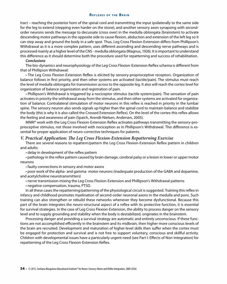
The MNRI® Leg Cross Flexion-Extension repatterning exercise, based on its sensory-mo-tor pathway, consists of four steps presented below (Masgutova, Akhmatova, 2004, 2007, 2010). Step 1Intervention A: Press on the center of the right foot’s quadratus plantae
muscle to simulate the sensory-proprioceptive stimulus of stepping on an object (See Figures 4 and 10). Hold for 5 – 7 seconds.
Reflex motor pattern: stretching and forced raising of the right arch (Sinel-nikov, 1942) caused by stepping on an object.
Intervention B: Press and stretch on lower 1/6th of the quadriceps to trig-ger extension of the knee and hip (Figure 10). Hold for 5 – 7 seconds.
Reflex motor pattern: extension of right knee and hip, stepping harder on the object
Main muscles: Quadratus plantae, quadriceps, adductors, poplitus. Qua-dratus plantae, abductor hallucis, flexor digitorum longus and brevis
Opposing muscles: tensor fasciae latae, sartorius, extensor hallucis brevis, extensor digitorum brevis
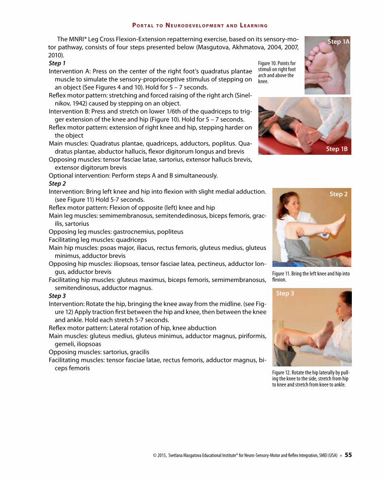
Optional intervention: Perform steps A and B simultaneously.Step 2 Intervention: Bring left knee and hip into flexion with slight medial adduction.
(see Figure 11) Hold 5-7 seconds.Reflex motor pattern: Flexion of opposite (left) knee and hipMain leg muscles: semimembranosus, semitendedinosus, biceps femoris, grac-
ilis, sartorius Opposing leg muscles: gastrocnemius, popliteusFacilitating leg muscles: quadriceps Main hip muscles: psoas major, iliacus, rectus femoris, gluteus medius, gluteus
minimus, adductor brevis Opposing hip muscles: iliopsoas, tensor fasciae latea, pectineus, adductor lon-
gus, adductor brevis Facilitating hip muscles: gluteus maximus, biceps femoris, semimembranosus,
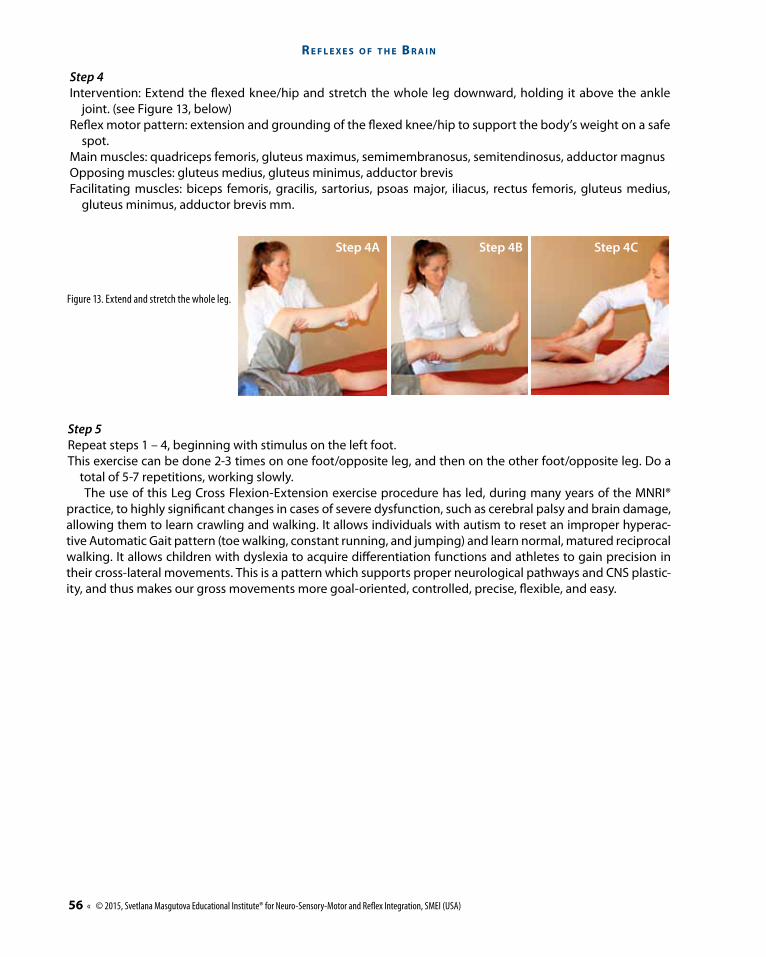
semitendinosus, adductor magnus.Step 3Intervention: Rotate the hip, bringing the knee away from the midline. (see Fig-
ure 12) Apply traction first between the hip and knee, then between the knee and ankle. Hold each stretch 5-7 seconds.
Reflex motor pattern: Lateral rotation of hip, knee abduction Main muscles: gluteus medius, gluteus minimus, adductor magnus, piriformis,
gemeli, iliopsoasOpposing muscles: sartorius, gracilis Facilitating muscles: tensor fasciae latae, rectus femoris, adductor magnus, bi-
ceps femoris
Figure 11. Bring the left knee and hip into flexion.
Figure 10. Points for stimuli on right foot arch and above the knee.
Figure 12. Rotate the hip laterally by pull-ing the knee to the side, stretch from hip to knee and stretch from knee to ankle.
Step 1a
Step 1B
Step 2
Step 3
R e f l e x e s o f t h e B R a i n
56 « © 2015, Svetlana Masgutova Educational Institute® for Neuro-Sensory-Motor and Reflex Integration, SMEI (USA)
Step 5 Repeat steps 1 – 4, beginning with stimulus on the left foot.This exercise can be done 2-3 times on one foot/opposite leg, and then on the other foot/opposite leg. Do a
total of 5-7 repetitions, working slowly.The use of this Leg Cross Flexion-Extension exercise procedure has led, during many years of the MNRI®
practice, to highly significant changes in cases of severe dysfunction, such as cerebral palsy and brain damage, allowing them to learn crawling and walking. It allows individuals with autism to reset an improper hyperac-tive Automatic Gait pattern (toe walking, constant running, and jumping) and learn normal, matured reciprocal walking. It allows children with dyslexia to acquire differentiation functions and athletes to gain precision in their cross-lateral movements. This is a pattern which supports proper neurological pathways and CNS plastic-ity, and thus makes our gross movements more goal-oriented, controlled, precise, flexible, and easy.
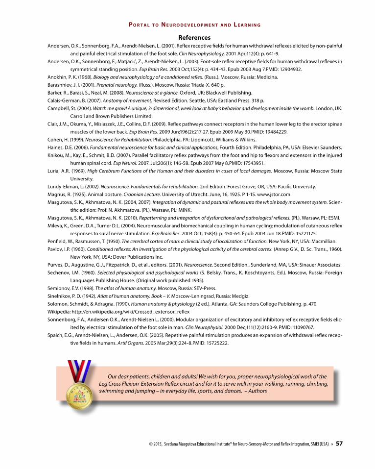
Step 4Intervention: Extend the flexed knee/hip and stretch the whole leg downward, holding it above the ankle
joint. (see Figure 13, below)Reflex motor pattern: extension and grounding of the flexed knee/hip to support the body’s weight on a safe
spot.Main muscles: quadriceps femoris, gluteus maximus, semimembranosus, semitendinosus, adductor magnus Opposing muscles: gluteus medius, gluteus minimus, adductor brevisFacilitating muscles: biceps femoris, gracilis, sartorius, psoas major, iliacus, rectus femoris, gluteus medius,
gluteus minimus, adductor brevis mm.
Figure 13. Extend and stretch the whole leg.
Step 4a Step 4B Step 4c
© 2015, Svetlana Masgutova Educational Institute® for Neuro-Sensory-Motor and Reflex Integration, SMEI (USA) » 57
Po R t a l t o n e u R o d e v e l o P m e n t a n d l e a R n i n g
ReferencesAndersen, O.K., Sonnenborg, F.A., Arendt-Nielsen, L. (2001). Reflex receptive fields for human withdrawal reflexes elicited by non-painful
and painful electrical stimulation of the foot sole. Clin Neurophysiology, 2001 Apr;112(4): p. 641-9. Andersen, O.K., Sonnenborg, F., Matjacić, Z., Arendt-Nielsen, L. (2003). Foot-sole reflex receptive fields for human withdrawal reflexes in
symmetrical standing position. Exp Brain Res. 2003 Oct;152(4): p. 434-43. Epub 2003 Aug 7.PMID: 12904932.Anokhin, P. K. (1968). Biology and neurophysiology of a conditioned reflex. (Russ.). Moscow, Russia: Medicina. Barashniev, J. I. (2001). Prenatal neurology. (Russ.). Moscow, Russia: Triada-X. 640 p.Barker, R., Barasi, S., Neal, M. (2008). Neuroscience at a glance. Oxford, UK: Blackwell Publishing. Calais-German, B. (2007). Anatomy of movement. Revised Edition. Seattle, USA: Eastland Press. 318 p. Campbell, St. (2004). Watch me grow! A unique, 3-dimensional, week look at baby’s behavior and development inside the womb. London, UK:
Carroll and Brown Publishers Limited.Clair, J.M., Okuma, Y., Misiaszek, J.E., Collins, D.F. (2009). Reflex pathways connect receptors in the human lower leg to the erector spinae
muscles of the lower back. Exp Brain Res. 2009 Jun;196(2):217-27. Epub 2009 May 30.PMID: 19484229. Cohen, H. (1999). Neuroscience for Rehabilitation. Philadelphia, PA: Lippincott, Williams & Wilkins. Haines, D.E. (2006). Fundamental neuroscience for basic and clinical applications, Fourth Edition. Philadelphia, PA, USA: Elsevier Saunders.Knikou, M., Kay, E., Schmit, B.D. (2007). Parallel facilitatory reflex pathways from the foot and hip to flexors and extensors in the injured
human spinal cord. Exp Neurol. 2007. Jul;206(1): 146-58. Epub 2007 May 8.PMID: 17543951.Luria, A.R. (1969). High Cerebrum Functions of the Human and their disorders in cases of local damages. Moscow, Russia: Moscow State
University. Lundy-Ekman, L. (2002). Neuroscience. Fundamentals for rehabilitation. 2nd Edition. Forest Grove, OR, USA: Pacific University. Magnus, R. (1925). Animal posture. Croonian Lecture. University of Utrecht. June, 16, 1925. P 1-15. www.jstor.comMasgutova, S. K., Akhmatova, N. K. (2004, 2007). Integration of dynamic and postural reflexes into the whole body movement system. Scien-
tific edition: Prof. N. Akhmatova. (Pl.). Warsaw, PL: MINK. Masgutova, S. K., Akhmatova, N. K. (2010). Repatterning and integration of dysfunctional and pathological reflexes. (Pl.). Warsaw, PL: ESMI.Mileva, K., Green, D.A., Turner D.L. (2004). Neuromuscular and biomechanical coupling in human cycling: modulation of cutaneous reflex
responses to sural nerve stimulation. Exp Brain Res. 2004 Oct; 158(4): p. 450-64. Epub 2004 Jun 18.PMID: 15221175.Penfield, W., Rasmussen, T. (1950). The cererbral cortex of man: a clinical study of localization of function. New York, NY, USA: Macmillian.Pavlov, I.P. (1960). Conditioned reflexes: An investigation of the physiological activity of the cerebral cortex. (Anrep G.V., D. Sc. Trans., 1960).
New York, NY, USA: Dover Publications Inc. Purves, D., Augustine, G.J., Fitzpatrick, D., et al., editors. (2001). Neuroscience. Second Edition., Sunderland, MA, USA: Sinauer Associates.Sechenov, I.M. (1960). Selected physiological and psychological works (S. Belsky, Trans., K. Koschtoyants, Ed.). Moscow, Russia: Foreign
Languages Publishing House. (Original work published 1935). Semionov, E.V. (1998). The atlas of human anatomy. Moscow, Russia: SEV-Press. Sinelnikov, P. D. (1942). Atlas of human anatomy. Book – V. Moscow-Leningrad, Russia: Medgiz. Solomon, Schmidt, & Adragna. (1990). Human anatomy & physiology (2 ed.). Atlanta, GA: Saunders College Publishing. p. 470. Wikipedia: http://en.wikipedia.org/wiki/Crossed_extensor_reflex Sonnenborg, F.A., Andersen O.K., Arendt-Nielsen L. (2000). Modular organization of excitatory and inhibitory reflex receptive fields elic-
ited by electrical stimulation of the foot sole in man. Clin Neurophysiol. 2000 Dec;111(12):2160-9. PMID: 11090767.Spaich, E.G., Arendt-Nielsen, L., Andersen, O.K. (2005). Repetitive painful stimulation produces an expansion of withdrawal reflex recep-
tive fields in humans. Artif Organs. 2005 Mar;29(3):224-8.PMID: 15725222.
Our dear patients, children and adults! We wish for you, proper neurophysiological work of the Leg Cross Flexion-Extension Reflex circuit and for it to serve well in your walking, running, climbing, swimming and jumping – in everyday life, sports, and dances. – Authors