Embed Size (px)
Citation preview
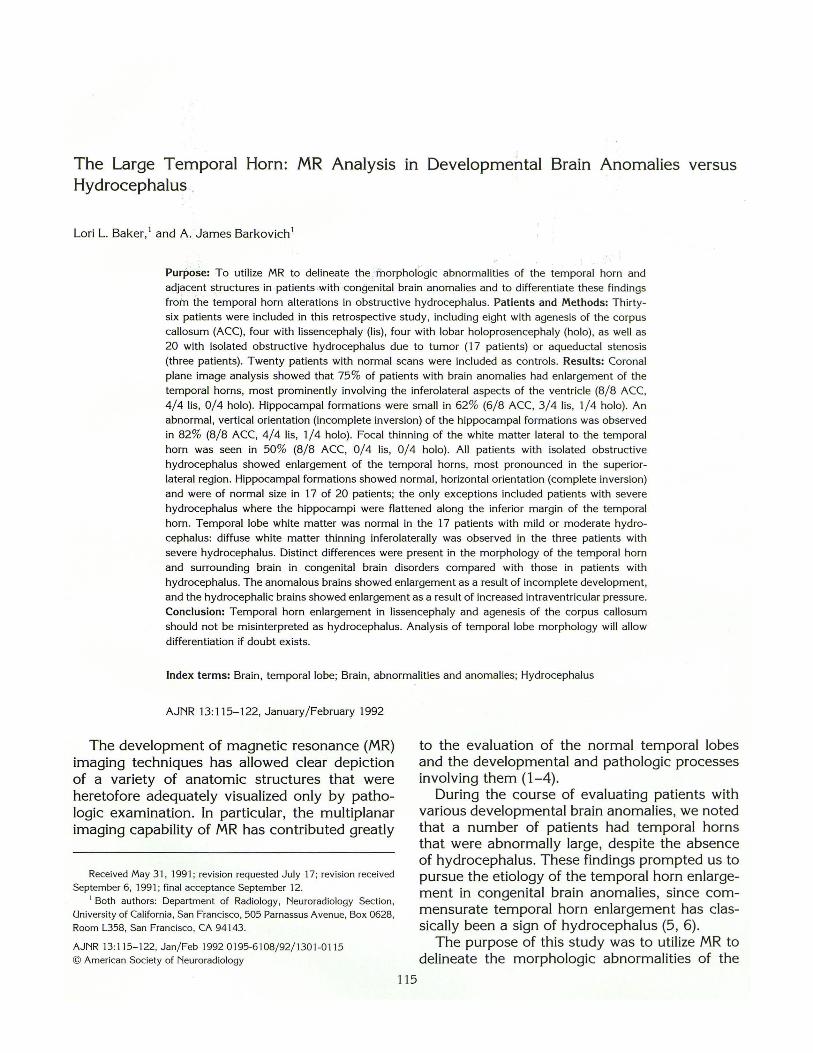
The Large Temporal Horn: MR Analysis in Developmental Brain Anomalies versus Hydrocephalus .
Lori L. Baker, 1 and A . James Barkovich 1
Purpose: To utilize MR to delineate the J norphologic abnormalities of the temporal horn and
adjacent structures in patients .with ·congenital brain anomalies and to differentiate these findings
froin the temporal horn alterations in obstructive hydrocephalus. Patients and Methods: Thirty
six patients were included in this retrospective study, including eight with agenesis of the corpus
callosum (ACC), four with lissencephaly (lis), four with lobar holoprosencephaly (holo), as well as
20 with isolated obstructive hydrocephalus due to tumor (17 patients) or aqueductal stenosis
(three patients). Twenty patients with normal scans were included as controls. Results: Coronal
plane image analysis showed that 75% of patients with brain anomalies had enlargement of the
temporal horns, most prominently involving the inferolateral aspects of the ventricle (8/ 8 ACC,
4/ 4 lis, 0/ 4 holo). Hippocampal formations were small in 62% (6/ 8 ACC, 3/ 4 lis, 1/4 holo). An abnormal , vertical orientation (incomplete inversion) of the hippocampal formations was observed
in 82% (8/ 8 ACC, 4/ 4 lis, 1/ 4 holo). Focal thinning of the white matter lateral to the temporal
horn was seen in 50% (8/ 8 ACC, 0/ 4 lis, 0/ 4 holo). All patients with isolated obstructive
hydrocephalus showed enlargement of the temporal horns, most pronounced in the superior
lateral region. Hippocampal formations showed normal , horizontal orientation (complete inversion)
and were of normal size in 17 of 20 patients; the only exceptions included pat ients with severe
hydrocephalus where the hippocampi were flattened along the inferior margin of the temporal
horn. Temporal lobe white matter was normal in the 17 patients with mild or moderate hydro
cephalus: diffuse white matter thinning inferolaterally was observed in the three patients with
severe hydrocephalus. Distinct differences were present in the morphology of the temporal horn
and surrounding brain in congenital brain disorders compared with those in patients with
hydrocephalus. The anomalous brains showed enlargement as a result of incomplete development,
and the hydrocephalic brains showed enlargement as a result of increased intraventricular pressure.
Conclusion: Temporal horn enlargement in lissencephaly and agenesis of the corpus callosum
should not be misinterpreted as hydrocephalus. Analysis of temporal lobe morphology will allow
differentiation if doubt exists.
Index terms: Brain, temporal lobe; Brain, abnormalities and anomalies; Hydrocephalus
AJNR 13:115-122, January/ February 1992
The development of magnetic resonance (MR) imaging techniques has allowed clear depiction of a variety of anatomic structures that were heretofore adequately visualized only by pathologic examination. In particular, the multiplanar imaging capability of MR has contributed greatly
Received May 3 1, 1991; revision requested July 17; revision received
September 6, 1991 ; final acceptance September 12. 1 Both authors: Department of Radiology , Neuroradiology Section,
University of California, San Francisco, 505 Parnassus Avenue, Box 0628,
Room L358, San Francisco, CA 94143.
AJNR 13:1 15-1 22, Jan/ Feb 1992 0 195-61 08/ 92/ 1301 -011 5 © American Society of Neuroradiology
115
to the evaluation of the normal temporal lobes and the developmental and pathologic processes involving them (1-4).
During the course of evaluating patients with various developmental brain anomalies, we noted that a number of patients had temporal horns that were abnormally large, despite the absence of hydrocephalus. These findings prompted us to pursue the etiology of the temporal horn enlargement in congenital brain anomalies , since commensurate temporal horn enlargement has classically been a sign of hydrocephalus (5 , 6).
The purpose of this study was to utilize MR to delineate the morphologic abnormalities of the
116
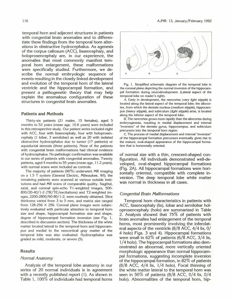
temporal horn and adjacent structures in patients with congenital brain anomalies and to differentiate these findings from the temporal horn alterations in obstructive hydrocephalus. As agenesis of the corpus callosum (ACC), lissencephaly, and holoprosencephaly are, in our experience, the anomalies that most commonly manifest temporal horn enlargement, these malformations were specifically studied. Furthermore, we describe the normal embryologic sequence of events resulting in the closely linked development and evolution of the temporal horn of the lateral ventricle and the hippocampal formation, and present a pathogenetic theory that may help explain the anomalous configuration of these structures in congenital brain anomalies.
Patients and Methods
Thirty-six patients (21 males, 15 females) , aged 5 months to 52 years (mean age, 10.6 years) were included in this retrospective study. Our patient series included eight with ACC, four with lissencephaly, four with holoprosencephaly (1 lobar, 3 semilobar) as well as 20 with isolated obstructive hydrocephalus due to tufT\or (17 patients) or aqueductal stenosis (three patients) . None of the patients with congenital brain malformations had clinical evidence of hydrocephalus. No pathologic confirmation was available in our series of patients with congenital anomalies. Twenty patients, aged 4 months to 55 years (mean age, 11 .2 years), with normal scans were included as controls.
The majority of patients (90%) underwent MR imaging on a 1.5 T system (General Electric, Milwaukee, WI); the remaining patients were scanned at various outside institutions and had MR scans of comparable quality. Sagittal, axial, and coronal spin-echo T1-weighted images, 500-800/20-40/1-2 (TR/TE/ excitations) and T2-weighted images, 2200-2800/60-80/1-2, were routinely obtained. Slice thickness varied from 3 to 5 mm, and matrix size ranged from 128-256 X 256. Coronal plane images were subjectively evaluated with particular attention to temporal horn size and shape, hippocampal formation size and shape, degree of hippocampal formation inversion (see Fig. 1, described in discussion section). The thickness of the white matter located lateral to the temporal horn and hippocampus and medial to the neocortical gray matter of the temporal lobe was also evaluated. Hydrocephalus was graded as mild, moderate, or severe (5) .
Results
Normal Anatomy
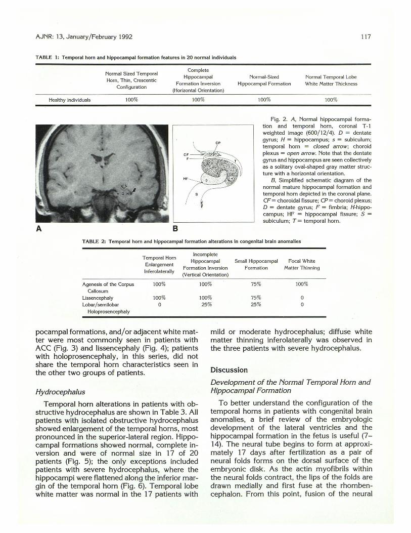
Analysis of the temporal lobe anatomy in our series of 20 normal individuals is in agreement with a recently published report (1). As shown in Table 1, 100% of individuals had temporal horns
AJNR: 13, January/February 1992
A. B. C.
Fig. 1. Simplified schematic diagram of the temporal lobe in the coronal plane depicting the normal inversion of the hippocampal formation during neurodevelopment. (Lateral aspect of the temporal lobe on reader 's right) .
A , Early in development, the neocortex (very light stipple) is located along the lateral aspect of the temporal lobe; the allocortex , from which the dentate nucleus (medium stipple), hippocampus (heavy stipple), and subiculum (light stipple) arise, is located along the inferior aspect of the temporal lobe.
B, The neocortex grows more rapidly than the allocortex during embryogenesis, resulting in medial displacement and internal "inversion" of the dentate gyrus, hippocampus, and subiculum precursors into the temporal horn region.
C, The process of medial displacement and internal "inversion" of the hippocampal formation precursors eventually gives rise to the mature, oval-shaped appearance of the hippocampal formation that is horizontally oriented.
of normal size with a thin, crescent-shaped configuration. All individuals demonstrated well-developed, oval-shaped hippocampal formations (Fig. 2A). All hippocampal formations were horizontally oriented, compatible with complete inversion. The deep temporal lobe white matter was normal in thickness in all cases.
Congenital Brain Malformations
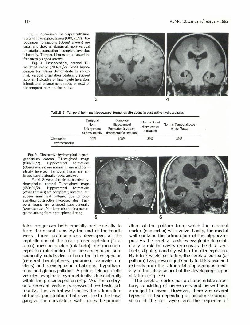
Temporal horn characteristics in patients with ACC, lissencephaly (lis), lobar and semilobar holoprosencephaly (holo) are summarized in Table 2. Analysis showed that 75% of patients with brain anomalies had enlargement of the temporal horns, most prominently involving the inferolateral aspects of the ventricle (8/8 ACC, 4/41is, 0/ 4 holo) Figs. 3 and 4). Hippocampal formations were small in 62% of patients (6/8 ACC, 3/4 lis, 1/4 holo). The hippocampal formations also demonstrated an abnormal, more vertically oriented morphologic appearance than normal hippocampal formations, suggesting incomplete inversion of the hippocampal formation, in 82% of patients (8/8 ACC, 4/4 lis, 1/4 holo). Focal thinning of the white matter lateral to the temporal horn was seen in 50% of patients (8/8 ACC, 0/4 lis, 0/4 holo). Abnormalities of the temporal horn, hip-
AJNR: 13, January/February 1992 117
TABLE 1: Temporal horn and hippocampal formation features in 20 normal individuals
Normal Sized Temporal Horn , Thin, Crescentic
Configuration
Complete Hippocampal
Formation Inversion (Horizontal Orientation)
Normal-Sized Hippocampal Formation
Normal Temporal Lobe White Matter Thickness
Healthy individuals 100% 100% 100% 100%
A B
Fig. 2 . A, Normal hippocampal formation and temporal horn , coronal T -1 weighted image (600/12/4). D = dentate gyrus; H = hippocampus; s = subiculum; temporal horn = closed arrow; choroid plexus = open arrow. Note that the dentate gyrus and hippocampus are seen collectively as a solitary oval-shaped gray matter structure with a horizontal orientation.
B, Simplified schematic diagram of the normal mature hippocampal formation and temporal horn depicted in the coronal plane. CF= choroidal fissure; CP= choroid plexus; D = dentate gyrus; F = fimbria; H-hippocampus; HF = hippocampal fissure; S = subiculum; T = temporal horn.
TABLE 2: Temporal horn and hippocampal formation alterations in congenital brain anomalies
Temporal Horn Incomplete
Enlargement Hippocampal Small Hippocampal Focal White
lnferolaterally Formation Inversion Formation Matter Thinning (Vertical Orientation)
Agenesis of the Corpus 100% Callosum
Lissencephaly 100% Lobar/ semilobar 0
Holoprosencephaly
pocampal formations, and/or adjacent white matter were most commonly seen in patients with ACC (Fig. 3) and lissencephaly (Fig. 4); patients with holoprosencephaly, in this series, did not share the temporal horn characteristics seen in the other two groups of patients.
Hydrocephalus
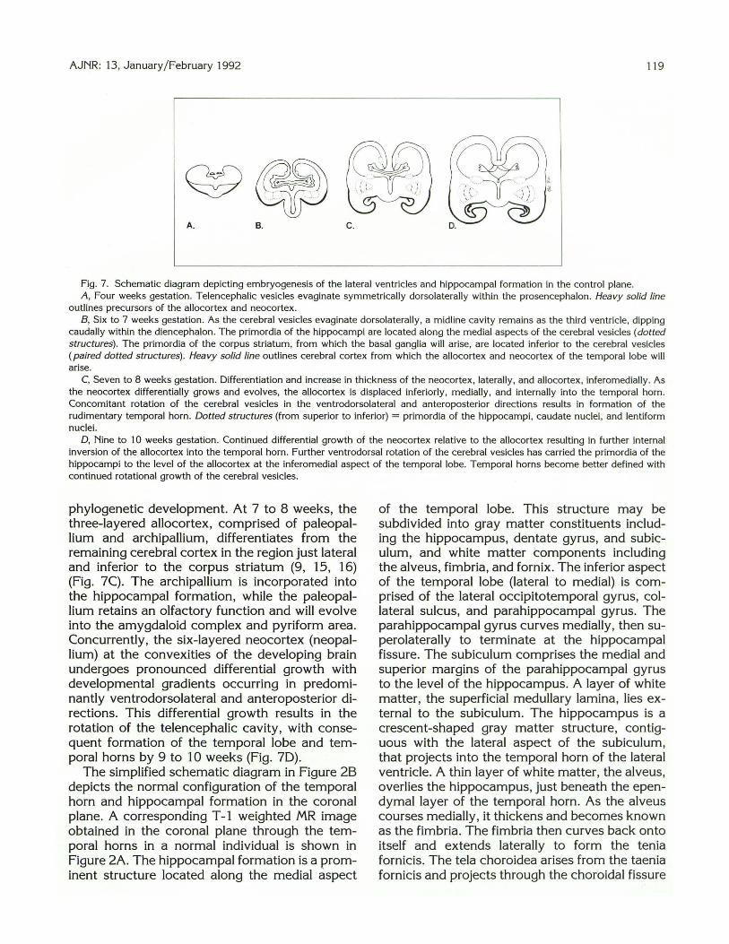
Temporal horn alterations in patients with obstructive hydrocephalus are shown in Table 3. All patients with isolated obstructive hydrocephalus showed enlargement of the temporal horns, most pronounced in the superior-lateral region. Hippocampal formations showed normal, complete inversion and were of normal size in 17 of 20 patients (Fig. 5); the only exceptions included patients with severe hydrocephalus, where the hippocampi were flattened along the inferior margin of the temporal horn (Fig. 6). Temporal lobe white matter was normal in the 17 patients with
100% 75% 100%
100% 75% 0 25% 25% 0
mild or moderate hydrocephalus; diffuse white matter thinning inferolaterally was observed in the three patients with severe hydrocephalus.
Discussion
Development of the Normal Temporal Horn and Hippocampal Formation
To better understand the configuration of the temporal horns in patients with congenital brain anomalies, a brief review of the embryologic development of the lateral ventricles and the hippocampal formation in the fetus is useful (7-14). The neural tube begins to form at approximately 17 days after fertilization as a pair of neural folds forms on the dorsal surface of the embryonic disk. As the actin myofibrils within the neural folds contract, the lips of the folds are drawn medially and first fuse at the rhombencephalon. From this point, fusion of the neural
118
Fig. 3. Agenesis of the corpus callosum, coronal Tl-weighted image (600/20/2). Hippocampal formations (closed arrows) are small and show an abnormal, more vertical orientation, suggesting incomplete inversion bilaterally. Temporal horns are enlarged inferolaterally (open arrows).
Fig. 4. Lissencephaly , coronal Tlweighted image (700/20/2). Small hippocampal formations demonstrate an abnormal, vertical orientation bilaterally (closed arrows), indicative of incomplete inversion. lnferolateral enlargement (open arrows) of the temporal horns is also noted.
AJNR: 13, January/ February 1992
3 4
TABLE 3: Temporal horn and hippocampal formation alterations in obstructive hydrocephalus
Complete Temporal
Horn
Enlargement
Superolaterally
Hippocampal Normal-Sized
Hippocampal Formation
Normal Temporal Lobe
White Matter Formation Inversion
(Horizonta l Orientation)
Obstructive
Hydrocephalus
100%
Fig. 5. Obstructive hydrocephalus, postgadolinium coronal Tl-weighted image (800/30/2). Hippocampal formations (closed arrows) are normal in size and completely inverted. Temporal horns are enlarged superolaterally (open arrows).
Fig. 6. Severe, chronic obstructive hydrocephalus, coronal Tl-weighted image (650/20/2). Hippocampal formations (closed arrows) are completely inverted, but appear small and flattened due to longstanding obstructive hydrocephalus. Temporal horns are enlarged superolaterally (open arrows). M =large obstructing meningioma arising from right sphenoid wing.
5
100%
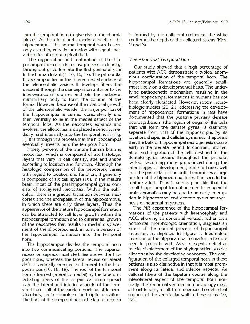
folds progresses both cranially and caudally to form the neural tube. By the end of the fourth week, three protuberances developed at the cephalic end of the tube: prosencephalon (forebrain) , mesencephalon (midbrain), and rhombencephalon (hindbrain). The prosencephalon subsequently subdivides to form the telencephalon (cerebral hemispheres, putamen, caudate nucleus) and diencephalon (thalamus, hypothalamus, and globus pallidus). A pair of telencephalic vesicles evaginate symmetrically dorsolaterally within the prosencephalon (Fig. 7 A). The embryonic cerebral vesicle possesses three basic primordia. The ventral wall carries the primordium of the corpus striatum that gives rise to the basal ganglia . The dorsolateral wall carries the primor-
85% 85%
6
dium of the pallium from which the cerebral cortex (neocortex) will evolve. Lastly, the medial wall contains the primordium of the hippocampus. As the cerebral vesicles evaginate dorsolaterally, a midline cavity remains as the third ventricle, dipping caudally within the diencephalon. By 6 to 7 weeks gestation, the cerebral cortex (or pallium) has grown significantly in thickness and extends from the primordial hippocampus medially to the lateral aspect of the developing corpus striatum (Fig. 7B).
The cerebral cortex has a characteristic structure, consisting of nerve cells and nerve fibers arranged in layers. However, there are several types of cortex depending on histologic composition of the cell layers and the sequence of
AJNR: 13, January/February 1992 119
A. B. C.
Fig. 7. Schematic diagram depicting embryogenesis of the lateral ventricles and hippocampal formation in the control plane. A, Four weeks gestation. Telencephalic vesicles evaginate symmetrically dorsolaterally within the prosencephalon. Heavy solid line
outlines precursors of the allocortex and neocortex. B, Six to 7 weeks gestation. As the cerebral vesicles evaginate dorsolaterally, a midline cavity remains as the third ventricle, dipping
caudally within the diencephalon. The primordia of the hippocampi are located along the medial aspects of the cerebral vesicles (dotted structures). The primordia of the corpus striatum, from which the basal ganglia will arise, are located inferior to the cerebral vesicles (paired dotted structures). Heavy solid line outlines cerebral cortex from which the allocortex and neocortex of the temporal lobe will arise.
C, Seven to 8 weeks gestation. Differentiation and increase in thickness of the neocortex, laterally, and allocortex, inferomedially. As the neocortex differentially grows and evolves, the allocortex is displaced inferiorly , medially, and internally into the temporal horn. Concomitant rotation of the cerebral vesicles in the ventrodorsolateral and anteroposterior directions results in formation of the rudimentary temporal horn. Dotted structures (from superior to inferior) = primordia of the hippocampi, caudate nuclei , and lentiform nuclei.
D, Nine to 10 weeks gestation. Continued differential growth of the neocortex relative to the allocortex resulting in further internal inversion of the allocortex into the temporal horn. Further ventrodorsal rotation of the cerebral vesicles has carried the primordia of the hippocampi to the level of the allocortex at the inferomedial aspect of the temporal lobe. Temporal horns become better defined with continued rotational growth of the cerebral vesicles.
phylogenetic development. At 7 to 8 weeks, the three-layered allocortex, comprised of paleopallium and archipallium, differentiates from the remaining cerebral cortex in the region just lateral and inferior to the corpus striatum (9, 15, 16) (Fig. 7C). The archipallium is incorporated into the hippocampal formation, while the paleopallium retains an olfactory function and will evolve into the amygdaloid complex and pyriform area. Concurrently, the six-layered neocortex (neopallium) at the convexities of the developing brain undergoes pronounced differential growth with developmental gradients occurring in predominantly ventrodorsolateral and anteroposterior directions. This differential growth results in the rotation of the telencephalic cavity, with consequent formation of the temporal lobe and temporal horns by 9 to 10 weeks (Fig. 7D).
The simplified schematic diagram in Figure 28 depicts the normal configuration of the temporal horn and hippocampal formation in the coronal plane. A corresponding T -1 weighted MR image obtained in the coronal plane through the temporal horns in a normal individual is shown in Figure 2A. The hippocampal formation is a prominent structure located along the medial aspect
of the temporal lobe. This structure may be subdivided into gray matter constituents including the hippocampus, dentate gyrus, and subiculum, and white matter components including the alveus, fimbria, and fornix. The inferior aspect of the temporal lobe (lateral to medial) is comprised of the lateral occipitotemporal gyrus, collateral sulcus, and parahippocampal gyrus. The parahippocampal gyrus curves medially, then superolaterally to terminate at the hippocampal fissure. The subiculum comprises the medial and superior margins of the parahippocampal gyrus to the level of the hippocampus. A layer of white matter, the superficial medullary lamina, lies external to the subiculum. The hippocampus is a crescent-shaped gray matter structure, contiguous with the lateral aspect of the subiculum, that projects into the temporal horn of the lateral ventricle. A thin layer of white matter, the alveus, overlies the hippocampus, just beneath the ependymal layer of the temporal horn. As the alveus courses medially, it thickens and becomes known as the fimbria. The fimbria then curves back onto itself and extends laterally to form the tenia fornicis. The tela choroidea arises from the taenia fornicis and projects through the choroidal fissure
120
into the temporal horn to give rise to the choroid plexus. At the lateral and superior aspects of the hippocampus, the normal temporal horn is seen only as a thin, curvilinear region with signal characteristics of cerebrospinal fluid.
The organization and maturation of the hippocampal formation is a slow process, extending throughout gestation into the first postnatal year in the human infant (7, 10, 16, 17). The primordial hippocampus lies in the infereomedial surface of the telencephalic vesicle. It develops fibers that descend through the diencephalon anterior to the interventricular foramen and join the ipsilateral mammillary body to form the column of the fornix . However, because of the rotational growth of the telencephalic vesicle, the major portion of the hippocampus is carried dorsolaterally and then ventrally to lie in the medial aspect of the temporal lobe. As the neocortex expands and evolves, the allocortex is displaced inferiorly, medially, and internally into the temporal horn (Fig. 1). It is through this process that the hippocampus eventually "inverts" into the temporal horn.
Ninety percent of the mature human brain is neocortex, which is composed of six histologic layers that vary in cell density, size and shape according to location and function. Although the histologic composition of the neocortex varies with regard to location and function , it generally is composed of six cell layers (16). In the mature brain, most of the parahippocampal gyrus consists of six-layered neocortex. Within the subiculum there is a gradual transition between neocortex and the archipallium of the hippocampus, in which there are only three layers. Thus the appearance of the mature hippocampal formation can be attributed to cell layer growth within the hippocampal formation and to differential growth of the neocortex that results in medial displacement of the allocortex and, in turn, inversion of the hippocampal formation into the temporal horn.
The hippocampus divides the temporal horn into two communicating portions. The superior recess or supracornual cleft lies above the hippocampus, whereas the lateral recess or lateral cleft is vertically oriented and lateral to the hippocampus (10, 18, 19). The roof of the temporal horn is formed (lateral to medial) by the tapetum, radiating fibers of the corpus callosum spread over the lateral and inferior aspects of the temporal horn , tail of the caudate nucleus, stria semicircularis, tenia choroidea, and optic radiation. The floor of the temporal horn (the lateral recess)
AJNR: 13, January / February 1992
is formed by the collateral eminence, the white matter at the depth of the collateral sulcus (Figs. 2 and 3).
The Abnormal Temporal Horn
Our study showed that a high percentage of patients with ACC demonstrate a typical anomalous configuration of the temporal horn. The hippocampal formations are generally small, most likely on a developmental basis. The underlying pathogenetic mechanism resulting in the small hippocampal formations in humans has not been clearly elucidated. However, recent neurobiologic studies (20, 21) addressing the development of hippocampal formations in rats have documented that the putative primary dentate neuroepithelium (the region of origin of the cells that will form the dentate gyrus) is distinctly separate from that of the hippocampus by its location, shape, and cellular dynamics. It appears that the bulk of hippocampal neurogenesis occurs early in the prenatal period. In contrast, proliferation and migration of the cells destined for the dentate gyrus occurs throughout the prenatal period, becoming more pronounced during the later stages of development, and continues well into the postnatal period until it comprises a large portion of the hippocampal formation seen in the mature adult. Thus it seems plausible that the small _ hippocampal formation seen in congenital brain anomalies may be due to an early interuption in hippocampal and dentate gyrus neurogenesis or neuronal migration.
The MR appearance of the hippocampal formations of the patients with lissencephaly and ACC, showing an abnormal vertical, rather than horizontal, morphologic orientation, suggests an arrest of the normal process of hippocampal inversion, as depicted in Figure 1. Incomplete inversion of the hippocampal formation, a feature seen in patients with ACC, suggests defective medial displacement of the phylogenetically older allocortex by the developing neocortex. The configuration of the enlarged temporal horn in these patients is also distinctive in that it is most prominent along its lateral and inferior aspects. As callosal fibers of the tapetum course along the inferolateral aspect of the temporal horn normally, the abnormal ventricular morphology may, at least in part, result from decreased mechanical support of the ventricular wall in these areas (10, 22).
AJNR: 13, January/ February 1992
In lissencephaly, a disorder of incomplete suication, the anomalous appearance of the temporal horn and hippocampal formation is similar to that seen in ACC. Interestingly, the pattern of temporal lobe anomalies in lissencephalic brains is similar to findings described in the pathology literature regarding premature infants, who also have poorly developed brains with incomplete sulcation (22, 23). Therefore, a lack of development of the neocortex and incomplete proliferation/migration of the dentate cells seem particularly likely in these patients (23).
With the exception of one individual, our group of patients with lobar and semilobar holoprosencephaly did not demonstrate incomplete development and inversion of the hippocampal formations or abnormal temporal horns. This finding was surprising because incomplete or lack of development of the hippocampal formations and temporal horns has been described in the sonographic (24) and pathology literature (23, 25) as a finding in holoprosencephaly. The discrepancy of our findings with these previous studies may reflect our population of patients with holoprosencephaly who had relatively well-developed brains (ie, lobar and semilobar holoprosencephaly); in contradistinction, many of the patients detected prenatally and studied pathologically have severe (alobar) holoprosencephaly and, presumably, less well-developed hippocampal formations.
The pattern of temporal horn enlargement observed in patients with isolated hydrocephalus differed from that seen in the patients with congenital malformations in that the morphology was grossly normal-the structures around the ventricle appeared merely compressed. The hippocampal formation was normal in size and completely inverted, and the temporal lobe white matter was of normal thickness in 85%. Exceptions to these generalizations were found in those patients with severe hydrocephalus; in this group, the hippocampal formations were completely inverted but flattened along the inferior aspect of the dilated temporal horn, presumably by the high intraventricular pressure. The pattern of temporal horn dilatation was also different compared to those patients with malformations. Hydrocephalic patients showed dilatation of the "natural" recesses of the temporal horn, ie, enlargement was most pronounced superiorly and laterally.
It would be interesting to study a group of patients with lissencephaly or ACC and hydro-
121
cephalus to see if the present analysis allows segregation of those patients with hydrocephalus and underlying brain malformations from those with uncomplicated hydrocephalus. Unfortunately, we were unable to find any such patients in our files . Nonetheless, it is obvious from this analysis that patients with lissencephaly and ACC can have temporal horn enlargement in the absence of hydrocephalus and that, if a question of hydrocephalus is present, it should be possible to confirm or eliminate that question based on the morphology of the temporal horn enlargement.
To summarize, we have compared the patterns of temporal horn enlargement in patients with congenital brain malformations to those in patients with hydrocephalus. Distinct differences were present in the morphology of the temporal horn and surrounding brain in these disorders, with the anomalous brains showing enlargement as a result of incomplete development and the hydrocephalic brains showing enlargement as a result of increased intraventricular pressure. Temporal horn enlargement in lissencephaly and ACC should not be misinterpreted as hydrocephalus; analysis of temporal lobe morphology will allow differentiation if any question exists.
Acknowledgment
We thank Clark Carrol , for contributing an MR study of a patient with lissencephaly (Fig. 5).
References
1. Naidich TP, Daniels DL, Haughton VM, Williams A, Pojunas K ,
Palacios E. Hippocampal formation and related structures of the
limbic lobe: anatomic-MR correlation. I. Surface features and coronal
sections. Radiology 1987;162:747- 754 2. Naidich TP, Daniels DL, Haughton VM, et al. Hippocampal formation
and related structures of the limbic lobe: anatomic-MR correlation. II.
Sagittal sections. Radiology 1987;162:755-761 3. A tlas SW, Zimmerman RA, Bilaniuk L T, et al. Corpus ca llosum and
limbic system: neuroanatomic MR evaluation of developmental
anomalies. Radiology 1986; 160:355- 362 4. Barkovich AJ . Pediatric neuroimaging. New York: Raven Press,
1990:77-121 5. Heinz ER, Ward A, Drayer BP, Dubois PJ. Distinction between
obstructive and atrophic dilatation of ventricles in children. J Comput
Assist Tomogr 1980;4:320-325 6. Sjaastad 0, Skalpe LO, Engeset A. The width of the temporal horn
and the differential diagnosis between pressure hydrocephalus and
hydrocephalus ex vacuo. Neurology 1969; 19:1087- 1093 7. Green JD. The hippocam pus. Physiol Rev 1964;44:561-608 8. Humphrey T . The development of the human hippocampal fissure. J
Anat 1967; 101:655-676 9. Kier EL. The cerebral ventricles: a phylogenetic and ontogenetic
study. In : Newton TH, Potts DG, eds. Radiology of the skull and brain:
anatomy and pathology. Vol. 3. Saint Louis, CV Mosby, 1977: 2787-2914
122
10. Deck MDF. The latera l ventricles. In: Newton TH , Potts DG, eds.
Radiology of the skull and brain: ventricles and cisterns. Vol. 4. Saint
Louis: CV Mosby, 1978:3489-3587
11 . Swanson LW. The hippocampus and the concept of the limbic
system. In : Seifert W, ed. Neurobiology of the hippocampus. New
York ; Academic Press, 1983:3-19
12. Angevine JB. Development of the hippocampal region. In : Isaacson
RL, Pribram KH, eds. Hippocampus. New York: Plenum Press,
1975:61-94
13. Bates JJ, Netsky MG. Developmenta l anomalies of the horns of the
lateral ventricles. J Neurapatho/ Exp Neural 1955; 14:3 16-325
14. Gilles FH. Telencephalon medium and the olfacto-cerebra l outpuch
ing. In: Gilles FH , Leviton A, Dooling EC, eds. The developing human
brain. Bristol, England: John Wright PSG Inc., 1983:59-86
15. Stanfield BB, Cowan WM. The development of the hippocampal
region. In: Peters A , Jones EG, eds. Cerebral cortex: development
and maturation of the cerebral cortex. Vol 7. New York: Plenum
Press, 1988:91-131
16. Barr ML. The human nervous system. Hagerstown , MD: Harper and
Row Publishers 1979:2 16-221
AJNR: 13, January/ February 1992
17. Hines M. Studie~ in the growth and differentiation of the telencephalon
in man: the fissura hippocampi. J Comp Neura/ 1922;34:73-171 18. Childe AE, Penfield W. Anatomic and pneumographic studies of the
temporal horn . Arch Neural Psychiatry 1937;37: 1021-1034 19. Lindgren E. A pneumoencephalographic study of the temporal horn
with special reference to tumors in the tempora l region. Acta Radio/
(Suppl) 1948;69: 1-151 20. Altman J , Bayer SA. Mosaic organization of the hippocampal neu
roephtielium and the multiple germinal sources of dentate granule
cells. J Comp Neuro/1990;310:325-342
21. Altman J , Bayer SA. Migration and distribution of two populations of
hippocampal granule cell precursors during the perinatal and post
natal periods. J Comp Neuro/1990;301:365-381 22. Probst FP. The prasencephalies. Berlin: Springer-Verlag, 1979:3- 65 23. Barkovich AJ, Koch TK, Carrol CL. The spectrum of lissencephaly :
report of ten cases analyzed by MRI. Ann Neural (in press)
24. Filly RA, Chinn DH, Callen PW. A lobar holoprosencephaly: ultraso
nographic prenatal diagnosis. Radiology 1984; 151:455-459 25. Yakovlev Pl. Pathoarchitectonic studies of cerebral malformation. Ill.
Arhinencephalies (holotelencephalies). J Neurapatho/ Exp Neural
1959; 18:22-55