Embed Size (px)
Citation preview
c o r t e x 4 7 ( 2 0 1 1 ) 6 5 9e6 7 3
ava i lab le at www.sc ienced i rec t . com
journa l homepage : www.e lsev ier . com/ loca te / cor tex
Research report
Are left fronto-temporal brain areas a prerequisitefor normal music-syntactic processing?
Daniela Sammler a,*, Stefan Koelsch a,b,** and Angela D. Friederici a
aMax Planck Institute for Human Cognitive and Brain Sciences, Leipzig, GermanybCluster of Excellence “Languages of Emotion”, Freie Universitat Berlin, Germany
a r t i c l e i n f o
Article history:
Received 29 July 2009
Reviewed 28 October 2009
Revised 5 January 2010
Accepted 7 April 2010
Action editor Stefano Cappa
Published online 1 May 2010
Keywords:
Music
Language
Syntax
ERAN
ELAN
Brain lesions
* Corresponding author. Department of Neur1a, 04103 Leipzig, Germany.** Corresponding author. Cluster of ExcellencGermany.
E-mail addresses: [email protected]/$ e see front matter ª 2010 Elsevdoi:10.1016/j.cortex.2010.04.007
a b s t r a c t
An increasing number of neuroimaging studies in music cognition research suggest that
“language areas” are involved in the processing of musical syntax, but none of these
studies clarified whether these areas are a prerequisite for normal syntax processing in
music. The present electrophysiological experiment tested whether patients with lesions
in Broca’s area (N¼ 6) or in the left anterior temporal lobe (N¼ 7) exhibit deficits in the
processing of structure in music compared to matched healthy controls (N¼ 13). A chord
sequence paradigmwas applied, and the amplitude and scalp topography of the Early Right
Anterior Negativity (ERAN) was examined, an electrophysiological marker of musical
syntax processing that correlates with activity in Broca’s area and its right hemisphere
homotope. Left inferior frontal gyrus (IFG) (but not anterior superior temporal gyrus e
aSTG) patients with lesions older than 4 years showed an ERAN with abnormal scalp
distribution, and subtle behavioural deficits in detecting music-syntactic irregularities. In
one IFG patient tested 7 months post-stroke, the ERAN was extinguished and the behav-
ioural performance remained at chance level. These combined results suggest that the left
IFG, known to be crucial for syntax processing in language, plays also a functional role in
the processing of musical syntax. Hence, the present findings are consistent with the
notion that Broca’s area supports the processing of syntax in a rather domain-general way.
ª 2010 Elsevier Srl. All rights reserved.
1. Introduction Right Anterior Negativity (ERAN), a component of the event-
In 2001, Maess et al. (2001) stated that “Musical syntax is
processed in Broca’s area”, and suggested that this regionmay
“process syntactic information that is less language-specific
than previously believed”. More precisely, the authors recon-
structed the sources of the magnetic equivalent of the Early
opsychology, Max Planck
e “Languages of Emotion”
(D. Sammler), stefan.koelier Srl. All rights reserved
related potential (ERP) elicited by harmonically unexpected
chords and taken as an index for early music-syntactic pro-
cessing (Koelsch et al., 2000; Koelsch, 2009). Most intriguingly,
the dipoles of the mERAN (i.e., the ERAN recorded with mag-
netoencephalography e MEG) were localised in Broca’s area
(inferior Brodman area 44), a brain region known to be
Institute for Human Cognitive and Brain Sciences, Stephanstrasse
, Freie Universitat Berlin, Habelschwerdter Allee 45, 14195 Berlin,
[email protected] (S. Koelsch)..
1 Patients with circumscribed lesions of the right IFG or rightaSTG show up only rarely in clinical settings, most probablybecause they do not develop those massive (language) deficits aspatients with comparable left hemispheric lesions. For example,no patient with focal right IFG lesion and spared aSTG was foundamong the more than 2000 records of our data base.
c o r t e x 4 7 ( 2 0 1 1 ) 6 5 9e6 7 3660
involved in syntactic analysis in language (Friederici, 2006;
Grodzinsky and Santi, 2008), and its right hemisphere homo-
tope. The present electroencephalography (EEG) study aimed
to strengthen the proposition of Maess et al. (2001) by testing
whether lesions in left fronto-temporal brain regions lead to
deficits in music-syntactic processing.
During the past 10 years, a series of studies has referred to
the neuroanatomical and functional parallels of syntax pro-
cessing in music and language (for reviews, see Koelsch, 2005;
Patel, 2008). Music, like language, is a progression of percep-
tually discrete elements (e.g., chords and words) that are
arranged according to specific rules (e.g., harmonic and mor-
phosyntactic principles) to form meaningful sequences (e.g.,
musical phrases and sentences; Lerdahl and Jackendoff, 1983;
Patel, 2003; Koelsch and Siebel, 2005). We effortlessly acquire
implicit representations of these syntactic systems by mere
exposure during early childhood (Koelsch et al., 2003; Kuhl,
2004; McMullen and Saffran, 2004; Oberecker et al., 2005;
Tillmann et al., 2000), and automatically access this knowl-
edge to smoothly and accurately integrate successively
incoming chords and words, and to predict forthcoming
elements when listening to music or speech.
Neuroimaging studies have suggested that these parallels
of syntax processing in music and language map onto a partly
common neural architecture. Broca’s area has been associ-
ated with syntactic and hierarchical processing in language
(e.g., Grodzinsky and Santi, 2008; Friederici et al., 2006;
Makuuchi et al., 2009; for a review see Grodzinsky and
Friederici, 2006). For example, the neural generators of the
language-related Early Left Anterior Negativity (ELAN), an ERP
component evoked by word-category errors in sentences
(Neville et al., 1991; Friederici et al., 1993; Hahne and Friederici,
1999; Lau et al., 2006) have been localised in Broca’s area and
its right hemisphere homologue (Friederici et al., 2000;
Knosche et al., 1999; additional sources of the ELAN were
found in the anterior superior temporal gyrus e aSTG bilat-
erally). As mentioned above, the dipoles of the music-related
ERAN were also located in the left and right inferior frontal
gyrus (IFG; Maess et al., 2001), in fact, close to those of the
ELAN. Likewise, functional magnetic resonance imaging
(fMRI) studies yielded activations in the IFG and the aSTG
during the presentation of both music-syntactic irregularities
(Koelsch et al., 2002a, 2005a; Tillmann et al., 2003, 2006;
Krumhansl, 2004; Minati et al., 2008), or syntactically incor-
rect sentences (Brauer and Friederici, 2007; Friederici et al.,
2003; Ruschemeyer et al., 2005), although activations were
mostly bilateral with right-hemispheric weighting during the
processing of music, and were clearly left-dominant during
the processing of language. These findings led to the
assumption that syntax processing in music and language
may partly overlap in fronto-temporal brain areas, particu-
larly in the language-dominant left hemisphere (Patel, 2003).
However, neither MEG nor fMRI are able to ultimately
clarify whether these brain regions are a prerequisite for
syntax processing. MEG source localisation has to face the
ambiguity of the inverse problem, and fMRI results usually
reflect correlations between brain activations and an assumed
function resulting from the subtraction of two conditions. In
other words, such data may indicate an involvement of the
IFG and aSTG, even if these are not obligatory to syntax
processing, thus rendering it premature to conclude that these
areas are crucial for the processing of musical and/or
linguistic syntax. This question can be resolved by testing
whether the respective functions are disrupted in patients
with lesions in these brain areas.
In the language domain, numerous lesion studies prove the
necessityof the left inferior frontalandthe leftanterior temporal
lobe for linguistic syntax processing (Friederici and Kotz, 2003;
Stowe et al., 2005). For example, Broca’s aphasics exhibit defi-
cits in comprehending semantically reversible passives such as
“The boy was kissed by the girl.” which can only be correctly
understood when relying on syntactic information (Caplan and
Futter, 1986; Caplan et al., 1996; Caramazza and Zurif, 1976;
Davis et al., 2008). Likewise, patients with left anterior
temporal brain lesions exhibit marked deficits in comprehend-
ing complex morphosyntactic structures (Dronkers et al., 1994;
Grossman et al., 1998). Further evidence comes from ERP
studies showing that theELAN(indexing initial phrase-structure
building in sentences) disappears if left fronto-lateral or left
anterior temporalbrainareasaredamaged (Friederici etal., 1998,
1999; Friederici and Kotz, 2003; Kotz et al., 2003), suggesting the
necessity of these left hemispheric brain regions for fast
syntactic procedures during language processing.
In the music domain, the clinical literature on music-
syntactic processing is restricted to four behavioural case
studies (for an overview, see Stewart et al., 2006) that do not,
however, yield a precise spatial localisation of the perturbed
functions, because they tested either split-brain patients
(Tramo and Bharucha, 1991) or patients with bilateral lesion
configurations (Peretz, 1993; Peretz et al., 1994; Tramo et al.,
1990). A recent study included patients with a homogeneous
syntactic comprehension deficit in language, but with variable
lesion sites in the left hemisphere, not always including
Broca’s area or temporal regions (Patel et al., 2008). Therefore,
it is currently unknown whether left inferior frontal and left
anterior temporal brain structures are relevant for the pro-
cessing of syntax in music.
The present EEG study was designed to fill this gap and
tested whether patients with lesions encompassing the left
IFG (Broca’s area) or the left aSTG exhibit deficits in the pro-
cessing of musical structure. Special care was taken that
either the IFG or the aSTG but not both structures were
lesioned, to allow a possible dissociation of their function. No
patients with similarly circumscribed lesions in the homotope
right-hemispheric areas were tested.1 The ERAN was used as
an electrophysiological marker for intact or disrupted music-
syntactic processing. It was hypothesised that both patient
groups would exhibit a reduced amplitude and/or a topog-
raphy shift of the ERAN, as well as behavioural deficits in
detecting music-syntactically irregular chords (compared to
healthy controls). Such a result would provide direct evidence
for the necessity of the left inferior frontal and the left anterior
temporal lobe in musical syntax processing.
c o r t e x 4 7 ( 2 0 1 1 ) 6 5 9e6 7 3 661
2. Method
2.1. Participants
2.1.1. PatientsBecause the particular aim of this experiment was the local-
isation of music-syntactic functions, all patients were chosen
according to lesion site, not according to functional deficits
(for a reverse approach, see Patel et al., 2008). Two patient
groups were studied. The first lesion group comprised six
patients (one woman) with lesions encompassing the left IFG
including the pars opercularis and the pars triangularis (but
sparing the left aSTG). The second lesion group contained
seven patients (two women) with lesions in the left anterior
temporal lobe including the aSTG and the planum polare (but
sparing the left IFG). Heschl’s gyrus was intact in all patients
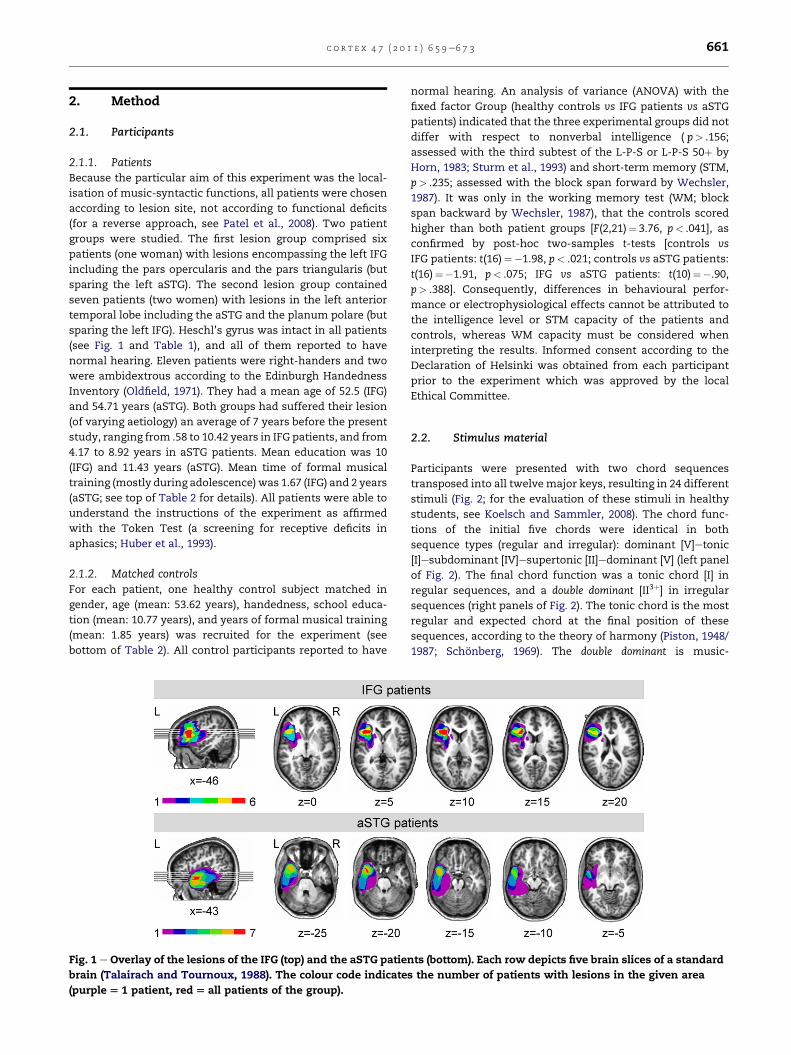
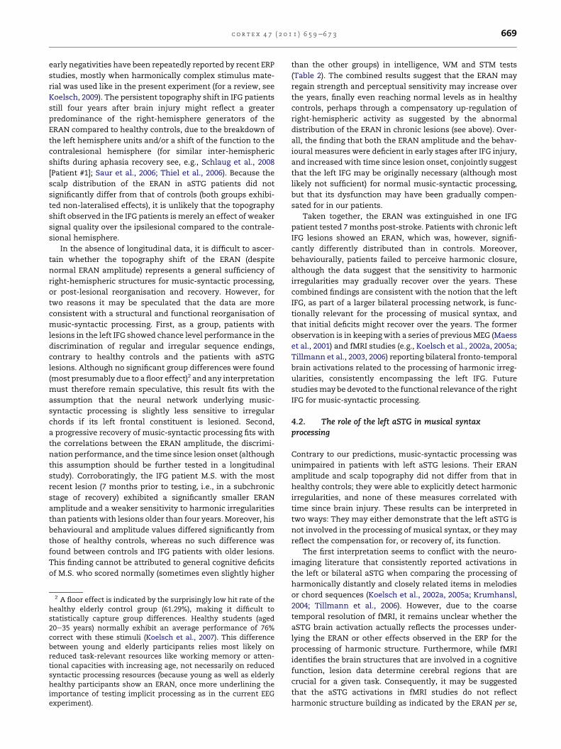
(see Fig. 1 and Table 1), and all of them reported to have
normal hearing. Eleven patients were right-handers and two
were ambidextrous according to the Edinburgh Handedness
Inventory (Oldfield, 1971). They had a mean age of 52.5 (IFG)
and 54.71 years (aSTG). Both groups had suffered their lesion
(of varying aetiology) an average of 7 years before the present
study, ranging from .58 to 10.42 years in IFG patients, and from
4.17 to 8.92 years in aSTG patients. Mean education was 10
(IFG) and 11.43 years (aSTG). Mean time of formal musical
training (mostly during adolescence) was 1.67 (IFG) and 2 years
(aSTG; see top of Table 2 for details). All patients were able to
understand the instructions of the experiment as affirmed
with the Token Test (a screening for receptive deficits in
aphasics; Huber et al., 1993).
2.1.2. Matched controlsFor each patient, one healthy control subject matched in
gender, age (mean: 53.62 years), handedness, school educa-
tion (mean: 10.77 years), and years of formal musical training
(mean: 1.85 years) was recruited for the experiment (see
bottom of Table 2). All control participants reported to have
Fig. 1 e Overlay of the lesions of the IFG (top) and the aSTG patie
brain (Talairach and Tournoux, 1988). The colour code indicate
(purple[ 1 patient, red[ all patients of the group).
normal hearing. An analysis of variance (ANOVA) with the
fixed factor Group (healthy controls vs IFG patients vs aSTG
patients) indicated that the three experimental groups did not
differ with respect to nonverbal intelligence ( p> .156;
assessed with the third subtest of the L-P-S or L-P-S 50þ by
Horn, 1983; Sturm et al., 1993) and short-term memory (STM,
p> .235; assessed with the block span forward by Wechsler,
1987). It was only in the working memory test (WM; block
span backward by Wechsler, 1987), that the controls scored
higher than both patient groups [F(2,21)¼ 3.76, p< .041], as
confirmed by post-hoc two-samples t-tests [controls vs
IFG patients: t(16)¼�1.98, p< .021; controls vs aSTG patients:
t(16)¼�1.91, p< .075; IFG vs aSTG patients: t(10)¼�.90,
p> .388]. Consequently, differences in behavioural perfor-
mance or electrophysiological effects cannot be attributed to
the intelligence level or STM capacity of the patients and
controls, whereas WM capacity must be considered when
interpreting the results. Informed consent according to the
Declaration of Helsinki was obtained from each participant
prior to the experiment which was approved by the local
Ethical Committee.
2.2. Stimulus material
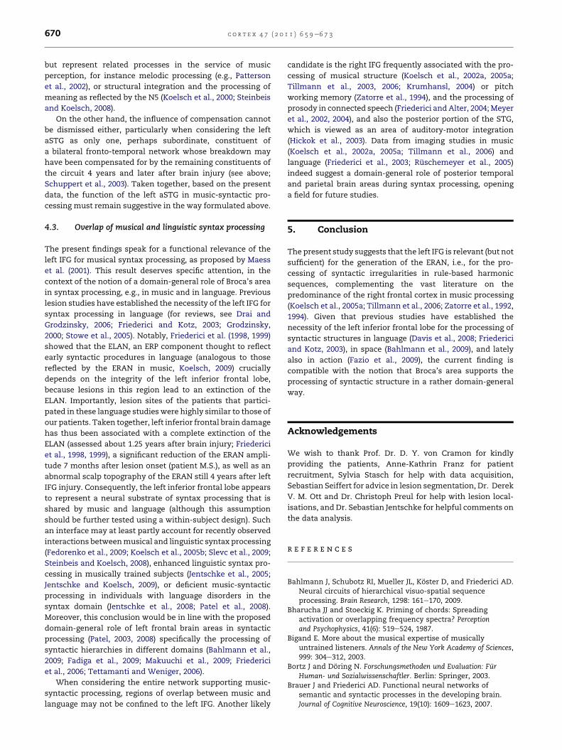
Participants were presented with two chord sequences
transposed into all twelvemajor keys, resulting in 24 different
stimuli (Fig. 2; for the evaluation of these stimuli in healthy
students, see Koelsch and Sammler, 2008). The chord func-
tions of the initial five chords were identical in both
sequence types (regular and irregular): dominant [V]etonic
[I]esubdominant [IV]esupertonic [II]edominant [V] (left panel
of Fig. 2). The final chord function was a tonic chord [I] in
regular sequences, and a double dominant [II3þ] in irregular
sequences (right panels of Fig. 2). The tonic chord is the most
regular and expected chord at the final position of these
sequences, according to the theory of harmony (Piston, 1948/
1987; Schonberg, 1969). The double dominant is music-
nts (bottom). Each row depicts five brain slices of a standard
s the number of patients with lesions in the given area
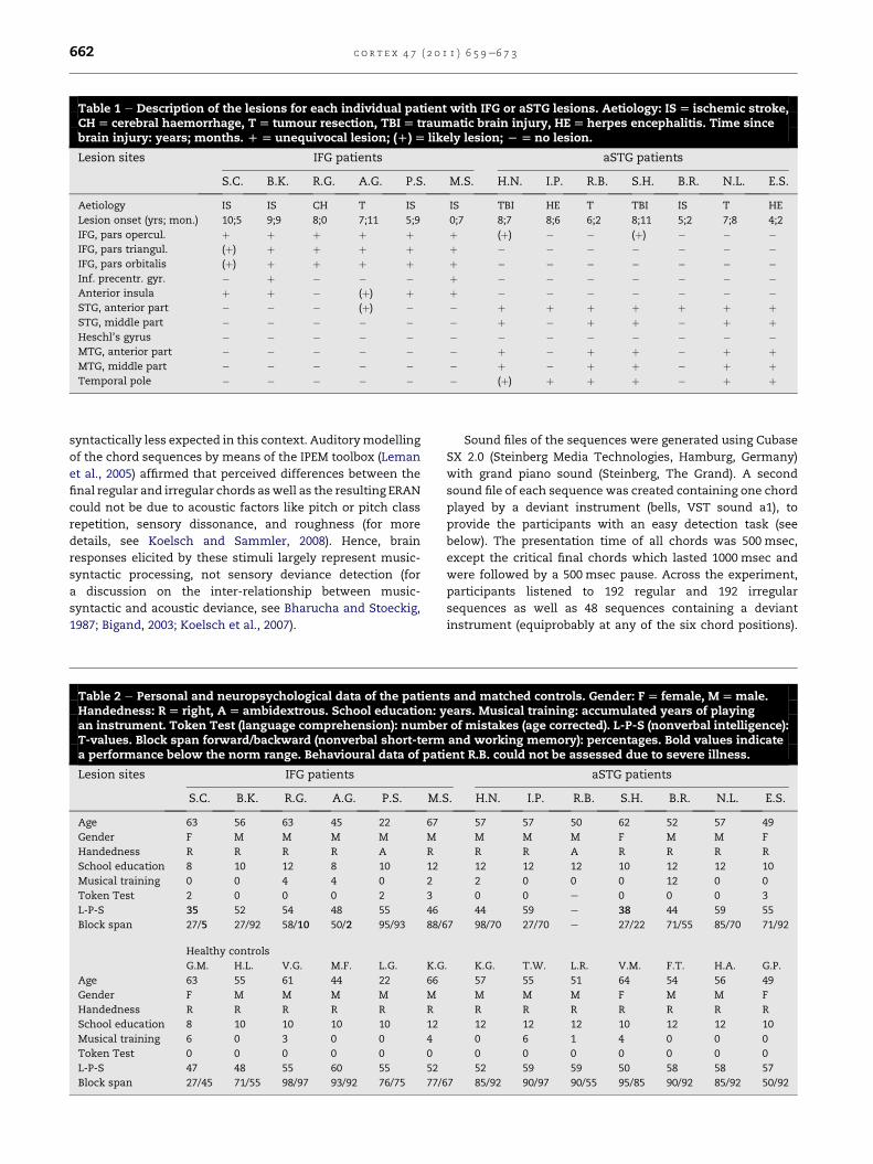
Table 1 e Description of the lesions for each individual patient with IFG or aSTG lesions. Aetiology: IS[ ischemic stroke,CH[ cerebral haemorrhage, T[ tumour resection, TBI[ traumatic brain injury, HE[herpes encephalitis. Time sincebrain injury: years; months. D[ unequivocal lesion; (D)[ likely lesion; L[no lesion.
Lesion sites IFG patients aSTG patients
S.C. B.K. R.G. A.G. P.S. M.S. H.N. I.P. R.B. S.H. B.R. N.L. E.S.
Aetiology IS IS CH T IS IS TBI HE T TBI IS T HE
Lesion onset (yrs; mon.) 10;5 9;9 8;0 7;11 5;9 0;7 8;7 8;6 6;2 8;11 5;2 7;8 4;2
IFG, pars opercul. þ þ þ þ þ þ (þ) � � (þ) � � �IFG, pars triangul. (þ) þ þ þ þ þ � � � � � � �IFG, pars orbitalis (þ) þ þ þ þ þ � � � � � � �Inf. precentr. gyr. � þ � � � þ � � � � � � �Anterior insula þ þ � (þ) þ þ � � � � � � �STG, anterior part � � � (þ) � � þ þ þ þ þ þ þSTG, middle part � � � � � � þ � þ þ � þ þHeschl’s gyrus � � � � � � � � � � � � �MTG, anterior part � � � � � � þ � þ þ � þ þMTG, middle part � � � � � � þ � þ þ � þ þTemporal pole � � � � � � (þ) þ þ þ � þ þ
c o r t e x 4 7 ( 2 0 1 1 ) 6 5 9e6 7 3662
syntactically less expected in this context. Auditorymodelling
of the chord sequences by means of the IPEM toolbox (Leman
et al., 2005) affirmed that perceived differences between the
final regular and irregular chords aswell as the resulting ERAN
could not be due to acoustic factors like pitch or pitch class
repetition, sensory dissonance, and roughness (for more
details, see Koelsch and Sammler, 2008). Hence, brain
responses elicited by these stimuli largely represent music-
syntactic processing, not sensory deviance detection (for
a discussion on the inter-relationship between music-
syntactic and acoustic deviance, see Bharucha and Stoeckig,
1987; Bigand, 2003; Koelsch et al., 2007).
Table 2 e Personal and neuropsychological data of the patientHandedness: R[ right, A[ ambidextrous. School education: yan instrument. Token Test (language comprehension): numbeT-values. Block span forward/backward (nonverbal short-terma performance below the norm range. Behavioural data of pati
Lesion sites IFG patients
S.C. B.K. R.G. A.G. P.S. M.
Age 63 56 63 45 22 67
Gender F M M M M M
Handedness R R R R A R
School education 8 10 12 8 10 12
Musical training 0 0 4 4 0 2
Token Test 2 0 0 0 2 3
L-P-S 35 52 54 48 55 46
Block span 27/5 27/92 58/10 50/2 95/93 88/
Healthy controls
G.M. H.L. V.G. M.F. L.G. K.G
Age 63 55 61 44 22 66
Gender F M M M M M
Handedness R R R R R R
School education 8 10 10 10 10 12
Musical training 6 0 3 0 0 4
Token Test 0 0 0 0 0 0
L-P-S 47 48 55 60 55 52
Block span 27/45 71/55 98/97 93/92 76/75 77/
Sound files of the sequences were generated using Cubase
SX 2.0 (Steinberg Media Technologies, Hamburg, Germany)
with grand piano sound (Steinberg, The Grand). A second
sound file of each sequence was created containing one chord
played by a deviant instrument (bells, VST sound a1), to
provide the participants with an easy detection task (see
below). The presentation time of all chords was 500 msec,
except the critical final chords which lasted 1000 msec and
were followed by a 500 msec pause. Across the experiment,
participants listened to 192 regular and 192 irregular
sequences as well as 48 sequences containing a deviant
instrument (equiprobably at any of the six chord positions).
s and matched controls. Gender: F[ female, M[male.ears. Musical training: accumulated years of playingr of mistakes (age corrected). L-P-S (nonverbal intelligence):and working memory): percentages. Bold values indicateent R.B. could not be assessed due to severe illness.
aSTG patients
S. H.N. I.P. R.B. S.H. B.R. N.L. E.S.
57 57 50 62 52 57 49
M M M F M M F
R R A R R R R
12 12 12 10 12 12 10
2 0 0 0 12 0 0
0 0 e 0 0 0 3
44 59 e 38 44 59 55
67 98/70 27/70 e 27/22 71/55 85/70 71/92
. K.G. T.W. L.R. V.M. F.T. H.A. G.P.
57 55 51 64 54 56 49
M M M F M M F
R R R R R R R
12 12 12 10 12 12 10
0 6 1 4 0 0 0
0 0 0 0 0 0 0
52 59 59 50 58 58 57
67 85/92 90/97 90/55 95/85 90/92 85/92 50/92
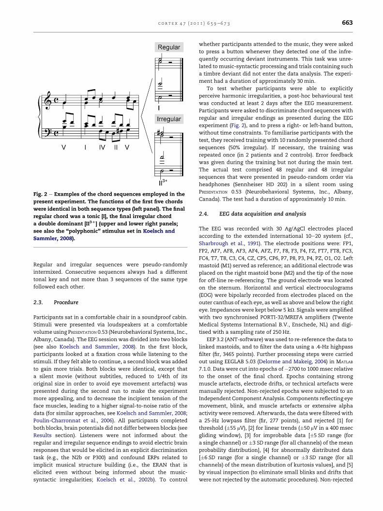
Fig. 2 e Examples of the chord sequences employed in the
present experiment. The functions of the first five chords
were identical in both sequence types (left panel). The final
regular chord was a tonic [I], the final irregular chord
a double dominant [II3D] (upper and lower right panels;
see also the “polyphonic” stimulus set in Koelsch and
Sammler, 2008).
c o r t e x 4 7 ( 2 0 1 1 ) 6 5 9e6 7 3 663
Regular and irregular sequences were pseudo-randomly
intermixed. Consecutive sequences always had a different
tonal key and not more than 3 sequences of the same type
followed each other.
2.3. Procedure
Participants sat in a comfortable chair in a soundproof cabin.
Stimuli were presented via loudspeakers at a comfortable
volumeusing PRESENTATION 0.53 (Neurobehavioral Systems, Inc.,
Albany, Canada). The EEG session was divided into two blocks
(see also Koelsch and Sammler, 2008). In the first block,
participants looked at a fixation cross while listening to the
stimuli. If they felt able to continue, a second block was added
to gain more trials. Both blocks were identical, except that
a silent movie (without subtitles, reduced to 1/4th of its
original size in order to avoid eye movement artefacts) was
presented during the second run to make the experiment
more appealing, and to decrease the incipient tension of the
face muscles, leading to a higher signal-to-noise ratio of the
data (for similar approaches, see Koelsch and Sammler, 2008;
Poulin-Charronnat et al., 2006). All participants completed
both blocks, brain potentials did not differ between blocks (see
Results section). Listeners were not informed about the
regular and irregular sequence endings to avoid electric brain
responses that would be elicited in an explicit discrimination
task (e.g., the N2b or P300) and confound ERPs related to
implicit musical structure building (i.e., the ERAN that is
elicited even without being informed about the music-
syntactic irregularities; Koelsch et al., 2002b). To control
whether participants attended to the music, they were asked
to press a button whenever they detected one of the infre-
quently occurring deviant instruments. This task was unre-
lated tomusic-syntactic processing and trials containing such
a timbre deviant did not enter the data analysis. The experi-
ment had a duration of approximately 30 min.
To test whether participants were able to explicitly
perceive harmonic irregularities, a post-hoc behavioural test
was conducted at least 2 days after the EEG measurement.
Participants were asked to discriminate chord sequences with
regular and irregular endings as presented during the EEG
experiment (Fig. 2), and to press a right- or left-hand button,
without time constraints. To familiarise participants with the
test, they received training with 10 randomly presented chord
sequences (50% irregular). If necessary, the training was
repeated once (in 2 patients and 2 controls). Error feedback
was given during the training but not during the main test.
The actual test comprised 48 regular and 48 irregular
sequences that were presented in pseudo-random order via
headphones (Sennheiser HD 202) in a silent room using
PRESENTATION 0.53 (Neurobehavioral Systems, Inc., Albany,
Canada). The test had a duration of approximately 10 min.
2.4. EEG data acquisition and analysis
The EEG was recorded with 30 Ag/AgCl electrodes placed
according to the extended international 10e20 system (cf.,
Sharbrough et al., 1991). The electrode positions were: FP1,
FP2, AF7, AF8, AF3, AF4, AFZ, F7, F8, F3, F4, FZ, FT7, FT8, FC3,
FC4, T7, T8, C3, C4, CZ, CP5, CP6, P7, P8, P3, P4, PZ, O1, O2. Left
mastoid (M1) served as reference; an additional electrode was
placed on the right mastoid bone (M2) and the tip of the nose
for off-line re-referencing. The ground electrode was located
on the sternum. Horizontal and vertical electrooculograms
(EOG) were bipolarly recorded from electrodes placed on the
outer canthus of each eye, aswell as above and below the right
eye. Impedances were kept below 5 kU. Signals were amplified
with two synchronised PORTI-32/MREFA amplifiers (Twente
Medical Systems International B.V., Enschede, NL) and digi-
tised with a sampling rate of 250 Hz.
EEP 3.2 (ANT-software) was used to re-reference the data to
linked mastoids, and to filter the data using a .4-Hz highpass
filter (fir, 3465 points). Further processing steps were carried
out using EEGLAB 5.03 (Delorme and Makeig, 2004) in MATLAB
7.1.0. Data were cut into epochs of�2700 to 1000 msec relative
to the onset of the final chord. Epochs containing strong
muscle artefacts, electrode drifts, or technical artefacts were
manually rejected. Non-rejected epochs were subjected to an
Independent Component Analysis. Components reflecting eye
movement, blink, and muscle artefacts or extensive alpha
activity were removed. Afterwards, the data were filteredwith
a 25-Hz lowpass filter (fir, 277 points), and rejected [1] for
threshold (�55 mV), [2] for linear trends (�50 mV in a 400 msec
gliding window), [3] for improbable data [�5 SD range (for
a single channel) or �3 SD range (for all channels) of the mean
probability distribution], [4] for abnormally distributed data
[�6 SD range (for a single channel) or �3 SD range (for all
channels) of the mean distribution of kurtosis values], and [5]
by visual inspection (to eliminate small blinks and drifts that
were not rejected by the automatic procedures). Non-rejected
c o r t e x 4 7 ( 2 0 1 1 ) 6 5 9e6 7 3664
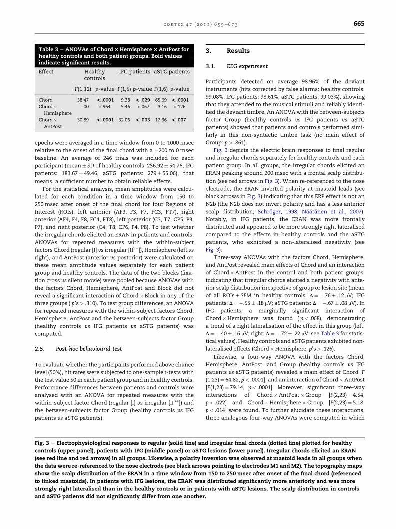
Table 3 e ANOVAs of Chord3Hemisphere3AntPost forhealthy controls and both patient groups. Bold valuesindicate significant results.
Effect Healthycontrols
IFG patients aSTG patients
F(1,12) p-value F(1,5) p-value F(1,6) p-value
Chord 38.47 <.0001 9.38 <.029 65.69 <.0001
Chord�Hemisphere
.00 >.964 5.46 <.067 3.16 >.126
Chord�AntPost
30.89 <.0001 32.06 <.003 17.36 <.007
c o r t e x 4 7 ( 2 0 1 1 ) 6 5 9e6 7 3 665
epochs were averaged in a time window from 0 to 1000 msec
relative to the onset of the final chord with a �200 to 0 msec
baseline. An average of 246 trials was included for each
participant (mean� SD of healthy controls: 256.92� 54.76, IFG
patients: 183.67� 49.46, aSTG patients: 279� 55.06), that
means, a sufficient number to obtain reliable effects.
For the statistical analysis, mean amplitudes were calcu-
lated for each condition in a time window from 150 to
250 msec after onset of the final chord for four Regions of
Interest (ROIs): left anterior (AF3, F3, F7, FC3, FT7), right
anterior (AF4, F4, F8, FC4, FT8), left posterior (C3, T7, CP5, P3,
P7), and right posterior (C4, T8, CP6, P4, P8). To test whether
the irregular chords elicited an ERAN in patients and controls,
ANOVAs for repeated measures with the within-subject
factors Chord (regular [I] vs irregular [II3þ]), Hemisphere (left vs
right), and AntPost (anterior vs posterior) were calculated on
these mean amplitude values separately for each patient
group and healthy controls. The data of the two blocks (fixa-
tion cross vs silent movie) were pooled because ANOVAs with
the factors Chord, Hemisphere, AntPost and Block did not
reveal a significant interaction of Chord� Block in any of the
three groups ( p’s> .310). To test group differences, an ANOVA
for repeated measures with the within-subject factors Chord,
Hemisphere, AntPost and the between-subjects factor Group
(healthy controls vs IFG patients vs aSTG patients) was
computed.
2.5. Post-hoc behavioural test
To evaluatewhether the participants performed above chance
level (50%), hit rates were subjected to one-sample t-tests with
the test value 50 in each patient group and in healthy controls.
Performance differences between patients and controls were
analysed with an ANOVA for repeated measures with the
within-subject factor Chord (regular [I] vs irregular [II3þ]) andthe between-subjects factor Group (healthy controls vs IFG
patients vs aSTG patients).
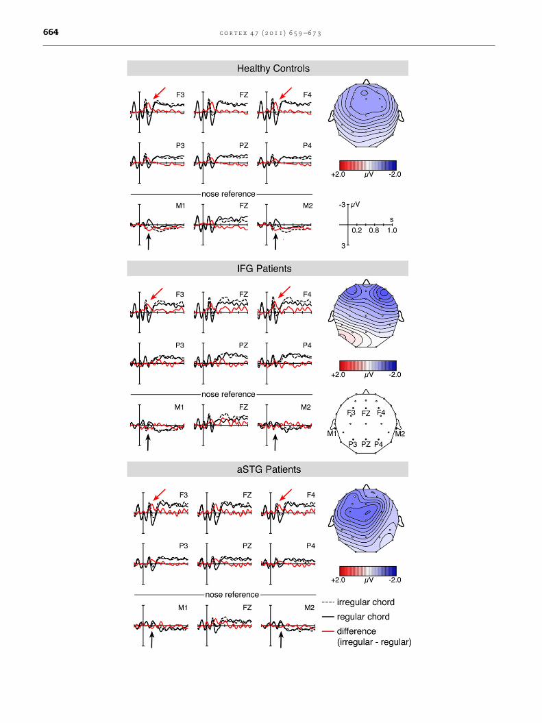
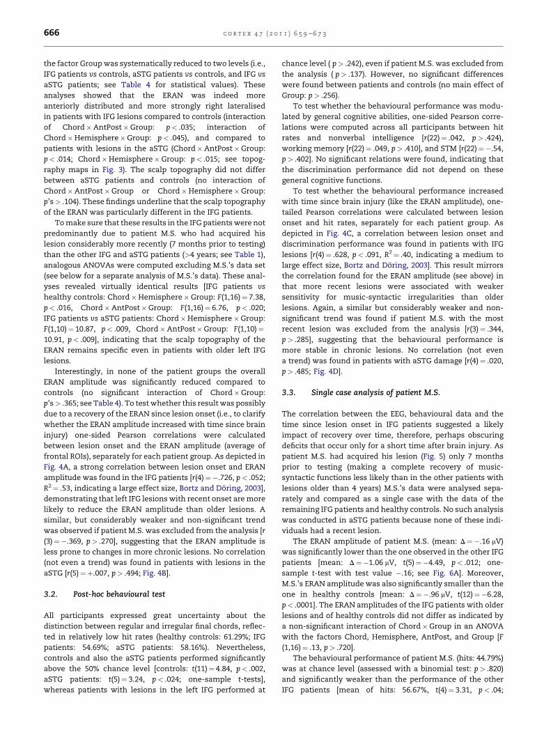
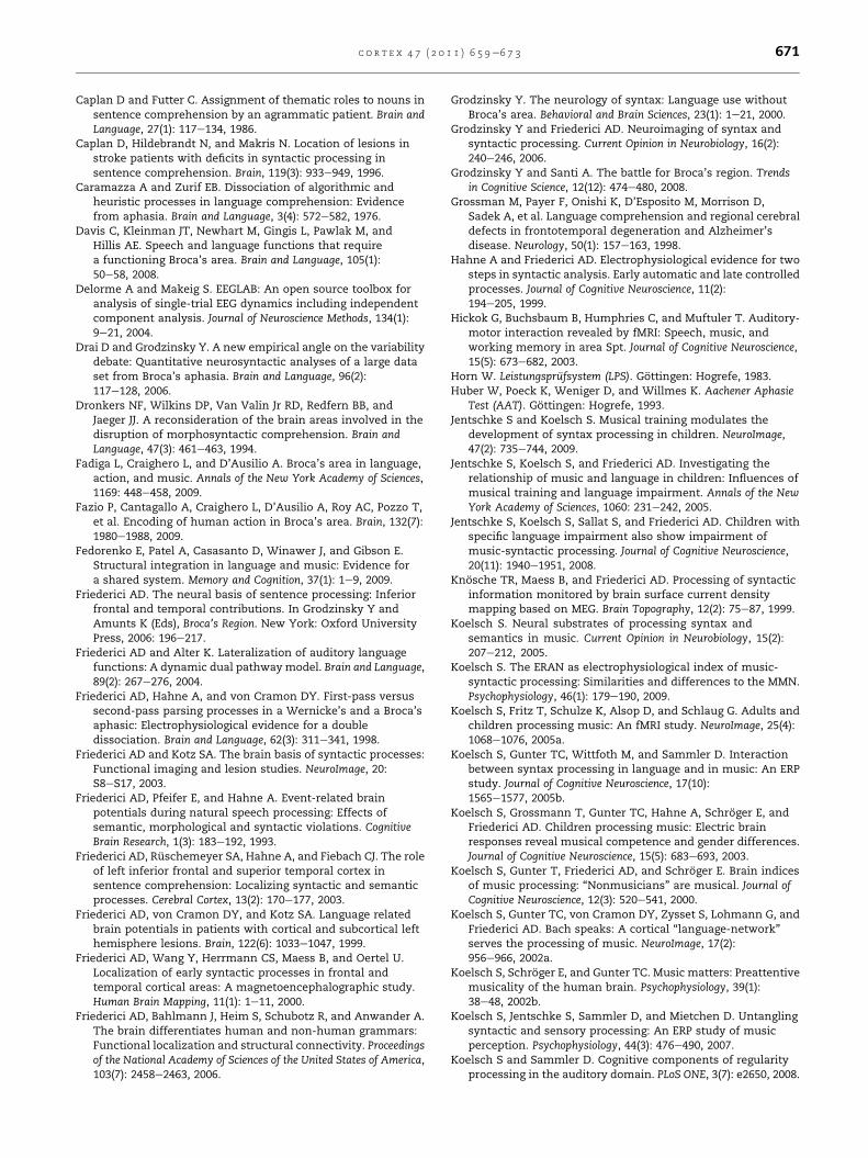
Fig. 3 e Electrophysiological responses to regular (solid line) an
controls (upper panel), patients with IFG (middle panel) or aSTG
(see red line and red arrows) in all groups. Likewise, a polarity in
the data were re-referenced to the nose electrode (see black arrow
show the scalp distribution of the ERAN in a time window from
to linked mastoids). In patients with IFG lesions, the ERAN was
strongly right lateralised than in the healthy controls or in pati
and aSTG patients did not significantly differ from one another
3. Results
3.1. EEG experiment
Participants detected on average 98.96% of the deviant
instruments (hits corrected by false alarms: healthy controls:
99.08%, IFG patients: 98.61%, aSTG patients: 99.03%), showing
that they attended to the musical stimuli and reliably identi-
fied the deviant timbre. An ANOVAwith the between-subjects
factor Group (healthy controls vs IFG patients vs aSTG
patients) showed that patients and controls performed simi-
larly in this non-syntactic timbre task (no main effect of
Group: p> .861).
Fig. 3 depicts the electric brain responses to final regular
and irregular chords separately for healthy controls and each
patient group. In all groups, the irregular chords elicited an
ERAN peaking around 200 msec with a frontal scalp distribu-
tion (see red arrows in Fig. 3). When re-referenced to the nose
electrode, the ERAN inverted polarity at mastoid leads (see
black arrows in Fig. 3) indicating that this ERP effect is not an
N2b (the N2b does not invert polarity and has a less anterior
scalp distribution; Schroger, 1998; Naatanen et al., 2007).
Notably, in IFG patients, the ERAN was more frontally
distributed and appeared to be more strongly right lateralised
compared to the effects in healthy controls and the aSTG
patients, who exhibited a non-lateralised negativity (see
Fig. 3).
Three-way ANOVAs with the factors Chord, Hemisphere,
and AntPost revealed main effects of Chord and an interaction
of Chord�AntPost in the control and both patient groups,
indicating that irregular chords elicited a negativity with ante-
rior scalp distribution irrespective of group or lesion site (mean
of all ROIs� SEM in healthy controls: D¼�.76� .12 mV; IFG
patients: D¼�.55� .18 mV; aSTG patients: D¼�.67� .08 mV). In
IFG patients, a marginally significant interaction of
Chord�Hemisphere was found ( p< .068), demonstrating
a trend of a right lateralisation of the effect in this group (left:
D¼�.40� .16 mV; right: D¼�.72� .22 mV; see Table 3 for statis-
tical values). Healthy controls and aSTGpatients exhibitednon-
lateralised effects (Chord�Hemisphere: p’s> .126).
Likewise, a four-way ANOVA with the factors Chord,
Hemisphere, AntPost, and Group (healthy controls vs IFG
patients vs aSTG patients) revealed a main effect of Chord [F
(1,23)¼ 64.82, p< .0001], and an interaction of Chord�AntPost
[F(1,23)¼ 79.14, p< .0001]. Moreover, significant three-way
interactions of Chord�AntPost�Group [F(2,23)¼ 4.54,
p< .022] and Chord�Hemisphere�Group [F(2,23)¼ 5.18,
p< .014] were found. To further elucidate these interactions,
three analogous four-way ANOVAs were computed in which
d irregular final chords (dotted line) plotted for healthy
lesions (lower panel). Irregular chords elicited an ERAN
version was observed at mastoid leads in all groups when
s pointing to electrodesM1 andM2). The topographymaps
150 to 250 msec after onset of the final chord (referenced
distributed significantly more anteriorly and was more
ents with aSTG lesions. The scalp distribution in controls
.
c o r t e x 4 7 ( 2 0 1 1 ) 6 5 9e6 7 3666
the factor Groupwas systematically reduced to two levels (i.e.,
IFG patients vs controls, aSTG patients vs controls, and IFG vs
aSTG patients; see Table 4 for statistical values). These
analyses showed that the ERAN was indeed more
anteriorly distributed and more strongly right lateralised
in patients with IFG lesions compared to controls (interaction
of Chord�AntPost�Group: p< .035; interaction of
Chord�Hemisphere�Group: p< .045), and compared to
patients with lesions in the aSTG (Chord�AntPost�Group:
p< .014; Chord�Hemisphere�Group: p< .015; see topog-
raphy maps in Fig. 3). The scalp topography did not differ
between aSTG patients and controls (no interaction of
Chord�AntPost�Group or Chord�Hemisphere�Group:
p’s> .104). These findings underline that the scalp topography
of the ERAN was particularly different in the IFG patients.
Tomake sure that these results in the IFG patientswere not
predominantly due to patient M.S. who had acquired his
lesion considerably more recently (7 months prior to testing)
than the other IFG and aSTG patients (>4 years; see Table 1),
analogous ANOVAs were computed excluding M.S.’s data set
(see below for a separate analysis of M.S.’s data). These anal-
yses revealed virtually identical results [IFG patients vs
healthy controls: Chord�Hemisphere�Group: F(1,16)¼ 7.38,
p< .016, Chord�AntPost�Group: F(1,16)¼ 6.76, p< .020;
IFG patients vs aSTG patients: Chord�Hemisphere�Group:
F(1,10)¼ 10.87, p< .009, Chord�AntPost�Group: F(1,10)¼10.91, p< .009], indicating that the scalp topography of the
ERAN remains specific even in patients with older left IFG
lesions.
Interestingly, in none of the patient groups the overall
ERAN amplitude was significantly reduced compared to
controls (no significant interaction of Chord�Group:
p’s> .365; see Table 4). To test whether this result was possibly
due to a recovery of the ERAN since lesion onset (i.e., to clarify
whether the ERAN amplitude increased with time since brain
injury) one-sided Pearson correlations were calculated
between lesion onset and the ERAN amplitude (average of
frontal ROIs), separately for each patient group. As depicted in
Fig. 4A, a strong correlation between lesion onset and ERAN
amplitude was found in the IFG patients [r(4)¼�.726, p< .052;
R2¼ .53, indicating a large effect size, Bortz and Doring, 2003],
demonstrating that left IFG lesions with recent onset aremore
likely to reduce the ERAN amplitude than older lesions. A
similar, but considerably weaker and non-significant trend
was observed if patient M.S. was excluded from the analysis [r
(3)¼�.369, p> .270], suggesting that the ERAN amplitude is
less prone to changes in more chronic lesions. No correlation
(not even a trend) was found in patients with lesions in the
aSTG [r(5)¼þ.007, p> .494; Fig. 4B].
3.2. Post-hoc behavioural test
All participants expressed great uncertainty about the
distinction between regular and irregular final chords, reflec-
ted in relatively low hit rates (healthy controls: 61.29%; IFG
patients: 54.69%; aSTG patients: 58.16%). Nevertheless,
controls and also the aSTG patients performed significantly
above the 50% chance level [controls: t(11)¼ 4.84, p< .002,
aSTG patients: t(5)¼ 3.24, p< .024; one-sample t-tests],
whereas patients with lesions in the left IFG performed at
chance level ( p> .242), even if patient M.S. was excluded from
the analysis ( p> .137). However, no significant differences
were found between patients and controls (no main effect of
Group: p> .256).
To test whether the behavioural performance was modu-
lated by general cognitive abilities, one-sided Pearson corre-
lations were computed across all participants between hit
rates and nonverbal intelligence [r(22)¼ .042, p> .424),
working memory [r(22)¼ .049, p> .410], and STM [r(22)¼�.54,
p> .402]. No significant relations were found, indicating that
the discrimination performance did not depend on these
general cognitive functions.
To test whether the behavioural performance increased
with time since brain injury (like the ERAN amplitude), one-
tailed Pearson correlations were calculated between lesion
onset and hit rates, separately for each patient group. As
depicted in Fig. 4C, a correlation between lesion onset and
discrimination performance was found in patients with IFG
lesions [r(4)¼ .628, p< .091, R2¼ .40, indicating a medium to
large effect size, Bortz and Doring, 2003]. This result mirrors
the correlation found for the ERAN amplitude (see above) in
that more recent lesions were associated with weaker
sensitivity for music-syntactic irregularities than older
lesions. Again, a similar but considerably weaker and non-
significant trend was found if patient M.S. with the most
recent lesion was excluded from the analysis [r(3)¼ .344,
p> .285], suggesting that the behavioural performance is
more stable in chronic lesions. No correlation (not even
a trend) was found in patients with aSTG damage [r(4)¼ .020,
p> .485; Fig. 4D].
3.3. Single case analysis of patient M.S.
The correlation between the EEG, behavioural data and the
time since lesion onset in IFG patients suggested a likely
impact of recovery over time, therefore, perhaps obscuring
deficits that occur only for a short time after brain injury. As
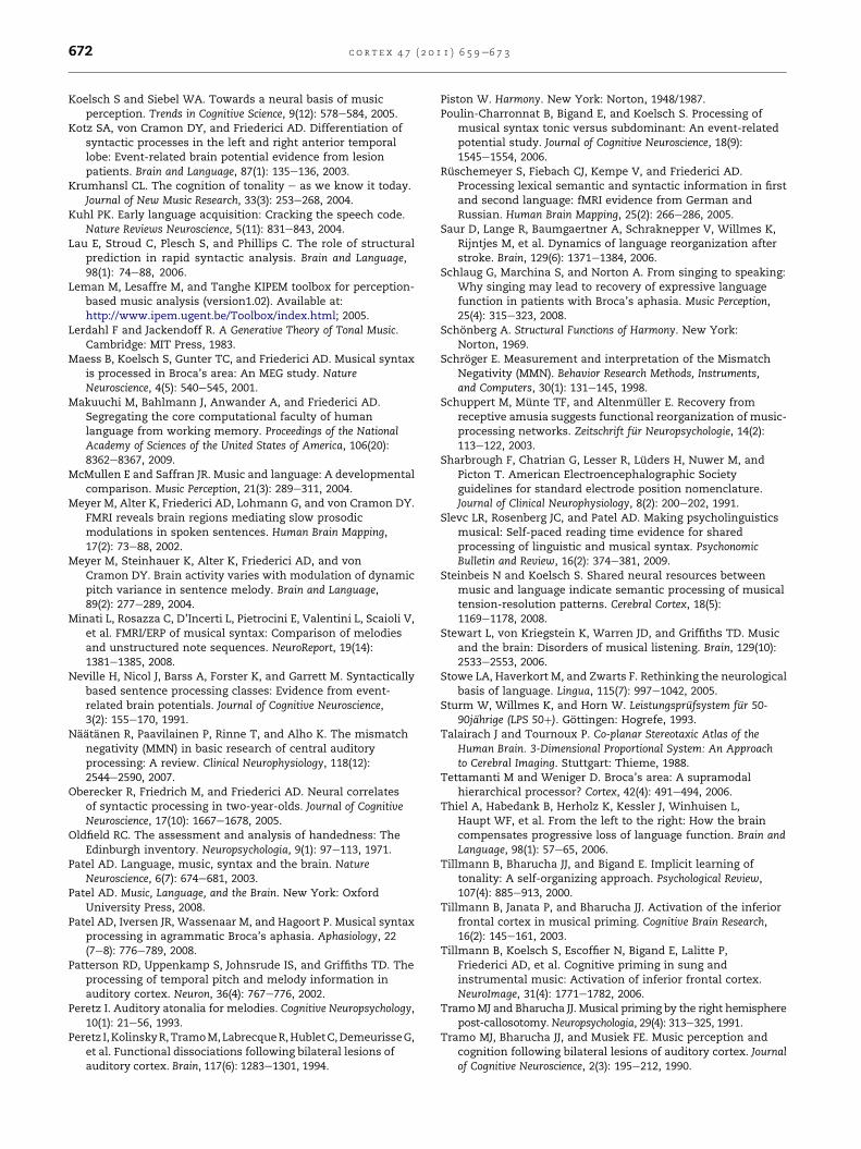
patient M.S. had acquired his lesion (Fig. 5) only 7 months
prior to testing (making a complete recovery of music-
syntactic functions less likely than in the other patients with
lesions older than 4 years) M.S.’s data were analysed sepa-
rately and compared as a single case with the data of the
remaining IFG patients and healthy controls. No such analysis
was conducted in aSTG patients because none of these indi-
viduals had a recent lesion.
The ERAN amplitude of patient M.S. (mean: D¼�.16 mV)
was significantly lower than the one observed in the other IFG
patients [mean: D¼�1.06 mV, t(5)¼�4.49, p< .012; one-
sample t-test with test value �.16; see Fig. 6A]. Moreover,
M.S.’s ERAN amplitude was also significantly smaller than the
one in healthy controls [mean: D¼�.96 mV, t(12)¼�6.28,
p< .0001]. The ERAN amplitudes of the IFG patients with older
lesions and of healthy controls did not differ as indicated by
a non-significant interaction of Chord�Group in an ANOVA
with the factors Chord, Hemisphere, AntPost, and Group [F
(1,16)¼ .13, p> .720].
The behavioural performance of patient M.S. (hits: 44.79%)
was at chance level (assessed with a binomial test: p> .820)
and significantly weaker than the performance of the other
IFG patients [mean of hits: 56.67%, t(4)¼ 3.31, p< .04;
Table 4 e ANOVAs of Chord3Hemisphere3AntPost3Group/LesionSite. Bold values indicate significant results.
IFG patients versushealthy controls
aSTG patients versushealthy controls
IFG patients versusaSTG patients
F(1,17) p-value F(1,18) p-value F(1,11) p-value
Chord 36.30 <.0001 63.60 <.0001 41.78 <.0001
Chord�Group .86 >.365 .26 >.616 .35 >.567
Chord�Hemisphere 4.90 <.041 2.76 >.114 .15 >.707
Chord�Hemisphere�Group 4.69 <.045 2.92 >.104 8.34 <.016
Chord�AntPost 67.24 <.0001 39.25 <.0001 52.61 <.0001
Chord�AntPost�Group 5.26 <.035 .76 >.394 8.55 <.015
c o r t e x 4 7 ( 2 0 1 1 ) 6 5 9e6 7 3 667
one-sample t-test with test value 44.79; see Fig. 6B], and
healthy controls [mean of hits: 61.29%, t(11)¼ 7.07, p< .0001].
No significant difference was found between the other IFG
patients and healthy controls [t(15)¼�1.08, p> .299;
two-samples t-test]. These combined results suggest that the
ERAN amplitude and the ability to detect harmonically irreg-
ular chords are impaired in early stages after left IFG injury,
but might have reached normal levels several years later
(despite a still abnormal scalp distribution of the ERAN; see
above).
Fig. 4 e Correlation of lesion onset, ERAN amplitude and behav
lesions, the ERAN amplitude (average of the frontal ROIs) increa
brain injury (A). Likewise, performance in explicitly discrimina
increased with time since lesion onset in IFG patients (C). These
most recent lesion) was excluded from the analysis. No correla
Patient R.B. is missing in D because he did not participate in th
4. Discussion
The present study investigated whether left inferior frontal
and left anterior temporal brain areas (known to be crucial for
syntax processing in language) are a prerequisite for normal
syntax processing in music. Patients with lesions including
the left IFG or left aSTG as well as healthy controls were tested
in a chord sequence paradigm. The behavioural performance
in detecting harmonically irregular chords, and the ERAN (an
ioural discrimination performance. In patients with IFG
sed linearly (i.e., became more negative) with time since
ting chord sequences with regular and irregular endings
correlations lost their significance if patient M.S. (with the
tions were found in patients with aSTG lesions (B and D).
e behavioural experiment due to severe illness.
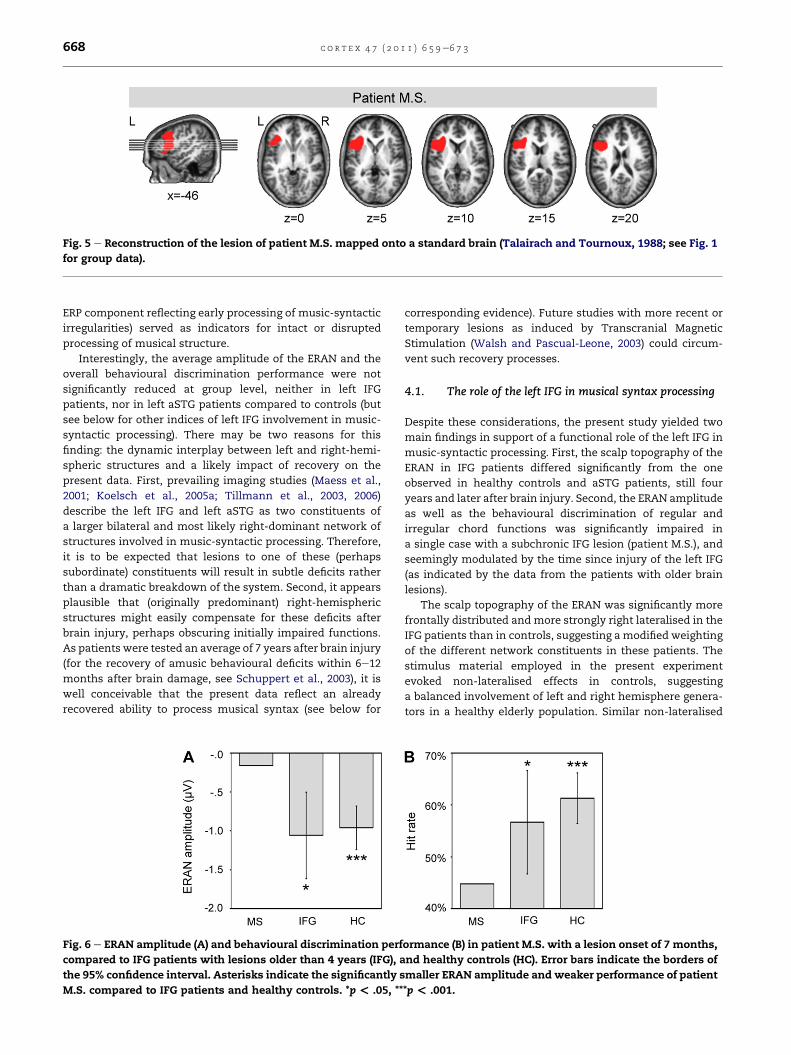
Fig. 5 e Reconstruction of the lesion of patient M.S. mapped onto a standard brain (Talairach and Tournoux, 1988; see Fig. 1
for group data).
c o r t e x 4 7 ( 2 0 1 1 ) 6 5 9e6 7 3668
ERP component reflecting early processing of music-syntactic
irregularities) served as indicators for intact or disrupted
processing of musical structure.
Interestingly, the average amplitude of the ERAN and the
overall behavioural discrimination performance were not
significantly reduced at group level, neither in left IFG
patients, nor in left aSTG patients compared to controls (but
see below for other indices of left IFG involvement in music-
syntactic processing). There may be two reasons for this
finding: the dynamic interplay between left and right-hemi-
spheric structures and a likely impact of recovery on the
present data. First, prevailing imaging studies (Maess et al.,
2001; Koelsch et al., 2005a; Tillmann et al., 2003, 2006)
describe the left IFG and left aSTG as two constituents of
a larger bilateral and most likely right-dominant network of
structures involved in music-syntactic processing. Therefore,
it is to be expected that lesions to one of these (perhaps
subordinate) constituents will result in subtle deficits rather
than a dramatic breakdown of the system. Second, it appears
plausible that (originally predominant) right-hemispheric
structures might easily compensate for these deficits after
brain injury, perhaps obscuring initially impaired functions.
As patients were tested an average of 7 years after brain injury
(for the recovery of amusic behavioural deficits within 6e12
months after brain damage, see Schuppert et al., 2003), it is
well conceivable that the present data reflect an already
recovered ability to process musical syntax (see below for
Fig. 6 e ERAN amplitude (A) and behavioural discrimination perf
compared to IFG patients with lesions older than 4 years (IFG), a
the 95% confidence interval. Asterisks indicate the significantly s
M.S. compared to IFG patients and healthy controls. *p< .05, **
corresponding evidence). Future studies with more recent or
temporary lesions as induced by Transcranial Magnetic
Stimulation (Walsh and Pascual-Leone, 2003) could circum-
vent such recovery processes.
4.1. The role of the left IFG in musical syntax processing
Despite these considerations, the present study yielded two
main findings in support of a functional role of the left IFG in
music-syntactic processing. First, the scalp topography of the
ERAN in IFG patients differed significantly from the one
observed in healthy controls and aSTG patients, still four
years and later after brain injury. Second, the ERAN amplitude
as well as the behavioural discrimination of regular and
irregular chord functions was significantly impaired in
a single case with a subchronic IFG lesion (patient M.S.), and
seemingly modulated by the time since injury of the left IFG
(as indicated by the data from the patients with older brain
lesions).
The scalp topography of the ERAN was significantly more
frontally distributed and more strongly right lateralised in the
IFG patients than in controls, suggesting a modified weighting
of the different network constituents in these patients. The
stimulus material employed in the present experiment
evoked non-lateralised effects in controls, suggesting
a balanced involvement of left and right hemisphere genera-
tors in a healthy elderly population. Similar non-lateralised
ormance (B) in patient M.S. with a lesion onset of 7 months,
nd healthy controls (HC). Error bars indicate the borders of
maller ERAN amplitude and weaker performance of patient
*p< .001.
c o r t e x 4 7 ( 2 0 1 1 ) 6 5 9e6 7 3 669
early negativities have been repeatedly reported by recent ERP
studies, mostly when harmonically complex stimulus mate-
rial was used like in the present experiment (for a review, see
Koelsch, 2009). The persistent topography shift in IFG patients
still four years after brain injury might reflect a greater
predominance of the right-hemisphere generators of the
ERAN compared to healthy controls, due to the breakdown of
the left hemisphere units and/or a shift of the function to the
contralesional hemisphere (for similar inter-hemispheric
shifts during aphasia recovery see, e.g., Schlaug et al., 2008
[Patient #1]; Saur et al., 2006; Thiel et al., 2006). Because the
scalp distribution of the ERAN in aSTG patients did not
significantly differ from that of controls (both groups exhibi-
ted non-lateralised effects), it is unlikely that the topography
shift observed in the IFG patients is merely an effect of weaker
signal quality over the ipsilesional compared to the contrale-
sional hemisphere.
In the absence of longitudinal data, it is difficult to ascer-
tain whether the topography shift of the ERAN (despite
normal ERAN amplitude) represents a general sufficiency of
right-hemispheric structures for music-syntactic processing,
or post-lesional reorganisation and recovery. However, for
two reasons it may be speculated that the data are more
consistent with a structural and functional reorganisation of
music-syntactic processing. First, as a group, patients with
lesions in the left IFG showed chance level performance in the
discrimination of regular and irregular sequence endings,
contrary to healthy controls and the patients with aSTG
lesions. Although no significant group differences were found
(most presumably due to a floor effect)2 and any interpretation
must therefore remain speculative, this result fits with the
assumption that the neural network underlying music-
syntactic processing is slightly less sensitive to irregular
chords if its left frontal constituent is lesioned. Second,
a progressive recovery of music-syntactic processing fits with
the correlations between the ERAN amplitude, the discrimi-
nation performance, and the time since lesion onset (although
this assumption should be further tested in a longitudinal
study). Corroboratingly, the IFG patient M.S. with the most
recent lesion (7 months prior to testing, i.e., in a subchronic
stage of recovery) exhibited a significantly smaller ERAN
amplitude and a weaker sensitivity to harmonic irregularities
than patients with lesions older than four years. Moreover, his
behavioural and amplitude values differed significantly from
those of healthy controls, whereas no such difference was
found between controls and IFG patients with older lesions.
This finding cannot be attributed to general cognitive deficits
of M.S. who scored normally (sometimes even slightly higher
2 A floor effect is indicated by the surprisingly low hit rate of thehealthy elderly control group (61.29%), making it difficult tostatistically capture group differences. Healthy students (aged20e35 years) normally exhibit an average performance of 76%correct with these stimuli (Koelsch et al., 2007). This differencebetween young and elderly participants relies most likely onreduced task-relevant resources like working memory or atten-tional capacities with increasing age, not necessarily on reducedsyntactic processing resources (because young as well as elderlyhealthy participants show an ERAN, once more underlining theimportance of testing implicit processing as in the current EEGexperiment).
than the other groups) in intelligence, WM and STM tests
(Table 2). The combined results suggest that the ERAN may
regain strength and perceptual sensitivity may increase over
the years, finally even reaching normal levels as in healthy
controls, perhaps through a compensatory up-regulation of
right-hemispheric activity as suggested by the abnormal
distribution of the ERAN in chronic lesions (see above). Over-
all, the finding that both the ERAN amplitude and the behav-
ioural measures were deficient in early stages after IFG injury,
and increased with time since lesion onset, conjointly suggest
that the left IFG may be originally necessary (although most
likely not sufficient) for normal music-syntactic processing,
but that its dysfunction may have been gradually compen-
sated for in our patients.
Taken together, the ERAN was extinguished in one IFG
patient tested 7months post-stroke. Patients with chronic left
IFG lesions showed an ERAN, which was, however, signifi-
cantly differently distributed than in controls. Moreover,
behaviourally, patients failed to perceive harmonic closure,
although the data suggest that the sensitivity to harmonic
irregularities may gradually recover over the years. These
combined findings are consistent with the notion that the left
IFG, as part of a larger bilateral processing network, is func-
tionally relevant for the processing of musical syntax, and
that initial deficits might recover over the years. The former
observation is in keepingwith a series of previousMEG (Maess
et al., 2001) and fMRI studies (e.g., Koelsch et al., 2002a, 2005a;
Tillmann et al., 2003, 2006) reporting bilateral fronto-temporal
brain activations related to the processing of harmonic irreg-
ularities, consistently encompassing the left IFG. Future
studiesmay be devoted to the functional relevance of the right
IFG for music-syntactic processing.
4.2. The role of the left aSTG in musical syntaxprocessing
Contrary to our predictions, music-syntactic processing was
unimpaired in patients with left aSTG lesions. Their ERAN
amplitude and scalp topography did not differ from that in
healthy controls; they were able to explicitly detect harmonic
irregularities, and none of these measures correlated with
time since brain injury. These results can be interpreted in
two ways: They may either demonstrate that the left aSTG is
not involved in the processing of musical syntax, or they may
reflect the compensation for, or recovery of, its function.
The first interpretation seems to conflict with the neuro-
imaging literature that consistently reported activations in
the left or bilateral aSTG when comparing the processing of
harmonically distantly and closely related items in melodies
or chord sequences (Koelsch et al., 2002a, 2005a; Krumhansl,
2004; Tillmann et al., 2006). However, due to the coarse
temporal resolution of fMRI, it remains unclear whether the
aSTG brain activation actually reflects the processes under-
lying the ERAN or other effects observed in the ERP for the
processing of harmonic structure. Furthermore, while fMRI
identifies the brain structures that are involved in a cognitive
function, lesion data determine cerebral regions that are
crucial for a given task. Consequently, it may be suggested
that the aSTG activations in fMRI studies do not reflect
harmonic structure building as indicated by the ERAN per se,
c o r t e x 4 7 ( 2 0 1 1 ) 6 5 9e6 7 3670
but represent related processes in the service of music
perception, for instance melodic processing (e.g., Patterson
et al., 2002), or structural integration and the processing of
meaning as reflected by the N5 (Koelsch et al., 2000; Steinbeis
and Koelsch, 2008).
On the other hand, the influence of compensation cannot
be dismissed either, particularly when considering the left
aSTG as only one, perhaps subordinate, constituent of
a bilateral fronto-temporal network whose breakdown may
have been compensated for by the remaining constituents of
the circuit 4 years and later after brain injury (see above;
Schuppert et al., 2003). Taken together, based on the present
data, the function of the left aSTG in music-syntactic pro-
cessing must remain suggestive in the way formulated above.
4.3. Overlap of musical and linguistic syntax processing
The present findings speak for a functional relevance of the
left IFG for musical syntax processing, as proposed by Maess
et al. (2001). This result deserves specific attention, in the
context of the notion of a domain-general role of Broca’s area
in syntax processing, e.g., in music and in language. Previous
lesion studies have established the necessity of the left IFG for
syntax processing in language (for reviews, see Drai and
Grodzinsky, 2006; Friederici and Kotz, 2003; Grodzinsky,
2000; Stowe et al., 2005). Notably, Friederici et al. (1998, 1999)
showed that the ELAN, an ERP component thought to reflect
early syntactic procedures in language (analogous to those
reflected by the ERAN in music, Koelsch, 2009) crucially
depends on the integrity of the left inferior frontal lobe,
because lesions in this region lead to an extinction of the
ELAN. Importantly, lesion sites of the patients that partici-
pated in these language studies were highly similar to those of
our patients. Taken together, left inferior frontal brain damage
has thus been associated with a complete extinction of the
ELAN (assessed about 1.25 years after brain injury; Friederici
et al., 1998, 1999), a significant reduction of the ERAN ampli-
tude 7 months after lesion onset (patient M.S.), as well as an
abnormal scalp topography of the ERAN still 4 years after left
IFG injury. Consequently, the left inferior frontal lobe appears
to represent a neural substrate of syntax processing that is
shared by music and language (although this assumption
should be further tested using a within-subject design). Such
an interface may at least partly account for recently observed
interactions betweenmusical and linguistic syntax processing
(Fedorenko et al., 2009; Koelsch et al., 2005b; Slevc et al., 2009;
Steinbeis and Koelsch, 2008), enhanced linguistic syntax pro-
cessing in musically trained subjects (Jentschke et al., 2005;
Jentschke and Koelsch, 2009), or deficient music-syntactic
processing in individuals with language disorders in the
syntax domain (Jentschke et al., 2008; Patel et al., 2008).
Moreover, this conclusion would be in line with the proposed
domain-general role of left frontal brain areas in syntactic
processing (Patel, 2003, 2008) specifically the processing of
syntactic hierarchies in different domains (Bahlmann et al.,
2009; Fadiga et al., 2009; Makuuchi et al., 2009; Friederici
et al., 2006; Tettamanti and Weniger, 2006).
When considering the entire network supporting music-
syntactic processing, regions of overlap between music and
language may not be confined to the left IFG. Another likely
candidate is the right IFG frequently associated with the pro-
cessing of musical structure (Koelsch et al., 2002a, 2005a;
Tillmann et al., 2003, 2006; Krumhansl, 2004) or pitch
working memory (Zatorre et al., 1994), and the processing of
prosody in connected speech (Friederici and Alter, 2004;Meyer
et al., 2002, 2004), and also the posterior portion of the STG,
which is viewed as an area of auditory-motor integration
(Hickok et al., 2003). Data from imaging studies in music
(Koelsch et al., 2002a, 2005a; Tillmann et al., 2006) and
language (Friederici et al., 2003; Ruschemeyer et al., 2005)
indeed suggest a domain-general role of posterior temporal
and parietal brain areas during syntax processing, opening
a field for future studies.
5. Conclusion
The present study suggests that the left IFG is relevant (but not
sufficient) for the generation of the ERAN, i.e., for the pro-
cessing of syntactic irregularities in rule-based harmonic
sequences, complementing the vast literature on the
predominance of the right frontal cortex in music processing
(Koelsch et al., 2005a; Tillmann et al., 2006; Zatorre et al., 1992,
1994). Given that previous studies have established the
necessity of the left inferior frontal lobe for the processing of
syntactic structures in language (Davis et al., 2008; Friederici
and Kotz, 2003), in space (Bahlmann et al., 2009), and lately
also in action (Fazio et al., 2009), the current finding is
compatible with the notion that Broca’s area supports the
processing of syntactic structure in a rather domain-general
way.
Acknowledgements
We wish to thank Prof. Dr. D. Y. von Cramon for kindly
providing the patients, Anne-Kathrin Franz for patient
recruitment, Sylvia Stasch for help with data acquisition,
Sebastian Seiffert for advice in lesion segmentation, Dr. Derek
V. M. Ott and Dr. Christoph Preul for help with lesion local-
isations, and Dr. Sebastian Jentschke for helpful comments on
the data analysis.
r e f e r e n c e s
Bahlmann J, Schubotz RI, Mueller JL, Koster D, and Friederici AD.Neural circuits of hierarchical visuo-spatial sequenceprocessing. Brain Research, 1298: 161e170, 2009.
Bharucha JJ and Stoeckig K. Priming of chords: Spreadingactivation or overlapping frequency spectra? Perceptionand Psychophysics, 41(6): 519e524, 1987.
Bigand E. More about the musical expertise of musicallyuntrained listeners. Annals of the New York Academy of Sciences,999: 304e312, 2003.
Bortz J and Doring N. Forschungsmethoden und Evaluation: FurHuman- und Sozialwissenschaftler. Berlin: Springer, 2003.
Brauer J and Friederici AD. Functional neural networks ofsemantic and syntactic processes in the developing brain.Journal of Cognitive Neuroscience, 19(10): 1609e1623, 2007.
c o r t e x 4 7 ( 2 0 1 1 ) 6 5 9e6 7 3 671
Caplan D and Futter C. Assignment of thematic roles to nouns insentence comprehension by an agrammatic patient. Brain andLanguage, 27(1): 117e134, 1986.
Caplan D, Hildebrandt N, and Makris N. Location of lesions instroke patients with deficits in syntactic processing insentence comprehension. Brain, 119(3): 933e949, 1996.
Caramazza A and Zurif EB. Dissociation of algorithmic andheuristic processes in language comprehension: Evidencefrom aphasia. Brain and Language, 3(4): 572e582, 1976.
Davis C, Kleinman JT, Newhart M, Gingis L, Pawlak M, andHillis AE. Speech and language functions that requirea functioning Broca’s area. Brain and Language, 105(1):50e58, 2008.
Delorme A and Makeig S. EEGLAB: An open source toolbox foranalysis of single-trial EEG dynamics including independentcomponent analysis. Journal of Neuroscience Methods, 134(1):9e21, 2004.
Drai D and Grodzinsky Y. A new empirical angle on the variabilitydebate: Quantitative neurosyntactic analyses of a large dataset from Broca’s aphasia. Brain and Language, 96(2):117e128, 2006.
Dronkers NF, Wilkins DP, Van Valin Jr RD, Redfern BB, andJaeger JJ. A reconsideration of the brain areas involved in thedisruption of morphosyntactic comprehension. Brain andLanguage, 47(3): 461e463, 1994.
Fadiga L, Craighero L, and D’Ausilio A. Broca’s area in language,action, and music. Annals of the New York Academy of Sciences,1169: 448e458, 2009.
Fazio P, Cantagallo A, Craighero L, D’Ausilio A, Roy AC, Pozzo T,et al. Encoding of human action in Broca’s area. Brain, 132(7):1980e1988, 2009.
Fedorenko E, Patel A, Casasanto D, Winawer J, and Gibson E.Structural integration in language and music: Evidence fora shared system. Memory and Cognition, 37(1): 1e9, 2009.
Friederici AD. The neural basis of sentence processing: Inferiorfrontal and temporal contributions. In Grodzinsky Y andAmunts K (Eds), Broca’s Region. New York: Oxford UniversityPress, 2006: 196e217.
Friederici AD and Alter K. Lateralization of auditory languagefunctions: A dynamic dual pathway model. Brain and Language,89(2): 267e276, 2004.
Friederici AD, Hahne A, and von Cramon DY. First-pass versussecond-pass parsing processes in a Wernicke’s and a Broca’saphasic: Electrophysiological evidence for a doubledissociation. Brain and Language, 62(3): 311e341, 1998.
Friederici AD and Kotz SA. The brain basis of syntactic processes:Functional imaging and lesion studies. NeuroImage, 20:S8eS17, 2003.
Friederici AD, Pfeifer E, and Hahne A. Event-related brainpotentials during natural speech processing: Effects ofsemantic, morphological and syntactic violations. CognitiveBrain Research, 1(3): 183e192, 1993.
Friederici AD, Ruschemeyer SA, Hahne A, and Fiebach CJ. The roleof left inferior frontal and superior temporal cortex insentence comprehension: Localizing syntactic and semanticprocesses. Cerebral Cortex, 13(2): 170e177, 2003.
Friederici AD, von Cramon DY, and Kotz SA. Language relatedbrain potentials in patients with cortical and subcortical lefthemisphere lesions. Brain, 122(6): 1033e1047, 1999.
Friederici AD, Wang Y, Herrmann CS, Maess B, and Oertel U.Localization of early syntactic processes in frontal andtemporal cortical areas: A magnetoencephalographic study.Human Brain Mapping, 11(1): 1e11, 2000.
Friederici AD, Bahlmann J, Heim S, Schubotz R, and Anwander A.The brain differentiates human and non-human grammars:Functional localization and structural connectivity. Proceedingsof the National Academy of Sciences of the United States of America,103(7): 2458e2463, 2006.
Grodzinsky Y. The neurology of syntax: Language use withoutBroca’s area. Behavioral and Brain Sciences, 23(1): 1e21, 2000.
Grodzinsky Y and Friederici AD. Neuroimaging of syntax andsyntactic processing. Current Opinion in Neurobiology, 16(2):240e246, 2006.
Grodzinsky Y and Santi A. The battle for Broca’s region. Trendsin Cognitive Science, 12(12): 474e480, 2008.
Grossman M, Payer F, Onishi K, D’Esposito M, Morrison D,Sadek A, et al. Language comprehension and regional cerebraldefects in frontotemporal degeneration and Alzheimer’sdisease. Neurology, 50(1): 157e163, 1998.
Hahne A and Friederici AD. Electrophysiological evidence for twosteps in syntactic analysis. Early automatic and late controlledprocesses. Journal of Cognitive Neuroscience, 11(2):194e205, 1999.
Hickok G, Buchsbaum B, Humphries C, and Muftuler T. Auditory-motor interaction revealed by fMRI: Speech, music, andworking memory in area Spt. Journal of Cognitive Neuroscience,15(5): 673e682, 2003.
Horn W. Leistungsprufsystem (LPS). Gottingen: Hogrefe, 1983.Huber W, Poeck K, Weniger D, and Willmes K. Aachener Aphasie
Test (AAT). Gottingen: Hogrefe, 1993.Jentschke S and Koelsch S. Musical training modulates the
development of syntax processing in children. NeuroImage,47(2): 735e744, 2009.
Jentschke S, Koelsch S, and Friederici AD. Investigating therelationship of music and language in children: Influences ofmusical training and language impairment. Annals of the NewYork Academy of Sciences, 1060: 231e242, 2005.
Jentschke S, Koelsch S, Sallat S, and Friederici AD. Children withspecific language impairment also show impairment ofmusic-syntactic processing. Journal of Cognitive Neuroscience,20(11): 1940e1951, 2008.
Knosche TR, Maess B, and Friederici AD. Processing of syntacticinformation monitored by brain surface current densitymapping based on MEG. Brain Topography, 12(2): 75e87, 1999.
Koelsch S. Neural substrates of processing syntax andsemantics in music. Current Opinion in Neurobiology, 15(2):207e212, 2005.
Koelsch S. The ERAN as electrophysiological index of music-syntactic processing: Similarities and differences to the MMN.Psychophysiology, 46(1): 179e190, 2009.
Koelsch S, Fritz T, Schulze K, Alsop D, and Schlaug G. Adults andchildren processing music: An fMRI study. NeuroImage, 25(4):1068e1076, 2005a.
Koelsch S, Gunter TC, Wittfoth M, and Sammler D. Interactionbetween syntax processing in language and in music: An ERPstudy. Journal of Cognitive Neuroscience, 17(10):1565e1577, 2005b.
Koelsch S, Grossmann T, Gunter TC, Hahne A, Schroger E, andFriederici AD. Children processing music: Electric brainresponses reveal musical competence and gender differences.Journal of Cognitive Neuroscience, 15(5): 683e693, 2003.
Koelsch S, Gunter T, Friederici AD, and Schroger E. Brain indicesof music processing: “Nonmusicians” are musical. Journal ofCognitive Neuroscience, 12(3): 520e541, 2000.
Koelsch S, Gunter TC, von Cramon DY, Zysset S, Lohmann G, andFriederici AD. Bach speaks: A cortical “language-network”serves the processing of music. NeuroImage, 17(2):956e966, 2002a.
Koelsch S, Schroger E, and Gunter TC. Music matters: Preattentivemusicality of the human brain. Psychophysiology, 39(1):38e48, 2002b.
Koelsch S, Jentschke S, Sammler D, and Mietchen D. Untanglingsyntactic and sensory processing: An ERP study of musicperception. Psychophysiology, 44(3): 476e490, 2007.
Koelsch S and Sammler D. Cognitive components of regularityprocessing in the auditory domain. PLoS ONE, 3(7): e2650, 2008.
c o r t e x 4 7 ( 2 0 1 1 ) 6 5 9e6 7 3672
Koelsch S and Siebel WA. Towards a neural basis of musicperception. Trends in Cognitive Science, 9(12): 578e584, 2005.
Kotz SA, von Cramon DY, and Friederici AD. Differentiation ofsyntactic processes in the left and right anterior temporallobe: Event-related brain potential evidence from lesionpatients. Brain and Language, 87(1): 135e136, 2003.
Krumhansl CL. The cognition of tonality e as we know it today.Journal of New Music Research, 33(3): 253e268, 2004.
Kuhl PK. Early language acquisition: Cracking the speech code.Nature Reviews Neuroscience, 5(11): 831e843, 2004.
Lau E, Stroud C, Plesch S, and Phillips C. The role of structuralprediction in rapid syntactic analysis. Brain and Language,98(1): 74e88, 2006.
Leman M, Lesaffre M, and Tanghe KIPEM toolbox for perception-based music analysis (version1.02). Available at:http://www.ipem.ugent.be/Toolbox/index.html; 2005.
Lerdahl F and Jackendoff R. A Generative Theory of Tonal Music.Cambridge: MIT Press, 1983.
Maess B, Koelsch S, Gunter TC, and Friederici AD. Musical syntaxis processed in Broca’s area: An MEG study. NatureNeuroscience, 4(5): 540e545, 2001.
Makuuchi M, Bahlmann J, Anwander A, and Friederici AD.Segregating the core computational faculty of humanlanguage from working memory. Proceedings of the NationalAcademy of Sciences of the United States of America, 106(20):8362e8367, 2009.
McMullen E and Saffran JR. Music and language: A developmentalcomparison. Music Perception, 21(3): 289e311, 2004.
Meyer M, Alter K, Friederici AD, Lohmann G, and von Cramon DY.FMRI reveals brain regions mediating slow prosodicmodulations in spoken sentences. Human Brain Mapping,17(2): 73e88, 2002.
Meyer M, Steinhauer K, Alter K, Friederici AD, and vonCramon DY. Brain activity varies with modulation of dynamicpitch variance in sentence melody. Brain and Language,89(2): 277e289, 2004.
Minati L, Rosazza C, D’Incerti L, Pietrocini E, Valentini L, Scaioli V,et al. FMRI/ERP of musical syntax: Comparison of melodiesand unstructured note sequences. NeuroReport, 19(14):1381e1385, 2008.
Neville H, Nicol J, Barss A, Forster K, and Garrett M. Syntacticallybased sentence processing classes: Evidence from event-related brain potentials. Journal of Cognitive Neuroscience,3(2): 155e170, 1991.
Naatanen R, Paavilainen P, Rinne T, and Alho K. The mismatchnegativity (MMN) in basic research of central auditoryprocessing: A review. Clinical Neurophysiology, 118(12):2544e2590, 2007.
Oberecker R, Friedrich M, and Friederici AD. Neural correlatesof syntactic processing in two-year-olds. Journal of CognitiveNeuroscience, 17(10): 1667e1678, 2005.
Oldfield RC. The assessment and analysis of handedness: TheEdinburgh inventory. Neuropsychologia, 9(1): 97e113, 1971.
Patel AD. Language, music, syntax and the brain. NatureNeuroscience, 6(7): 674e681, 2003.
Patel AD. Music, Language, and the Brain. New York: OxfordUniversity Press, 2008.
Patel AD, Iversen JR, Wassenaar M, and Hagoort P. Musical syntaxprocessing in agrammatic Broca’s aphasia. Aphasiology, 22(7e8): 776e789, 2008.
Patterson RD, Uppenkamp S, Johnsrude IS, and Griffiths TD. Theprocessing of temporal pitch and melody information inauditory cortex. Neuron, 36(4): 767e776, 2002.
Peretz I. Auditory atonalia for melodies. Cognitive Neuropsychology,10(1): 21e56, 1993.
Peretz I,KolinskyR,TramoM,LabrecqueR,HubletC,DemeurisseG,et al. Functional dissociations following bilateral lesions ofauditory cortex. Brain, 117(6): 1283e1301, 1994.
Piston W. Harmony. New York: Norton, 1948/1987.Poulin-Charronnat B, Bigand E, and Koelsch S. Processing of
musical syntax tonic versus subdominant: An event-relatedpotential study. Journal of Cognitive Neuroscience, 18(9):1545e1554, 2006.
Ruschemeyer S, Fiebach CJ, Kempe V, and Friederici AD.Processing lexical semantic and syntactic information in firstand second language: fMRI evidence from German andRussian. Human Brain Mapping, 25(2): 266e286, 2005.
Saur D, Lange R, Baumgaertner A, Schraknepper V, Willmes K,Rijntjes M, et al. Dynamics of language reorganization afterstroke. Brain, 129(6): 1371e1384, 2006.
Schlaug G, Marchina S, and Norton A. From singing to speaking:Why singing may lead to recovery of expressive languagefunction in patients with Broca’s aphasia. Music Perception,25(4): 315e323, 2008.
Schonberg A. Structural Functions of Harmony. New York:Norton, 1969.
Schroger E. Measurement and interpretation of the MismatchNegativity (MMN). Behavior Research Methods, Instruments,and Computers, 30(1): 131e145, 1998.
Schuppert M, Munte TF, and Altenmuller E. Recovery fromreceptive amusia suggests functional reorganization of music-processing networks. Zeitschrift fur Neuropsychologie, 14(2):113e122, 2003.
Sharbrough F, Chatrian G, Lesser R, Luders H, Nuwer M, andPicton T. American Electroencephalographic Societyguidelines for standard electrode position nomenclature.Journal of Clinical Neurophysiology, 8(2): 200e202, 1991.
Slevc LR, Rosenberg JC, and Patel AD. Making psycholinguisticsmusical: Self-paced reading time evidence for sharedprocessing of linguistic and musical syntax. PsychonomicBulletin and Review, 16(2): 374e381, 2009.
Steinbeis N and Koelsch S. Shared neural resources betweenmusic and language indicate semantic processing of musicaltension-resolution patterns. Cerebral Cortex, 18(5):1169e1178, 2008.
Stewart L, von Kriegstein K, Warren JD, and Griffiths TD. Musicand the brain: Disorders of musical listening. Brain, 129(10):2533e2553, 2006.
Stowe LA, Haverkort M, and Zwarts F. Rethinking the neurologicalbasis of language. Lingua, 115(7): 997e1042, 2005.
Sturm W, Willmes K, and Horn W. Leistungsprufsystem fur 50-90jahrige (LPS 50þ). Gottingen: Hogrefe, 1993.
Talairach J and Tournoux P. Co-planar Stereotaxic Atlas of theHuman Brain. 3-Dimensional Proportional System: An Approachto Cerebral Imaging. Stuttgart: Thieme, 1988.
Tettamanti M and Weniger D. Broca’s area: A supramodalhierarchical processor? Cortex, 42(4): 491e494, 2006.
Thiel A, Habedank B, Herholz K, Kessler J, Winhuisen L,Haupt WF, et al. From the left to the right: How the braincompensates progressive loss of language function. Brain andLanguage, 98(1): 57e65, 2006.
Tillmann B, Bharucha JJ, and Bigand E. Implicit learning oftonality: A self-organizing approach. Psychological Review,107(4): 885e913, 2000.
Tillmann B, Janata P, and Bharucha JJ. Activation of the inferiorfrontal cortex in musical priming. Cognitive Brain Research,16(2): 145e161, 2003.
Tillmann B, Koelsch S, Escoffier N, Bigand E, Lalitte P,Friederici AD, et al. Cognitive priming in sung andinstrumental music: Activation of inferior frontal cortex.NeuroImage, 31(4): 1771e1782, 2006.
TramoMJ and Bharucha JJ. Musical priming by the right hemispherepost-callosotomy. Neuropsychologia, 29(4): 313e325, 1991.
Tramo MJ, Bharucha JJ, and Musiek FE. Music perception andcognition following bilateral lesions of auditory cortex. Journalof Cognitive Neuroscience, 2(3): 195e212, 1990.

c o r t e x 4 7 ( 2 0 1 1 ) 6 5 9e6 7 3 673
Walsh V and Pascual-Leone A. Transcranial MagneticStimulation e A Neurochronometrics of Mind. Cambridge:The MIT Press, 2003.
Wechsler D. Wechsler Memory Scale e Revised (WMS-R). SanAntonio, CA: Psychological Corporation/Harcourt BraceJovanovich, 1987.
Zatorre RJ, Evans AC, and Meyer E. Neural mechanismsunderlying melodic perception and memory for pitch. Journalof Neuroscience, 14(4): 1908e1919, 1994.
Zatorre RJ, Evans AC, Meyer E, and Gjedde A. Lateralization ofphonetic and pitch discrimination in speech processing.Science, 256(5058): 846e849, 1992.