Embed Size (px)
Citation preview

IntroductionHypertrophic cardiomyopathy is a prevalent autosomaldominant human disorder caused by mutations in con-tractile protein genes (for reviews, see refs. 1, 2). Over 200DNA defects have been identified in ten disease genes thatencode constituents of the cardiac muscle sarcomere.While much is known about these primary causes ofhypertrophic cardiomyopathy, little is understood aboutthe signaling events initiated by the mutant sarcomerethat result in the hypertrophic response. Notably, there isa considerable latency period in the clinical expression ofhypertrophic cardiomyopathy; ventricular hypertrophyevolves insidiously in genetically affected individuals,often beginning around pubescence. However, once estab-lished, the hypertrophic phenotype remains throughoutlife and causes a range of symptoms including chest pain,palpitations, and, in some, sudden death (3, 4). Suddendeath can be the presenting manifestation of disease, andunrecognized hypertrophic cardiomyopathy remains themost frequent cause of sudden cardiac death in athletes(5, 6). Although a variety of pharmacologic agents are usedto treat symptoms and arrhythmias in hypertrophic car-diomyopathy, therapies have not been identified that
prevent expression of disease in individuals with sarcom-ere protein gene mutations. Study of animal models ofhypertrophic cardiomyopathy (7–12) has indicated poten-tial signaling pathways that may contribute to compo-nents of the cardiac pathology caused by sarcomere genemutations. However, the identification of key early molec-ular events in disease pathogenesis has remained elusive.To define the early molecular steps in the pathogenesis ofhypertrophic cardiomyopathy, αMHC403/+ mice were stud-ied. αMHC403/+ mice carry an Arg403Gln missense muta-tion in one allele of the cardiac myosin heavy chain andmodel a severe human disease mutation (13). Like affect-ed humans, αMHC403/+ mice gradually develop myocardialhypertrophy, myocyte disarray, and fibrosis (14). Bio-chemical and biophysical studies indicate that αMHC403/+
mice have abnormal Ca2+ homeostasis: myofibrillar prepa-rations from mutant mice require higher Ca2+ concentra-tions to achieve the same contractile force as controls (15),and αMHC403/+ mice treated with agents that affectmyocyte Ca2+ levels exhibit dramatically accelerated hyper-trophic pathology and decreased survival (16). Precise reg-ulation of Ca2+ is essential for myocyte function (17).Cyclic fluxes of Ca2+ between three compartments —
The Journal of Clinical Investigation | April 2002 | Volume 109 | Number 8 1013
The L-type calcium channel inhibitor diltiazem prevents cardiomyopathy in a mouse model
Christopher Semsarian,1 Imran Ahmad,1 Michael Giewat,1 Dimitrios Georgakopoulos,2
Joachim P. Schmitt,1 Bradley K. McConnell,1 Steven Reiken,3 Ulrike Mende,4
Andrew R. Marks,3 David A. Kass,2 Christine E. Seidman,1,5 and J.G. Seidman1
1Department of Genetics, Howard Hughes Medical Institute and Harvard Medical School, Boston, Massachusetts, USA2Division of Cardiology, Department of Medicine, The Johns Hopkins Medical Institutions, Baltimore, Maryland, USA3Center for Molecular Cardiology, College of Physicians and Surgeons of Columbia University, New York, New York, USA4Cardiovascular Division, Brigham and Women’s Hospital, Boston, Massachusetts, USA5Cardiovascular Division and Howard Hughes Medical Institute, Brigham and Women’s Hospital, Boston, Massachusetts, USA
Address correspondence to: J.G. Seidman, Department of Genetics, Harvard Medical School, 200 Longwood Avenue, Boston, Massachusetts 02115, USA. Phone: (617) 432-7871; Fax: (617) 432-7832; E-mail: [email protected].
Received for publication November 20, 2001, and accepted in revised form March 11, 2002.
Dominant mutations in sarcomere protein genes cause hypertrophic cardiomyopathy, an inheritedhuman disorder with increased ventricular wall thickness, myocyte hypertrophy, and disarray. To under-stand the early consequences of mutant sarcomere proteins, we have studied mice (designated αMHC403/+)bearing an Arg403Gln missense mutation in the α cardiac myosin heavy chain. We demonstrate that Ca2+
is reduced in the sarcoplasmic reticulum of αMHC403/+ mice, and levels of the sarcoplasmic reticulumCa2+-binding protein calsequestrin are diminished in advance of changes in cardiac histology or mor-phology. Further evidence for dysregulation of sarcoplasmic reticulum Ca2+ in these animals is seen intheir decreased expression of the ryanodine receptor Ca2+-release channel and its associated membraneproteins and in an increase in ryanodine receptor phosphorylation. Early administration of the L-typeCa2+ channel inhibitor diltiazem restores normal levels of these sarcoplasmic reticular proteins and pre-vents the development of pathology in αMHC403/+ mice. We conclude that disruption of sarcoplasmicreticulum Ca2+ homeostasis is an important early event in the pathogenesis of this disorder and suggestthat the use of Ca2+ channel blockers in advance of established clinical disease could prevent hypertrophiccardiomyopathy caused by sarcomere protein gene mutations.
J. Clin. Invest. 109:1013–1020 (2002). DOI:10.1172/JCI200214677.

cytoplasm, sarcoplasmic reticulum (SR), and sarcomere —account for excitation-contraction coupling. Depolariza-tion triggers entry of small amounts of Ca2+ through theL-type Ca2+ channels located on the cell membrane, whichin turn prompts SR Ca2+ release by cardiac ryanodinereceptors (RyR’s), a process termed calcium-induced Ca2+
release. The resulting rapid rise in cytosolic levels fostersCa2+–troponin-C interactions and triggers sarcomere con-traction. Activation of the ATP-dependent calcium pump(SERCA) recycles cytosolic Ca2+ into the SR to restore sar-comere relaxation. To understand the mechanism bywhich calcium dysregulation occurs in αMHC403/+
myocytes, we studied Ca2+-binding proteins in specificmyocyte compartments and monitored the hypertrophicresponse to the Arg403Gln missense mutation. We reportthat changes in Ca2+-binding protein levels occur inadvance of disease, and we demonstrate that restorationof these protein levels by the L-type Ca2+ channel inhibitordiltiazem prevents clinical expression of hypertrophic car-diomyopathy in αMHC403/+ mice.
MethodsMice. αMHC403/+ mice were generated as described (13)and were bred and maintained on the 129/SvEv geneticbackground. Selected mice were treated with diltiazemadded to their drinking water (450 mg/l) correspondingto 1.8 mg of diltiazem per day. Short-term studiesinvolved treatment of mice with either enalapril, atenolol,or fludrocortisone added to their drinking water toachieve a dose of 25 mg/kg/d. All mice were maintainedaccording to protocols approved by the Institutional Ani-mal Care and Use Committee of Harvard Medical School.
RNA and protein analyses. Northern blot analyses wereperformed as described previously (14). Total RNA wasisolated from the left ventricle using Trizol (GIBCO BRL;Life Technologies Inc., Carlsbad, California, USA) andanalyzed by standard Northern blot procedures usingoligonucleotide probes previously described (14). Myofib-rillar-rich protein extracts were made from whole heartsusing a high-salt extraction method as previouslydescribed (18, 19). In brief, isolated hearts were rapidlytransferred to ice-cold extraction buffer containing 50mM KH2PO4, 70 mM NaF, 5 mM EDTA, and proteaseinhibitors. The samples were homogenized and cen-trifuged at 15,000 g for 10 minutes at 4°C. The pellet wasresuspended in extraction buffer containing 1% Triton X-100 and centrifuged at 5,000 g for 5 minutes at roomtemperature. The resultant pellet was resuspended inextraction buffer and protein concentration determined.Samples were analyzed by standard Western blotting pro-cedures (20). SR extracts were also prepared using differ-ential centrifugation (21) and used for Western blotanalyses. The following antibodies were used: rabbit poly-clonal and mouse monoclonal anti-calsequestrin (both1:1000; Affinity BioReagents Inc., Golden, Colorado,USA), mouse monoclonal anti-myosin (1:500; BiogenesisLtd., Poole, United Kingdom), mouse monoclonal anti-triadin (1:500; Affinity BioReagents Inc.), and mouseanti-RyR and rabbit anti-junctin antibodies (dilutions of
1:500 and 1:250, respectively; provided by L. Jones, Indi-ana University School of Medicine, Indianapolis, Indiana,USA). RNA and protein blots were quantitated by den-sitometry (NIH Image Analysis software, version 1.60).
Immunoprecipitation of RyR. Whole-cell homogenateswere prepared as previously described (21). After deter-mining the protein concentration, we aliquoted the super-natants and stored them at –80°C until use. Ryanodinereceptor-2 (RyR2) was immunoprecipitated from cardiacsamples by incubating 500 mg of homogenate with anti-RyR antibody in 0.5 ml of a modified radioimmunopre-cipitation buffer (50 mM Tris-HCl [pH 7.4], 0.9% NaCl, 1.0mM NaF, 1.0 mM Na3PO4, 0.25% Triton X-100, and pro-tease inhibitors) overnight at 4°C. Protein A-Sepharosebeads were added, incubated at 4°C for 1 hour. Immuno-precipitated proteins were separated by PAGE and ana-lyzed for RyR2 and FKBP12.6 by immunoblotting.
Back-phosphorylation of RyR2. RyR2 was immunoprecip-itated from cardiac homogenates as described above.The protein A beads were washed with 1× phosphoryla-tion buffer (8 mM MgCl2, 10 mM EGTA, and 50 mMTris/PIPES, pH 6.8), and resuspended in 10 ml of a 1.5×phosphorylation buffer containing either protein kinaseA (PKA) catalytic subunit (Sigma Aldrich Co., St. Louis,Missouri, USA) or PKA plus a PKA inhibitor (PKI5-24,500 nM; Calbiochem-Novabiochem Corp., San Diego,California, USA). Back-phosphorylation of immuno-precipitated RyR2 was initiated with 33 µM Mg-ATPcontaining 10% [γ-32P]ATP (NEN Life Sciences, Boston,Massachusetts, USA) and terminated after 5 minutes atroom temperature with 5 ml of stop solution (4% SDSand 0.25 M DTT). Samples were heated to 95°C andsize-fractionated on 6% SDS-PAGE, and RyR2 radioac-tivity was quantified using a PhosphorImager (Molecu-lar Dynamics, Sunnyvale, California, USA) and Image-Quant software (Amersham Pharmacia Biotech,Piscataway, New Jersey, USA). Nonspecific phosphoryla-tion (not inhibited by PKA inhibitor) was subtracted,and the resulting value was divided by the amount ofRyR2 protein (determined by immunoblotting and den-sitometry) and expressed as the inverse of the PKA-dependent [γ-32P]ATP signal.
Histopathology. Cardiac tissue was treated for histolog-ic and morphologic examination as described (13, 14).In brief, the heart was excised from wild-type andαMHC403/+ mice, washed in 37°C Dulbecco’s PBS, andarrested in 50 mM KCl. Sections from fixed, embeddedtissues were stained with either hematoxylin and eosinor Masson’s Trichrome. The amount of fibrosis wasquantitated using IP Lab Scientific Imaging Software(version 3.5; Scanalytics Inc., Fairfax, Virginia, USA).
Mouse echocardiography. Echocardiographic studies wereperformed on mice using a 6- to 15-MHz linear arrayprobe with a Sonos 4500 ultrasonograph (Hewlett-Packard, Andover, Massachusetts, USA) as described (14,16). Mice were anesthetized with 2.5% tribromoethanol(0.010 ml/g), warmed with a heating pad, and attachedto an ECG monitor. Left ventricular end-diastolic(LVDD) and end-systolic (LVSD) chamber dimensions
1014 The Journal of Clinical Investigation | April 2002 | Volume 109 | Number 8

and wall thickness were obtained from M-mode tracingsusing measurements averaged from three separate car-diac cycles. Left ventricular fractional shortening (%) wasderived as follows: (LVDD – LVSD)/LVDD × 100. A sin-gle observer, who did not know the mouse’s genotype,made all echocardiographic measurements. Heart rateswere determined from electrocardiographic recordingsperformed during echocardiography.
Cardiac hemodynamics. Left ventricular hemodynamicstudies were performed in mice as described previously(22). In brief, anesthetized mice were intubated and arti-ficially ventilated, and real-time left ventricular pres-sure-volume relationships were measured using a newlydeveloped miniaturized impedance/micromanometercatheter (Millar Instruments Inc., Houston, Texas,USA). Aortic flow was measured by an ultrasoundperivascular probe (Transonic Systems Inc., Ithaca, NewYork, USA) placed around the thoracic aorta. Pressure-volume signals were recorded at steady-state and duringtransient reduction of cardiac preload achieved byocclusion of the inferior vena cava. Data were digitizedat 2 kHz for subsequent analysis.
Electrocardiogram telemetry. Electrocardiograms (ECGs)were recorded telemetrically in conscious, freely movingmice by monitoring signals from surgically implantedtransmitters (model ETA-F20; Data Sciences Interna-tional, St. Paul, Minnesota, USA). Studies on wild-typeand αMHC403/+ age- and sex-matched mice in a 129/SvEvgenetic background were started 48 hours after comple-tion of surgery. Receiver plates and analogue ECGadapters (Data Sciences International) were used to col-lect data into a MacLab charts program (ADInstru-ments, Grand Junction, Colorado, USA).
Mouse myocyte preparation and Ca2+ measurements. Isolat-ed adult myocytes were prepared as described (16) withminor modifications. Hearts were digested with collage-nase type I (230 U/ml; Worthington Biochemical Corp.,Lakewood, New Jersey, USA) in physiological buffer con-taining (in mM): 126 NaCl, 4.4 KCl, 1 MgCl2, 4 NaHCO3,10 HEPES, 11 glucose, pH 7.35, supplemented with 25µM CaCl2 and 30 mM 2,3-butanedione (BDM). Thereleased myocytes from both ventricles were graduallysubjected to increased calcium and decreased BDM con-centrations. Cells were loaded with the fluorescent Ca2+
indicator fura-2 (1 mM), and fluorescence was measuredusing a dual-excitation fluorescence imaging/contrac-tility recording system (IonOptix Corp., Milton, Massa-chusetts, USA) according to established procedures (16).Caffeine was administered at a dose of 10 mM to pro-duce Ca2+ release from the SR; maximum fluorescencewas used as a measure of SR Ca2+ (23).
Immunohistochemistry. Immunostaining was performedin isolated adult myocytes, which were allowed to settleon polyglycine coverslips and fixed with methanol at–20°C. Calsequestrin primary antibodies used were thesame as for Western blotting at a dilution of 1:500 forboth. Mouse anti-myosin antibody (Biogenesis Ltd.) wasused at a dilution of 1:200. Fluorophore-labeled (AlexaFluor 488; Molecular Probes Inc., Eugene, Oregon, USA)
secondary antibodies at a dilution of 1:500–1:1000 wereused for calsequestrin; a rhodamine (red-labeled) sec-ondary antibody at a dilution of 1:200 was used formyosin. Immunostained myocytes were analyzed byboth compound and confocal microscopy.
Statistical analysis. The statistical significance of differ-ences between groups of wild-type and αMHC403/+ micein continuous variables was determined by unpairedStudent t test and ANOVA for determination of drugand genotype effects. Data are expressed as mean ± SD.A P value less than 0.05 was considered significant.
ResultsAttenuated Ca2+ responses were previously observed inαMHC403/+ mice treated with two pharmacologically dis-tinct agents, cyclosporin A and minoxidil (16). Thehypothesis was therefore made that this deficientresponse indicated decreased SR Ca2+ stores caused by thesarcomere gene mutation. To test this hypothesis, isolat-ed cardiac myocytes from wild-type and untreatedαMHC403/+ mice were fura-2–loaded and exposed to caf-feine, which promotes SR Ca2+ release via RyR2. Theamount of cytosolic Ca2+ was monitored by emission offluorescence at specified wavelengths. A 10-mM caffeinebolus produced prompt and significant Ca2+ release into
The Journal of Clinical Investigation | April 2002 | Volume 109 | Number 8 1015
Figure 1SR Ca2+ storage and altered Ca2+-related protein expression. (a) Ca2+
changes, assessed in wild-type (+/+) and αMHC403/+ (403/+) myocytes,in response to a bolus administration of 10 mM caffeine (vertical arrowsindicate time of administration). (b) Western blot analysis of calse-questrin (CSQ) and components of the quaternary complex in myofib-rillar protein extracts from wild-type and αMHC403/+ mice aged 30–50weeks. Coomassie staining indicates loading of samples in each lane.Treatment of αMHC403/+ mice with diltiazem led to normalization of allfour components of the RyR2 complex — calsequestrin (8 days treat-ment), triadin, junctin, and RyR2 (30 weeks treatment). (c) Time coursestudy of changes in calsequestrin protein expression in equal amountsof myofibrillar extracts from mice aged 2, 4, 6, 8, 12, and 30 weeks.

cytoplasm from wild-type myocytes (maximum height =158.8 ± 55 units; n = 28) but a smaller response (maxi-mum height = 133.1 ± 41 units; n = 25) from αMHC403/+
myocytes (Figure 1a and data not shown; P = 0.007).These data suggest that the SRs of wild-type myocytescontain more Ca2+ than do those of mutant myocytes.
Ca2+-binding proteins from whole-cell preparations,myofibrillar extracts, and SR extracts were assessed byWestern blot analysis. Several proteins were specificallyreduced in myofibrillar extracts from mutant myocytesas compared with wild-type extracts. The amounts ofcalmodulin, calcineurin (PP2B-Aα and -Aβ), SERCA2,and phospholamban in myofibrillar extracts were com-parable in wild-type and αMHC403/+ myocytes (data notshown). However, calsequestrin, the primary Ca2+ stor-age protein in the SR, was markedly reduced in myofib-rillar preparations and SR extracts of αMHC403/+ ventri-cles. A 56% ± 19% reduction of normal calsequestrinlevels was observed in myofibrillar preparations (n = 8)from mutant mice (Figure 1b). Comparable low calse-questrin levels were observed in 15- to 20-week-old and30- to 50-week-old αMHC403/+ mice (data not shown).
Calsequestrin protein levels were then examined inyounger αMHC403/+ mice. Although calsequestrin levelsin myofibrillar extracts from neonates and 2-week-oldmutant mice (Figure 1c) were comparable to levels foundin wild-type mice, calsequestrin was decreased in extractsfrom 4-week-old mutant mice. This decrease in calse-questrin level paralleled the augmented expression of themutant α isoform of cardiac myosin heavy chain, whichoccurs 1–2 weeks after birth (24). Notably, this timelineprecedes the onset of left ventricular hypertrophy bymany weeks (14, 25). These data indicate that changes incalsequestrin levels are an early molecular event in thepathogenesis of hypertrophic cardiomyopathy.
Calsequestrin forms a quaternary complex in the SRmembrane, with the cardiac RyR2 channel and two othermembrane proteins, junctin and triadin (26). Levels ofthese proteins were examined by Western blotting. Eachof these components was significantly decreased inmyofibrillar preparations from the SR of αMHC403/+
myocytes compared with wild-type cells (Figure 1b;decrease of 63% ± 20%, 48% ± 4%, and 38% ± 5% for RyR2,triadin, and junctin respectively; n = 3 in each group;
1016 The Journal of Clinical Investigation | April 2002 | Volume 109 | Number 8
Figure 2Calsequestrin localization withinmyocytes. (a) Immunostaining of iso-lated wild-type (+/+) and αMHC403/+
(403/+) myocytes with calsequestrin(CSQ) and myosin primary antibodies.Calsequestrin primary antibodies weredetected with a fluoro-conjugated sec-ondary antibody, and myosin antibod-ies were visualized with a rhodamine-red secondary antibody (confocalimages at ×40 magnification). (b) Cor-responding images analyzed at highermagnification (×200), demonstratingloss of normal distribution of calse-questrin within αMHC403/+ myocytes.
Table 1Left ventricular function of wild-type and αMHC403/+ mice aged 30–50 weeks with or without diltiazem treatment
No treatment Diltiazem-treated Statistics
Parameter WT αMHC403/+ WT αMHC403/+ ANOVA t test
Number of mice 5 5 3 5 P value P valueHeart rate (bpm) 627 ± 48 641 ± 34 600 ± 34 576 ± 12 NS 0.0018
Systolic
ESV (µl) 6.3 ± 2.2 2.5 ± 1.6 13.6 ± 4.9 17.7 ± 5.2 0.006 < 0.001EDV (µl) 27.3 ± 7.8 16.0 ± 6.5 35.3 ± 3.1 40.0 ± 4.3 0.017 < 0.001EF (%) 74.6 ± 15.4 81.8 ± 14.6 62.3 ± 10.4 56.4 ± 9.6 NS 0.010EES (mmHg/µl) 16.8 ± 4.3 50.4 ± 6.4 3.07 ± 0.5 3.67 ± 3.5 < 0.001 < 0.001dP/dtmax (mmHg/s) 15,967 ± 1,952 20,862 ± 2,407 14,291 ± 510 14,862 ± 1048 0.023 < 0.001
Diastolic
dP/dtmin (mmHg/s) –14,087 ± 6,231 –9,879 ± 1,400 –14,555 ± 661 –10,290 ± 661 NS NSβ 0.029 ± 0.007 0.118 ± 0.060 0.130 ± 0.063 0.122 ± 0.021 0.011 NStau g (ms) 7.36 ± 1.5 12.3 ± 3.3 7.54 ± 0.42 11.7 ± 0.22 NS NSPressure1/2 time (mmHg) 3.97 ± 0.66 5.04 ± 0.85 4.04 ± 0.14 6.00 ± 0.16 NS 0.0380
Parameters are grouped into measures of systolic (contractile) and diastolic (relaxation). WT, wild-type; bpm, beats per minute; ESV, end-systolic volume; EDV,end-diastolic volume; EF, ejection fraction; EES, elastance end-systolic; β, diastolic stiffness; tau g, tau Glantz; NS, not significant. ANOVA assesses drug and geno-type effects; unpaired t test compares αMHC403/+ mice with and without diltiazem treatment. dP/dtmax, maximal change in pressure over change in time.

P < 0.001 vs. wild-type). RyR2 channel phosphorylationwas also assessed. (RyR2 was also decreased in SR extractsfrom αMHC403/+ hearts; data not shown.) PKA phospho-rylation of the RyR2 channel was increased almost three-fold in SR preparations from αMHC403/+ mouse heartscompared with wild-type (2.8 ± 0.2 units vs. 1.0 ± 0.1 unitsin wild-type; n = 3 in each group; P < 0.001). Consistentwith the PKA hyperphosphorylation of RyR2 channels,coimmunoprecipitation assays showed that the amountof FKBP12.6 bound to the RyR2 channel was reduced by40% (0.6 ± 0.1 units vs. 1.0 ± 0.1 units in wild-type; n = 3 ineach group; P < 0.001). Collectively these data indicatethat the RyR2 channels are both reduced in number andfunctionally modified in αMHC403/+ mice, such that thesensitivity of the channels to Ca2+ and the channels’ gat-ing properties are altered.
To further evaluate SR binding proteins in αMHC403/+
myocytes, immunohistochemical studies were per-formed. In wild-type myocytes, antibody-labeled SR calse-questrin exists in close association with the sarcomere(27) (Figure 2a). Immunocytochemistry of calsequestrin,like myosin and other sarcomere proteins, exhibited ahighly registered pattern of cross-striations, demarcatedby the two nuclei characteristic of cardiac myocytes (Fig-ure 2, a and b). αMHC403/+ myocytes are more irregularlyshaped than wild-type cells and exhibited a nonuniformpattern of calsequestrin immunohistochemistry (Figure2a). The normal alignment of calsequestrin along sar-comere cross-striations observed in wild-type cardiacmyocytes was observed in approximately half ofαMHC403/+ myocytes (data not shown). The remainingαMHC403/+ myocytes demonstrated no associationbetween calsequestrin and sarcomere cross-striations,despite having normal sarcomere structure based onnormal sarcomeric patterns with myosin (Figure 2, a andb) and actin antibodies (data not shown). High-powermagnification of αMHC403/+ myocytes confirmed a dis-ordered alignment of SR calsequestrin but did not showsarcomere disarray: cross-striations of the myosin-deco-rated sarcomere appeared normal in αMHC403/+ myocytes(Figure 2, a and b). Together with the reduction of calse-questrin levels in myofibrillar extracts, this indicates thatboth structural and functional alteration of SR Ca2+-binding proteins occurs in αMHC403/+ myocytes.
In an attempt to correct aberrant Ca2+ handling inαMHC403/+ myocytes, several pharmacologic agents wereadministered. Diltiazem, an L-type Ca2+ channelinhibitor; atenolol, a β-adrenergic inhibitor; enalapril, anangiotensin-converting enzyme inhibitor; and fludro-cortisone, a mineralocorticoid that alters cardiac volume,were orally administered (see Methods) to young (age 6–8weeks) male wild-type and αMHC403/+ mice (n = 6 in dilti-azem treatment group; n = 2 each for other three agents)for 7–10 days. In addition to specific effects of each drug,these pharmacologic agents alter cardiac hemodynamics.Both diltiazem and atenolol decrease heart rate; these andenalapril can also reduce blood pressure. Fludrocortisonecan alter circulatory volume. Only the L-type Ca2+ chan-nel inhibitor diltiazem affected levels of the calsequestrin
complex in mutant mice, suggesting that the effect of dil-tiazem was independent of its effects on heart rate orblood pressure. Levels of calsequestrin, triadin, junctin,and RyR2 from myofibrillar extracts of diltiazem-treatedαMHC403/+ mice were higher than those of untreatedαMHC403/+ extracts (Figure 1b) and comparable to levelsfound in myofibrillar extracts from wild-type mice. Dilti-azem restoration of calsequestrin levels was brisk, occur-ring within 8 days of treatment, and sustained during 30weeks of treatment (Figure 1b). Furthermore, the amountof Ca2+ in the SR of myocytes, as measured by caffeinerelease (23), from diltiazem-treated wild-type and mutanthearts was not different (n = 16 wild-type myocytes and29 mutant myocytes; P = 0.23).
Previous studies had demonstrated that diltiazemabrogated the accelerated and exaggerated hypertrophicresponse of αMHC403/+ mice to cyclosporin A and minox-idil (16). To ascertain the effects of diltiazem on the nat-ural progression of disease, this calcium channelinhibitor was orally administered to male, prehyper-trophic αMHC403/+ mice (n = 8 wild-type and 12αMHC403/+) for 30 weeks. No differences were observedbetween treatment initiated in mice of age 6–8 weeks
The Journal of Clinical Investigation | April 2002 | Volume 109 | Number 8 1017
Figure 3Prevention of cardiac hypertrophy in αMHC403/+ mice. (a) Left ven-tricular anterior wall thickness (LVAW) was evaluated in wild-type andαMHC403/+ mice at 30–50 weeks of age using transthoracic echocar-diography. A left ventricular anterior wall thickness greater than 1.00mm, in mice 30–50 weeks of age, is considered to be hypertrophied.White bars indicate wild-type mice; black bars represent αMHC403/+
mice. *P < 0.01 vs. untreated αMHC403/+ mice. Two mice (one wild-type, one αMHC403/+ mouse) died during the study and were notincluded in the analysis. (b) Changes in RNA molecular markers ofcardiac hypertrophy. RNA expression in hearts from αMHC403/+ mice(30–50 weeks old) compared with expression in age-matched wild-type (WT) hearts, with and without diltiazem treatment. Northernblots of atrial natriuretic factor (ANF) and α-skeletal actin expressionare shown. RNA levels were standardized to 28S RNA band stainedwith ethidium bromide. The intensity of hybridizing species wasassessed by densitometry of the Northern blots, and the RNA levelswere normalized to the ANF level in wild-type left ventricle.
and in mice of age 10–12 weeks. Cardiac hemodynamicswere profiled in diltiazem-treated wild-type andαMHC403/+ mice. ECG telemetry demonstrated a 13.4%reduction in heart rates of both wild-type and mutantunanesthetized mice (582 ± 25 beats per minute inuntreated vs. 504 ± 1 beats per minute in diltiazem-treat-ed; P < 0.05). Diltiazem did not change atrioventricular(PR interval) or intraventricular (QRS) conduction ascompared with untreated mice, and no ventriculararrhythmias were observed (data not shown). Tail-cuffblood pressure measurements (n > 20) were compared infour conscious treated and untreated mutant mice; nodifferences were found in the mean systolic blood pres-sure (treated, 121 ± 6 mmHg; untreated, 120 ± 7 mmHg).
Invasive cardiac hemodynamic monitoring in dilti-azem-treated αMHC403/+ mice demonstrated improve-ments in contractile (systolic) properties. Increases inend-diastolic and end-systolic volumes compared withuntreated αMHC403/+ mice were observed, indicatingoverall improved cardiac function (Table 1). Maximalchange in pressure over change in time (dP/dtmax) val-ues and end-systolic elastance (EES) also indicatedenhanced ventricular contraction with diltiazem (P < 0.001 vs. untreated αMHC403/+ mice). Measures ofventricular relaxation, e.g., time to peak filling,
dP/dtmin, and tau, were unchanged in diltiazem-treatedcompared with untreated αMHC403/+ mice.
Ventricular hypertrophy is demonstrable by two-dimensional echocardiogram at 30 weeks in αMHC403/+
mice (14, 25). We performed echocardiography in dilti-azem-treated wild-type and αMHC403/+ mice at ages 30and 39 weeks and compared left ventricular wall meas-urements in untreated age-, sex-, and strain-matchedwild-type and αMHC403/+ mice (n = 23 total untreated).Ventricular chamber volumes, left atrial dimensions, andfractional shortening in αMHC403/+ mice were not affect-ed by diltiazem treatment (data not shown). Left ven-tricular wall thickness of wild-type mice was not affect-ed by diltiazem treatment (0.89 ± 0.03 vs. 0.87 ± 0.06 mmin untreated; P = not significant; Figure 3a). In contrast,the maximum left ventricular wall thickness of dilti-azem-treated αMHC403/+ mice was significantly less thanthat of untreated age-matched αMHC403/+ mice at 30weeks and at 39 weeks (1.01 ± 0.05 vs. 1.12 ± 0.07 mm; P < 0.001; Figure 3a). Wall thickness reduction was seenuniformly throughout the left ventricle, with compara-ble diminution of anterior and posterior left ventricularwall hypertrophy. Molecular markers of cardiac hyper-trophy were also assessed. Untreated adult αMHC403/+
mice had a three- to fivefold increase in RNA expressionof atrial natriuretic factor (ANF) and α-skeletal actincompared with wild-type mice (Figure 3b). In contrast,diltiazem-treated αMHC403/+ mice had approximately50% less ANF and α-skeletal actin RNA expression (Fig-ure 3b) than untreated mutant mice.
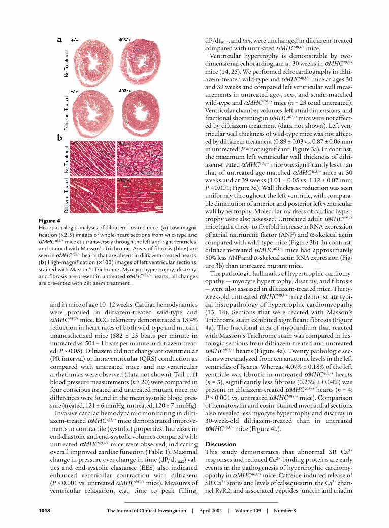
The pathologic hallmarks of hypertrophic cardiomy-opathy — myocyte hypertrophy, disarray, and fibrosis— were also assessed in diltiazem-treated mice. Thirty-week-old untreated αMHC403/+ mice demonstrate typi-cal histopathology of hypertrophic cardiomyopathy(13, 14). Sections that were reacted with Masson’sTrichrome stain exhibited significant fibrosis (Figure4a). The fractional area of myocardium that reactedwith Masson’s Trichrome stain was compared in his-tologic sections from diltiazem-treated and untreatedαMHC403/+ hearts (Figure 4a). Twenty pathologic sec-tions were analyzed from ten anatomic levels in the leftventricles of hearts. Whereas 4.07% ± 0.18% of the leftventricle was fibrotic in untreated αMHC403/+ hearts (n = 3), significantly less fibrosis (0.23% ± 0.04%) waspresent in diltiazem-treated αMHC403/+ hearts (n = 4; P < 0.001 vs. untreated αMHC403/+ mice). Comparisonof hematoxylin and eosin–stained myocardial sectionsalso revealed less myocyte hypertrophy and disarray in30-week-old diltiazem-treated than in untreatedαMHC403/+ mice (Figure 4b).
DiscussionThis study demonstrates that abnormal SR Ca2+
responses and reduced Ca2+-binding proteins are earlyevents in the pathogenesis of hypertrophic cardiomy-opathy in αMHC403/+ mice. Caffeine-induced release ofSR Ca2+ stores and levels of calsequestrin, the Ca2+ chan-nel RyR2, and associated peptides junctin and triadin
1018 The Journal of Clinical Investigation | April 2002 | Volume 109 | Number 8
Figure 4Histopathologic analyses of diltiazem-treated mice. (a) Low-magni-fication (×2.5) images of whole-heart sections from wild-type andαMHC403/+ mice cut transversely through the left and right ventricles,and stained with Masson’s Trichrome. Areas of fibrosis (blue) areseen in αMHC403/+ hearts that are absent in diltiazem-treated hearts.(b) High-magnification (×100) images of left ventricular sections,stained with Masson’s Trichrome. Myocyte hypertrophy, disarray,and fibrosis are present in untreated αMHC403/+ hearts; all changesare prevented with diltiazem treatment.

were decreased in advance of histopathology or echocar-diographic evidence of cardiac hypertrophy in αMHC403/+
mice. Restitution of normal Ca2+ storage protein levelsoccurred in response to diltiazem-mediated inhibitionof L-type Ca2+ channels, and when diltiazem was admin-istered early to αMHC403/+ mice, the cardiac histopathol-ogy and hemodynamic features of this sarcomere pro-tein mutation were significantly abated.
Previous studies demonstrated that levels of cytosolicCa2+ in αMHC403/+ myocytes are comparable to thosefound in wild-type cells (16). However, responses to phar-macologic agents such as cyclosporin A or minoxidil,which produce a brisk (30%) increase in diastolic Ca2+
levels in wild-type myocytes, are attenuated (<8% increas-es) in αMHC403/+ myocytes (16). This diminutiveresponse by αMHC403/+ myocytes is explained by the cur-rent studies. In response to the potent activation of RyR2channels by caffeine, the maximum and total Ca2+
release from the SR was less in αMHC403/+ myocytes thanin wild-type myocytes. This suggests that the SRs ofαMHC403/+ myocytes contain lower levels of Ca2+ than dothe SRs of wild-type myocytes (Figures 1 and 2).
Because αMHC403/+ myocyte SRs contained reducedamounts of Ca2+, we hypothesized that SR Ca2+ storageproteins would correspondingly be diminished. Decreasedlevels of calsequestrin and RyR2 in the SRs of αMHC403/+
myocytes were found. Calsequestrin is a 55-kDa high-capacity Ca2+-binding protein that both serves as a reser-voir for SR Ca2+ and, with junctin and triadin, forms acomplex that regulates Ca2+ release via RyR2 (26, 28, 29).Downregulation of calsequestrin and the other compo-nents of this quaternary RyR2 complex in αMHC403/+
myocytes is consistent with decreased SR Ca2+ levels. SRCa2+ levels may further be depleted by increased phos-phorylation of the RyR2 channel in αMHC403/+ myocytes.Phosphorylation decreases the amount of RyR2-associat-ed FKBP12.6, which makes the channel more sensitive tocalcium-induced activation and increases the probabilityof channel opening (21, 30, 31). Whereas only one of threepotential sites was phosphorylated in wild-type myocytes,most RyR2 channels from αMHC403/+ myocytes had twoor more phosphorylated sites.
Reduced SR Ca2+ levels but normal cytosolic Ca2+ levelsin αMHC403/+ myocytes are consistent with the modelthat the mutant sarcomere functions like an ion trap(Figure 5). Altered biophysical properties of a mutatedmyosin heavy chain could enhance Ca2+ retention by thesarcomere and deplete SR Ca2+ stores. Observations ofαMHC403/+ muscle physiology support this model:mutant fibers show greater-than-normal isometric ten-sion development at submaximal Ca2+ levels, and mutanthearts exhibit decreased rates of relaxation (15, 32).
The development of SR Ca2+ dysregulation followsmutant peptide expression and predates the hyper-trophic response in αMHC403/+ mice. By 4 weeks afterbirth, when αMHC403/+ myocytes express equal amountsof mutant and wild-type peptide (24), dysregulation ofSR calcium was apparent, and if it went uninterrupted,phenotypic manifestations of hypertrophic cardiomy-
opathy ensued (13, 14). L-type channel inhibition by dil-tiazem most likely interrupts this process by chronicattenuation of calcium-induced SR Ca2+ release, so as tolimit Ca2+ sequestration by the mutant sarcomere. Theconcurrent restoration of normal RyR2 and SR Ca2+-binding proteins levels and reduction of cardiac hyper-trophy and fibrosis in diltiazem-treated αMHC403/+ micefurther support a mechanistic link between SR Ca2+ dys-regulation and disease phenotype.
The L-type Ca2+ channel is the primary target of dilti-azem. However, diltiazem is known to have other cellu-lar targets, such as the mitochondrial Na+/Ca2+ exchang-er. Alteration in mitochondrial Ca2+ concentrationmight change αMHC403/+ myocyte energetics and there-by alter the hypertrophic response of the heart. Dilti-azem might reduce the hypertrophic response byimproving cardiac energetics. However, under normalconditions, diltiazem-treated αMHC403/+ hearts appearto do less work (lower heart rate, reduced stiffness [EES],and reduced dP/dtmax; see Table 1) than the untreated
The Journal of Clinical Investigation | April 2002 | Volume 109 | Number 8 1019
Figure 5Model of Ca2+ cycling in cardiac myocytes and the effects of a hyper-trophic cardiomyopathy–causing mutation. (a) Wild-type myocytesshowing normal Ca2+ regulation. Ca2+ enters the myocyte through L-type Ca2+ channels. Small entry of Ca2+ stimulates Ca release (cal-cium-induced Ca2+ release; CICR) from the SR via cardiac ryanodinereceptors (RyR2) to the sarcomere. Ca2+ returns to the SR via the sar-coplasmic/endoplasmic Ca2+ ATPase (SERCA) pump, which is regu-lated by phospholamban (PLB). Ca2+ cycling is “balanced” betweenthe sarcomere and SR. (b) Mutant (αMHC403/+) myocytes have amutation in the sarcomere (represented by an asterisk). The defec-tive sarcomere acts as an ion trap, resulting in accumulation of Ca2+.Less Ca2+ returns to the SR, resulting in decreased SR Ca2+ stores,decreased SR calsequestrin (CSQ), and reduced expression of RyR2.The net effect is a Ca2+ shift within the cell, with a relative Ca2+ excessin the sarcomere, and Ca2+ depletion in the SR.
αMHC403/+ heart, suggesting that even if cardiac energet-ics remain unaltered in αMHC403/+ hearts, the heartsshould benefit from diltiazem-mediated work reduction.
Other investigators have identified several importantintracellular Ca2+ sensors that can activate several signal-ing pathways to effect myocyte growth and function (forreview, see ref. 33). Based on its association with the SRand function in RyR2 channel phosphorylation, calci-um/calmodulin–dependent protein kinase (CamKII) is aparticularly intriguing candidate for signaling myocytegrowth (34). Levels of two well-characterized signalingmolecules, calmodulin and the calcium/calmodulin–acti-vated phosphatase calcineurin (PP2B-Aα and -Aβ), werenot altered in αMHC403/+ mice (data not shown). Whethertransient or compartmentalized changes in these or Ca2+
activation of other signaling molecules triggers hyper-trophy in αMHC403/+ mice remains unknown.
An important conclusion from these data is that earlyrestoration of SR Ca2+ decreases the hypertrophicresponse to a sarcomere mutation. Administration ofdiltiazem, in doses resulting in only modest hemody-namic effects, to prehypertrophic αMHC403/+ miceimproved cardiac function and provided long-term (39weeks) attenuation of hypertrophic pathology. Improve-ment in cardiac function was almost exclusively in sys-tolic function parameters, with little effect on diastolicfunction (Table 1). This may suggest that altering Ca2+
handling is important for contractile function, but thatdiastolic function is mediated by alternate mechanisms.In humans with clinical evidence of hypertrophic car-diomyopathy, L-type Ca2+ channel inhibitors such as dil-tiazem are used to treat symptoms. Our findings indi-cate that greater benefit should occur if these inhibitorsare administered in advance of disease expression. Inconjunction with gene-based, preclinical diagnosis ofhuman sarcomere protein mutations, evaluation of thepotential for diltiazem, and other pharmacologic agentsthat restore SR Ca2+ levels, to inhibit development ofhypertrophic cardiomyopathy appears warranted.
AcknowledgmentsWe thank Larry R. Jones for providing the RyR2 andjunctin antibodies, and Julie Zhang for myocyte isolation.The Howard Hughes Medical Institute supported thesestudies. C. Semsarian is the recipient of a National HeartFoundation of Australia grant. U. Mende is supported byan NIH Specialized Centers of Research grant and anAmerican Heart Association Scientist Developmentgrant. I. Ahmad is supported by a Sarnoff Fellowship.
1. Fatkin, D., Seidman, J.G., and Seidman, C.E. 2000. Hypertrophic car-diomyopathy. In Cardiovascular medicine. J.T. Willerson and J.N. Cohn, edi-tors. W.B. Saunders Co. Philadelphia, Pennsylvania, USA. 1055–1074.
2. Seidman, C.E., and Seidman, J.G. 2001. Hypertrophic cardiomyopathy. InThe metabolic and molecular bases of inherited disease. C.R. Scriver, A.L. Beaudet,W.S. Sly, and D. Valle, editors. McGraw-Hill. New York, New York, USA.5433–5452.
3. Maron, B.J., Bonow, R.O., Cannon, R.O., III, Leon, M.B., and Epstein, S.E.1987. Hypertrophic cardiomyopathy. Interrelations of clinical manifesta-tions, pathophysiology, and therapy (1). N. Engl. J. Med. 316:844–852.
4. Spirito, P., Seidman, C.E., McKenna, W.J., and Maron, B.J. 1997. The man-agement of hypertrophic cardiomyopathy. N. Engl. J. Med. 336:775–785.
5. Maron, B.J., Epstein, S.E., and Roberts, W.C. 1986. Causes of sudden death
in competitive athletes. J. Am. Coll. Cardiol. 7:204–214.6. Maron, B.J., et al. 1996. Sudden death in young competitive athletes. Clin-
ical, demographic, and pathological profiles. JAMA. 276:199–204.7. Vikstrom, K.L., Factor, S.M., and Leinwand, L.A. 1996. Mice expressing
mutant myosin heavy chains are a model for familial hypertrophic car-diomyopathy. Mol. Med. 2:556–567.
8. Kittleson, M.D., et al. 1999. Familial hypertrophic cardiomyopathy inmaine coon cats: an animal model of human disease. Circulation.99:3172–3180.
9. Marian, A.J., et al. 1999. A transgenic rabbit model for human hypertrophiccardiomyopathy. J. Clin. Invest. 104:1683–1692.
10. Oberst, L., et al. 1998. Dominant-negative effect of a mutant cardiac tro-ponin T on cardiac structure and function in transgenic mice. J. Clin. Invest.102:1498–1505.
11. Patel, R., et al. 2001. Simvastatin induces regression of cardiac hypertrophyand fibrosis and improves cardiac function in a transgenic rabbit model ofhuman hypertrophic cardiomyopathy. Circulation. 104:317–324.
12. Lim, D.S., et al. 2001. Angiotensin II blockade reverses myocardial fibrosisin a transgenic mouse model of human hypertrophic cardiomyopathy. Cir-culation. 103:789–791.
13. Geisterfer-Lowrance, A.A., et al. 1996. A mouse model of familial hyper-trophic cardiomyopathy. Science. 272:731–734.
14. McConnell, B.K., et al. 2001. Comparison of two murine models of famil-ial hypertrophic cardiomyopathy. Circ. Res. 88:383–389.
15. Blanchard, E., Seidman, C., Seidman, J.G., LeWinter, M., and Maughan, D.1999. Altered crossbridge kinetics in the αMHC403/+ mouse model of famil-ial hypertrophic cardiomyopathy. Circ. Res. 84:475–483.
16. Fatkin, D., et al. 2000. An abnormal Ca2+ response in mutant sarcomereprotein-mediated familial hypertrophic cardiomyopathy. J. Clin. Invest.106:1351–1359.
17. Bers, D.M. 2000. Calcium fluxes involved in control of cardiac myocyte con-traction. Circ. Res. 87:275–281.
18. Kim, S.J., et al. 1999. An α-cardiac myosin heavy chain gene mutationimpairs contraction and relaxation function of cardiac myocytes. Am. J.Physiol. 276:H1780–H1787.
19. McConnell, B.K., Moravec, C.S., and Bond, M. 1998. Troponin I phospho-rylation and myofilament calcium sensitivity during decompensated car-diac hypertrophy. Am. J. Physiol. 274:H385–H396.
20. McConnell, B.K., et al. 1999. Dilated cardiomyopathy in homozygousmyosin-binding protein-C mutant mice. J. Clin. Invest. 104:1235–1244.
21. Marx, S.O., et al. 2000. PKA phosphorylation dissociates FKBP12.6 fromthe calcium release channel (ryanodine receptor): defective regulation infailing hearts. Cell. 101:365–376.
22. Georgakopoulos, D., et al. 1999. The pathogenesis of familial hypertrophiccardiomyopathy: early and evolving effects from an α-cardiac myosin heavychain missense mutation. Nat. Med. 5:327–330.
23. Palmer, B.M., Lynch, J.M., Snyder, S.M., and Moore, R.L. 2001. Renal hyper-tension prevents run training modification of cardiomyocyte diastolicCa2+ regulation in male rats. J. Appl. Physiol. 90:2063–2069.
24. Fatkin, D., et al. 1999. Neonatal cardiomyopathy in mice homozygous forthe Arg403Gln mutation in the α-cardiac myosin heavy chain gene. J. Clin.Invest. 103:147–153.
25. Semsarian, C., et al. 2001. A polymorphic modifier gene alters the hyper-trophic response in a murine model of familial hypertrophic cardiomy-opathy. J. Mol. Cell. Cardiol. 33:2055–2060.
26. Zhang, L., Kelley, J., Schmeisser, G., Kobayashi, Y.M., and Jones, L.R. 1997.Complex formation between junctin, triadin, calsequestrin, and the ryan-odine receptor. Proteins of the cardiac junctional sarcoplasmic reticulummembrane. J. Biol. Chem. 272:23389–23397.
27. Scriven, D.R., Dan, P., and Moore, E.D. 2000. Distribution of proteinsimplicated in excitation-contraction coupling in rat ventricular myocytes.Biophys. J. 79:2682–2691.
28. Jones, L.R., et al. 1998. Regulation of Ca2+ signaling in transgenic mousecardiac myocytes overexpressing calsequestrin. J. Clin. Invest.101:1385–1393.
29. Niki, I., Yokokura, H., Sudo, T., Kato, M., and Hidaka, H. 1996. Ca2+ sig-naling and intracellular Ca2+ binding proteins. J. Biochem. (Tokyo).120:685–698.
30. Szegedi, C., Sarkozi, S., Herzog, A., Jona, I., and Varsanyi, M. 1999. Calse-questrin: more than ‘only’ a luminal Ca2+ buffer inside the sarcoplasmicreticulum. Biochem. J. 337:19–22.
31. Yano, K., and Zarain-Herzberg, A. 1994. Sarcoplasmic reticulum calse-questrins: structural and functional properties. Mol. Cell. Biochem.135:61–70.
32. Spindler, M., et al. 1998. Diastolic dysfunction and altered energetics in theαMHC403/+ mouse model of familial hypertrophic cardiomyopathy. J. Clin.Invest. 101:1775–1783.
33. Frey, N., McKinsey, T.A., and Olson, E.N. 2000. Decoding calcium signalsinvolved in cardiac growth and function. Nat. Med. 6:1221–1227.
34. Passier, R., et al. 2000. CaM kinase signaling induces cardiac hypertrophyand activates the MEF2 transcription factor in vivo. J. Clin. Invest.105:1395–1406.
1020 The Journal of Clinical Investigation | April 2002 | Volume 109 | Number 8