Embed Size (px)
Citation preview
The L-4F mimetic peptide prevents insulin resistancethrough increased levels of HO-1, pAMPK,and pAKT in obese mice1
Stephen J. Peterson,3,*,† Dong Hyun Kim,3,* Ming Li,* Vincenzo Positano,§ Luca Vanella,*Luigi F. Rodella,* Francesco Piccolomini,* Nitin Puri,* Amalia Gastaldelli,§
Claudia Kusmic,§ Antonio LʼAbbate,§,** and Nader G. Abraham2,*,†,††
Departments of Pharmacology* and Medicine,† New York Medical College, Valhalla, NY 10595; CentralNational Research Institute of Clinical Physiology§ and Scuola Superiore Sant’Anna and CNR Instituteof Clinical Research,** Pisa, Italy; and The Rockefeller University,†† New York 10021
Abstract We examined mechanisms by which L-4F reducesobesity and diabetes in obese (ob) diabetic mice. We hypoth-esized that L-4F reduces adiposity via increased pAMPK,pAKT, HO-1, and increased insulin receptor phosphoryla-tion in ob mice. Obese and lean mice were divided into fivegroups: lean, lean-L-4F-treated, ob, ob-L-4F-treated, and ob-L-4F-LY294002. Food intake, insulin, glucose adipocyte stemcells, pAMPK, pAKT, CB1, and insulin receptor phosphory-lation were determined. Subcutaneous (SAT) and visceraladipose tissue (VAT) were determined by MRI and hepaticlipid content by magnetic resonance spectroscopy. SAT andVAT volumes decreased in ob-L-4F-treated animals com-pared with control. L-4F treatment decreased hepatic lipidcontent and increased the numbers of small adipocytes(P , 0.05) and phosphorylation of insulin receptors. L-4Fdecreased CB1 in SAT and VAT and increased pAKT andpAMPK in endothelium. L-4F-mediated improvement in en-dothelium was prevented by LY294002. Inhibition of pAKTand pAMPK by LY294002 was associated with an increase inglucose levels. Upregulation of HO-1 by L-4F produced adi-pose remodeling and increased the number of small dif-ferentiated adipocytes. The anti-obesity effects of L-4Fare manifested by a decrease in visceral fat content with re-ciprocal increases in adiponectin, pAMPK, pAKT, and phos-phorylation of insulin receptors with improved insulinsensitivity.—Peterson, S. J., D. H. Kim, M. Li, V. Positano,L. Vanella, L. F. Rodella, F. Piccolomini, N. Puri, A.Gastaldelli, C. Kusmic, A. LʼAbbate, and N. G. Abraham.The L-4F mimetic peptide prevents insulin resistancethrough increased levels of HO-1, pAMPK, and pAKT inobese mice. J. Lipid Res. 2009. 50: 1293–1304.
Supplementary key words diabetes • adiponectin • adiposity • apolipo-protein A-I • heme oxygenase-1 • insulin receptor • insulin sensitivity •
obesity • endothelial dysfunction
Insulin resistance is the hallmark of type 2 diabetes andits associated cardiovascular disease (1, 2). Once hyper-glycemia is established, an increase in reactive oxygen species(ROS) occurs, resulting in a progressive deterioration invascular function with an elevation of inflammatory cyto-kines from adipose tissue (3). Obesity is also a risk factorfor cardiovascular disease and diabetes in humans (4).Obesity and insulin resistance are considered independentpredictors of cardiovascular disease (5, 6). However, re-cent studies have revealed the presence of myocardial per-fusion abnormalities, even in the absence of coronarylesions, in patients with asymptomatic diabetes mellitus(6, 7). This suggests that obesity and diabetes are stronglyassociated with endothelial dysfunction (8–13), increasedlipolysis, hepatic triglyceride secretion, and sterol-regulatoryelement binding protein-1 (SREBP-1) (14). SREBPs are themajor transcription factors that regulate genes involvedin fatty acid and cholesterol synthesis and are regulatedby pAMPK and pAKT in ob mice (14). In addition, ac-tivation of the endocannabinoid receptor-1 (CB1 receptor)was shown to increase the hepatic lipogenic transcriptionfactor and fatty acid synthesis (7). In visceral adipose tissue,CB1 receptor activity leads to a decrease in adiponectin andan increase in lipogenesis, both of which contribute toinsulin resistance.
Drug therapy for atherosclerosis, coronary artery disease,dyslipidemia, hypertension, and other diseases associatedwith highly inflammatory states have been shown to either
e Author s̓ Choice
This work was supported by National Institutes of Health Grants DK-068134,HL-55601, and HL-34300 (N.G.A.), by the CNR Medical Department andCardiopulmonary Project, and by the Scuola SantʼAnna.e Author s̓ Choice— Final version full access.
Manuscript received 24 November 2008 and in revised form 5 February 2009.
Published, JLR Papers in Press, March 26, 2009.DOI 10.1194/jlr.M800610-JLR200
Abbreviations: FR, fat resonance; HO, heme oxygenase; IOD,integratedoptical density; i.p., intraperitoneally;MRS,magnetic resonancespectroscopy; MSC, mesenchymal stem cell; ob, obese; ROS, reactiveoxygen species; SAT, subcutaneous fat tissue; SREBP-1, sterol-regulatoryelement binding protein-1; VAT, visceral fat tissue.
1 This work was presented in part at the American Heart Association,November, 2009.
2 To whom correspondence should be addressed.e-mail: [email protected]
3 S. J. Peterson and D. H. Kim contributed equally to this work.
Copyright © 2009 by the American Society for Biochemistry and Molecular Biology, Inc.
This article is available online at http://www.jlr.org Journal of Lipid Research Volume 50, 2009 1293
by on October 23, 2009
ww
w.jlr.org
Dow
nloaded from
directly or indirectly induce heme oxygenase (HO) activityand increase HO gene expression (10). These drugs in-clude aspirin, statins, losartan, immunosuppressive agents,such as cyclosporine A, and a host of other compounds, in-cluding 4F (10, 15). These 4F compounds are apolipo-protein A1 mimetic peptides, synthesized from eitherD (D-4F) or L (L-4F) amino acids that enhance the abilityof HDL to protect LDL against oxidation in atheroscleroticanimals (16–18). Oral administration of D-4F has beenreported to reduce atherosclerotic disease independent ofcholesterol levels (17, 18). D-4F causes HDL to become anti-inflammatory, stimulates HDL-mediated cholesterol efflux,and reverses cholesterol transport frommacrophages (16, 17).D-4F has been reported to reduce SREBP-1c mRNA levelsEO6 immunoreactivity and renal inflammation in LDLreceptor-null mice fed a Western diet (19). In a sickle celldisease model, apolipoprotein A1 improved vasoreactivityin LDL-receptor null mice (20). L-4F and D-4F improve vas-cular function and restore the balance between nitric oxideand superoxide anions due to increase in EC-SOD, HO-1,and peNOS levels (8, 20–23). Induction of HO-1 protein re-sults in a marked increase in serum levels of adiponectinand a decrease in the inflammatory cytokines IL-1, IL-6,and TNF and an increase in insulin sensitivity (24–26).
In this study, we examined the mechanism of L-4F treat-ment in the restoration of vascular function by measuringthe expression of pAMPK, pAKT, and insulin receptor phos-phorylation. L-4F treatment resulted in reduced adiposity,manifested by a decrease in visceral and subcutaneous fatlevels, decreased adipogenesis, and the increased presenceof smaller insulin sensitive adipocytes via an increase inpAMPK, pAKT, and insulin receptor phosphorylation.
MATERIALS AND METHODS
Animal protocolsMale obese (ob) mice (B6v-Lep ob/J) were purchased from
Harlan (Chicago, IL) at the age of 7 weeks and used at 8 weeksof age. Age- and sex-matched lean mice (B6.V, lean; Harlan)served as controls. Mice were fed a normal chow diet and hadfree access to water. Body weight of ob and lean mice at the be-ginning of the treatment were 346 5 g and 266 3 g, respectively.Glucose levels were 2486 21 and 1266 14 mg/dl for ob and leanmice, respectively. Glucose monitoring was performed using anautomated analyzer (Life Scan, Milpitas, CA). Beginning at9 weeks of age when all ob mice had established diabetes, L-4F(i.e., Ac-D-W-F-K-A-F-Y-D-K-V-A-E-K-F-K-E-A-F-NH2) synthesizedfrom L-amino acids as previously described (27) was injected ata dose of 200 mg/100 g daily in 200 ml vehicle), or vehicle (ABCT:ammonium bicarbonate buffer at pH 7.4 containing 0.01%Tween 20) were administered intraperitoneally (i.p.) for 6 weeks.At the time of sacrifice, subcutaneous and visceral fat visible inthe abdomen, mesenteric fat, fat around the liver, kidney spleen,and heart were dissected free, pooled for each mouse, andweighed. Blood was collected (50–100 ml) from the tail vein ofanesthetized ob mice following administration of either L-4F orvehicle. L-4F treated ob mice were divided into two groups, onetreated with L-4F and the P13 kinase AKT inhibitor, LY294002(Cell Signaling Technology, Boston, MA) 2-(4-morpholinyl)-8-phenyl-4H-1-benzopyran-4-HCl), which was dissolved in DMSO
and diluted with PBS and injected i.p. at a dose of 100 mg/kg,three times a week for the last three weeks of the study (28, 29).We have previously shown that LY294004 is an inhibitor of HO-1-mediated cell protection (30). The second group of ob-L-4F-treated mice were treated with the same volume of vehicle(PMSO1PBS) without LY294002. Six groups of animals werestudied: A) lean, B) lean-L-4F, C) lean-L-4F-LY-294002, D) ob con-trol, E) ob-L-4F, and F) ob-L-4F-LY294002. Food intake did notchange in the mice in any treatment group. The Animal Careand Use Committee of New York Medical College approvedall experiments.
MRI and magnetic resonance spectroscopySubcutaneous fat tissue (SAT) and visceral fat tissue (VAT)
were determined by MRI, while hepatic fat was determined bymagnetic resonance spectroscopy (MRS). Mice were imaged ina GE Excite 1.5T scanner using a knee coil with a T1-weightedspin-echo pulse sequence (TEC 9.0 ms, TR 5 540ms, NEX 5 4,FOVO 8 3 8 cm) and an image size of 224 3 192 pixels. Thewhole chest and abdomen of each mouse were covered with axialslices (thickness 3 mm, no spacing). Acquired images underwentsemiautomatic segmentation of SAT and VAT using the previouslyvalidated HIPPO FAT: tool (31–33).
The software computed three masks (background, fat, andnonfat tissues) using a fuzzy clustering segmentation. Externaland internal SAT boundaries were then defined by an active con-tour algorithm that exploited the previously computed masks asexternal force maps. A third contour was computed surroundingthe area where VAT was present together with other tissues. VATitself was assessed by the automated analysis of the signal histogramin the visceral region previously defined, by identifying the secondpeak of the signal histogram. This provided whole body volume(including the skeletal and soft tissues), total fat, SAT and VATvolumes, as well as VAT/SAT and fat/body volume ratios.
Liver lipid measurementsLocalized 1H MRS of the liver was acquired on the GE Excite
1.5T MRI scanner. The liver slice with the largest gross dimen-sions was chosen for the MRS study. MRS for water and fat quan-tification was accomplished using a point resolved spectroscopysequence. After line broadening and phase and baseline correc-tion, the peak area of the water at 4.77 ppm and fat resonance(FR) at 1.40 ppm were measured. Quantification of the fat con-tent was done by comparing the area of the FR with that of theunsuppressed water. Spectroscopic data were processed using theNUTS software. Hepatic fat percentage was calculated by dividing(FR) by the sum of FR and peak area of water. This technique ishighly reproducible, with a coefficient of variation ,2% in slicesstudied on eight separate occasions.
Determination of adipocyte cell size, HO-1, and CB1Subcutaneous adipose tissue and renal, liver, and aortic vis-
ceral fat tissue collected from untreated ob mice and L-4F-treatedob mice (n5 8 mice per group) were prepared for morphologicalanalysis. Samples were fixed in 4% paraformaldehyde for 24 h, cutinto small pieces, and embedded in paraffin for histological analy-sis. The samples were cut by microtome (5 mm thick), mounted onD-polylisinated glass slides, deparaffinizated in xylene, and stainedwith hematoxylin and eosin for the evaluation of adipocyte size orprocessed for CB-1 orHO-1 immunohistochemistry. Immunostain-ing for CB-1 was carried out using a goat polyclonal anti-CB-1 pri-mary antibody (22) (Santa Cruz Biotechnology, Santa Cruz, CA),and HO-1 immunostaining was carried out using a rabbit poly-clonal anti-HO-1 primary antibody (Stressgen Bioreagents, Victoria,BC, Canada). For each experimental group, five sections per animal
1294 Journal of Lipid Research Volume 50, 2009
by on October 23, 2009
ww
w.jlr.org
Dow
nloaded from
were stained. Sections were immersed in 3% hydrogen peroxideand diluted in methanol for 30 min to quench endogenousperoxidase activity. The sections were preincubated with 3%horse serum for 60 min followed by primary antibody anti-CB-1diluted 1:125 for 2 h at 37°C. The sections were then washedin TBS (0.1 M), incubated 30 min at room temperature withbiotinylated horse anti-goat immunoglobulin (Vector Labora-tories, Burlingame, CA), and then incubated for 30 min at roomtemperature with avidin-biotin-horseradish peroxidase complex(ABC complex; Vector Laboratories, Burlingame, CA). The re-action product was visualized using hydrogen peroxide anddiaminobenzidine (Sigma-Aldrich, St. Louis, MO) as the chro-mogen. All slides were dehydrated and mounted in DPX (Sigma-Aldrich). Negative controls for primary antibody with nonimmuneserum revealed no signal.
Determination of CB-1 and HO-1 fatCB-1 and HO-1 staining intensity was computed as integrated
optical density (IOD). Digitally fixed images of the slices (n5 5 peranimal) at 203 magnification were analyzed using an optical mi-croscope equipped with an image analyzer (Image Pro Plus; Im-maginie Computer, Milan, Italy). For quantitative analysis, IODwas calculated for arbitrary areas, measuring three fields with thesame area for each section.
Determination of adipocyte cell sizeDigital images of adipose tissue sections were captured using a
light microscope (Olympus) at 203 magnification. For eachgroup, three fields from each of five different hematoxylin-eosinstained sections per animal were analyzed. Individual adipocyteareas (mm2) within each field were determined using image anal-ysis software (Image Pro Plus). For the quantitative analysis, adi-pocyte areas were calculated in arbitrary fields, measuring50 adipocytes for each section.
Western blot analysis of liver, kidney, and adipocyte stemcells for HO-1, AMPK, pAMPK, AKT, pAKT, and insulinreceptor phosphorylation
At sacrifice, subcutaneous and visceral fat in the abdomen (thevisible mesenteric fat and fat around the liver, kidney, spleen, andthe heart) were dissected, pooled for each mouse, and used toisolate adipocyte stem cells. Specimens were stored at2140°Cuntilassayed. Frozen liver, kidney, heart, and fat tissues were pulverizedunder liquid nitrogen and placed in a homogenization buffer[mmol/l: 10 phosphate buffer, 250 sucrose, 1 EDTA, 0.1 PMSF,and 0.1% (v/v) tergitol, pH 7.5]. Homogenates were centrifugedat 27,000 g for 10 min at 4°C, supernatant was isolated, and proteinlevels were visualized by immunoblotting with antibodies. Anti-bodies against AMPK, pAMPK, AKT, and pAKT were obtainedfrom Cell Signaling Technology (Beverly, MA). Antibodies wereprepared by dilution of HO-1, pAMPK, pAKT, and insulin receptoras we described previously (23, 25, 26).
Measurement of fiber diameter and fat depositsSerial sections (8 mm thick) were cut by cryostat and stained
with hematoxylin-eosin for morphological evaluation (measure-ment of diameter) and analayzed for fat deposits with Oil Red Ostaining. Digital images were taken using a light microscope(Olympus) and then analyzed with a software program (Image-Pro Plus 4.5.1). Ten fibers from each muscle slice were randomlyselected to estimate fiber diameter; fat deposits were evaluatedthrough measurement of the percentage of Oil Red O stainedareas in five fields per randomly selected muscle sections. A totalof five sections per animal were analyzed.
Adipocyte mesenchymal stem cell isolation from SAT andVAT of lean, ob, and ob L-4F-treated mice
To isolate mouse adipocyte mesenchymal stem cells (MSCs), adi-pose tissues were washed with PBS and digested at 37°C for 30 minwith 0.075% type II collagenase (34). At 50% confluence, L-4F wasadded as indicated in the figure legends. HO-1 and adipogenesiswere measured, with the latter using Oil Red O staining (25).
Glucose tolerance testAfter a 12 h fast, mice were injected i.p. with glucose (2.0g/kg
body weight). Blood samples were taken at various time points(0–120 min) for measurement of blood glucose levels.
Human bone marrow-derived mesenchymal stemcells and adipocytes
Frozen bone marrow mononuclear cells were purchased fromAllcells (Emeryville, CA). After thawing the cells, mononuclearcells were cultured as previously described (23, 25, 26).
Adipogenic differentiation of human MSCs andeffect of L-4F
Adipogenic differentiation of human MSCs was induced by in-cubation in an adipogenesis inductionmedium [DMEM-high glucose(35, 36), supplemented with 10 mg/ml of insulin, 1 mmol/L of dexa-methasone, 0.2 mmol/L of indomethacin, 10% FBS, and 1% anti-biotic–antimycotic solution]. The medium was changed every3–4 days (35, 36) for vehicle and L4F. At 50% confluence, L-4F andvehicle solutions were added, andHO-1, adiponectin, pAMPK, pAKT,and adipogenesis were measured, the latter using Oil Red O as pre-viously described (25). MSC-derived adipocytes were treated withL-4F, 7.5 mM LY294002, a dose effective in inhibiting P 1-3 Kinase/AKT (29, 30). Addition of L-4F or LY294002 to adipocyte stem cell cul-ture does not affect cell viability as measured by trypan blue staining.
The effect of L-4F and LY2940002 in cultured adipocytestem cells on Oil Red O staining and lipid droplet size
For Oil Red O staining, 0.21% Oil Red O in 100% isopropanol(Sigma-Aldrich) was used. Briefly, adipocytes were fixed in 10%
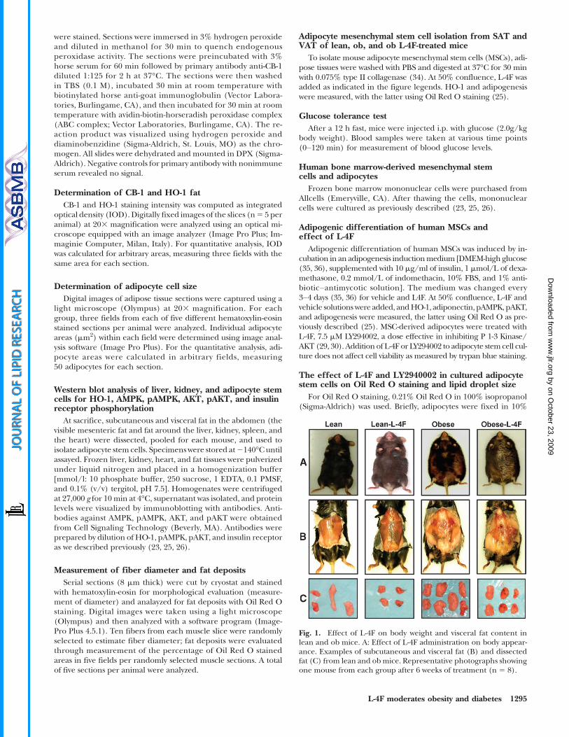
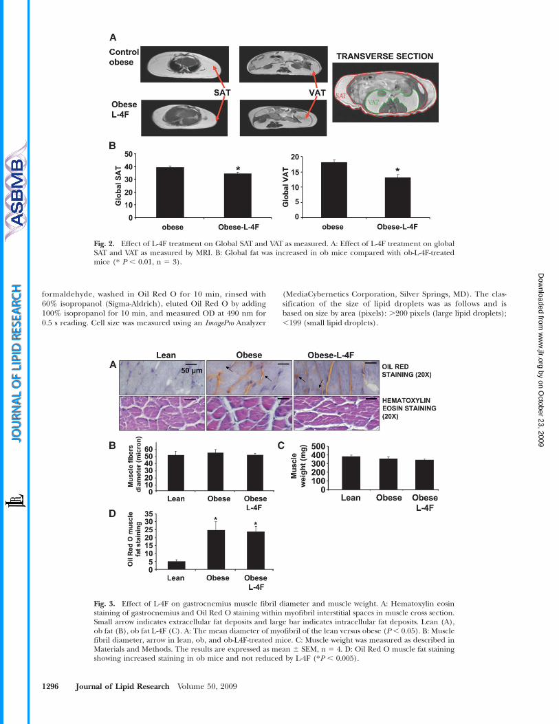
Fig. 1. Effect of L-4F on body weight and visceral fat content inlean and ob mice. A: Effect of L-4F administration on body appear-ance. Examples of subcutaneous and visceral fat (B) and dissectedfat (C) from lean and ob mice. Representative photographs showingone mouse from each group after 6 weeks of treatment (n 5 8).
L-4F moderates obesity and diabetes 1295
by on October 23, 2009
ww
w.jlr.org
Dow
nloaded from
formaldehyde, washed in Oil Red O for 10 min, rinsed with60% isopropanol (Sigma-Aldrich), eluted Oil Red O by adding100% isopropanol for 10 min, and measured OD at 490 nm for0.5 s reading. Cell size was measured using an ImagePro Analyzer
(MediaCybernetics Corporation, Silver Springs, MD). The clas-sification of the size of lipid droplets was as follows and isbased on size by area (pixels): .200 pixels (large lipid droplets);,199 (small lipid droplets).
Fig. 2. Effect of L-4F treatment on Global SAT and VAT as measured. A: Effect of L-4F treatment on globalSAT and VAT as measured by MRI. B: Global fat was increased in ob mice compared with ob-L-4F-treatedmice (* P , 0.01, n 5 3).
Fig. 3. Effect of L-4F on gastrocnemius muscle fibril diameter and muscle weight. A: Hematoxylin eosinstaining of gastrocnemius and Oil Red O staining within myofibril interstitial spaces in muscle cross section.Small arrow indicates extracellular fat deposits and large bar indicates intracellular fat deposits. Lean (A),ob fat (B), ob fat L-4F (C). A: The mean diameter of myofibril of the lean versus obese (P , 0.05). B: Musclefibril diameter, arrow in lean, ob, and ob-L4F-treated mice. C: Muscle weight was measured as described inMaterials and Methods. The results are expressed as mean 6 SEM, n 5 4. D: Oil Red O muscle fat stainingshowing increased staining in ob mice and not reduced by L-4F (*P , 0.005).
1296 Journal of Lipid Research Volume 50, 2009
by on October 23, 2009
ww
w.jlr.org
Dow
nloaded from
Statistical analysisStatistical significance between experimental groups was deter-
mined by the Fisher method of analysis of multiple comparisons(P , 0.05 was regarded as significant). For comparison betweentreatment groups, the null hypothesis was tested by either a single-factor ANOVA for multiple groups or unpaired t-test for two groups.Data are presented as mean 6 SEM except for cell size and IOD forCB1, which are presented asmean6 SD.Differences between experi-mental groups were evaluated with ANOVA with Bonferroni correc-tions. Statistical significance was regarded as significant at P , 0.05.
RESULTS
L-4F treatment reduces weight gain and fat contentWe examined the effect of L-4F treatment on body and
fat appearance. As seen in Fig. 1, L-4F treatment visiblyreduced weight gain in ob mice. The final weights after6 weeks of vehicle or L-4F treatment were (53 6 2.9 gand 43.26 1.7 g, respectively, P, 0.05). The L-4F-mediatedreduction of weight gain was reversible. When L-4F wasdiscontinued at week 10, ob mice gained weight at a fasterrate than ob vehicle treated animals (data not shown). Asseen in Fig. 1, visceral fat in obese mice was decreased byL-4F treatment. This provided semiquantitative estimatesof fat content, which was quantitated using MRI asdescribed below.
Effect of L-4F on fat content determined by MRIMRI was used to quantify SAT and VAT. As seen on Fig. 2,
global SAT and global VAT were significantly decreased byL4F, P , 0.05 and P , 0.01, respectively. Subcutaneous fat
in ob mice was decreased from 5.13 6 0.78 to 3.21 6 45g,and visceral fat in obese mice was decreased from 8.82 60.49 to 6.13 6 0.69 g. The decrease in visceral fat was ac-companied by a significant increase in adiponectin levelsand a decrease in the levels of the inflammatory cytokineIL-1 b. Prior to treatment, adiponectin levels in the ob micewere 2.73 6 0.51 mg/ml compared with 4.76 6 0.93 mg/ml(P , 0.029) in lean animals. L-4F treatment resulted ina significant increase (P , 0.01) in the levels of serumadiponectin 6.146 1.49 mg/ml in the ob mice. Obese miceexhibited a significant increase in serum IL-1b levels (123629 pg/ml) when compared with age-matched lean controls(486 31 pg/ml), P, 0.05. L-4F treatment resulted in a sig-nificant (P , 0.01) decrease in serum IL-1 levels in theob mice (39 6 26 pg/ml) compared with 106 6 21 pg/mlin untreated ob to levels seen in the lean mice, confirmingprevious data (23).
L-4F treatment on muscle weight and intramuscularOil Red O staining
The weight of gastrocnemius muscle in lean mice, un-treated ob mice, and L-4F-treated ob mice was 393 6 46 mg,350 6 52 mg, and 356 6 48 mg, respectively (Fig. 3). Asshown in Fig. 3, L-4F treatment did not affect muscle fiber,fat content, or muscle diameter. The diameters of musclefibers for the lean, ob untreated, and L-4F-treatedmice were50 6 4.7 mm, 53 6 4 mm, and 51 6 4 mm, respectively(Fig. 3B). Intramuscular lipid droplets were severalfoldhigher in ob compared with lean mice (P, 0.005) but werenot different in ob mice treated with L-4F when comparedwith untreated ob mice (Fig. 3D).
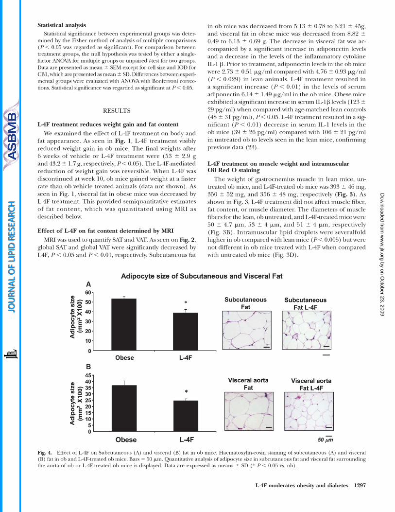
Fig. 4. Effect of L-4F on Subcutaneous (A) and visceral (B) fat in ob mice. Haematoxylin-eosin staining of subcutaneous (A) and visceral(B) fat in ob and L-4F-treated ob mice. Bars5 50 mm. Quantitative analysis of adipocyte size in subcutaneous fat and visceral fat surroundingthe aorta of ob or L-4F-treated ob mice is displayed. Data are expressed as means 6 SD (* P , 0.05 vs. ob).
L-4F moderates obesity and diabetes 1297
by on October 23, 2009
ww
w.jlr.org
Dow
nloaded from
Effect of L-4F on adipocyte sizeHistological examination of SAT and VAT revealed that
adipocyte cell size was significantly decreased (Fig. 4). Adi-pocyte cell size in the subcutaneous fat of control ob micewas 52 6 14 mm2 3100 (Fig. 4A). Adipocyte cell size wassignificantly lower in L-4F-treated ob mice, 42 6 11 mm2 3100 (P , 0.05). A similar reduction was seen with L-4Ftreatment in adipocyte cell size in visceral fat surroundingaortic fat tissue (Fig. 4B). Adipocyte cell size was propor-tionally lower in subcutaneous fat tissue. Adipocyte cell sizein lean animals was 37 6 9 mm2 3 100 compared with leanmice treatedwithL-4F (266 7mm23 100) (data not shown).
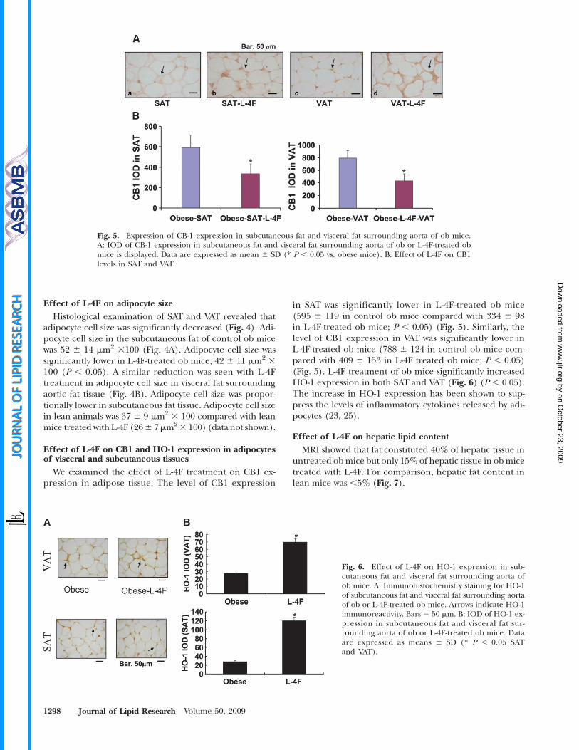
Effect of L-4F on CB1 and HO-1 expression in adipocytesof visceral and subcutaneous tissues
We examined the effect of L-4F treatment on CB1 ex-pression in adipose tissue. The level of CB1 expression
in SAT was significantly lower in L-4F-treated ob mice(595 6 119 in control ob mice compared with 334 6 98in L-4F-treated ob mice; P , 0.05) (Fig. 5). Similarly, thelevel of CB1 expression in VAT was significantly lower inL-4F-treated ob mice (788 6 124 in control ob mice com-pared with 409 6 153 in L-4F treated ob mice; P , 0.05)(Fig. 5). L-4F treatment of ob mice significantly increasedHO-1 expression in both SAT and VAT (Fig. 6) (P , 0.05).The increase in HO-1 expression has been shown to sup-press the levels of inflammatory cytokines released by adi-pocytes (23, 25).
Effect of L-4F on hepatic lipid contentMRI showed that fat constituted 40% of hepatic tissue in
untreated obmice but only 15% of hepatic tissue in obmicetreated with L-4F. For comparison, hepatic fat content inlean mice was ,5% (Fig. 7).
Fig. 6. Effect of L-4F on HO-1 expression in sub-cutaneous fat and visceral fat surrounding aorta ofob mice. A: Immunohistochemistry staining for HO-1of subcutaneous fat and visceral fat surrounding aortaof ob or L-4F-treated ob mice. Arrows indicate HO-1immunoreactivity. Bars 5 50 mm. B: IOD of HO-1 ex-pression in subcutaneous fat and visceral fat sur-rounding aorta of ob or L-4F-treated ob mice. Dataare expressed as means 6 SD (* P , 0.05 SATand VAT).
Fig. 5. Expression of CB-1 expression in subcutaneous fat and visceral fat surrounding aorta of ob mice.A: IOD of CB-1 expression in subcutaneous fat and visceral fat surrounding aorta of ob or L-4F-treated obmice is displayed. Data are expressed as mean 6 SD (* P , 0.05 vs. obese mice). B: Effect of L-4F on CB1levels in SAT and VAT.
1298 Journal of Lipid Research Volume 50, 2009
by on October 23, 2009
ww
w.jlr.org
Dow
nloaded from
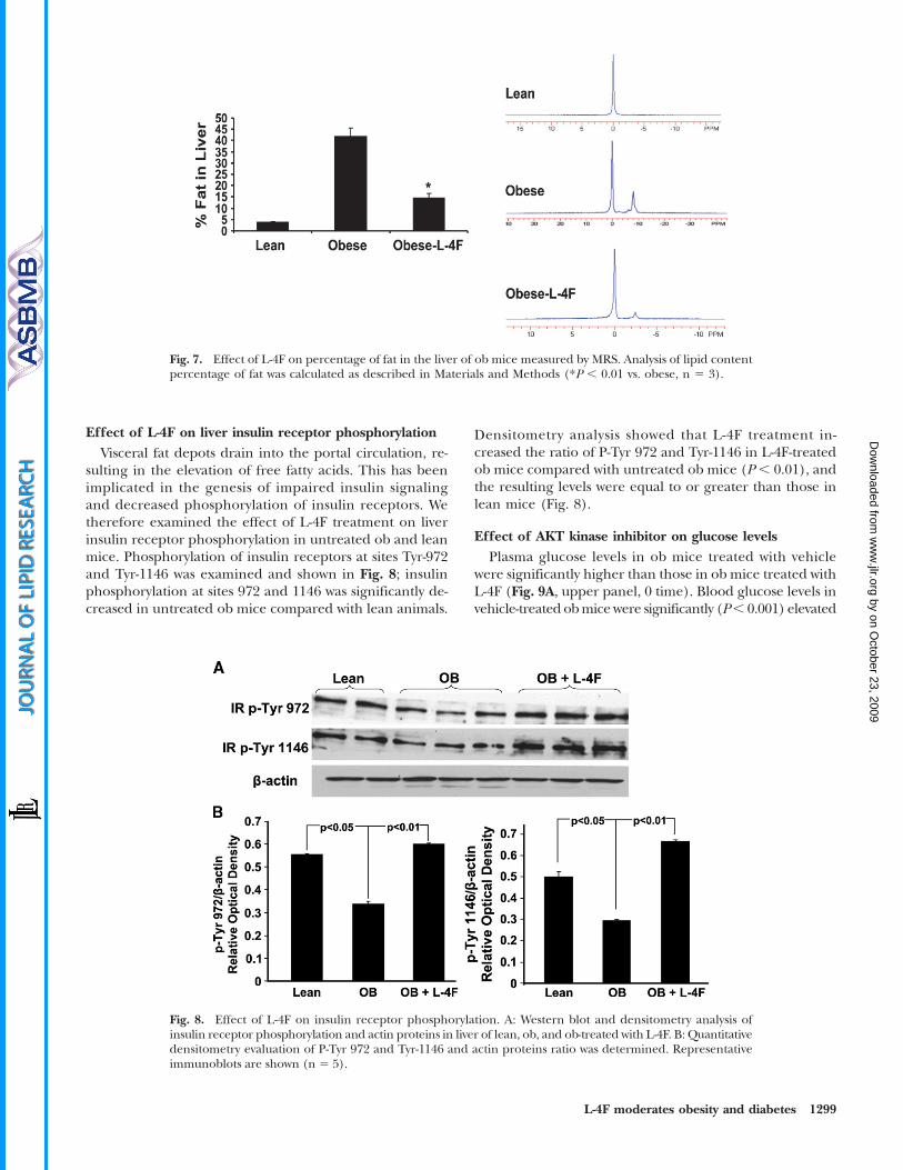
Effect of L-4F on liver insulin receptor phosphorylationVisceral fat depots drain into the portal circulation, re-
sulting in the elevation of free fatty acids. This has beenimplicated in the genesis of impaired insulin signalingand decreased phosphorylation of insulin receptors. Wetherefore examined the effect of L-4F treatment on liverinsulin receptor phosphorylation in untreated ob and leanmice. Phosphorylation of insulin receptors at sites Tyr-972and Tyr-1146 was examined and shown in Fig. 8; insulinphosphorylation at sites 972 and 1146 was significantly de-creased in untreated ob mice compared with lean animals.
Densitometry analysis showed that L-4F treatment in-creased the ratio of P-Tyr 972 and Tyr-1146 in L-4F-treatedob mice compared with untreated ob mice (P , 0.01), andthe resulting levels were equal to or greater than those inlean mice (Fig. 8).
Effect of AKT kinase inhibitor on glucose levelsPlasma glucose levels in ob mice treated with vehicle
were significantly higher than those in ob mice treated withL-4F (Fig. 9A, upper panel, 0 time). Blood glucose levels invehicle-treated obmice were significantly (P, 0.001) elevated
Fig. 8. Effect of L-4F on insulin receptor phosphorylation. A: Western blot and densitometry analysis ofinsulin receptor phosphorylation and actin proteins in liver of lean, ob, and ob-treated with L-4F. B: Quantitativedensitometry evaluation of P-Tyr 972 and Tyr-1146 and actin proteins ratio was determined. Representativeimmunoblots are shown (n 5 5).
Fig. 7. Effect of L-4F on percentage of fat in the liver of ob mice measured by MRS. Analysis of lipid contentpercentage of fat was calculated as described in Materials and Methods (*P , 0.01 vs. obese, n 5 3).
L-4F moderates obesity and diabetes 1299
by on October 23, 2009
ww
w.jlr.org
Dow
nloaded from
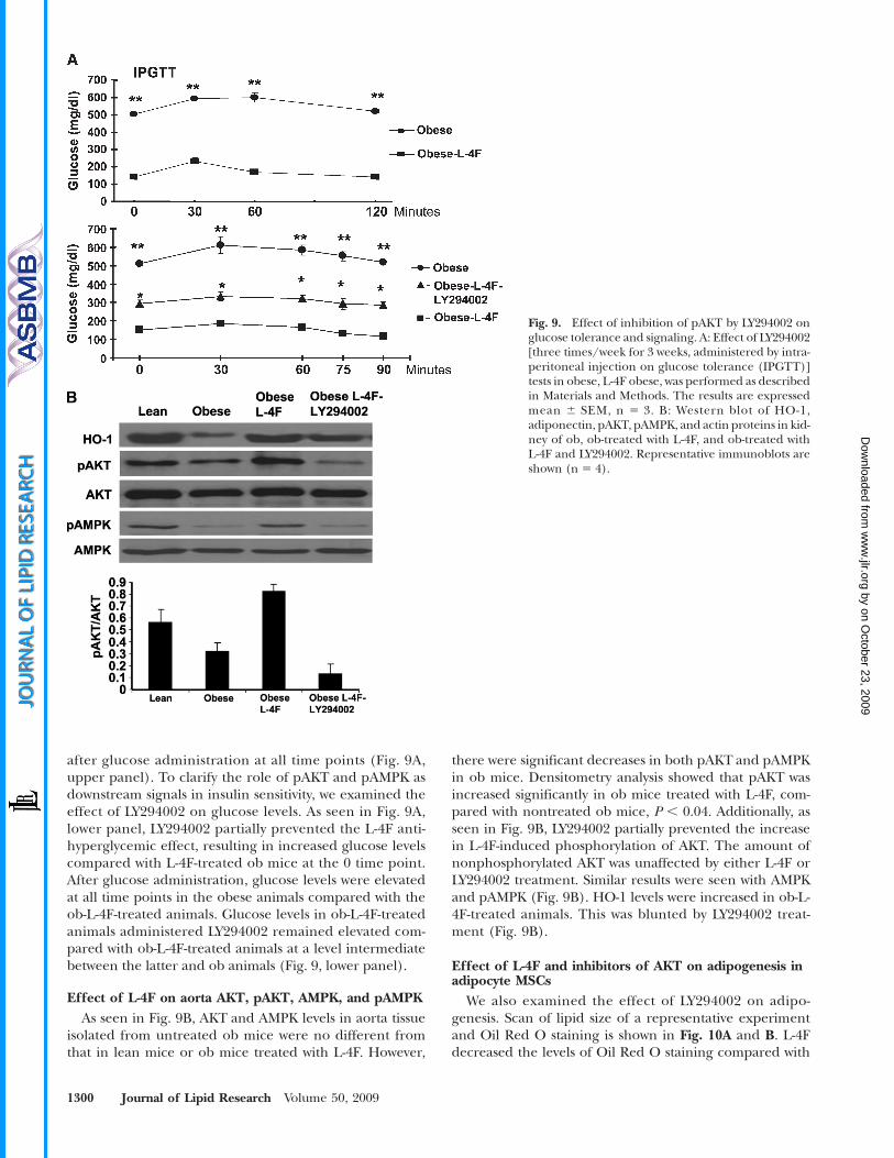
after glucose administration at all time points (Fig. 9A,upper panel). To clarify the role of pAKT and pAMPK asdownstream signals in insulin sensitivity, we examined theeffect of LY294002 on glucose levels. As seen in Fig. 9A,lower panel, LY294002 partially prevented the L-4F anti-hyperglycemic effect, resulting in increased glucose levelscompared with L-4F-treated ob mice at the 0 time point.After glucose administration, glucose levels were elevatedat all time points in the obese animals compared with theob-L-4F-treated animals. Glucose levels in ob-L-4F-treatedanimals administered LY294002 remained elevated com-pared with ob-L-4F-treated animals at a level intermediatebetween the latter and ob animals (Fig. 9, lower panel).
Effect of L-4F on aorta AKT, pAKT, AMPK, and pAMPKAs seen in Fig. 9B, AKT and AMPK levels in aorta tissue
isolated from untreated ob mice were no different fromthat in lean mice or ob mice treated with L-4F. However,
there were significant decreases in both pAKT and pAMPKin ob mice. Densitometry analysis showed that pAKT wasincreased significantly in ob mice treated with L-4F, com-pared with nontreated ob mice, P , 0.04. Additionally, asseen in Fig. 9B, LY294002 partially prevented the increasein L-4F-induced phosphorylation of AKT. The amount ofnonphosphorylated AKT was unaffected by either L-4F orLY294002 treatment. Similar results were seen with AMPKand pAMPK (Fig. 9B). HO-1 levels were increased in ob-L-4F-treated animals. This was blunted by LY294002 treat-ment (Fig. 9B).
Effect of L-4F and inhibitors of AKT on adipogenesis inadipocyte MSCs
We also examined the effect of LY294002 on adipo-genesis. Scan of lipid size of a representative experimentand Oil Red O staining is shown in Fig. 10A and B. L-4Fdecreased the levels of Oil Red O staining compared with
Fig. 9. Effect of inhibition of pAKT by LY294002 onglucose tolerance and signaling. A: Effect of LY294002[three times/week for 3 weeks, administered by intra-peritoneal injection on glucose tolerance (IPGTT)]tests in obese, L-4F obese, was performed as describedin Materials and Methods. The results are expressedmean 6 SEM, n 5 3. B: Western blot of HO-1,adiponectin, pAKT, pAMPK, and actin proteins in kid-ney of ob, ob-treated with L-4F, and ob-treated withL-4F and LY294002. Representative immunoblots areshown (n 5 4).
1300 Journal of Lipid Research Volume 50, 2009
by on October 23, 2009
ww
w.jlr.org
Dow
nloaded from
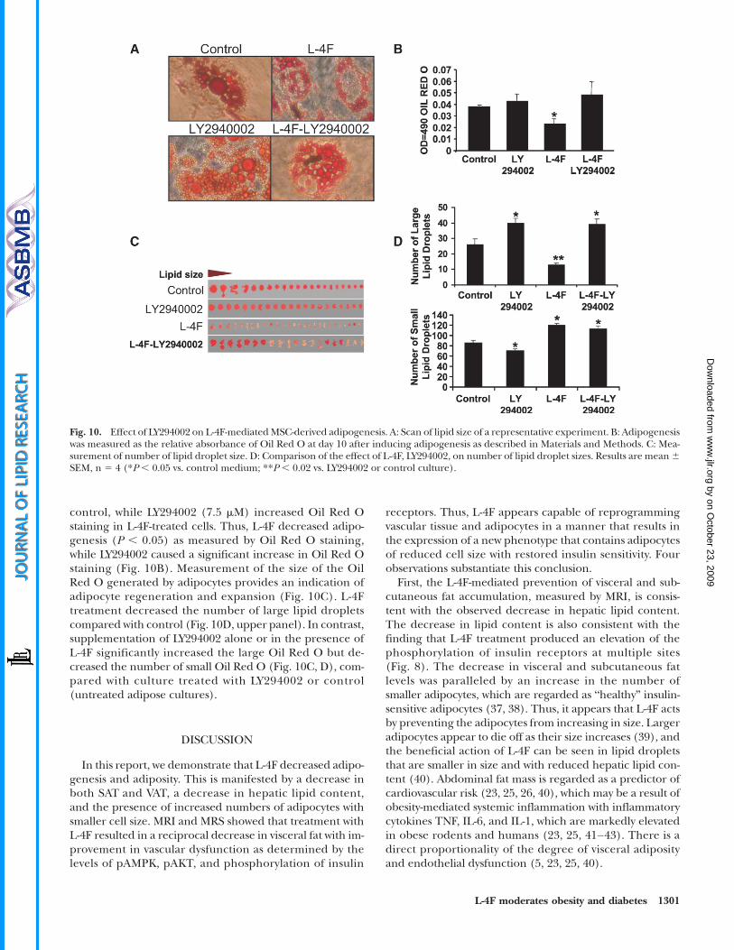
control, while LY294002 (7.5 mM) increased Oil Red Ostaining in L-4F-treated cells. Thus, L-4F decreased adipo-genesis (P , 0.05) as measured by Oil Red O staining,while LY294002 caused a significant increase in Oil Red Ostaining (Fig. 10B). Measurement of the size of the OilRed O generated by adipocytes provides an indication ofadipocyte regeneration and expansion (Fig. 10C). L-4Ftreatment decreased the number of large lipid dropletscompared with control (Fig. 10D, upper panel). In contrast,supplementation of LY294002 alone or in the presence ofL-4F significantly increased the large Oil Red O but de-creased the number of small Oil Red O (Fig. 10C, D), com-pared with culture treated with LY294002 or control(untreated adipose cultures).
DISCUSSION
In this report, we demonstrate that L-4F decreased adipo-genesis and adiposity. This is manifested by a decrease inboth SAT and VAT, a decrease in hepatic lipid content,and the presence of increased numbers of adipocytes withsmaller cell size. MRI and MRS showed that treatment withL-4F resulted in a reciprocal decrease in visceral fat with im-provement in vascular dysfunction as determined by thelevels of pAMPK, pAKT, and phosphorylation of insulin
receptors. Thus, L-4F appears capable of reprogrammingvascular tissue and adipocytes in a manner that results inthe expression of a new phenotype that contains adipocytesof reduced cell size with restored insulin sensitivity. Fourobservations substantiate this conclusion.
First, the L-4F-mediated prevention of visceral and sub-cutaneous fat accumulation, measured by MRI, is consis-tent with the observed decrease in hepatic lipid content.The decrease in lipid content is also consistent with thefinding that L-4F treatment produced an elevation of thephosphorylation of insulin receptors at multiple sites(Fig. 8). The decrease in visceral and subcutaneous fatlevels was paralleled by an increase in the number ofsmaller adipocytes, which are regarded as “healthy” insulin-sensitive adipocytes (37, 38). Thus, it appears that L-4F actsby preventing the adipocytes from increasing in size. Largeradipocytes appear to die off as their size increases (39), andthe beneficial action of L-4F can be seen in lipid dropletsthat are smaller in size and with reduced hepatic lipid con-tent (40). Abdominal fat mass is regarded as a predictor ofcardiovascular risk (23, 25, 26, 40), which may be a result ofobesity-mediated systemic inflammation with inflammatorycytokines TNF, IL-6, and IL-1, which are markedly elevatedin obese rodents and humans (23, 25, 41–43). There is adirect proportionality of the degree of visceral adiposityand endothelial dysfunction (5, 23, 25, 40).
Fig. 10. Effect of LY294002 on L-4F-mediatedMSC-derived adipogenesis. A: Scan of lipid size of a representative experiment. B: Adipogenesiswas measured as the relative absorbance of Oil Red O at day 10 after inducing adipogenesis as described in Materials and Methods. C: Mea-surement of number of lipid droplet size. D: Comparison of the effect of L-4F, LY294002, on number of lipid droplet sizes. Results are mean6SEM, n 5 4 (*P , 0.05 vs. control medium; **P , 0.02 vs. LY294002 or control culture).
L-4F moderates obesity and diabetes 1301
by on October 23, 2009
ww
w.jlr.org
Dow
nloaded from
Second, L-4F treatment decreased serum IL-1b and CB1receptor. The metabolic syndrome and obesity are charac-terized by increased serum levels of inflammatory cyto-kines, such as IL-1b, which decrease insulin sensitivity(44, 45). CB1 receptors in mice increase hepatic lipogenictranscription factor SREBP-1 and fatty acid synthesis (7).Therefore, the L-4F-mediated decreases in the levels ofIL-1b and CB1 receptors may contribute to the observedincrease in insulin sensitivity. In addition, downregulationof the peripheral endocannabinoid system, CB1, may con-tribute to the weight loss as well.
Third, L-4F administration has been shown to increaseHO-1 protein levels and adiponectin both in vitro andin vivo. Decreased levels of serum adiponectin are the re-sult of an increase in the levels of ROS and H2O2, therebycontributing to the pathogenesis of insulin resistance(46, 47). Previous studies have shown that increased levelsof HO-1 protein caused a marked increase in serumadiponectin levels in Zucker fat rats and ob mice (23, 25,26). An increase in adiponectin levels is an indication ofimprovement in the metabolic syndrome that, in turn,leads to increased insulin sensitivity and a subsequentdecrease in arterial disease and heart disease (37, 45,48–51). This supports the concept that expansion ofadipogenesis leads to an increased number of adipocytesof smaller cell size. Smaller adipocytes are considered tobe healthy, insulin-sensitive adipocyte cells that are capa-ble of producing adiponectin (37). While increases inobesity and diabetes are considered a risk factor for cardio-vascular complications (45), improvement in the diabeticphenotype, including improved insulin sensitivity andglucose tolerance, may occur through increased pre-adipocyte differentiation and increased adiponectin secre-tion (37, 52).
Fourth, the remarkable action of L-4F treatment in in-creased pAKT, pAMPK, and phosphorylation of insulin re-ceptors is associated in an improvement in glucosetolerance. Insulin resistance is an independent factor forthe development of vascular dysfunction (53, 54). There-fore, L-4F treatment improves vascular function on the basisof increases in both insulin sensitivity and insulin receptorphosphorylation. Others have shown that increased phos-phorylation of insulin receptors and vascular functionmay be a response to the increase in pAMPK and pAKTcrosstalk (55–57). Further activation of pAMPK and pAKTincreases glucose transport fatty acid oxidation and mito-chondrial function (58–60). In this study, inhibition ofpAKT by LY294002 administration in L-4F-treated mice re-versed glucose tolerance and insulin sensitivity.We previouslyreported that LY294002 inhibition of the AKT pathwaysprevented HO-1 in ameliorating cell protection againstROS (30).However, LY294002 administration in vivo inhibitedboth AKT and AMPK and decreased glucose tolerance andinsulin sensitivity. pAKTand AMPK act as fuel sensors in theregulation of energy balance, and the resultant crosstalkof AMPK-AKT has been shown to regulate nitric oxide bio-availability and vascular function (56, 61, 62). Furthermore,activated AMPK alone has been suggested as a therapeutictarget to ameliorate endothelial dysfunction (63–65). Thus,
the novel effect of L-4F on the HO-1-adiponectin-pAKT-pAMPK-module (i.e., an increase in HO-1) increasesadiponectin, and the subsequent increase in AKT-AMPKcrosstalk and signaling pathway provides a beneficialmechanistic basis for L-4F-mediated vascular protection.L-4F treatment offers great potential as a drug that couldbe employed therapeutically to address the metabolic de-rangements associated with obesity, themetabolic syndrome,and insulin-resistant diabetes to restore vascular function.
The authors are indebted toDr. Attallah Kappas and the BeatriceRenfield Foundation for their support. Ms. Daniele DeMarchiand Dr. Alessandro Pingipore are well appreciated for valuablehelp in the acquisition of Magnetic Resonance data.
REFERENCES
1. Morino, K., S. Neschen, S. Bilz, S. Sono, D. Tsirigotis, R. M. Reznick,I. Moore, Y. Nagai, V. Samuel, D. Sebastian, et al. 2008. Muscle spe-cific IRS-1 Ser-.Ala transgenic mice are protected from fat-inducedinsulin resistance in skeletal muscle. Diabetes. 57: 2644–2651.
2. Danial, N. N., L. D. Walensky, C. Y. Zhang, C. S. Choi, J. K. Fisher,A. J. Molina, S. R. Datta, K. L. Pitter, G. H. Bird, J. D. Wikstrom, et al.2008. Dual role of proapoptotic BAD in insulin secretion and betacell survival. Nat. Med. 14: 144–153.
3. Wellen, K. E., and G. S. Hotamisligil. 2003. Obesity-induced inflam-matory changes in adipose tissue. J. Clin. Invest. 112: 1785–1788.
4. Weisberg, S. P., D. Hunter, R. Huber, J. Lemieux, S. Slaymaker,K. Vaddi, I. Charo, R. L. Leibel, and A. W. Ferrante, Jr. 2006. CCR2modulates inflammatory and metabolic effects of high-fat feeding.J. Clin. Invest. 116: 115–124.
5. Eckel, R. H., D. A. York, S. Rossner, V. Hubbard, I. Caterson, S. T.St Jeor, L. L. Hayman, R. M. Mullis, and S. N. Blair. 2004. PreventionConference VII: Obesity, a worldwide epidemic related to heart dis-ease and stroke: executive summary. Circulation. 110: 2968–2975.
6. Tounian, P., Y. Aggoun, B. Dubern, V. Varille, B. Guy-Grand, D. Sidi,J. P. Girardet, and D. Bonnet. 2001. Presence of increased stiffnessof the common carotid artery and endothelial dysfunction in se-verely obese children: a prospective study. Lancet. 358: 1400–1404.
7. Osei-Hyiaman, D., M. DePetrillo, P. Pacher, J. Liu, S. Radaeva, S.Batkai, J. Harvey-White, K. Mackie, L. Offertaler, L. Wang, et al.2005. Endocannabinoid activation at hepatic CB1 receptors stimu-lates fatty acid synthesis and contributes to diet-induced obesity.J. Clin. Invest. 115: 1298–1305.
8. Kruger, A. L., S. Peterson, S. Turkseven, P. M. Kaminski, F. F. Zhang,S. Quan, M. S. Wolin, and N. G. Abraham. 2005. D-4F induces hemeoxygenase-1 and extracellular superoxide dismutase, decreasesendothelial cell sloughing, and improves vascular reactivity in ratmodel of diabetes. Circulation. 111: 3126–3134.
9. Kruger, A. L., S. J. Peterson, M. L. Schwartzman, H. Fusco, J. A.McClung, M. Weiss, S. Shenouda, A. I. Goodman, M. S. Goligorsky,A. Kappas, et al. 2006. Up-regulation of heme oxygenase providesvascular protection in an animal model of diabetes through itsantioxidant and antiapoptotic effects. J. Pharmacol. Exp. Ther. 319:1144–1152.
10. Abraham, N. G., and A. Kappas. 2008. Pharmacological and clinicalaspects of heme oxygenase. Pharmacol. Rev. 60: 79–127.
11. Steinberg, H. O., H. Chaker, R. Leaming, A. Johnson, G. Brechtel,and A. D. Baron. 1996. Obesity/insulin resistance is associated withendothelial dysfunction. Implications for the syndrome of insulinresistance. J. Clin. Invest. 97: 2601–2610.
12. Yudkin, J. S., M. Kumari, S. E. Humphries, and V. Mohamed-Ali.2000. Inflammation, obesity, stress and coronary heart disease: isinterleukin-6 the link? Atherosclerosis. 148: 209–214.
13. Zhang, F., J. I. Kaide, L. Yang, H. Jiang, S. Quan, R. Kemp, W. Gong,M. Balazy, N. G. Abraham, and A. Nasjletti. 2004. CO modulatespulmonary vascular response to acute hypoxia: relation to endothelin.Am. J. Physiol. Heart Circ. Physiol. 286: H137–H144.
14. Porstmann, T., B. Griffiths, Y. L. Chung, O. Delpuech, J. R. Griffiths,J. Downward, and A. Schulze. 2005. PKB/Akt induces transcription
1302 Journal of Lipid Research Volume 50, 2009
by on October 23, 2009
ww
w.jlr.org
Dow
nloaded from
of enzymes involved in cholesterol and fatty acid biosynthesis viaactivation of SREBP. Oncogene. 24: 6465–6481.
15. Lee, T. S., C. C. Chang, Y. Zhu, and J. Y. Shyy. 2004. Simvastatin in-duces heme oxygenase-1: a novel mechanism of vessel protection.Circulation. 110: 1296–1302.
16. Navab, M., G. M. Anantharamaiah, S. T. Reddy, S. Hama, G. Hough,V. R. Grijalva, A. C. Wagner, J. S. Frank, G. Datta, D. Garber, et al.2004. Oral D-4F causes formation of pre-beta high-density lipoproteinand improves high-density lipoprotein-mediated cholesterol effluxand reverse cholesterol transport from macrophages in apolipo-protein E-null mice. Circulation. 109: 3215–3220.
17. Navab, M., G. M. Ananthramaiah, S. T. Reddy, B. J. Van Lenten,B. J. Ansell, G. C. Fonarow, K. Vahabzadeh, S. Hama, G. Hough,N. Kamranpour, et al. 2004. The oxidation hypothesis of athero-genesis: the role of oxidized phospholipids and HDL. J. Lipid Res.45: 993–1007.
18. Navab, M., G. M. Anantharamaiah, S. T. Reddy, B. J. Van Lenten,G.Hough, A.Wagner, K.Nakamura,D.W.Garber,G.Datta, J. P. Segrest,et al. 2003. Human apolipoprotein AI mimetic peptides for the treat-ment of atherosclerosis. Curr. Opin. Investig. Drugs. 4: 1100–1104.
19. Buga, G. M., J. S. Frank, G. A. Mottino, A. Hakhamian, A. Narasimha,A. D.Watson, B. Yekta, M. Navab, S. T. Reddy, G.M. Anantharamaiah,et al. 2008. D-4F reduces EO6 immunoreactivity, SREBP-1c mRNAlevels, and renal inflammation in LDL receptor-null mice fed aWestern diet. J. Lipid Res. 49: 192–205.
20. Ou, J., Z.Ou,D.W. Jones, S.Holzhauer,O. A.Hatoum, A.W. Ackerman,D. W. Weihrauch, D. D. Gutterman, K. Guice, K. T. Oldham, et al.2003. L-4F, an apolipoprotein A-1 mimetic, dramatically improvesvasodilation in hypercholesterolemia and sickle cell disease. Circula-tion. 107: 2337–2341.
21. Ou, Z., J. Ou, A. W. Ackerman, K. T. Oldham, and K. A. Pritchard,Jr. 2003. L-4F, an apolipoprotein A-1 mimetic, restores nitric oxideand superoxide anion balance in low-density lipoprotein-treatedendothelial cells. Circulation. 107: 1520–1524.
22. Peterson, S. J., D. Husney, A. L. Kruger, R. Olszanecki, F. Ricci, L. F.Rodella, A. Stacchiotti, R. Rezzani, J. A. McClung, W. S. Aronow, et al.2007. Long-term treatment with the apolipoprotein A1mimetic pep-tide increases antioxidants and vascular repair in type I diabetic rats.J. Pharmacol. Exp. Ther. 322: 514–520.
23. Peterson, S. J., G. Drummond, K. D. Hyun, M. Li, A. L. Kruger,S. Ikehara, and N. G. Abraham. 2008. L-4F treatment reduces adi-posity, increases adiponectin levels and improves insulin sensitivityin obese mice. J. Lipid Res. 49: 1658–1669.
24. LʼAbbate, A., D. Neglia, C. Vecoli, M. Novelli, V. Ottaviano, S. Baldi,R. Barsacchi, A. Paolicchi, P. Masiello, G. S. Drummond, et al. 2007.Beneficial effect of heme oxygenase-1 expression on myocardialischemia-reperfusion involves an increase in adiponectin in mildlydiabetic rats. Am. J. Physiol. Heart Circ. Physiol. 293: H3532–H3541.
25. Li, M., D. H. Kim, P. L. Tsenovoy, S. J. Peterson, R. Rezzani, L. F.Rodella, W. S. Aronow, S. Ikehara, and N. G. Abraham. 2008. Treat-ment of obese diabetic mice with a heme oxygenase inducer reducesvisceral and subcutaneous adiposity, increases adiponectin levels,and improves insulin sensitivity and glucose tolerance. Diabetes. 57:1526–1535.
26. Kim, D. H., A. P. Burgess, M. Li, P. L. Tsenovoy, F. Addabbo, J. A.McClung, N. Puri, and N. G. Abraham. 2008. Heme oxygenase-mediated increases in adiponectin decrease fat content and inflam-matory cytokines, tumor necrosis factor-alpha and interleukin-6 inZucker rats and reduce adipogenesis in human mesenchymal stemcells. J. Pharmacol. Exp. Ther. 325: 833–840.
27. Navab, M., G. M. Anantharamaiah, S. Hama, D. W. Garber,M. Chaddha, G. Hough, R. Lallone, and A. M. Fogelman. 2002.Oral administration of an Apo A-I mimetic peptide synthesizedfrom D-amino acids dramatically reduces atherosclerosis in miceindependent of plasma cholesterol. Circulation. 105: 290–292.
28. Hu, L., J. Hofmann, and R. B. Jaffe. 2005. Phosphatidylinositol3-kinase mediates angiogenesis and vascular permeability associatedwith ovarian carcinoma. Clin. Cancer Res. 11: 8208–8212.
29. Hu, L., J. Hofmann, Y. Lu, G. B. Mills, and R. B. Jaffe. 2002. In-hibition of phosphatidylinositol 3′-kinase increases efficacy of pacli-taxel in in vitro and in vivo ovarian cancer models. Cancer Res. 62:1087–1092.
30. Salinas, M., R. Diaz, N. G. Abraham, C. M. Ruiz De Galarreta,and A. Cuadrado. 2003. Nerve growth factor protects against6-hydroxydopamine-induced oxidative stress by increasing expres-sion of heme oxygenase-1 in a phosphatidylinositol 3-kinase-depen-dent manner. J. Biol. Chem. 278: 13898–13904.
31. Positano, V., A. Gastaldelli, A. M. Sironi, M. F. Santarelli, M. Lombardi,andL. Landini. 2004. An accurate and robustmethod for unsupervisedassessment of abdominal fat by MRI. J. Magn. Reson. Imaging. 20:684–689.
32. Demerath, E.W., K. J. Ritter, W. A. Couch, N. L. Rogers, G.M.Moreno,A. Choh, M. Lee, K. Remsberg, S. A. Czerwinski, W. C. Chumlea, et al.2007. Validity of a new automated software program for visceraladipose tissue estimation. Int. J. Obes. (Lond). 31: 285–291.
33. Bonekamp, S., P. Ghosh, S. Crawford, S. F. Solga, A. Horska, F. L.Brancati, A. M. Diehl, S. Smith, and J. M. Clark. 2008. Quantitativecomparison and evaluation of software packages for assessment ofabdominal adipose tissue distribution bymagnetic resonance imaging.Int. J. Obes. (Lond). 32: 100–111.
34. Lee, R. H., B. Kim, I. Choi, H. Kim, H. S. Choi, K. Suh, Y. C. Bae,and J. S. Jung. 2004. Characterization and expression analysis ofmesenchymal stem cells fromhumanbonemarrow and adipose tissue.Cell. Physiol. Biochem. 14: 311–324.
35. Novikoff, A. B., P. M. Novikoff, O. M. Rosen, and C. S. Rubin. 1980.Organelle relationships in cultured 3T3–L1 preadipocytes. J. CellBiol. 87: 180–196.
36. Tondreau, T., N. Meuleman, A. Delforge, M. Dejeneffe, R. Leroy,M. Massy, C. Mortier, D. Bron, and L. Lagneaux. 2005. Mesenchymalstem cells derived from CD133-positive cells in mobilized peripheralblood and cord blood: proliferation, Oct4 expression, and plasticity.Stem Cells. 23: 1105–1112.
37. Kim, J. Y., E. van de Wall, M. Laplante, A. Azzara, M. E. Trujillo,S. M. Hofmann, T. Schraw, J. L. Durand, H. Li, G. Li, et al. 2007.Obesity-associated improvements in metabolic profile through ex-pansion of adipose tissue. J. Clin. Invest. 117: 2621–2637.
38. Nicolai, A., M. Li, D. H. Kim, S. J. Peterson, L. Vanella, V. Positano,A. Gastaldelli, R. Rezzani, L. F. Rodella, G. Drummond, et al. 2009.Heme Oxygenase-1 induction remodels adipose tissue and improvesinsulin sensitivity in obesity-induced diabetic rats. Hypertension. 53:508–515.
39. Khan, T., E. S. Muise, P. Iyengar, Z. V. Wang, M. Chandalia, N. Abate,B. B. Zhang, P. Bonaldo, S. Chua, and P. E. Scherer. 2009. Metabolicdysregulation and adipose tissue fibrosis: role of collagen VI.Mol. CellBiol. 29: 1575–1591.
40. Williams, I. L., P. J. Chowienczyk, S. B.Wheatcroft, A. Patel, R. Sherwood,A. Momin, A. M. Shah, and M. T. Kearney. 2006. Effect of fat distri-bution on endothelial-dependent and endothelial-independentvasodilatation in healthy humans. Diabetes Obes. Metab. 8: 296–301.
41. Bastard, J. P., M. Maachi, J. T. Van Nhieu, C. Jardel, E. Bruckert,A. Grimaldi, J. J. Robert, J. Capeau, and B. Hainque. 2002. Adiposetissue IL-6 content correlates with resistance to insulin activation ofglucose uptake both in vivo and in vitro. J. Clin. Endocrinol. Metab.87: 2084–2089.
42. Maachi, M., L. Pieroni, E. Bruckert, C. Jardel, S. Fellahi, B. Hainque,J. Capeau, and J. P. Bastard. 2004. Systemic low-grade inflammationis related to both circulating and adipose tissue TNFalpha, leptinand IL-6 levels in obese women. Int. J. Obes. Relat. Metab. Disord. 28:993–997.
43. Hingorani, A. D., J. Cross, R. K. Kharbanda, M. J. Mullen, K. Bhagat,M. Taylor, A. E. Donald, M. Palacios, G. E. Griffin, J. E. Deanfield, et al.2000. Acute systemic inflammation impairs endothelium-dependentdilatation in humans. Circulation. 102: 994–999.
44. Muse, E. D., T. K. Lam, P. E. Scherer, and L. Rossetti. 2007.Hypothalamic resistin induces hepatic insulin resistance. J. Clin. Invest.117: 1670–1678.
45. Lazar, M. A. 2005. How obesity causes diabetes: not a tall tale.Science. 307: 373–375.
46. Kondo, H., I. Shimomura, Y. Matsukawa, M. Kumada, M. Takahashi,M. Matsuda, N. Ouchi, S. Kihara, T. Kawamoto, S. Sumitsuji, et al.2002. Association of adiponectin mutation with type 2 diabetes: acandidate gene for the insulin resistance syndrome. Diabetes. 51:2325–2328.
47. Fasshauer, M., J. Klein, S. Neumann, M. Eszlinger, and R. Paschke.2001. Adiponectin gene expression is inhibited by beta-adrenergicstimulation via protein kinase A in 3T3–L1 adipocytes. FEBS Lett.507: 142–146.
48. Bahia, L., L. G. Aguiar, N. Villela, D. Bottino, A. F. Godoy-Matos, B.Geloneze, M. Tambascia, and E. Bouskela. 2006. Relationship be-tween adipokines, inflammation, and vascular reactivity in lean controlsand obese subjects with metabolic syndrome. Clinics. 61: 433–440.
49. Han, S. H., M. J. Quon, J. A. Kim, and K. K. Koh. 2007. Adiponectinand cardiovascular disease: response to therapeutic interventions.J. Am. Coll. Cardiol. 49: 531–538.
L-4F moderates obesity and diabetes 1303
by on October 23, 2009
ww
w.jlr.org
Dow
nloaded from
50. Fontana, L., J. C. Eagon, M. E. Trujillo, P. E. Scherer, and S. Klein.2007. Visceral fat adipokine secretion is associated with systemicinflammation in obese humans. Diabetes. 56: 1010–1013.
51. Iwashima, Y., T. Horio, Y. Suzuki, S. Kihara, H. Rakugi, K. Kangawa,T. Funahashi, T. Ogihara, and Y. Kawano. 2006. Adiponectin andinflammatory markers in peripheral arterial occlusive disease.Atherosclerosis. 188: 384–390.
52. Yamauchi, T., Y. Nio, T. Maki, M. Kobayashi, T. Takazawa, M. Iwabu,M. Okada-Iwabu, S. Kawamoto, N. Kubota, T. Kubota, et al. 2007.Targeted disruption of AdipoR1 and AdipoR2 causes abrogation ofadiponectin binding and metabolic actions. Nat. Med. 13: 332–339.
53. Kearney, M. T., E. R. Duncan, M. Kahn, and S. B. Wheatcroft. 2008.Insulin resistance and endothelial cell dysfunction: studies in mam-malian models. Exp. Physiol. 93: 158–163.
54. Duncan, E., P. Crossey, S.Walker, N. Anilkumar, L. Poston, G. Douglas,V. Ezzat, S. Wheatcroft, A. M. Shah, and M. Kearney. 2008. The effectof endothelium-specific insulin resistance on endothelial function invivo. Diabetes. 57: 3307–3314.
55. Fleming, I., C. Schulz, B. Fichtlscherer, B. E. Kemp, B. Fisslthaler,and R. Busse. 2003. AMP-activated protein kinase (AMPK) regulatesthe insulin-induced activation of the nitric oxide synthase in humanplatelets. Thromb. Haemost. 90: 863–871.
56. Kovacic, S., C. L. Soltys, A. J. Barr, I. Shiojima, K. Walsh, and J. R.Dyck. 2003. Akt activity negatively regulates phosphorylation of AMP-activated protein kinase in the heart. J. Biol. Chem. 278: 39422–39427.
57. Longnus, S. L., C. Segalen, J. Giudicelli, M. P. Sajan, R. V. Farese,and O. E. Van. 2005. Insulin signalling downstream of protein kinaseB is potentiated by 5′AMP-activated protein kinase in rat hearts invivo. Diabetologia. 48: 2591–2601.
58. Hardie, D. G. 2003. Minireview: the AMP-activated protein kinasecascade: the key sensor of cellular energy status. Endocrinology.144: 5179–5183.
59. Hardie, D. G. 2007. AMP-activated protein kinase as a drug target.Annu. Rev. Pharmacol. Toxicol. 47: 185–210.
60. Di Noia, M. A., D. S. Van, F. Palmieri, L. M. Yang, S. Quan, A. I.Goodman, and N. G. Abraham. 2006. Heme oxygenase-1 enhancesrenal mitochondrial transport carriers and cytochrome C oxidaseactivity in experimental diabetes. J. Biol. Chem. 281: 15687–15693.
61. Ouchi, N., H. Kobayashi, S. Kihara, M. Kumada, K. Sato, T. Inoue,T. Funahashi, andK.Walsh. 2004. Adiponectin stimulates angiogenesisby promoting cross-talk between AMP-activated protein kinase andAkt signaling in endothelial cells. J. Biol. Chem. 279: 1304–1309.
62. Sun, J. F., T. Phung, I. Shiojima, T. Felske, J. N. Upalakalin, D. Feng,T. Kornaga, T. Dor, A. M. Dvorak, K. Walsh, et al. 2005. Microvascularpatterning is controlled by fine-tuning the Akt signal. Proc. Natl. Acad.Sci. USA. 102: 128–133.
63. Schulz, E., J. Dopheide, S. Schuhmacher, S. R. Thomas, K. Chen,A. Daiber, P. Wenzel, T. Munzel, and J. F. Keaney, Jr. 2008. Suppres-sion of the JNK pathway by induction of a metabolic stress responseprevents vascular injury and dysfunction.Circulation. 118: 1347–1357.
64. Chen, K., M. T. Kirber, H. Xiao, Y. Yang, and J. F. Keaney, Jr. 2008.Regulation of ROS signal transduction by NADPH oxidase 4 locali-zation. J. Cell Biol. 181: 1129–1139.
65. Murabito, J. M., M. J. Keyes, C. Y. Guo, J. F. Keaney, Jr., R. S. Vasan,R. B. DʼAgostino, Sr., and E. J. Benjamin. 2008. Cross-sectional rela-tions of multiple inflammatory biomarkers to peripheral arterialdisease: The Framingham Offspring Study. Atherosclerosis. 203:509–514.
1304 Journal of Lipid Research Volume 50, 2009
by on October 23, 2009
ww
w.jlr.org
Dow
nloaded from

![Jean-Pierre Dupuy IMETIC THEORY AS SCIENCE+Mimetic+Theory.pdf · Jean-Pierre Dupuy 1. MIMETIC THEORY AS SCIENCE This paper is about Mimetic Theory [MT] and its efforts to constitute](https://img.pdfslide.us/doc/110x75/5e17950bec51206ecd3d09f1/jean-pierre-dupuy-imetic-theory-as-mimetictheorypdf-jean-pierre-dupuy-1-mimetic.jpg)