Embed Size (px)
DESCRIPTION
The Knee. Tibio-Femoral. Osteology. Distal Femur with Proximal Tibia Largest Joint Cavity in the Body A modified hinge joint with significant passive rotation Technically, one degree of freedom (Flexion/Extension) but passive rotary component is essential - PowerPoint PPT Presentation
Citation preview
The Knee
Tibio-Femoral
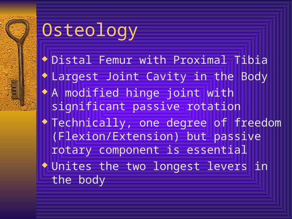
Osteology
Distal Femur with Proximal Tibia Largest Joint Cavity in the Body A modified hinge joint with significant
passive rotation Technically, one degree of freedom
(Flexion/Extension) but passive rotary component is essential
Unites the two longest levers in the body
Tibio-Femoral
Support
Knee supports the weight of the body and transmits forces from the ground
Functional stability of the joint is derived from the passive restraint of the ligaments, the active support of muscles, the joint geometry, and the compressive forces pushing the bones together
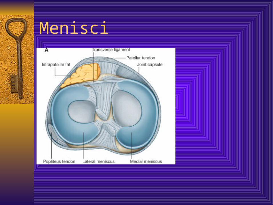
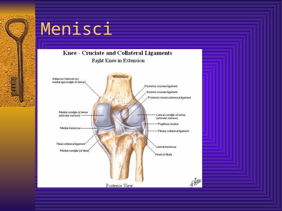
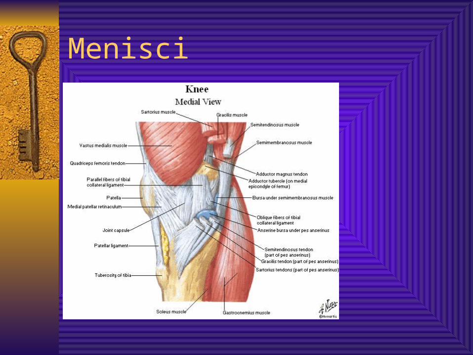
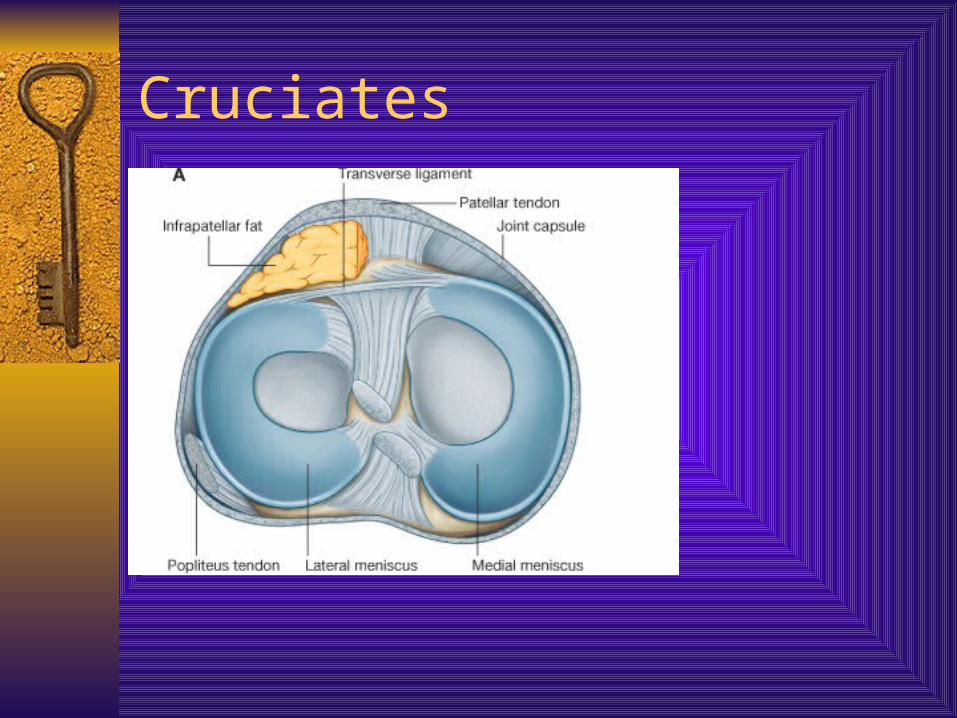
Menisci The surface of the tibia is covered by
fibrocartilaginous menisci - They:– Enhance the joint stability by deepening the contact
surface– They help with shock absorption by transmitting ½ of
weight bearing load in full extension and some in flexion as well
– They protect the articular cartilage– They transmit the load across the surface of the joint,
thus reducing the load per unit area on the tibio-femoral contact sites. The contact area in the joint is reduced 50% when the menisci are absent
Menisci
Menisci In hi load situations, 70% of the load is absorbed by the
menisci, especially the lateral meniscus The menisci assist in lubrication of the joint by acting
as a space filling mechanism, more fluid is dispersed to the surface of tibia and femur
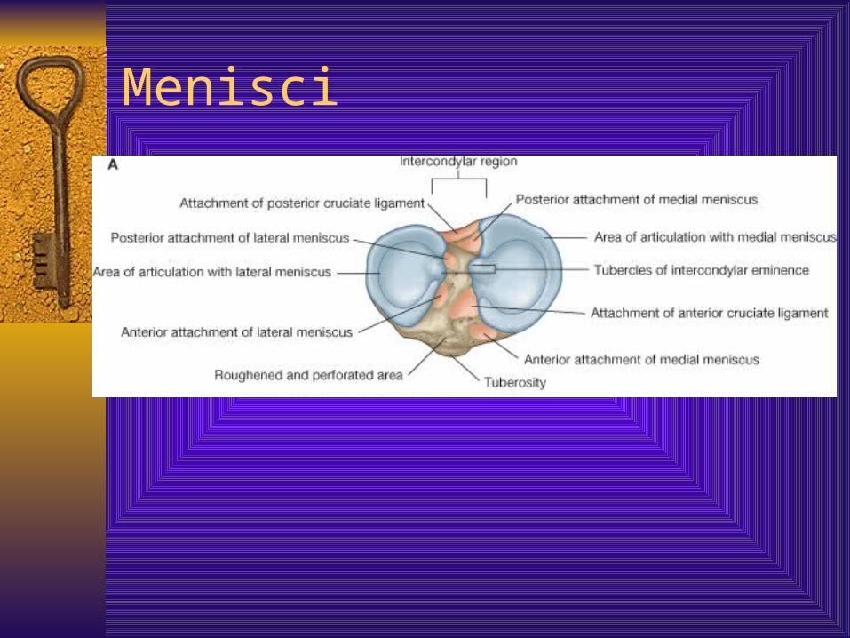
20% increase in friction following meniscal removal Medial Meniscus – larger, reflects the shape of medial
tibial condyle A + P horns – attached to medial collateral ligament and basically immobile
Lateral Meniscus – smaller, tighter, almost a complete circle A= P horns – NOT attached to lateral collateral ligament
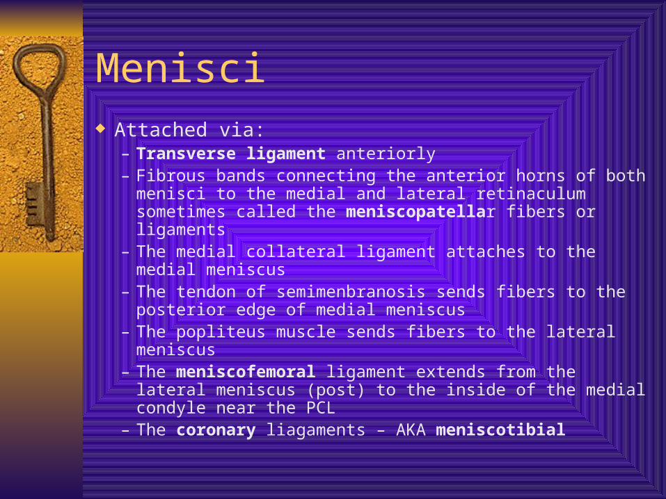
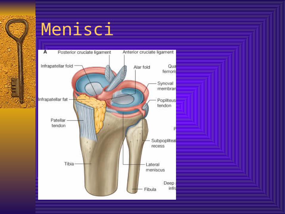
Menisci Attached via:
– Transverse ligament anteriorly– Fibrous bands connecting the anterior horns of both menisci to
the medial and lateral retinaculum sometimes called the meniscopatellar fibers or ligaments
– The medial collateral ligament attaches to the medial meniscus– The tendon of semimenbranosis sends fibers to the posterior
edge of medial meniscus– The popliteus muscle sends fibers to the lateral meniscus– The meniscofemoral ligament extends from the lateral
meniscus (post) to the inside of the medial condyle near the PCL
– The coronary liagaments – AKA meniscotibial
Menisci
Menisci
Menisci
Menisci
Menisci
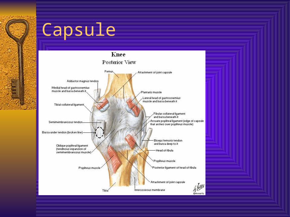
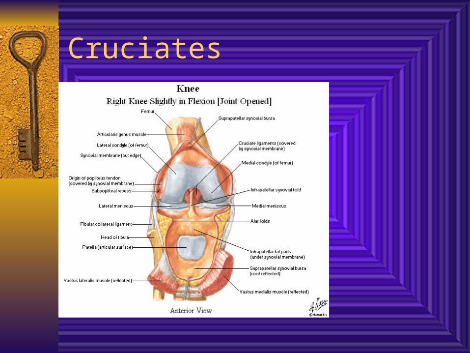
Joint Capsule Largest in body Surrounds entire joint, except anteriorly Originally (in utero) is three capsule that merge
into one MCL – flat band, attached above medial condyle
of the femur and below to the medial surface of the shaft of the tibia – resists lateral displacement
LCL – cordlike, attached above the lateral condyle of femur and below the head of the fibula – resists medial displacement
Capsule
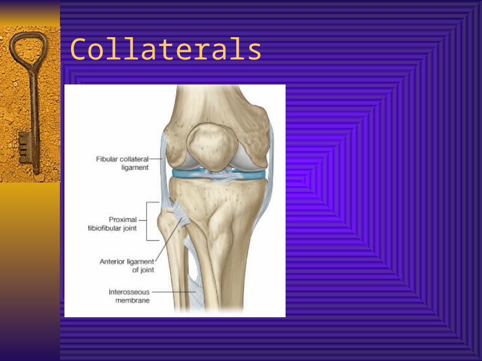
Collaterals
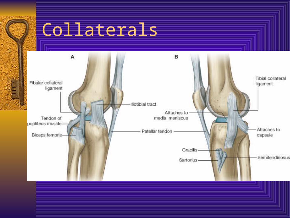
Collaterals
Capsule
Oblique Popliteal – derived from semimembranosus on posterior aspect of the capsule, runs from that tendon to medial aspect of the lateral femoral condyle (posteriorly)
Arcuate popliteal from head of fibula, runs over the popliteus muscle to attach into posterior joint capsule
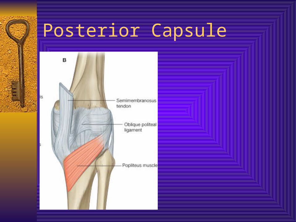
Posterior Capsule
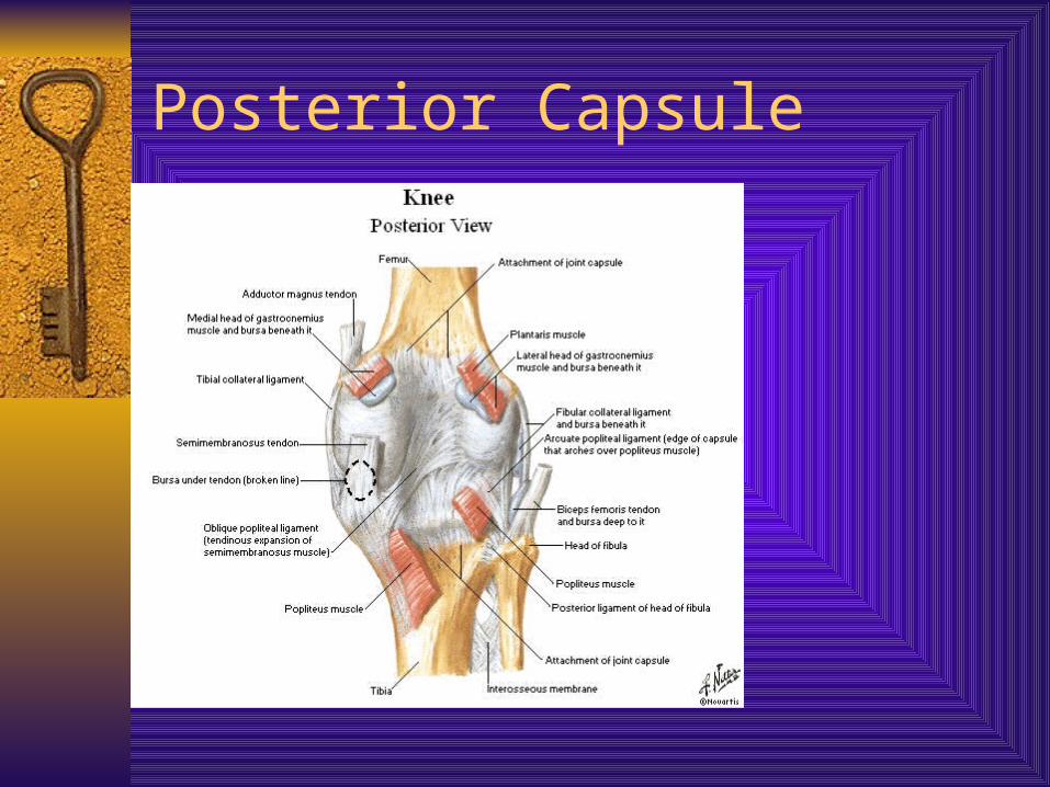
Posterior Capsule
Little Guys
Capsule
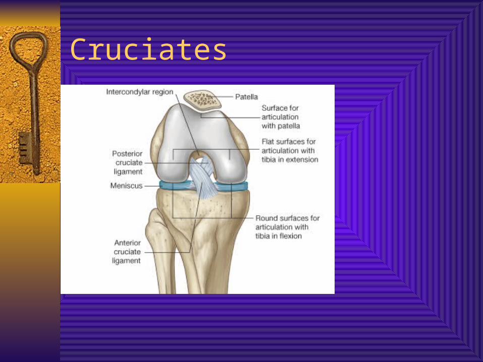
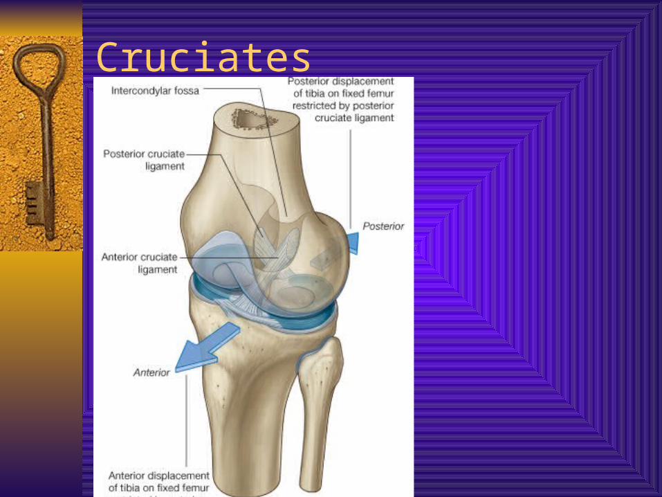
Cruciates – called intrinsic- note synovium ACL – attached to the anteriorly intercondylar
area of the tibia and passes upward, backward, and laterally to be attached to the posterior part of medial surface of the lateral femoral condyle.
ACL fibers run in three directions – anteromedial, intermediate and posterolateral directions
NWB this ligament prevents anterior displacement of tibia on femur
Cruciates
Cruciates
Cruciates
Cruciates
Capsule
PCL – attached to the posterior intercondylar area of the tibia and passes upward, forward, and medially to be attached to the anterior part of the lateral surface of the medial femoral condyle.
PCL fibers run in two directions, anteromedial and posterolateral directions
NWB prevents posterior displacement of tibia on femur
In closed chain, the role of cruciates changes
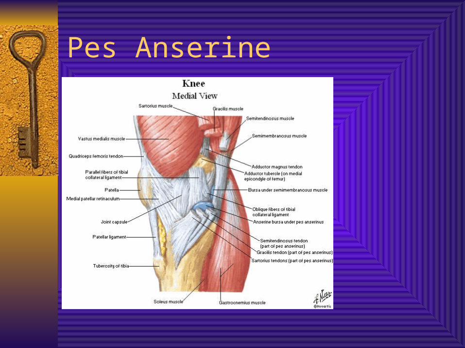
Pes Anserine
Unholy Triad
ACL, MCL, Medial Meniscus Mechanism of injury generally involves all
three at same time
Compartments of the Knee
Medial– Medial retinaculum– Pes anserine– Adductor Magnus– Semimenbranosus– Capsular ligaments – meniscofemoral,
meniscotibila, post. Oblique– MCL– PCL
Compartments Lateral
– Lateral Retinaculum– IT Band– Biceps Attachment– Popliteus MM– LCL– Lateral capsular ligaments – meniscofemoral, meniscotibial– Arcuate ligament– ACL
Arcuate Lig., Post. Lateral Capsule, LCL – Called Arcuate Complex
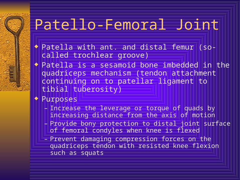
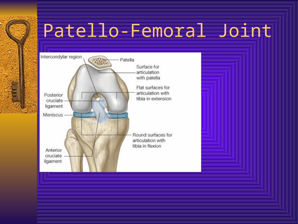
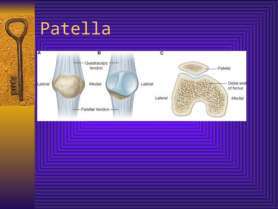
Patello-Femoral Joint Patella with ant. and distal femur (so-called trochlear
groove) Patella is a sesamoid bone imbedded in the
quadriceps mechanism (tendon attachment continuing on to patellar ligament to tibial tuberosity)
Purposes– Increase the leverage or torque of quads by increasing
distance from the axis of motion– Provide bony protection to distal joint surface of femoral
condyles when knee is flexed– Prevent damaging compression forces on the quadriceps
tendon with resisted knee flexion such as squats
Patello-Femoral Joint
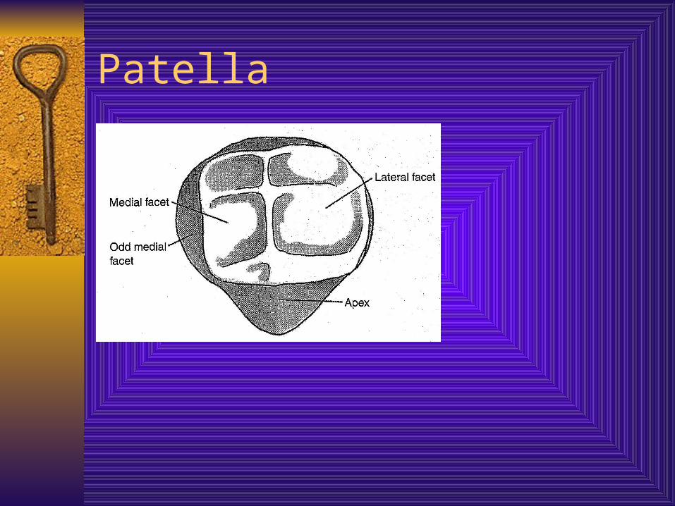
Patella Posterior surface covered with articular cartilage –
Thickest articular cartilage in body Facets – Medial side had medial facet and the odd
facet Lateral side has lateral facet Separated by vertical ridge Can divide med. and lat. facets to superior and
inferior Proximal part called the base, distal part the pole
or apex
Patella
Patella
Patella
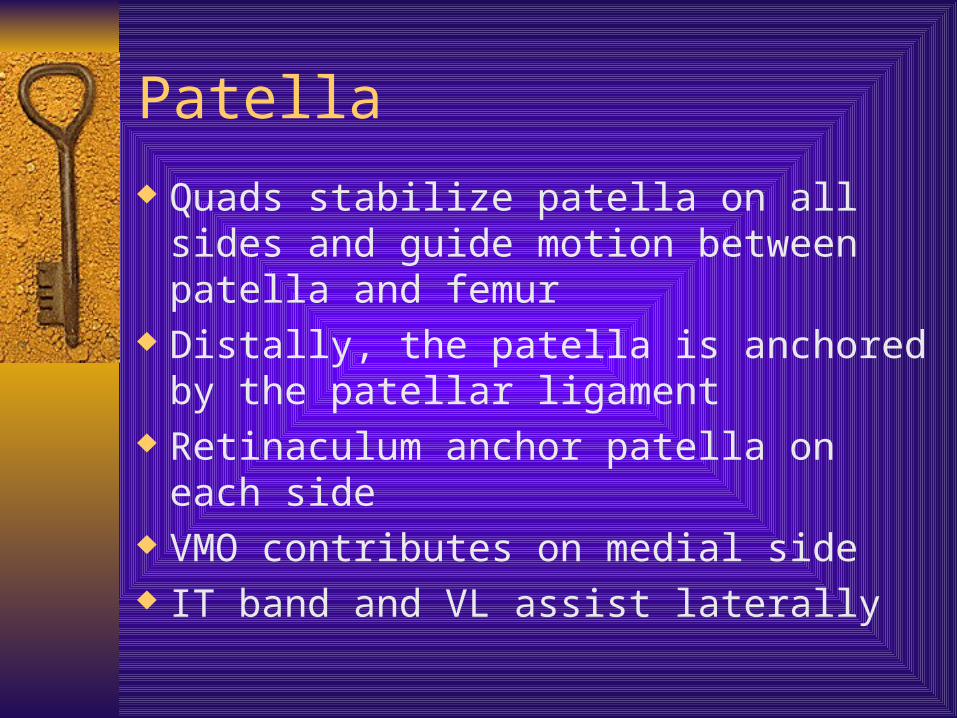
Quads stabilize patella on all sides and guide motion between patella and femur
Distally, the patella is anchored by the patellar ligament
Retinaculum anchor patella on each side VMO contributes on medial side IT band and VL assist laterally
Patella
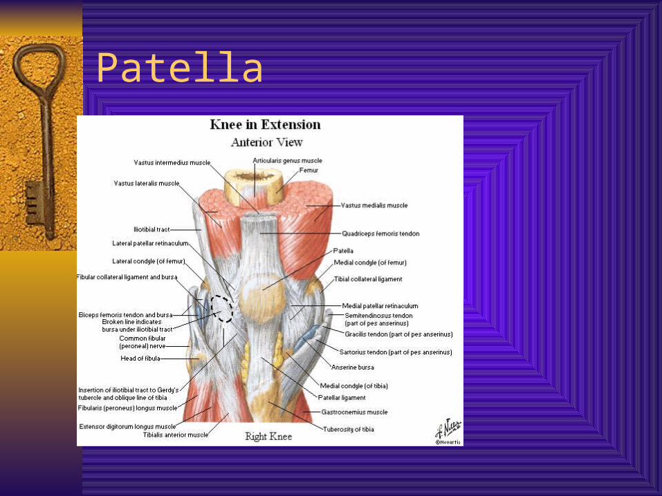
Patella
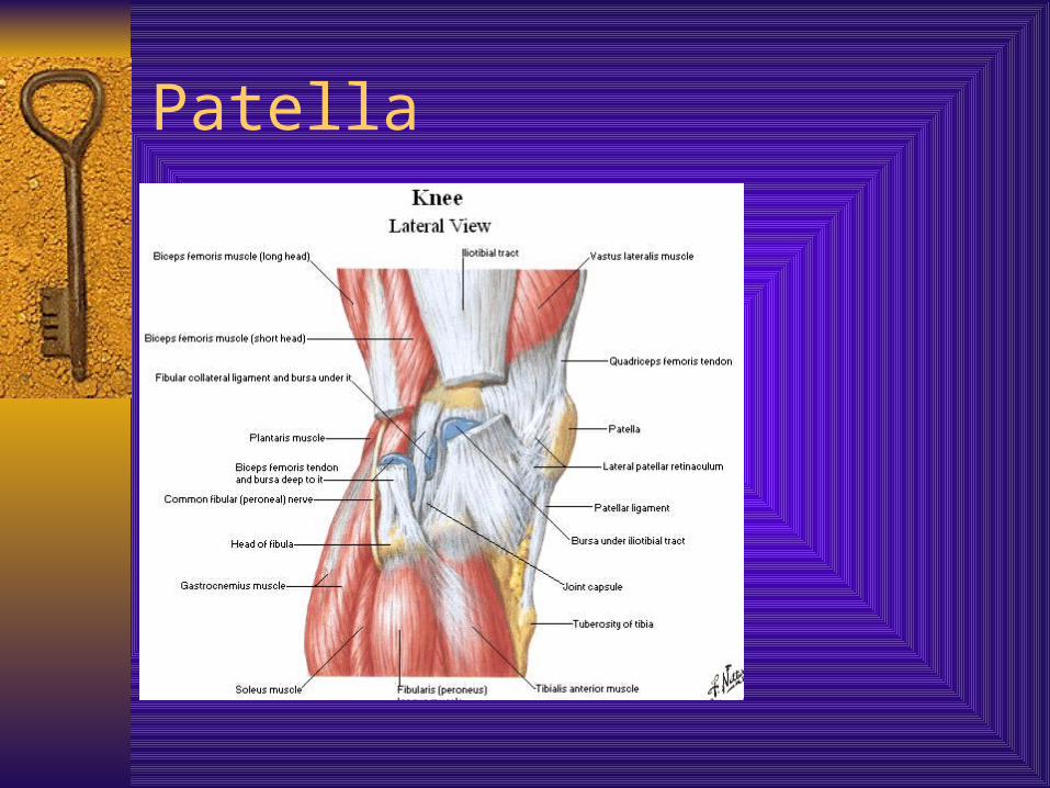
Patella
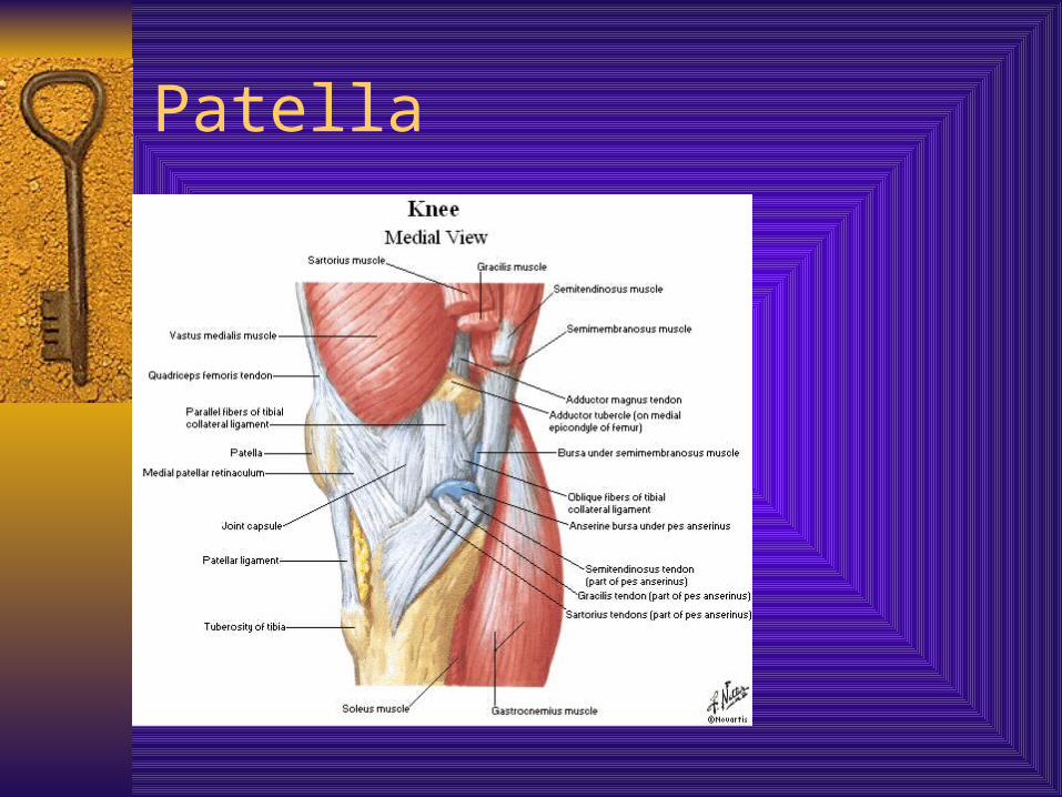
Patella
Patella
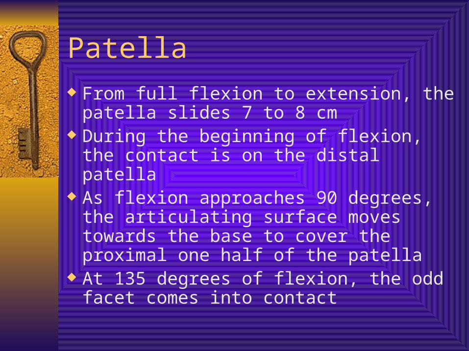
From full flexion to extension, the patella slides 7 to 8 cm
During the beginning of flexion, the contact is on the distal patella
As flexion approaches 90 degrees, the articulating surface moves towards the base to cover the proximal one half of the patella
At 135 degrees of flexion, the odd facet comes into contact
Patella
The odd facet is frequently the 1st part of patella to be affected in premature degeneration of articular cartilage
The load on the patella differs according to activity
In walking = 1/3 bdy weight Climbing stairs = 3 to 4 X body weight Squatting without weight = 7 to 8 X body
weight
Q Angle An angle found by drawing a line from ASIS to
middle of patella and a second line from mid patella to tibial tuberosity– Represents efficiency of Quads– Most efficient = 10 degrees– Males range from 10-14– Females from 15-17– Represents the valgus stress acting on knee and, if
excessive, can cause patello femoral problems Great than 17 degrees considered excessive, called
Genu Valgum or knock knees Very small angle causes genu varum
Girls Play Too
Terminal Rotation AKA Locking Home When the knee moves towards full extension, the
tibia external rotates about 20 degrees on the fixed femur – Explain relationship of condyles
Purely mechanical event, occurs with passive or active knee extension and can not be produced voluntarily
In closed chain motion, such as rising from sitting, terminal rotation is seen as internal rotation of the femur on fixed tibia
Knee Motion The long articulating surface of the femoral
condyles is about twice the length of the tibial condyles
Therefore the activity of flexion and extension can not be a pure hinge motion or simple rolling of one bone over the other
Instead the condyles execute both rolling and sliding motions
Rolling is predominant at the initiation of flexion and sliding occurs more at the end of flexion
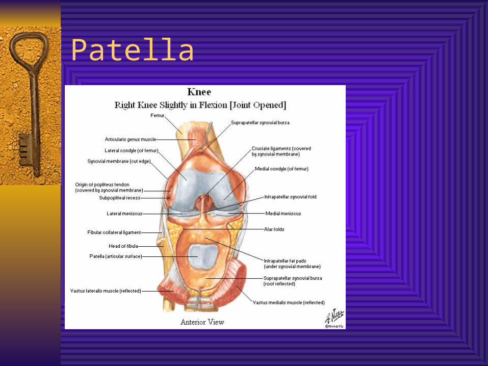
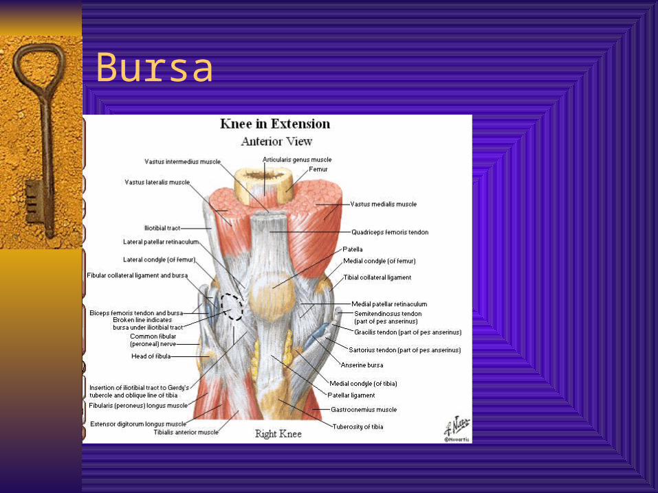
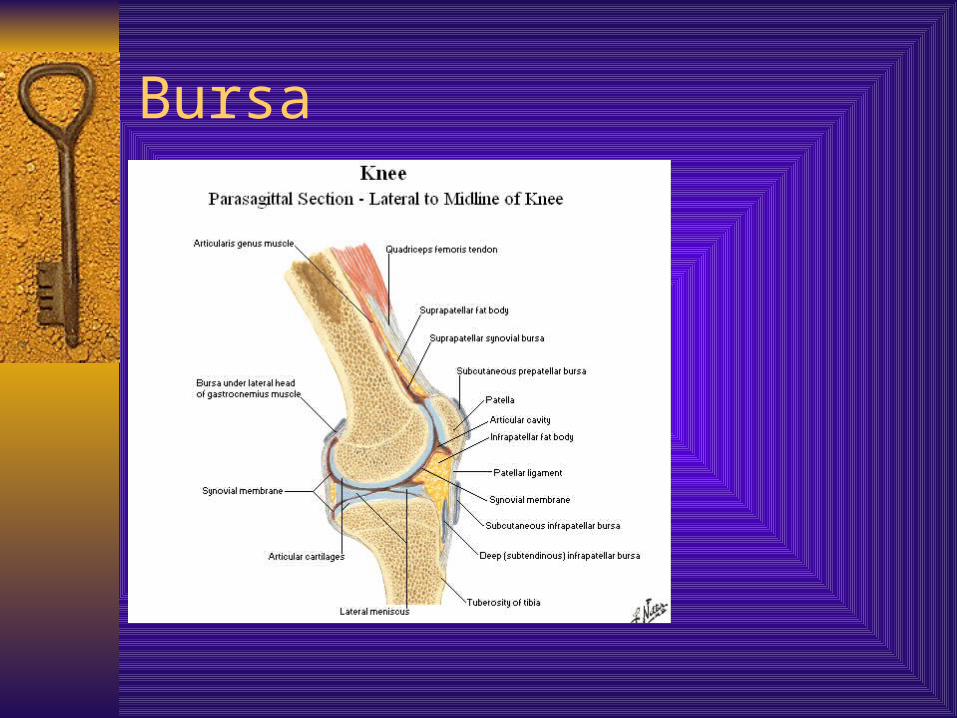
Bursa
20 + associated with the knee Most important Subcutaneous prepatellar Subcutaneous infrapatellar Deep infrapatellar Anserine bursa Bursa deep to iliotibial band Inferior subtendinous bursa of biceps
Bursa
Bursa
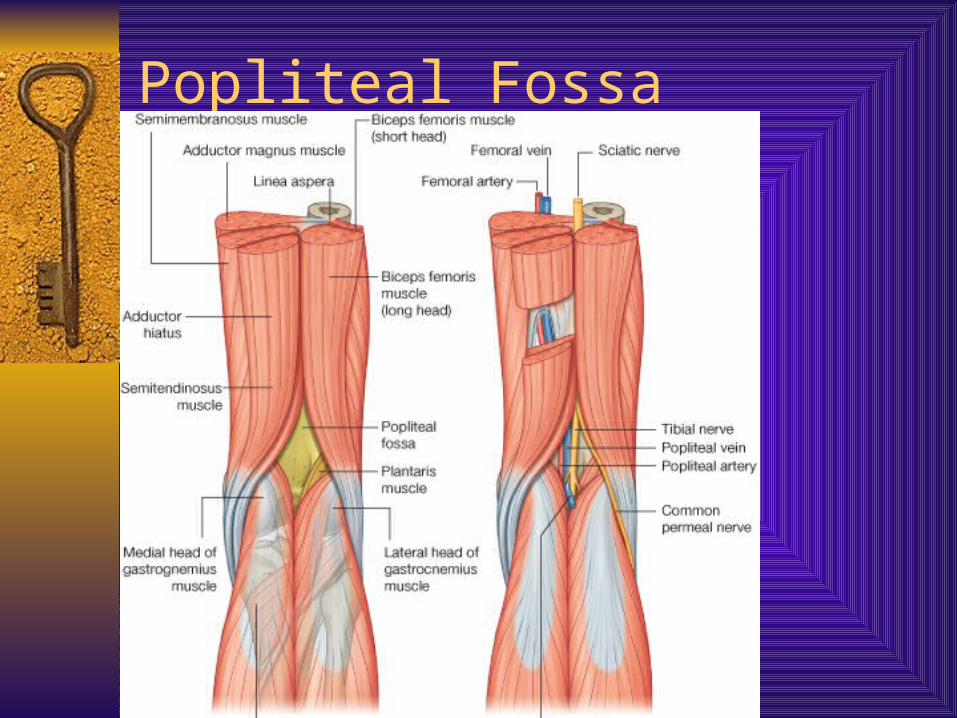
Popliteal Fossa
The diamond shaped region posterior to knee joint
Transition between thigh and leg Boundaries = sup. – biceprs laterally,
semitendinosis medially; inf. Medial and lateral gastrocnemius
Contents = popliteal artery and branches, popliteal vein, tibial nerve
Popliteal Fossa
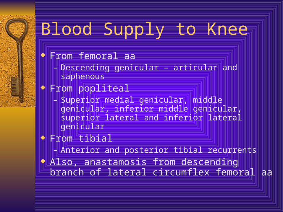
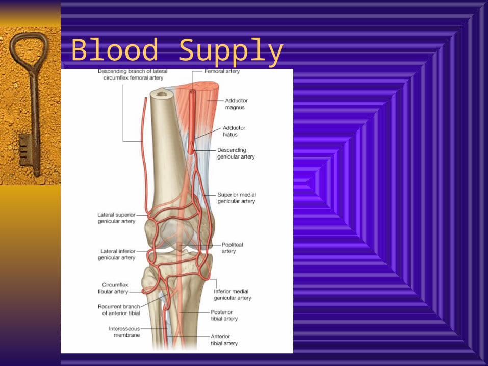
Blood Supply to Knee From femoral aa
– Descending genicular – articular and saphenous From popliteal
– Superior medial genicular, middle genicular, inferior middle genicular, superior lateral and inferior lateral genicular
From tibial– Anterior and posterior tibial recurrents
Also, anastamosis from descending branch of lateral circumflex femoral aa
Blood Supply
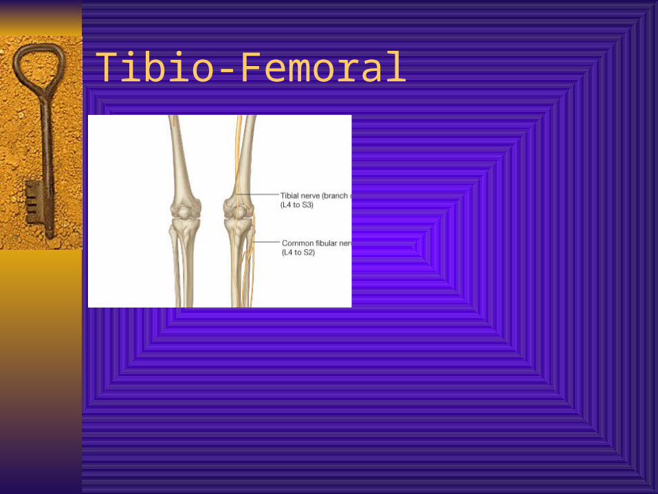
Innervation
Branches from saphenous, obturator (a stretch), tibial and common peroneal
Note the cutaneous coverage about the knee region
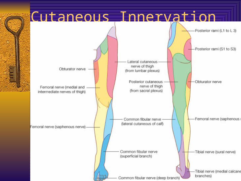
Cutaneous Innervation
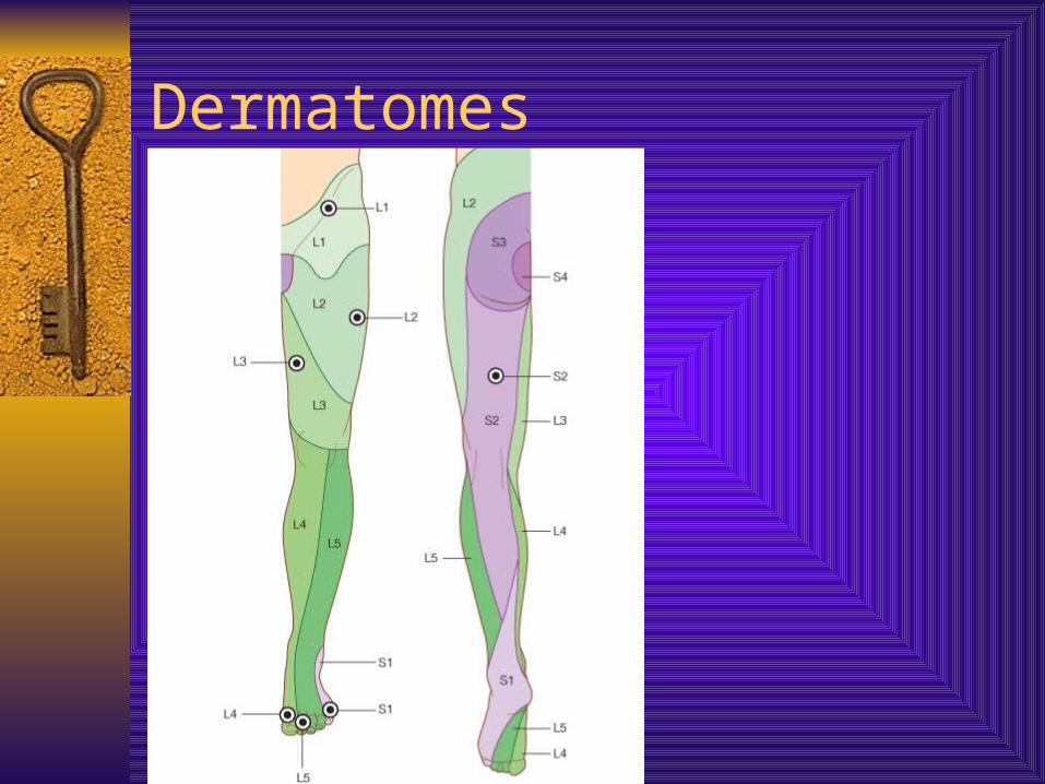
Dermatomes