Embed Size (px)
Citation preview
The Journal of Obstetrics and Gynaecology of the British Empire
TOXOPLASMOSIS AND PREGNANCY BY
SVANTE C : SON HOLMDAHL, M.D. Assistant Obstetrician and Gvnaecologist
Froin the Medical Department of the Goteborg Children's Hospital and the Gynaecologicul Departincnrs, the H Institute of Pathological Anutoiny and the Virus Laboratory of the Sahlgrenska Hospital,
Giiteborg, Sweden
INTRODUCTION A L A R ~ number of cases of infantile toxo- plasmosis, the disease defined by Wolf and his colleagues (Wolf, Cowen and Paige, 1939; Wolf and Cowen, 1939: Paige, Cowen and Wolf, 1942; Cowen and Wolf, 1945), have been reported during the last 10 years. Frenkel and Friedlander (1951), Sabin (1942, 1948). Piekarski (1950), Thalhammer (1951) and others have collected these cases and assembled the results of micro- biological research on animals.
The frequency of antenatal inception of toxoplasmosis in human beings has not yet been given thorough study. The present investigation aims to throw light on the question of how often toxoplasmosis is responsible for abortion, intra- uterine death of the foetus, neonatal disease and death, and infantile disease and malformation. The diagnostic value of serological tests for toxoplasmosis in antenatal care and obstetrics will also be discussed and likewise the manner of antenatal inception of toxoplasmosis. The possibilities of preventing intra-uterine toxo- plasmosis and whether or not antenatally diag- nosed toxoplasmosis justifies termination of pregnancy are two other problems which will be scrutinized.
SURVEY OF LITERATURE It has been established by experiments on
animals that the microbes are spread by the
blood to the different parts of the body during the first two weeks after infection. While this goes on, a rapidly increasing titre of antibodies can be demonstrated. It appears that the distribution of microbes by way of the maternal blood begins to cease when antibodies appear in the circulation and, when they reach a high level, the parasites are only found sporadically in the tissues and in an inactive form as intracellular aggregations (Cowen and Wolf, 1950, 1951).
Antibodies to Toxoplasma have been found in a large number of presumably healthy persons, the frequency varying from a few to 50 per cent of the subjects examined (Callahan et al., 1946; Holmdahl, 1949; Kunert and Juptner, 1952; Macdonald, 1949; Piekarski, 1950; Sabin, 1942). They can be demonstrated with several different methods. Only three of them will now be men- tioned. In Sabin's rabbit test and the egg test worked out independently by Alm (1948) and Macdonald (1950) the serum's capacity to inhibit the activity of living Toxoplasma is estimated. The egg test has proved to be the better of these two tests. In the dye test of Sabin and Feldman (1948), the method most used nowadays, the antibody strength is determined by a micro- biological staining method. The results of the egg and dye tests are equal as regards quality, but the dye test seems to give more accurate quantitative results (v. Zeipel and Linder, 1951).
After infection with toxoplasmosis. the dye 765
history-of-obgyn.com
766 JOURNAL OF OBSTETRICS AND GYNAECOLOGY
test titre rises to a high level (Bengtsson, 1950; Gard et al., 1949, 1952; Jelke, 1950; v. Zeipel and Linder, 1951). The high titres seem to per- sist for at least 6 months, and then sink slowly over a period of several years. It has been possible to demonstrate antibodies as late as 10 years after infection.
In the case of pregnancy there is obviously a risk of infection of the foetus during the acute phase when the parasites are present in the maternal blood. This has been borne out by several experimental studies on animals (Cowen and Wolf, 1950, 1951; Eichenwald, 1948; Jacobs and Jones, 1950). On the other hand, it is not certain whether the microbes can infect the foetus after the parasites have left the blood. Some authors maintain that a high antibody titre means a large risk of infection of the foetus. Others maintain that it protects the foetus from latent toxoplasmosis (Holmdahl, 1951, 1952; Macdonald, 1950), pointing out that the number of women with a high antibody titre is large in comparison with the few cases of infantile toxoplasmosis observed.
The clinical picture of infantile toxoplasmosis includes internal hydrocephalus, intracerebral calcification, chorioretinopathy and psycho- motor disorders. In acute and sub-acute cases there are also changes in the cerebrospinal fluid with monocytic pleocytosis and an elevated protein content. Occasionally, however, the picture is dominated by intestinal symptoms with enlargement of the liver and spleen and jaundice. These signs are often present at birth, but in many cases do not appear until weeks or months have passed. According to Sabin and Feldman (1949), intracerebral calcification is always present.
It is still uncertain whether toxoplasmosis can cause abortion. There are a few cases reported in which a woman had a miscarriage directly before or after giving birth to a baby with toxo- plasmosis. However, it has not been possible to prove that Toxoplasma caused the miscarriages. A number of cases of intra-uterine foetal death have been ascribed to toxoplasrnosis, the assumption being based on evidence of the infec- tion in the maternal serum. In my opinion, none of these cases is sufficiently authenticated.
Cowen and Wolf (1950, 1951) showed in animal experiments, however, that Toxoplasma can lead to specific inflammatory lesions in the foetus and intra-uterine death. Analysis of the respon- sibility of toxoplasmosis for human intra- uterine mortality must be done by statistical procedures, for autolysis leads to the obliteration of any inflammatory changes present.
Ever since cases of antenatally acquired toxo- plasmosis were first demonstrated, it has been assumed that the microbes are transferred to the foetus via the placenta, but not until recently has it been possible to establish this fact. Cowen and Wolf (1950, 1951) showed by extensive experimental studies on pregnant mice that specific inflammatory lesions occur in the placenta during the second week of infection. However, they could not find a single instance of renewed dissemination to the placenta by the blood stream after the infection had been checked by antibodies.
On the other hand, every attempt to transfer Toxoplasma by way of the placenta to the foetus in primates has failed. Even when pregnant monkeys were inoculated with Toxoplasma 2 or 3 weeks before delivery, the infection was not spread to the foetus. The negative results may be due to the fact that only small series of experimental animals could be used (Cowen and Wolf, 1950).
Despite the fact that hundreds of cases of congenital toxoplasmosis in humans are reported, only 2 are described in which the mother showed signs of acute infection during the latter part of pregnancy, the 7th and 8th month, respectively (Adams, 1948; Farquhar and Turner, 1949). These 2 women had multi- locular tender and inflamed lymph nodes and a finely mottled eruption. The signs and symptoms of toxoplasmosis in the adult do not seem to differ from those of a mild infection (Plaut, 1946; Tomlinson, 1945). The majority of persons with serological evidence of recent toxo- plasmosis can never relate anything of note.
PRESENT STUDY Materid
The present study covers the 23,260 babies born at the two maternity hospitals in the city of Goteborg during the years 1948 to and includ-
history-of-obgyn.com
TOXOPLASMOSIS AND PREGNANCY 767
ing 1951. They represent about 95 per cent of the children born in Goteborg during this period. Of these, 633 were stillborn or died within a week after birth. All the foetuses classed as stillborn were at least 35 cm. long. All these 633 children were autopsied. The central nervous system was examined with the naked eye and a few sections studied microscopically, except in cases of great maceration, anencephaly and cranioclasis. When inflammatory changes were present, more extensive microscopical study was
out-patient department for antenatal care. These women were examined with the dye test and 270 of them with repeated tests until their child was born. Furthermore, 93 women with abortions in the 2nd to 6th months were examined with the dye test, one test at the time of the miscarriage and another 2 to 6 weeks later.
In addition, 59 aborted ova and 158 placentas were examined histopathologically and any inflammatory changes discovered checked with the antibodies in the maternal serum.
Serological Methods The two serological tests for Toxoplasma-
neutralizing antibody used in this study are des- cribed by zeipel and Linder (1951) in a publication from our virological department. In the egg test, the strength of titre is given as the ratio between the average number of Toxo- plasma colonies in the negative serum controls and in the serum tested. In the dye test, the
with which at least 50 per cent of the microbes are injured by the antibodies.
made. All the living children were examined by
paediatricians on at least two occasions during the first week of life. About 21,000 of them were followed up regularly during the first year by paediatricians at the city child welfare clinics. On any signs of cerebral lesion the children were sent to the medical department of the Gijteborg Children’s Hospital.
a large number of wOmen. One series, Which I described in an earlier study (Holmdahl, 1949), includes the following, cases, collected succes-
The study also includes a Serological study Of titre is given as the highest dilution of Serum
- sively as they came to the maternity hospitals : 108 women with children born dead or dying soon after birth, 113 women giving premature birth, 101 women with an inexplicable abortion in the 2nd to 6th months, and 32 women giving birth to children with major developmental defects. The serological examination of these women was undertaken with the egg test. The children were examined on the lines just described. Whenever the mother had an antibody titre of 100 or more, however, the central nervous system of the child was given a more thorough histopathological examination if it was stillborn or died shortly after birth. If it was alive, its skull was roentgenographed and its eyes examined with the ophthalmoscope.
For control purposes, 233 women with a normal delivery and normal children were examined with the egg test as well as 94 pre- sumably healthy non-pregnant women. The latter were included to see whether pregnancy was associated with non-specific antibody formation.
The second series consists of 415 women chosen at random from the ones coming to the
Results of Investigation Two cases of infantile toxoplasmosis were
discovered in the 23,260 children born at the maternity hospitals in Goteborg during the years 1948 to and including 1951. They will now be briefly described.
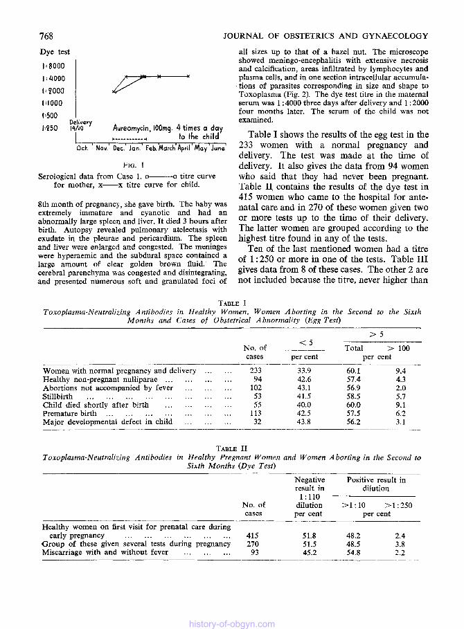
CASE 1. A primigravida aged 17 had a n uneventful pregnancy and a child weighing 3,400 g. was normally delivered at term. A week later it was reported to be well. The mother noticed shortly after birth, however, that one eye was smaller than the other. Breast-feeding was satisfactory and the child gained 500 g. in 7 weeks. At 8 weeks it was noted that the circumference of the skull had increased from 38 cm. at birth to 44.5 cm. Other abnormalities noted were bilateral micro- phthalmus, bilateral healed chorioretinitis and intra- cranial lunate calcifications. Ventriculograms revealed severe internal hydrocephalus. The cerebrospinal fluid was clear and contained a large amount of protein but a normal amount of cells and no Toxoplasma. The antibody titres of the mother and child can be seen from Fig. 1.
CASE 2. A multipara aged 32 had had 2 norma1 deliveries 12 and 6 years previously and both children were living and well. She suffered from sore throat on several occasions during pregnancy. A month before delivery the children brought home some baby birds which died in the house. The patient got another sore throat a t this juncture and a few weeks later, in the
history-of-obgyn.com
768 JOURNAL OF OBSTETRICS AND GYNAECOLOGY
Dye test
1:8000 1 1: 2000 1 P--- 1:500 l:'ooo I .~
Delivery i:P50 14/10 Aureomycin, 100rng. 4 times a day
I c- -_____-__ 4 to the child L--7 act. Nov.' Dec.' Jan.' Feb.'March'April 'May'June'
FIG. 1 Serological data from Case 1. 0- o titre curve
for mother, x-x titre curve for child.
8th month of pregnancy, she gave birth. The baby was extremely immature and cyanotic and had an abnormally large spleen and liver. It died 3 hours after birth. Autopsy revealed pulmonary atelectasis with exudate in the pleurae and pericardium. The spleen and liver were enlarged and congested. The meninges were hyperaemic and the subdural space contained a large amount of clear golden brown fluid. The cerebral parenchyma was congested and disintegrating, and presented numerous soft and granulated foci of
all sizes up to that of a hazel nut. The microscope showed meningo-encephalitis with extensive necrosis and calcification, areas infiltrated by lymphocytes and plasma cells, and in one section intracellular accumula- tions of parasites corresponding in size and shape to Toxoplasma (Fig. 2). The dye test titre in the maternal serum was 1 :4000 three days after delivery and 1 : 2000 four months later. The serum of the child was not examined.
Table I shows the results of the egg test in the 233 women with a normal pregnancy and delivery. The test was made at the time of delivery. It also gives the data from 94 women who said that they had never been pregnant. Table II contains the results of the dye test in 315 women who came to the hospital for ante- natal care and in 270 of these women given two or more tests up to the time of their delivery. The latter women are grouped according to the highest titre found in any of the tests.
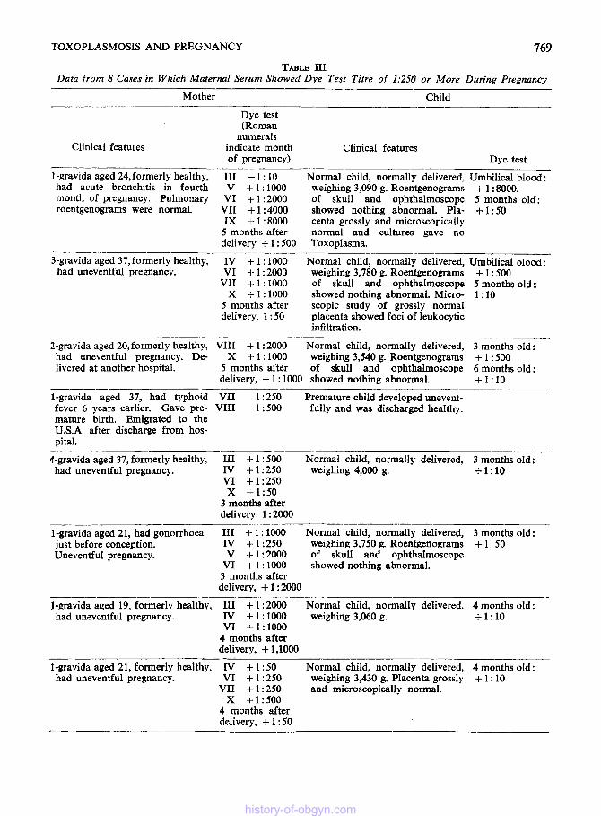
Ten of the last mentioned women had a titre of 1 :250 or more in one of the tests. Table I11 gives data from 8 of these cases. The other 2 are not included because the titre, never higher than
TABLE I Toxoplasma-Neutralizing Antibodies in Healthy Women, Women Aborting in the Second to the Sixrh
Months and Cases of Obstetrical A bnornzality (Egg Test)
> 5 t 5 ___
No. of _____ Total > 100 cases per cent per cent
Women with normal pregnancy and delivery . . . . . . 233 33.9 60.1 9.4 Healthy non-pregnant nulliparae . . . . . . . . . . . . 94 42.6 57.4 4.3 Abortions not accompanied by fever . . . . . . . . . 102 43.1 56.9 2.0 Stillbirth . . . . . . . . . . . . . . . . . . . . . . . . 53 41.5 58.5 5.7 Child died shortly after birth . . . . . . . . . . . . 55 40.0 60.0 9.1 Premature birth . . . . . . . . . . . . . . . . . . . . . 113 42.5 57.5 6.2 Major developmental defect in child . . . . . . . . . 32 43.8 56.2 3.1
TABLE I1 Toxoplasma-Neutralizing Antibodies in Healthy Pregnant Women and Women Aborting in the Second to
Sixth Months (Dye Test)
No. of cases
_____._
Healthy women on first visit for prenatal care during
Group of these given several tests during pregnancy early pregnancy . . . . . . . . . . . . . . . . . . 4 15
270 Miscarriage with and without fever . . . . . . . . . 93
Negative result in
1:llO dilution per cent
Positive result in dilution
>1:10 >1:250 per cent
51.8 51.5 45.2
48.2 2.4 48.5 3.8 54.8 1.2
history-of-obgyn.com
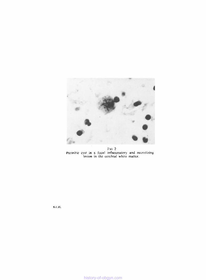
FIG 2 Parasitic cyst in a focal inflammatory and necrotizing
lesion in the cerebral white matter.
S.C.H.
history-of-obgyn.com
TOXOPLASMOSIS AND PREGNANCY 769 TABLE I11
Data from 8 Cases in Which Maternal Serum Showed Bye Test Titre of 1:250 or More During Pregnancy
Mother Child _ _ _ _ _ _ ~ _ _ _ _ _ _ _ _ _ _ _ _ _ _ _ ~
Dye test (Roman
numerals Clinical features indicate month Clinical features
of pregnancy) Dye test _ _ _ _ ~ __ _____ - 1-gravida aged 24,formerly healthy. I11 - 1 : 10 Normal child, normally delivered, Umbilical blood: had acute bronchitis in fourth V + 1 : 1000 weighing 3,090 g. Roentgenograms + 1 : 8000. month of pregnancy. Pulmonary VI + 1 :2000 of skull and ophthalmoscope 5 months old : roentgenograms were normal. VII + 1 : 4000 showed nothing abnormal. Pla- + 1 : 50
IX + 1 : 8000 centa grossly and microscopically 5 months after normal and cultures gave no delivery + 1 : 500 Toxoplasma.
Normal child, normally delivered, Umbilical blood : - _.____
3-gravida aged 37, formerly healthy, IV + 1 : 1000 had uneventful pregnancy. VI + 1 : 2000 weighing 3,780 g. Roentgenograms + 1 : 500
VII + 1 : 1000 of skull and ophthalmoscope 5 months old:
5 months after scopic study of grossly normal delivery, 1 : 50 placenta showed foci of leukocytic
infiltration.
X + 1 : 1000 showed nothing abnormal. Micro- 1 : 10
2-gravida aged 20,formerly healthy, VIII + 1 : 2000 Normal child, normally delivered, 3 months old : had uneventful pregnancy. De- X + 1 : 1000 weighing 3,540 g. Roentgenograms + 1 : 500 livered at another hospital. 5 months after of skull and ophthalmoscope 6 months old:
delivery, + 1 : 1000 showed nothing abnormal. +1:10 -__.- - 1-gravida aged 37, had typhoid VII 1 : 250 Premature child developed unevent- fever 6 years earlier. Gave pre- VIII 1 ~500 fully and was discharged healthy. mature birth. Emigrated to the U S A . after discharge from hos- pital.
___________ __ -~ 4-gravida aged 37, formerly healthy, I11 + 1 ; 500 Normal child, normally delivered, 3 months old : had uneventful pregnancy. IV + 1 : 250 weighing 4,000 g. +1:10
VI +1:250 X +1:50
3 months after delivery, 1 : 2000
- ~ __- 1-gravida aged 21, had gonorrhoea I11 + 1 ; 1000 Normal child, normally delivered, 3 months old: just before conception. IV + 1 : 250 weighing 3,750 g. Roentgenograms + 1 : 50 Uneventful pregnancy. V +1:2000 of skull and ophthalmoscope
VI + 1 : 1000 showed nothing abnormal. 3 months after delivery, + 1 : ZOO0
~~
1-gravida aged 19, formerly healthy, I11 + 1 : 2000 Normal child, normally delivered, 4 months old : had uneventful pregnancy. IV + 1 : 1000 weighing 3,060 g. +1:10
VI +1:1000 4 months after delivery, + 1,1000
1-gravida aged 21, formerly healthy, IV + 1 : 50 Normal child, normally delivered, 4 months old: had uneventful pregnancy. VI + 1 : 250 weighing 3,430 g. Placenta grossly + 1 : 10
VII + 1 : 250 and microscopically normal. x +1:500
4 months after delivery, + 1 : 50
history-of-obgyn.com
770 JOURNAL OF OBSTETRICS AND GYNAECOLOGY
1 : 250, decreased successively with every test. All the children of the women in Table I11 were healthy and exhibited no signs of toxoplasmosis.
The first woman in the table is particularly interesting as she seems to have acquired toxo- plasmosis during pregnancy. The first dye test gave negative results, but the following ones gave positive results, the titres rising continuously to 1 : 8000. The rise began in the 4th month in con- nection with acute bronchitis. Roentgenograms of the lungs were normal and none of the signs typical of acute toxoplasmosis in the adult was manifested.
Two separate series of women aborting in the 2nd to 6th month were examined, one with the egg test (Table I) and the other with the dye test (Table 11). The first group were examined with only one test after the abortion, the second one with at least two tests, one after the abortion and the others 2 to 6 weeks later to see whether the titre rose after the abortion. The first group had a temperature below 38°C. at the time of the abortion, but about half the other women had a temperature over 38°C. Cases in which the abortion could be traced to organic or functional defect in the genital organs were not included in these series.
The titres of the women with babies born dead or dying shortly after birth are seen in Table I. A single egg test was made directly after the delivery. The dead children were examined on
the lines previously described. Whenever the mother had a high antibody titre, however, several slides from different parts of the central nervous system were examined histologically. No inflammatory change pointing to toxo- plasmosis was found in any case.
The antibody level in the 113 women giving premature birth is also seen in Table I. The children weighed 2,500 g. or less and were examined as previously described. No signs of toxoplasmosis were seen in any case.
Table I also includes the 32 women whose children suffered from major developmental defect. In Table IV these cases are grouped according to the nature of the defect. No clinical or histological signs of toxoplasmosis were seen in any of these children. Two of them had internal hydrocephalus without meningocele. The maternal serum contained Toxoplasma-neutralizing antibodies in both these cases. The following is a brief description of them :
(1) A child born healthy at term began to show rapidly progressing hydrocephalus a few days after birth. It died at one year of age with an enormous head. There were no clinical signs of chorioretinitis or encephalo-meningitis, and no evidence of toxo- plasmosis was found on pathological examination. An egg test of the mother’s serum at the time of delivery revealed a titre of 50, and a titre of 22 in the child at the age of 6 weeks.
TABLE IV Toxoplasma-Neutralizing Antibodies in Women whose Children Suffered from Major Developmental
Defect ~
Defect
Egg test
> S Number of _ _ _ ~ -
cases t 5 Total > 100
Organic heart disease . . . . . . . . . . . . . . . . . . Myelomeningocele with or without hydrocephalus
Acrania . . . . . . . . . . . . . . . . . . . . . . . . Mongolism . . . . . . . . . . . . . . . . . . . . . . . .
Ocular aplasia . . . . . . . . . . . . . . . . . . . . . Diaphragmatic aplasia . . . . . . . . . . . . . . . . . . Atresia of oesophagus . . . . . . . . . . . . . . . . . . Umbilical hernia . . . . . . . . . . . . . . . . . . . . . Hydronephrosis . . . . . . . . . . . . . . . . . . . . . Fibrosis of pancreas . . . . . . . . . . . . . . . . . . Deformity of hands and feet . . . . . . . . . . . . . . . Internal hydrocephalus without meningocele . . . . . .
5 4 5 3 1 4 1 1 1 1 4 2
- 1
history-of-obgyn.com
TOXOPLASMOSIS AND PREGNANCY
( 2 ) A hydrocephalic child born at term died a few hours after birth. Autopsy revealed foetal chondro- dystrophy, internal hydrocephalus and intracerebral haemorrhage. No signs of inflammation or necrosis in the cerebral meninges or parenchyma were found on gross or microscopical examination, nor any evidence of toxoplasmosis. The maternal serum had an egg test titre of 140 ten days after delivery.
It is seen from Tables I and I1 that none of the various groups had a significantly increased percentage of women with Toxoplasma- neutralizing antibodies. It may be concluded that the differences between them were due to chance.
Fifty-nine ova from the cases of miscarriage in Table I were examined histologically. Infiltra- tion and necrosis were seen in 35 of them, but the changes showed no correlation to the anti- body titre in the maternal serum.
The placentas from 158 of the women in Table I were also examined histologically. No correla- tion was found between infiltration and necrosis in the placenta and the occurrence of antibodies in the maternal serum. The same was true of calcification in the placenta.
77 1
large proportion of women with antibodies in any of the selected series. Furthermore, not a single case of toxoplasmosis was found on pathological examination of the dead children or clinical examination of the living ones whose mothers had a high antibody titre. Because of this, I conclude that toxoplasmosis is only rarely a cause of abortion, intra-uterine foetal death, neonatal mortality, premature birth or defective development in the child.
DISCUSSION AND CONCLUSIONS Frequency of Antenatal Inception of Toxo-
Two cases of infantile toxoplasmosis were found in 23,260 newborn children. While some cases may have escaped notice, it is clear that manifest infantile toxoplasmosis is a rare disease.
It is possible, however, that intra-uterine toxoplasmosis may cause the death of the ovum and that the specific inflammatory changes are concealed by the autolytic disintegration occur- ring at abortion and intra-uterine foetal death during the last stage of pregnancy. It is likewise possible that the infection heals before the child is born under the influence of the maternal anti- bodies. Under such conditions the infection is of clinical interest only if it causes a disorder in development-premature birth or malformation. However, if toxoplasmosis is a common cause of these defects, it must be possible to demon- strate more mothers with antibodies in their case than in others.
As is seen in Tables I and 11, however, the serological studies did not reveal a significantly
plasmosis
Value of Serological Tests and Manner of
Antibodies were observed in the serum of about half the women examined. In view of this it may be reckoned that about 11,500 of the women in the entire series of 23,260 mothers had antibodies. About 700 of these had such a high antibody titre (dye test positive in 1 : 250 dilution or over) that they probably had had an infection within recent years. Nevertheless, only 2 cases of infantile toxoplasmosis were discovered. This shows that it is impossible to draw conclusions regarding diagnosis or prognosis from a single serological test, even in the case of a high titre.
By making repeated tests one can learn to what stage in the antibody curve the case has arrived. A persisting low dye test titre of 1 : 50 or less was found in 45 per cent of the 270 healthy pregnant women in Table 11. In these cases the antibodies were probably the result of an infection years ago. A small percentage of the women in Table I11 showed a persisting high titre. This was apparently caused by a recent infection, past the clinical stage, however. As mentioned before, all these women gave birth to normal children with no signs of toxoplasmosis. Only 1 woman showed serological evidence of fresh infection, but she, too, gave birth to a healthy child. After delivery the child's titre dropped rapidly, indicating that the antibodies were the result of passive immunization. This case of infection with Toxoplasma during preg- nancy seems to be the first of its kind to be reported. It shows that the same is true of humans as of monkeys, that the mother can be infected with Toxoplasma during pregnancy without the foetus being infected.
Several authors maintain that the frequent
Antenatal Inception
history-of-obgyn.com
712 JOURNAL OF OBSTETRICS AND GYNAECOLOGY
occurrence of antibodies in pregnant women is due to the lowering of resistance associated with pregnancy and that there is a risk of intra- uterine toxoplasmosis when the mother has a high antibody titre. It has been assumed that the conditions are analogous to those occurring in syphilis in pregnant women. A fact militating against this assumption is that only 2 cases of infantile toxoplasmosis were discovered in the present series, though about 700 women showed a dye test titre of 1 : 250 or more. In my opinion, there is reason to assume that the conditions are the same as in animals. A high antibody titre need not in itself mean that the maternal toxo- plasmosis is in an infectious stage. Only in the rare cases where the high titre represents a fresh infection is there a risk of the foetus being infected.
Apart from these exceptional cases, there seems to be no risk of the foetus being infected by Toxoplasma when the mother has a high titre of Toxoplasma-neutralizing antibodies. Instead, the antibodies seem to protect the foetus. For one reason, the antibodies have a strong inhibiting effect on the parasites in vitro.
Another is that, judging from the experimental investigations already mentioned, the microbes’ prospects of extracellular existence become so small when antibodies begin to appear in the blood that any inflammation soon heals.
For the same reasons there can be no risk of the foetus of a future pregnancy being infected by Toxoplasma, no matter what phase of the infection the mother is in prior to the pregnancy.
On the other hand, there is risk of infection of the foetus during the first 2 weeks of the disease, when the microbes are spread by the blood. This has been shown by a large number of animal experiments, and we may assume that the same is true of humans. How great the risk is, is impossible to say. Judging by experiments on monkeys and the results of this study, it would appear to be of little importance.
Possibifities of preventing Intra-uterine Toxo-
These facts must be taken into account when considering prophylactic measures against intra- uterine toxoplasmosis. The most important measure is to find every case of acute toxo-
plasmosis
plasmosis among the pregnant mothers. The signs and symptoms of acute toxoplasmosis are often of diffuse nature in the adult, the infection generally resembling those of a common infec- tion. Tender lymph nodes occur in some cases, a rash in others. In the case diagnosed sero- logically in the present series, the mother showed signs of acute bronchitis. For this reason, it is probably rarely that the disease can be diagnosed clinically. Serological evidence of its presence is not obtained until several tests in succession show a continuous rise in titre. In the mean- time there is a chance of the foetus being infected. It seems to be difficult, therefore, to prevent the occurrence of infantile toxoplasmosis antenatally.
Does Toxoplasnzosis justify Termilzation of
There is no need for legally induced abortion because of transplacental passage of toxo- plasmosis to the foetus in an earlier pregnancy, since neither among animals nor humans has infection of this kind been observed in two preg- nancies in succession. Nor, for reasons just given, is a high antibody titre at the beginning of the pregnancy any justification for its termination.
If fresh toxoplasmosis has been demonstrated, on the other hand, either by discovery of the microbes or a rapidly rising antibody titre, there is obviously a risk of the foetus being infected. Both the animal data and the results of this study would indicate that the risk is insignificant. In view of this fact, it is my opinion that for the time being the pregnancy should be allowed to proceed in these cases. The question cannot be settled until we have more clinical experience.
In every case of this kind in which there is a risk of the foetus being infected, the mother should be treated with sulphapyridine or aureo- mycin. It may be possible by this means to prevent transplacental infection or favourably affect a foetal infection already present.
In view of what has just been said, there is no reason to advise a woman against another pregnancy because she has borne a child with congenital toxoplasmosis or because she shows a high antibody titre.
Pregnancy?
history-of-obgyn.com
TOXOPLASMOSIS AND PREGNANCY
SUMMARY
Two cases of infantile toxoplasmosis were found among 23,260 newborn babies born in Goteborg during a period of 4 years. They are described. No other case was found, despite systematic examination of all the children in the 633 cases of stillbirth or neonatal death and follow-up examination of nearly all the living children during the first year.
Antibodies to Toxoplasma, estimated with the egg and dye test, were found in about 50 per cent of the pregnant women; 3 to 5 per cent of them had a high titre pointing to recent or current toxoplasmosis. Statistical study did not reveal a significantly greater proportion of anti- body carriers among the women whose preg- nancy resulted in abortion, intra-uterine foetal death, neonatal mortality or a defectively developed child.
Consequently, toxoplasmosis seems to occur rarely among the newborn of G6teborg. The study was likewise unable to prove that toxo- plasmosis could cause abortion or intra-uterine death of the foetus, though it did not belie the possibility.
A woman can be infected with toxoplasmosis while she is pregnant without the foetus becom- ing infected. This was shown by 1 case in which the results of the dye test were negative at the start of the pregnancy but changed to positive in the 4th month with a rapid rise in the titre to a high level.
Antibodies being present in the serum of a large number of completely healthy pregnant women who later bore healthy children, no con- clusions as regards diagnosis or prognosis can be drawn from the presence of antibodies, even in high titre. It seems impossible to prevent the occurrence of infantile toxoplasmosis antenatally with the diagnostic possibilities now at hand.
The author is of the opinion that a previous toxoplasmic infection in the mother does not incur any risk for the foetus of a present or future pregnancy. On the contrary, antibodies in the maternal serum probably protect the foetus against toxoplasmosis. The only time when there seems to be risk of the foetus being infected is when a woman without antibodies is infected during pregnancy. It is concluded that
773 as yet toxoplasmosis is not a sufficient indication for the termination of pregnancy.
This study could never have been made had it not been for the friendly co-operation and generous help of a large number of colleagues at the Sahlgrenska Hospital. I am particularly indebted to the following : the gynaecologists, Professor H. Fredrikson, Professor E. Jerlov, and Dr. A. Kjessler; the paediatricians, Drs. Y. Akerrkn and K. Holmdahl; the pathologists, Professor J. Mellgren and Dr. S. Olsson; the virologists, Drs. L. Alm, G. von Zeipel, and L. A. Linder; and the ophthalmologist, Dr. S. Holm.
My thanks are also due to a great number of other colleagues, so many that there is not room for them here, as well as to the Goteborg Medical Society and the Board of Directors of the Gtiteborg City Hospitals for generous financial support.
REFERENCES Adarns, F. H. (1948): Pediatrics, 2, 511. Alrn, L. (1948): Nord. Med., 40, 2109. Bengtsson, E. (1950) : Cardiologia, Bask, 17, 289. Callahan, W. P., Russell, W. D., and Smith, M. C.
Cowen, D., and Wolf, A. (1945): J . infect. Dis., 77,
Cowen, D., and Wolf, A. (1950): J . exp. Med., 92,
Cowen, D., and Wolf, A. (1951): J . Neuropath. exp.
Eichenwald, H. (1948): Amer. J . Dis. Child., 76, 307. Farquhar, H. G., and Turner, W. M. L. (1949): Arch.
Dis. Childh., 24, 118. Frenkel, J. K., and Friedlander, S. (1951): Toxoplas-
musis. U.S. Public Health Service, Washington. Publ. 141.
Gard, S., Magnusson, J. H., and Hagberg, E. (1952): Actu puediut., 41, 15.
Gard, S., Magnusson, J. H., Wahlgren, F., and Gille, G. (1949): Pediatrics, 4, 432.
Holrndahl, S. (1949) : Toxoplasmosis and pregnancy. Elanders, Gothenburg.
Holmdahl, S. (1951): Actu paediai., Suppl. 83, p. 153. Holrndahl, S. (1952): Zbl. Gynuk., 74, 1357. Jacobs, L., and Jones, F. E. (1950): J . infect. Dis.,
(1946): Medicine, 25, 343.
144.
393, 413.
Neurol., 10, 1.
87, 78.
history-of-obgyn.com
774 JOURNAL O F OBSTETRICS AND GYNAECOLOGY
Jelke, H. (1950): Ann. paediat., Basle, 175, 434. Kunert, H., and Jiiptner, H. (1952): Gerbutslz.
Macdonald, A. (1949): Lancet, 1, 950. Macdonald, A. (1950): Lancet, 2, 560. Paige, B. H., Cowen, D., and Wolf, A. (1942): Amer.
Parsons, L. G. (1946): J . Obstet. Gynaec. Brit. Emp.,
Piekarski, G. (1950): Z. Parasitenk., 14, 582. Plaut, A. (1946): Amer. J . Path., 22, 421. Sabin, A. B. (1942). in De Sanctis: Advances in
Sabin, A. B. (1948), in Brenneman: Practice of
Frauenheilk., 12, 910.
J . Dis. Child., 63, 414.
53, 13.
pediatrics, Vol. I. New York.
pediatrics. Vol. 4. Hagerstown.
Sabin, A. B., and Feldman, H. A. (1948): Science,
Sabin, A. B., and Feldman, H. A. (1949): Pediatrics.
Thalhammer, 0. (1951): Oest. 2. Kinderheilk., 6, 1. Tomlinson, W. J. (1945): A,mer. J . clin. Path., 15, 123. Wolf, A., and Cowen, D. (1939): Bull. Neitrol. Inst.,
Wolf, A., Cowen, D., and Paige, B. H. (1939): Arner.
Wolf, A., Cowen, D., and Paige, B. H. (1939): Arner.
von Zeipel, G., and Linder, L. A. (1951): Acta path.
108, 660.
4, 660.
N.Y., 6, 306.
J . Path., 15, 651.
93, 548.
microbiol. scand., 29, 222.
history-of-obgyn.com