Embed Size (px)
Citation preview
THE JOURNAL OF BIOLOGICAL CHEMISTRY 0 1986 by The American Society of Biological Chemists, Inc Vol. 261, No. 17, Issue of June 15, pp. 7934-7940,1966
Printed in U.S.A.
Binding of Drosophila Heat-shock Gene Transcription Factor to the hsp 70 Promoter EVIDENCE FOR SYMMETRIC AND DYNAMIC INTERACTIONS*
(Received for publication, October 30, 1985)
David J. ShueyS and Carl S. Parker From the Division of Chemistry and Chemical Engineering, California Institute of Technology, Pasadena, California 91125
A Drosophila heat-shock gene transcription factor (HSTF) has been shown to bind to three domains up- stream from the TATA homology on a hsp 70 gene. The domain closest to the TATA homology consists of two contiguous binding sites with different binding affinities. Occupancy of the TATA homology proximal site (site 1) coordinates HSTF binding to the neighbor- ing site (site 2) in a cooperative manner (Topol, J., Ruden, D. M., and Parker, C. S. (1985) Cell 42,527- 537). We have used alkylation interference and pro- tection experiments to determine which residues within the binding sites are closely contacted by the HSTF. The contacts inferred from these studies in- cluded the residues present in the consensus sequence found in all HSTF binding sites and exhibit rotational symmetry, suggestive of a multimeric HSTF. By em- ploying a gel electrophoresis separation technique we were able to resolve two protein-DNA complexes con- sisting of site 1 occupancy (complex A) and sites 1 and 2 occupancy (complex B). Analysis of these discrete species reveals that a subset of contacts within site 1 change upon HSTF binding to site 2, suggesting that a conformational change in the protein-DNA complex occurs. Implications for the activation of heat-shock gene transcription are discussed.
The Drosophila heat-shock genes are transcriptionally ac- tivated by a mechanism that utilizes, in part, a heat-shock gene-specific transcription factor (HSTF’) (2). It is known that the HSTF binds to several sites on the hsp 70 promoter (I) as well as to all of the other Drosophila heat-shock genes examined? Present within all of the high affinity binding sites is a rotationally symmetric consensus sequence: C _ _ G A A - - T T C - - G. Several laboratories have shown
that this sequence can confer heat inducibility when placed in an analogous position upstream of heterologous promoters (3, 4). Maximal activation of the hsp 70 gene in Drosophila, however, appears to require more than a single binding site. Dudler and Travers (5) have shown that sequences within approximately 100 base pairs from the start point of transcrip-
* This research was supported by a grant from the National Insti- tutes of General Medical Sciences and in part by funds from American Cancer Society Grant NP-482 (to C. S. P.). The costs of publication of this article were defrayed in part by the payment of page charges. This article must therefore be hereby marked “advertisement” in accordance with 18 U.S.C. Section 1734 solely to indicate this fact.
$ Supported by National Institutes of Health Predoctoral Train- eeship Grant GM 07616.
The abbreviations used are: HSTF, heat-shock gene-specific tran- scription factor; Hepes, 4-(2-hydroxyethyl)-1-piperazineethanesul- fonic acid.
C. S. Parker and J. Topol, manuscript in preparation.
tion are required for transcriptional activation of the Droso- phila hsp 70 gene in vivo. Within this 100-base element it has been determined that two HSTF binding sites are present, both of which must be present for maximal transcription of the hsp 70 genes in vitro. These two sites are occupied by a cooperative interaction where a high affinity site (site 1) is first occupied at low levels of HSTF followed by efficient HSTF binding to the critical second site (site 2 ) . The coop- erative binding to site 2 has been suggested to serve as a molecular switch that turns on the hsp 70 genes upon acti- vation (1).
In this study we have investigated the spatial distribution of the HSTF on binding sites 1 and 2 by performing a series of chemical DNA interference and protection experiments. These experiments have identified the purine and phosphate residues present within the two binding sites that are critical for HSTF binding. We have learned that many of the purine contacts occur within the consensus sequence and are found in a symmetrical arrangement. In addition, cooperative bind- ing to site 2 results in a series of interesting transitions in the contacts made by the HSTF within the binding domain. These contact transitions suggest that conformational changes in the protein-DNA complex arise as a result of site 2 occupancy. The potential significance of these changes in the activation of the hsp 70 genes is discussed.
MATERIALS AND METHODS
Isolation of Drosophila HSTF-Heat-shock transcription factor was partially purified from nonshocked Drosophila Kc cells as previ- ously described (2). In addition, phenylmethylsulfonyl fluoride was included at 1 mM during cell homogenation and nuclear lysis. We estimate that the HSTF used in these experiments represents ap- proximately 10% of the total protein. Several HSTF preparations were used in this study, each yielding very consistent results.
All data presented in this report were generated with HSTF puri- fied from nonshocked Drosophila Kc cells. We have, however, inves- tigated the possibility that heat-shocked Kc cells harbor a structurally distinct HSTF that may interact with the promoter differently. Methylation interference experiments were performed on each strand with HSTF isolated from heat-shocked cells. The interference prop- erties of this factor were found to be indistinguishable from those employing nonshocked factor?
Cloned DNA Template-All binding experiments described utilized the 101-base pair gel-purified HindIII-BamHI insert of plasmid aDm 3110 including Drosophila sequences between -11 and -103. This recombinant was constructed by ligating a HindIII-Sal1 fragment of a 5’ Bal 31 deletion construct: 5’-8-103 (1) into a HindIII-Sal1 restricted pUCS vector (6).
DNA templates were labeled at the 5’ end with [y-32P]ATP and polynucleotide kinase at the HindIII site (coding strand) or the BamHI site (noncoding strand). Following digestion with the second restriction enzyme, DNA fragments were gel purified and recovered from DE-81 membranes as previously described (1). Specific activities
D. Shuey, unpublished data.
7934
Binding of Drosophila HSTF to the hsp 70 Promoter 7935 were generally 2-5 X lo6 cpm/pmol.
Alkylation Interference Experiments-Labeled DNA fragments were methylated with dimethyl sulfate or ethylated with ethylnitro- sourea essentially as described by Sakonju and Brown (7). Approxi- mately 0.03 pmol (100,000 cpm) of the alkylated templates were individually incubated with various concentrations of purified HSTF (0-1 pg) in a 15-p1 reaction consisting of 20 mM Hepes, pH 7.6, 40 mM KCl, 5 mM MgC12, 4% Ficoll, 3% glycerol, and 50 pg/ml pBR322 plasmid DNA restricted with HinfI. Following incubation at 22 "C for 5 min the reaction mixtures were loaded onto a 1.5-mm thick 2.5% vertical agarose gel and electrophoresed a t 200 V for 60-75 min in 0.25 X TBE (25 mM Tris-OH, 25 mM boric acid, 0.25 mM EDTA). Bands representing unbound, complex A, and complex B species were identified by autoradiography and excised from the gel. DNA was extracted from the gel slices by incubating them at 90 "C in 150 pl of TE (10 mM Tris, pH 8.1, 1 mM EDTA) for 30 min followed by extraction with 70 "C phenol. The aqueous phase was extracted again with phenol and once with ether. All volumes were then adjusted to 200 p1 with TE. Base cleavage was carried out by the addition of 10 pl of 2 N NaOH and subsequent incubation at 90 "C for 30 min. Samples were chilled on ice, and 2 pg of salmon sperm DNA was added, and the DNA was precipitated with 3 volumes of 90% ethanol, 0.1 M NaOAc. The pellets were washed with 70% ethanol, dried, and resuspended in formamide-loading dyes. Cleavage products were dis- played on 12% acrylamide, 6 M urea sequencing gels using G + A and C + T specific reactions as markers (8).
DNase I Footprinting-Binding conditions were identical to those described above. Following a 5-min incubation at 22 "C, DNase I (Worthington) was added to each reaction to a final concentration of 1 pg/ml. After 30 s the digestion was stopped by the addition of EDTA to 10 mM and the mixture was applied directly onto an agarose gel. Labeled DNA was eluted, denatured, and run on a sequence gel as described above, omitting the base hydrolysis step.
Methylation (Dimethyl Sulfate) Footprinting-Increasing concen- trations of HSTF (0-1 pg) were incubated with unalkylated labeled templates in the binding conditions described above. After 5 min, dimethyl sulfate was added to a final concentration of 50 mM and the incubation was continued at 22 "C for 10-15 min. Reactions were terminated by loading the mixtures directly onto an agarose gel. Protein-DNA complex gel analysis, DNA recovery, cleavage, and display were performed as described above.
Computer Graphics-The HSTF-DNA contacts shown as dotted surfaces which depict the van der Waals radius of either guanine N- 7 or adenine N-3 were projected in three dimensions by Stephen L. Mayo at the Caltech Materials Simulation Facility (directed by Pro- fessor William A. Goddard, funded in part by the Energy Conversion and Utilization Technology Project of the Department of Energy). An Evans and Sutherland PS-3OO/DEC VAX 11/780 was used to display interactive color graphics in real time, using the BIOGRAF macromolecular modeling and analysis program written by S. L. Mayo, B. D. Olafson, and W. A. Goddard. The crystallographic coordinates of B-DNA established by Arnott and Huskins (9) were adapted for visual display by S. L. Mayo.
RESULTS
Isolation of HSTF-DNA Complexes-To determine the res- idues critical for HSTF binding we employed the chemical modification procedure developed by Gilbert and co-workers (10-13). This procedure identifies contacts between a se- quence-specific DNA-binding protein and its DNA target by partially alkylating the DNA and determining if the binding protein can specifically bind to the modified DNA fragment. Those alkylated residues that prevent binding are considered to be critical contacts. This approach requires that specifically bound DNA be separated from unbound DNA in order to determine the important contacts for protein binding. Meth- ods previously used to separate protein-DNA complexes from unbound DNA include nitrocellulose filtration (13) and anti- body precipitation (7 , 14). The nitrocellulose filtration pro- cedure was initially attempted with the HSTF but failed to resolve specific protein-DNA complexes from nonspecific pro- tein-DNA interactions. By using a modification of the pro- tein-DNA complex gel electrophoresis technique initially de- veloped by Garner and Revzin (15) and Fried and Crothers
(16), we have resolved HSTF-DNA complexes from unbound DNA fragments.
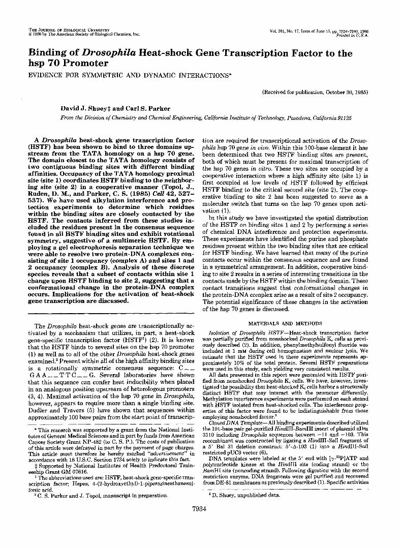
Shown in Fig. 1 are the results obtained by the addition of increasing amounts of partially purified HSTF to a 101-base pair end-labeled DNA fragment containing two contiguous HSTF-binding sites. All binding reactions contained a 400- fold mass excess of pBR322 plasmid DNA. As described previously (1) two slowly migrating protein-DNA complexes are observed. At low HSTF concentrations a protein-DNA complex (designated complex A in Fig. 1) is first observed followed by a more slowly migrating complex (designated complex B in Fig. 1) formed at higher HSTF concentrations. That specific protein-DNA interactions are responsible for the formation of these complexes was determined by subject- ing the protein-DNA complexes to DNase I digestion prior to agarose gel electrophoresis. The DNA in the complexes was excised and eluted from an agarose gel, denatured, and applied to a standard sequencing gel (see "Experimental Procedures"). In panel B of Fig. 1 the DNase I cleavage pattern obtained for unbound DNA (lune I), complex A (lune 2), and complex B (lune 3) is shown. The DNase I footprint clearly shows that complex A has HSTF bound only to site 1 (the TATA ho- mology-proximal binding site) while complex B has HSTF bound to both sites 1 and 2.
This two-step gel procedure consisting of a native agarose gel of the protein-DNA complex followed by a denaturing acrylamide gel of the eluted DNA was employed in each of the chemical modification experiments described below. We have analyzed only those complexes that formed at lower HSTF concentrations. This was done because saturating lev- els of HSTF resulted in a low level of nonspecific binding in
A. 1 2 3 4 5 6
-unbound
B. 1 2 3 R Y
site 1
site 2
FIG. 1. HSTF-DNA complex gel and DNase footprint anal- ysis. A, autoradiogram of a dried analytical complex gel showing complexes A and B. Lanes 1-6 represent 0.01,0.02, 0.05,0.1, and 0.2 pg of partially purified HSTF. A constant 0.005 pmol of template DNA was used in each binding reaction. B, DNase I cleavage pattern of template DNA eluted from a preparative complex gel. Lane I , minus protein; lune 2, complex A; lane 3, complex B. The fragment used consisted of the plasmid aDm 3110 insert labeled at the 5' end of the coding strand. Lanes R and Yare chemical cleavage reactions of purines and pyrimidines, respectively.
7936 Binding of Drosophila HSTF to the hsp 70 Promoter
complex B, masking the specific interactions. Presumably this is due to the presence of contaminating DNA-binding proteins in the HSTF preparations. All experiments described in this report employed the same DNA fragment (see "Exper- imental Procedures" for details).
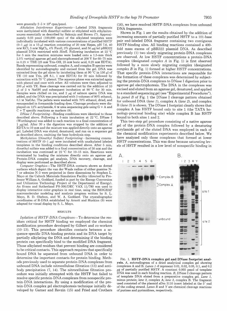
Interference of HSTF Binding by Purine Methylation- Double-stranded DNA methylation with dimethyl sulfate is specific for the N-7 position of guanine (located in the major groove) and the N-3 position of adenine (located in the minor groove). Alkylation of either base creates a positive charge on the purine ring, rendering the modified residue base labile.
A partially methylated template DNA was incubated under standard binding conditions with subsaturating levels of HSTF and subsequently electrophoresed on a native agarose gel. The DNA recovered from the protein-DNA complex gel was denatured, subjected to base cleavage, and displayed on a sequencing gel shown in Fig. 2. Panel A shows the results obtained when the coding (top) strand was labeled and panel B shows the results obtained for the noncoding (bottom) strand. Lanes 1 and 5 are control reactions where no HSTF was added, showing the dimethyl sulfate-induced cleavage pattern. The DNA eluted from complex A, complex B, and the unbound fragment are shown in lanes 2 and 6,3 and 7,4 and 8, respectively. Lanes R and Y represent purine and pyrimidine specific cleavage products, respectively, for each template. Arrows denote residues that, when methylated, in- terfere with specific HSTF binding. Such residues are iden- tified by the absence of a band in the protein-DNA complex, because their methylation prevents specific HSTF binding. All guanines within the consensus sequence (with the excep-
A . B. CODING STRAND NONCODING STRAND
(TOP 1 l 2 3 4 R Y
4
- ite 1
G-62-
4
G-55- 4
""- -
( Bottom 1 5 6 7 8 R Y
,ite 2
;i te 1
ite 2
FIG. 2. HSTF methylation interference. Methylation-specific cleavage products of template DNA eluted from preparative complex gels. Lanes I and 5, minus protein; lanes 2 and 6, complex A, lanes 3 and 7, complex B; and lanes 4 and 8, unbound fraction. Lanes R and Y represent chemical cleavage of purines and pyrimidines in each case. Approximate DNase I footprint boundaries are shown in brack- ets. Arrows denote critical contacts. Designated bases G-55 and G-62 represent contacts within site 1 that differ in complex A and complex B lanes.
tion of G-62 in complex A) are strong contacts. The interfer- ence properties of two residues, in particular, are altered in the transition from complex A to complex B. Base G-55 of the coding strand is only an important contact for site 1 occupancy in complex A. This contact is no longer essential for site 1 occupancy when site 2 is bound by HSTF to form complex B (compare lanes 2 and 3 in panel A of Fig. 2). Conversely, base G-62 of the noncoding strand is a critical contact for complex B formation but not complex A forma- tion. This is surprising because this residue is present within the consensus sequence of site 1. These observations suggest that a conformational change may occur to the HSTF-DNA complex upon cooperative HSTF binding to site 2 (see below for further results and discussion).
The unbound methylated DNA fragments derived from experiments employing subsaturating levels of HSTF are shown in lanes 4 and 8. These fragments are enriched with those residues critical for HSTF binding to site 1. We note that those residues critical for site 2 occupancy are not sig- nificantly enriched in the unbound fraction. Stable HSTF binding, to site 2 is dependent upon prior HSTF binding to site 1; thus methylated residues that prevent binding to site 1 also prevent binding to site 2. Methylation of critical resi- dues within site 2 has no effect on site 1 occupancy and will, therefore, appear in complex A. For these reasons a significant enrichment of site 1 but not site 2 contacts in the unbound fraction of DNA fragments is expected.
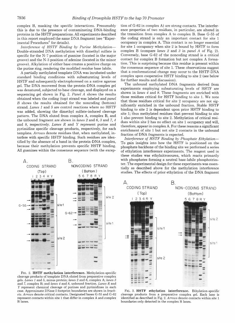
Interference of HSTF Binding by Phosphate Ethyhtion- To gain insights into how the HSTF is positioned on the phosphate backbone of the binding site we performed a series of ethylation interference experiments. The reagent used in these studies was ethylnitrosourea, which reacts primarily with phosphates forming a neutral base-labile phosphotries- ter. The experimental design for these experiments was essen- tially as described above for the methylation interference studies. The effects of prior ethylation of the DNA fragment
A. B. CODING STRAND NON-CODING STRAND
( Top) I 2 3 4 R Y
"-. , - e
site 1
(Bottom) 5 6 7 8 R Y
site 2
site 1
FIG. 3. HSTF ethylation interference. Ethylation-specific cleavage products from a preparative complex gel. Each lane is identified as described in Fig. 2. Arrows denote contacts within site 1 boundaries only detected in the complex B lanes.
Binding of Drosophila HSTF to the hsp 70 Promoter 7937
containing sites 1 and 2 on HSTF binding are shown in Fig. 3. Lanes 1 and 5 are the ethylated DNA fragments derived from control reactions where no HSTF was added. Complex A, B, and the unbound DNA fragments are shown in lanes 2 and 6, 3 and 7, 4 and 8, respectively. The cleared regions in complex A and complex B represent those phosphate residues that, when ethylated, prevent HSTF binding. Particularly worth noting are the strong phosphate contacts surrounding the consensus sequence central TTC elements in both sites 1 and 2 (lanes 2 and 3 in panel A and lanes 6 and 7 in panel B of Fig. 3). The position of these essential phosphate contacts suggests that the conserved bases may have important minor groove binding determinants. Consistent with this observa- tion we can detect partial interference of site 1 occupancy by methylation of the A-56 residue in the minor groove (Fig. 2, lanes 5-7). As described above for the methylation interfer- ence experiments, the interference of certain residues within site 1 is dependent on which protein-DNA complex is ana- lyzed. Arrows denote those phosphate residues within site l that only make contacts in complex B.
It must be stressed that this reagent is not completely phosphate specific (17) and may be reacting with ring nucleo- philes, particularly on guanines (compare minus protein, lanes 1 and 5, to purine markers, lane R) . Thus, all "phosphate" contacts immediately 5' of guanine residues are considered ambiguous. Alkylation interference results are summarized in Fig. 5A.
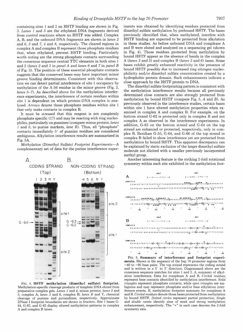
Methylation (Dimethyl Sulfate) Footprint Experiments-A complementary set of data for the purine interference exper-
A. B. CODING STRAND NON-CODING STRAND
(Top) I 2 3 R Y
site 1
G -63- G -62-
jite '2
(Bottom)
4 5 6 R Y
; i te 2
;ite 1
"
FIG. 4. HSTF methylation (dimethyl sulfate) footprint. Methylation-specific cleavage products of template DNA eluted from preparative complex gels. Lanes 1 and 4, minus protein; lanes 2 and 5, complex A; lanes 3 and 6, complex B; lanes R and Y, chemical cleavage of purines and pyrimidines, respectively. Approximate DNase I footprint boundaries are shown in brackets. Site 1 bases G- 64, G-63, and G-62 display altered methylation patterns in complex A and complex B lanes.
iments was obtained by identifying residues protected from dimethyl sulfate methylation by prebound HSTF. The bases previously identified that, when methylated, interfere with HSTF binding are expected to be protected from alkylation in these studies. As before unbound DNA and complexes A and B were eluted and analyzed on a sequencing gel (shown in Fig. 4). Those residues protected from methylation by bound HSTF appear as the absence of bands in the complex A (lanes 2 and 5) and complex B (lanes 3 and 6) lanes. Some bases exhibit greatly enhanced reactivity in the presence of bound HSTF possibly due to increased ring nitrogen nucleo- philicity and/or dimethyl sulfate concentration created by a hydrophobic protein domain. Such enhancements indicate a close approach by the HSTF protein to DNA.
The dimethyl sulfate footprinting pattern is consistent with the methylation interference results because all previously determined close contacts are also strongly protected from methylation by bound HSTF (compare Fig. 5, A and B). As previously observed in the interference studies, certain bases within site 1 have altered methylation properties when ex- amined in complex A and complex B. For example, on the bottom strand (2-62 is protected only in complex B and not complex A as observed in the interference experiments. In addition, G-63 on the bottom strand and G-64 on the top strand are enhanced or protected, respectively, only in com- plex B. Residues G-55, G-64, and G-66 of the top strand in complex B failed to show interference yet are protected from methylation by bound HSTF. This apparent discrepancy can be explained by steric exclusion of the larger dimethyl sulfate molecule not elicited with a smaller previously incorporated methyl group.
Another interesting feature is the striking 2-fold rotational symmetry within each site exhibited in the methylation foot-
A 41.2 r(k 1
C " G " " , T C " G C " G 6 4 " l l C " G
COUPLEX 4 ~ ~ ~ ~ ~ ~ ~ ~ ~ T ~ T C G T ~ G C T T C G D G ~ G ~ G C G C G C ' C ' ~ C ~ O ~ ~ ~ ~ ~ ~ ~ ' C G C ~ ~ ~ ~ ~ ~ ~ ~ ~ ~
c G l G C G 4 i T 1 . G L G C I 4 C G 4 4 G C l C l C l C G C G C G G 4 G C A ~ A ~ @ ~ 4 4 O ~ ~ ~ ~ ~ A ~ J ~ ~ ~ ~ ~
COUKEX B A c ~vavTv~'c.~v?@l ?$C.T.T'C~G &4 G 4 G c G'?G*c.?T'?@~ 4 ?G'T'?C G ?@a 4 4 4 G A G C G
c G T G c G D. T 4 A Q ~ G ~ A L L ~ A a02 CJ~C G c G c G@: G ~ C J J ~ @ ~ a G C.T T.TL C C G C
I l l , I I I I I 1 1 1 -90 -en -m " -50 -4
.It. 2 4b I
B C " G " " T T C " G C " G . . " T T C " G
UklPLEX 4 G C ~ C G C I ~ ~ T C T C G T T G C I T ~ G ~ G ~ G ~ G ~ G ~ G ~ ~ ~ ~ ~ ~ ~ ~ ~ ~ ~ ~ G ~ ~ ~ ~ ~ ~ G ~ G ~ G
A h
C G l G C G I l I I G 4 G C A 4 C G ~ A G C l C l C l C G C G C G G 4 ~ C l l @ C 4 4 ~ C ~ C T l l l C l C G C Y "
CMlPLEX B G c 4 c G c r 4 r r c r c ~ 3 r r O c r ~ c ~ ~ @ 4 ~ 4 G c ~ c ~ c c r c ( D 4 4 l ~ l l c ~ c @ 4 4 4 ~ G 4 G c G
c G T G C G 4 r 4 4 O 4 ~ c 4 4 C 8 4 4 ~ c l c l c l c G c G c ~ O ~ ~ c l l ~ c ~ 4 @ c ~ c l l l ~ c l c G c
FIG. 5. Summary of interference and footprint experi- ments. Shown is the sequence of the hsp 70 promoter regions from -40 to -95 base pairs. The top strand represents the coding strand and is written in a 5' to 3' direction. Diagrammed above are the consensus sequence matches for sites 1 and 2. A, summary of alkyl- ation interference. Data for complexes A and B. Circled residues represent base contacts identified by methylation interference. Solid triangles represent phosphate contacts, while open triangles are am- biguous and may represent phosphate and/or base ethylation inter- ference events. B, methylation footprint summary for complexes A and B. Circled residues denote those bases protected from methylation by bound HSTF. Dotted circles represent partial protection. Single and double carats identify sites of weak and strong methylation enhancement, respectively. The "+" in each case denotes the 2-fold symmetry axis.
" l I , I I l I , I I I I
-90 -80 -m " -5a -4
7938 Binding of Drosophila HSTF to the hsp 70 Promoter
tisp 70 Complex 6
Eompler 1 S i t e I
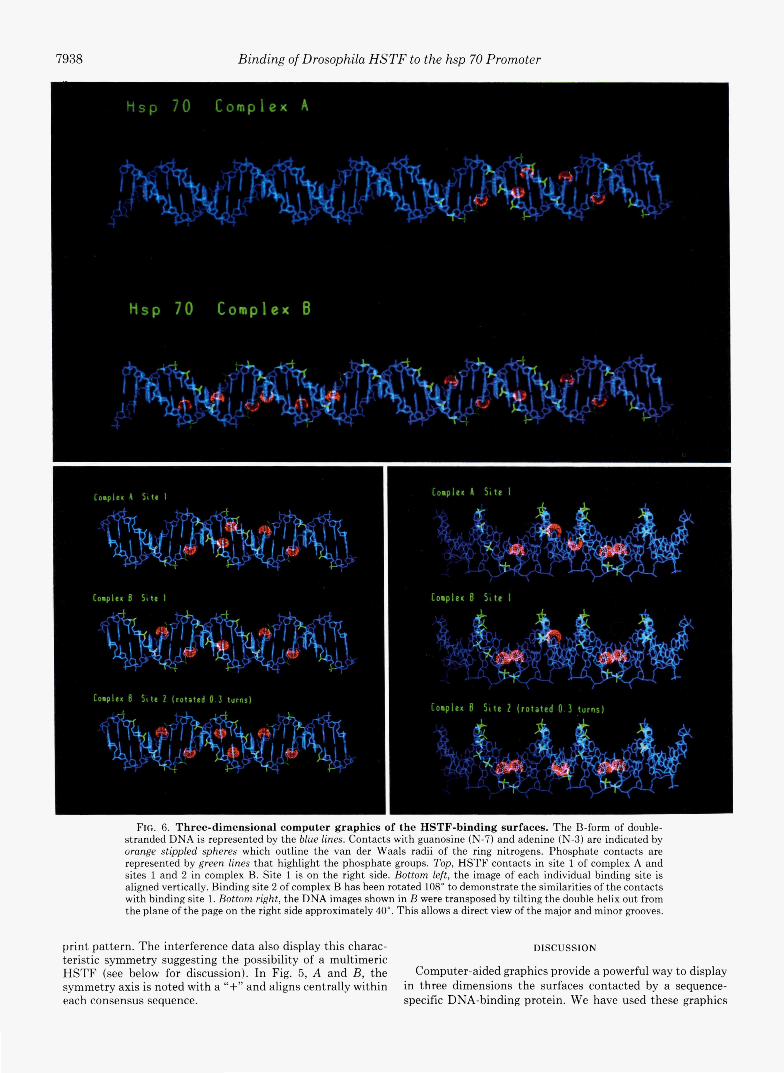
FIG. 6. Three-dimensional computer graphics of the HSTF-binding surfaces. The B-form of double- stranded DNA is represented by the blue lines. Contacts with guanosine (N-7) and adenine (N-3) are indicated by orange stippled spheres which outline the van der Waals radii of the ring nitrogens. Phosphate contacts are represented by green lines that highlight the phosphate groups. Top, HSTF contacts in site 1 of complex A and sites 1 and 2 in complex B. Site 1 is on the right side. Bottom left, the image of each individual binding site is aligned vertically. Binding site 2 of complex B has been rotated 108" to demonstrate the similarities of the contacts with binding site 1. Bottom right, the DNA images shown in B were transposed by tilting the double helix out from the plane of the page on the right side approximately 40". This allows a direct view of the major and minor grooves.
print pattern. The interference data also display this charac- DISCUSSION teristic symmetry suggesting the possibility of a multimeric HSTF (see below for discussion). In Fig. 5, A and B, the Computer-aided graphics provide a powerful way to display symmetry axis is noted with a "+" and aligns centrally within in three dimensions the surfaces contacted by a sequence- each consensus sequence. specific DNA-binding protein. We have used these graphics
Binding of Drosophila HSTF to the hsp 70 Promoter 7939
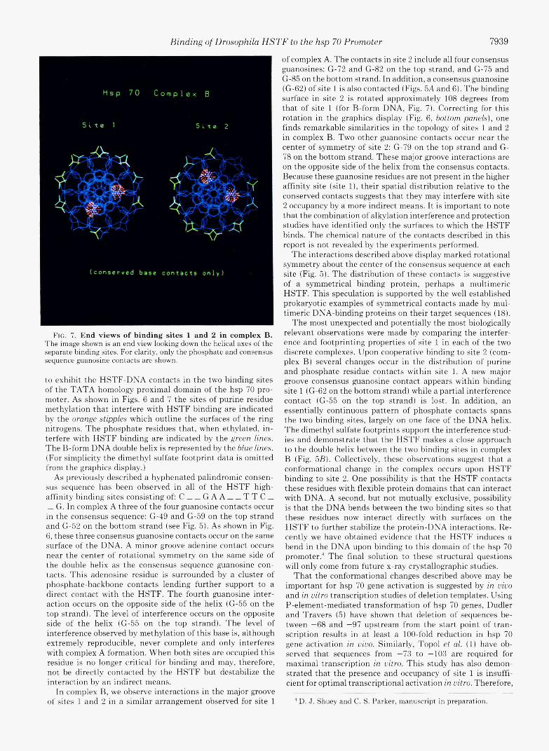
3
FIG. 7. End views of binding sites 1 and 2 in complex B. The image shown is an end view looking down the helical axes of the separate binding sites. For clarity, only the phosphate and consensus sequence guanosine contacts are shown.
t.o exhibit the HSTF-DNA contacts in the two binding sites of the TATA homology proximal domain of the hsp 70 pro- moter. As shown in Figs. 6 and 7 the sites of purine residue methylation that interfere with HSTF binding are indicated by the orange stipples which outline the surfaces of the ring nitrogens. The phosphate residues that, when ethylated, in- terfere with HSTF binding are indicated by the green lines. The B-form DNA double helix is represented by the blue lines. (For simp1icit.y the dimethyl sulfate footprint data is omitted from the graphics display.)
As previously described a hyphenated palindromic consen- sus sequence has been observed in all of t.he HSTF high- affinity binding sites consisting of: C - - G A A - - T T C - - G. In complex A three of the four guanosine contacts occur in the consensus sequence: G-49 and G-59 on the top strand and (2-52 on the bottom strand (see Fig. 5). As shown in Fig. 6, these three consensus guanosine contacts occur on the same surface of the DNA. A minor groove adenine cont.act occurs near the center of rotational symmetry on the same side of the double helix as the consensus sequence guanosine con- tacts. This adenosine residue is surrounded by a cluster of phosphate-backbone contacts lending further support to a direct contact with the HSTF. The fourth guanosine inter- action occurs on the opposite side of the helix ((2-55 on the top strand). The level of interference occurs on the opposite side of the helix (G-55 on the top strand). The level of interference observed by methylation of this base is, although extremely reproducible, never complete and only interferes with complex A formation. When both sites are occupied this residue is no longer critical for binding and may, therefore, not be directly contacted by the HSTF but destabilize the int.eract.ion by an indirect means.
In complex B, we observe interactions in the major groove of sites 1 and 2 in a similar arrangement, observed for site 1
of complex A. The contacts in site 2 include all four consensus guanosines: G-72 and (2-82 on the t.op strand, and G-75 and (2-85 on the bottom strand. In addition, a consensus guanosine ((3-62) of site 1 is also contacted (Figs. 5A and 6). The binding surface in site 2 is rotated approximately 108 degrees from that of site 1 (for B-form DNA, Fig. 7). Correcting for this rotation in the graphics display (Fig. 6, bottom panels), one finds remarkable similarities in the topology of sites 1 and 2 in complex B. Two other guanosine contacts occur near the center of symmetry of site 2: G-79 on the top strand and G- 78 on the bottom strand. These major groove interactions are on the opposite side of the helix from the consensus contacts. Because these guanosine residues are not present in the higher affinity site (site l), their spatial distribution relative to the conserved contacts suggests that. they may interfere with site 2 occupancy by a more indirect means. It is important to note that the combination of alkylation interference and protection studies have identified only the surfaces to which the HSTF binds. The chemical nature of the contacts described in this report is not revealed by the experiments performed.
The interactions described above display marked rotational symmetry about the center of the consensus sequence at each site (Fig. 5). The distribution of these contacts is suggestive of a symmetrical binding protein, perhaps a multimeric HSTF. This speculation is supported by the well established prokaryotic examples of symmetrical contacts made by mul- timeric DNA-binding proteins on their target sequences (18).
The most unexpected and potentially the most biologically relevant observations were made by comparing the interfer- ence and footprinting properties of site 1 in each of the two discrete complexes. Upon cooperative binding to site 2 (com- plex B) several changes occur in the distribution of purine and phosphate residue contacts within sit,e 1. A new major groove consensus guanosine contact appears within binding site 1 (G-62 on the bottom strand) while a partial interference contact (G-55 on the top strand) is lost. In addition, an essentially continuous pattern of phosphate contach spans the two binding sites, largely on one face of the DNA helix. The dimethyl sulfate fo0tprint.s support t.he interference stud- ies and demonstrate that the HSTF makes a close approach to the double helix between the two binding sites in complex B (Fig. 5B). Collectively, these observations suggest that a conformational change in the complex occurs upon HSTF binding to site 2. One possibility is that the HSTF contacts these residues with flexible protein domains that. can interact with DNA. A second, but not. mut.ually exclusive, possibility is that the DNA bends between the two binding sites so that these residues now interact directly with surfaces on the HSTF to further stabilize the protein-DNA interactions. Re- cently we have obtained evidence that the HSTF induces a bend in the DNA upon binding to this domain of the hsp 70 pr~moter .~ The final solution to these struct(ura1 questions will only come from future x-ray crystallographic studies.
That the conformational changes described above may be important for hsp 70 gene activation is suggested by in uiuo and in vitro transcription studies of deletion templates. Using P-element-mediated transformation of hsp 70 genes, Dudler and Travers (5) have shown that deletion of sequences be- tween -68 and -97 upstream from the start point of tran- scription results in at least a 100-fold reduction in hsp 70 gene activation in uiuo. Similarly, Topol et al. (1) have ob- served that sequences from -73 to -103 are required for maximal transcription in uitro. This study has also demon- strated that the presence and occupancy of site 1 is insuffi- cient for optimal transcriptional activation in uitro. Therefore,
D. J. Shuey and C. S. Parker, manuscript in preparation. ~"
7940 Binding of Drosophila HSTF to the hsp 70 Promoter
it appears that occupancy of site 2 by the HSTF results in the maximal activation of the hsp 70 promoter. Thus, a clear correlation exists between cooperative binding at site 2, ap- parent conformational changes, and activation of transcrip- tion. It is not clear how these protein-protein and protein- DNA interactions function mechanistically to activate tran- scription. One possibility is that HSTF in the “active” state (complex B) presents a complementary surface for interaction with other components of the transcriptional machinery (RNA polymerase, the TATA binding factor, etc.). These protein-protein and protein-DNA contacts may also be influ- enced by the conformation of the promoter itself. It is inter- esting to speculate that bending of the DNA by positive activators of transcription may be a general phenomenon. Support for this idea has come from Wu and Crothers (19) who have suggested that CAP binding to l ac promoter DNA induces a bend or a kink in the DNA. Clearly, other transcrip- tional activators must be examined before any generalization can be made.
An examination of the consensus sequences and DNase I protection boundaries present on the other heat-shock genes reveals that there are potentially two contiguous binding sites.’ The distance between the rotationally symmetric units on hsp 83, hsp 22, and possibly hsp 26 is, however, 20 base pairs and not 23 as for the hsp 70 gene. This would result in two adjacent HSTF molecules 2n the same surface of the double helix (approximately 10 A closer to each other than on the hsp 70 promoter, assuming B-form DNA). It has not yet been determined experimentally whether occupancy of both presumptive sites is required for maximal transcription of these heat-shock genes or whether binding to these sites is cooperative. It is important that this information be obtained before a uniformly applicable model can be formulated to explain the mechanism of HSTF action in the activation of heat-shock gene transcription.
Acknowledgments-We thank the members of the Parker Lab, Eric Davidson, Norman Davidson, and Stewart Scherer, for helpful com- ments on the manuscript. We are indebted to Stephen Mayo and Williain Goddard for their valuable time and expertise in preparing the computer-aided graphics presented in this report. We thank Phoebe Ray for her significant aid in the preparation of this manu- script.
1.
2. 3. 4.
5. 6. 7. 8.
9.
10.
11.
12. 13.
14. 15.
16.
17. 18.
19.
REFERENCES Topol, J., Ruden, D. M., and Parker, C. S. (1985) Cell 42 , 527-
Parker, C. S., and Topol, J. (1984) Cell 37, 273-283 Pelham, H. R. B. (1982) Cell 30,517-528 Mirault, M.-E., Southgate, R., and Delwart, E. (1982) EMBO J.
Dudler, R., and Travers, A. A. (1984) Cell 38, 391-398 Vieira, J., and Messing, J. (1982) Gene 19 , 259-268 Sakonju, S., and Brown, D. D. (1982) Cell 31,395-405 Maxam, A., and Gilbert, W. (1980) Methods Enzymol. 65, 499-
560 Amott, S., and Huskins, D. W. L. (1972) Bwchem. Biophys. Res.
Cornrnun. 47,1504-1509 Gilbert, W., Maxam, A., and Mirzabekov, A. (1976) in Control of
Ribosome Synthesis (Kjeldgaard, N. C., and Maaloe, O., eds), pp. 139-143, Munksgaard, Copenhagen
Ogata, R. T., and Gilbert, W. (1978) Proc. Nutl. Acud. Sci. U. S.
Johnsrud, L. (1978) Proc. Nutl. Acud. Sci. U. S. A. 75,5314-5318 Siebenlist, U., and Gilbert, W. (1980) Proc. Natl. Acad. Sci. U. S.
Jones, K. A., and Tjian, R. (1984) Cell 35 , 155-162 Garner, M. M., and Revzin, A. (1981) Nucleic Acids Res. 9,3047-
Fried, M., and Crothers, D. M. (1981) Nucleic Acids Res. 9,6505-
Sun, L., and Singer, B. (1975) Biochemistry 14, 1795-1802 Pabo, C . O., and Sauer, R. T. (1984) Annu. Reu. Biochem. 5 3 ,
Wu, H.-M., and Crothers, D. M. (1984) Nature 308 , 509-513
537
1,1279-1285
A. 75,5851-5854
A. 77, 122-126
3060
6525
293-321


![U1.1 lesson5[lo6]](https://img.pdfslide.us/doc/110x75/58eceb391a28ab8d308b462d/u11-lesson5lo6.jpg)