Embed Size (px)
Citation preview
Modulation of the Actin Cytoskeleton via Gelsolin RegulatesVacuolar H�-ATPase Recycling*
Received for publication, November 10, 2004Published, JBC Papers in Press, December 9, 2004, DOI 10.1074/jbc.M412750200
Valerie Beaulieu‡, Nicolas Da Silva‡, Nuria Pastor-Soler‡, Christopher R. Brown‡,Peter J. S. Smith§, Dennis Brown‡¶, and Sylvie Breton‡¶�
From the ‡Program in Membrane Biology, Massachusetts General Hospital, Charlestown, Massachusetts 02129, the¶Department of Medicine, Harvard Medical School, Boston, Massachusetts 02215, and the §BioCurrents Research Center,Marine Biological Laboratory, Woods Hole, Massachusetts 02543
The role of the actin cytoskeleton in regulating mem-brane protein trafficking is complex and depends on thecell type and protein being examined. Using the epidid-ymis as a model system in which luminal acidification iscrucial for sperm maturation and storage, we now re-port that modulation of the actin cytoskeleton by thecalcium-activated actin-capping and -severing proteingelsolin plays a key role in regulating vacuolar H�-ATPase (V-ATPase) recycling. Epididymal clear cellscontain abundant V-ATPase in their apical pole, and anincrease in their cell-surface V-ATPase expression cor-relates with an increase in luminal proton secretion. Wehave shown that apical membrane accumulation of V-ATPase is triggered by an elevation in cAMP followingactivation of bicarbonate-regulated soluble adenylyl cy-clase in response to alkaline luminal pH (Pastor-Soler,N., Beaulieu, V., Litvin, T. N., Da Silva, N., Chen, Y.,Brown, D., Buck, J., Levin, L. R., and Breton, S. (2003)J. Biol. Chem. 278, 49523–49529). Here, we show thatclear cells express high levels of gelsolin, indicating apotential role in the functional activity of these cells.When jasplakinolide was used to overcome the severingaction of gelsolin by polymerizing actin, complete inhi-bition of the alkaline pH- and cAMP-induced apicalmembrane accumulation of V-ATPase was observed.Conversely, when gelsolin-mediated actin filament elon-gation was inhibited using a 10-residue peptide (PBP10)derived from the phosphatidylinositol 4,5-bisphosphate-binding region (phosphoinositide-binding domain 2) ofgelsolin, significant V-ATPase apical membrane mobiliza-tion was induced, even at acidic luminal pH. In contrast,the calcium chelator 1,2-bis(2-aminophenoxy)ethane-N,N,N�,N�-tetraacetic acid tetrakis(acetoxymethyl ester)and the phospholipase C inhibitor U-73122 inhibited thealkaline pH-induced V-ATPase apical accumulation. Thus,maintenance of the actin cytoskeleton in a depolymerizedstate by gelsolin facilitates calcium-dependent apical accu-
mulation of V-ATPase in response to luminal pH alkaliniza-tion. Gelsolin is present in other cell types that express theV-ATPase in their plasma membrane and recycling vesicles,including kidney intercalated cells and osteoclasts. There-fore, modulation of the actin cortex by this severing andcapping protein may represent a common mechanism bywhich these cells regulate their rate of proton secretion.
The actin cytoskeleton is a dynamic structure that is in-volved in a variety of cellular processes, including cell motility(1–6), division (7), adhesion (8), and vesicle trafficking (9–20).In eukaryotic cells, a dense sheet of polymerized actin adjacentto the plasma membrane forms the actin cortex. The role of thecortical actin in regulating vesicle trafficking is extremely com-plex, and numerous studies have shown that depolymerizationof the actin cytoskeleton can either facilitate or impair vesiclerecycling. This topic has been extensively reviewed (20, 21),and the following examples illustrate the variability of resultsobtained upon actin cytoskeleton modification. Orci et al. (13)were among the first to demonstrate that the actin-depolymer-izing agent cytochalasin can either stimulate or inhibit exocy-tosis depending on the experimental conditions used. Actindepolymerization enhances catecholamine secretion in adrenalchromaffin cells (22, 23), suggesting that the actin cortex canform a barrier for exocytic vesicles. Although a similar stimu-lation of exocytosis was observed upon moderate actin depoly-merization in pancreatic acinar cells, severe depletion of F-actin inhibited exocytosis in these cells (18), indicating theneed for a minimal amount of polymerized actin. In intactmuscle, the insulin-stimulated cell-surface mobilization ofGLUT4 is inhibited after disruption of cortical F-actin (24). Incontrast, accumulation of the water channel AQP2 in theplasma membrane of primary cultured kidney collecting ductprincipal cells occurs after disruption of the actin cytoskeleton(25). Increase in surface expression of AQP2 is promoted byvasopressin in the mammalian kidney and toad bladder (26),and in the latter, this process was shown to be accompanied bydepolymerization of the actin cytoskeleton (27). More recently,actin filaments were proposed to facilitate endocytosis of waterchannels but not to be required for exocytosis in the toadbladder (28). In some cells, disruption of the actin cytoskeletoninhibits apical endocytosis without affecting basolateral endo-cytosis (29, 30). Another study showed variable effects on re-ceptor-mediated endocytosis after either depolymerization orpolymerization of actin filaments in different cell types (31). Ittherefore appears that opposite effects can be obtained follow-ing disruption of the actin cytoskeleton depending on the celltype or the nature of the recycling process examined.
* This work was supported by National Institutes of Health GrantsHD40793 (to S. B.), DK38452 (to D. B. and S. B.), DK42956 (to D. B.),P41-RR001395 (to P. J. S. S.), and KO8-HD45524 (to N.P.-S.) and Na-tional Research Service Award HD08684 (to N. P.-S.). The work per-formed in the Microscopy Core Facility of the Massachusetts GeneralHospital Program in Membrane Biology was supported by Center forthe Study of Inflammatory Bowel Disease Grant DK43351 and BostonArea Diabetes and Endocrinology Research Center Award DK57521.The costs of publication of this article were defrayed in part by thepayment of page charges. This article must therefore be hereby marked“advertisement” in accordance with 18 U.S.C. Section 1734 solely toindicate this fact.
� To whom correspondence and reprint requests should be addressed:Program in Membrane Biology, Massachusetts General Hospital East,149 13th St., Charlestown, MA 02129. Tel.: 617-726-5785; Fax: 617-726-5669; E-mail: [email protected].
THE JOURNAL OF BIOLOGICAL CHEMISTRY Vol. 280, No. 9, Issue of March 4, pp. 8452–8463, 2005© 2005 by The American Society for Biochemistry and Molecular Biology, Inc. Printed in U.S.A.
This paper is available on line at http://www.jbc.org8452
by on April 19, 2007
ww
w.jbc.org
Dow
nloaded from
Remodeling of the actin cytoskeleton is controlled by severalintracellular proteins, including the calcium-activated actin-severing proteins villin, gelsolin, fragmin, severin, and ad-severin (scinderin) (32, 33). Gelsolin severs actin filaments andcaps their fast-growing barbed ends, thereby promoting rapidactin filament fragmentation and blocking their elongation (34,35). The binding of gelsolin to actin is activated by calcium andby low pH, whereas phosphatidylinositol 4,5-bisphosphate(PIP2)1 and some other phosphoinositides remove gelsolin fromthe barbed ends of actin filaments (36, 37). Two phosphoinosit-ide-binding domains (PBDs) of gelsolin (PBD1 and PBD2;10–20 amino acids each) have been identified (38). The actin-severing activity of gelsolin is located in the N-terminal half ofgelsolin (38). Thus, modulation of the actin cytoskeleton bygelsolin is a regulated and complex process that involves boththe severing and capping properties of gelsolin.
Gelsolin was shown to be highly expressed in a variety of celltypes involved in active plasma membrane recycling, includingosteoclasts, kidney intercalated cells, and spermatozoa (39–41). It was proposed that active remodeling of the actin cy-toskeleton by gelsolin might regulate the endo/exocytic activityin these cells. In this study, we show that gelsolin is highlyexpressed in another cell type involved in active recycling, theclear cells of the epididymis. Furthermore, osteoclasts, kidneyintercalated cells, and epididymal clear cells all express highlevels of the vacuolar H�-ATPase (V-ATPase) on intracellularvesicles and the plasma membrane (42–46). Active proton se-cretion by these cells via the V-ATPase is responsible for boneresorption (osteoclasts) (46, 47), systemic acid/base homeosta-sis (intercalated cells) (48), and maintenance of sperm in adormant state during maturation and storage in the epididy-mis (clear cells) (43).
The V-ATPase is a complex enzyme composed of numeroussubunits, some of which are present in multiple copies in eachholoenzyme (49, 50). It is ubiquitously expressed in eukaryoticcells, where it is located in intracellular acidic organelles, in-cluding lysosomes, the Golgi apparatus, secretory vesicles, andendosomes. In some specialized cells such as osteoclasts, kid-ney intercalated cells, and epididymal clear cells, the V-ATPaseis also expressed in the plasma membrane (46, 48, 51, 52). Inthese cells, the V-ATPase rapidly recycles between intracellu-lar vesicles and the plasma membrane, and an increase inproton secretion correlates with an increase in V-ATPase sur-face expression (48, 53–55).
We (59) and others (55–58) have shown that the V-ATPasecan bind either directly or indirectly to actin via two of itssubunits, B and C, located on the cytoplasmic side of the mem-brane. The B subunit of the V-ATPase exists in two formsknown as B1 and B2. The B1 subunit is predominant in kidneyintercalated cells and epididymal clear cells (44, 45, 60),whereas osteoclasts express the B2 isoform (47). These iso-forms are highly homologous, and they both bind directly toactin via their cytoplasmic N termini (56, 58). In addition, theB1 subunit, but not the B2 subunit, contains a DTAL motif inits C terminus, which allows for binding to the PDZ domain-containing protein NHERF1 (59). NHERF1, which was origi-nally identified as a cofactor for the regulation of the Na�/H�
exchanger NHE3 (61), binds to the ezrin/radixin/moesin familyof actin-binding proteins and links many membrane proteins tothe actin cytoskeleton (62). Thus, NHERF1 may allow for in-
direct interaction between V-ATPase and actin. It was pro-posed that interaction between the V-ATPase and F-actin isrequired for V-ATPase-dependent proton secretion by oste-oclasts (55), but whether this is also the case in other cell typesremains to be elucidated. In addition, although the role of theactin cytoskeleton in regulating clathrin-mediated endocytosisis clearly emerging (20, 63), its role in the recycling of V-ATPase, which utilizes unique clathrin- and caveolin-inde-pendent mechanisms (51, 64), has not been demonstrated yet.
In this study, we used epididymal clear cells as a modelsystem to examine the role of actin cytoskeleton modulation viagelsolin in regulating V-ATPase-dependent proton secretion.This model allows the study of V-ATPase recycling in intactcells while they reside in their native epithelium. The epidid-ymis establishes a low luminal pH of 6.5–6.8 to maintainspermatozoa in a quiescent state during their maturation andstorage in this organ. We have shown previously that clear cellsare key players in this acidification process (43). In addition, wehave shown that V-ATPase recycling in these cells is stronglydependent on luminal pH and that a bicarbonate-regulatedsoluble adenylyl cyclase-dependent rise in cAMP in response toalkaline luminal pH induces an accumulation of V-ATPase inthe apical membrane, leading to significant elongation of V-ATPase-containing microvilli (65). The resulting increase inproton secretion by clear cells would re-establish the pH of thelumen to its resting acidic value. We now show that polymeri-zation of the actin cytoskeleton inhibits the alkaline pH-in-duced V-ATPase apical cell-surface mobilization in gelsolin-rich clear cells. In addition, a PIP2-binding peptide thatcompetes for the binding of PIP2 with gelsolin, thereby prevent-ing uncapping of gelsolin from actin filament barbed ends,favors the apical membrane accumulation of V-ATPase. Wealso show that the phospholipase C (PLC) calcium signalingpathway is involved in the regulation of V-ATPase trafficking.These data point to a major role for the actin cytoskeleton-remodeling protein gelsolin in regulating V-ATPase endocyto-sis, a process that is broadly used by specialized acidifyingV-ATPase-expressing cells in various tissues and organs.
EXPERIMENTAL PROCEDURES
Tissue Fixation and Immunofluorescence—Adult male Sprague-Dawley rats (Charles River Laboratories, Wilmington, MA) were anes-thetized using an intraperitoneal sodium pentobarbital injection of 10mg/100 g of body weight. They were then perfused through the leftventricle of the heart with phosphate-buffered saline (PBS; 12 mM
phosphate buffer containing 137 mM NaCl and 2.7 mM KCl, pH 7.4),followed by PLP fixative containing 10 mM sodium periodate, 75 mM
lysine, 4% paraformaldehyde, and 5% sucrose. Epididymides were har-vested and further fixed overnight at 4 °C in PLP fixative. Tissues werethen washed in PBS and stored in PBS containing 0.02% sodium azide.
PLP-fixed epididymides were cryoprotected in a solution of 30%sucrose in PBS, and 5-�m cryostat sections were cut and picked up ontoFisher Superfrost Plus microscope slides as described previously (42,65). Sections were hydrated for 15 min in PBS and treated with 1%SDS, an antigen retrieval technique that we have described previously(66). Nonspecific binding of the antibody was blocked by preincubationof sections with 1% bovine serum album in PBS containing 0.02%sodium azide for 15 min. Sections were then incubated with the primaryantibody for 90 min at room temperature, followed by two washes of 5min in high salt PBS (2.7% NaCl) to reduce nonspecific staining and onewash in normal PBS. The secondary antibody was applied for 1 h atroom temperature and washed as described above. Double labeling wasperformed by subsequent incubation of the sections with additionalprimary and secondary antibodies.
The primary antibodies used were as follows. Affinity-purifiedchicken or rabbit antibodies raised against the last 10 C-terminalamino acids (C-GANANRKFLD) of the E subunit of the V-ATPase wereused at a 1:5 or 1:50 dilution, respectively, and have been characterizedpreviously (59, 65). A rabbit polyclonal antibody against gelsolin, kindlyprovided and characterized previously by Dr. David J. Kwiatkowski(39), was used at a 1:1200 dilution. A monoclonal antibody raised
1 The abbreviations used are: PIP2, phosphatidylinositol 4,5-bisphos-phate; PBD, phosphoinositide-binding domain; V-ATPase, vacuolar H�-ATPase; PLC, phospholipase C; PBS, phosphate-buffered saline; HRP,horseradish peroxidase; BAPTA-AM, 1,2-bis(2-aminophenoxy)ethane-N,N,N�,N�-tetraacetic acid tetrakis(acetoxymethyl ester); CPT-cAMP,8-(4-chlorophenylthio)-cAMP; sAC, soluble adenylyl cyclase.
Actin Cytoskeleton and V-ATPase Recycling 8453
by on April 19, 2007
ww
w.jbc.org
Dow
nloaded from

against chicken gizzard actin (Chemicon International, Inc., Temecula,CA) was used at a 1:100 dilution. The secondary antibodies used weredonkey anti-chicken antibody coupled to fluorescein isothiocyanate (7.5�g/ml), goat anti-rabbit IgG coupled to Cy3 (2 �g/ml) or fluoresceinisothiocyanate (7.5 �g/ml), and goat anti-mouse IgG coupled to Cy3 (2�g/ml) and were purchased from Jackson ImmunoResearch Laborato-ries, Inc. (West Grove, PA) . All antibodies were diluted in Dako anti-body diluent. The slides were mounted in Vectashield (Vector Labora-tories, Burlingame, CA) diluted 1:1 with 1.5 M Tris buffer, pH 8.5. Someslides were mounted in Vectashield containing the DNA marker 4�,6-diamidino-2-phenylindole to stain the nucleus. Images were acquiredeither with a Hamamatsu Orca charge-coupled device camera mountedon a Nikon E800 epifluorescence microscope using IPLab Spectrumsoftware (Scanalytics, Inc., Fairfax, VA) or with a Zeiss Axioplan mi-croscope equipped with a Radiance 2000 confocal laser scanning system(Bio-Rad). Final images were imported into and printed from AdobePhotoshop.
Western Blotting—Homogenate from total epididymis was resuspendedin Laemmli sample buffer (Bio-Rad) and boiled for 5 min in Laemmlisample buffer containing 2.5% �-mercaptoethanol. Electrophoresis wasperformed using Tris/glycine/SDS running buffer (Boston BioProducts,Inc., Worcester, MA) on 4–20% polyacrylamide gel (Cambrex Bio ScienceWalkersville, Inc., Walkersville, MD). After transfer to Immun-Blot poly-vinylidene difluoride membrane (Bio-Rad), an overnight incubation wasperformed at 4 °C with anti-gelsolin antibody at a dilution of 1:10,000.Horseradish peroxidase (HRP)-conjugated goat anti-rabbit IgG (Pierce)was then applied at a 1:10,000 dilution for 1 h at room temperature.Proteins were detected using Western Lightning Western Blot Chemilu-minescence Reagent Plus (PerkinElmer Life Sciences).
Detection of Proton Secretion Using a Self-referencing Proton-selec-tive Electrode—The effect of actin polymerization by jasplakinolide onproton secretion from intact vas deferens was measured using an ex-tracellular proton-selective electrode as we have described previously(42, 43, 67). The proximal vas deferens was dissected, cut open to exposethe apical surface of the epithelium, and bathed in a PBS solution of lowbuffering capacity (2 mM phosphate). No bath perfusion was performedto allow a proton gradient to build up near the apical surface of V-ATPase-rich cells. The proton-selective electrode was positioned nearthe surface of the tissue, and oscillations of the electrode were per-formed perpendicularly to the apical membrane with an amplitude of 50�m and a frequency of 0.3 Hz. The difference in proton equilibriumpotentials, measured at the extreme points of oscillation, is proportionalto the proton flux generated by the vas deferens. The Nernstian slope ofthe electrode was determined before and after each experiment usingcalibration solutions adjusted to pH 6.0, 7.0, and 8.0. The effect ofjasplakinolide was examined on seven vas deferens. A Student’s t testfor paired experiments was performed, and differences were consideredsignificant at p � 0.05. The effect of the vehicle (methanol) was alsoassessed in a separate group of three vas deferens.
In Vivo Perfusion of the Distal Cauda Epididymidis: V-ATPase Lo-calization and Detection of Endocytosis—Rats were anesthetized asdescribed above. The vas deferens and epididymis were exposed, andthe vas deferens was perfused through the lumen using a microcannula(0.4-mm outer diameter, 0.2-mm inner diameter; Kent Scientific,Torrington, CT) as described previously (65). A small incision was madein the distal portion of the cauda epididymidis to allow the perfusate toflow out of the tubule. The vas deferens and distal cauda epididymidiswere retrograde-perfused at a rate of 45 �l/min using a syringe pump(Model 100, KD Scientific Inc., Holliston, MA). The lumen was initiallywashed free of sperm in PBS adjusted to either pH 6.5 or 7.8 (controlconditions) as indicated. To detect endocytosis, HRP (Sigma) was addedto PBS at a concentration of 5 mg/ml for 15 min. The lumen was thenflushed for 3 min with ice-cold PBS adjusted to the appropriate pH toremove non-endocytosed HRP. The vas deferens and cauda epididymi-dis were harvested and fixed by immersion either for 4 h at roomtemperature or overnight at 4 °C in PLP fixative. Tissues were washedin PBS and stored in PBS containing 0.02% azide at 4 °C until furtherutilization.
Some epididymides were subjected to different experimental condi-tions as described in detail under “Results.” To induce actin filamentpolymerization, 10 �M jasplakinolide (Molecular Probes, Inc., Eugene,Oregon), which binds to F-actin and prevents its depolymerization, wasadded to the luminal perfusate. To inhibit actin filament assembly, weused a 10-residue peptide derived from the PBD2 PIP2-binding region ofgelsolin (PBP10, 20 �M), provided by Dr. John H. Hartwig (68). Thispeptide was made membrane-permeable by coupling to rhodamine Band was shown to potentially bind to PIP2. By competing with gelsolinfor binding PIP2, it prevents the uncapping of gelsolin from F-actin and
the gelsolin-dependent desequestering of actin monomers, which areboth required for actin assembly (68). Thus, the net effect induced bythe PBP10 peptide is depolymerization of the actin cytoskeleton. Inaddition, to reduce the intracellular calcium concentration, BAPTA-AM(5 �M; Sigma) was added to the luminal perfusate in the absence orpresence of the permeant cAMP analog 8-(4-chlorophenylthio)-cAMP(CPT-cAMP; 1 mM; Sigma). Finally, the inhibitor U-73122 (10 �M;Sigma) was used to inhibit PLC.
V-ATPase and HRP internalization was detected by immunofluores-cence (65). 5-�m cryostat sections were processed as described above,but an additional permeabilization procedure with 0.1% Triton X-100was included between the hydration and blocking steps to detect HRPin endosomes. After incubation with chicken anti-V-ATPase antibodyand then donkey anti-chicken IgG coupled to fluorescein isothiocyanate,the sections were incubated with anti-HRP polyclonal antibody (Jack-son ImmunoResearch Laboratories, Inc.) at a 1:5000 dilution, followedby Cy3-conjugated goat anti-rabbit IgG.
Quantification of V-ATPase Apical Membrane Accumulation—Thelevel of accumulation of V-ATPase in microvilli was quantified by meas-uring the area occupied by V-ATPase-labeled microvilli as we havedescribed previously (65). Confocal images were imported into IPLabsoftware as TIFF files, and the segmentation procedure was used tomeasure the area of V-ATPase-positive microvilli. This value was nor-malized for the width of the cell at the apical pole. At least threeepididymides from different animals were perfused for each condition,and a minimum of 10 cells/tissues were examined for a total of at least30 cells/condition. Analysis of variance and Student’s t test were per-formed. Differences were considered significant at p � 0.05.
Confirmation of Actin Polymerization by Jasplakinolide—Rats wereanesthetized, and vas deferens were harvested, cut open, and washedfree of sperm. The epithelium was peeled off the surrounding connectiveand muscle tissues as we have described previously (69) and incubatedeither with 10 �M jasplakinolide in PBS or with the vehicle alone(PBS/methanol) for 45 min at room temperature. Triton X-100-solubleand -insoluble fractions were prepared based on a previous publication(70). The epithelia from both control and jasplakinolide-treated vasdeferens were freeze-thawed three times in 100 �l of PBS containingprotease inhibitors (Complete mini, Roche Diagnostics) and then cen-trifuged at 16,000 � g for 17 min. The supernatant was kept anddesignated as the soluble fraction. The pellet was homogenized with100 �l of PBS containing 1% Triton X-100 and protease inhibitors. Thisfraction was designated as the insoluble fraction.
Equal volumes of each fraction were subjected to electrophoresis toassess the level of polymerized insoluble actin and monomeric solubleactin. Fractions were resuspended in Laemmli sample buffer and boiledfor 5 min in Laemmli sample buffer containing 2.5% �-mercaptoetha-nol. Electrophoresis was performed as described above. Western blot-ting was performed using monoclonal antibody raised against chickengizzard actin (1:2000 dilution), followed by HRP-conjugated goat anti-mouse IgG (1:10,000 dilution; Pierce).
Effect of Jasplakinolide on V-ATPase Expression—The vas deferensand distal cauda epididymidis were perfused in vivo as described abovewith PBS containing either 10 �M jasplakinolide or the vehicle alone(methanol). After a 45-min perfusion period, the vas deferens and caudaepididymidis were harvested and immediately frozen in liquid nitrogenfor protein extraction. 20 �g of proteins were subjected to electrophore-sis as described above. Western blotting using the affinity-purifiedanti-chicken anti-V-ATPase antibody at a dilution of 1:500 was per-formed as described above. As a loading control, the same membraneswere blotted with �-tubulin monoclonal antibody (1:20,000 dilution;Chemicon International, Inc.).
RESULTS
High Expression of Gelsolin in Clear Cells of theEpididymis
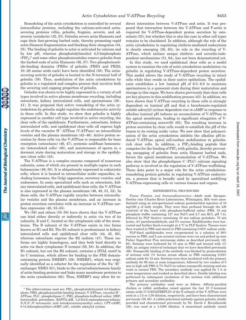
Gelsolin was localized by immunofluorescence in cryostatsections of PLP-fixed epididymis. High levels of gelsolin weredetected in clear cells (Fig. 1A, red), identified by their V-ATPase expression (yellow). In contrast, adjacent principalcells were negative for gelsolin. Higher magnification imagesshow that whereas gelsolin was present throughout the cyto-plasm of clear cells, it was very abundant in their apical mi-crovilli (Fig. 1B, green). Double labeling revealed very strongactin staining in the stereocilia of principal cells, but little or nodetectable actin in clear cell microvilli (Fig. 1B, red), indicating
Actin Cytoskeleton and V-ATPase Recycling8454
by on April 19, 2007
ww
w.jbc.org
Dow
nloaded from
that the actin cytoskeleton is much less organized in the apicalpole of clear cells compared with principal cells. We postulatedthat the low level of actin polymerization in clear cells is at-tributed to their very high gelsolin content. A strong band at�90 kDa corresponding to gelsolin (71) was detected by West-ern blotting (Fig. 1C).
Effect of Jasplakinolide on Net Proton Secretion
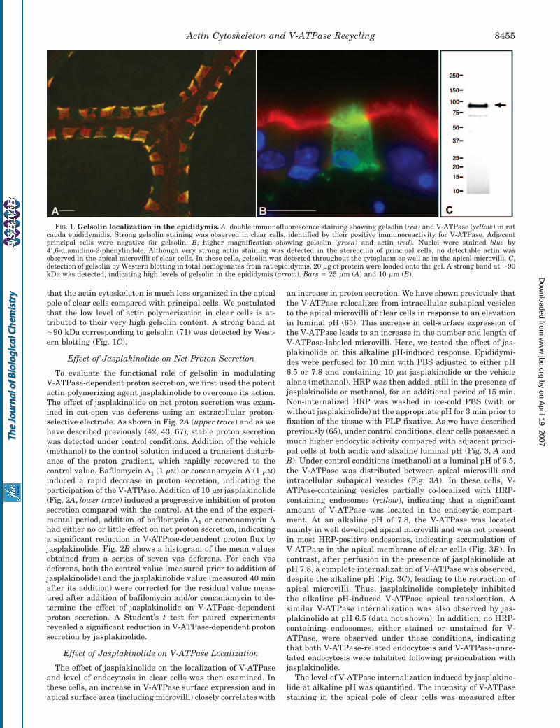
To evaluate the functional role of gelsolin in modulatingV-ATPase-dependent proton secretion, we first used the potentactin polymerizing agent jasplakinolide to overcome its action.The effect of jasplakinolide on net proton secretion was exam-ined in cut-open vas deferens using an extracellular proton-selective electrode. As shown in Fig. 2A (upper trace) and as wehave described previously (42, 43, 67), stable proton secretionwas detected under control conditions. Addition of the vehicle(methanol) to the control solution induced a transient disturb-ance of the proton gradient, which rapidly recovered to thecontrol value. Bafilomycin A1 (1 �M) or concanamycin A (1 �M)induced a rapid decrease in proton secretion, indicating theparticipation of the V-ATPase. Addition of 10 �M jasplakinolide(Fig. 2A, lower trace) induced a progressive inhibition of protonsecretion compared with the control. At the end of the experi-mental period, addition of bafilomycin A1 or concanamycin Ahad either no or little effect on net proton secretion, indicatinga significant reduction in V-ATPase-dependent proton flux byjasplakinolide. Fig. 2B shows a histogram of the mean valuesobtained from a series of seven vas deferens. For each vasdeferens, both the control value (measured prior to addition ofjasplakinolide) and the jasplakinolide value (measured 40 minafter its addition) were corrected for the residual value meas-ured after addition of bafilomycin and/or concanamycin to de-termine the effect of jasplakinolide on V-ATPase-dependentproton secretion. A Student’s t test for paired experimentsrevealed a significant reduction in V-ATPase-dependent protonsecretion by jasplakinolide.
Effect of Jasplakinolide on V-ATPase Localization
The effect of jasplakinolide on the localization of V-ATPaseand level of endocytosis in clear cells was then examined. Inthese cells, an increase in V-ATPase surface expression and inapical surface area (including microvilli) closely correlates with
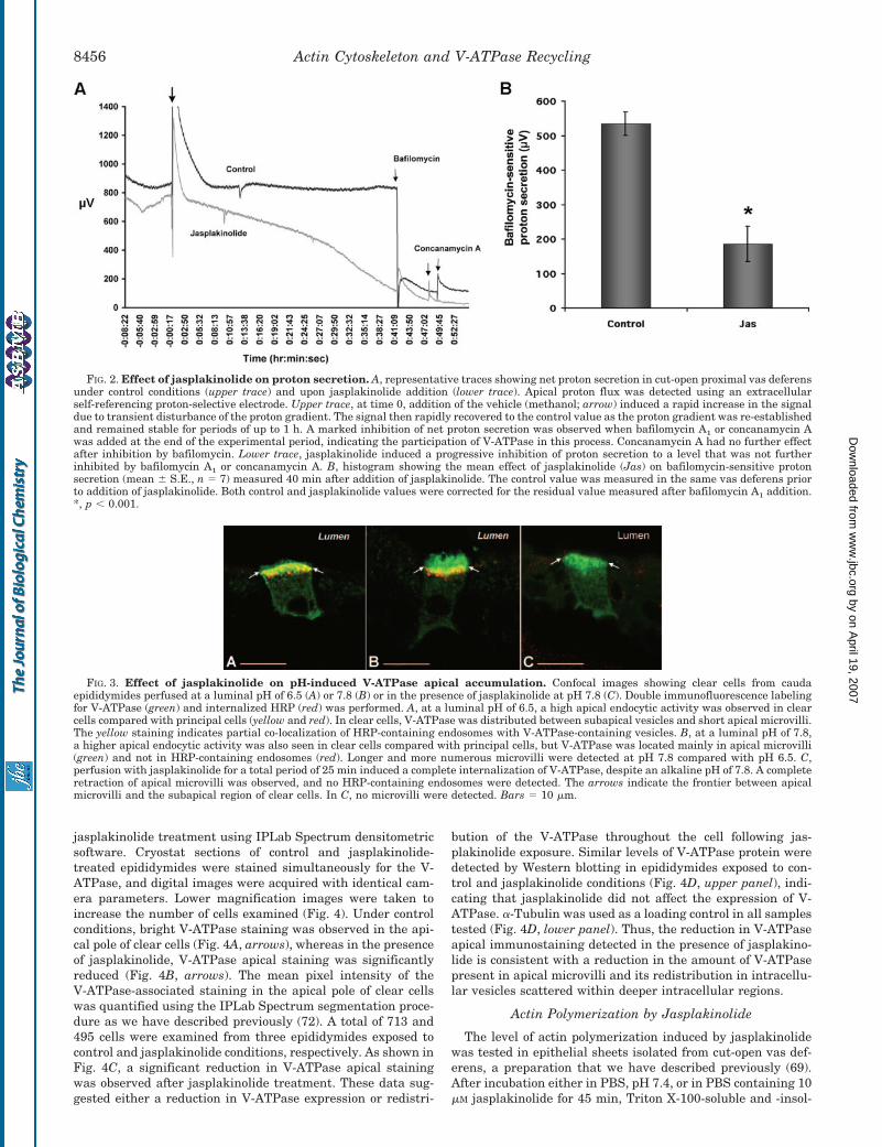
an increase in proton secretion. We have shown previously thatthe V-ATPase relocalizes from intracellular subapical vesiclesto the apical microvilli of clear cells in response to an elevationin luminal pH (65). This increase in cell-surface expression ofthe V-ATPase leads to an increase in the number and length ofV-ATPase-labeled microvilli. Here, we tested the effect of jas-plakinolide on this alkaline pH-induced response. Epididymi-des were perfused for 10 min with PBS adjusted to either pH6.5 or 7.8 and containing 10 �M jasplakinolide or the vehiclealone (methanol). HRP was then added, still in the presence ofjasplakinolide or methanol, for an additional period of 15 min.Non-internalized HRP was washed in ice-cold PBS (with orwithout jasplakinolide) at the appropriate pH for 3 min prior tofixation of the tissue with PLP fixative. As we have describedpreviously (65), under control conditions, clear cells possessed amuch higher endocytic activity compared with adjacent princi-pal cells at both acidic and alkaline luminal pH (Fig. 3, A andB). Under control conditions (methanol) at a luminal pH of 6.5,the V-ATPase was distributed between apical microvilli andintracellular subapical vesicles (Fig. 3A). In these cells, V-ATPase-containing vesicles partially co-localized with HRP-containing endosomes (yellow), indicating that a significantamount of V-ATPase was located in the endocytic compart-ment. At an alkaline pH of 7.8, the V-ATPase was locatedmainly in well developed apical microvilli and was not presentin most HRP-positive endosomes, indicating accumulation ofV-ATPase in the apical membrane of clear cells (Fig. 3B). Incontrast, after perfusion in the presence of jasplakinolide atpH 7.8, a complete internalization of V-ATPase was observed,despite the alkaline pH (Fig. 3C), leading to the retraction ofapical microvilli. Thus, jasplakinolide completely inhibitedthe alkaline pH-induced V-ATPase apical translocation. Asimilar V-ATPase internalization was also observed by jas-plakinolide at pH 6.5 (data not shown). In addition, no HRP-containing endosomes, either stained or unstained for V-ATPase, were observed under these conditions, indicatingthat both V-ATPase-related endocytosis and V-ATPase-unre-lated endocytosis were inhibited following preincubation withjasplakinolide.
The level of V-ATPase internalization induced by jasplakino-lide at alkaline pH was quantified. The intensity of V-ATPasestaining in the apical pole of clear cells was measured after
FIG. 1. Gelsolin localization in the epididymis. A, double immunofluorescence staining showing gelsolin (red) and V-ATPase (yellow) in ratcauda epididymidis. Strong gelsolin staining was observed in clear cells, identified by their positive immunoreactivity for V-ATPase. Adjacentprincipal cells were negative for gelsolin. B, higher magnification showing gelsolin (green) and actin (red). Nuclei were stained blue by4�,6-diamidino-2-phenylindole. Although very strong actin staining was detected in the stereocilia of principal cells, no detectable actin wasobserved in the apical microvilli of clear cells. In these cells, gelsolin was detected throughout the cytoplasm as well as in the apical microvilli. C,detection of gelsolin by Western blotting in total homogenates from rat epididymis. 20 �g of protein were loaded onto the gel. A strong band at �90kDa was detected, indicating high levels of gelsolin in the epididymis (arrow). Bars � 25 �m (A) and 10 �m (B).
Actin Cytoskeleton and V-ATPase Recycling 8455
by on April 19, 2007
ww
w.jbc.org
Dow
nloaded from
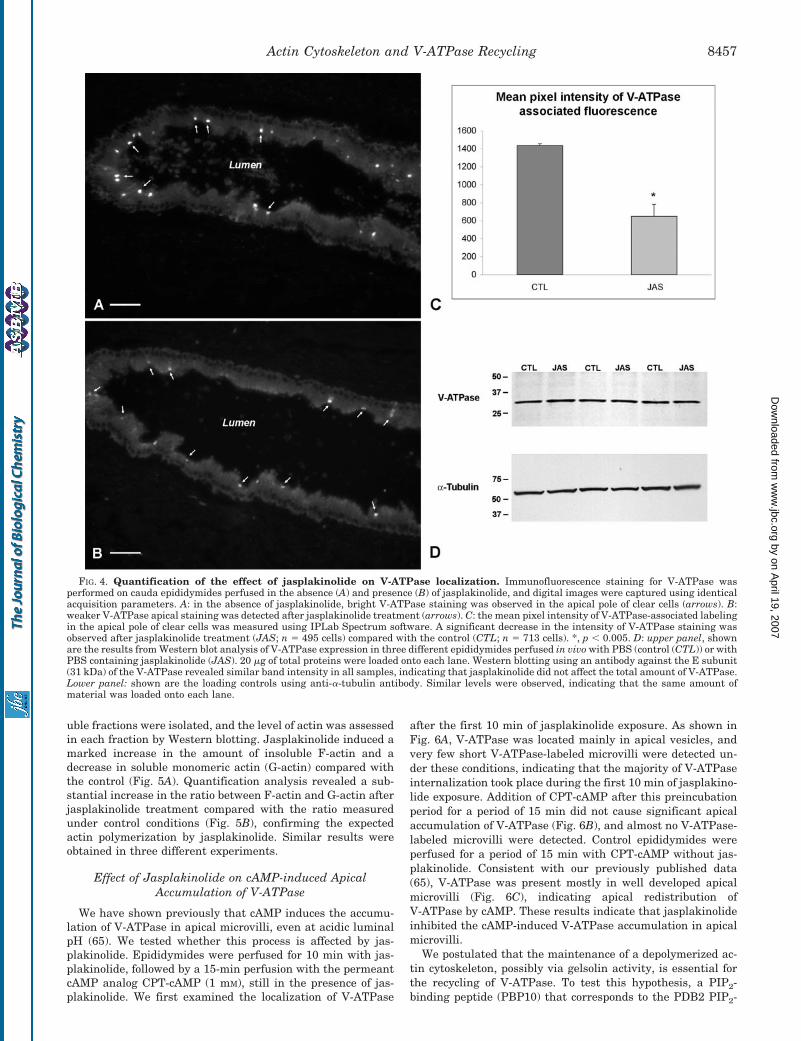
jasplakinolide treatment using IPLab Spectrum densitometricsoftware. Cryostat sections of control and jasplakinolide-treated epididymides were stained simultaneously for the V-ATPase, and digital images were acquired with identical cam-era parameters. Lower magnification images were taken toincrease the number of cells examined (Fig. 4). Under controlconditions, bright V-ATPase staining was observed in the api-cal pole of clear cells (Fig. 4A, arrows), whereas in the presenceof jasplakinolide, V-ATPase apical staining was significantlyreduced (Fig. 4B, arrows). The mean pixel intensity of theV-ATPase-associated staining in the apical pole of clear cellswas quantified using the IPLab Spectrum segmentation proce-dure as we have described previously (72). A total of 713 and495 cells were examined from three epididymides exposed tocontrol and jasplakinolide conditions, respectively. As shown inFig. 4C, a significant reduction in V-ATPase apical stainingwas observed after jasplakinolide treatment. These data sug-gested either a reduction in V-ATPase expression or redistri-
bution of the V-ATPase throughout the cell following jas-plakinolide exposure. Similar levels of V-ATPase protein weredetected by Western blotting in epididymides exposed to con-trol and jasplakinolide conditions (Fig. 4D, upper panel), indi-cating that jasplakinolide did not affect the expression of V-ATPase. �-Tubulin was used as a loading control in all samplestested (Fig. 4D, lower panel). Thus, the reduction in V-ATPaseapical immunostaining detected in the presence of jasplakino-lide is consistent with a reduction in the amount of V-ATPasepresent in apical microvilli and its redistribution in intracellu-lar vesicles scattered within deeper intracellular regions.
Actin Polymerization by Jasplakinolide
The level of actin polymerization induced by jasplakinolidewas tested in epithelial sheets isolated from cut-open vas def-erens, a preparation that we have described previously (69).After incubation either in PBS, pH 7.4, or in PBS containing 10�M jasplakinolide for 45 min, Triton X-100-soluble and -insol-
FIG. 2. Effect of jasplakinolide on proton secretion. A, representative traces showing net proton secretion in cut-open proximal vas deferensunder control conditions (upper trace) and upon jasplakinolide addition (lower trace). Apical proton flux was detected using an extracellularself-referencing proton-selective electrode. Upper trace, at time 0, addition of the vehicle (methanol; arrow) induced a rapid increase in the signaldue to transient disturbance of the proton gradient. The signal then rapidly recovered to the control value as the proton gradient was re-establishedand remained stable for periods of up to 1 h. A marked inhibition of net proton secretion was observed when bafilomycin A1 or concanamycin Awas added at the end of the experimental period, indicating the participation of V-ATPase in this process. Concanamycin A had no further effectafter inhibition by bafilomycin. Lower trace, jasplakinolide induced a progressive inhibition of proton secretion to a level that was not furtherinhibited by bafilomycin A1 or concanamycin A. B, histogram showing the mean effect of jasplakinolide (Jas) on bafilomycin-sensitive protonsecretion (mean � S.E., n � 7) measured 40 min after addition of jasplakinolide. The control value was measured in the same vas deferens priorto addition of jasplakinolide. Both control and jasplakinolide values were corrected for the residual value measured after bafilomycin A1 addition.*, p � 0.001.
FIG. 3. Effect of jasplakinolide on pH-induced V-ATPase apical accumulation. Confocal images showing clear cells from caudaepididymides perfused at a luminal pH of 6.5 (A) or 7.8 (B) or in the presence of jasplakinolide at pH 7.8 (C). Double immunofluorescence labelingfor V-ATPase (green) and internalized HRP (red) was performed. A, at a luminal pH of 6.5, a high apical endocytic activity was observed in clearcells compared with principal cells (yellow and red). In clear cells, V-ATPase was distributed between subapical vesicles and short apical microvilli.The yellow staining indicates partial co-localization of HRP-containing endosomes with V-ATPase-containing vesicles. B, at a luminal pH of 7.8,a higher apical endocytic activity was also seen in clear cells compared with principal cells, but V-ATPase was located mainly in apical microvilli(green) and not in HRP-containing endosomes (red). Longer and more numerous microvilli were detected at pH 7.8 compared with pH 6.5. C,perfusion with jasplakinolide for a total period of 25 min induced a complete internalization of V-ATPase, despite an alkaline pH of 7.8. A completeretraction of apical microvilli was observed, and no HRP-containing endosomes were detected. The arrows indicate the frontier between apicalmicrovilli and the subapical region of clear cells. In C, no microvilli were detected. Bars � 10 �m.
Actin Cytoskeleton and V-ATPase Recycling8456
by on April 19, 2007
ww
w.jbc.org
Dow
nloaded from
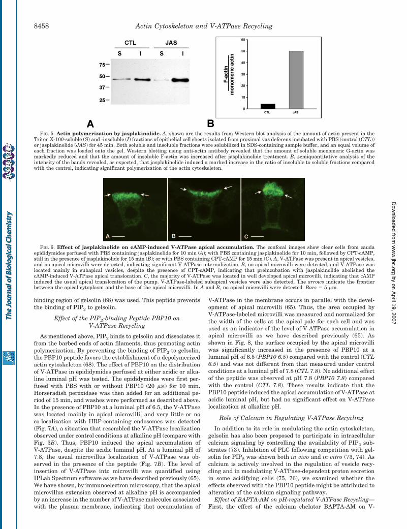
uble fractions were isolated, and the level of actin was assessedin each fraction by Western blotting. Jasplakinolide induced amarked increase in the amount of insoluble F-actin and adecrease in soluble monomeric actin (G-actin) compared withthe control (Fig. 5A). Quantification analysis revealed a sub-stantial increase in the ratio between F-actin and G-actin afterjasplakinolide treatment compared with the ratio measuredunder control conditions (Fig. 5B), confirming the expectedactin polymerization by jasplakinolide. Similar results wereobtained in three different experiments.
Effect of Jasplakinolide on cAMP-induced ApicalAccumulation of V-ATPase
We have shown previously that cAMP induces the accumu-lation of V-ATPase in apical microvilli, even at acidic luminalpH (65). We tested whether this process is affected by jas-plakinolide. Epididymides were perfused for 10 min with jas-plakinolide, followed by a 15-min perfusion with the permeantcAMP analog CPT-cAMP (1 mM), still in the presence of jas-plakinolide. We first examined the localization of V-ATPase
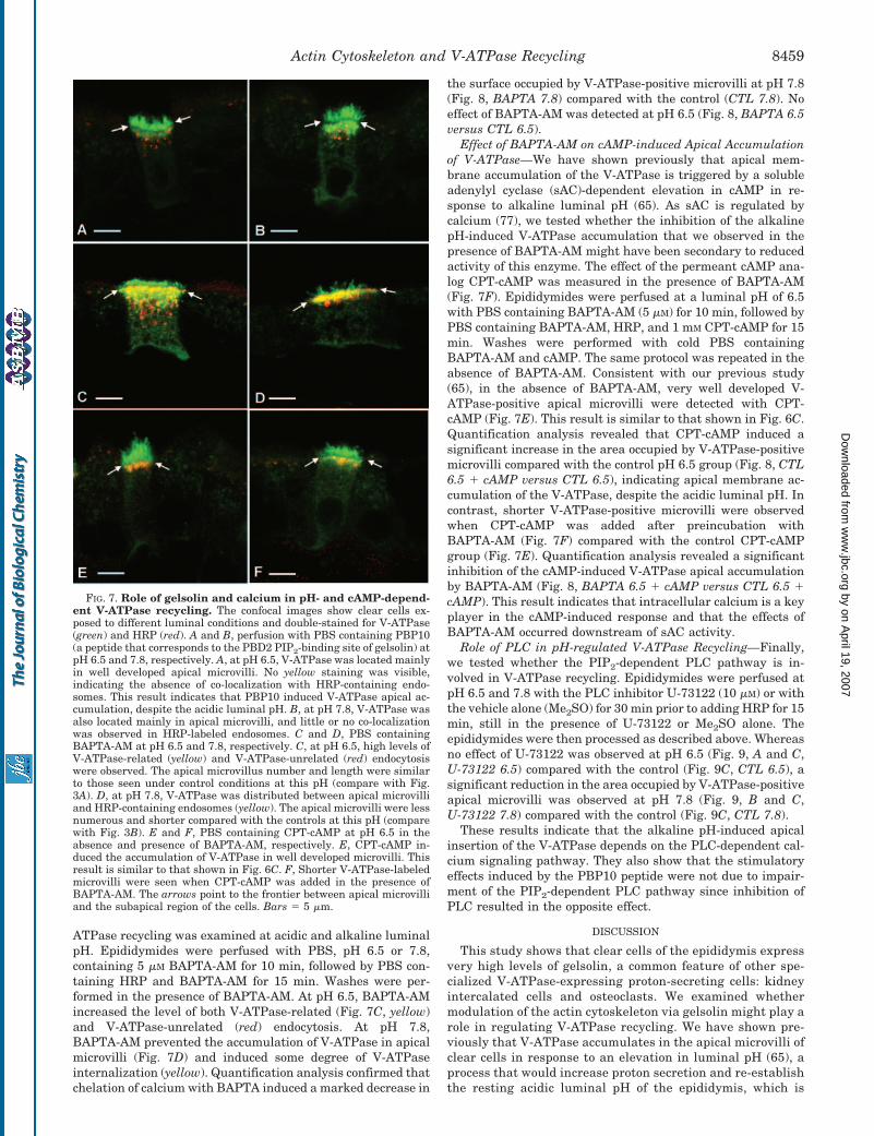
after the first 10 min of jasplakinolide exposure. As shown inFig. 6A, V-ATPase was located mainly in apical vesicles, andvery few short V-ATPase-labeled microvilli were detected un-der these conditions, indicating that the majority of V-ATPaseinternalization took place during the first 10 min of jasplakino-lide exposure. Addition of CPT-cAMP after this preincubationperiod for a period of 15 min did not cause significant apicalaccumulation of V-ATPase (Fig. 6B), and almost no V-ATPase-labeled microvilli were detected. Control epididymides wereperfused for a period of 15 min with CPT-cAMP without jas-plakinolide. Consistent with our previously published data(65), V-ATPase was present mostly in well developed apicalmicrovilli (Fig. 6C), indicating apical redistribution ofV-ATPase by cAMP. These results indicate that jasplakinolideinhibited the cAMP-induced V-ATPase accumulation in apicalmicrovilli.
We postulated that the maintenance of a depolymerized ac-tin cytoskeleton, possibly via gelsolin activity, is essential forthe recycling of V-ATPase. To test this hypothesis, a PIP2-binding peptide (PBP10) that corresponds to the PDB2 PIP2-
FIG. 4. Quantification of the effect of jasplakinolide on V-ATPase localization. Immunofluorescence staining for V-ATPase wasperformed on cauda epididymides perfused in the absence (A) and presence (B) of jasplakinolide, and digital images were captured using identicalacquisition parameters. A: in the absence of jasplakinolide, bright V-ATPase staining was observed in the apical pole of clear cells (arrows). B:weaker V-ATPase apical staining was detected after jasplakinolide treatment (arrows). C: the mean pixel intensity of V-ATPase-associated labelingin the apical pole of clear cells was measured using IPLab Spectrum software. A significant decrease in the intensity of V-ATPase staining wasobserved after jasplakinolide treatment (JAS; n � 495 cells) compared with the control (CTL; n � 713 cells). *, p � 0.005. D: upper panel, shownare the results from Western blot analysis of V-ATPase expression in three different epididymides perfused in vivo with PBS (control (CTL)) or withPBS containing jasplakinolide (JAS). 20 �g of total proteins were loaded onto each lane. Western blotting using an antibody against the E subunit(31 kDa) of the V-ATPase revealed similar band intensity in all samples, indicating that jasplakinolide did not affect the total amount of V-ATPase.Lower panel: shown are the loading controls using anti-�-tubulin antibody. Similar levels were observed, indicating that the same amount ofmaterial was loaded onto each lane.
Actin Cytoskeleton and V-ATPase Recycling 8457
by on April 19, 2007
ww
w.jbc.org
Dow
nloaded from
binding region of gelsolin (68) was used. This peptide preventsthe binding of PIP2 to gelsolin.
Effect of the PIP2-binding Peptide PBP10 onV-ATPase Recycling
As mentioned above, PIP2 binds to gelsolin and dissociates itfrom the barbed ends of actin filaments, thus promoting actinpolymerization. By preventing the binding of PIP2 to gelsolin,the PBP10 peptide favors the establishment of a depolymerizedactin cytoskeleton (68). The effect of PBP10 on the distributionof V-ATPase in epididymides perfused at either acidic or alka-line luminal pH was tested. The epididymides were first per-fused with PBS with or without PBP10 (20 �M) for 10 min.Horseradish peroxidase was then added for an additional pe-riod of 15 min, and washes were performed as described above.In the presence of PBP10 at a luminal pH of 6.5, the V-ATPasewas located mainly in apical microvilli, and very little or noco-localization with HRP-containing endosomes was detected(Fig. 7A), a situation that resembled the V-ATPase localizationobserved under control conditions at alkaline pH (compare withFig. 3B). Thus, PBP10 induced the apical accumulation ofV-ATPase, despite the acidic luminal pH. At a luminal pH of7.8, the usual microvillus localization of V-ATPase was ob-served in the presence of the peptide (Fig. 7B). The level ofinsertion of V-ATPase into microvilli was quantified usingIPLab Spectrum software as we have described previously (65).We have shown, by immunoelectron microscopy, that the apicalmicrovillus extension observed at alkaline pH is accompaniedby an increase in the number of V-ATPase molecules associatedwith the plasma membrane, indicating that accumulation of
V-ATPase in the membrane occurs in parallel with the devel-opment of apical microvilli (65). Thus, the area occupied byV-ATPase-labeled microvilli was measured and normalized forthe width of the cells at the apical pole for each cell and wasused as an indicator of the level of V-ATPase accumulation inapical microvilli as we have described previously (65). Asshown in Fig. 8, the surface occupied by the apical microvilliwas significantly increased in the presence of PBP10 at aluminal pH of 6.5 (PBP10 6.5) compared with the control (CTL6.5) and was not different from that measured under controlconditions at a luminal pH of 7.8 (CTL 7.8). No additional effectof the peptide was observed at pH 7.8 (PBP10 7.8) comparedwith the control (CTL 7.8). These results indicate that thePBP10 peptide induced the apical accumulation of V-ATPase atacidic luminal pH, but had no significant effect on V-ATPaselocalization at alkaline pH.
Role of Calcium in Regulating V-ATPase Recycling
In addition to its role in modulating the actin cytoskeleton,gelsolin has also been proposed to participate in intracellularcalcium signaling by controlling the availability of PIP2 sub-strates (73). Inhibition of PLC following competition with gel-solin for PIP2 was shown both in vivo and in vitro (73, 74). Ascalcium is actively involved in the regulation of vesicle recy-cling and in modulating V-ATPase-dependent proton secretionin some acidifying cells (75, 76), we examined whether theeffects observed with the PBP10 peptide might be attributed toalteration of the calcium signaling pathway.
Effect of BAPTA-AM on pH-regulated V-ATPase Recycling—First, the effect of the calcium chelator BAPTA-AM on V-
FIG. 5. Actin polymerization by jasplakinolide. A, shown are the results from Western blot analysis of the amount of actin present in theTriton X-100-soluble (S) and -insoluble (I) fractions of epithelial cell sheets isolated from proximal vas deferens incubated with PBS (control (CTL))or jasplakinolide (JAS) for 45 min. Both soluble and insoluble fractions were solubilized in SDS-containing sample buffer, and an equal volume ofeach fraction was loaded onto the gel. Western blotting using anti-actin antibody revealed that the amount of soluble monomeric G-actin wasmarkedly reduced and that the amount of insoluble F-actin was increased after jasplakinolide treatment. B, semiquantitative analysis of theintensity of the bands revealed, as expected, that jasplakinolide induced a marked increase in the ratio of insoluble to soluble fractions comparedwith the control, indicating significant polymerization of the actin cytoskeleton.
FIG. 6. Effect of jasplakinolide on cAMP-induced V-ATPase apical accumulation. The confocal images show clear cells from caudaepididymides perfused with PBS containing jasplakinolide for 10 min (A); with PBS containing jasplakinolide for 10 min, followed by CPT-cAMP,still in the presence of jasplakinolide for 15 min (B); or with PBS containing CPT-cAMP for 15 min (C). A, V-ATPase was present in apical vesicles,and no apical microvilli were detected, indicating significant V-ATPase internalization. B, no apical microvilli were detected, and V-ATPase waslocated mainly in subapical vesicles, despite the presence of CPT-cAMP, indicating that preincubation with jasplakinolide abolished thecAMP-induced V-ATPase apical translocation. C, the majority of V-ATPase was located in well developed apical microvilli, indicating that cAMPinduced the usual apical translocation of the pump. V-ATPase-labeled subapical vesicles were also detected. The arrows indicate the frontierbetween the apical cytoplasm and the base of the apical microvilli. In A and B, no apical microvilli were detected. Bars � 5 �m.
Actin Cytoskeleton and V-ATPase Recycling8458
by on April 19, 2007
ww
w.jbc.org
Dow
nloaded from
ATPase recycling was examined at acidic and alkaline luminalpH. Epididymides were perfused with PBS, pH 6.5 or 7.8,containing 5 �M BAPTA-AM for 10 min, followed by PBS con-taining HRP and BAPTA-AM for 15 min. Washes were per-formed in the presence of BAPTA-AM. At pH 6.5, BAPTA-AMincreased the level of both V-ATPase-related (Fig. 7C, yellow)and V-ATPase-unrelated (red) endocytosis. At pH 7.8,BAPTA-AM prevented the accumulation of V-ATPase in apicalmicrovilli (Fig. 7D) and induced some degree of V-ATPaseinternalization (yellow). Quantification analysis confirmed thatchelation of calcium with BAPTA induced a marked decrease in
the surface occupied by V-ATPase-positive microvilli at pH 7.8(Fig. 8, BAPTA 7.8) compared with the control (CTL 7.8). Noeffect of BAPTA-AM was detected at pH 6.5 (Fig. 8, BAPTA 6.5versus CTL 6.5).
Effect of BAPTA-AM on cAMP-induced Apical Accumulationof V-ATPase—We have shown previously that apical mem-brane accumulation of the V-ATPase is triggered by a solubleadenylyl cyclase (sAC)-dependent elevation in cAMP in re-sponse to alkaline luminal pH (65). As sAC is regulated bycalcium (77), we tested whether the inhibition of the alkalinepH-induced V-ATPase accumulation that we observed in thepresence of BAPTA-AM might have been secondary to reducedactivity of this enzyme. The effect of the permeant cAMP ana-log CPT-cAMP was measured in the presence of BAPTA-AM(Fig. 7F). Epididymides were perfused at a luminal pH of 6.5with PBS containing BAPTA-AM (5 �M) for 10 min, followed byPBS containing BAPTA-AM, HRP, and 1 mM CPT-cAMP for 15min. Washes were performed with cold PBS containingBAPTA-AM and cAMP. The same protocol was repeated in theabsence of BAPTA-AM. Consistent with our previous study(65), in the absence of BAPTA-AM, very well developed V-ATPase-positive apical microvilli were detected with CPT-cAMP (Fig. 7E). This result is similar to that shown in Fig. 6C.Quantification analysis revealed that CPT-cAMP induced asignificant increase in the area occupied by V-ATPase-positivemicrovilli compared with the control pH 6.5 group (Fig. 8, CTL6.5 � cAMP versus CTL 6.5), indicating apical membrane ac-cumulation of the V-ATPase, despite the acidic luminal pH. Incontrast, shorter V-ATPase-positive microvilli were observedwhen CPT-cAMP was added after preincubation withBAPTA-AM (Fig. 7F) compared with the control CPT-cAMPgroup (Fig. 7E). Quantification analysis revealed a significantinhibition of the cAMP-induced V-ATPase apical accumulationby BAPTA-AM (Fig. 8, BAPTA 6.5 � cAMP versus CTL 6.5 �cAMP). This result indicates that intracellular calcium is a keyplayer in the cAMP-induced response and that the effects ofBAPTA-AM occurred downstream of sAC activity.
Role of PLC in pH-regulated V-ATPase Recycling—Finally,we tested whether the PIP2-dependent PLC pathway is in-volved in V-ATPase recycling. Epididymides were perfused atpH 6.5 and 7.8 with the PLC inhibitor U-73122 (10 �M) or withthe vehicle alone (Me2SO) for 30 min prior to adding HRP for 15min, still in the presence of U-73122 or Me2SO alone. Theepididymides were then processed as described above. Whereasno effect of U-73122 was observed at pH 6.5 (Fig. 9, A and C,U-73122 6.5) compared with the control (Fig. 9C, CTL 6.5), asignificant reduction in the area occupied by V-ATPase-positiveapical microvilli was observed at pH 7.8 (Fig. 9, B and C,U-73122 7.8) compared with the control (Fig. 9C, CTL 7.8).
These results indicate that the alkaline pH-induced apicalinsertion of the V-ATPase depends on the PLC-dependent cal-cium signaling pathway. They also show that the stimulatoryeffects induced by the PBP10 peptide were not due to impair-ment of the PIP2-dependent PLC pathway since inhibition ofPLC resulted in the opposite effect.
DISCUSSION
This study shows that clear cells of the epididymis expressvery high levels of gelsolin, a common feature of other spe-cialized V-ATPase-expressing proton-secreting cells: kidneyintercalated cells and osteoclasts. We examined whethermodulation of the actin cytoskeleton via gelsolin might play arole in regulating V-ATPase recycling. We have shown pre-viously that V-ATPase accumulates in the apical microvilli ofclear cells in response to an elevation in luminal pH (65), aprocess that would increase proton secretion and re-establishthe resting acidic luminal pH of the epididymis, which is
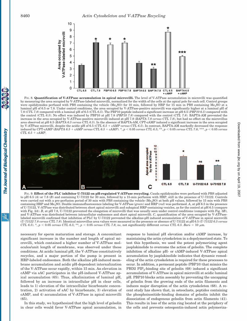
FIG. 7. Role of gelsolin and calcium in pH- and cAMP-depend-ent V-ATPase recycling. The confocal images show clear cells ex-posed to different luminal conditions and double-stained for V-ATPase(green) and HRP (red). A and B, perfusion with PBS containing PBP10(a peptide that corresponds to the PBD2 PIP2-binding site of gelsolin) atpH 6.5 and 7.8, respectively. A, at pH 6.5, V-ATPase was located mainlyin well developed apical microvilli. No yellow staining was visible,indicating the absence of co-localization with HRP-containing endo-somes. This result indicates that PBP10 induced V-ATPase apical ac-cumulation, despite the acidic luminal pH. B, at pH 7.8, V-ATPase wasalso located mainly in apical microvilli, and little or no co-localizationwas observed in HRP-labeled endosomes. C and D, PBS containingBAPTA-AM at pH 6.5 and 7.8, respectively. C, at pH 6.5, high levels ofV-ATPase-related (yellow) and V-ATPase-unrelated (red) endocytosiswere observed. The apical microvillus number and length were similarto those seen under control conditions at this pH (compare with Fig.3A). D, at pH 7.8, V-ATPase was distributed between apical microvilliand HRP-containing endosomes (yellow). The apical microvilli were lessnumerous and shorter compared with the controls at this pH (comparewith Fig. 3B). E and F, PBS containing CPT-cAMP at pH 6.5 in theabsence and presence of BAPTA-AM, respectively. E, CPT-cAMP in-duced the accumulation of V-ATPase in well developed microvilli. Thisresult is similar to that shown in Fig. 6C. F, Shorter V-ATPase-labeledmicrovilli were seen when CPT-cAMP was added in the presence ofBAPTA-AM. The arrows point to the frontier between apical microvilliand the subapical region of the cells. Bars � 5 �m.
Actin Cytoskeleton and V-ATPase Recycling 8459
by on April 19, 2007
ww
w.jbc.org
Dow
nloaded from
necessary for sperm maturation and storage. A concomitantsignificant increase in the number and length of apical mi-crovilli, which contained a higher number of V-ATPase mol-ecules/unit length of membrane, was observed under theseconditions. At acidic luminal pH, the V-ATPase constitutivelyrecycles, and a major portion of the pump is present inHRP-labeled endosomes. Both the alkaline pH-induced mem-brane accumulation and acidic pH-dependent internalizationof the V-ATPase occur rapidly, within 15 min. An elevation incAMP via sAC participates in the pH-induced V-ATPase ap-ical accumulation (65). Thus, alkalinization of luminal pH,followed by an increase in intracellular pH in clear cells,leads to 1) elevation of the intracellular bicarbonate concen-tration, 2) activation of sAC by bicarbonate, 3) elevation ofcAMP, and 4) accumulation of V-ATPase in apical microvilli(65).
In this study, we hypothesized that the high level of gelsolinin clear cells would favor V-ATPase apical accumulation, in
response to luminal pH elevation and/or cAMP increase, bymaintaining the actin cytoskeleton in a depolymerized state. Totest this hypothesis, we used the potent polymerizing agentjasplakinolide to overcome the action of gelsolin. The completeinhibition of alkaline pH- or cAMP-induced V-ATPase apicalaccumulation by jasplakinolide indicates that dynamic remod-eling of the actin cytoskeleton is required for these processes tooccur. In addition, a permeant peptide (PBP10) that mimics thePBD2 PIP2-binding site of gelsolin (68) induced a significantaccumulation of V-ATPase in apical microvilli at acidic luminalpH. PBP10 blocks actin assembly by preventing the uncappingof gelsolin from the growing ends of the actin filaments andinduces major disruption of the actin cytoskeleton (68). A re-cent study has shown that, in osteoclasts, peptides containingthe phosphoinositide-binding domains of gelsolin inhibit thedissociation of endogenous gelsolin from actin filaments (41).This results in loss of the actin ring located at the periphery ofthe cells and prevents osteopontin-induced actin polymeriza-
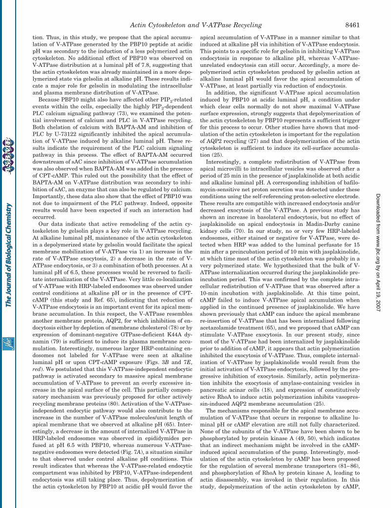
FIG. 8. Quantification of V-ATPase accumulation in apical microvilli. The level of V-ATPase accumulation in microvilli was quantifiedby measuring the area occupied by V-ATPase-labeled microvilli, normalized for the width of the cells at the apical pole for each cell. Control groupswere epididymides perfused with PBS containing the vehicle (Me2SO) for 10 min, followed by HRP for 15 min in PBS containing Me2SO at aluminal pH of 6.5 or 7.8. Under control conditions, the area occupied by V-ATPase-positive microvilli was significantly higher at a luminal pH of7.8 (CTL 7.8) compared with a luminal pH of 6.5 (CTL 6.5). The PBP10 peptide induced a significant increase at pH 6.5 (PBP10 6.5) compared withthe control (CTL 6.5). No effect was induced by PBP10 at pH 7.8 (PBP10 7.8) compared with the control (CTL 7.8). BAPTA-AM prevented theincrease in the area occupied by V-ATPase-positive microvilli induced at pH 7.8 (BAPTA 7.8 versus CTL 7.8), but had no effect on the microvillusarea observed at pH 6.5 (BAPTA 6.5 versus CTL 6.5). In the absence of BAPTA-AM, CPT-cAMP induced a significant increase in the area occupiedby V-ATPase microvilli, despite the acidic pH of 6.5 (CTL 6.5 � cAMP versus CTL 6.5). In contrast, BAPTA-AM markedly decreased the responseinduced by CPT-cAMP (BAPTA 6.5 � cAMP versus CTL 6.5 � cAMP). *, p � 0.05 versus CTL 6.5; **, p � 0.05 versus CTL 7.8; ***, p � 0.05 versusCTL 6.5 � cAMP.
FIG. 9. Effect of the PLC inhibitor U-73122 on pH-regulated V-ATPase recycling. Cauda epididymides were perfused with PBS adjustedto pH 6.5 (A) or 7.8 (B) and containing U-73122 for 30 min, followed by a 15-min perfusion with HRP, still in the presence of U-73122. Controlswere carried out with a pre-perfusion period of 30 min with PBS containing the vehicle (Me2SO) at both pH values, followed by 15 min with PBScontaining HRP and Me2SO. Double immunofluorescence labeling for V-ATPase (green) and HRP (red) was performed. A, at pH 6.5 in the presenceof U-73122, V-ATPase was distributed between short apical microvilli and subapical HRP-containing vesicles, as for the control at pH 6.5 (comparewith Fig. 3A). B, at pH 7.8, U-73122 prevented the elongation of microvilli that was usually seen under control conditions (compare with Fig. 3B),and V-ATPase was distributed between intracellular endosomes and short apical microvilli. C, quantification of the area occupied by V-ATPase-labeled microvilli confirmed that inhibition of PLC by U-73122 prevented the alkaline-pH induced accumulation of V-ATPase in apical microvilli(U-73122 7.8 versus CTL 7.8). Identical microvillus area values were measured in the presence or absence of U-73122 at pH 6.5 (U-73122 6.5 versusCTL 6.5). *, p � 0.05 versus CTL 6.5; **, p � 0.05 versus CTL 7.8; ns, not significantly different versus CTL 6.5. Bars � 10 �m.
Actin Cytoskeleton and V-ATPase Recycling8460
by on April 19, 2007
ww
w.jbc.org
Dow
nloaded from
tion. Thus, in this study, we propose that the apical accumu-lation of V-ATPase generated by the PBP10 peptide at acidicpH was secondary to the induction of a less polymerized actincytoskeleton. No additional effect of PBP10 was observed onV-ATPase distribution at a luminal pH of 7.8, suggesting thatthe actin cytoskeleton was already maintained in a more depo-lymerized state via gelsolin at alkaline pH. These results indi-cate a major role for gelsolin in modulating the intracellularand plasma membrane distribution of V-ATPase.
Because PBP10 might also have affected other PIP2-relatedevents within the cells, especially the highly PIP2-dependentPLC calcium signaling pathway (73), we examined the poten-tial involvement of calcium and PLC in V-ATPase recycling.Both chelation of calcium with BAPTA-AM and inhibition ofPLC by U-73122 significantly inhibited the apical accumula-tion of V-ATPase induced by alkaline luminal pH. These re-sults indicate the requirement of the PLC calcium signalingpathway in this process. The effect of BAPTA-AM occurreddownstream of sAC since inhibition of V-ATPase accumulationwas also observed when BAPTA-AM was added in the presenceof CPT-cAMP. This ruled out the possibility that the effect ofBAPTA-AM on V-ATPase distribution was secondary to inhi-bition of sAC, an enzyme that can also be regulated by calcium.Importantly, these data also show that the effect of PBP10 wasnot due to impairment of the PLC pathway. Indeed, oppositeresults would have been expected if such an interaction hadoccurred.
Our data indicate that active remodeling of the actin cy-toskeleton by gelsolin plays a key role in V-ATPase recycling.At alkaline luminal pH, maintenance of the actin cytoskeletonin a depolymerized state by gelsolin would facilitate the apicalmembrane mobilization of V-ATPase via 1) an increase in therate of V-ATPase exocytosis, 2) a decrease in the rate of V-ATPase endocytosis, or 3) a combination of both processes. At aluminal pH of 6.5, these processes would be reversed to facili-tate internalization of the V-ATPase. Very little co-localizationof V-ATPase with HRP-labeled endosomes was observed undercontrol conditions at alkaline pH or in the presence of CPT-cAMP (this study and Ref. 65), indicating that reduction ofV-ATPase endocytosis is an important event for its apical mem-brane accumulation. In this respect, the V-ATPase resemblesanother membrane protein, AQP2, for which inhibition of en-docytosis either by depletion of membrane cholesterol (78) or byexpression of dominant-negative GTPase-deficient K44A dy-namin (79) is sufficient to induce its plasma membrane accu-mulation. Interestingly, numerous larger HRP-containing en-dosomes not labeled for V-ATPase were seen at alkalineluminal pH or upon CPT-cAMP exposure (Figs. 3B and 7E,red). We postulated that this V-ATPase-independent endocyticpathway is activated secondary to massive apical membraneaccumulation of V-ATPase to prevent an overly excessive in-crease in the apical surface of the cell. This partially compen-satory mechanism was previously proposed for other activelyrecycling membrane proteins (80). Activation of the V-ATPase-independent endocytic pathway would also contribute to theincrease in the number of V-ATPase molecules/unit length ofapical membrane that we observed at alkaline pH (65). Inter-estingly, a decrease in the amount of internalized V-ATPase inHRP-labeled endosomes was observed in epididymides per-fused at pH 6.5 with PBP10, whereas numerous V-ATPase-negative endosomes were detected (Fig. 7A), a situation similarto that observed under control alkaline pH conditions. Thisresult indicates that whereas the V-ATPase-related endocyticcompartment was inhibited by PBP10, V-ATPase-independentendocytosis was still taking place. Thus, depolymerization ofthe actin cytoskeleton by PBP10 at acidic pH would favor the
apical accumulation of V-ATPase in a manner similar to thatinduced at alkaline pH via inhibition of V-ATPase endocytosis.This points to a specific role for gelsolin in inhibiting V-ATPaseendocytosis in response to alkaline pH, whereas V-ATPase-unrelated endocytosis can still occur. Accordingly, a more de-polymerized actin cytoskeleton produced by gelsolin action atalkaline luminal pH would favor the apical accumulation ofV-ATPase, at least partially via reduction of endocytosis.
In addition, the significant V-ATPase apical accumulationinduced by PBP10 at acidic luminal pH, a condition underwhich clear cells normally do not show maximal V-ATPasesurface expression, strongly suggests that depolymerization ofthe actin cytoskeleton by PBP10 represents a sufficient triggerfor this process to occur. Other studies have shown that mod-ulation of the actin cytoskeleton is important for the regulationof AQP2 recycling (27) and that depolymerization of the actincytoskeleton is sufficient to induce its cell-surface accumula-tion (25).
Interestingly, a complete redistribution of V-ATPase fromapical microvilli to intracellular vesicles was observed after aperiod of 25 min in the presence of jasplakinolide at both acidicand alkaline luminal pH. A corresponding inhibition of bafilo-mycin-sensitive net proton secretion was detected under theseconditions using the self-referencing proton-selective electrode.These results are compatible with increased endocytosis and/ordecreased exocytosis of the V-ATPase. A previous study hasshown an increase in basolateral endocytosis, but no effect ofjasplakinolide on apical endocytosis in Madin-Darby caninekidney cells (70). In our study, no or very few HRP-labeledendosomes, either stained or negative for V-ATPase, were de-tected when HRP was added to the luminal perfusate for 15min after a preincubation period of 10 min with jasplakinolide,at which time most of the actin cytoskeleton was probably in avery polymerized state. We hypothesized that the bulk of V-ATPase internalization occurred during the jasplakinolide pre-incubation period. This was confirmed by the complete intra-cellular redistribution of V-ATPase that was observed after a10-min incubation with jasplakinolide. At this time point,cAMP failed to induce V-ATPase apical accumulation whenapplied in the continued presence of jasplakinolide. We haveshown previously that cAMP can induce the apical membranere-insertion of V-ATPase that has been internalized followingacetazolamide treatment (65), and we proposed that cAMP canstimulate V-ATPase exocytosis. In our present study, sincemost of the V-ATPase had been internalized by jasplakinolideprior to addition of cAMP, it appears that actin polymerizationinhibited the exocytosis of V-ATPase. Thus, complete internal-ization of V-ATPase by jasplakinolide would result from theinitial activation of V-ATPase endocytosis, followed by the pro-gressive inhibition of exocytosis. Similarly, actin polymeriza-tion inhibits the exocytosis of amylase-containing vesicles inpancreatic acinar cells (18), and expression of constitutivelyactive RhoA to induce actin polymerization inhibits vasopres-sin-induced AQP2 membrane accumulation (25).
The mechanisms responsible for the apical membrane accu-mulation of V-ATPase that occurs in response to alkaline lu-minal pH or cAMP elevation are still not fully characterized.None of the subunits of the V-ATPase have been shown to bephosphorylated by protein kinase A (49, 50), which indicatesthat an indirect mechanism might be involved in the cAMP-induced apical accumulation of the pump. Interestingly, mod-ulation of the actin cytoskeleton by cAMP has been proposedfor the regulation of several membrane transporters (81–86),and phosphorylation of RhoA by protein kinase A, leading toactin disassembly, was invoked in their regulation. In thisstudy, depolymerization of the actin cytoskeleton by cAMP,
Actin Cytoskeleton and V-ATPase Recycling 8461
by on April 19, 2007
ww
w.jbc.org
Dow
nloaded from
leading to inhibition of V-ATPase endocytosis, would be com-patible with the apical accumulation of V-ATPase induced bycAMP. Thus, cAMP would exert its action via both an increasein V-ATPase exocytosis and a decrease in V-ATPase endocyto-sis. Whether cAMP acts via modulation of the actin cytoskele-ton or via an independent pathway or whether both processestake place still remains to be elucidated.
Our results point to a significant role for gelsolin in regulat-ing V-ATPase recycling via modulation of the actin cytoskele-ton. The severing activity of gelsolin is strongly dependent onintracellular calcium (37, 87, 88). Although high calcium con-centrations were initially reported to be required for gelsolinactivation, a conformational switch that reduces considerablythe required concentrations of calcium to nanomolar rangeshas recently been described in villin (89), a protein homologousto gelsolin (90, 91). It was proposed that both villin and gelsolinare maintained in an autoinhibited conformation and that,upon tyrosine phosphorylation, a conformational modificationoccurs that reveals high affinity sites for calcium. This model isin agreement with our results showing the absolute require-ment of calcium for the alkaline pH-induced accumulation ofV-ATPase. To determine whether tyrosine phosphorylation isinvolved in this process will require further studies. The cal-cium requirement of gelsolin was shown to depend on pH (92),but the pH necessary to regulate gelsolin in vitro is very low(pH �6.0) and is unlikely to be reached intracellularly.
We (59) and others (55–58) have shown that the V-ATPaseinteracts directly or indirectly with actin. V-ATPase binds toF-actin, but not to G-actin, and in osteoclasts, internalization ofV-ATPase is correlated with increased interaction with actin(56). We are currently investigating the possibility that theinteraction between V-ATPase and actin can also be modulatedin clear cells.
In conclusion, this study shows high expression of gelsolin inclear cells of the epididymis and that modulation of the actincytoskeleton by gelsolin plays a key role in the regulation ofV-ATPase surface expression. As gelsolin is also expressed inother cells expressing the V-ATPase in their plasma mem-brane, including kidney intercalated cells and osteoclasts, wepropose that modulation of the actin cytoskeleton by this sev-ering and capping protein represents a common mechanism bywhich these cells can regulate their rate of proton secretion.
REFERENCES
1. Mitchison, T. J., and Cramer, L. P. (1996) Cell 84, 371–3792. Hartwig, J. H., and Kwiatkowski, D. J. (1991) Curr. Opin. Cell Biol. 3, 87–973. Lauffenburger, D. A., and Horwitz, A. F. (1996) Cell 84, 359–3694. Blanchoin, L., Amann, K. J., Higgs, H. N., Marchand, J. B., Kaiser, D. A., and
Pollard, T. D. (2000) Nature 404, 1007–10115. Machesky, L. M., and Insall, R. H. (1999) J. Cell Biol. 146, 267–2726. Cunningham, C. C., Stossel, T. P., and Kwiatkowski, D. J. (1991) Science 251,
1233–12367. Wong, G. K., Allen, P. G., and Begg, D. A. (1997) Cell Motil. Cytoskeleton 36,
30–428. Pavalko, F. M., and Otey, C. A. (1994) Proc. Soc. Exp. Biol. Med. 205, 282–2939. Bernstein, B. W., DeWit, M., and Bamburg, J. R. (1998) Mol. Brain Res. 53,
236–25110. Lang, T., Wacker, I., Wunderlich, I., Rohrbach, A., Giese, G., Soldati, T., and
Almers, W. (2000) Biophys. J. 78, 2863–287711. Kjeken, R., Egeberg, M., Habermann, A., Kuehnel, M., Peyron, P., Floeten-
meyer, M., Walther, P., Jahraus, A., Defacque, H., Kuznetsov, S. A., andGriffiths, G. (2004) Mol. Biol. Cell 15, 345–358
12. Hartwig, J. H., Brown, D., Ausiello, D. A., Stossel, T. P., and Orci, L. (1990)J. Histochem. Cytochem. 38, 1145–1153
13. Orci, L., Gabbay, K. H., and Malaisse, W. J. (1972) Science 175, 1128–113014. Fath, K. R., Mamajiwalla, S. N., and Burgess, D. R. (1993) J. Cell Sci. Suppl.
17, 65–7315. Durrbach, A., Louvard, D., and Coudrier, E. (1996) J. Cell Sci. 109, 457–46516. Riezman, H., Munn, A., Geli, M. I., and Hicke, L. (1996) Experientia (Basel) 52,
1033–104117. Buss, F., Luzio, J. P., and Kendrick-Jones, J. (2001) FEBS Lett. 508, 295–29918. Muallem, S., Kwiatkowska, K., Xu, X., and Yin, H. L. (1995) J. Cell Biol. 128,
589–59819. Brown, D. (2003) Am. J. Physiol. 284, F893–F90120. Apodaca, G. (2001) Traffic 2, 149–15921. Hoekstra, D., Tyteca, D., and van IJzendoorn, S. C. (2004) J. Cell Sci. 117,
2183–2192S. C. D.22. Sontag, J. M., Aunis, D., and Bader, M. F. (1988) Eur. J. Cell Biol. 46, 316–32623. Morita, K., Oka, M., and Hamano, S. (1988) Biochem. Pharmacol. 37,
3357–335924. Brozinick, J. T., Jr., Hawkins, E. D., Strawbridge, A. B., and Elmendorf, J. S.
(2004) J. Biol. Chem. 279, 40699–4070625. Klussmann, E., Tamma, G., Lorenz, D., Wiesner, B., Maric, K., Hofmann, F.,
Aktories, K., Valenti, G., and Rosenthal, W. (2001) J. Biol. Chem. 276,20451–20457
26. Ausiello, D. A., Hartwig, J., and Brown, D. (1987) Soc. Gen. Physiol. Ser. 42,259–275
27. Hays, R. M., Condeelis, J., Gao, Y., Simon, H., Ding, G., and Franki, N. (1993)Pediatr. Nephrol. 7, 672–679
28. Dibas, A., Mia, A., and Yorio, T. (2000) Proc. Soc. Exp. Biol. Med. 223, 203–20929. Gottlieb, T. A., Ivanov, I. E., Adesnik, M., and Sabatini, D. D. (1993) J. Cell
Biol. 120, 695–71030. Jackman, M. R., Shurety, W., Ellis, J. A., and Luzio, J. P. (1994) J. Cell Sci.
107, 2547–255631. Fujimoto, L. M., Roth, R., Heuser, J. E., and Schmid, S. L. (2000) Traffic 1,
161–17132. Janmey, P. A. (1994) Annu. Rev. Physiol. 56, 169–19133. Dos Remedios, C. G., Chhabra, D., Kekic, M., Dedova, I. V., Tsubakihara, M.,
Berry, D. A., and Nosworthy, N. J. (2003) Physiol. Rev. 83, 433–473I. V.34. Kwiatkowski, D. J. (1999) Curr. Opin. Cell Biol. 11, 103–10835. Azuma, T., Witke, W., Stossel, T. P., Hartwig, J. H., and Kwiatkowski, D. J.
(1998) EMBO J. 17, 1362–137036. Hartwig, J. H., Bokoch, G. M., Carpenter, C. L., Janmey, P. A., Taylor, L. A.,
Toker, A., and Stossel, T. P. (1995) Cell 82, 643–65337. Janmey, P. A., and Stossel, T. P. (1987) Nature 325, 362–36438. Kwiatkowski, D. J., Janmey, P. A., and Yin, H. L. (1989) J. Cell Biol. 108,
1717–172639. Lueck, A., Brown, D., and Kwiatkowski, D. J. (1998) J. Cell Sci. 111,
3633–364340. Cabello-Agueros, J. F., Hernandez-Gonzalez, E. O., and Mujica, A. (2003) Cell
Motil. Cytoskeleton 56, 94–10841. Biswas, R. S., Baker, D. A., Hruska, K. A., and Chellaiah, M. A. (2004) BMC
Cell Biol. http://www.biomedcentral.com/1471-2121/5/1942. Breton, S., Nsumu, N. N., Galli, T., Sabolic, I., Smith, P. J., and Brown, D.
(2000) Am. J. Physiol. 278, F717–F72543. Breton, S., Smith, P. J., Lui, B., and Brown, D. (1996) Nat. Med. 2, 470–47244. Brown, D., Lui, B., Gluck, S., and Sabolic, I. (1992) Am. J. Physiol. 263,
C913–C91645. Brown, D., Hirsch, S., and Gluck, S. (1988) J. Clin. Investig. 82, 2114–212646. Gluck, S. L., Lee, B. S., Wang, S. P., Underhill, D., Nemoto, J., and Holliday,
L. S. (1998) Acta Physiol. Scand. Suppl. 643, 203–21247. Lee, B. S., Holliday, L. S., Ojikutu, B., Krits, I., and Gluck, S. L. (1996) Am. J.
Physiol. 270, C382–C38848. Brown, D., and Breton, S. (2000) J. Exp. Biol. 203, 137–14549. Wagner, C. A., Finberg, K. E., Breton, S., Marshansky, V., Brown, D., and
Geibel, J. P. (2004) Physiol. Rev. 84, 1263–131450. Sun-Wada, G. H., Wada, Y., and Futai, M. (2004) Biochim. Biophys. Acta 1658,
106–11451. Brown, D., and Breton, S. (1996) J. Exp. Biol. 199, 2345–235852. Toyomura, T., Oka, T., Yamaguchi, C., Wada, Y., and Futai, M. (2000) J. Biol.
Chem. 275, 8760–876553. Madsen, K. M., Verlander, J. W., Kim, J., and Tisher, C. C. (1991) Kidney Int.
Suppl. 33, S57–S6354. Steinmetz, P. R. (1986) Am. J. Physiol. 251, F173–F18755. Lee, B. S., Gluck, S. L., and Holliday, L. S. (1999) J. Biol. Chem. 274,
29164–2917156. Chen, S. H., Bubb, M. R., Yarmola, E. G., Zuo, J., Jiang, J., Lee, B. S., Lu, M.,
Gluck, S. L., Hurst, I. R., and Holliday, L. S. (2004) J. Biol. Chem. 279,7988–7998
57. Vitavska, O., Wieczorek, H., and Merzendorfer, H. (2003) J. Biol. Chem. 278,18499–18505
58. Holliday, L. S., Lu, M., Lee, B. S., Nelson, R. D., Solivan, S., Zhang, L., andGluck, S. L. (2000) J. Biol. Chem. 275, 32331–32337
59. Breton, S., Wiederhold, T., Marshansky, V., Nsumu, N. N., Ramesh, V., andBrown, D. (2000) J. Biol. Chem. 275, 18219–18224
60. Paunescu, T. G., Da Silva, N., Marshansky, V., McKee, M., Breton, S., andBrown, D. (2004) Am. J. Physiol. 287, C149–C162
61. Weinman, E. J., Steplock, D., Tate, K., Hall, R. A., Spurney, R. F., andShenolikar, S. (1998) J. Clin. Investig. 101, 2199–2206
62. Shenolikar, S., and Weinman, E. J. (2001) Am. J. Physiol. 280, F389–F39563. Guilherme, A., Soriano, N. A., Bose, S., Holik, J., Bose, A., Pomerleau, D. P.,
Furcinitti, P., Leszyk, J., Corvera, S., and Czech, M. P. (2004) J. Biol. Chem.279, 10593–10605
64. Breton, S., Lisanti, M. P., Tyszkowski, R., McLaughlin, M., and Brown, D.(1998) J. Histochem. Cytochem. 46, 205–214
65. Pastor-Soler, N., Beaulieu, V., Litvin, T. N., Da Silva, N., Chen, Y., Brown, D.,Buck, J., Levin, L. R., and Breton, S. (2003) J. Biol. Chem. 278,49523–49529
66. Brown, D., Lydon, J., McLaughlin, M., Stuart-Tilley, A., Tyszkowski, R., andAlper, S. (1996) Histochem. Cell Biol. 105, 261–267
67. Breton, S., Hammar, K., Smith, P. J. S., and Brown, D. (1998) Am. J. Physiol.275, C1134–C1142
68. Cunningham, C. C., Vegners, R., Bucki, R., Funaki, M., Korde, N., Hartwig,J. H., Stossel, T. P., and Janmey, P. A. (2001) J. Biol. Chem. 276,43390–43399
69. Stevens, A. L., Breton, S., Gustafson, C. E., Bouley, R., Nelson, R. D., Kohan,D. E., and Brown, D. (2000) Am. J. Physiol. 278, C791–C802
70. Shurety, W., Stewart, N. L., and Stow, J. L. (1998) Mol. Biol. Cell 9, 957–97571. Kwiatkowski, D. J., Stossel, T. P., Orkin, S. H., Mole, J. E., Colten, H. R., and
Actin Cytoskeleton and V-ATPase Recycling8462
by on April 19, 2007
ww
w.jbc.org
Dow
nloaded from
Yin, H. L. (1986) Nature 323, 455–45872. Pastor-Soler, N., Isnard-Bagnis, C., Herak-Kramberger, C., Sabolic, I., Van
Hoek, A., Brown, D., and Breton, S. (2002) Biol. Reprod. 66, 1716–172273. Sun, H., Lin, K., and Yin, H. L. (1997) J. Cell Biol. 138, 811–82074. Banno, Y., Nakashima, T., Kumada, T., Ebisawa, K., Nonomura, Y., and
Nozawa, Y. (1992) J. Biol. Chem. 267, 6488–649475. Schwartz, J. H., Masino, S. A., Nichols, R. D., and Alexander, E. A. (1994)
Am. J. Physiol. 266, F94–F10176. Cannon, C., van Adelsberg, J., Kelly, S., and Al-Awqati, Q. (1985) Nature 314,
443–44677. Litvin, T. N., Kamenetsky, M., Zarifyan, A., Buck, J., and Levin, L. R. (2003)
J. Biol. Chem. 278, 15922–1592678. Lu, H., Sun, T. X., Bouley, R., Blackburn, K., McLaughlin, M., and Brown, D.
(2004) Am. J. Physiol. 286, F233–F24379. Sun, T. X., Van Hoek, A., Huang, Y., Bouley, R., McLaughlin, M., and Brown,
D. (2002) Am. J. Physiol. 282, F998–F101180. Gundelfinger, E. D., Kessels, M. M., and Qualmann, B. (2003) Nat. Rev. Mol.
Cell Biol. 4, 127–13981. Szaszi, K., Kurashima, K., Kaibuchi, K., Grinstein, S., and Orlowski, J. (2001)
J. Biol. Chem. 276, 40761–40768
82. Shapiro, M., Matthews, J., Hecht, G., Delp, C., and Madara, J. L. (1991)J. Clin. Investig. 87, 1903–1909
83. Wu, M. S., Bens, M., Cluzeaud, F., and Vandewalle, A. (1994) J. Membr. Biol.142, 323–336
84. Reshkin, S. J., and Murer, H. (1992) Am. J. Physiol. 262, F572–F57785. Prat, A. G., Cunningham, C. C., Jackson, G. R., Jr., Borkan, S. C., Wang, Y.,
Ausiello, D. A., and Cantiello, H. F. (1999) Am. J. Physiol. 277,C1160–C1169
86. Tamma, G., Klussmann, E., Procino, G., Svelto, M., Rosenthal, W., and Val-enti, G. (2003) J. Cell Sci. 116, 1519–1525
87. Lin, K. M., Wenegieme, E., Lu, P. J., Chen, C. S., and Yin, H. L. (1997) J. Biol.Chem. 272, 20443–20450
88. Yin, H. L., and Stossel, T. P. (1979) Nature 281, 583–58689. Kumar, N., and Khurana, S. (2004) J. Biol. Chem. 279, 24915–2491890. Arpin, M., Pringault, E., Finidori, J., Garcia, A., Jeltsch, J. M., Vandekerck-
hove, J., and Louvard, D. (1988) J. Cell Biol. 107, 1759–176691. Finidori, J., Friederich, E., Kwiatkowski, D. J., and Louvard, D. (1992) J. Cell
Biol. 116, 1145–115592. Lamb, J. A., Allen, P. G., Tuan, B. Y., and Janmey, P. A. (1993) J. Biol. Chem.
268, 8999–9004
Actin Cytoskeleton and V-ATPase Recycling 8463
by on April 19, 2007
ww
w.jbc.org
Dow
nloaded from