Embed Size (px)
Citation preview
THE JOURNAL OF BIOLCGICAL. CHEMISTRY 0 1994 by The American Society for Biochemistry and Molecular Biology, Inc
Vol. 269, No. 14, Issue of April 8, pp. 10370-10377, 1994 Printed in U.S.A.
Mutational Analysis of the Traffic ATPase (ABC) Transporters Involved in Uptake of Eye Pigment Precursors in Drosophila melanogaster IMPLICATIONS FOR STRUCTURE-FUNCTION RELATIONSHIPS*
(Received for publication, November 17, 1993)
Gary D. EwartSI, David CannellS5, Graeme B. Cox*, and Anthony J. Howells5 From the $Division of Biochemistry and Molecular Biology, John Curtin School of Medical Research and the §Division of Biochemistry and Molecular Biology, Faculty of Science, The Australian National University, P 0. Box 4, Canberra City, 2600, Australia
The white, brown, and scarlet genes of Drosophila melanogaster encode three proteins that belong to the Traffic ATPase superfamily of transmembrane per- meases and are involved in the transport of guanine and tryptophan (precursors of the red and brown eye pig- ments). We have determined the nucleotide sequences of two mutant white alleles (wco2 and USw.) that cause re- duced red pigmentation but have no effect on brown pigmentation. In wco2 the effect is only observed when interacting with the bw‘ allele or a newly isolated allele (bwT5’). These alleles of the brown gene were cloned and sequenced. In wco2 the codon for glycine 588 is changed to encode serine; in dlux the triplet ATC encoding iso- leucine 581 is deleted; asparagine 638 is changed to threonine in bw6, and glycine 578 is changed to aspartate in bwT5’. No other relevant changes to the gene struc- tures were detected. P-element-mediated germline transduction was used to construct a fly strain contain- ing a white gene with a mutation of the nucleotide bind- ing domain. Such flies had white eyes, indicating that the mutated white gene was unable to support either guanine or tryptophan transport. The implications of these mutations are discussed in terms of a model of the Drosophila pigment precursor transport system.
The red-brown eye color of wild-type Drosophila melano- gaster is due to the biosynthesis and deposition, in the pigment cells of the eyes, of two pigment types; drosopterins, which are red colored and are synthesized from GTP, and ommochromes, which are brown and are synthesized from tryptophan (Sum- mers et al., 1982). According to a current model (discussed below), the precursors for these pathways, guanine and tryp- tophan, are transported into the pigment cells by separate membrane permeases that are members of the Traffic ATPase (terminology ofAmes et al. (1990)) orAl3C (terminology of Hyde et al. (1990)) family of membrane transporters.
Early work on the physiological characterization of Dro- sophila eye color mutant strains by Sullivan’s group (Sullivan et al., 1979, 1980; Sullivan and Sullivan, 1975) identified three genes, white (w+), scarlet (st’), and brown (bw+), as being in- volved in the uptake of the pigment precursors by cells in developing eyes. Explanted tissues from mutants with null al-
* This work was supported by a grant from the Australian Research Council. Financial support was also obtained from Sir John Proud, The
Warman, and The Bruce and Joy Reid Foundation. The costs of publi- Raymond E. F’urves Foundation, The James N. Kirby Foundation, c . H.
charges. This article must therefore be hereby marked “advertisement” cation of this article were defrayed in part by the payment of page
in accordance with 18 U.S.C. Section 1734 solely to indicate this fact.
leles at either scarlet (st’) or white (w’) had reduced capability to take up tryptophan, and tissue from mutants with null al- leles at brown (bw’) or white (w’) had reduced capability to transport guanine. When subsequent cloning and sequencing of the cDNAs revealed that the w+, bw+, and st+ genes encode three related proteins belonging to the Traffic ATPase family with hydropathies characteristic of membrane proteins, it was proposed that the guanine transporter contains heterodimers of subunits encoded by the white and brown genes, and the tryptophan transporter contains heterodimers of subunits en- coded by the white and scarlet genes (O’Hare et al., 1984; Pepling and Mount, 1990; Dreesen et al., 1988; Tearle et al., 1989).
Trafiic ATPase transporters in general are involved in a wide variety of membrane transport processes in nature and include periplasmic permeases of bacteria (Ames, 1986; Cox et al., 1988; Dassa and Hofnung, 1985); the yeast STE 6 protein, which transports the a-factor mating pheromone (Berkower and Michealis, 1991); as well as human proteins of medical significance; the cystic fibrosis transmembrane conductance regulator (CFTR)’ (Collins, 1992); the P-glycoprotein respon- sible for development of multidrug resistance in tumor cells (Higgins and Gottesman, 1992; Chen et al., 1986; Gros et al., 1986); and the proteins involved in delivering antigenic oli- gopeptides to the major histocompatability complexes in the endoplasmic reticulum (Powis et al., 1992). The guanine and tryptophan transporters of Drosophila are unusual among the eukaryotic Traffic ATPases in that they transport their sub- strates into the cell rather than pumping molecules out of the cell (see Higgins, 1992).
Comparison among Traffic ATPase proteins reveals that, al- though the conservation of amino acid sequence may be low between any two members of the family, the overall predicted structural arrangement is highly conserved (Higgins, 1992). Consequently, the general model of these transporters indi- cates that they contain four domains; two nucleotide binding folds located in a hydrophilic portion of the complex and two membrane spanning regions where the polypeptide chain is predicted to form five or six membrane spanning a-helices (Hyde et al., 1990). An additional domain on a separate subunit, called the periplasmic substrate binding protein, is present in the bacterial periplasmic permease complexes, but involvement of such a subunit has not been established for any eukaryotic members of the family. The nucleotide binding folds are con- served in many proteins that bind ATP or GTP and they are
The abbreviations used are: CFTR, cystic fibrosis transmembrane conductance regulator; PCR, polymerase chain reaction; kb, kilobase paids); bp, base paids).
10370

Drosophila Eye Pigmentation Mutants 10371
characterized by a stretch of around 200 amino acids contain- ing two highly conserved smaller sequence elements corre- sponding to the Walker A and B motifs (Walker et al., 1982). The role of the nucleotide binding folds in the Traffic ATPase transporters is presumably to provide the energy, by hydrolysis of ATP, to drive conformational changes in the proteins that are necessary for the transport mechanism. The requirement for ATP hydrolysis has been convincingly demonstrated for the histidine permease of S. typhimurium (Ames and Joshi, 1990) and nonhydrolyzable analogues of ATP have been shown to be incapable of supporting chloride ion transport by CFTR (Ander- son et al., 1991a; Anderson and Welsh, 1992).
Although the four domains are always present, they can be located on from one to four polypeptides, depending on the individual transporter. In the case of the Drosophila trypto- phan and guanine transporters, each of the putative subunits encoded by the white, brown, and scarlet genes contains one nucleotide binding fold and one transmembrane domain. The apparent requirement for at least two copies of each domain in the native Traffic ATPase transporters is consistent with the proposed heterodimeric composition of the Drosophila trans- porters discussed above (see also Dreesen et al., 1988).
In this paper we describe the identification of nucleotide changes in two alleles each of white and brown that affect function of the Drosophila guanine transporter, but not the tryptophan transporter. The corresponding changes to the amino acid sequences are all located in the C-terminal hydro- phobic portion of the two proteins. In addition, we report site- directed alterations of highly conserved amino acids in the nucleotide binding domain of the white-encoded subunit that cause loss of function of both the guanine and tryptophan transporters.
EXPERIMENTAL PROCEDURES Fly Strains-The wco2 u,bw6 fly strain was obtained from Dr. James
Farmer (Brigham Young University, Provo, UT). The strain containing
tute of Technology, Pasadena, CA. The strain T50, which contained the novel bwT50 allele, came from the wild-type stock collection of Prof. J . Gibson (The Australian National University, Canberra, Australia).
Amplification of white and brown Gene Fragments fiom Genomic DNA-Genomic DNA was isolated from approximately 100 flies accord- ing to the method of Lifton as modified from Bender et al. (1983) and was quantitated by measurement of A,. Oligonucleotides to be used as primers for PCR and sequencing experiments (see Table I) were de- signed by reference to the white gene sequence of OHare et al. (1984) or the brown cDNA sequence of Dreesen et al. (1988). PCR amplifications were performed on a Perkin-Elmer Cetus DNA thermocycler in 50-111 reaction volumes containing template genomic DNA (250 ng), primers (20 pmol of each), dNTPs (200 pv each), and thermostable DNA po- lymerase from Pyrococcus furiosis (native Pfu polymerase; 1.25 units; from Stratagene) in the recommended buffer supplied with the enzyme. The temperature profiles programmed to the thermocycler for amplifi- cation experiments were as follows: for white gene fragments, 30 cycles of 94 "C for 45 s, 55 "C for 1 min, 72 "C for 3 min; for brown gene fragments, 35 cycles of 94 "C for 45 s,63 "C for 1 min, 72 "C for 3 min.
WBwr Recombinant DNA Technology Using Escherichia coli-The wM2, , bw6, and bwT5O gene fragments produced by PCR were excised
from agarose gels and purified using the Geneclean I1 kit (BIO 101, Inc.). They were then treated with suitable restriction enzymes to cleave the sites incorporated into 5' ends of the oligonucleotide primers (see Table I) and ligated into the pBluescript SK' (Stratagene) vector which had been prepared by cleavage with the same enzymes. Plasmid DNA for double-stranded sequencing was prepared from three to five independent XL1-Blue (recA1, endAl, gyrA96, thi-1, hsdR17, supE44, rek41, lac, [F'proAB, lacPZAM15, TnlO(tet')l) transformant colonies by the alkaline lysis method of Ish-Horowicz (Sambrook et al., 1989).
DNA Sequencing-The Sequenase version 2 kit (U. S. Biochemicals Corp.) was used to perform DNA sequencing of alkali-denatured plas- mid DNA using the dideoxy chain-terminating method of Sanger et al. (1977). Routine sequence reactions were performed with [U-~'S]~ATP, and the 7-deaza-dGTP labeling and termination mixes were used to
wBUZ was obtained from the Drosophila Stock Center, California Insti-
minimize the number of regions containing compressions. Any remain- ing compressions were resolved with the dITP-containing mixtures. TO prepare the template for each sequencing reaction, the double-stranded DNA (-4 pg) was incubated for 5 min at room temperature in the presence of 200 ~ l l ~ NaOH, 0.2 ~ l l ~ EDTAin a final volume of 20 pl. Then ammonium acetate was added (2 pl of a 2 M solution at pH 4.6) to neutralize the reaction, and the DNA was precipitated with 3 volumes of ethanol. Prior to sequencing, the denatured plasmid was further purified using the Geneclean I1 kit, omitting the agarose gel electro- phoresis step. This final step was found to improve the quality and consistency of the sequence data. The sequencing strategies for the white and brown alleles are depicted in Figs. 1 and 2, respectively.
Although the thermostable Pfu DNA polymerase is known to be a high fidelity polymerase due to the presence of a proofreading function (Lundberg et al., 1991), all the mutations observed were confirmed in at least three independent clones of the relevant PCR fragment to protect against the possibility that they were changes introduced by the en- zyme during PCR.
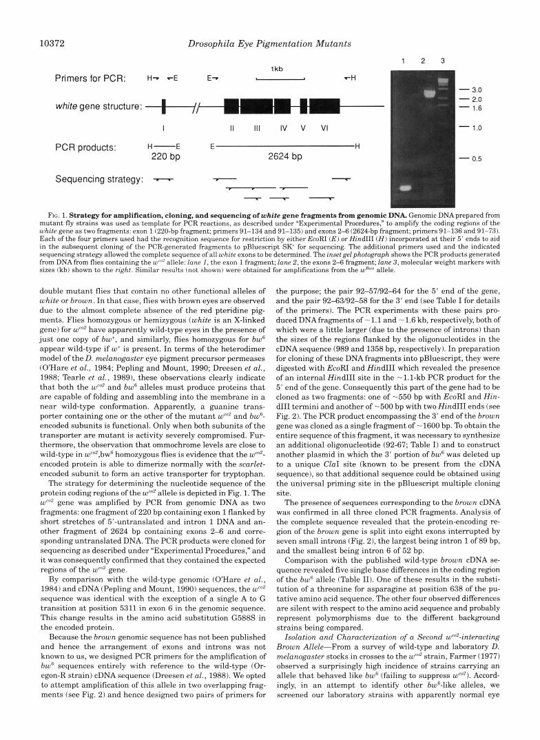
Site-directed Mutagenesis of the white Gene and P-element-mediated Dunsformation of Drosophila-The E. coli plasmid pRK3 was obtained from Dr. P. Schedl (Princeton University, Princeton, NJ). It harbors a P-element vector containing the white minigene and regulatory se- quences flanked by the boundary domains scs and scs' that insulate the white gene against chromosomal position effects (Kellem and Schedl, 1991). To construct a P-element transformation vector with a selectable marker independent of the white gene, the bacterial neomycin resist- ance gene under control of the Drosophila hsp70 promoter was isolated from the plasmid pUChsneo (Steller and Pirotta, 1985) as a 1.6-kb AseUHindIII fragment and ligated into the unique XhoI site of pRK3 between the scs element and the white regulatory sequences. After treatment with Klenow to create blunt termini the vector and insert DNA fragments were ligated together, the resultant vector is called pRK3hsneo (Fig. 3).
Site-directed mutagenesis of the ATP binding site of the white gene was performed using Amersham's in uitro mutagenesis kit (version 2.1). A 1.8-kb XbaUSacI fragment of pRK3hsneo containing the 5' region of the white gene was subcloned into the phagemid pBluescript SK', and single-stranded DNAwas isolated in the strain XL1-Blue after infection with M13 KO7 helper phage (Vieira and Messing, 1987). The mutagenic oligonucleotide 91-122 (Table I) was annealed and all reactions were performed as described in the kit's instruction manual, with the excep- tion that PuuI was used for the nicking step prior to T5 exonuclease treatment. AnXbaUSacI fragment containing the desired mutation was then subcloned back into pRWhsneo, replacing the corresponding wild- type sequence and producing the plasmid pRK3hsneo.mutl.
P-element-mediated transformation of D. rnelanogaster was per- formed essentially as described by Spradling (1986). Freshly laid eggs were obtained from a w; PLry'(A23)I (99B) strain and injected with either pRK3hsneo (P[w+; neo'l) or pRK3hsneo.mutl (P[w"""; neo'l). Sur- viving GO flies were crossed to a w1ll8 strain, and germline transfor- mants were selected on the basis of eye color andor G418 (0.2 mg/ml) resistance, as described under "Results." This level of the drug was initially chosen for the selection of transformants, on the basis of pre- liminary experiments to determine the minimum concentration of G418 that neither of the wo background strains were able to survive. It was subsequently endorsed by the result that when the G1 progeny of P[w+, neo'l recipient flies that had wild-type eye color were crossed to the wI1l8
strain and the G2 progeny grown on 0.2 mg/ml G418, only flies with colored eyes survived. To confirm the presence of the white P-element construct in transformants, genomic DNA was isolated from individual flies (Jowett, 1986), and PCR was performed using a Corbett capillary thermocycler as detailed in the legend of Fig. 4.
Materials-All enzymes and chemicals used were of the highest qual- ity available. Oligonucleotides were synthesized by the Biomolecular Resource Facility, The Australian National University, Canberra, Aus- tralia. [~r-~~sldATP was obtained from Amersham (Australia) Pty. Ltd. All standard molecular biological methods not otherwise described were performed essentially as described in Sambrook et al. (1989).
RESULTS
Amino Acid Substitutions in the Znteracting Alleles wco2 and bw6-The specific interaction between the eye pigmentation alleles weo2 and bw6 was first described by Farmer and Fair- banks (Farmer, 1977; Farmer and Fairbanks, 1986). Charac- terization of the mutations in these alleles is of particular in- terest because both are phenotypically silent except in wm2,bw6
10372 Drosophila Eye Pigmentation Mutants
1 kb Primersfor PCR: H-. --E E-. , I "H
white gene structure: ,+,LL- I I I 1 1 1 IV v VI
PCR products: H- E E H 220 bp 2624 bp
Sequencing strategy: - 7 - - " -
1 2 3
- 3.0 - 2.0 - 1.6
- 1.0
- 0.5
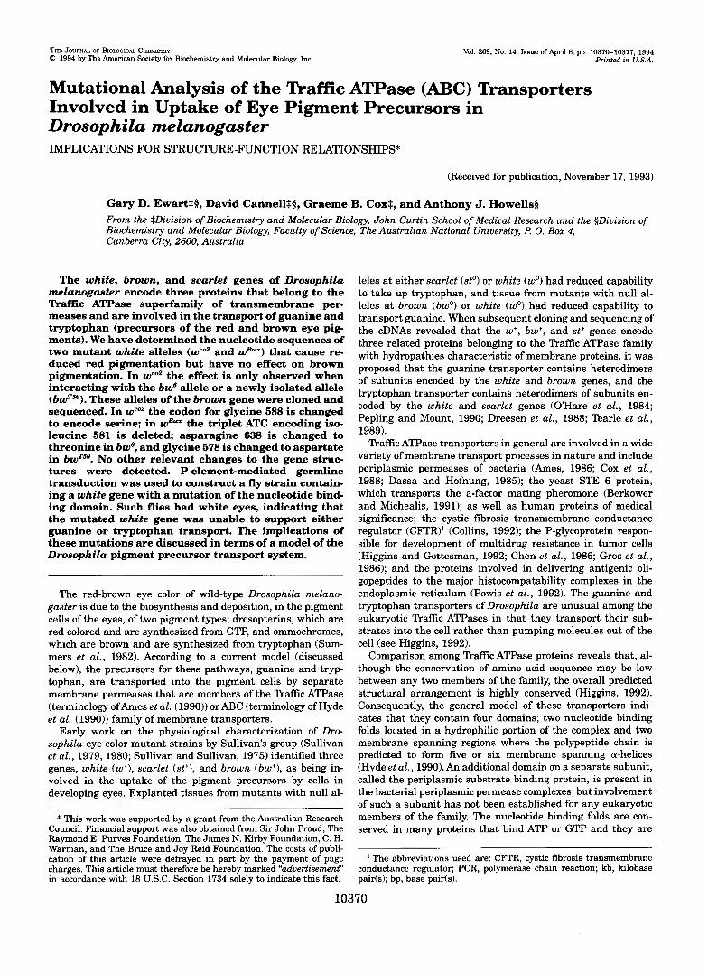
FIG. 1. Strategy for amplification, cloning, and sequencing of white gene fragments from genomic DNA. Genomic DNA prepared from mutant fly strains was used as template for PCR reactions, as described under "Experimental Procedures," to amplify the coding regions of the white gene as two fragments: exon 1 (220-bp fragment; primers 91-134 and 91-135) and exons 2-6 (2624-bp fragment; primers 91-136 and 91-73). Each of the four primers used had the recognition sequence for restriction by either EcoRI (E) or Hind111 ( H ) incorporated at their 5' ends to aid in the subsequent cloning of the PCR-generated fragments to pBluescript SK for sequencing. The additional primers used and the indicated sequencing strategy allowed the complete sequence of all white exons to be determined. The inset gel photograph shows the PCR products generated from DNA from flies containing the wCn2 allele: lane I , the exon 1 fragment; lane 2, the exons 2-6 fragment; lane 3, molecular weight markers with sizes (kb) shown to the right. Similar resu!ts (not shown) were obtained for amplifications from the w"~* allele.
double mutant flies that contain no other functional alleles of white or brown. In that case, flies with brown eyes are observed due to the almost complete absence of the red pteridine pig- ments. Flies homozygous or hemizygous (white is an X-linked gene) for wm2 have apparently wild-type eyes in the presence of just one copy of bw+, and similarly, flies homozygous for bw6 appear wild-type if w+ is present. In terms of the heterodimer model of the D. melanogaster eye pigment precursor permeases (OHare et al., 1984; Pepling and Mount, 1990; Dreesen et al., 1988; Tearle et al., 1989), these observations clearly indicate that both the wco2 and bw6 alleles must produce proteins that are capable of folding and assembling into the membrane in a near wild-type conformation. Apparently, a guanine trans- porter containing one or the other of the mutant wm2 and bw6- encoded subunits is functional. Only when both subunits of the transporter are mutant is activity severely compromised. Fur- thermore, the observation that ommochrome levels are close to wild-type in wco2,bwfi homozygous flies is evidence that the wm2- encoded protein is able to dimerize normally with the scarlet- encoded subunit to form an active transporter for tryptophan.
The strategy for determining the nucleotide sequence of the protein coding regions of the wen' allele is depicted in Fig. 1. The w"' gene was amplified by PCR from genomic DNA as two fragments: one fragment of 220 bp containing exon 1 flanked by short stretches of 5"untranslated and intron 1 DNA and an- other fragment of 2624 bp containing exons 2-6 and corre- sponding untranslated DNA. The PCR products were cloned for sequencing as described under "Experimental Procedures," and it was consequently confirmed that they contained the expected regions of the wCo2 gene.
By comparison with the wild-type genomic (OHare et al., 1984) and cDNA (Pepling and Mount, 1990) sequences, the wco2 sequence was identical with the exception of a single A to G transition a t position 5311 in exon 6 in the genomic sequence. This change results in the amino acid substitution G588S in the encoded protein.
Because the brown genomic sequence has not been published and hence the arrangement of exons and introns was not known to us, we designed PCR primers for the amplification of bwfi sequences entirely with reference to the wild-type (Or- egon-R strain) cDNA sequence (Dreesen et al., 1988). We opted to attempt amplification of this allele in two overlapping frag- ments (see Fig. 2) and hence designed two pairs of primers for
the purpose; the pair 92-57/9244 for the 5' end of the gene, and the pair 92-63192-58 for the 3' end (see Table I for details of the primers). The PCR experiments with these pairs pro- duced DNA fragments of - 1.1 and - 1.6 kb, respectively, both of which were a little larger (due to the presence of introns) than the sizes of the regions flanked by the oligonucleotides in the cDNA sequence (989 and 1358 bp, respectively). In preparation for cloning of these DNA fragments into pBluescript, they were digested with EcoRI and HindIII which revealed the presence of an internal HindIII site in the -1.1-kb PCR product for the 5' end of the gene. Consequently this part of the gene had to be cloned as two fragments: one of -550 bp with EcoRI and Hin- dIII termini and another of -500 bp with two HindIII ends (see Fig. 2). The PCR product encompassing the 3' end of the brown gene was cloned as a single fragment of -1600 bp. To obtain the entire sequence of this fragment, it was necessary to synthesize an additional oligonucleotide (92-67; Table I) and to construct another plasmid in which the 3' portion of bw6 was deleted up to a unique ClaI site (known to be present from the cDNA sequence), so that additional sequence could be obtained using the universal priming site in the pBluescript multiple cloning site.
The presence of sequences corresponding to the brown cDNA was confirmed in all three cloned PCR fragments. Analysis of the complete sequence revealed that the protein-encoding re- gion of the brown gene is split into eight exons interrupted by seven small introns (Fig. 2), the largest being intron 1 of 89 bp, and the smallest being intron 6 of 52 bp.
Comparison with the published wild-type brown cDNA se- quence revealed five single base differences in the coding region of the bw6 allele (Table 11). One of these results in the substi- tution of a threonine for asparagine at position 638 of the pu- tative amino acid sequence. The other four observed differences are silent with respect to the amino acid sequence and probably represent polymorphisms due to the different background strains being compared.
Isolation and Characterization of a Second wm2-interacting Brown Allele-From a survey of wild-type and laboratory D. melanogaster stocks in crosses to the wm2 strain, Farmer (1977) observed a surprisingly high incidence of strains carrying an allele that behaved like bw' (failing to suppress w"'). Accord- ingly, in an attempt to identify other bw6-like alleles, we screened our laboratory strains with apparently normal eye

Drosophila Eye Pigmentation Mutants
Primers for PCR: E-. "H
brown cDNA:
PCR products: E -1600 bp H n
E-. .-H
E L H -1150 bp : - 2.0
- 1.6
10373
Sequencing stategy: I - ;I - 1 .o
Hindlll Cla 1 - 0.5
brown gene structure: x I II I l l IV V VI VI1 Vl l l
1 kb
FIG. 2. Strategy for amplification, cloning, and sequencing of brown gene fragments from genomic DNA The relative locations of binding in the cDNA are shown for the two pairs of primers used for amplification (92-57 and 92-64 for the 5' end and 92-63 and 92-58 for the 3' end) of bw6 and b ~ ~ . ~ ~ allele sequences. The EcoRI ( E ) or HindIII ( H ) restriction enzyme recognition sequences incorporated in their 5' ends are also indicated. In the diagram of cDNA structure, the thick line indicates the protein coding sequences. The bwfi PCR products, after digestion with EcoRI and HindIII, are shown in the inset gel photograph: lane I , the 3'-terminal fragment; lane 2, size markers; lane 3, the 5"terminal fragments (an additional site for HindIII in the -1150 bp product was seen). The three fragments were cloned to pBluescript SK+ and sequenced as discussed in the text. The region sequenced from the primer 92-67 is indicated as starting with the asterisk. All other sequence data was obtained using either the PCR primers or the universal or reverse sequencing primers that have binding sites in the pBluescript vector. The deduced arrangement of exons and introns in the brown gene is illustrated in the lower portion of the diagram.
TABLE I Primers used in PCR, mutagenesis, and sequencing reactions
Primer Sequence (5' to 3')" base bindmg* Site of 3' Used for.. Strand'
white gene 91-134 TTGAAGCTTGAGTGATTGGGGTG -51 Exon 1 PCR + 91-135 GCAGAGAATTCGATGTTGCAATCGC 123 Exon 1 PCR -
91-73 GATGAAGCTTATCTTGTTTTTATTGGCAC 5714 Exon 2-6 PCR - 91-136 AACCGAATTCGTAGGATACTTCG 3132 Exon 2-6 PCR +
9 1-TW5 CGGCAGCTGGTCAACCGGACA 3409 Sequencing + 9 1-68 CCACGACATCTGACCTATCG 3824 Sequencing + 91-69 ACACCTACAAGGCCACCTGG 4886 Sequencing + 91-70 GATCGTGTGCTGACATTTGC 3872 SequencingPCR - 9 1-7 1 CTTTTACGAGGAGTGGTTCC 4537 Sequencing - 91-72 GATGTGCAGCTAATTTCGCC 543 1 Sequencing - 91-122 GTTCCGGTGCCCTGCAGACGACCCTG 3615 Mutagenesis +
brown gene 92-57 GGCGAGAATTCGGCACATCACATAGC -60 5' end PCR + 92-64 TGAAAAGCTTGAAGATGTCCGACGTCG 878 5' end PCR - 92-63 GCAGGAATTCCATCGAGATGGAGGTCG 759 3' end PCR +
92-67 TAGTAGGCGGACAGGCTGTAGGTG 1464 Sequencing - 92-58 AAATTAAGCTTAAGCAAGTTCTG~AAC 2066 3' end PCR -
a Recognition sequences for restriction by EcoRI, HindIII and PstI endonucleases are underlined. * Binding sites are numbered with +1 as the Aof the translation start codon in the published white genomic (O'Hare et al., 1984) and brown cDNA
(Dreesen et al., 1988) sequences. + indicates the coding strand; - indicates the complementary strand.
TABLE I1 Seauence differences between the mutant and wild-tvoe white and brown alleles
Allele Phenotype Mutation in DNA" Effect on amino acid sequence
wCOZ Interaction with bw6 and bwT.w reduces red pigments G to A (5311) Glyw to Ser bw' Interaction with wcoz reduces red pigments A to C (1913) A m M 8 to Thr
A to T (822) A to C (843)
Silent
A to G (1335) Silent Silent
A to C (1365) Silent bWT.50 Interaction with wCn2 reduces red pigments G to A (1733) a y 5 7 * to ASP WRI?
" The nucleotide positions in parentheses are relative to the first base of the start codon in the published white genomic (O'Hare et al., 1984) and
Brown eye color due to reduced red pigments A(529O)ATC Ilesx' deleted
brown cDNA (Dreesen et al., 1988) sequences.
color, as follows. Male flies from our various strains (bw'lbw'; where * indicates an unidentified allele) were mated to female wea2 v;bw6 flies and eye color of the male progeny (wco2 vly; bw6/bw') was assessed. Males with reduced red pigments were carriers of a potentially novel w'"'-interacting allele. The ver- milion ( v ) marker was incorporated in the screen to block om-
mochrome synthesis and thereby enable small changes in the level of pteridine pigments to be more easily observed.
From a total of the 36 strains that were screened in this way, one of the crosses produced male flies with yellow eyes among the progeny. These flies contained a bw6-like allele, which we have named bwT5' (after the strain T50 from which it was
10374 Drosophila Eye Pigmentation Mutants
isolated). To rule out the possibility that this new allele was a brown null, flies from the T50 strain were also crossed to bwo (w+ u') flies. All of the resulting progeny had wild-type eye color, consistent with the bwT5' allele requiring a specific interaction with wCo2 to produce a detectable reduction in pteridine pigmen- tation. If bwTSO had been a null allele, that proportion of the progeny that were bw0/bwT5' should have had brown eyes.
For characterization of the mutation in the bwT5' allele, DNA was isolated from four of the male flies with yellow eyes pro- duced in the initial cross to wm2 v;bw6 females. The brown gene fragments were amplified and cloned for sequencing as de- scribed for bw6 in the previous section (results not shown).
Sequencing revealed that the bwT5' allele was not identical to bw6. The new allele contains a single missense mutation with a G to A transition corresponding to position 1733 of the cDNA sequence. This results in the substitution of glycine 578 by aspartate.
Identification of a Single Codon Deletion in the wBwX Allele: Another Mutation in White That Affects the Guanine Duns- porter but Not the Wptophan Zkansporter-Although the white-encoded subunit is involved in transport of both guanine and tryptophan, in wBWX there is a preferential reduction in the pteridine level, whereas the ommochrome level is virtually un- affected (Zachar and Bingham, 1982; Lindsay and Grell, 1968). As for wco2, this indicates that the wBwx allele produces a protein that is capable of inserting in the membrane in such a way that it interacts with the scarlet-encoded subunit, to form an active tryptophan transporter in Drosophila eyes. However, clearly the mutant form of the wBwx protein is unable to support normal function of the guanine transporter.
To identify any mutations in the wB"-encoded subunit, we amplified and sequenced the allele from genomic DNA as de- scribed for the wco2 allele (results not shown). The sequence data showed that the triplet ATC, encoding Ile5*', has been deleted from the wBwr gene (Table 11). Otherwise, the sequence of the entire coding region and all splice donor and acceptor sites was identical to the published white sequences.
A Site-directed Mutation of Residues in the Nucleotide Bind- ing Domain of the White-encoded Subunit That Results in Loss of Function of Both Zkansporters-The invariant amino acid doublet Gly-Lys of the Walker motif A has been proposed to be directly involved in binding of ATP in the nucleotide binding fold (Parsonage et al., 1987), and mutations to these residues in various members of the Traffic ATPase family are known to eliminate or severely reduce function (Berkower and Michealis, 1991; Azzaria et al., 1989; Cox et al., 1989). To establish the significance of the nucleotide binding fold in the white-encoded subunit of Drosophila guanine and tryptophan transporters, we used oligonucleotide-directed mutagenesis to alter the codons for the Gly'35-Lys136 pair in the putative nucleotide bind- ing fold to encode Leu-Gln. The mutated white minigene re- placed the wild-type gene in pRK3hsneo (P[w+, neo'l) (Fig. 31, to create the plasmid pRK3hsneo.mutl (P[wmufz, neo'l). Plasmid constructs containing either the wild-type or mutated white genes were subsequently used in P-element-mediated germline transduction experiments as described under "Experimental Procedures." The eye color of the G1 progeny was assessed and then G2 flies were selected for resistance to G418 at 0.2 mg/ml. Two of the GO flies from embryos injected with the P[w+, neorl construct produced G1 progeny with the expected wild-type eye color. In contrast, none of the G1 progeny from P[wmutl, neorl recipient GO flies made any detectable pigments in their eyes. However, when the P[wmutl, near] G1 flies were allowed to in- terbreed and lay eggs in medium containing 0.2 mg/ml G418, fertile G2 adult flies were recovered that had escaped the drug selection.
To confirm that the P[wmufz, ned] G2 survivors of G418 se-
P
pUC8 (Amp')
pRK3hsneo (approx.12 Kb)
white regulatory
FIG. 3. The plasmid pFtK3hsneo used in P-element-mediated transformation of D. melanogaster. The arrangement and approxi- mate relative sizes is shown for genes and DNAmotifs referred to in the text that are flanked by the P-element inverted repeat sequences. The unique XbaI and Sac1 restriction endonuclease sites, used for cloning the equivalent fragment of the white minigene containing the site- directed mutation in pRK3hsneo.mutl (see "Experimental Proce- dures"), are also shown. The E. coli origin of replication derived from pUC8 and the 0-lactamase gene (conferring ampicillin resistance) are located in the segment external to the P-element termini.
lection were authentic transformants, PCR was used to amplify the 5' end of the white minigene from genomic DNA isolated from individual G418 survivors. One primer was designed to bind to the 5"nontranslated region, and the other was designed to bind in exon 3, downstream of the mutation site. The pro- duction of a 1.2-kb fragment aRer PCR confirmed that the white minigene (containing a shortened intron 1) was present in the genome of the flies, and digestion of the PCR product by PstI into two fragments of approximately 940 and 300 base pairs was proof that the gene contained the site-directed mu- tation (see Fig. 4).
Therefore, we conclude that a white gene with a G l ~ ' ~ ~ - L y s ' ~ ~ / Leu-Gln mutation in the putative nucleotide binding fold is unable to complement eye color in recipient flies with a defec- tive white gene. This result also demonstrates that a functional nucleotide binding fold on the white-encoded subunit is neces- sary for activity of both the guanine and tryptophan transport- ers of D. melanogaster. The nucleotide binding folds of neither the scarlet nor brown gene products are able to support trans- port on their own.
DISCUSSION
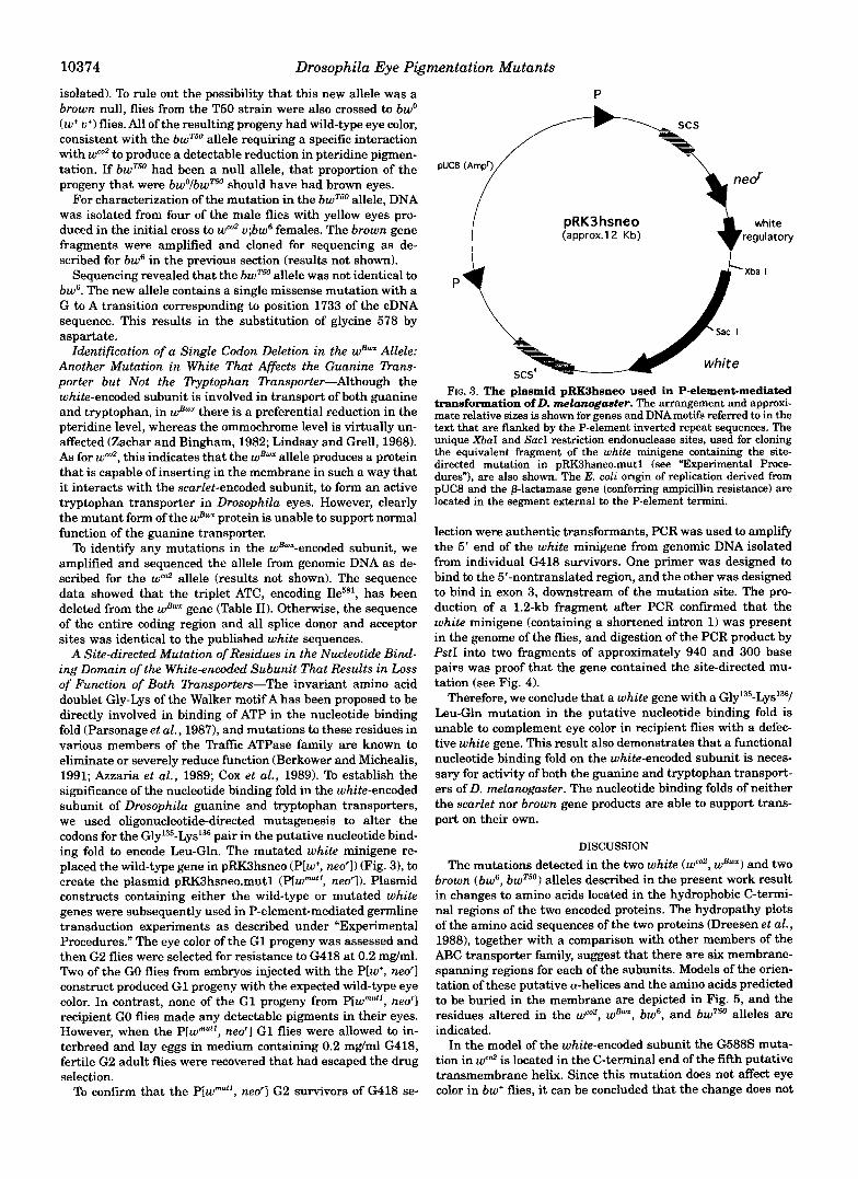
The mutations detected in the two white (wco2, wB") and two brown (bw6, bwTS0) alleles described in the present work result in changes to amino acids located in the hydrophobic C-termi- nal regions of the two encoded proteins. The hydropathy plots of the amino acid sequences of the two proteins (Dreesen et al., 1988), together with a comparison with other members of the ABC transporter family, suggest that there are six membrane- spanning regions for each of the subunits. Models of the orien- tation of these putative a-helices and the amino acids predicted to be buried in the membrane are depicted in Fig. 5 , and the residues altered in the wco2, wBwx, bw', and bwTSO alleles are indicated.
In the model of the white-encoded subunit the G588S muta- tion in wfo2 is located in the C-terminal end of the fifth putative transmembrane helix. Since this mutation does not affect eye color in bw' flies, it can be concluded that the change does not

Drosophila Eye Pigmentation Mutants 10375
P[w+, ned] or ~ [ v ~ u t 7 9 ned]
Injection into w-, 42-3 host
J.
J.
3/
GO fly x w 1 7 '8 flies
5;l flies (assess eye colour) G418 (0.2 mg/ml) selection
62 ned Iransformants
PCR test on genomic DNA from single flies t o confirm mutant white mini-gene is present
J/ 1 2 3 4 5 6 7 ""
- + - + - + - +
- 2036 - 1636
- 1028
- 506/517
- 200 154/136
FIG. 4. The strategy for construction, selection, and PCR screening of P[w"""; neo'l transformant flies. For the PCR test, genomic DNA was isolated from single flies and approximately 100 ng was used in PCR reactions with oligonucleotides 91-134 and 91-70 to amplify the 5' portion of the white minigene (across the shortened intron 1). A product of 1.2 kb indicates that the flies contained the P-element-borne white gene. The PCR products were separated from unreacted primers and "primer dimers" by the GenecleanTM method and were then tested for the presence of an internal PstI site that would have been introduced by successful mutagenesis with oligonucleotide 91-122. Lanes 1 and 7, DNAmarkers with sizes (bp) indicated on the far right-hand side. Lanes 2-6, purified PCR products, before (-) and after (+) digestion with PstI, amplified from the following flies: lane 2, a noninjected w, A223 fly; lane 3, a G2 fly (with wild-type eye color) derived from an egg injected with pRK3hsneo. Lanes 4-6, three G2 flies (white-eyed) derived from eggs injected with pRK3hsneo.mutl.
appreciably affect the ability of the wCo2-encoded subunit to function in active guanine and tryptophan transporters. Therefore, the global structure of the mutant subunit must be nearly identical to wild-type, and any structural alterations due to the introduction of a hydroxyl moiety or the increased size of the serine residue compared with glycine must be rela- tively localized and insignificant with respect to function of the heterodimers. Similarly, it can be argued that the amino acid substitutions in either bw' (N638T) or bwT5" (G578D) do not cause disruption of subunit structure or function in het- erodimers with w'-encoded subunits. As for wco2, the mutant residues in the bw6 and bwT5"-encoded subunits are predicted to be near the external surface of the membrane. In burT5" the G578D mutation is in a similar position in transmembrane helix 5 as the change in wco2, and in bw6 the N638T change is
located near the membrane surface in transmembrane helix 6. Notably, the three mutations all give rise to residues capable of H-bonding, and it would seem likely that H-bonding be- tween serine a t position 588 in the white-encoded subunit and either aspartate 578 or threonine 638 in the brown-encoded subunit is responsible for the observed phenotype although other explanations are possible. These interactions would im- ply that helix 5 of the white protein is close to helices 5 and 6 of the brown protein (see Fig. 5). As such interactions cause loss of function, yet individually have no effect, it is possible that some dynamic aspect of transporter function has been af- fected. Furthermore, with reference to the folding models, the predicted location of all three mutations near the external surface of the membrane makes it tempting to speculate that function associated with the mouth of the pore through which the substrate passes may be affected.
The wEwx allele, containing a mutation that results in the deletion of Ile5*', causes almost no pteridine pigment to be synthesized while leaving the ommochrome levels unaffected (Zachar and Bingham, 1982; Lindsay and Grell, 1968). This is an identical phenotype to that produced by the wco2 bw6 inter- action, and again it indicates that the mutant white-encoded subunit is capable of interacting with the scarlet-encoded sub- unit to form an active tryptophan transporter and is therefore assembled and is suficiently structurally similar to the wild- type subunit to form this functional interaction. We propose, however, that the deletion of Ile5*' from transmembrane helix 5 in the WE""-encoded subunit causes loss of a functional inter- action with the brown protein. Deletion of an amino acid from an a-helix changes the faces of the helix due to the rotation in position, by 1 amino acid residue, of the helical surface on the C-terminal side of the deletion relative to the surface on the N-terminal side. Obviously, this could have significant effects on the interactions of that helix with the other transmembrane helices of the complex. Furthermore, if a face of the helix lined the channel through which the substrate is transported, then the deletion may alter the chemical properties of the channel surface by changing the positions of residue side chains which might be critical to the transport mechanism. Therefore, we propose that transmembrane helix 5 of the white-encoded sub- unit is involved in specific interactions with the brown-encoded subunit in formation of the guanine transporter. This conclu- sion is consistent with the interaction between helices 5 and 6 of these subunits proposed on the basis of the changes observed in the wco2, bw', and bwT5' alleles, and it may be of relevance that the amino acid deleted is just two turns of an a-helix away from the interacting residue affected in wco2. A corollary to this deduction is that a similar interaction between transmembrane helix 5 of the white-encoded subunit and the scarlet-encoded subunit does not occur in the tryptophan transporter, since ommochrome levels are wild-type in both wBW* and wW2,bw6 flies.
Although the existence of additional mutations in the non- coding DNA of the wEwx allele has not been ruled out in this work, it seems unlikely that the phenotype could be caused by effects on the regulation of gene expression. I t should be noted that previous molecular investigation of the wEW* allele using Southern blotting has shown it to have a gross gene structure identical to the wild-type gene with the exception of a 150-bp deletion between positions -18 and -19.5 on the gene map of white (Zachar and Bingham, 1982). However, a causative effect of this deletion on the expression of the gene is excluded by the results of Hazelrigg et al. (1984) who showed that flies trans- formed with a white gene construct which does not contain DNA that far downstream of the coding region regulate expres- sion of white normally.
Another important property of the wEw' allele which indi-

10376 Drosophila Eye Pigmentation Mutants
a OUTSIDE
FIG. 5. a, model of the white-encoded subunit. b, model of the brown-encoded subunit. The amino acids predicted to be in the transmembrane helices in the white- and brown-encoded subunits of the guanine transporter are as indicated. The numbers inside the intra-helical loops in- dicate the number of amino acids in the loop. The relative distribution of charged amino acids is indicated by + or -. Amino acid residues that were found to be al- tered in the mutant alleles sequenced are in boldface type and have their residue position superscripted (see Table 11). The putative nucleotide binding fold (NBF) is discussed in the text.
t
-A: B I N D I N G
b OUTSIDE
COOH
CYTOPLASM
+d- BINDING
n - I DOMAIN? (
CYTOPLASM
COOH
Drosophila Eye Pigmentation Mutants 10377
cates that the mutation in the protein is responsible for the phenotype is that it has a semidominant effect on pteridine production (Lindsay and Grell, 1968): wBwxlw+ heterozygotes have an eye color that is noticeably less red than wild-type. This is in contrast to the majority of white alleles in which the eye color phenotype is wild-type in heterozygotes. This suggests the interesting possibility that the native permease complex might be tetrameric (or multimeric), possessing two or more copies each of the white-encoded and brown-encoded subunits. Then it is conceivable that the presence of just one copy of the wB""-encoded subunit in the oligomeric complex may be sum- cient to disrupt the function of the transporter and that partial pigmentation results from the activity of the small proportion of complexes that form containing only the wild-type white- encoded subunit.
Function-altering mutations within the transmembrane hel- ices have been identified in the MDR (Devine et al., 1992; Gros et al., 1991; Safa et al., 1990) and CFTR ("sui, 1992; Anderson et al., 1991b) proteins. In the case of the CFTR protein, 58 missense mutations and 3 single amino acid deletions have been reported that probably cause cystic fibrosis ("sui, 1992). Although the majority (32 mutants) of these are associated with either of the two nucleotide binding domains, other ap- parent hotspots for mutations are a predicted cytoplasmic loop in the C-terminal hydrophobic domain (nine mutants) and transmembrane helices 5 and 6 (six mutants). Interestingly, it has been suggested previously that these helices may have roles in substrate association and the mechanism of transport in Traffic ATPases (Devine et al., 1992; Gros et al., 1991; Webb et al., 1992).
The effects of the site-directed mutation resulting in Gly- Lys being replaced by Leu-Gln in the Walker motif A of the white protein is consistent with the effects of similar muta- tions on other ABC transporters and on the F,F,-ATPase (Berkower and Michealis, 1991; Parsonage et al., 1987; Az- zaria et al., 1989; Cox et al., 1989). Structural analysis of ad- enylate kinase has shown that these amino acids are directly involved in binding of the phosphoryl groups of the nucleotide (Hyde et al., 1990). Similar changes in the P-subunit of F,- ATPase affect the binding and hydrolysis of ATP (Parsonage et al., 1987). The results reported here are therefore consistent with the proposition that ABC transporters are energized by ATP hydrolysis. With respect to the models depicted in Fig. 5, the location of the nucleotide binding domains to the N-termi- nal side of the transmembrane helices in the Drosophila transporters is unusual among the eukaryotic ABC per- meases. Similarly, the Drosophila transporters are also un- usual among eukaryotic ABC permeases so far described, in that they transport their substrates into the cell instead of pumping molecules out of the cell (Higgins, 1992). As more and more examples of ABC permeases are characterized it will be interesting to see whether a correlation persists be- tween the order of the hydrophilic and hydrophobic domains on the polypeptides and the direction of pumping.
REFERENCES
Ames, G. F.-L. (1986) Annu. Rev. Biochem. 55,397-425 Ames, G. F.-L., and Joshi, A. K. (1990) J. Bacteriol. 172,4133-4137 Ames, G. F.-L., Mimura, C. S. , and Shyamala, V. (1990) FEMS Microbiol. Reu. 75,
Anderson, M. P., and Welsh, M. J. (1992) Science 257, 1701-1703 Anderson, M. P., Berger, H. A., Rich, D. P., Gregory, R. J., Smith, A. E., and Welsh,
Anderson, M. P., Gregory, R. J., Thompson, S., Souza, D. W., Paul, S. , Mulligan, R.
Azzaria, M., Schurr, E., and Gros, P. (1989) Mol. Cell. Biol. 9, 5289-5297 Bender, W., Spierer, P., and Hogness, D. S . (1983) J. Mol. Biol. 168, 17-33 Berkower, C., and Michaelis, S . (1991) EMBO J. 10,37773785 Chen, C., Chin, J. E., Ueda, K., Clark, D. P., Pastan, I., Gottesman, M. M., and
Collins, E S . (1992) Science 256, 774-779 Roninson, I. B. (1986) Cell 47,381-389
Cox, G. B., Webb, D., and Rosenberg, H. (1989) J. Bacteriol. 171, 1531-1534 Cox. G. B., Webb, D. C., Zimmermann, J. G., and Rosenberg, H. (1988) J. Bacteriol.
429-446
M. J. (1991a) Cell 67,775-784
C., Smith, A. E., and Welsh, M. J . (1991b) Science 253,202-205
Dassa, E., and Hofnung, M. (1985) EMBO J. 4,2287-2293 Devine, S . E., Ling, V., and Melera, P. W. (1992) Proc. Natl. Acad. Sci. U. S. A. 89,
170,2283-2286
Dreesen, T. D., Johnson, D. H., and Henikoff, S . (1988) Mol. Cell. Biol. 8, 5206
Farmer, J. L. (1977) Heredity 39.297303 Farmer, J. L., and Fairbanks, D. J. (1986) Dros. Info. S e n . 63,50-51 Gros, P., Croop, J., and Housman, D. (1986) Cell 47,371380 Gros, P., Dhir, R., Croop, J., and Talbot, F. (1991) P m . Natl. Acad. Sci. U. S. A. 88,
Hazelrigg, T., Levis, R., and Rubin, G. M. (1984) Cell 36, 469481 Higgins, C. F. (1992) Annu. Rev. Cell Bid. 8, 67-113 Higgins, C. F., and Gottesman, M. M. (1992) 7 k n d s Biochem. Sci. 17, 1%21 Hyde, S . C., Emsley, P., Hartshorn, M. J., Mimmack, M. M., Gileadi, U., Pearce, S .
346,362-365 R., Gallagher, M. P., Gill, D. R., Hubbard, R. E., and Higgins, C. F. (199O)Nature
Jowett, T. (1986) in Drosophila: A Practical Approach (Roberts, D. B., ed) p. 278, IRL Press, Oxford
Kellum, R., and Schedl, P. (1991) Cell 64, 941-950 Lindsay, D. L., and Grell, E. H. (1968) Genetic Variation in Drosophila melano-
Lundberg, K. S . , Shoemaker, D. D., Adams, M. W. W. Short, J. M. Sorge, J. A,, and
O'Hare, K., Murphy, C., Levis, R., and Rubin, G. M. (1984) J. Mol. Biol. 180,
Parsonage, D., Wilke-Mounts, S . , and Senior, A. E. (1987) J. Bid . Chem. 262,
Pepling, M., and Mount, S . M. (1990) Nucleic Acids Res. 18, 1633 Powis, S . J., Deverson, E. V. Coadwell, W. J. Ciruela, A,, Huskisson, N. S., Smith,
Safa, A. R., Stern, R. K., Choi, K., Agresti, M., Tamai, I., Mehta, N. D., and
Sambrwk, J., Fritsch, E. F., and Maniatis, T. (1989) Mokcular Cloning: A him-
Sanger, F., Nicklen, S . , and Coulson, A. R. (1977) Proc. Natl. Acad. Sci. U. S. A. 74,
Spradling, A. C. (1986) inDrosophi2a:A Practical Approach (Roberts, D. B., ed) pp.
Steller, H., and Pirrotta, V. (1985) EMBO J. 4, 167-171 Sullivan, D. T., and Sullivan, M. C. (1975) Biochem. Genet. 13, 603-613 Sullivan, D. T., Bell, L. A., Paton, D. R., and Sullivan, M. C. (1979) Biochem. Genet.
Sullivan, D. T., Bell, L. A., Paton, D. R., and Sullivan, M. C. (1980) Biochem. Genet.
Summers, K. M., Howells, A. J., and Pyliotis, N. A. (1982) Adu. Insect Physiol. 16,
Tearle, R. G., Belote, J. M., McKeown, M., Baker, B. S . , and Howells, A. J. (1989)
Tsui, L:C. (1992) Human Mutation 1, 197-203 Vieira, J., and Messing, J. (1987) Methods Enzymol. 163, 3-11 Walker, J. E., Saraste, M., Runswick, M. J., and Gay, N. J . (1982) EMBO J. 1,
Webb, D. C., Rosenberg, H., and Cox, G. B. (1992) J. Biol. Chem. 267,24661-24668 Zachar, Z., and Bingham, P. M. (1982) Cell 30,529-541
4564-4568
5215
7289-7293
gaster, Publication 627, Carnegie Institute, Washington, D. C.
Mathur, E. J. (1991) Gene (Amst.) 108, 1-6
437-455
8022-8026
H. Butcher, G. W., and Howard, J. C. (1992) Nature 367, 211-215
Roninson, I. €3. (1990) Proc. Natl. Acad. Sci. U. S. A. 87, 7225-7229
ratory Manual, Cold Spring Harbor Laboratory, Cold Spring Harbor, NY
5463-5467
175-197, IRL Press, Oxford
17,565573
18, 1109-1130
119-166
Genetics 122, 595406
94L951