Embed Size (px)
Citation preview
cells
Review
The JNK Signaling Pathway in Inflammatory SkinDisorders and Cancer †
Manel B. Hammouda 1, Amy E. Ford 1, Yuan Liu 1 and Jennifer Y. Zhang 1,2,*1 Department of Dermatology, Duke University Medical Center, Durham, NC 27710, USA;
[email protected] (M.B.H.); [email protected] (A.E.F.); [email protected] (Y.L.)2 Department of Pathology, Duke University Medical Center, Durham, NC 27710, USA* Correspondence: [email protected]; Tel.: +1-919-684-6794† Running Title: JNK Contribution to Skin Diseases.
Received: 20 February 2020; Accepted: 26 March 2020; Published: 2 April 2020�����������������
Abstract: The c-Jun N-terminal kinases (JNKs), with its members JNK1, JNK2, and JNK3, is asubfamily of (MAPK) mitogen-activated protein kinases. JNK signaling regulates a wide range ofcellular processes, including cell proliferation, differentiation, survival, apoptosis, and inflammation.Dysregulation of JNK pathway is associated with a wide range of immune disorders and cancer.Our objective is to provide a review of JNK proteins and their upstream regulators and downstreameffector molecules in common skin disorders, including psoriasis, dermal fibrosis, scleroderma, basalcell carcinoma (BCC), squamous cell carcinoma (SCC), and melanoma.
Keywords: JNK; skin inflammation; keratinocytes; BCC; SCC; melanoma; psoriasis; fibrosis;scleroderma
1. The c-Jun N-Terminal Kinase (JNK) Signaling Pathway
1.1. JNK Pathway Components
JNK, also known as stress-activated protein kinases (SAPK), represents a subfamily of the canonicalMAPK signal transduction pathway [1], which along with cyclin-dependent kinases (CDKs), glycogensynthase kinase 3 (GSK3), and CDK-like kinases (CLKs), constitutes a larger family referred to asthe CMGC Ser/Thr group kinases [1–3]. JNK proteins, JNK1, JNK2, and JNK3, are encoded by threeseparate genes Mapk8, Mapk9, and Mapk10, respectively [4]. Each is alternatively spliced to create atleast ten variants that were detected by Western blotting at approximately 46 kDa (e.g., JNK1α1 andJNK1β1) and 55 kDa (e.g., JNK1α2 and JNK1β2) molecular weights [5].
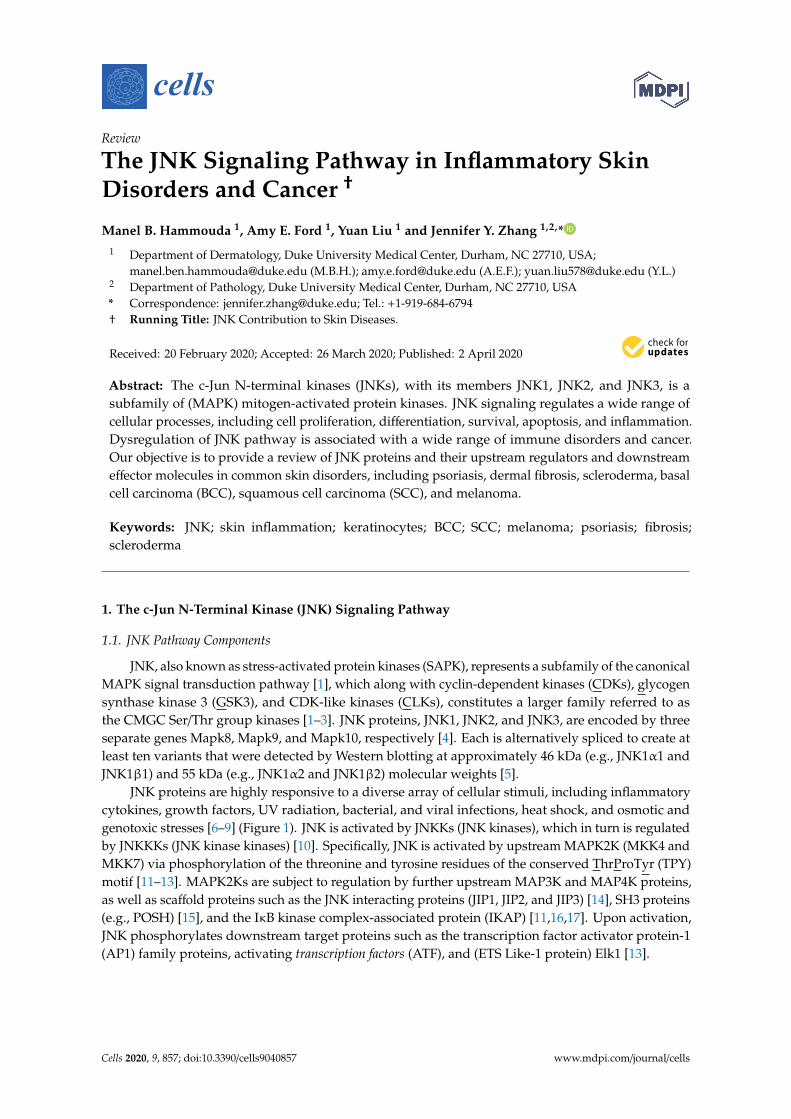
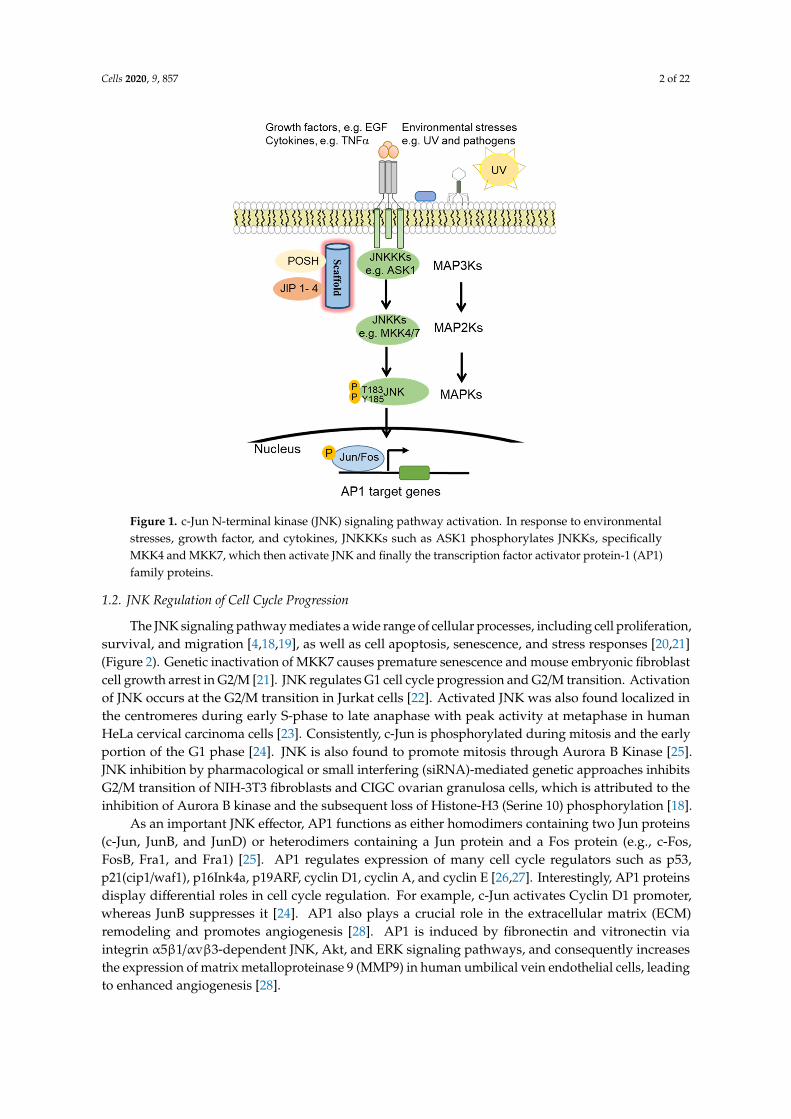
JNK proteins are highly responsive to a diverse array of cellular stimuli, including inflammatorycytokines, growth factors, UV radiation, bacterial, and viral infections, heat shock, and osmotic andgenotoxic stresses [6–9] (Figure 1). JNK is activated by JNKKs (JNK kinases), which in turn is regulatedby JNKKKs (JNK kinase kinases) [10]. Specifically, JNK is activated by upstream MAPK2K (MKK4 andMKK7) via phosphorylation of the threonine and tyrosine residues of the conserved ThrProTyr (TPY)motif [11–13]. MAPK2Ks are subject to regulation by further upstream MAP3K and MAP4K proteins,as well as scaffold proteins such as the JNK interacting proteins (JIP1, JIP2, and JIP3) [14], SH3 proteins(e.g., POSH) [15], and the IκB kinase complex-associated protein (IKAP) [11,16,17]. Upon activation,JNK phosphorylates downstream target proteins such as the transcription factor activator protein-1(AP1) family proteins, activating transcription factors (ATF), and (ETS Like-1 protein) Elk1 [13].
Cells 2020, 9, 857; doi:10.3390/cells9040857 www.mdpi.com/journal/cells
Cells 2020, 9, 857 2 of 22Cells 2020, 9, x FOR PEER REVIEW 2 of 22
Figure 1. c-Jun N-terminal kinase (JNK) signaling pathway activation. In response to environmental stresses, growth factor, and cytokines, JNKKKs such as ASK1 phosphorylates JNKKs, specifically MKK4 and MKK7, which then activate JNK and finally the transcription factor activator protein-1 (AP1) family proteins.
1.2. JNK Regulation of Cell Cycle Progression
The JNK signaling pathway mediates a wide range of cellular processes, including cell proliferation, survival, and migration [4,18,19], as well as cell apoptosis, senescence, and stress responses [20,21] (Figure 2). Genetic inactivation of MKK7 causes premature senescence and mouse embryonic fibroblast cell growth arrest in G2/M [21]. JNK regulates G1 cell cycle progression and G2/M transition. Activation of JNK occurs at the G2/M transition in Jurkat cells [22]. Activated JNK was also found localized in the centromeres during early S-phase to late anaphase with peak activity at metaphase in human HeLa cervical carcinoma cells [23]. Consistently, c-Jun is phosphorylated during mitosis and the early portion of the G1 phase [24]. JNK is also found to promote mitosis through Aurora B Kinase [25]. JNK inhibition by pharmacological or small interfering (siRNA)-mediated genetic approaches inhibits G2/M transition of NIH-3T3 fibroblasts and CIGC ovarian granulosa cells, which is attributed to the inhibition of Aurora B kinase and the subsequent loss of Histone-H3 (Serine 10) phosphorylation [18].
As an important JNK effector, AP1 functions as either homodimers containing two Jun proteins (c-Jun, JunB, and JunD) or heterodimers containing a Jun protein and a Fos protein (e.g., c-Fos, FosB, Fra1, and Fra1) [25]. AP1 regulates expression of many cell cycle regulators such as p53, p21(cip1/waf1), p16Ink4a, p19ARF, cyclin D1, cyclin A, and cyclin E [26,27]. Interestingly, AP1 proteins display differential roles in cell cycle regulation. For example, c-Jun activates Cyclin D1 promoter, whereas JunB suppresses it [24]. AP1 also plays a crucial role in the extracellular matrix (ECM) remodeling and promotes angiogenesis [28]. AP1 is induced by fibronectin and vitronectin via integrin α5β1/αvβ3-dependent JNK, Akt, and ERK signaling pathways, and consequently increases the expression of matrix metalloproteinase 9 (MMP9) in human umbilical vein endothelial cells, leading to enhanced angiogenesis [28].
Figure 1. c-Jun N-terminal kinase (JNK) signaling pathway activation. In response to environmentalstresses, growth factor, and cytokines, JNKKKs such as ASK1 phosphorylates JNKKs, specificallyMKK4 and MKK7, which then activate JNK and finally the transcription factor activator protein-1 (AP1)family proteins.
1.2. JNK Regulation of Cell Cycle Progression
The JNK signaling pathway mediates a wide range of cellular processes, including cell proliferation,survival, and migration [4,18,19], as well as cell apoptosis, senescence, and stress responses [20,21](Figure 2). Genetic inactivation of MKK7 causes premature senescence and mouse embryonic fibroblastcell growth arrest in G2/M [21]. JNK regulates G1 cell cycle progression and G2/M transition. Activationof JNK occurs at the G2/M transition in Jurkat cells [22]. Activated JNK was also found localized inthe centromeres during early S-phase to late anaphase with peak activity at metaphase in humanHeLa cervical carcinoma cells [23]. Consistently, c-Jun is phosphorylated during mitosis and the earlyportion of the G1 phase [24]. JNK is also found to promote mitosis through Aurora B Kinase [25].JNK inhibition by pharmacological or small interfering (siRNA)-mediated genetic approaches inhibitsG2/M transition of NIH-3T3 fibroblasts and CIGC ovarian granulosa cells, which is attributed to theinhibition of Aurora B kinase and the subsequent loss of Histone-H3 (Serine 10) phosphorylation [18].
As an important JNK effector, AP1 functions as either homodimers containing two Jun proteins(c-Jun, JunB, and JunD) or heterodimers containing a Jun protein and a Fos protein (e.g., c-Fos,FosB, Fra1, and Fra1) [25]. AP1 regulates expression of many cell cycle regulators such as p53,p21(cip1/waf1), p16Ink4a, p19ARF, cyclin D1, cyclin A, and cyclin E [26,27]. Interestingly, AP1 proteinsdisplay differential roles in cell cycle regulation. For example, c-Jun activates Cyclin D1 promoter,whereas JunB suppresses it [24]. AP1 also plays a crucial role in the extracellular matrix (ECM)remodeling and promotes angiogenesis [28]. AP1 is induced by fibronectin and vitronectin viaintegrin α5β1/αvβ3-dependent JNK, Akt, and ERK signaling pathways, and consequently increasesthe expression of matrix metalloproteinase 9 (MMP9) in human umbilical vein endothelial cells, leadingto enhanced angiogenesis [28].
Cells 2020, 9, 857 3 of 22Cells 2020, 9, x FOR PEER REVIEW 3 of 22
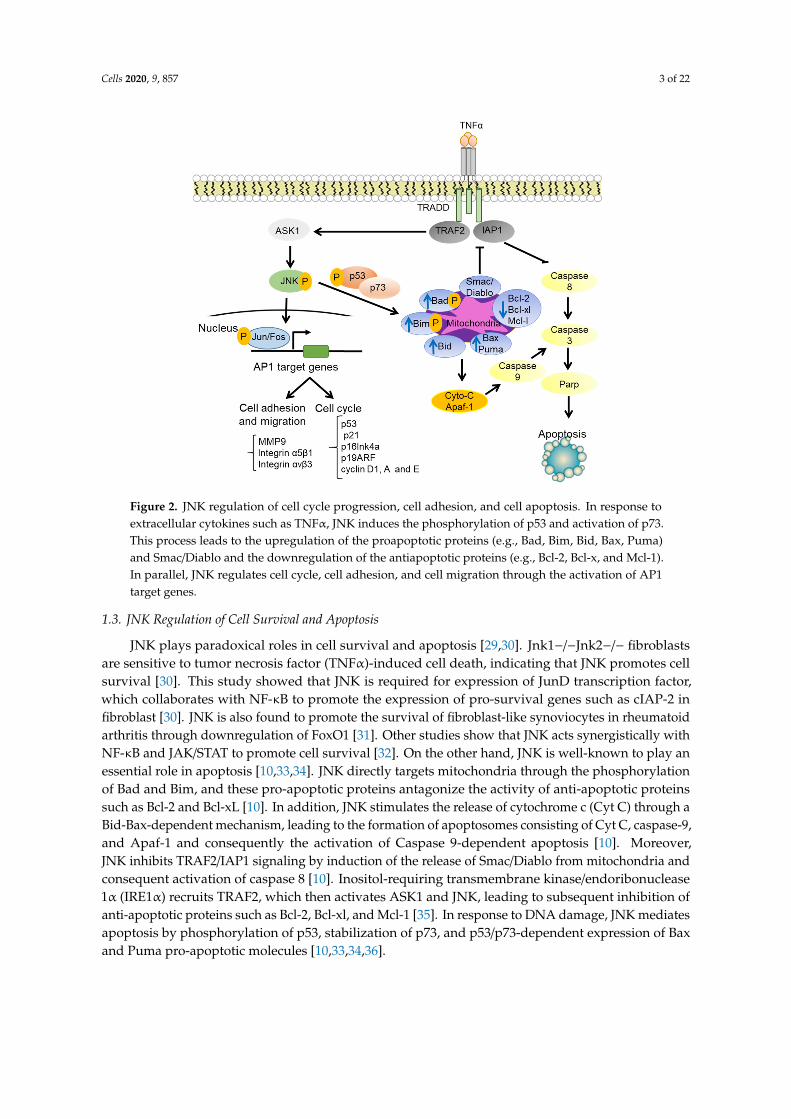
Figure 2. JNK regulation of cell cycle progression, cell adhesion, and cell apoptosis. In response to extracellular cytokines such as TNFα, JNK induces the phosphorylation of p53 and activation of p73. This process leads to the upregulation of the proapoptotic proteins (e.g., Bad, Bim, Bid, Bax, Puma) and Smac/Diablo and the downregulation of the antiapoptotic proteins (e.g., Bcl-2, Bcl-x, and Mcl-1). In parallel, JNK regulates cell cycle, cell adhesion, and cell migration through the activation of AP1 target genes.
1.3. JNK Regulation of Cell Survival and Apoptosis
JNK plays paradoxical roles in cell survival and apoptosis [29,30]. Jnk1−/−Jnk2−/− fibroblasts are sensitive to tumor necrosis factor (TNFα)-induced cell death, indicating that JNK promotes cell survival [30]. This study showed that JNK is required for expression of JunD transcription factor, which collaborates with NF-κB to promote the expression of pro-survival genes such as cIAP-2 in fibroblast [30]. JNK is also found to promote the survival of fibroblast-like synoviocytes in rheumatoid arthritis through downregulation of FoxO1 [31]. Other studies show that JNK acts synergistically with NF-κB and JAK/STAT to promote cell survival [32]. On the other hand, JNK is well-known to play an essential role in apoptosis [10,33,34]. JNK directly targets mitochondria through the phosphorylation of Bad and Bim, and these pro-apoptotic proteins antagonize the activity of anti-apoptotic proteins such as Bcl-2 and Bcl-xL [10]. In addition, JNK stimulates the release of cytochrome c (Cyt C) through a Bid-Bax-dependent mechanism, leading to the formation of apoptosomes consisting of Cyt C, caspase-9, and Apaf-1 and consequently the activation of Caspase 9-dependent apoptosis [10]. Moreover, JNK inhibits TRAF2/IAP1 signaling by induction of the release of Smac/Diablo from mitochondria and consequent activation of caspase 8 [10]. Inositol-requiring transmembrane kinase/endoribonuclease 1α (IRE1α) recruits TRAF2, which then activates ASK1 and JNK, leading to subsequent inhibition of anti-apoptotic proteins such as Bcl-2, Bcl-xl, and Mcl-1 [35]. In response to DNA damage, JNK mediates apoptosis by phosphorylation of p53, stabilization of p73, and p53/p73-dependent expression of Bax and Puma pro-apoptotic molecules [10,33,34,36].
Figure 2. JNK regulation of cell cycle progression, cell adhesion, and cell apoptosis. In response toextracellular cytokines such as TNFα, JNK induces the phosphorylation of p53 and activation of p73.This process leads to the upregulation of the proapoptotic proteins (e.g., Bad, Bim, Bid, Bax, Puma)and Smac/Diablo and the downregulation of the antiapoptotic proteins (e.g., Bcl-2, Bcl-x, and Mcl-1).In parallel, JNK regulates cell cycle, cell adhesion, and cell migration through the activation of AP1target genes.
1.3. JNK Regulation of Cell Survival and Apoptosis
JNK plays paradoxical roles in cell survival and apoptosis [29,30]. Jnk1−/−Jnk2−/− fibroblastsare sensitive to tumor necrosis factor (TNFα)-induced cell death, indicating that JNK promotes cellsurvival [30]. This study showed that JNK is required for expression of JunD transcription factor,which collaborates with NF-κB to promote the expression of pro-survival genes such as cIAP-2 infibroblast [30]. JNK is also found to promote the survival of fibroblast-like synoviocytes in rheumatoidarthritis through downregulation of FoxO1 [31]. Other studies show that JNK acts synergistically withNF-κB and JAK/STAT to promote cell survival [32]. On the other hand, JNK is well-known to play anessential role in apoptosis [10,33,34]. JNK directly targets mitochondria through the phosphorylationof Bad and Bim, and these pro-apoptotic proteins antagonize the activity of anti-apoptotic proteinssuch as Bcl-2 and Bcl-xL [10]. In addition, JNK stimulates the release of cytochrome c (Cyt C) through aBid-Bax-dependent mechanism, leading to the formation of apoptosomes consisting of Cyt C, caspase-9,and Apaf-1 and consequently the activation of Caspase 9-dependent apoptosis [10]. Moreover,JNK inhibits TRAF2/IAP1 signaling by induction of the release of Smac/Diablo from mitochondria andconsequent activation of caspase 8 [10]. Inositol-requiring transmembrane kinase/endoribonuclease1α (IRE1α) recruits TRAF2, which then activates ASK1 and JNK, leading to subsequent inhibition ofanti-apoptotic proteins such as Bcl-2, Bcl-xl, and Mcl-1 [35]. In response to DNA damage, JNK mediatesapoptosis by phosphorylation of p53, stabilization of p73, and p53/p73-dependent expression of Baxand Puma pro-apoptotic molecules [10,33,34,36].
Cells 2020, 9, 857 4 of 22
2. JNK Signaling in Immunological Skin Disorders
2.1. JNK Regulation of Immune Responses
Immune responses are often divided into Th1, Th2, and Th17 T helper lymphocyte immunity [37].Th1 cells release IL-2, interferon (IFN)-γ [38], and tumor necrosis factor (TNF)-β cytokines [39] andmediates responses against viral and bacterial infections and elimination of cancer cells [40]. Th2 cellsproduce interleukin IL-4, IL-5, IL-10, IL-13, and IL-33, and mediate humoral responses, B cell activation,and antibody production. Th17 cells secrete IL-17, IL-6, IL-22, and TNF-α. Th2 and Th17 play crucialroles in tissue repair and regeneration [41]. The balance between Th1 and Th2 immune responses iscrucial for normal tissue homeostasis [41]; this is known as the Th1 and Th2 paradigm.
JNK plays a significant role in the innate and adaptive immune responses [42–47]. JNK activationpromotes apoptosis of developing thymocytes and regulates T-cell differentiation and survival [45,48].The Src-family tyrosine kinase Lck is associated with the CD4 and CD8 co-receptors. Lck expressionis reduced in Th2 cells compared Th1 cells; Ectopic expression of Lck in Th2 cells led to increasedexpression of CD4 co-receptor and c-Jun phosphorylation at Serine 73 via concerted actions of JNK andERK signaling [49], suggesting that Lck promotes Th1 polarization in part through a JNK-dependentprocess. JNK/c-Jun signaling is at least partly responsible for CD27-induced suppression of IL-17and CCR6 expression and consequently reduced Th17 cell development and differentiation [50].In CD8+ T cells, JNK1 is activated by POSH-mediated complex formation with MLK3, MKK7, andJIP-1, and thereby regulates cell proliferation and function [48]. In B-cells, JNK/c-Jun is activatedby CD154-induced CD40 internalization in a JIP-dependent manner and regulates memory B-celldevelopment [14].
As a critical regulator of immune cell differentiation and activation, JNK is implicated in manyimmune-related skin disorders. In this section, we will review the role of JNK in the development ofpsoriasis and dermal fibrosis.
2.2. JNK Contribution to Psoriasis
2.2.1. Pathogenesis of Psoriasis
Psoriasis is one of the most common skin diseases affecting adults at approximately 2–3% ofthe world population [51]. Psoriasis is a chronic and dynamic disease where skin lesion morphologychanges and advances over time, leading to a systemic disorder within the blood mirroring the highlevels of cytokines and immune cells found in the skin lesions. Such severe systemic forms can extendto other organs, such as the musculoskeletal system in psoriatic arthritis [52]. Psoriasis involvesdysregulation of epidermal cell proliferation and differentiation, blood vessel dilation, infiltration ofT-cells and neutrophils, and an imbalance between CD4+ T effector cells, specifically the T helper(Th17) subset and regulatory T cells (Tregs) [52–56].
The pathogenesis of psoriasis is complex involving environmental triggers and geneticcontributions [51]. Recent genome-wide association analyses have identified multiple psoriasissusceptibility loci (PSORS). Among these are PSORS1 which maps to HLA-Cw6 on the majorhistocompatibility chromosomal region 6p21.3, PSORS2 which maps to CARD14 gene in thechromosomal region 17q25-qter, and other genes involved in the regulation of interferon (IFN),NF-κβ and JNK signaling pathways [55,57–60]. Environmental triggers of psoriasis are less defined,but there is substantial evidence linking psoriasis to drug treatments and the microbiome [51,61,62].
2.2.2. JNK and NF-κB Pathway Regulators in Psoriasis
CARD14 is highly expressed in epidermal keratinocytes, and its mutation is detected in bothfamilial and non-familial psoriasis [63]. Overexpression of psoriasis-associated mutants of CARD14 inkeratinocytes results in enhanced NF-κβ activation and upregulation of psoriasis-associated chemokines(e.g., CCL20 and IL-8) [63]. CARD14 shares structural similarity with CARD10 and CARD11, both of
Cells 2020, 9, 857 5 of 22
which act as scaffolds for signaling molecules such as the Mucosa-associated lymphoid tissue lymphomatranslocation protein 1, MALT1, to mediate downstream signaling pathways, including JNK andNF-κβ [64,65]. Like CARD10 and CARD11, overexpression of the wild type or a shortened splice variantof CARD14 induces JNK/c-Jun phosphorylation, and c-Jun accumulation and CARD14 co-expressionwith MALT1 further enhances JNK activation [63]. These results indicate that psoriasis-associatedCARD14 mutations induce inflammatory cytokines via MALT1-mediated aberrant activation of NF-κβand JNK signaling pathways. In immune cells, JNK is shown to regulate FOXP3, an importanttranscription factor and, a master regulator of Treg development and function [66,67]. Mutationsin FOXP3 impair nuclear localization and consequently loss-of-function of FOXP3 transcriptionalactivity [68]. High-level cytoplasmic retention of FOXP3 is associated with high IL-17 levels and diseaseseverity [69]. Inhibition of JNK with SP600125 or siRNA knockdown in CD4+CD25+ T-cells resulted inincreased cytoplasmic levels of FOXP3. FOXP3 nuclear translocation was mediated by an interactionwith pc-Jun induced by JNK, and it is speculated that mutations in FOXP3 prevent its interaction withc-Jun and nuclear translocation, leading to Treg dysfunction and promotion of psoriasis [70].
2.2.3. JNK Regulation of Dermal and Epidermal Interactions
Cysteine-rich angiogenic inducer 61 (Cyr61/CCN1) is a cell matrix chemokine found greatlyenhanced in lesional skin of psoriatic patients [71,72]. CCN1 produced by fibroblasts of theimiquimod/IL-23-induced psoriasis mice aggravates epidermal hyperplasia and inflammation throughJNK-mediated upregulation of CCL20 and IL-8 and subsequent recruitment of CCR6+ dendriticcells and T-cells into inflamed skin tissue [72]. The human β-defensin 2 (hβD-2) is an antimicrobialpeptide produced by both keratinocyte and immune cells, and promotes keratinocyte proliferationand recruitment of Th1 and Th17 CCR6+ immune cells [73,74]. hβD-2 was found to act through JNK,MEK/ERK, and PI3K/Akt signaling pathways to increase T-cell production of Th1-associated cytokines,including IFNγ, TNFα, IL-1β, IL-6, and IL-22, and decrease expression of IL-17 [75]. In return, thesecytokines modulate the expression of hβD-2, forming a positive feedback signaling loop. SerumhβD-2 levels were significantly increased in patients with psoriasis compared to healthy individuals,supporting a driver role of hβD-2 in psoriatic disease [75].
Conversely, CCL27, a cutaneous T-cell attracting chemokine, is found downregulated in lesionalpsoriatic skin via IL-17 and IFNγ partially through JNK regulation of cyclooxygenase-2 (COX-2) [76].In healthy skin fibroblasts, COX-2 induces the production of prostaglandin E2 (PGE2), which thensuppresses immunity by increasing the expression of IL-10 and reducing pro-inflammatory cytokinessuch as IL-23 and TNFα [77]. Fibroblasts derived from psoriatic plaques were found defective inJNK signaling and PGE2 production in response to IL-1β stimulation, both of which were correlatedwith reduced COX-2 expression. JNK inhibition with SP600125 reduced IL-1β-mediated COX-2mRNA levels in normal fibroblasts, indicating that JNK is directly involved in the regulation of COX-2expression. Together, these findings implicate that defective JNK function in fibroblasts contributes topsoriasis linked to deficient PGE2 function [77].
2.2.4. JNK as an Effector of Neuropeptide-Induced Inflammation
Neuropeptide signals have also been shown to play a role in psoriasis by mediating neurogenic skininflammation [78]. Calcitonin gene-related peptide (CGRP) is one of the most abundant neuropeptidesin human skin and is shown to act as a growth factor to induce human keratinocyte proliferation througha rapid increase in phosphorylation of MAPK signaling kinases including ERK1/2, p38, and JNK [79].CGRP levels and nerve fibers are elevated in epidermal psoriatic lesions [80,81]. Another neuropeptide,substance P (SP), is increased in lesional psoriatic skin. SP acts in part through JNK signaling to promoteinflammation synergistically with IL-33-mediated human mast cell activation, which release vascularendothelial growth factor (VEGF) [80,82]. CGRP and SP are frequently co-expressed, and they bothcounteract beneficial denervation treatment in a psoriasis mouse model [79,83]. Vasoactive intestinal
Cells 2020, 9, 857 6 of 22
peptide (VIP) is another neuropeptide strongly associated with psoriasis [80]. Unlike CGRP and SP,VIP induces inflammatory cytokines and VEGF through p38 and ERK but not JNK signaling [84].
2.2.5. JNK Links Gap Junction Defects to Psoriasis
Gap junctions consist of transmembrane proteins called connexins (e.g., Cx43, Cx26) that allowfor ions, small molecules, and secondary messengers to pass between cells [85]. Such intercellularcommunications are important for regulation of cellular proliferation, migration, apoptosis, andinflammatory and immune responses. Mutations in connexins (e.g., Cx43 and Cx26) result in decreasedprotein stability and phosphorylation and thus loss-of-function and such mutations are associated withpsoriasis [86]. The proinflammatory cytokine IL-22 was found to downregulate Cx43 gene transcriptionand promote keratinocyte proliferation and migration through a JNK-dependent manner [85].
2.2.6. JNK Regulation of Barrier Protein Defects
Epidermal barrier proteins, including filaggrin (FLG) and loricrin (LOR) are often downregulatedin lesional psoriatic skin, and their downregulation is in part linked to TNFα-JNK signaling [87].β-galactosidase binding lectin (Gal3) is an anti-microbial peptide predominantly expressed in theepidermis of normal skin. Gal3 was significantly decreased in imiquimod- and IL-23-induced mousemodel psoriatic lesions. Gal3−/−mice exhibited epidermal hyperplasia accompanied by an extensiveneutrophil accumulation, increased expression of psoriasis-associated proinflammatory moleculessuch as IL-1β, IL-22, and TNFα, and reduced expression of differentiation markers such as FLG.The abnormal phenotypes observed in Gal3−/−mice were linked to increased JNK activation [88].
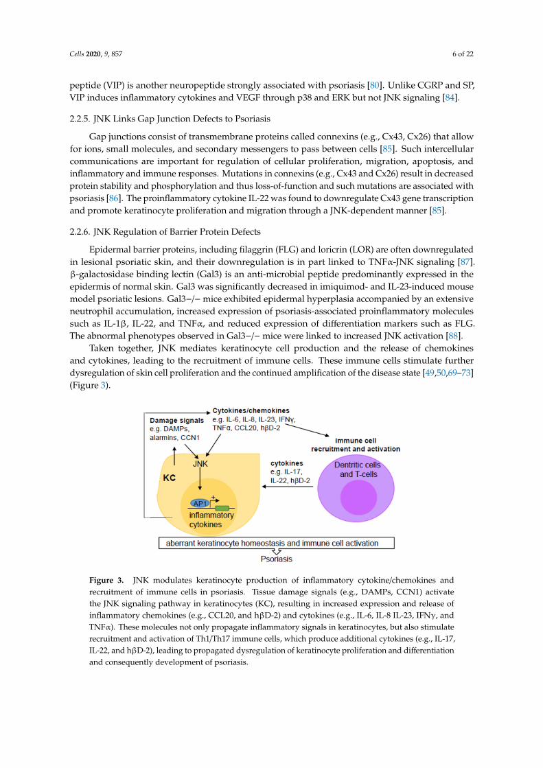
Taken together, JNK mediates keratinocyte cell production and the release of chemokinesand cytokines, leading to the recruitment of immune cells. These immune cells stimulate furtherdysregulation of skin cell proliferation and the continued amplification of the disease state [49,50,69–73](Figure 3).
Cells 2020, 9, x FOR PEER REVIEW 6 of 22
2.2.5. JNK Links Gap Junction Defects to Psoriasis
Gap junctions consist of transmembrane proteins called connexins (e.g., Cx43, Cx26) that allow for ions, small molecules, and secondary messengers to pass between cells [85]. Such intercellular communications are important for regulation of cellular proliferation, migration, apoptosis, and inflammatory and immune responses. Mutations in connexins (e.g., Cx43 and Cx26) result in decreased protein stability and phosphorylation and thus loss-of-function and such mutations are associated with psoriasis [86]. The proinflammatory cytokine IL-22 was found to downregulate Cx43 gene transcription and promote keratinocyte proliferation and migration through a JNK-dependent manner [85].
2.2.6. JNK Regulation of Barrier Protein Defects
Epidermal barrier proteins, including filaggrin (FLG) and loricrin (LOR) are often downregulated in lesional psoriatic skin, and their downregulation is in part linked to TNFα-JNK signaling [87]. β-galactosidase binding lectin (Gal3) is an anti-microbial peptide predominantly expressed in the epidermis of normal skin. Gal3 was significantly decreased in imiquimod- and IL-23-induced mouse model psoriatic lesions. Gal3−/− mice exhibited epidermal hyperplasia accompanied by an extensive neutrophil accumulation, increased expression of psoriasis-associated proinflammatory molecules such as IL-1β, IL-22, and TNFα, and reduced expression of differentiation markers such as FLG. The abnormal phenotypes observed in Gal3−/− mice were linked to increased JNK activation [88].
Taken together, JNK mediates keratinocyte cell production and the release of chemokines and cytokines, leading to the recruitment of immune cells. These immune cells stimulate further dysregulation of skin cell proliferation and the continued amplification of the disease state [49,50,69–73] (Figure 3).
Figure 3. JNK modulates keratinocyte production of inflammatory cytokine/chemokines and recruitment of immune cells in psoriasis. Tissue damage signals (e.g., DAMPs, CCN1) activate the JNK signaling pathway in keratinocytes (KC), resulting in increased expression and release of inflammatory chemokines (e.g., CCL20, and hβD-2) and cytokines (e.g., IL-6, IL-8 IL-23, IFNγ, and TNFα). These molecules not only propagate inflammatory signals in keratinocytes, but also stimulate recruitment and activation of Th1/Th17 immune cells, which produce additional cytokines (e.g., IL-17, IL-22, and hβD-2), leading to propagated dysregulation of keratinocyte proliferation and differentiation and consequently development of psoriasis.
Figure 3. JNK modulates keratinocyte production of inflammatory cytokine/chemokines andrecruitment of immune cells in psoriasis. Tissue damage signals (e.g., DAMPs, CCN1) activatethe JNK signaling pathway in keratinocytes (KC), resulting in increased expression and release ofinflammatory chemokines (e.g., CCL20, and hβD-2) and cytokines (e.g., IL-6, IL-8 IL-23, IFNγ, andTNFα). These molecules not only propagate inflammatory signals in keratinocytes, but also stimulaterecruitment and activation of Th1/Th17 immune cells, which produce additional cytokines (e.g., IL-17,IL-22, and hβD-2), leading to propagated dysregulation of keratinocyte proliferation and differentiationand consequently development of psoriasis.
Cells 2020, 9, 857 7 of 22
2.3. Dermal Fibrosis
2.3.1. Pathogenesis of Dermal Fibrosis
The fibrotic response is an integral component of normal wound healing and the repair process;however, the overactivation of the Th2 inflammatory response leads to fibrosis [89]. Scleroderma is anautoimmune disorder characterized by the hardening and tightening of the connective tissues [90,91].The etiology of scleroderma is complicated. It involves vascular injuries, immune activation, andconsequently excessive fibrosis of the skin and internal organs, including lung, gastrointestinal tract,and heart [92,93]. Central to the development and progression of fibrosis is the activation of residentfibroblasts, namely their differentiation into myofibroblasts, resulting in overproduction and impaireddegradation of extracellular matrix (ECM) components [93–96]. Myofibroblast differentiation isinitiated by profibrotic cytokines such as transforming growth factor-beta (TGFβ) and platelet-derivedgrowth factor (PDGF) [92,97–100].
2.3.2. JNK Connections with TGFβ and PDGF in Dermal Fibrosis
The mitogen-activated protein kinases (MAPKs), including JNK, have been linked to the aberrantactivation of fibroblasts and subsequent fibrosis [92,101]. JNK is activated by TGFβ and PDGFin Systemic sclerosis (SSc) fibroblasts. Inhibition of JNK by the selective small-molecule inhibitorCC-930 inhibited the release of extracellular matrix proteins by cultured fibroblasts, prevented fibrosisinduced by bleomycin, and in tight skin 1 (TSK1) mice, and most importantly induced regression ofpre-established fibrosis [92,102].
JNK phosphorylates c-Jun, leading to stabilization of c-Jun and enhanced transactivationactivity [103]. pc-Jun is increased in lesional skin biopsies from SSc patients compared with that ofhealthy control tissues and the increased staining was particularly observed in fibroblasts and theendothelial cells of small blood vessels of SSc samples [104]. JNK inhibition using CC-930 reduced thestimulatory effects of TGFβ and PDGF on c-Jun phosphorylation [92]. c-Jun activity was elevated inhuman SSc lesional skins, as well as mouse lesional skins induced by bleomycin or adenovirus-mediatedexpression of a constitutively active TGFβ receptor type I protein [104]. Similarly, pJNK is expressed atan elevated level in monocytes and neutrophils of scleroderma tissues compared to normal tissues [105].Conversely, ablation of JNK1 but not JNK2 globally or in airway epithelia resulted in a strong protectionfrom bleomycin and adenovirus-mediated expression of the active TGFβ [106,107]. Further, deletionof the Jnk1 allele in fibrotic skin induced a reversal of the fibrotic phenotype [107].
2.3.3. JNK Connections with STAT3 and WNT Signaling Pathways in Dermal Fibrosis
JNK mediates activation of the signal transducer and activator of transcription 3 (STAT3), which is amember of the STAT protein family implicated in tissue fibrosis [96]. STAT3 signaling is hyperactivatedin a TGFβ-dependent manner, and this activation is mediated by the combined actions of JAK, SRC,c-ABL, and JNK kinases in SSc fibroblasts [96]. Immunofluorescent staining detected elevated levelsof pJNK in fibroblasts of SSc skin compared to that of healthy skin. Inhibition of JNK by eithersiRNA-mediated gene knockdown or the small molecule inhibitor SP600125 inhibited TGFβ-inducedphosphorylation of STAT3, indicating that JNK plays an important role in TGFβ signaling andfibrosis [96].
JNK also mediates fibrosis driven by the Wnt signaling pathway [106,108,109]. Wnt signalingstimulates the release of collagen via JNK/c-Jun independent of the canonical Wnt/β-cateninsignaling [108]. Fibroblasts are the major source of canonical and non-canonical Wnt proteinssuch as Wnt-1, Wnt-10b, and Wnt-5a in SSc [109,110]. Evenness interrupted (EVI) is a multipasstransmembrane protein localized in the Golgi and at the cell surface, and it is essential for secretion ofcanonical and non-canonical Wnt ligands [109,111]. Knockdown of EVI in fibroblasts prevented therelease of Wnt ligands, accumulation of β-catenin, and phosphorylation of JNK/c-Jun [109].
Cells 2020, 9, 857 8 of 22
2.3.4. JNK Regulation of Extracellular Matrix Proteins in Dermal Fibrosis
Tissue fibrosis is a result of an imbalance of ECM deposition and degradation. TGFβupregulates type I collagen and TIMP metalloproteinase inhibitor 1(TIMP-1) and downregulatesmetalloproteinase-1(MMP1) [112]. MMP1 is a collagenase that breaks down interstitial and type I, II,and III collagens and critical for ECM remodeling [113,114]. JNK/AP1 (c-Fos/c-Jun), along with otherMAPKs and NF-κB, is crucial for IL-17 and rapamycin-induced MMP1 production in human dermal,cardiac, and lung fibroblasts [94,115,116]. JNK inhibition by SP600125 prevented the upregulation ofMMP1 by rapamycin and UVB in SSc dermal fibroblasts [93,117]. While AP1 is required for MMP1expression in SSc fibroblasts, AP1 inhibition with the small molecule compound T-5224 is found toincrease MMP1 mRNA levels in fibroblasts derived from healthy individuals [104]. However, anotherstudy showed that JNK inhibition in normal human dermal fibroblasts prevented UVB-induction ofMMP3, which promotes activation of other MMPs, including MMP1 and pro-MMPs and degradationof type I collagen [117]. Another cytokine linking JNK to fibrosis is monocyte chemoattractant protein1 (MCP-1, also known as CCL2), which is produced by SSc fibroblasts and promotes the inductionof MMP1 [118]. The secretion of MCP-1 is dependent on JNK-mediated signals and regulated byproteasomal degradation [118].
Besides actions downstream of TGFβ, JNK augments TGFβ gene transcription, induces expressionof enzymes responsible for activation of the latent form of TGFβ, and directly phosphorylates SMAD3,leading to enhanced transcription of pro-fibrotic molecules [119]. Consistently, blocking JNK activationsuppresses TGFβ1-induced fibrosis and inflammation [120].
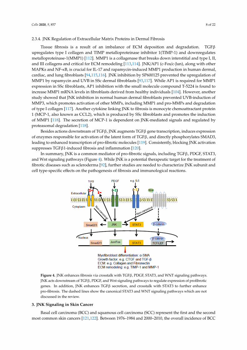
In summary, JNK is a common mediator of pro-fibrotic signals, including TGFβ, PDGF, STAT3,and Wnt signaling pathways (Figure 4). While JNK is a potential therapeutic target for the treatment offibrotic diseases such as scleroderma [92], further studies are needed to characterize JNK subunit andcell type-specific effects on the pathogenesis of fibrosis and immunological reactions.
Cells 2020, 9, x FOR PEER REVIEW 8 of 22
2.3.4. JNK Regulation of Extracellular Matrix Proteins in Dermal Fibrosis
Tissue fibrosis is a result of an imbalance of ECM deposition and degradation. TGFβ upregulates type I collagen and TIMP metalloproteinase inhibitor 1(TIMP-1) and downregulates metalloproteinase-1(MMP1) [112]. MMP1 is a collagenase that breaks down interstitial and type I, II, and III collagens and critical for ECM remodeling [113,114]. JNK/AP1 (c-Fos/c-Jun), along with other MAPKs and NF-κB, is crucial for IL-17 and rapamycin-induced MMP1 production in human dermal, cardiac, and lung fibroblasts [94,115,116]. JNK inhibition by SP600125 prevented the upregulation of MMP1 by rapamycin and UVB in SSc dermal fibroblasts [93,117]. While AP1 is required for MMP1 expression in SSc fibroblasts, AP1 inhibition with the small molecule compound T-5224 is found to increase MMP1 mRNA levels in fibroblasts derived from healthy individuals [104]. However, another study showed that JNK inhibition in normal human dermal fibroblasts prevented UVB-induction of MMP3, which promotes activation of other MMPs, including MMP1 and pro-MMPs and degradation of type I collagen [117]. Another cytokine linking JNK to fibrosis is monocyte chemoattractant protein 1 (MCP-1, also known as CCL2), which is produced by SSc fibroblasts and promotes the induction of MMP1 [118]. The secretion of MCP-1 is dependent on JNK-mediated signals and regulated by proteasomal degradation [118].
Besides actions downstream of TGFβ, JNK augments TGFβ gene transcription, induces expression of enzymes responsible for activation of the latent form of TGFβ, and directly phosphorylates SMAD3, leading to enhanced transcription of pro-fibrotic molecules [119]. Consistently, blocking JNK activation suppresses TGFβ1-induced fibrosis and inflammation [120].
In summary, JNK is a common mediator of pro-fibrotic signals, including TGFβ, PDGF, STAT3, and Wnt signaling pathways (Figure 4). While JNK is a potential therapeutic target for the treatment of fibrotic diseases such as scleroderma [92], further studies are needed to characterize JNK subunit and cell type-specific effects on the pathogenesis of fibrosis and immunological reactions.
Figure 4. JNK enhances fibrosis via crosstalk with TGFβ, PDGF, STAT3, and WNT signaling pathways. JNK acts downstream of TGFβ, PDGF, and Wnt signaling pathways to regulate expression of profibrotic genes. In addition, JNK enhances TGFβ secretion, and crosstalk with STAT3 to further enhance pro-fibrosis. The dashed lines show the canonical STAT3 and WNT signaling pathways which are not discussed in the review.
3. JNK Signaling in Skin Cancer
Basal cell carcinoma (BCC) and squamous cell carcinoma (SCC) represent the first and the second most common skin cancers [121,122]. Between 1976–1984 and 2000–2010, the overall incidence
Figure 4. JNK enhances fibrosis via crosstalk with TGFβ, PDGF, STAT3, and WNT signaling pathways.JNK acts downstream of TGFβ, PDGF, and Wnt signaling pathways to regulate expression of profibroticgenes. In addition, JNK enhances TGFβ secretion, and crosstalk with STAT3 to further enhancepro-fibrosis. The dashed lines show the canonical STAT3 and WNT signaling pathways which are notdiscussed in the review.
3. JNK Signaling in Skin Cancer
Basal cell carcinoma (BCC) and squamous cell carcinoma (SCC) represent the first and the secondmost common skin cancers [121,122]. Between 1976–1984 and 2000–2010, the overall incidence of BCC
Cells 2020, 9, 857 9 of 22
and SCC was increased by 145% and 263%, respectively [123]. Approximately 3 million cases of BCCand SCC were diagnosed in the US in 2019 [124,125]. Melanoma is the fifth most common cancer inmen and the sixth most common cancer in women [126]. An estimate of 192,310 new cases of melanomawas diagnosed in the US in 2019, with about 50% of them being invasive [125,127]. Common riskfactors for skin cancer include ultraviolet (UV), ionizing radiation, arsenic exposure, viral infection, andwounding [128–132]. JNK, as a dominant responder of these environmental stimuli, plays paradoxicalroles in cancer development with both oncogenic and tumor suppressor properties [133,134].
3.1. Differential Roles of JNK1 and JNK2 in SCC
JNK activation is frequently observed in SCC [135,136]. Specifically, JNK2 phosphorylation isincreased in SCC cell lines and tissues compared to normal keratinocytes and healthy skin samples,respectively [135,137]. Jnk2 deficient mice were resistant to skin cancer development followinginduction by the DMBA (7,12-dimethylbenz[α]anthracene)/TPA (12-O-tetradecanoylphorbol-13-acetate)two-stage carcinogenesis protocol, indicating that JNK2 functions as a promoter of skin cancer [138].Consistently, compared to WT mice, Mkk4 deficient mice displayed significantly reduced numbersof skin tumors after 20 weeks of DMBA/TPA treatment, which was attributed to reduced JNK2activity [139]. In contrast to JNK2, JNK1 showed a tumor suppressor function. Jnk1 deficient micedisplayed a higher papilloma incidence than that of wild-type mice [140]. In agreement with thesefindings, constitutively active MKK7 and MKK7-JNK2 fusion proteins, but not MKK7-JNK1, are ableto couple with the oncogenic Ras(V12) to transform normal keratinocytes into SCC-likes lesions [139]and this required intact c-Jun-function [135] and this required intact c-Jun-function [137]. In addition,c-Jun but not JunB can couple with Ras to induce epidermal malignancy [141]. Lastly, squamous cellcarcinoma antigen 1 (SCCA1) prevents keratinocytes from apoptotic cell death through inhibitionof JNK1 [142]. These data indicate that MKK7, JNK2, and c-Jun, but not JNK1 and JunB, promoteepidermal malignancy.
Epidermis-targeted expression of a catalytically deficient CYLD mutant (CYLDm) in K14-CYLDmtransgenic mice increased JNK activation and lysine-63 (K63)-ubiquitination and phosphorylation ofc-Jun and c-Fos transcription factors [143]. After DMBA/TPA treatment, K14-CYLDm mice developedincreased numbers of papilloma, with 66% of them developed into SCC and metastasis by week 32.Topical treatment of the JNK inhibitor SP600125 significantly reduced DMBA/TPA-induced tumorincidence and abolished skin cancer metastasis to lymph nodes in K14-CYLDm mice [143]. KDM4A isa demethylase that specifically demethylates the Lysine 9 and 36 residues of histone H3. In correlationwith increased KDM4A expression, c-Jun, and FOSL1 (Fra1), protein levels were increased in metastatichuman SCC tissues compared to primary SCC tissues [144]. Further, FRA1 was found to enhancehead and neck SCC cell proliferation and migration in a c-Jun-dependent manner [145].
3.2. JNK as a Key Mediator of the SHH, YAP, and WNT Signaling Pathways in BCC
The sonic hedgehog (SHH)/Gli signaling pathway plays a dominant role in BCC [146].JNK inhibition with SP600125 and siRNA knockdown of c-Jun inhibited Gli-induced cell cycleprogression, indicating that JNK and c-Jun are important for Hedgehog (HH)/Gli-driven BCC [147,148].In HaCaT keratinocytes, increased JNK expression was linked to the BCC-like phenotype inducedby SHH expression [149]. Interestingly, another study showed that the SHH/Gli signaling pathwayacts in synergy with the epidermal growth factor receptor (EGFR) to promote BCC, which requiresc-Jun activation by MEK/ERK, but not JNK [150]. In addition, c-Jun and Fos transcription factorsinteract with phosphorylated ATF2, and are required for ATF2-driven transformation of epidermalcells into BCC [151,152]. Moreover, in a BCC tumor model generated via subcutaneous injectionof TetON inducible CRISPR-Yap ASZ mouse cells into immunocompromised (nu/nu) mice, it wasfound that, after one-week treatment of Doxycycline, the Yap null tumors displayed reduced pJNK1/2and pJun(S63/S73) levels compared to those of WT BCC tumors [153]. In addition, c-Jun mRNA wassignificantly decreased in YAP-negative BCC clones and BCC cells treated with SP600125. Lastly,
Cells 2020, 9, 857 10 of 22
WNT16B, a member of the WNT gene family, was found upregulated in BCC tissues s and itsincreased expression enhanced proliferation of primary and immortalized human keratinocytes in aJNK-dependent manner [154]. Taken together, these data indicate that the JNK signaling pathway is acritical mediator acting downstream or in collaboration with SHH, YAP, and WNT signaling pathwaysto promote BCC [153,154].
3.3. Melanoma
3.3.1. JNK1 and JNK2 in Melanoma Growth and Progression
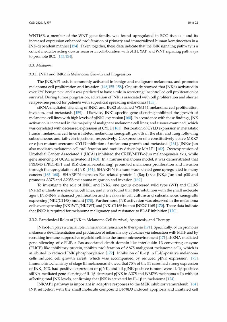
The JNK/AP1 axis is commonly activated in benign and malignant melanoma, and promotesmelanoma cell proliferation and invasion [148,155–158]. One study showed that JNK is activated inover 75% benign nevi and it was predicted to have a role in restricting uncontrolled cell proliferation orsurvival. During tumor progression, activation of JNK is associated with cell proliferation and shorterrelapse-free period for patients with superficial spreading melanomas [155].
siRNA-mediated silencing of JNK1 and JNK2 abolished WM164 melanoma cell proliferation,invasion, and metastasis [159]. Likewise, JNK1-specific gene silencing inhibited the growth ofmelanoma cell lines with high levels of pJNK1 expression [160]. In accordance with these findings, JNKactivation is increased in the majority of malignant melanoma cell lines, and tissues examined, whichwas correlated with decreased expression of CYLD [161]. Restoration of CYLD expression in metastatichuman melanoma cell lines inhibited melanoma xenograft growth in the skin and lung followingsubcutaneous and tail-vein injections, respectively. Coexpression of a constitutively active MKK7or c-Jun mutant overcame CYLD-inhibition of melanoma growth and metastasis [161]. JNK/c-Junalso mediates melanoma cell proliferation and motility driven by MALT1 [162]. Overexpression ofUrothelial Cancer Associated 1 (UCA1) inhibited the CREB/MITF/c-Jun melanogenesis axis, whilegene silencing of UCA1 activated it [163]. In a murine melanoma model, it was demonstrated thatPRDM5 (PRDI-BF1 and RIZ domain-containing) promoted melanoma proliferation and invasionthrough the upregulation of JNK [164]. SHARPIN is a tumor-associated gene upregulated in manycancers [165–168]. SHARPIN increases Ras-related protein 1 (Rap1) via JNK/c-Jun and p38 andpromotes A375 and A2058 melanoma migration and invasion [169].
To investigate the role of JNK1 and JNK2, one group expressed wild type (WT) and C116SJNK1/2 mutants in melanoma cell lines, and it was found that JNK inhibition with the small moleculeagent JNK-IN-8 enhanced proliferation and invasion in cell culture and subcutaneous xenograftsexpressing JNK2(C116S) mutant [170]. Furthermore, JNK activation was observed in the melanomacells overexpressing JNK1WT, JNK2WT, and JNK1C116S but not JNK2C116S [170]. These data indicatethat JNK2 is required for melanoma malignancy and resistance to BRAF inhibition [170].
3.3.2. Paradoxical Roles of JNK in Melanoma Cell Survival, Apoptosis, and Therapy
JNK/c-Jun plays a crucial role in melanoma resistance to therapies [171]. Specifically, c-Jun promotesmelanoma de-differentiation and production of inflammatory cytokines via interaction with MITF and byrecruiting immune-suppressive myeloid cells into the tumor microenvironment [171]. shRNA-mediatedgene silencing of c-FLIP, a Fas-associated death domain-like interleukin-1β-converting enzyme(FLICE)-like inhibitory protein, inhibits proliferation of A875 malignant melanoma cells, which isattributed to reduced JNK phosphorylation [172]. Inhibition of IL-1β in IL-lβ-positive melanomacells induced cell growth arrest, which was accompanied by reduced pJNK expression [173].Immunohistochemistry of stage III melanomas showed that 75% of the 51 cases had strong expressionof JNK, 20% had positive expression of pJNK, and all pJNK-positive tumors were IL-1β-positive.siRNA-mediated gene silencing of IL-1β decreased pJNK in A375 and WM793 melanoma cells withoutaffecting total JNK levels, confirming that JNK is activated by IL-1β in melanoma [174].
JNK/AP1 pathway is important in adaptive responses to the MEK inhibitor vemurafenib [164].JNK inhibition with the small molecule compound BI-78D3 induced apoptosis and inhibited cell
Cells 2020, 9, 857 11 of 22
growth of BRAF inhibitor-naive and resistant melanoma cells [175]. Melanoma cells treated with thePAK inhibitor PF-3758309 displayed increased activation of JNK, β-catenin, and the mTOR signalingpathway and shRNA-mediated gene silencing JNK and β-catenin further decreased melanoma cellviability [176]. In addition, JNK pathway inhibition sensitized BRAF mutant melanoma cells togenetically modified vaccinia virus-mediated cell death [163].
Paradoxically, JNK is also found to induce melanoma cell apoptosis. Quercetin, a plant-derivedpolyphenol compound, induced apoptosis of A375SM and A375P melanoma cells by increasing pJNKexpression both in vitro and in vivo [168]. Similarly, treatment of human SKMel-188 melanoma cell linewith Coriolus versicolor fungus-derived protein-bound polysaccharides induced apoptosis, and increasedROS levels, both of which were inhibited by SP600125 [169].
In summary, JNK proteins play important and distinct roles in different skin cancers (Figure 5).In BCC, JNK1/2 activates Jun/Fos, and enhances their interaction with phosphorylated ATF2, which thenenhances SHH/Gli induced tumorigenesis. In SCC, MKK4/7 activates JNK1 and JNK2. JNK1 inducesapoptosis, whereas JNK2 promotes carcinogenesis in an AP1-dependent manner. SCCA1 promotes SCCvia inhibition of JNK1, while CYLD inhibits SCC via suppression of JNK2/AP1 cascade. In melanoma,the MALT1, MKK4/7, and JNK/AP1 signaling cascade promotes melanoma cell proliferation andmigration, whereas CYLD inhibits it.
Cells 2020, 9, x FOR PEER REVIEW 11 of 22
PAK inhibitor PF-3758309 displayed increased activation of JNK, β-catenin, and the mTOR signaling pathway and shRNA-mediated gene silencing JNK and β-catenin further decreased melanoma cell viability [176]. In addition, JNK pathway inhibition sensitized BRAF mutant melanoma cells to genetically modified vaccinia virus-mediated cell death [163].
Paradoxically, JNK is also found to induce melanoma cell apoptosis. Quercetin, a plant-derived polyphenol compound, induced apoptosis of A375SM and A375P melanoma cells by increasing pJNK expression both in vitro and in vivo [168]. Similarly, treatment of human SKMel-188 melanoma cell line with Coriolus versicolor fungus-derived protein-bound polysaccharides induced apoptosis, and increased ROS levels, both of which were inhibited by SP600125 [169].
In summary, JNK proteins play important and distinct roles in different skin cancers (Figure 5). In BCC, JNK1/2 activates Jun/Fos, and enhances their interaction with phosphorylated ATF2, which then enhances SHH/Gli induced tumorigenesis. In SCC, MKK4/7 activates JNK1 and JNK2. JNK1 induces apoptosis, whereas JNK2 promotes carcinogenesis in an AP1-dependent manner. SCCA1 promotes SCC via inhibition of JNK1, while CYLD inhibits SCC via suppression of JNK2/AP1 cascade. In melanoma, the MALT1, MKK4/7, and JNK/AP1 signaling cascade promotes melanoma cell proliferation and migration, whereas CYLD inhibits it.
Figure 5. Differential roles of JNK1 and JNK2 in skin cancers. In BCC, JNK1/2 activates Jun/Fos, and enhances their interaction with phosphorylated ATF2, which then enhances SHH/Gli induced tumorigenesis. In SCC, MKK4/7 activates JNK1 and JNK2. JNK1 induces apoptosis, whereas JNK2 promotes carcinogenesis in an AP1-dependent manner. SCCA1 promotes SCC via inhibition of JNK1 and CYLD inhibits SCC via suppression of JNK2/AP1 cascade. In melanoma, the MALT1, MKK4/7, and JNK/AP1 signaling cascade promotes melanoma cell proliferation and migration, whereas CYLD inhibits it..
4. JNK as a Therapeutic Target
JNK has been recognized as a potential therapeutic target for many diseases. A number of peptides and small molecule inhibitors have been developed to either directly target JNK [136,165–167] or indirectly through inhibition of canonical and non-canonical JNK activators [168].
The ATP-competitive JNK inhibitors include small molecules from various scaffolds such as indazoles, pyridine carboxamides, aminopyrazoles, aminopyridines, benzothien-2-ylamides, benzothiazol-2-yl acetonitriles, quinoline derivatives, and aminopyrimidines [102,169,177–179]. Among these, CEP-1347, also named KT7515 or 3,9 bis [(ethylthio)methyl]-K252a, is a derivative from the natural compound K252a. CEP-1347 was found to prevent the death of neurons both in vitro and in vivo [180]. CEP-1347 induced differentiation and inhibited proliferation of human cancer cells, including glioblastoma (GS-Y01, GS-Y03, and GS-NCC01), pancreatic (PANC-1 CSLC), and ovarian (A2780 CSLC, and TOV21G CSLC) cancer cells [181]. Further, systemic administration of CEP-1347 at 1.5 mg/kg/day for 10 days significantly reduced tumor-initiating cancer stem cells within established tumors and prolonged the survival of mice receiving orthotopic implantation of glioma stem cells [181].
Figure 5. Differential roles of JNK1 and JNK2 in skin cancers. In BCC, JNK1/2 activates Jun/Fos,and enhances their interaction with phosphorylated ATF2, which then enhances SHH/Gli inducedtumorigenesis. In SCC, MKK4/7 activates JNK1 and JNK2. JNK1 induces apoptosis, whereas JNK2promotes carcinogenesis in an AP1-dependent manner. SCCA1 promotes SCC via inhibition of JNK1and CYLD inhibits SCC via suppression of JNK2/AP1 cascade. In melanoma, the MALT1, MKK4/7,and JNK/AP1 signaling cascade promotes melanoma cell proliferation and migration, whereas CYLDinhibits it.
4. JNK as a Therapeutic Target
JNK has been recognized as a potential therapeutic target for many diseases. A number ofpeptides and small molecule inhibitors have been developed to either directly target JNK [136,165–167]or indirectly through inhibition of canonical and non-canonical JNK activators [168].
The ATP-competitive JNK inhibitors include small molecules from various scaffolds suchas indazoles, pyridine carboxamides, aminopyrazoles, aminopyridines, benzothien-2-ylamides,benzothiazol-2-yl acetonitriles, quinoline derivatives, and aminopyrimidines [102,169,177–179].Among these, CEP-1347, also named KT7515 or 3,9 bis [(ethylthio)methyl]-K252a, is a derivative fromthe natural compound K252a. CEP-1347 was found to prevent the death of neurons both in vitroand in vivo [180]. CEP-1347 induced differentiation and inhibited proliferation of human cancer cells,including glioblastoma (GS-Y01, GS-Y03, and GS-NCC01), pancreatic (PANC-1 CSLC), and ovarian(A2780 CSLC, and TOV21G CSLC) cancer cells [181]. Further, systemic administration of CEP-1347 at
Cells 2020, 9, 857 12 of 22
1.5 mg/kg/day for 10 days significantly reduced tumor-initiating cancer stem cells within establishedtumors and prolonged the survival of mice receiving orthotopic implantation of glioma stem cells [181].
SP600125 is one of the most studied ATP-competitive JNK inhibitors derived fromanthrapyrazolone [182]. Its in vivo activity was demonstrated in mouse models of Parkinson’sdisease [183] and skin cancer (K14-CYLDm) [143]. SP600125 induced melanoma apoptosis and cellcycle arrest via the induction of p53, Bad, and Bax levels in 1205Lu and WM983B melanoma cells [160].Another drug, benzothiazolone AS601245, showed neuroprotective effects after focal cerebral ischemiain rats [184] and ischemia-reperfusion injury [185]. JNK-IN-8 is a novel compound that forms a covalentbond between the conserved cysteine in the ATP sites, leading to irreversible inhibition of all threeJNK proteins [136]. CC-930 is a potent JNK inhibitor that showed efficacy in inhibiting preclinicalmodels of dermal fibrosis induced by bleomycin and in the tight skin 1 (TSK1) mouse model [92,102].A phase I clinical study showed that CC-930 was well-tolerated in healthy volunteer patients, andinduced a dose-dependent reduction of dermal fibrosis in SSc diseases [186]. The phase II clinicaltrial of CC-930 in patients with idiopathic pulmonary fibrosis (IPF) showed similar pharmacokineticparameters to those found in the phase I [187]. Unfortunately, further preclinical trial (NCT01203943)of this compound was terminated due to the increased risk of liver damage [187].
Peptide inhibitors target protein-protein interactions between JNK and substrates such as c-Junand adaptor proteins such as JIP [188]. D-JNK-1 is a potent and membrane-permeable peptide inhibitorderived from the minimal JNK-binding region of JIP1 [189–191]. D-JNK-1 showed a neuroprotectiveeffect on animal models of stroke [180,192]. TI-JIP, another peptide derived from the JNK-bindingdomain of JIP-1 (amino acids 143–153), showed potent inhibition of JNK activity towards recombinantATF2, c-Jun, and Elk [190,191].
JNK inhibitors showed promising results in preclinical models, but their clinical benefit has notbeen appreciated so far. A major challenge with small molecular inhibitors is the non-specific sideeffects, as they target the highly conserved ATP-binding site, which are present in many differentMAPKs. For example, at higher concentrations, SP600125 not only inhibits the three JNK proteins [169],but also affects the closely related ERKs and p38 MAPKs [182,193].
5. Conclusions
JNK proteins regulate a multitude of cellular processes, including cell cycle, cell differentiation,cell proliferation, apoptosis, and inflammatory responses. Dysregulation of JNK signaling is inherentlylinked to psoriasis, skin fibrosis, and non-melanoma and melanoma skin cancers. Nevertheless,our understanding of JNK functions in these diseases is still limited and complicated by theisoform-specific and cell type specific responses. Further studies are needed to address JNKisoform-specific functions in a tissue type-specific manner and to better understand JNK upstream anddownstream molecules in various disease settings.
Author Contributions: All authors have read and agreed to the published version of the manuscript.
Funding: This work was in part supported by NIH/NIAMS grant to Jennifer Zhang (AR073858).
Conflicts of Interest: The authors declare no conflicts of interest.
References
1. Zeke, A.; Misheva, M.; Reményi, A.; Bogoyevitch, M.A. JNK Signaling: Regulation and Functions Based onComplex Protein-Protein Partnerships. Microbiol. Mol. Biol. Rev. 2016, 80, 793–835. [CrossRef] [PubMed]
2. Kannan, N.; Neuwald, A.F. Evolutionary constraints associated with functional specificity of the CMGCprotein kinases MAPK, CDK, GSK, SRPK, DYRK, and CK2alpha. Protein Sci. 2004, 13, 2059–2077. [CrossRef][PubMed]
3. Fedorov, O.; Marsden, B.; Pogacic, V.; Rellos, P.; Müller, S.; Bullock, A.N.; Schwaller, J.; Sundström, M.;Knapp, S. A systematic interaction map of validated kinase inhibitors with Ser/Thr kinases. Proc. Natl. Acad.Sci. USA 2007, 104, 20523–20528. [CrossRef]
Cells 2020, 9, 857 13 of 22
4. Bode, A.M.; Dong, Z. The functional contrariety of JNK. Mol. Carcinog. 2007, 46, 591–598. [CrossRef]5. Gupta, S.; Barrett, T.; Whitmarsh, A.J.; Cavanagh, J.; Sluss, H.K.; Dérijard, B.; Davis, R.J. Selective interaction
of JNK protein kinase isoforms with transcription factors. EMBO J. 1996, 15, 2760–2770. [CrossRef]6. Bogoyevitch, M.A.; Kobe, B. Uses for JNK: The many and varied substrates of the c-Jun N-terminal kinases.
Microbiol. Mol. Biol. Rev. 2006, 70, 1061–1095. [CrossRef]7. Biteau, B.; Karpac, J.; Hwangbo, D.; Jasper, H. Regulation of Drosophila lifespan by JNK signaling.
Exp. Gerontol. 2011, 46, 349–354. [CrossRef]8. Seki, E.; Brenner, D.A.; Karin, M. A Liver Full of JNK: Signaling in Regulation of Cell Function and Disease
Pathogenesis, and Clinical Approaches. Gastroenterology 2012, 143, 307–320. [CrossRef]9. Kusumaningrum, N.; Lee, D.H.; Yoon, H.-S.; Kim, Y.K.; Park, C.-H.; Chung, J.H. Gasdermin C is induced by
ultraviolet light and contributes to MMP-1 expression via activation of ERK and JNK pathways. J. Dermatol.Sci. 2018, 90, 180–189. [CrossRef]
10. Dhanasekaran, D.N.; Reddy, E.P. JNK signaling in apoptosis. Oncogene 2008, 27, 6245–6251. [CrossRef]11. Kyriakis, J.M.; Avruch, J. Mammalian Mitogen-Activated Protein Kinase Signal Transduction Pathways
Activated by Stress and Inflammation. Physiol. Rev. 2001, 81, 807–869. [CrossRef] [PubMed]12. Wang, X.; Destrument, A.; Tournier, C. Physiological Roles of MKK4 and MKK7: Insights from Animal
Models. Biochim. Biophys. Acta 2007, 1773, 1349–1357. [CrossRef] [PubMed]13. Chadee, D.N.; Kyriakis, J.M. Activation of SAPK/JNKs in vitro. Methods Mol. Biol. 2010, 661, 59–73.
[CrossRef] [PubMed]14. Wang, H.M.; Yan, Q.; Yang, T.; Cheng, H.; Du, J.; Yoshioka, K.; Kung, S.K.P.; Ding, G.H. Scaffold protein JLP
is critical for CD40 signaling in B lymphocytes. J. Biol. Chem. 2015, 290, 5256–5266. [CrossRef] [PubMed]15. Wilhelm, M.; Kukekov, N.V.; Schmit, T.L.; Biagas, K.V.; Sproul, A.A.; Gire, S.; Maes, M.E.; Xu, Z.; Greene, L.A.
Sh3rf2/POSHER protein promotes cell survival by ring-mediated proteasomal degradation of the c-JunN-terminal kinase scaffold POSH (plenty of SH3s) protein. J. Biol. Chem. 2012, 287, 2247–2256. [CrossRef][PubMed]
16. Morrison, D.K.; Davis, R.J. Regulation of MAP Kinase Signaling Modules by Scaffold Proteins in Mammals.Annu. Rev. Cell Dev. Biol. 2003, 19, 91–118. [CrossRef]
17. Yoshioka, K. Scaffold Proteins in Mammalian MAP Kinase Cascades. J. Biochem. 2004, 135, 657–661.[CrossRef]
18. Kutluk Oktay, E.B.O.O.M.O.; Filippo, G.G. The c-Jun N-terminal kinase JNK functions upstream of Aurora Bto promote entry into mitosis. Cell Cyle 2008, 7, 533–541. [CrossRef]
19. Ramsdale, R.; Jorissen, R.N.; Li, F.Z.; Al-Obaidi, S.; Ward, T.; Sheppard, K.E.; Bukczynska, P.E.; Young, R.J.;Boyle, S.E.; Shackleton, M.; et al. The transcription cofactor c-JUN mediates phenotype switching and BRAFinhibitor resistance in melanoma. Sci. Signal. 2015, 8, ra82. [CrossRef]
20. Yarza, R.; Vela, S.; Solas, M.; Ramirez, M.J. c-Jun N-terminal Kinase (JNK) Signaling as a Therapeutic Targetfor Alzheimer’s Disease. Front. Pharmacol. 2015, 6, 321. [CrossRef]
21. Wada, T.; Joza, N.; Cheng, H.-Y.M.; Sasaki, T.; Kozieradzki, I.; Bachmaier, K.; Katada, T.; Schreiber, M.;Wagner, E.F.; Nishina, H.; et al. MKK7 couples stress signalling to G2/M cell-cycle progression and cellularsenescence. Nat. Cell Biol. 2004, 6, 215–226. [CrossRef] [PubMed]
22. Yamamoto, K.; Ichijo, H.; Korsmeyer, S.J. BCL-2 is phosphorylated and inactivated by an ASK1/Jun N-terminalprotein kinase pathway normally activated at G(2)/M. Mol. Cell. Biol. 1999, 19, 8469–8478. [CrossRef][PubMed]
23. MacCorkle-Chosnek, R.A.; VanHooser, A.; Goodrich, D.W.; Brinkley, B.R.; Tan, T.-H. Cell Cycle Regulation ofc-Jun N-Terminal Kinase Activity at the Centrosomes. Biochem. Biophys. Res. Commun. 2001, 289, 173–180.[CrossRef] [PubMed]
24. Bakiri, L.; Lallemand, D.; Bossy-Wetzel, E.; Yaniv, M. Cell cycle-dependent variations in c-Jun and JunBphosphorylation: A role in the control of cyclin D1 expression. EMBO J. 2000, 19, 2056–2068. [CrossRef][PubMed]
25. Gazon, H.; Barbeau, B.; Mesnard, J.M.; Peloponese, J.M. Hijacking of the AP-1 Signaling Pathway duringDevelopment of ATL; Frontiers Media S.A.: Lausanne, Switzerland, 2018; Volume 8.
26. Feehan, R.P.; Shantz, L.M. Molecular signaling cascades involved in nonmelanoma skin carcinogenesis.Biochem. J. 2016, 473, 2973–2994. [CrossRef]
27. Shaulian, E.; Karin, M. AP-1 in Cell Proliferation and Survival. Oncogene 2001, 20, 2390–2400. [CrossRef]
Cells 2020, 9, 857 14 of 22
28. Jin, Y.-J.; Park, I.; Hong, I.-K.; Byun, H.-J.; Choi, J.; Kim, Y.-M.; Lee, H. Fibronectin and vitronectin induceAP-1-mediated matrix metalloproteinase-9 expression through integrin α5β1/αvβ3-dependent Akt, ERK andJNK signaling pathways in human umbilical vein endothelial cells. Cell. Signal. 2011, 23, 125–134. [CrossRef]
29. Chen, Y.R.; Tan, T.H. The c-Jun N-Terminal Kinase Pathway and Apoptotic Signaling (Review). Int. J. Oncol.2000, 16, 651–662. [CrossRef]
30. Lamb, J.A.; Ventura, J.-J.; Hess, P.; Flavell, R.A.; Davis, R.J. JunD mediates survival signaling by the JNKsignal transduction pathway. Mol. Cell 2003, 11, 1479–1489. [CrossRef]
31. Grabiec, A.M.; Angiolilli, C.; Hartkamp, L.M.; Van Baarsen, L.G.M.; Tak, P.P.; Reedquist, K.A. JNK-dependentdownregulation of FoxO1 is required to promote the survival of fibroblast-like synoviocytes in rheumatoidarthritis. Ann. Rheum. Dis. 2015, 74, 1763–1771. [CrossRef]
32. Wu, Q.; Wu, W.; Fu, B.; Shi, L.; Wang, X.; Kuca, K. JNK Signaling in Cancer Cell Survival; John Wiley and SonsInc.: Hoboken, NJ, USA, 2019; Volume 39, pp. 2082–2104.
33. Jones, E.V.; Dickman, M.J.; Whitmarsh, A.J. Regulation of p73-mediated apoptosis by c-Jun N-terminal kinase.Biochem. J. 2007, 405, 617–623. [CrossRef] [PubMed]
34. Wolf, E.R.; McAtarsney, C.P.; Bredhold, K.E.; Kline, A.M.; Mayo, L.D. Mutant and wild-type p53 formcomplexes with p73 upon phosphorylation by the kinase JNK. Sci. Signal. 2018, 11, eaao4170. [CrossRef][PubMed]
35. Hu, L.; Zhang, T.; Liu, D.; Guan, G.; Huang, J.; Proksch, P.; Chen, X.; Lin, W. Notoamide-type alkaloidinduced apoptosis and autophagy: Via a P38/JNK signaling pathway in hepatocellular carcinoma cells.RSC Adv. 2019, 9, 19855–19868. [CrossRef]
36. Gong, X.; Wang, M.; Tashiro, S.-I.; Onodera, S.; Ikejima, T. Involvement of JNK-Initiated p53 Accumulationand Phosphorylation of p53 in Pseudolaric Acid B Induced Cell Death. Exp. Mol. Med. 2006, 428–434.[CrossRef]
37. Annunziato, F.; Romagnani, C.; Romagnani, S. The 3 major types of innate and adaptive cell-mediatedeffector immunity. J. Allergy Clin. Immunol. 2015, 135, 626–635. [CrossRef] [PubMed]
38. van Oosterhout, A.J.M.; Motta, A.C. Th1/Th2 paradigm: Not seeing the forest for the trees? Eur. Respir. J.2005, 25, 591. [CrossRef]
39. Kaiko, G.E.; Horvat, J.C.; Beagley, K.W.; Hansbro, P.M. Immunological decision-making: How does theimmune system decide to mount a helper T-cell response? Immunology 2008, 123, 326–338. [CrossRef]
40. Kidd, P. Th1/Th2 balance: The hypothesis, its limitations, and implications for health and disease. Altern. Med.Rev. 2003, 8, 223–246.
41. Gieseck, R.L.; Wilson, M.S.; Wynn, T.A. Type 2 Immunity in Tissue Repair and Fibrosis; Nature PublishingGroup: Berlin, Germany, 2018; Volume 18, pp. 62–76.
42. Davis, R.J. Signal transduction by the JNK group of MAP kinases. Cell 2000, 103, 239–252. [CrossRef]43. Dong, C.; Davis, R.J.; Flavell, R.A. MAP K INASES IN THE I MMUNE R ESPONSE. Annu. Rev. Immunol.
2002, 20, 55–72. [CrossRef]44. Ghosh, S.; Karin, M. Missing pieces in the NF-kappaB puzzle. Cell 2002, 109, S81–S96. [CrossRef]45. Rincón, M.; Davis, R.J. Regulation of the immune response by stress-activated protein kinases. Immunol. Rev.
2009, 228, 212–224. [CrossRef] [PubMed]46. Huang, G.; Shi, L.Z.; Chi, H. Regulation of JNK and p38 MAPK in the immune system: Signal integration,
propagation and termination. Cytokine 2009, 48, 161–169. [CrossRef] [PubMed]47. Han, M.S.; Jung, D.Y.; Morel, C.; Lakhani, S.A.; Kim, J.K.; Flavell, R.A.; Davis, R.J. JNK Expression by
Macrophages Promotes Obesity-Induced Insulin Resistance and Inflammation. Science 2013, 339, 218–222.[CrossRef] [PubMed]
48. Cunningham, C.A.; Cardwell, L.N.; Guan, Y.; Teixeiro, E.; Daniels, M.A. POSH Regulates CD4+ T CellDifferentiation and Survival. J. Immunol. 2016, 196, 4003–4013. [CrossRef] [PubMed]
49. Shebzukhov, Y.V.; Stanislawiak, S.; Bezhaeva, T.R.; Nedospasov, S.A.; Kuprash, D.V. Low level of Lck kinasein Th2 cells limits expression of CD4 co-receptor and S73 phosphorylation of transcription factor c-Jun.Sci. Rep. 2017, 7, 2339. [CrossRef] [PubMed]
50. Coquet, J.M.; Middendorp, S.; van der Horst, G.; Kind, J.; Veraar, E.A.M.; Xiao, Y.; Jacobs, H.; Borst, J.The CD27 and CD70 Costimulatory Pathway Inhibits Effector Function of T Helper 17 Cells and AttenuatesAssociated Autoimmunity. Immunity 2013, 38, 53–65. [CrossRef]
51. Di Meglio, P.; Villanova, F.; Nestle, F.O. Psoriasis. Cold Spring Harb. Perspect. Med. 2014, 4, a015354. [CrossRef]
Cells 2020, 9, 857 15 of 22
52. Boehncke, W.-H.; Schön, M.P. Psoriasis. Lancet 2015, 386, 983–994. [CrossRef]53. Lowes, M.A.; Kikuchi, T.; Fuentes-Duculan, J.; Cardinale, I.; Zaba, L.C.; Haider, A.S.; Bowman, E.P.;
Krueger, J.G. Psoriasis vulgaris lesions contain discrete populations of Th1 and Th17 T cells. J. Investig.Dermatol. 2008, 128, 1207–1211. [CrossRef]
54. Chiricozzi, A.; Romanelli, P.; Volpe, E.; Borsellino, G.; Romanelli, M. Scanning the Immunopathogenesis ofPsoriasis. Int. J. Mol. Sci. 2018, 19, 179. [CrossRef] [PubMed]
55. Lowes, M.A.; Suárez-Fariñas, M.; Krueger, J.G. Immunology of Psoriasis. Annu. Rev. Immunol. 2014, 32,227–255. [CrossRef] [PubMed]
56. Kotb, I.S.; Lewis, B.J.; Barker, R.N.; Ormerod, A.D. Differential effects of phototherapy, adalimumab andbetamethasone-calcipotriol on effector and regulatory T cells in psoriasis. Br. J. Derm. 2018, 179, 127–135.[CrossRef] [PubMed]
57. Elder, J.T. PSORS1: Linking Genetics and Immunology. J. Investig. Dermatol. 2006, 126, 1205–1206. [CrossRef]58. Jordan, C.T.; Cao, L.; Roberson, E.D.O.; Pierson, K.C.; Yang, C.F.; Joyce, C.E.; Ryan, C.; Duan, S.; Helms, C.A.;
Liu, Y.; et al. PSORS2 is due to mutations in CARD14. Am. J. Hum. Genet. 2012, 90, 784–795. [CrossRef]59. Tsoi, L.C.; Spain, S.L.; Knight, J.; Ellinghaus, E.; Stuart, P.E.; Capon, F.; Ding, J.; Li, Y.; Tejasvi, T.;
Gudjonsson, J.E.; et al. Identification of 15 new psoriasis susceptibility loci highlights the role of innateimmunity. Nat. Genet. 2012, 44, 1341–1348. [CrossRef]
60. Singh, S.; Pradhan, D.; Puri, P.; Ramesh, V.; Aggarwal, S.; Nayek, A.; Jain, A.K. Genomic Alterations DrivingPsoriasis Pathogenesis; Elsevier B.V.: Amsterdam, The Netherlands, 2019; Volume 683, pp. 61–71.
61. Wang, H.; Chan, H.H.; Ni, M.Y.; Lam, W.W.; Chan, W.M.M.; Pang, H. Bacteriophage of the Skin Microbiomein Patients with Psoriasis and Healthy Family Controls. J. Investig. Dermatol. 2020, 140, 182–190 e185.[CrossRef]
62. Loesche, M.A.; Farahi, K.; Capone, K.; Fakharzadeh, S.; Blauvelt, A.; Duffin, K.C.; DePrimo, S.E.;Muñoz-Elías, E.J.; Brodmerkel, C.; Dasgupta, B.; et al. Longitudinal Study of the Psoriasis-Associated SkinMicrobiome during Therapy with Ustekinumab in a Randomized Phase 3b Clinical Trial. J. Investig. Dermatol.2018, 138, 1973–1981. [CrossRef]
63. Afonina, I.S.; Van Nuffel, E.; Baudelet, G.; Driege, Y.; Kreike, M.; Staal, J.; Beyaert, R. The paracaspase MALT1mediates CARD14-induced signaling in keratinocytes. EMBO Rep. 2016, 17, 914–927. [CrossRef]
64. Blonska, M.; Lin, X. NF-κB Signaling Pathways Regulated by CARMA Family of Scaffold Proteins. Cell Res.2011, 21, 55–70. [CrossRef]
65. Hulpiau, P.; Driege, Y.; Staal, J.; Beyaert, R. MALT1 is not alone after all: Identification of novel paracaspases.Cell. Mol. Life Sci. 2016, 73, 1103–1116. [CrossRef] [PubMed]
66. Hori, S.; Nomura, T.; Sakaguchi, S. Control of Regulatory T Cell Development by the Transcription FactorFoxp3. Science 2003, 299, 1057. [CrossRef] [PubMed]
67. Mu, J.; Tai, X.; Iyer, S.S.; Weissman, J.D.; Singer, A.; Singer, D.S. Regulation of MHC Class I Expression byFoxp3 and Its Effect on Regulatory T Cell Function. J. Immunol. 2014, 192, 2892–2903. [CrossRef] [PubMed]
68. Lopes, J.E.; Torgerson, T.R.; Schubert, L.A.; Anover, S.D.; Ocheltree, E.L.; Ochs, H.D.; Ziegler, S.F. Analysis ofFOXP3 Reveals Multiple Domains Required for Its Function as a Transcriptional Repressor. J. Immunol. 2006,177, 3133–3142. [CrossRef]
69. Chen, L.; Wu, J.; Ren, W.; Yang, X.; Shen, Z. c-Jun N-terminal kinase (JNK)-phospho-c-JUN (ser63/73) pathwayis essential for FOXP3 nuclear translocation in psoriasis. J. Dermatol. Sci. 2013, 69, 114–121. [CrossRef]
70. Gao, L.; Li, K.; Li, F.; Li, H.; Liu, L.; Wang, L.; Zhang, Z.; Gao, T.; Liu, Y. Polymorphisms in the FOXP3 gene inHan Chinese psoriasis patients. J. Dermatol. Sci. 2010, 57, 51–56. [CrossRef]
71. Wu, P.; Ma, G.; Zhu, X.; Gu, T.; Zhang, J.; Sun, Y.; Xu, H.; Huo, R.; Wang, B.; Shen, B.; et al. Cyr61/CCN1is involved in the pathogenesis of psoriasis vulgaris via promoting IL-8 production by keratinocytes in aJNK/NF-κB pathway. Clin. Immunol. 2017, 174, 53–62. [CrossRef]
72. Li, H.; Li, H.; Huo, R.; Wu, P.; Shen, Z.; Xu, H.; Shen, B.; Li, N. Cyr61/CCN1 induces CCL20 productionby keratinocyte via activating p38 and JNK/AP-1 pathway in psoriasis. J. Dermatol. Sci. 2017, 88, 46–56.[CrossRef]
73. Yang, D.; Chertov, O.; Bykovskaia, S.N.; Chen, Q.; Buffo, M.J.; Shogan, J.; Anderson, M.; Schröder, J.M.;Wang, J.M.; Howard, O.M.Z.; et al. β-Defensins: Linking innate and adaptive immunity through dendriticand T cell CCR6. Science 1999, 286, 525–528. [CrossRef]
Cells 2020, 9, 857 16 of 22
74. Niyonsaba, F.; Ushio, H.; Nakano, N.; Ng, W.; Sayama, K.; Hashimoto, K.; Nagaoka, I.; Okumura, K.; Ogawa, H.Antimicrobial peptides human β-defensins stimulate epidermal keratinocyte migration, proliferation andproduction of proinflammatory cytokines and chemokines. J. Investig. Dermatol. 2007, 127, 594–604.[CrossRef]
75. Kanda, N.; Kamata, M.; Tada, Y.; Ishikawa, T.; Sato, S.; Watanabe, S. Human β-defensin-2 enhances IFN-γand IL-10 production and suppresses IL-17 production in T cells. J. Leukoc. Biol. 2011, 89, 935–944. [CrossRef][PubMed]
76. Karakawa, M.; Komine, M.; Hanakawa, Y.; Tsuda, H.; Sayama, K.; Tamaki, K.; Ohtsuki, M. CCL27 isdownregulated by interferon gamma via epidermal growth factor receptor in normal human epidermalkeratinocytes. J. Cell. Physiol. 2014, 229, 1935–1945. [CrossRef] [PubMed]
77. Arasa, J.; Terencio, M.C.; Andrés, R.M.; Marín-Castejón, A.; Valcuende-Cavero, F.; Payá, M.; Montesinos, M.C.Defective Induction of COX-2 Expression by Psoriatic Fibroblasts Promotes Pro-inflammatory Activation ofMacrophages. Front. Immunol. 2019, 10, 536. [CrossRef] [PubMed]
78. Lotti, T.; D’Erme, A.M.; Hercogová, J. The Role of Neuropeptides in the Control of Regional Immunity; ElsevierInc.: Amsterdam, The Netherlands, 2014; Volume 32, pp. 633–645.
79. Granstein, R.D.; Wagner, J.A.; Stohl, L.L.; Ding, W. Calcitonin gene-related peptide: Key regulator ofcutaneous immunity. Acta Physiol. 2015, 213, 586–594. [CrossRef]
80. Reich, A.; Orda, A.; Wisnicka, B.; Szepietowski, J.C. Plasma concentration of selected neuropeptides inpatients suffering from psoriasis. Exp. Dermatol. 2007, 16, 421–428. [CrossRef]
81. Yu, X.J.; Li, C.Y.; Xu, Y.H.; Chen, L.M.; Zhou, C.L. Calcitonin gene-related peptide increases proliferation ofhuman HaCaT keratinocytes by activation of MAP kinases. Cell Biol. Int. 2009, 33, 1144–1148. [CrossRef]
82. Theoharides, T.C.; Zhang, B.; Kempuraj, D.; Tagen, M.; Vasiadi, M.; Angelidou, A.; Alysandratos, K.D.;Kalogeromitros, D.; Asadi, S.; Stavrianeas, N.; et al. IL-33 augments substance P-induced VEGF secretionfrom human mast cells and is increased in psoriatic skin. Proc. Natl. Acad. Sci. USA 2010, 107, 4448–4453.[CrossRef]
83. Ostrowski, S.M.; Belkadi, A.; Loyd, C.M.; Diaconu, D.; Ward, N.L. Cutaneous denervation of psoriasiformmouse skin improves acanthosis and inflammation in a sensory neuropeptide-dependent manner. J. Investig.Dermatol. 2011, 131, 1530–1538. [CrossRef]
84. Yu, X.J.; Ren, X.H.; Xu, Y.H.; Chen, L.M.; Zhou, C.L.; Li, C.Y. Vasoactive intestinal peptide induces vascularendothelial growth factor production in human HaCaT keratinocytes via MAPK pathway. Neuropeptides2010, 44, 407–411. [CrossRef]
85. Liang, J.; Chen, P.; Li, C.; Li, D.; Wang, J.; Xue, R.; Zhang, S.; Ruan, J.; Zhang, X. IL-22 Down-Regulates Cx43Expression and Decreases Gap Junctional Intercellular Communication by Activating the JNK Pathway inPsoriasis. J. Investig. Dermatol. 2019, 139, 400–411. [CrossRef]
86. Langlois, S.; Maher, A.C.; Manias, J.L.; Shao, Q.; Kidder, G.M.; Laird, D.W. Connexin levels regulatekeratinocyte differentiation in the epidermis. J. Biol. Chem. 2007, 282, 30171–30180. [CrossRef]
87. Kim, B.E.; Howell, M.D.; Guttman, E.; Gilleaudeau, P.M.; Cardinale, I.R.; Boguniewicz, M.; Krueger, J.G.;Leung, D.Y.M. TNF-α downregulates filaggrin and loricrin through c-Jun N-terminal kinase: Role for TNF-αantagonists to improve skin barrier. J. Investig. Dermatol. 2011, 131, 1272–1279. [CrossRef] [PubMed]
88. Shi, Z.R.; Tan, G.Z.; Cao, C.X.; Han, Y.F.; Meng, Z.; Man, X.Y.; Jiang, Z.X.; Zhang, Y.P.; Dang, N.N.; Wei, K.H.;et al. Decrease of galectin-3 in keratinocytes: A potential diagnostic marker and a critical contributor to thepathogenesis of psoriasis. J. Autoimmun. 2018, 89, 30–40. [CrossRef] [PubMed]
89. Wynn, T.A. Fibrotic disease and the T(H)1/T(H)2 paradigm. Nat. Rev. Immunol 2004, 4, 583–594. [CrossRef][PubMed]
90. Thomas, R.M.; Worswick, S.; Aleshin, M. Retinoic acid for treatment of systemic sclerosis and morphea:A literature review. Dermatol. Ther. 2017, 30. [CrossRef]
91. Sharma, A. Scleroderma-like Disorders. Curr. Rheumatol. Rev. 2018, 14, 22–27. [CrossRef]92. Reich, N.; Tomcik, M.; Zerr, P.; Lang, V.; Dees, C.; Avouac, J.; Palumbo, K.; Horn, A.; Akhmetshina, A.;
Beyer, C.; et al. Jun N-terminal kinase as a potential molecular target for prevention and treatment of dermalfibrosis. Ann. Rheum. Dis. 2012, 71, 737–745. [CrossRef]
93. Tamaki, Z.; Asano, Y.; Kubo, M.; Ihn, H.; Tada, Y.; Sugaya, M.; Kadono, T.; Sato, S. Effects of theimmunosuppressant rapamycin on the expression of human alpha2(I) collagen and matrix metalloproteinase1 genes in scleroderma dermal fibroblasts. J. Dermatol. Sci. 2014, 74, 251–259. [CrossRef]
Cells 2020, 9, 857 17 of 22
94. Brembilla, N.C.; Montanari, E.; Truchetet, M.E.; Raschi, E.; Meroni, P.; Chizzolini, C. Th17 cells favorinflammatory responses while inhibiting type I collagen deposition by dermal fibroblasts: Differential effectsin healthy and systemic sclerosis fibroblasts. Arthritis Res. Ther. 2013, 15, R151. [CrossRef]
95. Gilbane, A.J.; Denton, C.P.; Holmes, A.M. Scleroderma pathogenesis: A pivotal role for fibroblasts as effectorcells. Arthritis Res. Ther. 2013, 15, 215. [CrossRef]
96. Chakraborty, D.; Sumova, B.; Mallano, T.; Chen, C.W.; Distler, A.; Bergmann, C.; Ludolph, I.; Horch, R.E.;Gelse, K.; Ramming, A.; et al. Activation of STAT3 integrates common profibrotic pathways to promotefibroblast activation and tissue fibrosis. Nat. Commun. 2017, 8, 1130. [CrossRef]
97. Hu, H.H.; Chen, D.Q.; Wang, Y.N.; Feng, Y.L.; Cao, G.; Vaziri, N.D.; Zhao, Y.Y. New insights intoTGF-beta/Smad signaling in tissue fibrosis. Chem. -Biol. Interact. 2018, 292, 76–83. [CrossRef] [PubMed]
98. Meng, X.M.; Nikolic-Paterson, D.J.; Lan, H.Y. TGF-beta: The master regulator of fibrosis. Nat. Rev. Nephrol.2016, 12, 325–338. [CrossRef] [PubMed]
99. Klinkhammer, B.M.; Floege, J.; Boor, P. PDGF in organ fibrosis. Mol. Asp. Med. 2018, 62, 44–62. [CrossRef][PubMed]
100. Ying, H.Z.; Chen, Q.; Zhang, W.Y.; Zhang, H.H.; Ma, Y.; Zhang, S.Z.; Fang, J.; Yu, C.H. PDGF signalingpathway in hepatic fibrosis pathogenesis and therapeutics (Review). Mol. Med. Rep. 2017, 16, 7879–7889.[CrossRef] [PubMed]
101. Finnson, K.W.; Almadani, Y.; Philip, A. Non-canonical (non-SMAD2/3) TGF-beta signaling in fibrosis:Mechanisms and targets. Semin. Cell Dev. Biol. 2019, in press. [CrossRef]
102. Plantevin Krenitsky, V.; Nadolny, L.; Delgado, M.; Ayala, L.; Clareen, S.S.; Hilgraf, R.; Albers, R.; Hegde, S.;D’Sidocky, N.; Sapienza, J.; et al. Discovery of CC-930, an orally active anti-fibrotic JNK inhibitor. BioorganicMed. Chem. Lett. 2012, 22, 1433–1438. [CrossRef]
103. Sabapathy, K. Role of the JNK pathway in human diseases. Prog. Mol. Biol. Transl. Sci. 2012, 106, 145–169.[CrossRef]
104. Avouac, J.; Palumbo, K.; Tomcik, M.; Zerr, P.; Dees, C.; Horn, A.; Maurer, B.; Akhmetshina, A.; Beyer, C.;Sadowski, A.; et al. Inhibition of activator protein 1 signaling abrogates transforming growth factorbeta-mediated activation of fibroblasts and prevents experimental fibrosis. Arthritis Rheum. 2012, 64,1642–1652. [CrossRef]
105. Tourkina, E.; Richard, M.; Oates, J.; Hofbauer, A.; Bonner, M.; Gooz, P.; Visconti, R.; Zhang, J.; Znoyko, S.;Hatfield, C.M.; et al. Caveolin-1 regulates leucocyte behaviour in fibrotic lung disease. Ann. Rheum. Dis.2010, 69, 1220–1226. [CrossRef]
106. Beyer, C.; Reichert, H.; Akan, H.; Mallano, T.; Schramm, A.; Dees, C.; Palumbo-Zerr, K.; Lin, N.Y.; Distler, A.;Gelse, K.; et al. Blockade of canonical Wnt signalling ameliorates experimental dermal fibrosis. Ann. Rheum.Dis. 2013, 72, 1255–1258. [CrossRef] [PubMed]
107. van der Velden, J.L.; Alcorn, J.F.; Chapman, D.G.; Lundblad, L.K.A.; Irvin, C.G.; Davis, R.J.; Butnor, K.;Janssen-Heininger, Y.M.W. Airway epithelial specific deletion of Jun-N-terminal kinase 1 attenuatespulmonary fibrosis in two independent mouse models. PLoS ONE 2020, 15, e0226904. [CrossRef] [PubMed]
108. Zhang, P.; Cai, Y.; Soofi, A.; Dressler, G.R. Activation of Wnt11 by transforming growth factor-beta drivesmesenchymal gene expression through non-canonical Wnt protein signaling in renal epithelial cells. J. Biol.Chem. 2012, 287, 21290–21302. [CrossRef] [PubMed]
109. Distler, A.; Ziemer, C.; Beyer, C.; Lin, N.Y.; Chen, C.W.; Palumbo-Zerr, K.; Dees, C.; Weidemann, A.; Distler, O.;Schett, G.; et al. Inactivation of evenness interrupted (EVI) reduces experimental fibrosis by combinedinhibition of canonical and non-canonical Wnt signalling. Ann. Rheum. Dis. 2014, 73, 624–627. [CrossRef][PubMed]
110. Akhmetshina, A.; Palumbo, K.; Dees, C.; Bergmann, C.; Venalis, P.; Zerr, P.; Horn, A.; Kireva, T.; Beyer, C.;Zwerina, J.; et al. Activation of canonical Wnt signalling is required for TGF-beta-mediated fibrosis.Nat. Commun. 2012, 3, 735. [CrossRef] [PubMed]
111. Bartscherer, K.; Pelte, N.; Ingelfinger, D.; Boutros, M. Secretion of Wnt ligands requires Evi, a conservedtransmembrane protein. Cell 2006, 125, 523–533. [CrossRef] [PubMed]
112. Fineschi, S.; Reith, W.; Guerne, P.A.; Dayer, J.M.; Chizzolini, C. Proteasome blockade exerts an antifibroticactivity by coordinately down-regulating type I collagen and tissue inhibitor of metalloproteinase-1 andup-regulating metalloproteinase-1 production in human dermal fibroblasts. FASEB J. 2006, 20, 562–564.[CrossRef]
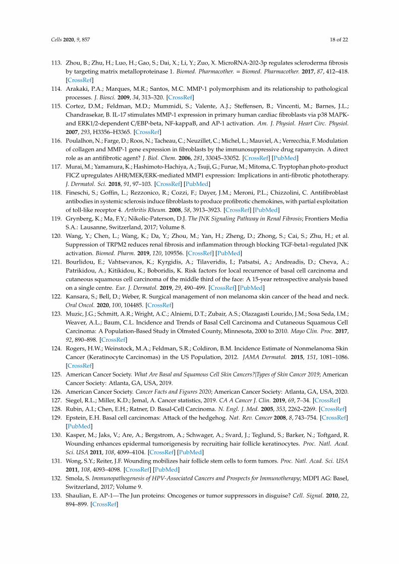
Cells 2020, 9, 857 18 of 22
113. Zhou, B.; Zhu, H.; Luo, H.; Gao, S.; Dai, X.; Li, Y.; Zuo, X. MicroRNA-202-3p regulates scleroderma fibrosisby targeting matrix metalloproteinase 1. Biomed. Pharmacother. = Biomed. Pharmacother. 2017, 87, 412–418.[CrossRef]
114. Arakaki, P.A.; Marques, M.R.; Santos, M.C. MMP-1 polymorphism and its relationship to pathologicalprocesses. J. Biosci. 2009, 34, 313–320. [CrossRef]
115. Cortez, D.M.; Feldman, M.D.; Mummidi, S.; Valente, A.J.; Steffensen, B.; Vincenti, M.; Barnes, J.L.;Chandrasekar, B. IL-17 stimulates MMP-1 expression in primary human cardiac fibroblasts via p38 MAPK-and ERK1/2-dependent C/EBP-beta, NF-kappaB, and AP-1 activation. Am. J. Physiol. Heart Circ. Physiol.2007, 293, H3356–H3365. [CrossRef]
116. Poulalhon, N.; Farge, D.; Roos, N.; Tacheau, C.; Neuzillet, C.; Michel, L.; Mauviel, A.; Verrecchia, F. Modulationof collagen and MMP-1 gene expression in fibroblasts by the immunosuppressive drug rapamycin. A directrole as an antifibrotic agent? J. Biol. Chem. 2006, 281, 33045–33052. [CrossRef] [PubMed]
117. Murai, M.; Yamamura, K.; Hashimoto-Hachiya, A.; Tsuji, G.; Furue, M.; Mitoma, C. Tryptophan photo-productFICZ upregulates AHR/MEK/ERK-mediated MMP1 expression: Implications in anti-fibrotic phototherapy.J. Dermatol. Sci. 2018, 91, 97–103. [CrossRef] [PubMed]
118. Fineschi, S.; Goffin, L.; Rezzonico, R.; Cozzi, F.; Dayer, J.M.; Meroni, P.L.; Chizzolini, C. Antifibroblastantibodies in systemic sclerosis induce fibroblasts to produce profibrotic chemokines, with partial exploitationof toll-like receptor 4. Arthritis Rheum. 2008, 58, 3913–3923. [CrossRef] [PubMed]
119. Grynberg, K.; Ma, F.Y.; Nikolic-Paterson, D.J. The JNK Signaling Pathway in Renal Fibrosis; Frontiers MediaS.A.: Lausanne, Switzerland, 2017; Volume 8.
120. Wang, Y.; Chen, L.; Wang, K.; Da, Y.; Zhou, M.; Yan, H.; Zheng, D.; Zhong, S.; Cai, S.; Zhu, H.; et al.Suppression of TRPM2 reduces renal fibrosis and inflammation through blocking TGF-beta1-regulated JNKactivation. Biomed. Pharm. 2019, 120, 109556. [CrossRef] [PubMed]
121. Bourlidou, E.; Vahtsevanos, K.; Kyrgidis, A.; Tilaveridis, I.; Patsatsi, A.; Andreadis, D.; Cheva, A.;Patrikidou, A.; Kitikidou, K.; Boboridis, K. Risk factors for local recurrence of basal cell carcinoma andcutaneous squamous cell carcinoma of the middle third of the face: A 15-year retrospective analysis basedon a single centre. Eur. J. Dermatol. 2019, 29, 490–499. [CrossRef] [PubMed]
122. Kansara, S.; Bell, D.; Weber, R. Surgical management of non melanoma skin cancer of the head and neck.Oral Oncol. 2020, 100, 104485. [CrossRef]
123. Muzic, J.G.; Schmitt, A.R.; Wright, A.C.; Alniemi, D.T.; Zubair, A.S.; Olazagasti Lourido, J.M.; Sosa Seda, I.M.;Weaver, A.L.; Baum, C.L. Incidence and Trends of Basal Cell Carcinoma and Cutaneous Squamous CellCarcinoma: A Population-Based Study in Olmsted County, Minnesota, 2000 to 2010. Mayo Clin. Proc. 2017,92, 890–898. [CrossRef]
124. Rogers, H.W.; Weinstock, M.A.; Feldman, S.R.; Coldiron, B.M. Incidence Estimate of Nonmelanoma SkinCancer (Keratinocyte Carcinomas) in the US Population, 2012. JAMA Dermatol. 2015, 151, 1081–1086.[CrossRef]
125. American Cancer Society. What Are Basal and Squamous Cell Skin Cancers?|Types of Skin Cancer 2019; AmericanCancer Society: Atlanta, GA, USA, 2019.
126. American Cancer Society. Cancer Facts and Figures 2020; American Cancer Society: Atlanta, GA, USA, 2020.127. Siegel, R.L.; Miller, K.D.; Jemal, A. Cancer statistics, 2019. CA A Cancer J. Clin. 2019, 69, 7–34. [CrossRef]128. Rubin, A.I.; Chen, E.H.; Ratner, D. Basal-Cell Carcinoma. N. Engl. J. Med. 2005, 353, 2262–2269. [CrossRef]129. Epstein, E.H. Basal cell carcinomas: Attack of the hedgehog. Nat. Rev. Cancer 2008, 8, 743–754. [CrossRef]
[PubMed]130. Kasper, M.; Jaks, V.; Are, A.; Bergstrom, A.; Schwager, A.; Svard, J.; Teglund, S.; Barker, N.; Toftgard, R.
Wounding enhances epidermal tumorigenesis by recruiting hair follicle keratinocytes. Proc. Natl. Acad.Sci. USA 2011, 108, 4099–4104. [CrossRef] [PubMed]
131. Wong, S.Y.; Reiter, J.F. Wounding mobilizes hair follicle stem cells to form tumors. Proc. Natl. Acad. Sci. USA2011, 108, 4093–4098. [CrossRef] [PubMed]
132. Smola, S. Immunopathogenesis of HPV-Associated Cancers and Prospects for Immunotherapy; MDPI AG: Basel,Switzerland, 2017; Volume 9.
133. Shaulian, E. AP-1—The Jun proteins: Oncogenes or tumor suppressors in disguise? Cell. Signal. 2010, 22,894–899. [CrossRef]
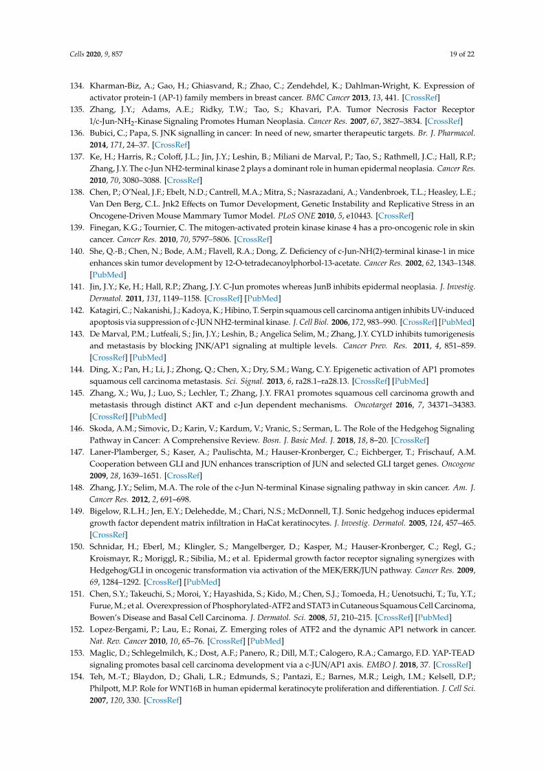
Cells 2020, 9, 857 19 of 22
134. Kharman-Biz, A.; Gao, H.; Ghiasvand, R.; Zhao, C.; Zendehdel, K.; Dahlman-Wright, K. Expression ofactivator protein-1 (AP-1) family members in breast cancer. BMC Cancer 2013, 13, 441. [CrossRef]
135. Zhang, J.Y.; Adams, A.E.; Ridky, T.W.; Tao, S.; Khavari, P.A. Tumor Necrosis Factor Receptor1/c-Jun-NH2-Kinase Signaling Promotes Human Neoplasia. Cancer Res. 2007, 67, 3827–3834. [CrossRef]
136. Bubici, C.; Papa, S. JNK signalling in cancer: In need of new, smarter therapeutic targets. Br. J. Pharmacol.2014, 171, 24–37. [CrossRef]
137. Ke, H.; Harris, R.; Coloff, J.L.; Jin, J.Y.; Leshin, B.; Miliani de Marval, P.; Tao, S.; Rathmell, J.C.; Hall, R.P.;Zhang, J.Y. The c-Jun NH2-terminal kinase 2 plays a dominant role in human epidermal neoplasia. Cancer Res.2010, 70, 3080–3088. [CrossRef]
138. Chen, P.; O’Neal, J.F.; Ebelt, N.D.; Cantrell, M.A.; Mitra, S.; Nasrazadani, A.; Vandenbroek, T.L.; Heasley, L.E.;Van Den Berg, C.L. Jnk2 Effects on Tumor Development, Genetic Instability and Replicative Stress in anOncogene-Driven Mouse Mammary Tumor Model. PLoS ONE 2010, 5, e10443. [CrossRef]
139. Finegan, K.G.; Tournier, C. The mitogen-activated protein kinase kinase 4 has a pro-oncogenic role in skincancer. Cancer Res. 2010, 70, 5797–5806. [CrossRef]
140. She, Q.-B.; Chen, N.; Bode, A.M.; Flavell, R.A.; Dong, Z. Deficiency of c-Jun-NH(2)-terminal kinase-1 in miceenhances skin tumor development by 12-O-tetradecanoylphorbol-13-acetate. Cancer Res. 2002, 62, 1343–1348.[PubMed]
141. Jin, J.Y.; Ke, H.; Hall, R.P.; Zhang, J.Y. C-Jun promotes whereas JunB inhibits epidermal neoplasia. J. Investig.Dermatol. 2011, 131, 1149–1158. [CrossRef] [PubMed]
142. Katagiri, C.; Nakanishi, J.; Kadoya, K.; Hibino, T. Serpin squamous cell carcinoma antigen inhibits UV-inducedapoptosis via suppression of c-JUN NH2-terminal kinase. J. Cell Biol. 2006, 172, 983–990. [CrossRef] [PubMed]
143. De Marval, P.M.; Lutfeali, S.; Jin, J.Y.; Leshin, B.; Angelica Selim, M.; Zhang, J.Y. CYLD inhibits tumorigenesisand metastasis by blocking JNK/AP1 signaling at multiple levels. Cancer Prev. Res. 2011, 4, 851–859.[CrossRef] [PubMed]
144. Ding, X.; Pan, H.; Li, J.; Zhong, Q.; Chen, X.; Dry, S.M.; Wang, C.Y. Epigenetic activation of AP1 promotessquamous cell carcinoma metastasis. Sci. Signal. 2013, 6, ra28.1–ra28.13. [CrossRef] [PubMed]
145. Zhang, X.; Wu, J.; Luo, S.; Lechler, T.; Zhang, J.Y. FRA1 promotes squamous cell carcinoma growth andmetastasis through distinct AKT and c-Jun dependent mechanisms. Oncotarget 2016, 7, 34371–34383.[CrossRef] [PubMed]
146. Skoda, A.M.; Simovic, D.; Karin, V.; Kardum, V.; Vranic, S.; Serman, L. The Role of the Hedgehog SignalingPathway in Cancer: A Comprehensive Review. Bosn. J. Basic Med. J. 2018, 18, 8–20. [CrossRef]
147. Laner-Plamberger, S.; Kaser, A.; Paulischta, M.; Hauser-Kronberger, C.; Eichberger, T.; Frischauf, A.M.Cooperation between GLI and JUN enhances transcription of JUN and selected GLI target genes. Oncogene2009, 28, 1639–1651. [CrossRef]
148. Zhang, J.Y.; Selim, M.A. The role of the c-Jun N-terminal Kinase signaling pathway in skin cancer. Am. J.Cancer Res. 2012, 2, 691–698.
149. Bigelow, R.L.H.; Jen, E.Y.; Delehedde, M.; Chari, N.S.; McDonnell, T.J. Sonic hedgehog induces epidermalgrowth factor dependent matrix infiltration in HaCat keratinocytes. J. Investig. Dermatol. 2005, 124, 457–465.[CrossRef]
150. Schnidar, H.; Eberl, M.; Klingler, S.; Mangelberger, D.; Kasper, M.; Hauser-Kronberger, C.; Regl, G.;Kroismayr, R.; Moriggl, R.; Sibilia, M.; et al. Epidermal growth factor receptor signaling synergizes withHedgehog/GLI in oncogenic transformation via activation of the MEK/ERK/JUN pathway. Cancer Res. 2009,69, 1284–1292. [CrossRef] [PubMed]
151. Chen, S.Y.; Takeuchi, S.; Moroi, Y.; Hayashida, S.; Kido, M.; Chen, S.J.; Tomoeda, H.; Uenotsuchi, T.; Tu, Y.T.;Furue, M.; et al. Overexpression of Phosphorylated-ATF2 and STAT3 in Cutaneous Squamous Cell Carcinoma,Bowen’s Disease and Basal Cell Carcinoma. J. Dermatol. Sci. 2008, 51, 210–215. [CrossRef] [PubMed]
152. Lopez-Bergami, P.; Lau, E.; Ronai, Z. Emerging roles of ATF2 and the dynamic AP1 network in cancer.Nat. Rev. Cancer 2010, 10, 65–76. [CrossRef] [PubMed]
153. Maglic, D.; Schlegelmilch, K.; Dost, A.F.; Panero, R.; Dill, M.T.; Calogero, R.A.; Camargo, F.D. YAP-TEADsignaling promotes basal cell carcinoma development via a c-JUN/AP1 axis. EMBO J. 2018, 37. [CrossRef]
154. Teh, M.-T.; Blaydon, D.; Ghali, L.R.; Edmunds, S.; Pantazi, E.; Barnes, M.R.; Leigh, I.M.; Kelsell, D.P.;Philpott, M.P. Role for WNT16B in human epidermal keratinocyte proliferation and differentiation. J. Cell Sci.2007, 120, 330. [CrossRef]
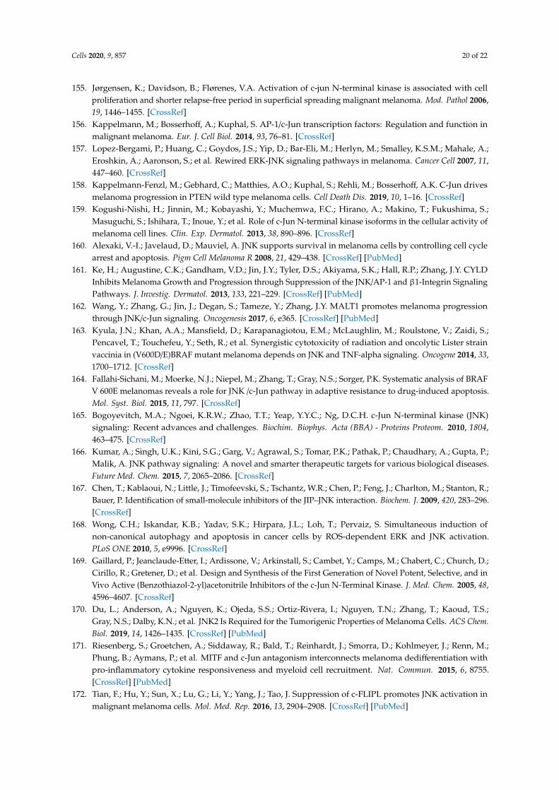
Cells 2020, 9, 857 20 of 22
155. Jørgensen, K.; Davidson, B.; Flørenes, V.A. Activation of c-jun N-terminal kinase is associated with cellproliferation and shorter relapse-free period in superficial spreading malignant melanoma. Mod. Pathol 2006,19, 1446–1455. [CrossRef]
156. Kappelmann, M.; Bosserhoff, A.; Kuphal, S. AP-1/c-Jun transcription factors: Regulation and function inmalignant melanoma. Eur. J. Cell Biol. 2014, 93, 76–81. [CrossRef]
157. Lopez-Bergami, P.; Huang, C.; Goydos, J.S.; Yip, D.; Bar-Eli, M.; Herlyn, M.; Smalley, K.S.M.; Mahale, A.;Eroshkin, A.; Aaronson, S.; et al. Rewired ERK-JNK signaling pathways in melanoma. Cancer Cell 2007, 11,447–460. [CrossRef]
158. Kappelmann-Fenzl, M.; Gebhard, C.; Matthies, A.O.; Kuphal, S.; Rehli, M.; Bosserhoff, A.K. C-Jun drivesmelanoma progression in PTEN wild type melanoma cells. Cell Death Dis. 2019, 10, 1–16. [CrossRef]
159. Kogushi-Nishi, H.; Jinnin, M.; Kobayashi, Y.; Muchemwa, F.C.; Hirano, A.; Makino, T.; Fukushima, S.;Masuguchi, S.; Ishihara, T.; Inoue, Y.; et al. Role of c-Jun N-terminal kinase isoforms in the cellular activity ofmelanoma cell lines. Clin. Exp. Dermatol. 2013, 38, 890–896. [CrossRef]
160. Alexaki, V.-I.; Javelaud, D.; Mauviel, A. JNK supports survival in melanoma cells by controlling cell cyclearrest and apoptosis. Pigm Cell Melanoma R 2008, 21, 429–438. [CrossRef] [PubMed]
161. Ke, H.; Augustine, C.K.; Gandham, V.D.; Jin, J.Y.; Tyler, D.S.; Akiyama, S.K.; Hall, R.P.; Zhang, J.Y. CYLDInhibits Melanoma Growth and Progression through Suppression of the JNK/AP-1 and β1-Integrin SignalingPathways. J. Investig. Dermatol. 2013, 133, 221–229. [CrossRef] [PubMed]
162. Wang, Y.; Zhang, G.; Jin, J.; Degan, S.; Tameze, Y.; Zhang, J.Y. MALT1 promotes melanoma progressionthrough JNK/c-Jun signaling. Oncogenesis 2017, 6, e365. [CrossRef] [PubMed]
163. Kyula, J.N.; Khan, A.A.; Mansfield, D.; Karapanagiotou, E.M.; McLaughlin, M.; Roulstone, V.; Zaidi, S.;Pencavel, T.; Touchefeu, Y.; Seth, R.; et al. Synergistic cytotoxicity of radiation and oncolytic Lister strainvaccinia in (V600D/E)BRAF mutant melanoma depends on JNK and TNF-alpha signaling. Oncogene 2014, 33,1700–1712. [CrossRef]
164. Fallahi-Sichani, M.; Moerke, N.J.; Niepel, M.; Zhang, T.; Gray, N.S.; Sorger, P.K. Systematic analysis of BRAFV 600E melanomas reveals a role for JNK /c-Jun pathway in adaptive resistance to drug-induced apoptosis.Mol. Syst. Biol. 2015, 11, 797. [CrossRef]
165. Bogoyevitch, M.A.; Ngoei, K.R.W.; Zhao, T.T.; Yeap, Y.Y.C.; Ng, D.C.H. c-Jun N-terminal kinase (JNK)signaling: Recent advances and challenges. Biochim. Biophys. Acta (BBA) - Proteins Proteom. 2010, 1804,463–475. [CrossRef]
166. Kumar, A.; Singh, U.K.; Kini, S.G.; Garg, V.; Agrawal, S.; Tomar, P.K.; Pathak, P.; Chaudhary, A.; Gupta, P.;Malik, A. JNK pathway signaling: A novel and smarter therapeutic targets for various biological diseases.Future Med. Chem. 2015, 7, 2065–2086. [CrossRef]
167. Chen, T.; Kablaoui, N.; Little, J.; Timofeevski, S.; Tschantz, W.R.; Chen, P.; Feng, J.; Charlton, M.; Stanton, R.;Bauer, P. Identification of small-molecule inhibitors of the JIP–JNK interaction. Biochem. J. 2009, 420, 283–296.[CrossRef]
168. Wong, C.H.; Iskandar, K.B.; Yadav, S.K.; Hirpara, J.L.; Loh, T.; Pervaiz, S. Simultaneous induction ofnon-canonical autophagy and apoptosis in cancer cells by ROS-dependent ERK and JNK activation.PLoS ONE 2010, 5, e9996. [CrossRef]
169. Gaillard, P.; Jeanclaude-Etter, I.; Ardissone, V.; Arkinstall, S.; Cambet, Y.; Camps, M.; Chabert, C.; Church, D.;Cirillo, R.; Gretener, D.; et al. Design and Synthesis of the First Generation of Novel Potent, Selective, and inVivo Active (Benzothiazol-2-yl)acetonitrile Inhibitors of the c-Jun N-Terminal Kinase. J. Med. Chem. 2005, 48,4596–4607. [CrossRef]
170. Du, L.; Anderson, A.; Nguyen, K.; Ojeda, S.S.; Ortiz-Rivera, I.; Nguyen, T.N.; Zhang, T.; Kaoud, T.S.;Gray, N.S.; Dalby, K.N.; et al. JNK2 Is Required for the Tumorigenic Properties of Melanoma Cells. ACS Chem.Biol. 2019, 14, 1426–1435. [CrossRef] [PubMed]
171. Riesenberg, S.; Groetchen, A.; Siddaway, R.; Bald, T.; Reinhardt, J.; Smorra, D.; Kohlmeyer, J.; Renn, M.;Phung, B.; Aymans, P.; et al. MITF and c-Jun antagonism interconnects melanoma dedifferentiation withpro-inflammatory cytokine responsiveness and myeloid cell recruitment. Nat. Commun. 2015, 6, 8755.[CrossRef] [PubMed]
172. Tian, F.; Hu, Y.; Sun, X.; Lu, G.; Li, Y.; Yang, J.; Tao, J. Suppression of c-FLIPL promotes JNK activation inmalignant melanoma cells. Mol. Med. Rep. 2016, 13, 2904–2908. [CrossRef] [PubMed]
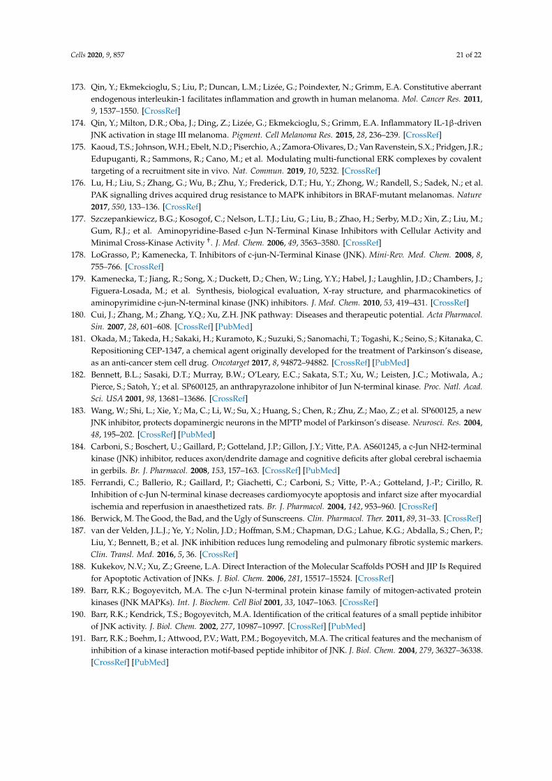
Cells 2020, 9, 857 21 of 22
173. Qin, Y.; Ekmekcioglu, S.; Liu, P.; Duncan, L.M.; Lizée, G.; Poindexter, N.; Grimm, E.A. Constitutive aberrantendogenous interleukin-1 facilitates inflammation and growth in human melanoma. Mol. Cancer Res. 2011,9, 1537–1550. [CrossRef]
174. Qin, Y.; Milton, D.R.; Oba, J.; Ding, Z.; Lizée, G.; Ekmekcioglu, S.; Grimm, E.A. Inflammatory IL-1β-drivenJNK activation in stage III melanoma. Pigment. Cell Melanoma Res. 2015, 28, 236–239. [CrossRef]
175. Kaoud, T.S.; Johnson, W.H.; Ebelt, N.D.; Piserchio, A.; Zamora-Olivares, D.; Van Ravenstein, S.X.; Pridgen, J.R.;Edupuganti, R.; Sammons, R.; Cano, M.; et al. Modulating multi-functional ERK complexes by covalenttargeting of a recruitment site in vivo. Nat. Commun. 2019, 10, 5232. [CrossRef]
176. Lu, H.; Liu, S.; Zhang, G.; Wu, B.; Zhu, Y.; Frederick, D.T.; Hu, Y.; Zhong, W.; Randell, S.; Sadek, N.; et al.PAK signalling drives acquired drug resistance to MAPK inhibitors in BRAF-mutant melanomas. Nature2017, 550, 133–136. [CrossRef]
177. Szczepankiewicz, B.G.; Kosogof, C.; Nelson, L.T.J.; Liu, G.; Liu, B.; Zhao, H.; Serby, M.D.; Xin, Z.; Liu, M.;Gum, R.J.; et al. Aminopyridine-Based c-Jun N-Terminal Kinase Inhibitors with Cellular Activity andMinimal Cross-Kinase Activity †. J. Med. Chem. 2006, 49, 3563–3580. [CrossRef]
178. LoGrasso, P.; Kamenecka, T. Inhibitors of c-jun-N-Terminal Kinase (JNK). Mini-Rev. Med. Chem. 2008, 8,755–766. [CrossRef]
179. Kamenecka, T.; Jiang, R.; Song, X.; Duckett, D.; Chen, W.; Ling, Y.Y.; Habel, J.; Laughlin, J.D.; Chambers, J.;Figuera-Losada, M.; et al. Synthesis, biological evaluation, X-ray structure, and pharmacokinetics ofaminopyrimidine c-jun-N-terminal kinase (JNK) inhibitors. J. Med. Chem. 2010, 53, 419–431. [CrossRef]
180. Cui, J.; Zhang, M.; Zhang, Y.Q.; Xu, Z.H. JNK pathway: Diseases and therapeutic potential. Acta Pharmacol.Sin. 2007, 28, 601–608. [CrossRef] [PubMed]
181. Okada, M.; Takeda, H.; Sakaki, H.; Kuramoto, K.; Suzuki, S.; Sanomachi, T.; Togashi, K.; Seino, S.; Kitanaka, C.Repositioning CEP-1347, a chemical agent originally developed for the treatment of Parkinson’s disease,as an anti-cancer stem cell drug. Oncotarget 2017, 8, 94872–94882. [CrossRef] [PubMed]
182. Bennett, B.L.; Sasaki, D.T.; Murray, B.W.; O’Leary, E.C.; Sakata, S.T.; Xu, W.; Leisten, J.C.; Motiwala, A.;Pierce, S.; Satoh, Y.; et al. SP600125, an anthrapyrazolone inhibitor of Jun N-terminal kinase. Proc. Natl. Acad.Sci. USA 2001, 98, 13681–13686. [CrossRef]
183. Wang, W.; Shi, L.; Xie, Y.; Ma, C.; Li, W.; Su, X.; Huang, S.; Chen, R.; Zhu, Z.; Mao, Z.; et al. SP600125, a newJNK inhibitor, protects dopaminergic neurons in the MPTP model of Parkinson’s disease. Neurosci. Res. 2004,48, 195–202. [CrossRef] [PubMed]
184. Carboni, S.; Boschert, U.; Gaillard, P.; Gotteland, J.P.; Gillon, J.Y.; Vitte, P.A. AS601245, a c-Jun NH2-terminalkinase (JNK) inhibitor, reduces axon/dendrite damage and cognitive deficits after global cerebral ischaemiain gerbils. Br. J. Pharmacol. 2008, 153, 157–163. [CrossRef] [PubMed]
185. Ferrandi, C.; Ballerio, R.; Gaillard, P.; Giachetti, C.; Carboni, S.; Vitte, P.-A.; Gotteland, J.-P.; Cirillo, R.Inhibition of c-Jun N-terminal kinase decreases cardiomyocyte apoptosis and infarct size after myocardialischemia and reperfusion in anaesthetized rats. Br. J. Pharmacol. 2004, 142, 953–960. [CrossRef]
186. Berwick, M. The Good, the Bad, and the Ugly of Sunscreens. Clin. Pharmacol. Ther. 2011, 89, 31–33. [CrossRef]187. van der Velden, J.L.J.; Ye, Y.; Nolin, J.D.; Hoffman, S.M.; Chapman, D.G.; Lahue, K.G.; Abdalla, S.; Chen, P.;
Liu, Y.; Bennett, B.; et al. JNK inhibition reduces lung remodeling and pulmonary fibrotic systemic markers.Clin. Transl. Med. 2016, 5, 36. [CrossRef]
188. Kukekov, N.V.; Xu, Z.; Greene, L.A. Direct Interaction of the Molecular Scaffolds POSH and JIP Is Requiredfor Apoptotic Activation of JNKs. J. Biol. Chem. 2006, 281, 15517–15524. [CrossRef]
189. Barr, R.K.; Bogoyevitch, M.A. The c-Jun N-terminal protein kinase family of mitogen-activated proteinkinases (JNK MAPKs). Int. J. Biochem. Cell Biol 2001, 33, 1047–1063. [CrossRef]
190. Barr, R.K.; Kendrick, T.S.; Bogoyevitch, M.A. Identification of the critical features of a small peptide inhibitorof JNK activity. J. Biol. Chem. 2002, 277, 10987–10997. [CrossRef] [PubMed]
191. Barr, R.K.; Boehm, I.; Attwood, P.V.; Watt, P.M.; Bogoyevitch, M.A. The critical features and the mechanism ofinhibition of a kinase interaction motif-based peptide inhibitor of JNK. J. Biol. Chem. 2004, 279, 36327–36338.[CrossRef] [PubMed]

Cells 2020, 9, 857 22 of 22
192. Gow, W.R.; Campbell, K.; Meade, A.J.; Watt, P.M.; Milech, N.; Knuckey, N.W.; Meloni, B.P. Lackof neuroprotection of inhibitory peptides targeting Jun/JNK after transient focal cerebral ischemia inspontaneously hypertensive rats. J. Cereb. Blood Flow Metab. 2011, 31, e1–e8. [CrossRef] [PubMed]
193. Bain, J.; McLauchlan, H.; Elliott, M.; Cohen, P. The specificities of protein kinase inhibitors: An update.Biochem. J. 2003, 371, 199–204. [CrossRef]
© 2020 by the authors. Licensee MDPI, Basel, Switzerland. This article is an open accessarticle distributed under the terms and conditions of the Creative Commons Attribution(CC BY) license (http://creativecommons.org/licenses/by/4.0/).


![Original Article Puerarin modulates autophagy to …signaling pathway with JNK inhibitor SP600125 can produce protective effects against CIRI [18]. Thrombolysis, antiplatelet agents,](https://img.pdfslide.us/doc/110x75/5e39305f90bc3d6d0c3b72a7/original-article-puerarin-modulates-autophagy-to-signaling-pathway-with-jnk-inhibitor.jpg)