Embed Size (px)
Citation preview

_earn C.E. creditConservative dentistry achieved through a multi-disciplinary approach
_techniqueA convenient cement for CAD block inlays
_opinionIn-office CAD/CAM isn’t for me … or is it?
32011
CAD/CAM the international magazine of digital dentistry
i s sn 2161-6574 U.S. Edition • Vol. 1 • Issue 3/2011

CAD/CAM 3_2011 I 03
guest editorial _ CAD/CAM I
CAD/CAM in dentistry
Dr. Gordon J. Christensen, top,
and Dr. Paul L. Child Jr.
_The concept of CAD/CAM has roots in antiquity. However, in the 1950s, CAD/CAM technology started to have some of the characteristics of what we now know as CAD/CAM. In the 1970s, computer drafting was popular, and by the 1980s, engineering applications of the concept became useful. Currently, CAD/CAM is used in almost every industry with great success.
Dentistry has been relatively slow to adapt to the concept, even though other industries have trans-formed most of their manufacturing processes to CAD/CAM because of its improved efficiency, repeat-ability and predictability. It was only about 25 years ago the CAD/CAM concept for milling restorations was introduced into dentistry with devices that were time consuming and difficult to manipulate. Many thought the concept would never replace the time honored “lost wax” casting technique and the com-monly used direct restorative techniques.
_How wrong they were …
Currently, CAD/CAM in dentistry is not only used, it is preferred by a growing and enthusiastic group of dental practitioners who have discovered its value and who have learned how to integrate the concept into mainstream dental practice. CAD/CAM in dentistry is beyond the phase of early adopters and those who buy everything new — it’s now moving into the “early majority.”
The dental CAD/CAM technologies now available, and the clinical results that are available to the profession and the patients they serve, would have seemed impossible only a few years ago. A realistic appraisal of some of them includes:
•Afteraconservativelearningperiod,imagingandmillingofcrownsandonlayswithaccuracyandclinical longevity prove to be superior to conventional restorations.
•Withexperience,fabricationofinlaysandveneersrivaltraditionaltechniques.•Expandeduseofcompetent,well-educatedstaffpersonsaccomplishesmuchoftheclinicalproce-
dure, allowing dentists to do other diverse techniques at the same time the restorations are being made.•Theyprovidepatientsasingle-appointment,relativelysimpleprocedurethatisinterestingandexcit-
ing to them while also being a practice builder for the practitioner.•Thereisanewvistaofcommunicationbetweendentists,dentaltechnologistsandassistantsasthe
“sky” is opened to the CAD/CAM concept.•AlthoughCAD/CAMwaspioneeredbyCEREC,E4Dandothersarenowprovidingprovenalternatives
in this growing area .•Movingrapidlybeyondjustfixedrestorationsintoaconvergenceofmultipledisciplinesindentistry,
CAD/CAM now includes implant dentistry, orthodontics, occlusion and surgery.There is no question that dentistry has awakened to the CAD/CAM concept. It is providing unprec-
edented service to millions of patients and deserves the attention of practitioners. Our basic and clinical researchover20yearsinCliniciansReport® (www.cliniciansreport.org;formerlyCRA)validatesitsuseandfuture potential for the profession.
Gordon J. Christensen, DDS, MSD, PhDPaul L. Child Jr., DMD, CDT
04 I
I content _ CAD/CAM
CAD/CAM3_2011
I C.E. article06 Conservative dentistry achieved through
a multi-disciplinary approach_Tom Colina, DMD
10 Welcome to the ‘Block Party’_Curtis Jansen, DDS
I clinical14 Bringing it all together with CAD/CAM
_Paresh Shah, DMD, MS, Cert. Esthetic Dentistry
20 CAD/CAM-processed lithium disilicate restorations: the replacement for PFM restorations _Jeff Scott, DMD
26 A CAD/CAM gallery_Graeme Milicich, BDS
I technique30 A convenient cement for CAD/CAM inlays
_John Cranham, DDS
I practice matters34 ‘The million dollar PPO’ _Matthew Krieger, DMD
I opinion36 In-office CAD/CAM isn’t for me … or is it?
_Charles Regalado, DDS
41 Time to lose ‘the wait’!_Dean Saiki, DDS
44 The perception vs. reality of chairside CAD/CAM dentistry _Alex Touchstone, DDS
47 Today’s in-office CAD/CAM is how to add ‘Wow!’ to your practice _Robert Stanton, DMD
I about the publisher49 _submissions 50 _imprint
page 26
page 34 page 41
I on the coverCover image provided by D4D Technologies
page 30
page 06 page 12
06 I
I C.E. article_ conservative dentistry
_Complex treatment needs can necessitate oral rehabilitation of patients. Often these patients will require a multi-disciplinary approach to correct problems.Whenpatientshavesignificantconcerns,such as severe malocclusions or destruction of dental tissue, oral rehabilitation can entail extensive treat-ment that may involve reconstructions.
To return the patient to optimal function, regain normal form and address possible concerns such as esthetics, an integrated approach that involves vari-ous disciplines needs to be taken. The challenge posed to a particular treatment plan may involve the treat-ment of many teeth and possibly the need to prepare a significant number of teeth and corresponding dental tissue.
Another challenge in reconstruction cases is the cost associated with the restoration of numerous teeth. Cost may be a factor for patients. There are often many options and approaches that can lead to the same successful treatment outcome. The variety of options can be at different ends of the spectrum. Diagnostic tools, including tomograms and the use of CAD/CAM systems, are useful in achieving complex treatment goals. This paper presents a treatment option that is an alternative to the reconstruction approach through the innovative application of multiple disciplines and current technology.
_Case presentation
A 31-year-old male patient presented with the chief complaint of his upper front teeth restorations breaking off a few months after being placed. He has had the front teeth restored numerous times with the same outcome. A comprehensive examination and records revealed the following findings.
_Medical history and functional concerns
There is a history of arthritis in the family. The patient experiences transient pain from his back, neck and shoulders. He has noted he clenches and grinds
CAD/CAM3_2011
Conservative dentistry achieved through a multi-disciplinary approachAuthor_Thomas Colina, DMD
This article qualifies for C.E. credit. To take the C.E. quiz, log on to www.dtstudyclub.com.
_c.e. credit part 1
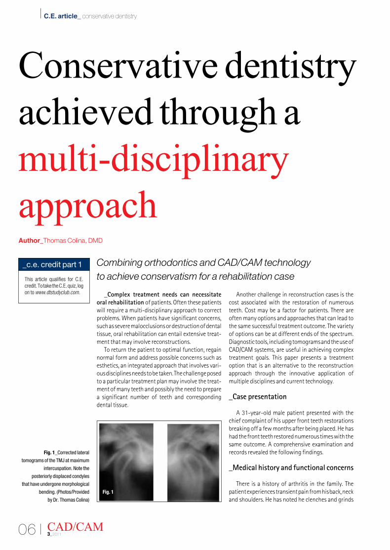
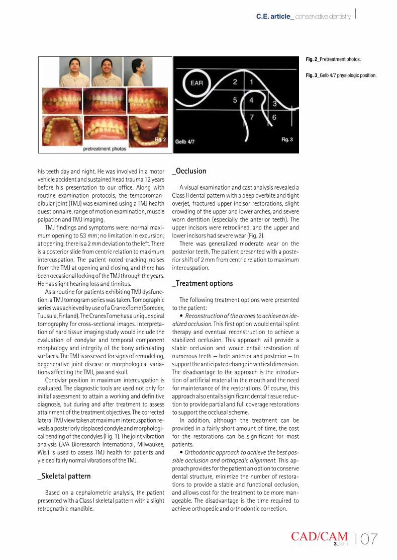
Fig. 1_Corrected lateral
tomograms of the TMJ at maximum
intercuspation. Note the
posteriorly displaced condyles
that have undergone morphological
bending. (Photos/Provided
by Dr. Thomas Colina)
Combining orthodontics and CAD/CAM technology to achieve conservatism for a rehabilitation case
Fig. 1
I 07CAD/CAM 3_2011
C.E. article_ conservative dentistry I
his teeth day and night. He was involved in a motor vehicle accident and sustained head trauma 12 years before his presentation to our office. Along with routine examination protocols, the temporoman-dibularjoint(TMJ)wasexaminedusingaTMJhealthquestionnaire, range of motion examination, muscle palpation and TMJ imaging.
TMJ findings and symptoms were: normal maxi-mum opening to 53 mm; no limitation in excursion; at opening, there is a 2 mm deviation to the left. There is a posterior slide from centric relation to maximum intercuspation. The patient noted cracking noises from the TMJ at opening and closing, and there has been occasional locking of the TMJ through the years. He has slight hearing loss and tinnitus.
As a routine for patients exhibiting TMJ dysfunc-tion, a TMJ tomogram series was taken. Tomographic series was achieved by use of a CranexTome (Soredex, Tuusula,Finland).TheCranexTomehasauniquespiraltomography for cross-sectional images. Interpreta-tion of hard tissue imaging study would include the evaluation of condylar and temporal component morphology and integrity of the bony articulating surfaces. The TMJ is assessed for signs of remodeling, degenerative joint disease or morphological varia-tions affecting the TMJ, jaw and skull.
Condylar position in maximum intercuspation is evaluated. The diagnostic tools are used not only for initial assessment to attain a working and definitive diagnosis, but during and after treatment to assess attainment of the treatment objectives. The corrected lateral TMJ view taken at maximum intercuspation re-veals a posteriorly displaced condyle and morphologi-calbendingofthecondyles(Fig.1).Thejointvibrationanalysis (JVA Bioresearch International, Milwaukee, Wis.) isusedtoassessTMJhealthforpatientsandyielded fairly normal vibrations of the TMJ.
_Skeletal pattern
Based on a cephalometric analysis, the patient presented with a Class I skeletal pattern with a slight retrognathic mandible.
_Occlusion
A visual examination and cast analysis revealed a Class II dental pattern with a deep overbite and tight overjet, fractured upper incisor restorations, slight crowding of the upper and lower arches, and severe worndentition (especially theanterior teeth). Theupper incisors were retroclined, and the upper and lowerincisorshadseverewear(Fig.2).
There was generalized moderate wear on the posterior teeth. The patient presented with a poste-rior shift of 2 mm from centric relation to maximum intercuspation.
_Treatment options
The following treatment options were presented to the patient:
•Reconstruction of the arches to achieve an ide-alized occlusion. This first option would entail splint therapy and eventual reconstruction to achieve a stabilized occlusion. This approach will provide a stable occlusion and would entail restoration of numerous teeth — both anterior and posterior — to support the anticipated change in vertical dimension. The disadvantage to the approach is the introduc-tion of artificial material in the mouth and the need for maintenance of the restorations. Of course, this approach also entails significant dental tissue reduc-tion to provide partial and full coverage restorations to support the occlusal scheme.
In addition, although the treatment can be provided in a fairly short amount of time, the cost for the restorations can be significant for most patients.
•Orthodontic approach to achieve the best pos-sible occlusion and orthopedic alignment. This ap-proach provides for the patient an option to conserve dental structure, minimize the number of restora-tions to provide a stable and functional occlusion, and allows cost for the treatment to be more man-ageable. The disadvantage is the time required to achieve orthopedic and orthodontic correction.
Fig. 2_Pretreatment photos.
Fig. 3_Gelb 4/7 physiologic position.
Fig. 2 Fig. 3

08 I
I C.E. article_ conservative dentistry
_Treatment plan details
Straight wire appliance treatment (SWA) wasproposed to attain ideal inter- and intra-arch align-ment augmented by a mandibular repositioning mechanics by way of posterior build-ups and elastics or a fixed orthotic or use of a Twin Force Appliance. This phase of treatment was anticipated to last 20 months. After the orthodontic treatment, restora-tion of the six anterior maxillary teeth with porcelain restorations would follow. The lower incisors will be evaluated for the need of restorations. The need for an upper bruxing appliance would also be evaluated after the completion of the restorations.
_Discussion of the treatment
The first phase of the treatment was the provision of orthodontic therapy using GAC Innovation C Self Ligating Bracket System. The Innovation C bracket system has a highly translucent porcelain structure and a rhodium coated clip, which provide superb esthetics as well as a high-torque component for the incisors of 17 degrees for the upper central and 10 degrees for the upper lateral incisors. One of the main goals for the treatment was the correction of the maxillary incisor torque. The retroclined upper incisors had contributed significantly to the severe wear of the anterior teeth and had resulted in an intercuspation that produced a posteriorly displaced condyle. The correction of the incisor torque brought about a natural repositioning of the mandible, which was a treatment goal for the patient. The JVA, which has been proven effective in discriminating joint vibrations to assess TMJ1,2 conditions, was utilized to evaluate the TMJ during and after treatment. Ante-rior repositioning of the mandible has been described in the literature as a viable approach in the treatment of Class II malocclusions and TMJ dysfunction.
Woodside3 and McNamara4 describe a functional approach to the correction of the Class II malocclu-sion. Anterior repositioning therapy has had a history
of more than 50 years. Gelb5 referred to his reposi-tioningappliancein1959anddescribedtheGelb4/7position, which is currently accepted in the litera-ture and recognized by many practitioners treating TMJ dysfunction to correlate with the physiologic positionofthecondyleinthefossa(Fig.3).Severalfunctional appliance designs and their efficacy of improving TMJ dysfunction through mandibular re-positioning have been described in later literature.6,7 Simmons8 further describes the alleviation of symp-toms after mandibular repositioning.
As noted, there was a natural anterior reposition-ing of the mandible upon removal of the centric interference in this patient, and appliance therapy was unnecessary. Posterior resin build-ups with Class II elastic therapy were sufficient to erupt the posterior teeth to achieve stability of the posterior segment. The condylar position was evaluated by use of progress tomograms and was supported and accompanied with the alleviation of TMJ related symptoms. To address concerns over the color of the teeth, the patient opted to whiten the teeth before the provision of the definitive restorations for the an-terior teeth. Upon evaluation of the post-orthodontic occlusion, to provide an occlusion with anterior guidance at protrusion and canine guidance at lateral excursion, it was adequate to provide restorations for onlytheupperincisor(Figs.4,5).Theupperincisorswere prepared conservatively and restored with por-celain IPS e.max CAD lithium disilicate veneers milled with the chairside E4D Dentist CAD/CAM system(D4DTechnologies,Richardson,Texas)(Figs.6,7).
There are numerous systems that are currently available. Systems are available chairside or labo-ratory based. The E4D Dentist system allows therestorative dentist to have complete control of design and delivery of restorations. The system uses a laser capture to acquire a digital impression. The information is condensed, aided by computer, to an intuitive format that allows the restorative dentist to modify the design and send the design to a precise automated milling unit that uses robotic technology.
CAD/CAM3_2011
Fig. 4_Debracket photos.
Fig. 5_Veneer post insert photos.
Fig. 4 Fig. 5

I 09CAD/CAM 3_2011
C.E. article_ conservative dentistry I
The system essentially automates many of the more mechanical and labor intensive procedures, such as waxing, investing, burnout, casting and/or press-ing involved in conventional fabrication of dental restorations.9Lithiumdisilicate(IPSe.max)hasthesuperiorflexuralstrengthof360MPato400MPa,ascompared to the strength of ceramic for PFM crowns, which has the strength of 80 MPa to 100 MPa; ve-neered zirconia, which has a flexural strength of 100 MPa; and leucite glass, which has the strength of ap-proximately 150 MPa to160 MPa. Lithium disilicate is a highly esthetic, high-strength material that can be conventionally cemented or adhesively bonded.10 The pressable lithium disilicate is indicated for inlays, onlays, thin veneers, veneers, partial crowns, three-unit anterior bridges, three-unit premolar bridges, telescope primary crowns and implant restoration while the machinable lithium disilicate is indicated for all the previous applications except bridges.11–14
_Summary
Reconstructivetreatmentusuallyentailssignifi-cant correction of malocclusion and the maxilloman-dibular relationship. Many patients requiring recon-struction commonly present with varying functional concerns, including TMJ dysfunction and associated symptoms. Technology, such as tomogram series and the use of JVA, could serve as standard equipment in the diagnosis and treatment of these patients as well as aid in objectively evaluating the TMJ condi-tion during and after the treatment. The goal of any treatment is to provide the patient with good esthet-ics, comfort and long-term function. The innovative melding of disciplines and the use of current materi-als and technology can allow conservation of dental tissue that is irreversibly altered and removed using the traditional reconstructive approaches._
_References
1. Ishigaki S, Bessette RW, Maruyama T. Diagnostic Ability of the Surface Vibration Analysis of Temporomandibular Joint. Abstract. IADR. Seattle, WA, March, 1994.
2. Knutson M, Radke J. Artificial Neural Network Classification of TMJ Internal Derangement. Abstract. J Dent Res 74 (AADR Abstracts) March, 1995.
3. Woodside DG, Metaxas A, Altuna G, The Influence
of Functional Appliance Therapy on Glenoid Fossa Remodelling. American Journal of Orthodontics and Dentofacial Orthopedics. 1987 Sep;92(3):181–198.
4. McNamara, Jr. JA, Carlson DS. Quantitative Analysis of Temporomandibular Joint Adaptations to Protrusive Function, AJO, 1979;76: 593–611.
5. Gelb H, Arnold GE. Syndromes of the Head and Neck of Dental Origin. American Medical Association Archives of Otolaryngology, Vol. 70, December 1959; 681–691.
6. Clark WJ. The Twin Block traction technique, European Journal of Orthodontics, 4, 129–138, 1982; and Lund, DI and Sandler, PJ, The effects of Twin Blocks: a prospective controlled study, American Journal of Orthodontics and Dentofacial Orthopaedics, 113, 104–110. 1998.
7. Simmons HC, 3rd, Gibbs SJ. Anterior repositioning appliance therapy for TMJ disorders: specific symptoms relieved and relationship to disk status on MRI.J Tenn Dent Assoc. 2009 Fall;89(4):22–30)
8. Severance G, Swann L. The Take CARE Approach to Treatment Planning, Preparation and Design for CAD/CAM Restorations. Oral Health. March 2009, 47–52.
9. Fabianelli A, Goracci C, Bertelli E, Davidson CL, Ferrari MA. A Clinical Trial of Empress II Porcelain Inlays Luted to Vital Teeth with Dual-Curing Adhesive System and a Self-Curing Resin Cement. Journal of Adhesive Dentistry, 2006 Dec;8(6):427–431.
10. Tysowsky G. The Science Behind Lithium Disilicate: Today’s Surprisingly Versatile, Esthetic and Durable Metal Free Alternative, Oral Health. March 2009. 93–97
11. Sorenson JA, Cruz M, Mito WT, Raffeiner O, Meredith HR, Foser, HP. A Clinical Investigation on Three-Unit Fixed Partial Dentures Fabricated with Lithium Disilicate Glass Ceramic, Pract Periodontics Aesthet Dent. 1999 Jan-Feb;11(1):95–106.
12. Holland W, Schweiger M, Frank M, Rheinberger V. A Comparison of the Microstructure and Properties of the IPS Empress 2 and the IPS Empress Glass Ceramics. Journal of Biomedical Material Research, 2000;53(4):297–303.
13. Kheradmandan S, Koutayas SO, Bernhard M, Strub JR. Fracture Strength of Four Different Types of Anterior Bridges After Thermomechanical Fatigue in the Dual-Axis Chewing Stimulator, Journal of Oral Rehabilitation. 2001 Apr;28(4):361-369.
14. Kheradmandan, S., Koutayas, S.O., Bernhard, M., Strub, J.R., Fracture Strength of Four Different Types of Anterior Bridges After Thermomechanical Fatigue in the Dual-Axis Chewing Stimulator, Journal of Oral Rehabilitation. 2001 Apr; 28 (4): 361-369.
Figs. 6, 7_ E4D veneer design
for teeth #22, #21, #11 and #12.
Conservative design achieved,
made possible with post-
orthodontic idealized occlusion.
Fig. 8_ Reflected frontal closeup.
Fig. 6 Fig. 7 Fig. 8
Thomas Colina, DMD, is a general dentist practicing in Winnipeg, Manitoba, Can-ada. He graduated from the University of Manitoba Faculty of Dentistry in 1989. His focus on providing comprehensive dental care often entails a multi- and interdisciplinary ap-proach. Colina is a member of the Manitoba Dental Asso-ciation, Canadian Dental As-sociation, Academy of General Dentistry and the International Association for Orthodon-tics. He is a senior certified instructor for the International Association for Orthodontics as well as a clinical instructor for the Department of Dental Diagnostic and Surgical Sci-ences, University of Manitoba Dental Faculty.
1-737 Keewatin St.Winnipeg, ManitobaCanada R2X [email protected]
CAD/CAM _contact

10 I
I C.E. article_ daily digital dentistry
Restorative clinicians have been spoiled in thepast regarding materials for direct and indirect resto-rations.We’vehadthegreatluxuryofseeinganadina journal, getting an offer in the mail or online, or at-tendingaC.E.courseaboutanewproduct,techniqueor service, and then immediately or the next day, we could take action. If we saw a new restorative mate-rial for fabricating restorations, we would simply write the request on a lab slip for the new material and expect to get it back in a couple weeks.
Think of the poor laboratory technician on the other end, reading perhaps for the first time, the method you want used to fabricate your restoration or a specific new material or a mix of materials and techniques.Remember,alaboratorysliporprescrip-tion is a work authorization, and if you write one, the laboratory technician has to comply. If we change our minds for the next restoration, we simply prescribe something else. I’m sure technicians sometimes
feel as if they’re chasing their tails with all the new materials, techniques and requests. Consider the investment in materials, systems, training and the learning curve they have to endure every time a new material is prescribed.
To the relief of patients, dentists, team members and technicians comes CAD/CAM dentistry and a little bit of sense and sensibility regarding dental ma-terials. Dental material manufacturers need to invest in the technology, methodology and product design, as well as the material evolution to the restoration (blocks,mandrels,discs),inordertointroduceanewmaterial for CAD/CAM dentistry. Then, in collabora-tion,dentalCAD(computer-aideddesign)anddentalCAM (computer-aided manufacturing) developersmust work with that material to produce consistent optimized results. This takes time and effort. Only those materials proven through economic evalua-tion, clinical validity and proven demand will make it to the final stages and into the software of the CAD systems and into the mills of the CAM systems and ultimately into our patients mouths.
CAD/CAM also requires the dentist to take more control of all facets of patient care; it requires more thought than a whim and a handwritten prescrip-tion to choose the right material. CAD/CAM requires thinking through the restorative and esthetic process before proceeding with a restoration, all better things for the dental professional as a whole. As more and more laboratories and dentists invest in digital den-tistry, everyone gains.
I’m “all in” for “daily digital dentistry.” I have dig-ital impression-only systems and a chairside CAD/CAMSystem,E4DDentist(Fig.1).Therestillisn’tjustone system that can complete all of the restorative indications we have in dentistry. It is my preference to select the techniques and materials that excel in a particular area, rather than compromise to have one system that says it does a little of everything. For me
CAD/CAM3_2011
Welcome to the ‘Block Party’Author_Curtis Jansen, DDS
This article qualifies for C.E. credit. To take the C.E. quiz, log on to www.dtstudyclub.com.
_c.e. credit part 2
Fig. 1(Photos/Provided by
Dr. Curtis Jansen)
I 11CAD/CAM 3_2011
C.E. article_ daily digital dentistry I
and my practice (a prosthodontic practice located in Monterey,Calif.),allofmysingle-unitrestorationsarefabricatedusingtheE4DDentistsystem.Inaddi-tion,withtheopeningofE4DSky™ Network and the newestversionof theE4D’sDentaLogic software,more and more of my total restorative care will be touched by digital technologies on a daily basis.
WhenyouarefirstintroducedtoCAD/CAMchair-side dentistry, you have the opportunity to refine your thinking on restorative care. You’ll no doubt become a better diagnostician and clinician — be-cause of looking at your preoperative conditions and preparations on a large monitor — but also a better and more confident provider of when to do what in different clinical situations.
Given the number of restorative materials avail-able at your fingertips, you’ll make better-educated decisions with each particular patient situation. UsingtheE4DDentistsystem,youhaveaccesstoanumberofprovenmaterials(blocks),eachwitheitheranIvoclarVivadentor3MESPElogoonit,soyouknow exactly what you are getting. The abundance of material options allows you to select the best one for the given clinical situation. A quick review of what is available follows.
_Block Party attendees
ResinIn the category of resin, you have the option to
select the Paradigm MZ100 block from 3M ESPE.Complementing the success of the direct restorative Filtek Z100, this block contains ceramic particles with an average size of 0.6 microns with cross-linked monomers that provide the ideal wear resistance, strength and radiopacity necessary for posterior use. I use it primarily for partial coverage restorations as well as some full coverage restorations on implants. The use of this resin for indirect restorations requires placement using an adhesive cementation protocol. I personally have an onlay restored with MZ100 in my own mouth, tooth #3.
When compared to conventional feldspathicporcelain restorations fabricated with chairside CAD/CAM, the Paradigm MZ100 restorations showed better color match through 10 years.1 This same study also showed no difference in margin finish, surface finish, anatomic form, caries or sensitivity. The authors actually concluded that “the composite inlays performed as well as the porcelain inlays with less bulk inlay fracture.” In an in vitro fatigue study on occlusal veneer restorations,2 Paradigm MZ100 had significantly higher fatigue resistance (100 percent survivalat185,000cyclesupto1400Nloads)com-pared to CAD/CAM feldspathic porcelain (0 percent survival).
Resin nano ceramicA new category for chairside CAD/CAM dentistry
is the resin nano ceramic created with the introduc-tion of the new Lava™ Ultimate block. This material defines a new category, resin nano ceramic, which provides some unique and beneficial characteristics forustohaveforchairside.Weallknowthat3MESPEanditsLavabrandhavebecomesynonymouswith zirconia restorations and they’ve expanded this technology to additional digital applications. Lava Ultimate material contains a blend of three fillers: zirconia and silica nanoparticles agglomerated into clusters, individually bonded silica nanoparticles and individually bonded zirconia nanoparticles.3
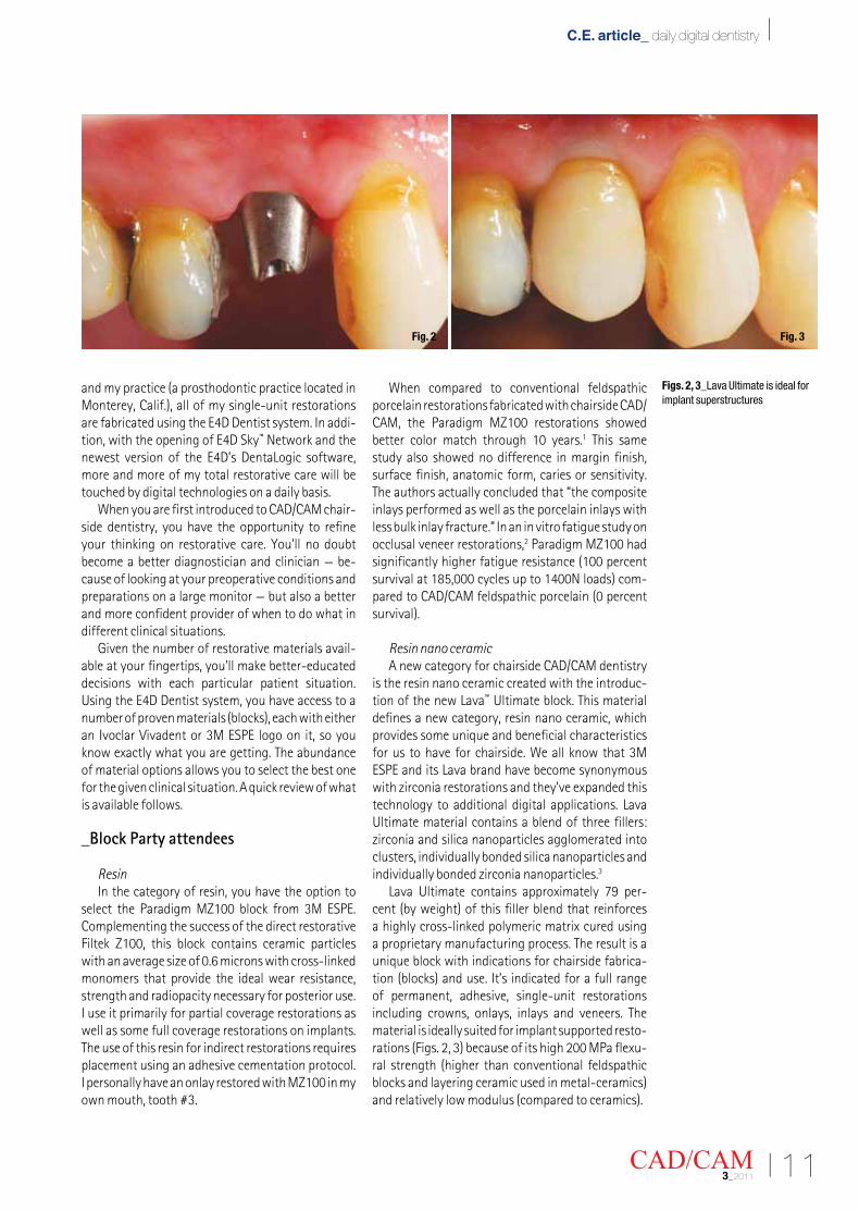
Lava Ultimate contains approximately 79 per-cent(byweight)ofthisfillerblendthatreinforcesa highly cross-linked polymeric matrix cured using a proprietary manufacturing process. The result is a unique block with indications for chairside fabrica-tion(blocks)anduse.It’sindicatedforafullrangeof permanent, adhesive, single-unit restorations including crowns, onlays, inlays and veneers. The material is ideally suited for implant supported resto-rations(Figs.2,3)becauseofitshigh200MPaflexu-ral strength (higher than conventional feldspathic blocksandlayeringceramicusedinmetal-ceramics)andrelativelylowmodulus(comparedtoceramics).
Figs. 2, 3_Lava Ultimate is ideal for implant superstructures
Fig. 2 Fig. 3
12 I
I C.E. article_ daily digital dentistry
From a time management standpoint, the use of resin or resin-ceramic system provides faster milling times and no need for an additional step of sinter-ing or firing. As a sign of its full confidence in this newcategoryofmaterial,3MESPEhasintroduceda unique 10-year warranty on the use of the Lava Ultimateblock.The3MESPELavaUltimateblockisoffered in eight shades with two translucency op-tions(LTandHT).
Glass ceramicIntheglassceramiccategory,withE4DDentist
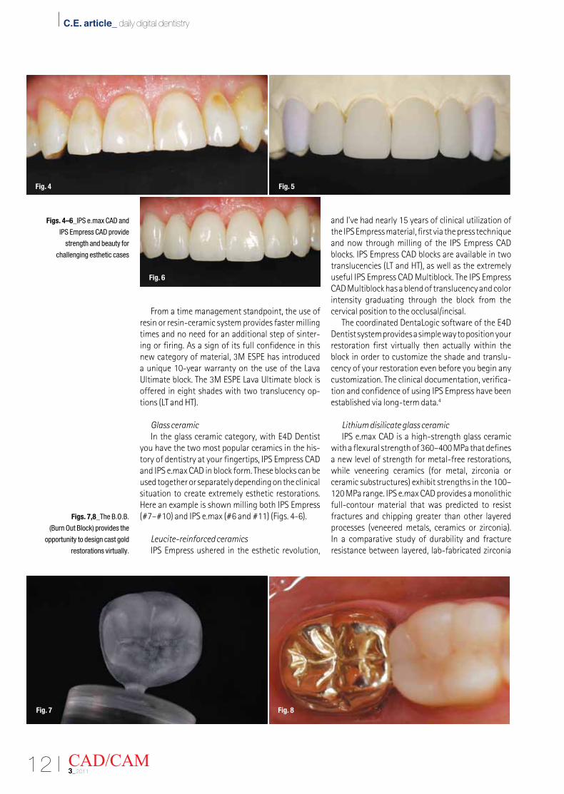
you have the two most popular ceramics in the his-toryofdentistryatyourfingertips,IPSEmpressCADand IPS e.max CAD in block form. These blocks can be used together or separately depending on the clinical situation to create extremely esthetic restorations. HereanexampleisshownmillingbothIPSEmpress(#7–#10)andIPSe.max(#6and#11)(Figs.4-6).
Leucite-reinforced ceramicsIPSEmpressusheredintheestheticrevolution,
and I’ve had nearly 15 years of clinical utilization of theIPSEmpressmaterial,firstviathepresstechniqueandnowthroughmillingoftheIPSEmpressCADblocks.IPSEmpressCADblocksareavailableintwotranslucencies(LTandHT),aswellastheextremelyusefulIPSEmpressCADMultiblock.TheIPSEmpressCAD Multiblock has a blend of translucency and color intensity graduating through the block from the cervical position to the occlusal/incisal.
ThecoordinatedDentaLogicsoftwareoftheE4DDentist system provides a simple way to position your restoration first virtually then actually within the block in order to customize the shade and translu-cency of your restoration even before you begin any customization. The clinical documentation, verifica-tionandconfidenceofusingIPSEmpresshavebeenestablished via long-term data.4
Lithium disilicate glass ceramicIPS e.max CAD is a high-strength glass ceramic
withaflexuralstrengthof360–400MPathatdefinesa new level of strength for metal-free restorations, while veneering ceramics (for metal, zirconia or ceramicsubstructures)exhibitstrengthsinthe100–120 MPa range. IPS e.max CAD provides a monolithic full-contour material that was predicted to resist fractures and chipping greater than other layered processes (veneered metals, ceramics or zirconia).In a comparative study of durability and fracture resistance between layered, lab-fabricated zirconia
CAD/CAM3_2011
Figs. 4–6_IPS e.max CAD and
IPS Empress CAD provide
strength and beauty for
challenging esthetic cases
Figs. 7,8_The B.O.B.
(Burn Out Block) provides the
opportunity to design cast gold
restorations virtually.
Fig. 4
Fig. 6
Fig. 5
Fig. 7 Fig. 8
I 13CAD/CAM 3_2011
C.E. article_ daily digital dentistry I
restorations and monolithic IPS e.max restorations, the IPS e.max restorations provided reduced fracture and more durable results.5
IPS e.max CAD blocks have the unique characteris-tic of being distributed in a partially crystallized stage (bluetovioletcolored).Thismeansthat,aftermilling,the IPS e.max CAD blocks need to be fully crystallized inatwo-stageceramicoven(e.g.ProgramatCS)priorto final delivery. This provides a major benefit to the entire procedure, with the advantages that the IPS e.max CAD milled restoration can be tried in the mouth and contacts verified before final crystallization and characterization. This makes the final delivery of the restoration more predictable and consistent.
The introduction of DentaLogic software ver-sion 2.0 coincides with the availability of additional shades of IPS e.max CAD for chairside use. IPS e.max CAD Impulse introduces five new shades, three Value and two Opal shades. Because of the different bright-ness values of the three Value blocks, restorations can be optimally integrated into the surrounding tooth structure in terms of their shade. The two Opal blocks allow clinicians to imitate the lifelike opalescent effect, which is desired in anterior restorations. The Opal blocks are ideally suited for the fabrication of veneers and thin veneers.
IPS e.max CAD blocks can also be seated with adhesive or conventional protocol depending on the retentive characteristics of the preparation following approvedguidelines(seebox).
AcrylicEven though thepriceofgoldhas reachedan
all-time high,6 if nostalgia and/or clinical concern of adequate clearance, margin design or material preference steer you toward metal-based restora-tions, you can still take advantage of digital scanning and designing benefits while providing you or your
laboratory with a simplified fabrication process for metal-based(gold)restorations.TheB.O.B(BurnOutBlock)blockfromD4DTechnologiescanbeselectedfor any preparation style and then scanned and milled for presentation to a laboratory for investment, burn-outandcasting(orpressing),thusprovidingyouwithconsistency in design, contacts and contour for your skilleddesignapplications(Figs.7,8).
_Conclusion
Chairside CAD/CAM systems have provided clini-cians with a new level of control in the practice of dentistry. From diagnosis through preparation and material selection, clinicians now have the capability of selecting materials with proven clinical perform-ance and delivering restorations with unmatched efficiency and productivity. The categories of resin, resin ceramic and glass ceramic give today’s modern practices the ability to offer solutions for the majority of crown and bridge indications right in the office._
_References
1. Fasbinder DJ, Dennison J, Heys D. Clinical Evaluation of CAD/CAM-Generated Composite Inlays: Ten-Year Report. J. Dent. Res. 90 (Spec Iss A): #378, 2011.
2. Magne P, Schlichting LH, Maia HP, Baratieri LN. In vitro fatigue resistance of CAD/CAM composite resin and ceramic posterior occlusal veneers. J Prosthet Dent 2010;104:149–157.
3. 3M ESPE Lava Ultimate CAD/CAM Restorative Technical Product Profile (3M ESPE).
4. Scientific documentation, IPS Empress CAD (Ivoclar Vivadent).
5. Petra C Guess, Ricardo Zavanelli, Nelson Silva and Van P Thomson. NYU Mouth Motion Fatigue and Durability Study.
6. www.money.cnn.com, August 22, 2011.
It should be noted that the proper and successful utilization of any of the metal-free types of materials (resin, resin ceramic, glass ceramic) require following approved preparation guidelines. These are simply providing proper clearance for the particular material — typically 1.5–2 mm occlusally (2 mm for implant restorations) and 1 mm axially; heavy chamfer or shoulder; rounded internal angles and butt joint margins — which need to be visible! All digital capture systems today can only capture what they see, and if you clinically can’t see the margins, don’t try to capture them digitally — first gain visualization through proper soft-tissue management. With all these materials, the preparation is of the utmost importance!
Concern has been raised by those without firsthand experi-
ence about the esthetic limitations of mono-block restorations or the limited longevity of surface-characterized (glazed) metal-free restorations. It should be noted it is often the dental bur that removes the glazed surface and not natural wear; one need only walk on 2,000-year-old tiles in Europe to realize the natural fu-sion of the glazed material into the base ceramic. Proper design, record (bite) taking and attention to detail in the use of various software packages, along with the replication of the virtual de-sign in ceramic after choosing the correct shade and translu-cency, quickly relieve any hesitations about esthetics and reinforce the benefits of doing more and more chairside restorative treat-ment.
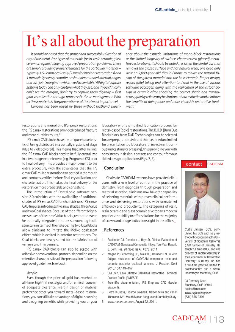
Curtis Jansen, DDS, com-pleted his DDS and his pros-thodontic education at the Uni-versity of Southern California (USC) School of Dentistry. He taught full time at USC and was director of implant dentistry in the Department of Restorative Dentistry. Currently, he has a full-time practice limited to prosthodontics and a dental laboratory in Monterey, Calif.
34 Dormody CourtMonterey, Calif. 93940 [email protected](831) 656-9394
CAD/CAM _contact
It’s all about the preparation
14 I
I clinical_ the restorative process
_Today’s dental professionals have the unique ability to provide patients with advanced re-storative treatments in a single appointment using state-of-the-art CAD/CAM technologies and es-thetic all-ceramic materials.1,2 Available for chairside use, convenient CAD/CAM systems offer greater simplicity and efficiency than earlier technologies. Combined with the exceptionally strong and esthetic all-ceramic materials currently available, chairside fabrication allows dentists to reduce the number of follow-up appointments and the amount of chair-time required to prepare, design, fabricate and seat restorations.1,2
Patients benefit from these advances through lowered cost and same-day dental treatment with-out the need for multiple follow-up visits.
_Advanced CAD/CAM Technology
Among the various CAD/CAM systems available, theE4DDentistsystem(D4DTechnologies,Richard-son,Texas)enablesdentalprofessionalstofabricaterestorations chairside in a single appointment.3,4 The unique three-dimensional software (DentaLogic™, D4DTechnologies)oftheE4DSystemwasdesignedwith dentist input to ensure the greatest intuitive-ness. Additionally, the combined equipment and software enables dentists to design and mill multiple restorationsatthesametime.Further,theE4DSys-tem and DentaLogic software can be learned quickly and easily.
Using the E4D technology, dental profession-als now have the ability to scan the hard and soft tissues of the oral cavity without using scanning powder.3,4 The scanner was developed with three scanning capabilities, including intraoral digital im-pressions, mouth impressions and models. Multiple design tools assist the dentist and team in creating restorations after preparations have been scanned. Once a restoration has been designed in the three-
dimensional rendering software, the restoration design is then transferred to the milling unit via wire-less networking. The dual-spindle milling technology of the system then uses fine diamond burs to shape a variety of materials into dental restorations that meet the precise case requirements. The accuracy of the milling system produces restorations demon-strating excellent fit and higher strength.3,4
TheE4Dsystemalsoprovidesdentalprofessionalsthe best in customer service. Utilizing the system’s software and advanced wireless networking capa-bilities, dental professionals gain access to a broad range of live and remote support from dedicated D4Dstaffmembers.Onceconnected,E4Dexpertscan assist users in a variety of tasks, from designing to troubleshooting. Just as quickly as this technology has evolved, so have the materials available to fabri-cate CAD/CAM restorations.
_Lithium Disilicate Glass Ceramic
IPSe.maxCAD(IvoclarVivadent,Amherst,N.Y.)isalithium disilicate glass ceramic material designed for CAD/CAM milling that demonstrates high strength and natural optical qualities. Once milled in the office or in the dental laboratory, IPS e.max CAD lithium-disilicate restorations achieve a monolithic strength of 360 MPa, which ensures a long-lasting result.5,6
Adhesive cementation using a separate dental conditioner is not indicated when ceramic thickness is 1.5 mm or greater because the material demon-strates high strength.5,6 IPS e.max CAD restorations may be seated using self-adhesive resin cements and self-adhesive composite cements to ensure all case requirements are met.
The lithium disilicate glass ceramic material also demonstrates excellent optical qualities when placed intraorally. After CAD/CAM processing, restorations milled from IPS e.max CAD require further charac-terization. When added before the crystallization
CAD/CAM3_2011
Bringing it all together with CAD/CAMAuthor_Paresh Shah, DMD, MS, Cert. Esthetic Dentistry
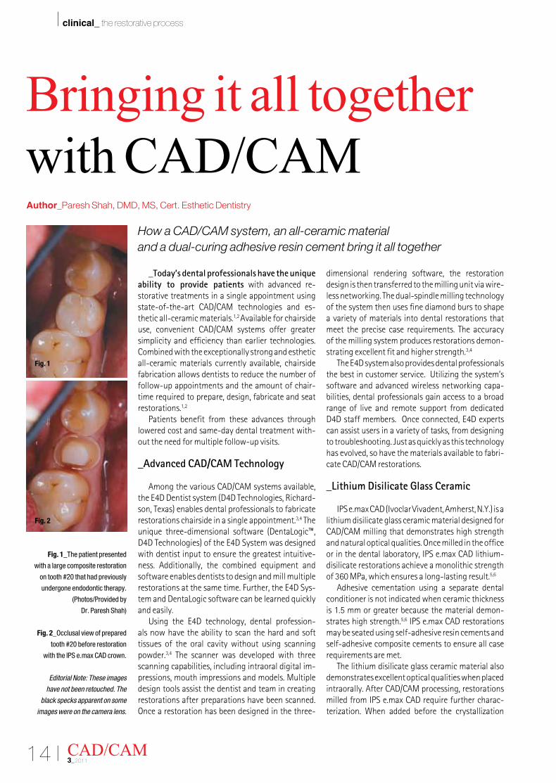
Fig. 1_The patient presented
with a large composite restoration
on tooth #20 that had previously
undergone endodontic therapy.
(Photos/Provided by
Dr. Paresh Shah)
Fig. 2_Occlusal view of prepared
tooth #20 before restoration
with the IPS e.max CAD crown.
Editorial Note: These images
have not been retouched. The
black specks apparent on some
images were on the camera lens.
How a CAD/CAM system, an all-ceramic material and a dual-curing adhesive resin cement bring it all together
Fig. 1
Fig. 2
I 15CAD/CAM 3_2011
clinical_ the restorative process I
process, subsequent polishing is unnecessary. Ad-ditionally, IPS e.max CAD is available in low translu-cency(LT)andhightranslucency(HT)blockstoensurerestorations appear lifelike and indistinguishable from surrounding dentition.5,6
Because of its optical properties and strength, IPS e.max CAD may be used for restoration of both the anterior and posterior dentition. Further, the material offers a restorative solution in cases requiring inlays andonlays,thinveneers(0.3mm),partialandfullcrowns, as well as implant superstructures.5,6
_Case presentation
A male presented with concerns regarding the health and esthetics of tooth #20. The patient had previously undergone endodontic therapy on tooth #20, and a large and unesthetic composite restora-tionhadbeenplaced(Fig1).
To address the patient’s esthetic concerns and restore health to the tooth, the treatment plan included restoration of tooth #20 with a highly esthetic and strong lithium disilicate glass ceramic crown(IPSe.maxCAD).ThecrownwouldbeCAD/CAM processed chairside and seated the same day using self-adhesive resin cement (Multilink Automix, IvoclarVivadent).
_Clinical protocol
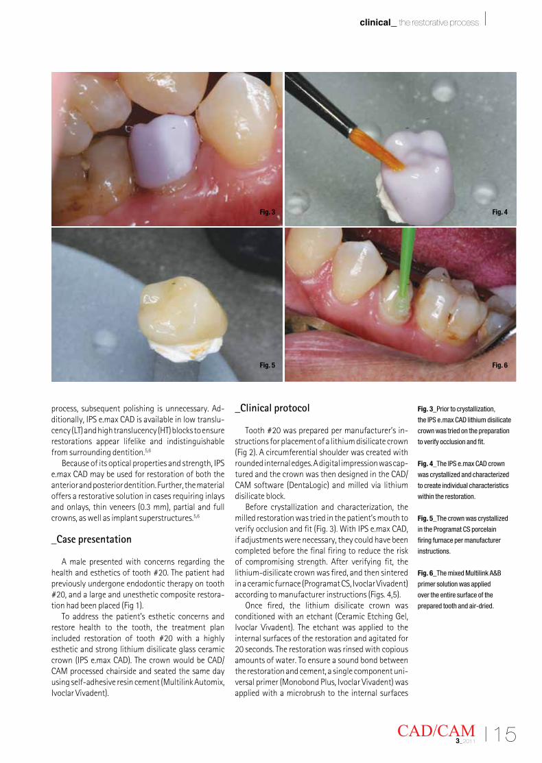
Tooth #20 was prepared per manufacturer’s in-structions for placement of a lithium disilicate crown (Fig2).Acircumferentialshoulderwascreatedwithrounded internal edges. A digital impression was cap-tured and the crown was then designed in the CAD/CAMsoftware(DentaLogic)andmilledvia lithiumdisilicate block.
Before crystallization and characterization, the milled restoration was tried in the patient’s mouth to verifyocclusionandfit(Fig.3).WithIPSe.maxCAD,if adjustments were necessary, they could have been completed before the final firing to reduce the risk of compromising strength. After verifying fit, the lithium-disilicate crown was fired, and then sintered inaceramicfurnace(ProgramatCS,IvoclarVivadent)accordingtomanufacturerinstructions(Figs.4,5).
Once fired, the lithium disilicate crown was conditionedwithanetchant(CeramicEtchingGel,Ivoclar Vivadent). The etchant was applied to theinternal surfaces of the restoration and agitated for 20 seconds. The restoration was rinsed with copious amounts of water. To ensure a sound bond between the restoration and cement, a single component uni-versalprimer(MonobondPlus,IvoclarVivadent)wasapplied with a microbrush to the internal surfaces
Fig. 3_Prior to crystallization,
the IPS e.max CAD lithium disilicate
crown was tried on the preparation
to verify occlusion and fit.
Fig. 4_The IPS e.max CAD crown
was crystallized and characterized
to create individual characteristics
within the restoration.
Fig. 5_The crown was crystallized
in the Programat CS porcelain
firing furnace per manufacturer
instructions.
Fig. 6_The mixed Multilink A&B
primer solution was applied
over the entire surface of the
prepared tooth and air-dried.
Fig. 3
Fig. 5
Fig. 4
Fig. 6
16 I
I clinical_ the restorative process
of the restoration. The primer was then air dried to remove any excess.
Prior to cementation, tooth #20 was isolated to control moisture and other contaminants. A two-component enamel-dentin primer (Multilink A/B, IvoclarVivadent)wasthenplacedonthepreparation.The primers were mixed in a 1:1 ratio. The mixed prim-ers were applied to all surfaces of the prepared tooth andairdried(Fig.6).
Dual-curing adhesive resin cement (Multilink Au-tomix,IvoclarVivadent)wasthenextrudedfromtheautomix syringe and placed on the internal surfaces of the crown. Available in yellow, transparent and white-opaque shades, the adhesive cement enables dentists to further enhance the esthetics of the resto-ration. The resin cement also facilitates fast and easy cleanup of excess. The automix tip simplified place-ment and allowed exact dosing to minimize waste.7,8
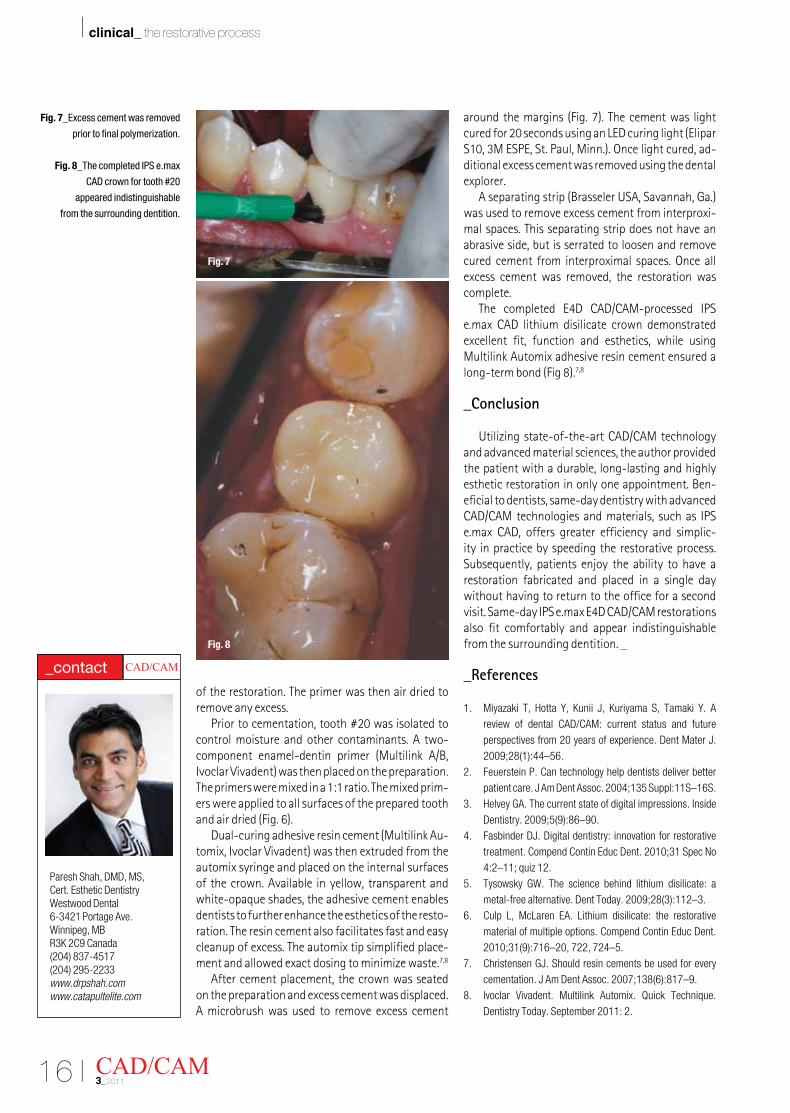
After cement placement, the crown was seated on the preparation and excess cement was displaced. A microbrush was used to remove excess cement
aroundthemargins(Fig.7).Thecementwaslightcuredfor20secondsusinganLEDcuringlight(EliparS10,3MESPE,St.Paul,Minn.).Oncelightcured,ad-ditional excess cement was removed using the dental explorer.
Aseparatingstrip(BrasselerUSA,Savannah,Ga.)was used to remove excess cement from interproxi-mal spaces. This separating strip does not have an abrasive side, but is serrated to loosen and remove cured cement from interproximal spaces. Once all excess cement was removed, the restoration was complete.
The completed E4D CAD/CAM-processed IPSe.max CAD lithium disilicate crown demonstrated excellent fit, function and esthetics, while using Multilink Automix adhesive resin cement ensured a long-termbond(Fig8).7,8
_Conclusion
Utilizing state-of-the-art CAD/CAM technology and advanced material sciences, the author provided the patient with a durable, long-lasting and highly esthetic restoration in only one appointment. Ben-eficial to dentists, same-day dentistry with advanced CAD/CAM technologies and materials, such as IPS e.max CAD, offers greater efficiency and simplic-ity in practice by speeding the restorative process. Subsequently, patients enjoy the ability to have a restoration fabricated and placed in a single day without having to return to the office for a second visit.Same-dayIPSe.maxE4DCAD/CAMrestorationsalso fit comfortably and appear indistinguishable from the surrounding dentition. _
_References
1. Miyazaki T, Hotta Y, Kunii J, Kuriyama S, Tamaki Y. A review of dental CAD/CAM: current status and future perspectives from 20 years of experience. Dent Mater J. 2009;28(1):44–56.
2. Feuerstein P. Can technology help dentists deliver better patient care. J Am Dent Assoc. 2004;135 Suppl:11S–16S.
3. Helvey GA. The current state of digital impressions. Inside Dentistry. 2009;5(9):86–90.
4. Fasbinder DJ. Digital dentistry: innovation for restorative treatment. Compend Contin Educ Dent. 2010;31 Spec No 4:2–11; quiz 12.
5. Tysowsky GW. The science behind lithium disilicate: a metal-free alternative. Dent Today. 2009;28(3):112–3.
6. Culp L, McLaren EA. Lithium disilicate: the restorative material of multiple options. Compend Contin Educ Dent. 2010;31(9):716–20, 722, 724–5.
7. Christensen GJ. Should resin cements be used for every cementation. J Am Dent Assoc. 2007;138(6):817–9.
8. Ivoclar Vivadent. Multilink Automix. Quick Technique. Dentistry Today. September 2011: 2.
CAD/CAM3_2011
Fig. 7_Excess cement was removed
prior to final polymerization.
Fig. 8_The completed IPS e.max
CAD crown for tooth #20
appeared indistinguishable
from the surrounding dentition.
Paresh Shah, DMD, MS, Cert. Esthetic DentistryWestwood Dental6-3421 Portage Ave.Winnipeg, MB R3K 2C9 Canada(204) 837-4517(204) 295-2233www.drpshah.comwww.catapultelite.com
CAD/CAM _contact
Fig. 7
Fig. 8

20 I
I clinical_ meeting patient expectations
_The demand for lifelike dental restorations has increased with media coverage promoting “smile makeovers.” Dental patients seek perfection in increasing numbers and have become willing to undergo elective procedures to achieve the perfect “Hollywood smile.” With an emphasis on naturalesthetics, dentists have often struggled to marry ef-fective conventional restorative materials and tech-niques that deliver proper function and oral health with patient expectations for the esthetic results.1,2
_CAD/CAM
Collaboration between technician and dentist in comprehensive esthetic, severe wear and complex implant cases is critical to successful restoration. Although cases fabricated in the laboratory are still
considered ideal in many instances, dentists now have the option to achieve everything from basic posterior restorations all the way to complete com-prehensive restorative cases in-office using chairside CAD/CAM technology and materials. Restorationsmay now be designed, milled and seated in a single appointment, increasing the level of service to pa-tients while reducing cost and the amount of time required for treatment.1,2
_E4D System
TheE4DDentistsystem(D4DTechnologies,Rich-ardson,Texas)offersmanyadvantageousfeaturesover CAD/CAM systems of the past. The unique three-dimensionalsoftware(DentaLogic,™D4DTechnolo-gies)of theE4DSystemwasdesigned toprovidedentists the ability to design/mill multiple restora-tions at the same time. Additionally, restorations may be fabricated chairside and in many cases, in a single appointment.3,4
Utilizing a three-dimensional high-speed laser scanner, theE4DSystemcanscanbothhardandsoft tissues for intraoral digital impressions, mouth impressions and models, without the use of conven-tional scanning powders. The system also incorpo-rates multiple design tools to assist the dentist when fabricating restorations in a single appointment. After rendering the restorative design, it may then be sent wirelessly to the precision milling system that uses dual spindles and fine diamond burs to effi-ciently fabricate various CAD materials into restora-tions that demonstrate excellent fit, higher strength and lifelike esthetics.3,4
To further aid dentists, D4D provides remoteDesign-on-Site(DOS)andSupport-on-Sight(SOS)services from various company specialists, who can
CAD/CAM3_2011
CAD/CAM-processed lithium disilicate restorations: the replacement for PFM restorationsAuthor_Jeff Scott, DMD
Fig. 1_A preoperative shade
photograph of tooth #21 was taken
to aid in determining the proper
shade of the IPS e.max CAD lithium
disilicate glass ceramic block.
(Photos/Provided by Dr. Jeff Scott)
Fig. 1
I 21CAD/CAM 3_2011
clinical_ meeting patient expectations I
then observe, assist or troubleshoot during the scan-ning, designing and milling process.3,4
_Material considerations
Historically, porcelain-fused-to-metal (PFM)crowns have been considered the ideal restoration in the posterior region of the oral cavity. Although PFM crowns may function for extended periods of time and demonstrate acceptable esthetics, dental pro-fessionals have often experienced numerous clinical challenges with this class of restoration.5
Among the many various challenges, in order to achieve acceptable esthetic and functional results, the preparation design for PFM restorations requires more aggressive removal of sound tooth structure. Additionally, PFM restorations require metal copings that are costly and notoriously difficult to mask with conventional porcelain materials.5,6 Therefore, high opacity ceramics were used and esthetics sacrificed when attempting to cover metal substrates. Also contributing to the lack of esthetics, the gingival-porcelain margins of PFM restorations typically appear unnatural.5
Because of this, restorative materials that can be fabricated quickly and more cost-effectively with innovative chairside CAD/CAM technologies and materials are slowly replacing PFM restorations.5
_IPS e.max CAD
Among these materials, IPS e.max CAD lithium disilicate glass ceramic (Ivoclar Vivadent, Amherst, N.Y.)isaninnovativeall-ceramicmaterialthatdem-onstrates exceptional monolithic strength. CAD/CAM processed in-office or in the dental laboratory, IPS e.max CAD blocks demonstrate a high flexural strength of 360 MPa. Strong and durable, the all-ceramic material is highly resistant to the forces of mastication over the long term. The material may be used for a variety of indications in the anterior and posterior, including inlays and onlays, veneers, par-
tial and full crowns, and implant superstructures.7,8
IPS e.max CAD restorations also demonstrate lifelike optical properties and naturally appearing esthetics. However, additional characterization may be instilled in IPS e.max CAD restorations using stains and glazes to ensure that restorations appear indistinguishable from the surrounding dentition. Offering a variety of esthetic solutions, IPS e.max CAD lithium disilicate glass ceramic blocks are available inhigh-translucency(HT)andlow-translucency(LT)formats, and are now also available in IPS e.max CAD Impulse shades. The LT blocks are considered ideal when fabricating crowns and implant-retained res-torations, while inlay and partial crown restorations are best completed with HT blocks. The new IPS e.max Impulse blocks are supplied in three Values (Value 1,2, 3)andtwoOpalshades(Opal1,2).Theyaremainlyused to fabricate thin veneers, partials, and single crowns. The suitable block can be selected depending on the preferred fabrication technique (staining, cut-backorlayeringtechnique)andtheindividualpatientsituation at hand. 7,8
The following case examples demonstrate the use ofE4DCAD/CAM-processedIPSe.maxCADlithiumdisilicate glass ceramic restorations and adhesive resin cements (Multilink Automix, Ivoclar Vivadent; UNICEM2Automix,3MESPE,St.Paul,Minn.)tore-store function and oral health, while still meeting the esthetic demands of patients.
_Case No. 1
A patient presented with recurrent decay sur-rounding a large amalgam restoration, along with a deep non-carious cervical lesion in the lower left first premolar. The patient’s second premolar and first molar had been restored many years before with PFM crowns.
Although the patient expressed dissatisfaction with the appearance of the old PFM restorations, the crowns lacked pathology and were still function-ing. Therefore, she decided to only restore the first
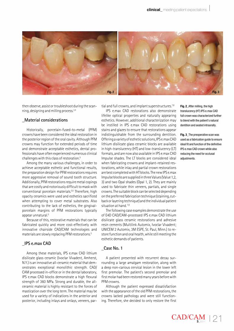
Fig. 2_After milling, the high
translucency (HT) IPS e.max CAD
full crown was characterized further
to blend with the patient’s natural
dentition and seated intraorally.
Fig. 3_The preoperative scan was
used as a fabrication guide to ensure
ideal fit and function of the definitive
IPS e.max CAD crown while also
reducing the need for occlusal
adjustments.
Fig. 3Fig. 2
22 I
I clinical_ meeting patient expectations
CAD/CAM3_2011
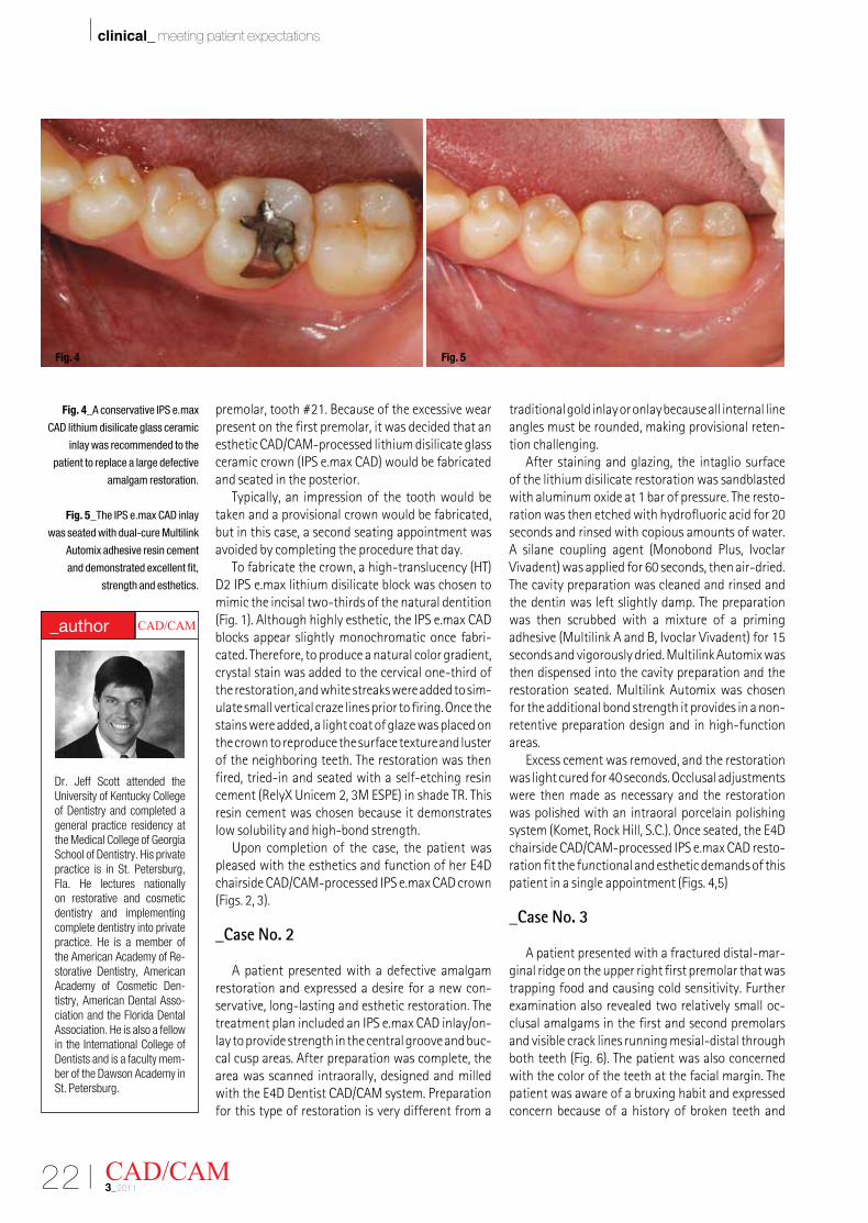
Fig. 4_A conservative IPS e.max
CAD lithium disilicate glass ceramic
inlay was recommended to the
patient to replace a large defective
amalgam restoration.
Fig. 5_The IPS e.max CAD inlay
was seated with dual-cure Multilink
Automix adhesive resin cement
and demonstrated excellent fit,
strength and esthetics.
premolar, tooth #21. Because of the excessive wear present on the first premolar, it was decided that an esthetic CAD/CAM-processed lithium disilicate glass ceramiccrown(IPSe.maxCAD)wouldbefabricatedand seated in the posterior.
Typically, an impression of the tooth would be taken and a provisional crown would be fabricated, but in this case, a second seating appointment was avoided by completing the procedure that day.
Tofabricatethecrown,ahigh-translucency(HT)D2 IPS e.max lithium disilicate block was chosen to mimic the incisal two-thirds of the natural dentition (Fig.1).Althoughhighlyesthetic,theIPSe.maxCADblocks appear slightly monochromatic once fabri-cated. Therefore, to produce a natural color gradient, crystal stain was added to the cervical one-third of the restoration, and white streaks were added to sim-ulate small vertical craze lines prior to firing. Once the stains were added, a light coat of glaze was placed on the crown to reproduce the surface texture and luster of the neighboring teeth. The restoration was then fired, tried-in and seated with a self-etching resin cement(RelyXUnicem2,3MESPE)inshadeTR.Thisresin cement was chosen because it demonstrates low solubility and high-bond strength.
Upon completion of the case, the patient was pleasedwiththeestheticsandfunctionofherE4Dchairside CAD/CAM-processed IPS e.max CAD crown (Figs.2,3).
_Case No. 2
A patient presented with a defective amalgam restoration and expressed a desire for a new con-servative, long-lasting and esthetic restoration. The treatment plan included an IPS e.max CAD inlay/on-lay to provide strength in the central groove and buc-cal cusp areas. After preparation was complete, the area was scanned intraorally, designed and milled withtheE4DDentistCAD/CAMsystem.Preparationfor this type of restoration is very different from a
traditional gold inlay or onlay because all internal line angles must be rounded, making provisional reten-tion challenging.
After staining and glazing, the intaglio surface of the lithium disilicate restoration was sandblasted with aluminum oxide at 1 bar of pressure. The resto-ration was then etched with hydrofluoric acid for 20 seconds and rinsed with copious amounts of water. A silane coupling agent (Monobond Plus, Ivoclar Vivadent)wasappliedfor60seconds,thenair-dried.The cavity preparation was cleaned and rinsed and the dentin was left slightly damp. The preparation was then scrubbed with a mixture of a priming adhesive(MultilinkAandB,IvoclarVivadent)for15seconds and vigorously dried. Multilink Automix was then dispensed into the cavity preparation and the restoration seated. Multilink Automix was chosen for the additional bond strength it provides in a non-retentive preparation design and in high-function areas.
Excesscementwasremoved,andtherestorationwaslightcuredfor40seconds.Occlusaladjustmentswere then made as necessary and the restoration was polished with an intraoral porcelain polishing system(Komet,RockHill,S.C.).Onceseated,theE4Dchairside CAD/CAM-processed IPS e.max CAD resto-ration fit the functional and esthetic demands of this patientinasingleappointment(Figs.4,5)
_Case No. 3
A patient presented with a fractured distal-mar-ginal ridge on the upper right first premolar that was trapping food and causing cold sensitivity. Further examination also revealed two relatively small oc-clusal amalgams in the first and second premolars and visible crack lines running mesial-distal through bothteeth(Fig.6).Thepatientwasalsoconcernedwith the color of the teeth at the facial margin. The patient was aware of a bruxing habit and expressed concern because of a history of broken teeth and
Fig. 5Fig. 4
Dr. Jeff Scott attended the University of Kentucky College of Dentistry and completed a general practice residency at the Medical College of Georgia School of Dentistry. His private practice is in St. Petersburg, Fla. He lectures nationally on restorative and cosmetic dentistry and implementing complete dentistry into private practice. He is a member of the American Academy of Re-storative Dentistry, American Academy of Cosmetic Den-tistry, American Dental Asso-ciation and the Florida Dental Association. He is also a fellow in the International College of Dentists and is a faculty mem-ber of the Dawson Academy in St. Petersburg.
CAD/CAM _author

24 I
I clinical_ meeting patient expectations
porcelain from previous crowns. The treatment plan for this patient included a nighttime appliance to pre-vent further damage from bruxing, as well as high-strength monolithic esthetic restorations. Because of a somewhat dark dentin color, a low-translucency (LT)IPSe.maxCADA1blockwaschosen.
Initially, a preoperative quadrant alginate impres-sion was taken. The affected dentition was then pre-pared, and another series of impressions taken with aquick-settingpolyvinyl-siloxane(PVS)impressionmaterial(Quick-Step,3MESPE).Abisacrylprovisionalrestoration(HenryScheinDental,Melville,N.Y.)wasseated with temporary cement (Fynal, Dentsply Caulk,Milford,Del.).Afterscanningthemodels,theIPS e.max CAD restorations were designed and milled with theE4DCAD/CAMsystem. The crownswerethen fitted to the solid model of the preparations and a laboratory microscope was used to confirm the marginal fit.
To speed-up seating, the interproximal contacts were adjusted on the laboratory bench. Further, the E4Dsoftwareallowsthepreoperativemodeltobescanned. The preparation model can then be super-imposed onto the design of the proposed crowns. This minimizes the occlusal adjustment necessary at placement while maintaining the staining and
characterization that has been built into the ceramic surface. Upon completion of firing and minimal adjustments, Multilink Automix dual-cure adhesive resin cement was used to seat the definitive restora-tionsintraorally(Fig.7).
The patient was pleased with the strength andestheticresultsoftheE4DDentistCAD/CAM-processed IPS e.max CAD crowns, which appeared indistinguishable from the surrounding dentition (Fig8).
_Conclusion
Although CAD/CAM technology promotes greater ease-of-use and efficiency, there is a learning curve and the technology must be mastered to be utilized to its full potential. To fully implement CAD/CAM into the dental practice, dentists must be determined and committed to training themselves as well as their teammembers.E4DDentistoffersbasictoadvancedtraining courses and excellent phone support to facilitate this process.
Additionally, dentists must understand that ma-terial selection is crucial in the success of CAD/CAM integration. Once familiar with the intricacies of CAD/CAM technologies and materials, the dentist, team members and, most importantly, the patient, will benefit tremendously.1,2_
_References
1. Ahmad I. Risk management in clinical practice. Part 5. Ethical considerations for dental enhancement procedures. Br Dent J. 2010;209(5):207˜14.
2. Marzola R, Derbabian K, Donovan TE, Arcidiacono A. The science of communicating the art of esthetic dentistry. part i: patient-dentist-patient communication. J Esthet Dent. 2000;12(3):131–8.
3. Helvey GA. The current state of digital impressions. Inside Dentistry. 2009;5(9):86–90.
4. Fasbinder DJ. Digital dentistry: innovation for restorative treatment. Compend Contin Educ Dent. 2010;31 Spec No 4:2-11; quiz 12.
5. Leinfelder KF. Porcelain esthetics for the 21st century. J Am Dent Assoc. 2000;131 Suppl:47S–51S.
6. Mjör IA. Problems and benefits associated with restorative materials: side-effects and long-term cost. Adv Dent Res. 1992;6:7–16.
7. Tysowsky GW. The science behind lithium disilicate: a metal-free alternative. Dent Today. 2009;28(3):112˜113.
8. Culp L, McLaren EA. Lithium disilicate: the restorative material of multiple options. Compend Contin Educ Dent. 2010;31(9):716-20, 722, 724–725.
9. Christensen GJ. Should resin cements be used for every cementation. J Am Dent Assoc. 2007;138(6):817–9.
10. Ivoclar Vivadent. Multilink Automix. Quick Technique. Dentistry Today. September 2011:2.
CAD/CAM3_2011
Fig. 6_A patient with a history
of bruxism presented to the office
seeking an esthetic solution for a
fractured distal-marginal ridge on
tooth #5 and visible mesial-distal
crack lines running through
both teeth #4 and #5.
Fig. 7_IPS e.max CAD lithium
disilicate glass ceramic blocks were
CAD/CAM-processed with the E4D
System into esthetic restorations for
teeth #4 and #5, then seated with
Multilink Automix.
Fig. 8_The facial margins of
the restorations demonstrated
excellent marginal integrity and
lifelike esthetics in the buccal
photograph that was taken
immediately after seating.
Jeff Scott, DMD239 2nd Ave. South, Ste. #100St. Petersburg, Fla. 33701(727) 898-8585jeff.scott@jeffscottdentistry.comwww.JeffScottDentistry.com
CAD/CAM _contact
Fig. 8
Fig. 6 Fig. 7
26 I
I clinical_ surmounting challenges
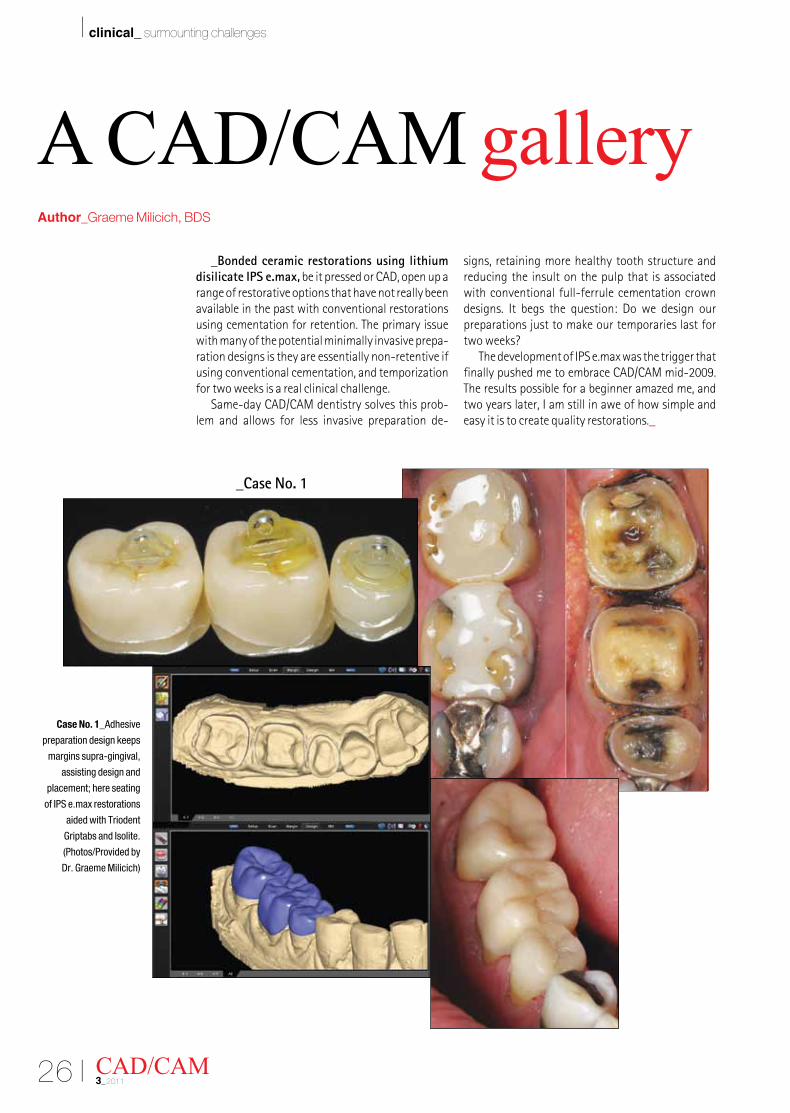
_Bonded ceramic restorations using lithium disilicate IPS e.max, be it pressed or CAD, open up a range of restorative options that have not really been available in the past with conventional restorations using cementation for retention. The primary issue with many of the potential minimally invasive prepa-ration designs is they are essentially non-retentive if using conventional cementation, and temporization for two weeks is a real clinical challenge.
Same-day CAD/CAM dentistry solves this prob-lem and allows for less invasive preparation de-
signs, retaining more healthy tooth structure and reducing the insult on the pulp that is associated with conventional full-ferrule cementation crown designs. It begs the question: Do we design our preparations just to make our temporaries last for two weeks?
The development of IPS e.max was the trigger that finally pushed me to embrace CAD/CAM mid-2009. The results possible for a beginner amazed me, and two years later, I am still in awe of how simple and easy it is to create quality restorations._
CAD/CAM3_2011
A CAD/CAM galleryAuthor_Graeme Milicich, BDS
Case No. 1_Adhesive
preparation design keeps
margins supra-gingival,
assisting design and
placement; here seating
of IPS e.max restorations
aided with Triodent
Griptabs and Isolite.
(Photos/Provided by
Dr. Graeme Milicich)
_Case No. 1
I 27CAD/CAM 3_2011
clinical_ surmounting challenges I
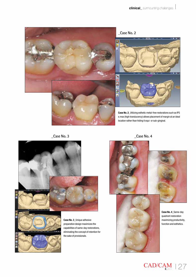
_Case No. 2
_Case No. 3 _Case No. 4
Case No. 2_Utilizing esthetic metal-free restorations such as IPS
e.max (high translucency) allows placement of margin at an ideal
location rather than hiding it equi- or sub-gingival.
Case No. 3_Unique adhesive
preparation design maximizes the
capabilities of same-day restorations,
eliminating the concept of retention for
the sake of provisionals.
Case No. 4_Same-day
quadrant restoration
maximizing productivity,
function and esthetics.
28 I
I clinical_ surmounting challenges
CAD/CAM3_2011
_Case No. 5
_Case No. 6 _Case No. 7
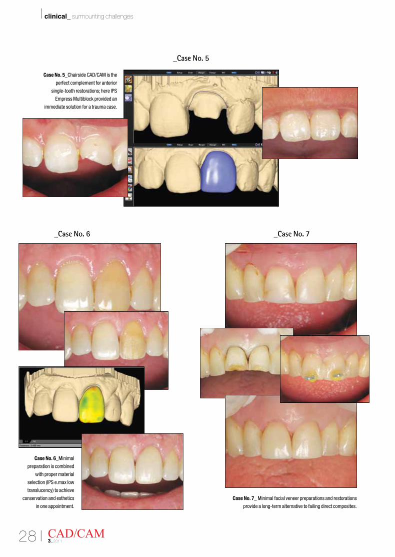
Case No. 5_Chairside CAD/CAM is the
perfect complement for anterior
single-tooth restorations; here IPS
Empress Multiblock provided an
immediate solution for a trauma case.
Case No. 6_Minimal
preparation is combined
with proper material
selection (IPS e.max low
translucency) to achieve
conservation and esthetics
in one appointment.
Case No. 7_ Minimal facial veneer preparations and restorations
provide a long-term alternative to failing direct composites.
I 29CAD/CAM 3_2011
clinical_ surmounting challenges I
Graeme Milicich, BDS, has been a dentist in Hamilton, New Zealand, since he graduated in 1976 and is one of three dentists at Anglesea Clinic Dental Care in the center of Hamilton. He has more than 12 years of experience in interna-tional lecturing and research in minimal intervention, lasers and restorative techniques. In conjunction with lecturing, he has also developed nine educational CD-ROMs to assist fellow professionals, their staff and their patients in easily understanding the various concepts associated with the ever-expanding field of minimally invasive dentistry. His newest CD is “Anterior Single Crown Aesthetics, using CAD/CAM,” teaching how to create the results seen in these cases. (See www.advancedental-ltd.com for more information.) Milicich introduced the E4D Dentist chairside CAD/CAM system into his practice in July 2009.
Graeme Milicich, BDSAnglesea Clinic Dental Care72 Braid Road, Hamilton, Waikato, New Zealand(647) [email protected]
_about the author CAD/CAM
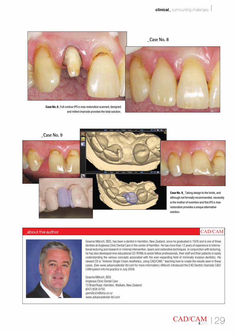
Case No. 9_ Taking design to the limits, and
although not formally recommended, necessity
is the mother of invention and this IPS e.max
restoration provides a unique alternative
solution.
_Case No. 8
Case No. 8_Full contour IPS e.max restoration scanned, designed
and milled chairside provides the total solution.
_Case No. 9
30 I
I technique_ CAD/CAM inlays
_For any indirect restorative procedure, use of the right cement is critical to ensure longevity of the restoration and patient satisfaction. In my practice, we utilize three different types of cement, each one carefully selected to best suit the specific material and clinical situation. To place gold PFMs or zirconia restorations in cases with good retention and resist-ance form, I utilize a resin-reinforced glass ionomer, such as 3M™ESPE™RelyX™ Luting Plus Cement. To place veneers or onlay restorations in cases without good retention and resistance form, I prefer a resin system, combined with a fourth- or fifth-generation adhesive.
In these cases, I typically utilize a combination of phosphoric acid, 3M ESPE Adper™ Single Bond PlusAdhesiveand3MESPERelyXVeneerCement,ensuring an adequate light cure at the appropriate steps. Finally, to maximize bond strength for inlays andonlayscreatedwithamillableblockfortheE4Dsystem in cases with acceptable retention and resist-anceform,Iutilize3MESPERelyXUnicem™ 2 Self-
AdhesiveResinCement.ThiscasewilldemonstratetheuseofRelyXUnicem2cementandanIPSe.maxCAD block.
_Case presentation
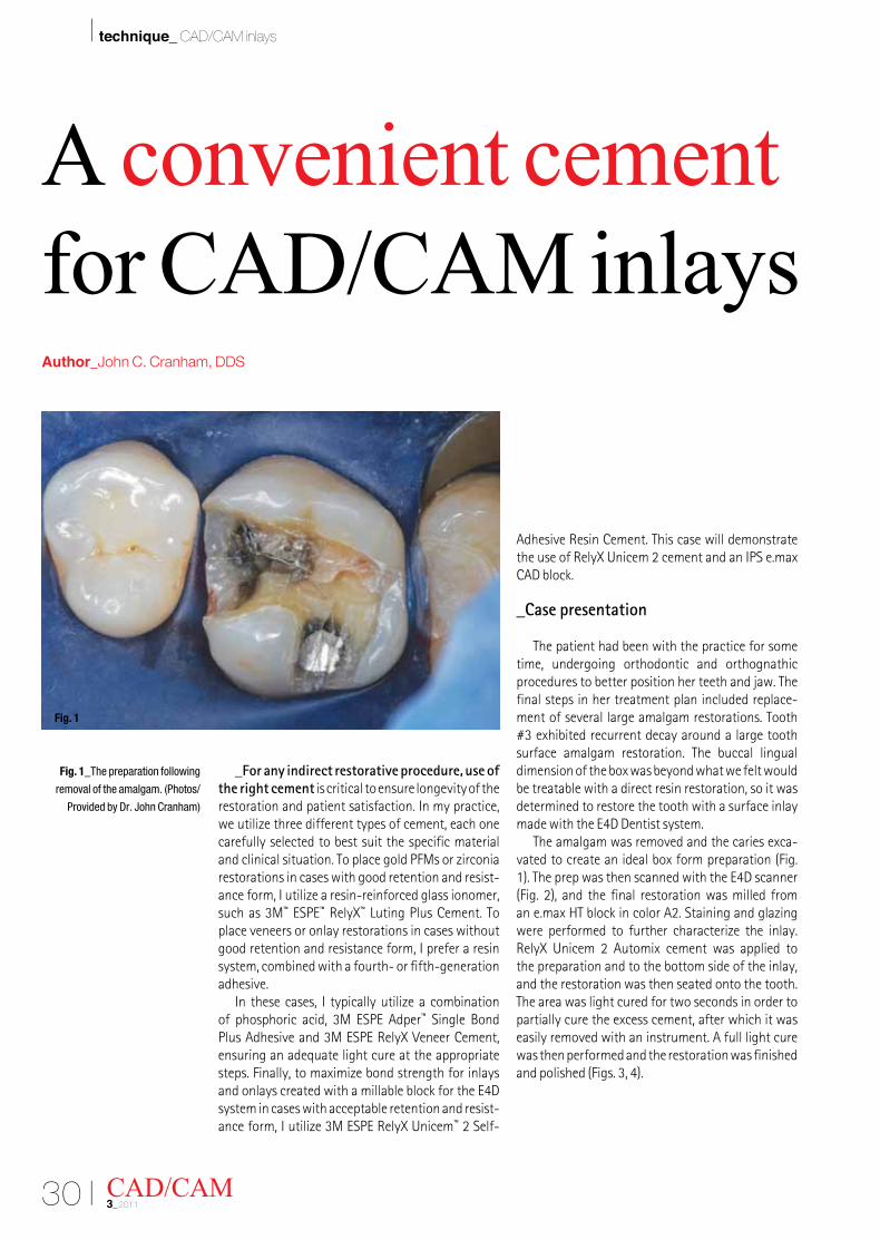
The patient had been with the practice for some time, undergoing orthodontic and orthognathic procedures to better position her teeth and jaw. The final steps in her treatment plan included replace-ment of several large amalgam restorations. Tooth #3 exhibited recurrent decay around a large tooth surface amalgam restoration. The buccal lingual dimension of the box was beyond what we felt would be treatable with a direct resin restoration, so it was determined to restore the tooth with a surface inlay madewiththeE4DDentistsystem.
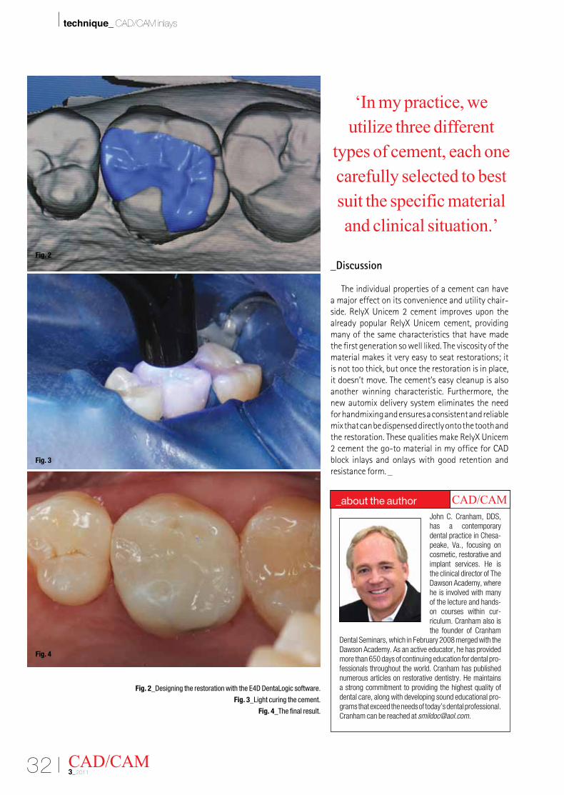
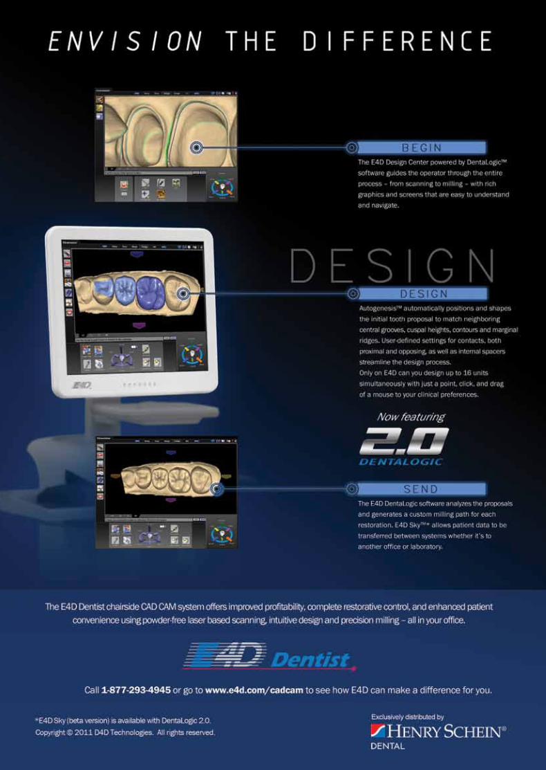
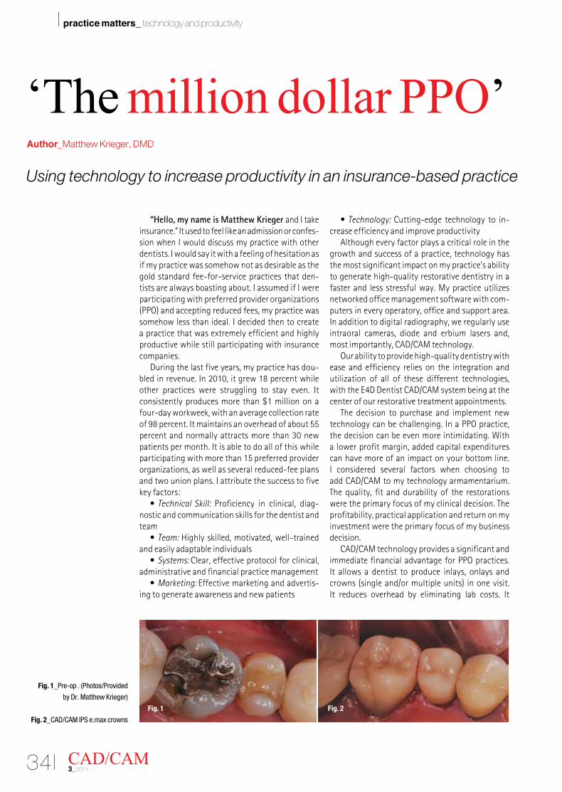
The amalgam was removed and the caries exca-vated to create an ideal box form preparation (Fig. 1).TheprepwasthenscannedwiththeE4Dscanner(Fig.2),andthe final restorationwasmilled froman e.max HT block in color A2. Staining and glazing were performed to further characterize the inlay. RelyX Unicem 2 Automix cement was applied tothe preparation and to the bottom side of the inlay, and the restoration was then seated onto the tooth. The area was light cured for two seconds in order to partially cure the excess cement, after which it was easily removed with an instrument. A full light cure was then performed and the restoration was finished andpolished(Figs.3,4).
CAD/CAM3_2011
A convenient cement for CAD/CAM inlaysAuthor_John C. Cranham, DDS
Fig. 1_The preparation following
removal of the amalgam. (Photos/
Provided by Dr. John Cranham)
Fig. 1
32 I
I technique_ CAD/CAM inlays
_Discussion
The individual properties of a cement can have a major effect on its convenience and utility chair-side.RelyXUnicem2 cement improvesupon thealready popular RelyX Unicem cement, providingmany of the same characteristics that have made the first generation so well liked. The viscosity of the material makes it very easy to seat restorations; it is not too thick, but once the restoration is in place, it doesn’t move. The cement’s easy cleanup is also another winning characteristic. Furthermore, the new automix delivery system eliminates the need for handmixing and ensures a consistent and reliable mix that can be dispensed directly onto the tooth and therestoration.ThesequalitiesmakeRelyXUnicem2 cement the go-to material in my office for CAD block inlays and onlays with good retention and resistance form. _
CAD/CAM3_2011
Fig. 2_Designing the restoration with the E4D DentaLogic software.
Fig. 3_Light curing the cement.
Fig. 4_The final result.
John C. Cranham, DDS, has a contemporary dental practice in Chesa-peake, Va., focusing on cosmetic, restorative and implant services. He is the clinical director of The Dawson Academy, where he is involved with many of the lecture and hands-on courses within cur-riculum. Cranham also is the founder of Cranham
Dental Seminars, which in February 2008 merged with the Dawson Academy. As an active educator, he has provided more than 650 days of continuing education for dental pro-fessionals throughout the world. Cranham has published numerous articles on restorative dentistry. He maintains a strong commitment to providing the highest quality of dental care, along with developing sound educational pro-grams that exceed the needs of today’s dental professional. Cranham can be reached at [email protected].
_about the author CAD/CAM
‘In my practice, we utilize three different
types of cement, each one carefully selected to best suit the specific material and clinical situation.’
Fig. 2
Fig. 3
Fig. 4
34I
I practice matters_ technology and productivity
“Hello, my name is Matthew Krieger and I take insurance.” It used to feel like an admission or confes-sion when I would discuss my practice with other dentists. I would say it with a feeling of hesitation as if my practice was somehow not as desirable as the gold standard fee-for-service practices that den-tists are always boasting about. I assumed if I were participating with preferred provider organizations (PPO)andacceptingreducedfees,mypracticewassomehow less than ideal. I decided then to create a practice that was extremely efficient and highly productive while still participating with insurance companies.
During the last five years, my practice has dou-bled in revenue. In 2010, it grew 18 percent while other practices were struggling to stay even. It consistently produces more than $1 million on a four-day workweek, with an average collection rate of 98 percent. It maintains an overhead of about 55 percent and normally attracts more than 30 new patients per month. It is able to do all of this while participating with more than 15 preferred provider organizations, as well as several reduced-fee plans and two union plans. I attribute the success to five key factors:
•Technical Skill: Proficiency in clinical, diag-nostic and communication skills for the dentist and team
•Team: Highly skilled, motivated, well-trained and easily adaptable individuals
•Systems: Clear, effective protocol for clinical, administrative and financial practice management
•Marketing: Effectivemarketingandadvertis-ing to generate awareness and new patients
•Technology: Cutting-edge technology to in-crease efficiency and improve productivity
Although every factor plays a critical role in the growth and success of a practice, technology has the most significant impact on my practice’s ability to generate high-quality restorative dentistry in a faster and less stressful way. My practice utilizes networked office management software with com-puters in every operatory, office and support area. In addition to digital radiography, we regularly use intraoral cameras, diode and erbium lasers and, most importantly, CAD/CAM technology.
Our ability to provide high-quality dentistry with ease and efficiency relies on the integration and utilization of all of these different technologies, withtheE4DDentistCAD/CAMsystembeingatthecenter of our restorative treatment appointments.
The decision to purchase and implement new technology can be challenging. In a PPO practice, thedecisioncanbeevenmoreintimidating.Witha lower profit margin, added capital expenditures can have more of an impact on your bottom line. I considered several factors when choosing to add CAD/CAM to my technology armamentarium. The quality, fit and durability of the restorations were the primary focus of my clinical decision. The profitability, practical application and return on my investment were the primary focus of my business decision.
CAD/CAM technology provides a significant and immediate financial advantage for PPO practices. It allows a dentist to produce inlays, onlays and crowns(singleand/ormultipleunits)inonevisit.It reduces overhead by eliminating lab costs. It
CAD/CAM3_2011
‘The million dollar PPO’Author_Matthew Krieger, DMD
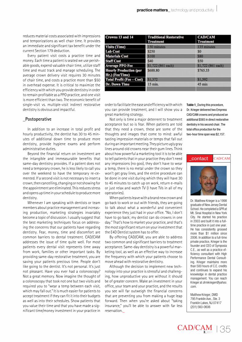
Fig. 1_Pre-op . (Photos/Provided
by Dr. Matthew Krieger)
Fig. 2_CAD/CAM IPS e.max crowns
Using technology to increase productivity in an insurance-based practice
Fig. 1 Fig. 2
I 35CAD/CAM 3_2011
practice matters_ technology and productivity I
reduces material costs associated with impressions and temporizations as well chair time. It provides an immediate and significant tax benefit under the current Section 179 deduction.
Every patient visit costs a practice time andmoney.Eachtimeapatientisseatedweuseperish-able goods, expend valuable chair time, utilize staff time and must track and manage scheduling. The average crown delivery visit requires 30 minutes of chair time, and costs a practice more than $50 in overhead expense. It is critical to maximize the efficiency with which you provide dentistry in order to remain profitable as a PPO practice, and one visit is more efficient than two. The economic benefit of single-visit vs. multiple-visit indirect restorative dentistry is obvious and impactful.
_Postoperative
In addition to an increase in total profit and hourlyproductivity,thedentisthas30to45min-utes of additional down time to produce more dentistry, provide hygiene exams and perform administrative duties.
Beyond the financial return on investment are the intangible and immeasurable benefits that same-day dentistry provides. If a patient does not need a temporary crown, he is less likely to call you over the weekend to have the temporary re-ce-mented. If a second visit is not necessary to insert a crown, then cancelling, changing or not showing for the appointment are eliminated. This reduces stress and opens up time in your schedule to produce more dentistry.
WheneverIamspeakingwithdentistsorteammembers about practice management and increas-ing production, marketing strategies invariably become a topic of discussion. I usually suggest that the best marketing techniques focus on address-ing the concerns that our patients have regarding dentistry. Fear, money, time and discomfort are common barriers to dental treatment. CAD/CAM addresses the issue of time quite well. For most patients every dental visit represents time away from work, families or other important tasks. By providing same-day restorative treatment, you are saving your patients precious time. People don’t like going to the dentist. It’s not personal. It’s just not pleasant. Have you ever had a colonoscopy? Not a great memory. Now imagine the thought of a colonoscopy that took not one but two visits and required you to “wear a temp between each visit, which may fall out.” It is much easier for patients to accept treatment if they can fit it into their budgets as well as into their schedules. Show patients that you value their time and that you have made a sig-nificant time/money investment in your practice in
order to facilitate the ease and efficiency with which you can provide treatment, and I will show you a great marketing strategy.
Not only is time a major deterrent to treatment acceptancebutsoisfear.Whenpatientsaretoldthat they need a crown, these are some of the thoughts and images that come to mind: awful tasting impression materials or temps that fall out during an important meeting. They picture ugly gray lines around old crowns near their gum lines. Think about how powerful a marketing tool it is to be able to tell patients that in your practice they don’t need anyimpressions(nogoo),theydon’thavetoweara temp, there is no metal under the crown so they won’t get gray lines, and the entire procedure can be done in one visit during which they will have 30 to45minutestocatchuponwork,returne-mailsor just relax and watch TV (I have TVs in all of my operatories).
Whenpatientsleavewithabrandnewcrownandgo back to work or out with friends, they are going to talk about what a wonderful and convenient experience they just had in your office. “No, I don’t have to go back; my dentist can do crowns in one day.” That’s how to market your practice, and that’s the most significant return on your investment that theE4DDentistsystemhastooffer.
By offering CAD/CAM, you are able to address two common and significant barriers to treatment acceptance. Same-day dentistry is a powerful mar-keting tool, as well as an effective way to increase the frequency with which your patients choose to move ahead with restorative dentistry.
Although the decision to implement new tech-nology into your practice is stressful and challeng-ing, how unproductive you are without it should be of greater concern. Make an investment in your office, your team and your practice, and the results you see will far outweigh the financial concerns that are preventing you from making a huge leap forward. Then when you’re asked about “taking insurance,” you’ll be able to answer with far less reservation._
Dr. Matthew Krieger is a 1998 graduate of New Jersey Dental School. He completed a GPR at Mt. Sinai Hospital in New York City. He started his practice in 2003 and built it into a full-time practice in just one year. He has consistently grossed more than $1 million since 2006. In addition to a full-time private practice, Krieger is the founder and CEO of Symposia C.E., as well as a practice ef-ficiency consultant with High Performance Dental Consult-ing. Krieger maintains more than 500 hours of C.E. credits and continues to expand his knowledge in dental practice management. You can reach Krieger at [email protected].
Matthew Krieger, DMD795 Franklin Ave., Ste. 3Franklin Lakes, NJ 07417(201) 560-0606
CAD/CAM _contact
Table 1_During this procedure,
Dr. Krieger delivered two Empress
CAD/CAM crowns and produced an
additional $565 in direct restorative
dentistry in his second chair. The
total office production for the
two-hour time span was $2,157.
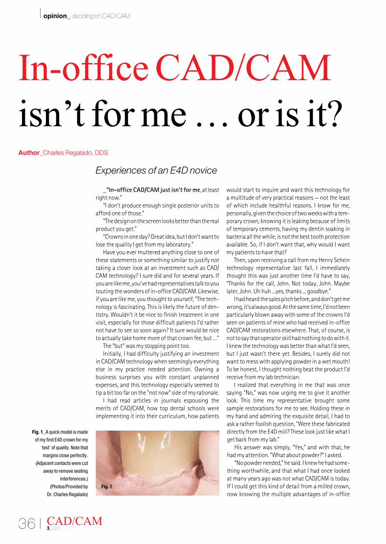
36 I
I opinion_ deciding on CAD/CAM
_“In-office CAD/CAM just isn’t for me, at least right now.”
“I don’t produce enough single posterior units to afford one of those.”
“The design on the screen looks better than the real product you get.”
“Crowns in one day? Great idea, but I don’t want to lose the quality I get from my laboratory.”
Have you ever muttered anything close to one of these statements or something similar to justify not taking a closer look at an investment such as CAD/CAM technology? I sure did and for several years. If you are like me, you’ve had representatives talk to you touting the wonders of in-office CAD/CAM. Likewise, if you are like me, you thought to yourself, “The tech-nology is fascinating. This is likely the future of den-tistry.Wouldn’titbenicetofinishtreatmentinonevisit, especially for those difficult patients I’d rather not have to see so soon again? It sure would be nice to actually take home more of that crown fee, but …”
The “but” was my stopping point too.Initially, I had difficulty justifying an investment
in CAD/CAM technology when seemingly everything else in my practice needed attention. Owning a business surprises you with constant unplanned expenses, and this technology especially seemed to tip a bit too far on the “not now” side of my rationale.
I had read articles in journals espousing the merits of CAD/CAM, how top dental schools were implementing it into their curriculum, how patients
would start to inquire and want this technology for a multitude of very practical reasons — not the least of which include healthful reasons. I know for me, personally, given the choice of two weeks with a tem-porary crown, knowing it is leaking because of limits of temporary cements, having my dentin soaking in bacteria all the while, is not the best tooth protection available. So, if I don’t want that, why would I want my patients to have that?
Then, upon receiving a call from my Henry Schein technology representative last fall, I immediately thought this was just another time I’d have to say, “Thanks for the call, John. Not today, John. Maybe later, John. Uh huh ...yes, thanks ... goodbye.”
I had heard the sales pitch before, and don’t get me wrong, it’s always good. At the same time, I’d not been particularly blown away with some of the crowns I’d seen on patients of mine who had received in-office CAD/CAM restorations elsewhere. That, of course, is not to say that operator skill had nothing to do with it. I knew the technology was better than what I’d seen, but I just wasn’t there yet. Besides, I surely did not want to mess with applying powder in a wet mouth! To be honest, I thought nothing beat the product I’d receive from my lab technician.
I realized that everything in me that was once saying “No,” was now urging me to give it another look. This time my representative brought some sample restorations for me to see. Holding these in my hand and admiring the exquisite detail, I had to askaratherfoolishquestion,“WerethesefabricateddirectlyfromtheE4Dmill?TheselookjustlikewhatIget back from my lab.”
His answer was simply, “Yes,” and with that, he hadmyattention.“Whataboutpowder?”Iasked.
“No powder needed,” he said. I knew he had some-thing worthwhile, and that what I had once looked at many years ago was not what CAD/CAM is today. If I could get this kind of detail from a milled crown, now knowing the multiple advantages of in-office
CAD/CAM3_2011
In-office CAD/CAM isn’t for me … or is it?Author_Charles Regalado, DDS
Fig. 1_A quick model is made
of my first E4D crown for my
‘test’ of quality. Note that
margins close perfectly.
(Adjacent contacts were cut
away to remove seating
interferences.)
(Photos/Provided by
Dr. Charles Regalado)
Experiences of an E4D novice
Fig. 1
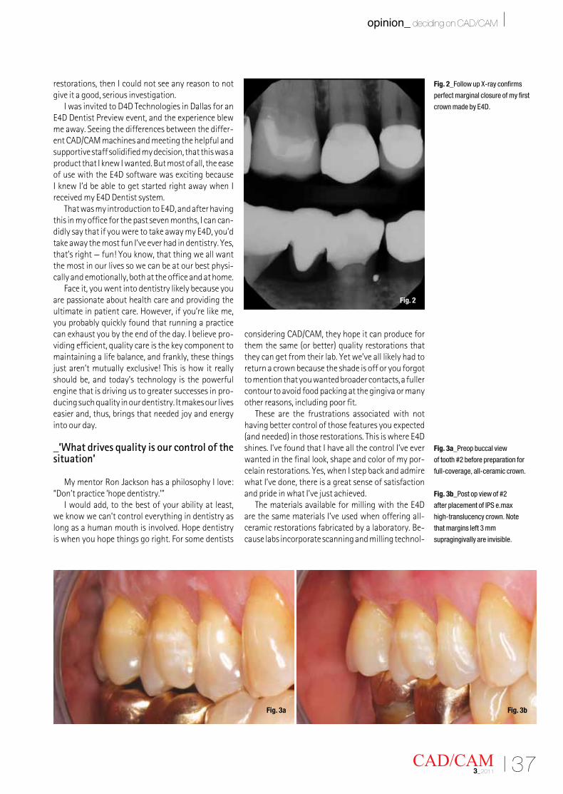
I 37CAD/CAM 3_2011
opinion_ deciding on CAD/CAM I
restorations, then I could not see any reason to not give it a good, serious investigation.
IwasinvitedtoD4DTechnologiesinDallasforanE4DDentistPreviewevent,andtheexperienceblewme away. Seeing the differences between the differ-ent CAD/CAM machines and meeting the helpful and supportive staff solidified my decision, that this was a product that I knew I wanted. But most of all, the ease ofusewiththeE4DsoftwarewasexcitingbecauseI knew I’d be able to get started right away when I receivedmyE4DDentistsystem.
ThatwasmyintroductiontoE4D,andafterhavingthis in my office for the past seven months, I can can-didlysaythatifyouweretotakeawaymyE4D,you’dtake away the most fun I’ve ever had in dentistry. Yes, that’s right — fun! You know, that thing we all want the most in our lives so we can be at our best physi-cally and emotionally, both at the office and at home.
Face it, you went into dentistry likely because you are passionate about health care and providing the ultimate in patient care. However, if you’re like me, you probably quickly found that running a practice can exhaust you by the end of the day. I believe pro-viding efficient, quality care is the key component to maintaining a life balance, and frankly, these things just aren’t mutually exclusive! This is how it really should be, and today’s technology is the powerful engine that is driving us to greater successes in pro-ducing such quality in our dentistry. It makes our lives easier and, thus, brings that needed joy and energy into our day.
_‘What drives quality is our control of the situation’
MymentorRonJacksonhasaphilosophyIlove:“Don’t practice ‘hope dentistry.’”
I would add, to the best of your ability at least, we know we can’t control everything in dentistry as long as a human mouth is involved. Hope dentistry is when you hope things go right. For some dentists
considering CAD/CAM, they hope it can produce for themthesame(orbetter)qualityrestorationsthatthey can get from their lab. Yet we’ve all likely had to return a crown because the shade is off or you forgot to mention that you wanted broader contacts, a fuller contour to avoid food packing at the gingiva or many other reasons, including poor fit.
These are the frustrations associated with not having better control of those features you expected (andneeded)inthoserestorations.ThisiswhereE4Dshines. I’ve found that I have all the control I’ve ever wanted in the final look, shape and color of my por-celain restorations. Yes, when I step back and admire what I’ve done, there is a great sense of satisfaction and pride in what I’ve just achieved.
ThematerialsavailableformillingwiththeE4Dare the same materials I’ve used when offering all-ceramic restorations fabricated by a laboratory. Be-cause labs incorporate scanning and milling technol-
Fig. 2_Follow up X-ray confirms
perfect marginal closure of my first
crown made by E4D.
Fig. 3a_Preop buccal view
of tooth #2 before preparation for
full-coverage, all-ceramic crown.
Fig. 3b_Post op view of #2
after placement of IPS e.max
high-translucency crown. Note
that margins left 3 mm
supragingivally are invisible.
Fig. 2
Fig. 3a Fig. 3b

38 I
I opinion_ deciding on CAD/CAM
ogy, it only made sense that I could benefit from CAD/CAM too, without coming up short on material qual-ity, durability and beauty. In fact, quality can actually increase because the intuitive DentaLogic™ software quickly illustrates areas where your preparation fails tomeettherequiredthickness.Ratherthanpayingfor a restoration that is too thin or have the patient return for a second preparation appointment, every-thing is easily corrected in the first appointment with the patient in the chair.
_‘But what about those margins?’
Peopleaskmeallthetime,“DoestheE4Dproducemargins as well as a lab-fabricated crown?” My an-swer is, it will produce as good a margin as you want it to give you. The old adage “garbage in, garbage out” ringstruewiththiskindoftechnology.GivetheE4Dagreatpreparationandyou’llgetagreatfit.What’smore, I no longer shy away from taking a postopera-tive radiograph, because I’ve simply learned to trust this technology.
I’ll be the first to admit that although I embraced CAD/CAM technology, there was still a nagging skep-tical side of me, so naturally I spent time testing each crown at the beginning of my learning period just to make sure things fit as well as I wanted them to. By simply pouring the impression of a tooth scanned in the mouth, a model is quickly made, and then I could seeformyselfhowtheE4Dtrulyperformed(Fig.1).It did not take long to convince me that I had little to worryabout(Figs.2–3b).
_‘That’s nice, but I don’t produce enough single units to make it worthwhile’
This is precisely what I thought and why I never really ventured to dive into in-office CAD/CAM. Now, I’ll be candid here: Many clinicians may not practice like I do. I chose long ago to significantly limit my number of treatment rooms and patients seen per day. I probably see a fraction of what the average dental office sees in a day. Moreover, that should encourage anyone considering in-office CAD/CAM because if it can be profitable in a smaller, limited-capacity office, then surely any office will reap huge financial benefits. How do I know? Because even in my smaller practice, looking over my expenses in the firstthreemonthsalonesinceintegratingtheE4D,Ihad saved more than $10,000 in fabrication costs! I don’t need to tell you how much more average of-fices would save more than that amount. I have no reservations in saying that my purchase has proven to be a “no-brainer” financially.
_‘What about anteriors?’
This was a key concern of mine regarding the abilities of CAD/CAM. I was under the impression that while posterior teeth were a good indication for in-office milled crowns, anterior teeth would remain the domain of laboratories. Because my practice involved anterior cases quite frequently, unless I could be con-vinced I could create and custom-shade anterior teeth, myusefortheE4Dwouldbetoolimited(Figs.4a,b).
CAD/CAM3_2011
Figs. 4a, 4b_Pre-op views. Patient
disliked rotations and wear of #10,
midline slant, improper proportions
of all incisors and facial composite
veneer of lingually positioned #8
done previously to bring facial plane
into arch. She also thought #6 was
slightly short for her liking.
Figs. 5a, 5b_Final veneers on
model. Note small incisal ‘cap’ milled
by E4D for cuspid #6. The milling
capability of E4D is nothing short of
amazing. Thinness of veneers can
be seen by black show-through on
certain restorations.
Fig. 4a
Fig. 5a
Fig. 4b
Fig. 5b
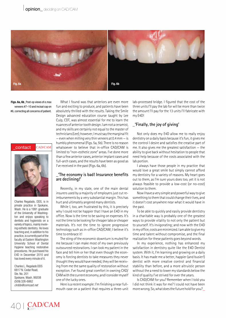
40 I
I opinion_ deciding on CAD/CAM
WhatIfoundwasthatanteriorsareevenmorefun and exciting to produce, and patients have been absolutely thrilled with the results. Taking the Smile Design advanced education course taught by Lee Culp, CDT, was almost essential for me to learn the nuances of anterior tooth design. I am not a ceramist, and my skills are certainly not equal to the master of technicians(Lee);however,Imustsaythemarginalfit—evenwhenmillingverythinveneersat0.4mm—ishumblyphenomenal(Figs.5a,5b).Thereisnoreasonwhatsoever to believe that in-office CAD/CAM is limited to “non-esthetic zone” areas. I’ve done more than a few anterior cases, anterior implant cases and full-arch cases, and the results have been as good as I’vereceivedinthepast(Figs.6a,6b).
_‘The economy is bad! Insurance benefits are declining!’
Recently, in my state, one of the main dentalinsurers used by a majority of employers just cut re-imbursements by a very substantial margin. This has hurt and ultimately angered many dentists.
WhileI,too,amfrustratedbythis,itispreciselywhyIcouldnotbehappierthatIhaveanE4Dinmyoffice. Now is the time to be saving on expenses. It’s not the time to be looking for cheaper labs or cheaper materials. It’s not the time to ignore progressive technology such as in-office CAD/CAM; I believe it’s time to embrace it!
The sting of the economic downturn is muted for me because I can make most of my own previously outsourced restorations. I can look my patient in the face and tell him or her that even though the econ-omy is forcing dentists to take measures they never thought they would have needed, they will be receiv-ing from me the same quality of restoration without exception. I’ve found great comfort in owning CAD/CAM with the current economy, and I consider myself one of the lucky ones.
Here is a recent example: I’m finishing a near full-mouth case on a patient that requires a three-unit
lab-processed bridge. I figured that the cost of the three units I’ll pay the lab for will be more than twice the amount I’ll pay for the 13 units I’ll fabricate with myE4D!
_‘Finally, the joy of giving’
NotonlydoesmyE4Dallowmetoreallyenjoydentistry on a daily basis because it’s fun, it gives me the control I desire and satisfies the creative part of me. It also gives me the greatest satisfaction — the ability to give back without hesitation to people that need help because of the costs associated with the lab portion.
I always have those people in my practice that would love a great smile but simply cannot afford my dentistry for a variety of reasons. My heart goes out to them, as I’m sure yours does too, yet it is not alwaysfeasibletoprovidea low-cost(orno-cost)solution to them.
Now I have a very simple and powerful way to give something to them that could change their lives, and it doesn’t cost anywhere near what it would have in the past.
To be able to quickly and easily provide dentistry in a charitable way is probably one of the greatest ways to provide vitality to not only the patient but toyourself.It’sinvigorating;andnowwiththeE4Din my office, costs are minimized. I am able to give my time and talent without compromise, and the final realization for these patients goes beyond words.
In my experience, nothing has enhanced my satisfactionindentistryquiteliketheE4DDentistsystem.Withit,I’mlearningandgrowingonadailybasis.Ithasmademeabetter,happier(andbusier!)dentist with more creative control and financial stability than before, and a more altruistic person without the a need to lower my standards below the kind of quality I’ve strived for over the years.
IsCAD/CAMforyou?RememberwhenItoldyouI did not think it was for me? I could not have been more wrong. So, what does the future hold for you? _
CAD/CAM3_2011
Figs. 6a, 6b_Post-op views of e.max
veneers #7–10 and incisal cap on
#6, correcting all concerns of patient.
Charles Regalado, DDS, is in private practice in Spokane, Wash. He is a 1991 graduate of the University of Washing-ton and enjoys speaking to dentists and hygienists on a variety of topics, mainly involv-ing esthetic dentistry. He loves teaching and, in addition to his practice, is currently part of the faculty at Eastern Washington University School of Dental Hygiene teaching restorative procedures. He purchased his E4D in December 2010 and has loved every minute of it.
Charles L. Regalado DDS6817 N. Cedar Road, Ste. No. 201Spokane, Wash. 99208(509) [email protected]
CAD/CAM _contact
Fig. 6a Fig. 6b
I 41CAD/CAM 3_2011
opinion_ economic hurdles I
_Elective services are a large part of dental procedures. Yet, according to the article “How Den-tistryWeathersaSlowEconomy”byRogerP.Levin,DDS, while patients tend to keep preventive care appointments, elective services decline during times of financial struggle. To protect against a stagnant economy, dentists need to consider ways to increase patientflowandsatisfaction.IfoundtheE4D(D4DTechnologies,Richardson,Texas)in-officeCAD/CAMsystem increased the vitality of my practice finan-cially, professionally and personally at a time when I hear my peers struggling to maintain their own and their staff’s motivation. Although it’s tempting to avoid a financial investment, now is the ideal time to place your practice a step above the competition. Don’t wait, and here’s why.
_Boosting practice economics
WhenfirstintroducedtoCAD/CAMtechnology,I knew it would change the future of dentistry, but I resisted the investment. It took 10 years to pay off my practice, and I wasn’t eager to jump back into debt.
As a solo practitioner, I was apprehensive. I knew in the long run it would be to my advantage, but I neededtobecomecomfortablewiththeidea.WhenIrealized the increasing cost of laboratory bills, I began talking with colleagues who had purchased other CAD/CAM systems, as well as those who invested in theE4D.
Of the systems available, what convinced me to chooseE4Dwastheopportunitytopreviewthesys-tem in action. I had a behind-the-scenes look at how the machine was developed, created and “real time” supportedbyD4D.ThethoughtofinvestinginCAD/CAM technology and being left to your own devices tolearnhowtouseitcanbeintimidating.D4Delimi-nates the uncertainty with its comprehensive edu-cational resources, continuing education courses, on-site setup and training and remote Support on
Sight.WhenIrealizedIwouldn’tbeforgottenonceIpurchasedit,IknewE4Dwasforme.
ChoosingtoembraceE4Ddigitaldentistryisoneof the best professional decisions I’ve made. It’s fast, much easier to use than you would think and the re-sultsarephenomenal.BeforeE4D,twoweekswouldpass between my patient’s first appointment and seatingtheircrown.WithE4D,ittakeslessthantwohours,1hourand40minutesonaverage.Becausethetime factor for fabrications has been so accelerated withtheE4D,ithasincreasedthenumberofpatientswe can see in a day and increased patient acceptance. This, in turn, has increased our production and col-lection numbers, all while reducing laboratory costs.
TheE4Dhasdefinitelyhadapositiveeconomicimpact on my practice and, as a result, is paying for itself. At a time when I felt my practice was coasting andIwasworriedabouttheeconomy,theE4Dhasproven to be a huge advantage, and not only finan-cially.Whilemanyofmypeersstrugglethroughtherecession, my practice continues to earn and grow in numerous other ways.
_Professional satisfaction
The personal and professional satisfaction I have experienced since incorporating the E4D into mypractice is unprecedented in my experience. It has
Time to lose ‘the wait’!Author_Dean Saiki, DDS
42 I
I opinion_ economic hurdles
sparked my enthusiasm. I enjoy coming to work and providing my patients with state-of-the-art technology and innovative services that I couldn’t in the past. For my patients, visiting the dentist today is a completely different experience than before I investedintheE4D.Mypatientscanwatchafabrica-tion from behind the scenes and see it being designed and created all in a single visit. If a patient comes in at 8a.m.,he(orshe)canbeoutby9:40a.m.Withinthattime, I sit down with the patient to explain the process and show him how we design the restorations. I love designing my own restorations. It’s both challenging andrewarding.TheE4Dsystemhaschangedmyroleas a dentist, as well as the dynamics between my patients and me.
MyteammembershavealsoembracedtheE4Dtechnology.Whenapatientisapprehensiveaboutundergoing a restoration, they enthusiastically an-swer their questions and describe the procedure. They love to inform our patients that a restoration can now be completed in a single visit and that there is no longer a need for the icky, gooey, impression material. They are excited that we can provide better service, more comfortably and faster, and in the event that there is an unscheduled emergency, my assist-ants are very adept at fabricating crowns.
It would be a shame to invest in new technology only to spend your time fumbling around trying to learn how to use it. You would lose money, time and patients. That’swhy I believe thatD4D shouldbecreditednotonlyforcreatingtheE4Dbutalsoforstanding behind it as extensively as it does. In two and a half years, I have not had a problem or glitch that wasn’t immediately addressed to my complete satisfaction. Help is available whenever we need it - something we couldn’t do without. The support thatD4DprovidesmakestheE4Dsounique.IamalsoimpressedwithhowsupportiveD4Disofourthoughts, ideas and suggestions concerning changes andupdatesregardingtheE4Dsystem.
Dentistry has come a long way from 15 years ago: newtechnology,digitalX-rays,intraoralcameras.Forpatients, it has become quick, easy and educational, and you can see their excitement and satisfaction. Investing in new technology has had a positive impact on my staff and me both personally and professionally.
_Personal success
Although I have always loved what I do, I have definitely noticed a difference since incorporating new technology into my practice. Offering innova-tive technology to our patients and mastering it has given both my staff and me personal satisfaction. Weenjoycomingtoworkandservingourpatients.Witheachnewrestoration,welearnhowtodothingsfaster and better. It’s simply more exciting. Knowing that we are providing our patients with cutting-edge services and that financially we are doing well is very gratifying.
Another personal advantage for me is that if I can’t be in the office because of illness, family emergency or vacation, my staff members can bring the dentist who is filling in for me up to speed regarding the new technology. They are eager and proud to share our new technology and their expertise with the new doctors.
_Conclusion
InvestingintheE4DDentistsystemhasadvancedmy operations and services and given my practice a competitive edge. Looking back, I realize my re-luctance to invest denied not only me but also my patients and staff the many advantages the E4Din-office CAD/CAM system can provide. Based on my experience, I would advise my colleagues not to wait likeIdid.TheE4Dhasdefinitelyhelpedmypracticeweather the economic storm. As the saying goes: Hindsight is 20/20 vision. Don’t wait. Now’s the time toinvestinE4D._
CAD/CAM3_2011
Dr. Dean Saiki graduated from the University of South-ern California School of Dentistry in 1988. He has been practicing in San Diego North County since 1989. He’s been a member of the ADA, CDA and San Diego Dental Society for more than 20 years. He attends regular bimonthly study club meetings to continue the ongo-ing educational process and loves training in the latest dental technologies. He has maintained his practice in Oceanside since 1990.
Dr. Dean Saiki 3231 Waring Court Ste. COceanside, Calif. 92056(760) 845-3456 [email protected]
_about the author CAD/CAM
‘Offering innovativetechnology to our patientsand mastering it has given
both my staff and mepersonal satisfaction.’
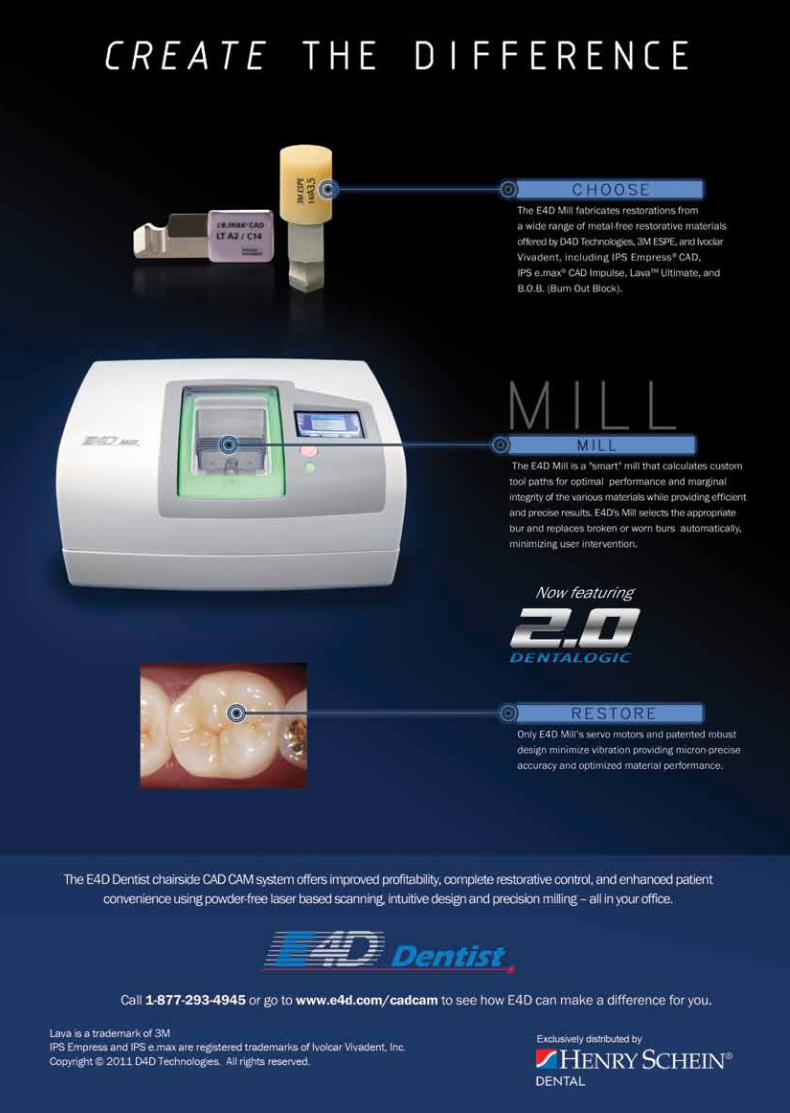
44 I
I opinion_ dispelling misconceptions
_I’ve been practicing restorative dentistry for more than 17 years, and during all but three months of that time, I’ve utilized chairside CAD/CAM dentistry on nearly a daily basis in my practice. Three months out of dental school, I purchased my firstchairsideCAD/CAMsystem—theCEREC2—onthe Internet. The system arrived in my office with an owner’s manual written in German; so that was my introduction, initiation and education in chairside CAD/CAM dentistry. During the next 17 years, I upgraded my system, improved my technique and began to teach others.
I trained clinicians, technicians and dental assist-ants on the basics, advanced techniques and even the future of CAD/CAM dentistry. I’ve trained thou-sands of dental professionals during the years and have heard and seen it all: the good and the bad, the perceptions and the realities. I’ve recently switched brands of systems, but not the quality of care.
Chairside CAD/CAM dentistry is only a vehicle to the result: quality patient care. The fundamentals of restorative dentistry, clinical skills, material under-standing, proper diagnosis and treatment planning are required to drive any system to excellence. Allow me to share with you some of the common state-ments relating to CAD/CAM dentistry that I’ve heard, which like most things, are based on a lack of under-standing, personal biases or a lack of firsthand expe-rience that often generates these misconceptions.
_ Perception: ‘Only one system works’
I’ve recently switched systems; specifically, from theCERECsystemtotheE4DDentistsystem.Icantellyou both systems can create beautiful, well-fitting restorations. Each one does it in a little differentmethod — one uses powder, one takes more images and gathers more data — but both are capable of successfully changing everything in a restorative practice: control of variables, convenience for the patient, dental team motivation and the bottom line. There are other differences in the systems — such as in software, milling, manufacturers, education, support and distributors — but they both work probably better than you’ve ever imagined. You owe it to yourself, your practice, your team and your patients to check them both out.
More important than the system is you: your atti-tudeandabilitytochange(forthebetter)yourteam’sinvolvement and your clinical skills. The incorporation ofanytechnology (digitalX-rays,patientmanage-ment, lasers, magnification) typically makes you a“better” clinician by providing more information to make better decisions, and chairside CAD/CAM den-tistry is no different. Being able to see your prepara-tions on a large screen, to rotate, zoom and view from any angle will improve your evaluation of your prepa-rations and your soft-tissue management. Yet, it isn’t the system, it’s the dentistry. Both systems will allow you to get where you want to go, but you have to drive.
Reality: It’s not the system; it’s the dentist and the team.
_Perception: ‘I can figure it out by myself’
Seventeen years ago, the only CAD/CAM educa-tion I could find was by trying to translate an owner’s manual from German just so I could turn on the CAD/CAM system. Today there is a plethora of educational opportunities: you can join proprietary websites with video lessons; you can purchase complete DVD sets of interactive learning opportunities; or attend basic training sessions, most of which are set-up by private clinicians that offer to show you “their” way. More than fouryearsago,D4DTechnologies (theE4Dguys)clearlyrecognizeddeficienciesinmanyofthese methods and introduced a novel approach to educationandsupport.Fromtheverybeginning,D4Dinstituted mandatory training and education and of-fered unlimited remote support with every purchase. The company also implemented the “team approach” to chairside CAD/CAM dentistry by offering a revolu-tionary certification program for dental assistants calledtheCAD/CAMDentalDesigner(CDD).
Knowing dentists, I’m sure that when most of them first hear they are required to attend training before they can get the system in their office, they look at that as an unnecessary hassle. However, let me tell you that education and support is the most important aspect of your successful journey to CAD/CAM dentistry — not only for you, but also for yourteam.IncludedineachE4Dsystempurchaseis not only training for you, but also for one of your assistants; and this includes everything, i.e., trans-
CAD/CAM3_2011
The perception vs. reality of chairside CAD/CAM dentistryAuthor_Alex Touchstone, DDS
I 45CAD/CAM 3_2011
opinion_ dispelling misconceptions I
portation, accommodations and a guarantee for a great experience. Too often, I’ve seen compromised basic education in order to up-sell or move someone toward advanced education opportunities. Whilethere is a right time for advanced courses, it varies by individual and by experiences and shouldn’t be predetermined at basic training.
In addition, most educational experiences en-lighten the students during the course, but then fall short on follow-up and compliance once the course isover.D4DTechnologiesaddressesthiswithathirdday of education in your office called integration. Soon after your system is set up, a clinical “integra-tion” specialist will arrive at your office and assist you for “the first day of the rest of your life with digital dentistry.” Then the true support kicks in with Sup-portonSight(SOS),whichisalsoincludedwiththesystem. One of the advantages of being introduced toE4DCAD/CAMdentistryduringtheInternetageis the requirement and advantage of having every customer online, which means an Internet access connection is mandatory for every E4D System.That’s so dental technicians, dental assistants and hardwareandsoftwareexperts,partoftheE4DSOSteam,canlogintoyoursystem(whenrequested)andremotely provide customer support. No more trying to tell someone over the phone what your screen looks like because the SOS team can take control of your system, help with designs, diagnosis of your hardware and also answer any questions about the operation of your system.
Reality: Education and support makes all the dif-ference in the world of CAD/CAM.
_Perception: ‘My patients don’t care about the benefits of chairside dentistry’
If you feel this way, it’s a good bet you haven’t asked them or you aren’t a patient-centered practice. It isn’t just being able to offer these restorations in one appointment, in one day, the next day or by the weekend, but more about being able to say “Yes!” to any patient inquiry. Chairside CAD/CAM dentistry gives you complete control of the entire process as well as many more options to provide quality care to yourpatientsinsteadofalwayshavingtosay,“We’llsee you in two weeks.”
Whilethereisnothingwrongwiththetwo-weekturnaround we’ve become accustomed to (or better said,lockedinto),whyisthattheonlyoption?
Disregarding when you are dealing with sensitiv-ity or changing the occlusal scheme, why not offer the patient the option of getting it all done today, or “Let’s prep on your way into work and seat on your way back from work.” In today’s economic times, being attentive to patients’ needs, concerns (leaving worktoooften)andconveniencefactors(running
errandswhiletherestorationsaremilling)iswhatwilldelineate those practices that flourish vs. simply sur-vive. The same reasons you’re using to consider stay-ing open late on some nights, adding weekend hours or adding an associate to cover more opportunities for patient care are the exact same reason you should consider offering additional treatment options and chairside CAD/CAM dentistry to your patient base.
Reality: All patients appreciate convenience.
_Perception: ‘Faster is better’
I cringe a bit when I hear some debates about the use of new technology that focus on how minutes can be saved through “bypassing this” or choosing a certain setting vs. the “correct one.” It’s curious because it’s as if our entire dental career has been spent waiting weeks for restorations and now we’re concernedaboutseconds.Whiletimeismoney, itworks both ways, on the front end and the back end. If by choosing a setting or making a material decision simply because it saves time on the front end (the dentalappointment),youloseonthebackendbycompromising the ultimate success of the restora-tion(longevityandperformance),whichmeansthatone’s priorities are now upside down.
Whilethisquestionismostlikelyanecdotalandwe may never have the true answer, I’d caution everyone to pause when they are told to select one material, system or procedure over the other simply because of a quantity of time saved. My advice is to choose based on the quality of the result and not the quantityofminutesyou’llsave(orlose).
Reality: Dentistry is not a race; seek quality over quantity.
_Perception: ‘It costs too much’
Whileweallliketolookatthepricetag,ininvest-ments and in technology you need to look at the “real costvs.benefit”orthereturnoninvestment(ROI)toyour practice. It is much easier for us to casually pass by a $200 lab bill 30 times each month vs. the serious-ness and hesitation we give to a $100,000 purchase. Take a few minutes with your accountant (or your spouse)tototalthefabricationfeesyou’vepaid,orintend to pay in the next five years, and compare that to investing and making monthly payments on a technology that will allow you to most likely fabricate 70 percent of those cases yourself (essentially all the singleunits).
Next, add in all the tax benefits of Section 179 and capital equipment and you’ll likely discover that the reality is a real eye opener. In the majority of situations,thecost(money,timeandpersonnel)offabricating single units yourself with a chairside CAD/CAM system is probably less than what you are doing
Alex Touchstone, DDS, has 17 years clinical experience in ap-plication of CAD/CAM dentistry in his dental practice. He holds two U.S. patents in the area of shade control for translucent dental restorations. During the past several years, he has had the privilege of teaching hundreds of dentists every year how to grow their practices through proper integration of carefully selected technolo-gies. He currently has the privilege of serving as the team leader of the online education and networking portal, www.CADCAMCAN.com, created for the benefit of the E4D com-munity. He can be reached at [email protected].
CAD/CAM _contact
46 I
I opinion_ dispelling misconceptions
CAD/CAM3_2011
now. In fact, doing dentistry the way you have been doing is what “costs too much.”
Reality: Most likely, a chairside CAD/CAM system costs less than what you are doing now.
_Perception: ‘Oh, I looked at it before and it’s not for me.’
Having spent 17 years defending and promoting chairside CAD/CAM dentistry, I’m pretty sure I’ve heard most of the excuses/reasons for not consider-ingit.Whiletherearecertainlysomevalidreasonswhy it isn’t for everyone, some comments should be reconsidered: “Oh, I’ve seen patients come through my office that have hideous looking CAD/CAM res-torations — no anatomy and just ugly — I’d never do those.” “They fit like socks on a rooster.” “I know Bob bought one, and now it sits in the corner.”
Indeed, technological development has gone through massive advances and what was once de-scribed as an “island of ceramic in a sea of cement” has now evolved to restorations that can have a marginal fit equal to or better than lab-fabricated restorations. So yes, early CAD/CAM promises don’t come close to today’s capabilities.
On the other hand, in that same practice that noticed the one CAD/CAM restoration, there were
probably hundreds of chairside CAD/CAM restora-tions that went through undetected and assumed to be lab-fabricated restorations. The difference between a noticeable and an undetected CAD/CAM restoration is probably not the technology but rather the clinician. Wehavetounderstandthatnorestorationgoesintothe mouth without the conscious decision and the professional skill of the clinician. If a less than ideal res-toration is permanently placed, it isn’t so much a factor of one system or technique vs. another, but rather the clinical skill or judgment of the clinician.
For those who have simply taken their peers’ opinions, read blogs or had good and bad experiences but haven’t checked out today’s chairside CAD/CAM dentistry for themselves, I encourage you to do so. The first cell phone was introduced the same decade as the first CAD/CAM chairside dentistry system, and neither bears any resemblance to the technologies offered today. Take a fresh look for yourself.
Reality: If you haven’t looked at chairside CAD/CAM in the last year, you haven’t looked at all.
If you’ve read this far, hopefully it means that even if you may have shared some of the perceptions listed above, you’re now willing to consider the possibility that technology can offer improvements in restora-tive dentistry._
AD
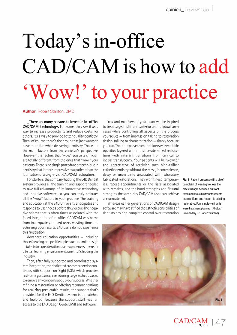
I 47CAD/CAM 3_2011
opinion_ the ‘wow!’ factor I
_There are many reasons to invest in in-office CAD/CAM technology. For some, they see it as a way to increase productivity and reduce costs. For others, it’s a way to provide better quality dentistry. Then, of course, there’s the group that just wants to have more fun while delivering dentistry. Those are the main factors from the clinician’s perspective. However, the factors that “wow” you as a clinician are totally different from the ones that “wow” your patients. There is no single procedure or technique in dentistry that is more impressive to a patient than the fabrication of a single-visit CAD/CAM restoration.
Forstarters,thecompanybackingtheE4DDentistsystem provides all the training and support needed to take full advantage of its innovative technology and intuitive software, so you can truly embrace all the “wow” factors in your practice. The training andeducationattheE4DUniversityanticipatesandresponds to user needs before they occur. The nega-tive stigma that is often times associated with the failed integration of in-office CAD/CAM was borne from inadequately trained users wasting time and achievingpoorresults.E4Dusersdonotexperiencethis frustration.
Advanced education opportunities — including those focusing on specific topics such as smile design — take into consideration user experiences to create a better learning environment, one that’s leading the industry.
Then, after fully supported and coordinated sys-tem integration, the dedicated customer service con-tinueswithSupport-on-Sight(SOS),whichprovidesreal-time guidance, even during large esthetic cases, toremoveanyconcernsaboutyoursuccess.Whetherrefining a restoration or offering recommendations for realizing predictable results, the support that’s providedfortheE4DDentistsystemisunmatchedand foolproof because the support staff has full accesstotheE4DDesignCenter,Millandsoftware.
You and members of your team will be inspired to treat large, multi-unit anterior and full/dual-arch cases while controlling all aspects of the process yourselves — from impression taking to restoration design, milling to characterization — simply because you can. There are polychromatic blocks with variable opacities layered within that create milled restora-tions with inherent transitions from cervical to incisal translucency. Your patients will be “wowed” and appreciative of receiving such high-caliber esthetic dentistry without the mess, inconvenience, delay or uncertainty associated with laboratory fabricated restorations. They won’t need temporar-ies, repeat appointments or the risks associated with remakes, and the bond strengths and flexural strengths the same-day CAD/CAM user can achieve are unmatched.
WhereasearliergenerationsofCAD/CAMdesignsoftware may have stifled the esthetic sensibilities of dentists desiring complete control over restoration
Today’s in-office CAD/CAM is how to add ‘Wow!’ to your practiceAuthor_Robert Stanton, DMD
Fig. 1_Patient presents with a chief
complaint of wanting to close the
black triangle between his front
teeth and make his front four teeth
more uniform and match his existing
restorative. Four single-visit units
were treatment planned. (Photos/
Provided by Dr. Robert Stanton)
Fig. 1
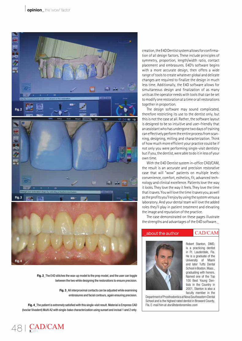
48 I
I opinion_ the ‘wow!’ factor
creation,theE4DDentistsystemallowsforconfirma-tion of all design factors. These include principles of symmetry, proportion, length/width ratio, contact placementandembrasures.E4D’ssoftwarebeginswith a more accurate design, then offers a wide range of tools to create whatever global and delicate changes are required to finalize the design in much lesstime.Additionally,theE4Dsoftwareallowsforsimultaneous design and finalization of as many units as the operator needs with tools that can be set to modify one restoration at a time or all restorations together in proportion.
The design software may sound complicated, therefore restricting its use to the dentist only, but thisisnotthecaseatall.Rather,thesoftwarelayoutis designed to be so intuitive and user-friendly that an assistant who has undergone two days of training can effectively perform the entire process from scan-ning, designing, milling and characterization. Think of how much more efficient your practice could be if not only you were performing single-visit dentistry but if you, the dentist, were able to do it in less of your own time.
WiththeE4DDentistsystemin-officeCAD/CAM,the result is an accurate and precision restorative case that will “wow” patients on multiple levels: convenience, comfort, esthetics, fit, advanced tech-nology and clinical excellence. Patients love the way it looks. They love the way it feels. They love the time that it saves. You will love the time it saves you, as well as the profits you’ll enjoy by using the system versus a laboratory. And your dental team will love the added roles they’ll play in patient treatment and elevating the image and reputation of the practice.
The case demonstrated on these pages illustrate thestrengthsandadvantagesoftheE4Dsoftware._
CAD/CAM3_2011
Robert Stanton, DMD, is a practicing dentist in Ft. Lauderdale, Fla. He is a graduate of the University of Miami and later Tufts Dental School in Boston, Mass., graduating with honors. Named one of the Top 100 Best Young Den-tists in the Country in 2001, Stanton is also a faculty member in the
Department of Prosthodontics at Nova Southeastern Dental School and is the highest rated dentist in Broward County, Fla. E-mail him at [email protected]
_about the author CAD/CAM
Fig. 2_The E4D stitches the wax-up model to the prep model, and the user can toggle
between the two while designing the restorations to ensure precision.
Fig. 3_All interproximal contacts can be adjusted while examining
embrasures and facial contours, again ensuring precision.
Fig. 4_The patient is extremely satisfied with this single-visit result. Material is Empress CAD
(Ivoclar Vivadent) Multi A2 with single-bake characterization using sunset and incisal 1 and 2 only.
Fig. 2
Fig. 3
Fig. 4
I 49CAD/CAM 3_2011
about the publisher _ submissions I
submissions:formatting requirements
Please note that all the textual elements of your submission: _the complete article, _all the figure captions, _the complete literature list and _ contact info (bio, mailing address,
e-mailaddress,etc.)
must be combined into one text document. Please do not submit multiple files for each of these items.
In addition, images (tables, charts, pho-tographs,etc.)mustnotbeembeddedinthetext document. All images must be submit-ted separately, and details about how to do this appear below.
If you are interested in submitting a C.E. article, contact us for additional instruc-tions before you make your submission.
_Text length
Article lengths can vary greatly — from 1,500 to 5,500 words — depending on the subject matter. Our approach is that if you need more or less words to do the topic jus-tice then please make the article as long or as short as necessary.
Wecanrunanextralongarticleinmul-tiple parts, but this is usually discussing a subject matter where each part can stand alone because it contains so much informa-tion. In addition, we do run multi-part series on various topics.
In short, we do not want to limit you in terms of article length, so please use the word count above as a general guideline and if you have specific questions, please do not hesitate to contact us.
_Text formatting
Please use single spacing and un-indented paragraphs for your text. Please do not put a blank line between paragraphs.
Wealsoaskthatyouforegoanyspecialformatting beyond the use of italics and boldface, and make sure that all text is left justified.
If you would like to emphasize certain words within the text, please only use italics (donotuseunderliningoralargerfontsize).Boldface is reserved for article headers.
Please do not “center” text on the page, add special tab stops, or use underlining as all of this must be removed before layout. If you require a special layout, please let the word processing program you are using help you to do this formatting rather than doing it by hand on your own.
If you need to make a list or add foot-notes or endnotes, please let the word processing program do it for you automati-cally. There are menus in every program that will help you to do this.
The fact is that no matter how careful one might be, errors have a way of creeping in when you try to hand number footnotes and literature lists.
_Image requirements
Please number images consecutively throughout the article by using a new number for each image. If it is imperative that certain images are grouped together, then use lowercase letters to designate the images in a group (i.e., Fig. 2a, Fig. 2b, Fig.2c).
Please put figure references in your arti-cle wherever they are appropriate, whether that is in the middle or end of a sentence but before the period.
If you are not directly mentioning the figure in the body of your article, when it ap-pears at the end of the sentence the figure reference should be enclosed within paren-thesis and appear before the final period.
In addition, please note:_ WerequireimagesinTIForJPEGformat._ These images must be no smaller than
4x4inchesinsizeat300DPI._ Images cannot be any smaller than 80 KB
in size (or they will print the size of a post-agestamp).
Larger images are always better, and something on the order of 1 MB is best. Thus, if you have an image that is greater than 1 MB, please do not bother “sizing it down” to meet our requirements, but send us the largest file size available.
The larger the starting image is in terms of bytes, the more leeway the designer has in terms of resizing the image to fill up more space should there be room avail-able).
Also, please remember that you should not embed the images into the body of the text document you submit. Images must be submitted separately from the textual submission.
You may submit images through a zipped file via e-mail, unzipped individual files via e-mail or post a CD containing your images directly to us (please contact us for the mailing address as this will depend upon where in the world you will bemailingthemfrom).
Please do not forget to send us a head shot photo of yourself that also fits the parameters above so that it can be printed along with your article.
_Abstracts
An abstract of your article is not required. However, if you choose to provide us with one, we will print it in a separate box.
_Contact info
At the end of every article is a contact info box with contact information along with a head shot of the author.
Please note at the end of your article the exact information you would like to appear in this box and format it according to the previously mentioned standards.
Ashortbio(60wordsorless)maypre-cede the contact info if you provide us with the necessary information.
_Questions? Comments?
Please do not hesitate to contact us for our InternationalC.E.MagazineAuthorKitorifyou have other questions/comments about the article submission process:
ManagingEditor Fred [email protected]
50 I CAD/CAM3_2011
I about the publisher _ imprint
CAD/CAMthe international C.E. magazine of digital dentistry
CAD/CAM_Copyright Regulations
_the international C.E. magazine of CAD/CAM published by Dental Tribune America is printed quarterly. The magazine’s articles and illustra-tions are protected by copyright. Reprintsofanykind,includingdigitalmediums, without the prior consent of the publisher are inadmissible and liable to prosecution. This also applies to duplicate copies, translations, microfilms and storage and processing in electronic systems. Reproductions,includingexcerpts, may only be made with the permission of the publisher.
All submissions to the editorial department are understood to be the original work of the author, meaning that he or she is the sole copyright holderandnootherindividual(s)orpublisher(s)holdsthecopyrighttothematerial. The editorial department reserves the right to review all editorial submissions for factual errors and to make amendments if necessary.
Dental Tribune America does not accept the submission of unsolicited books and manuscripts in printed or electronic form and such items will be disposed of unread should they be received.
Dental Tribune strives to maintain the utmost accuracy in its clinical articles. If you find a factual error or content that requires clarifica-tion,pleasecontactGroupEditorRobinGoodmanatr.goodman@dental-tribune.com. Opinions expressed by authors are their own and may not reflect those of Dental Tribune America and its employees.
Dental Tribune cannot assume responsibility for the validity of product claims or for typographical errors. The publisher also does not assume responsibility for product names or statements made by advertisers.
The responsibility for advertisements and other specially labeled items shall not be borne by the editorial department. Likewise, no respon-sibility shall be assumed for information published about associations, companies and commercial markets. All cases of consequential liability arising from inaccurate or faulty representation are excluded. General terms and conditions apply, and the legal venue is New York, New York.
Dental Tribune America is the official media partner of:
U.S. HeadquartersDental Tribune America 116West23rdStreet,Ste.500New York, NY 10011Tel.:(212)244-7181Fax:(212)[email protected]
PublisherTorstenR.Oemus [email protected]
Chief Operating Officer EricSeid [email protected]
Group Editor RobinGoodman [email protected]
Managing EditorFred Michmershuizen f.michmershuizen @dental-tribune.com
DesignerKristine Colker [email protected]
DesignerSierraRendon [email protected]
C.E. DirectorJulia [email protected]
C.E. International Sales ManagerChristiane [email protected]
Marketing ManagerAnna [email protected]
Marketing AssistantLorrie Young [email protected]
AccountingMelissa Chan [email protected]
List Manager Christopher Ceparano [email protected]
Product/Account ManagerMarkEisen [email protected]
Product/Account ManagerHumbertoEstrada [email protected]
Product/Account Manager & InteractiveGina Davison [email protected]
International Product/Account ManagerJan [email protected]
Editorial BoardMarcia Martins Marques, Leonardo Silberman,EminaIbrahimi,IgorCernavin,DanielHeysselaer,RoelanddeMoor,Julia Kamenova, T. Dostalova, Christliebe Pasini, Peter Steen Hansen, Aisha Sultan, Ahmed A Hassan, Marita Luomanen, Patrick Maher, Marie France Bertrand, Frederic Gaultier, Antonis Kallis, Dimitris Strakas, Kenneth Luk, MukulJain,RezaFekrazad,Sharonit Sahar-Helft, Lajos Gaspar, Paolo Vescovi, Marina Vitale, Carlo Fornaini, Kenji Yoshida, Hideaki Suda, Ki-Suk Kim, Liang Ling Seow, ShaymantSinghMakhan,EnriqueTrevino,Ahmed Kabir, Blanca de Grande, José Correia de Campos, Carmen Todea, Saleh Ghabban StephenHsu,AntoniEspanaTost,JosepArnabat, Ahmed Abdullah, Boris Gaspirc, Peter Fahlstedt, Claes Larsson, Michel Vock, Hsin-Cheng Liu, Sajee Sattayut, Ferda Tasar, Sevil Gurgan, Cem Sener, Christopher Mercer, Valentin Preve, Ali Obeidi, Anna-Maria Yannikou,SuchetanPradhan,RyanSeto,JoyceFong, Ingmar Ingenegeren, Peter Kleemann, Iris Brader, Masoud Mojahedi, Gerd Volland, GabrieleSchindler,RalfBorchers,StefanGrümer, Joachim Schiffer, Detlef Klotz, Herbert Deppe, Friedrich Lampert, Jörg Meister,ReneFranzen,AndreasBraun,SabineSennhenn-Kirchner, Siegfried Jänicke, Olaf Oberhofer and Thorsten Kleinert