Embed Size (px)
Citation preview
BiomaterialsScience
PAPER
Cite this: Biomater. Sci., 2021, 9,7194
Received 14th June 2021,Accepted 13th September 2021
DOI: 10.1039/d1bm00936b
rsc.li/biomaterials-science
The innate immune response of self-assemblingsilk fibroin hydrogels
Natalia Gorenkova,a,b,c Manfred F. Maitz,d Georg Böhme,b,d Hani A. Alhadrami,a,e
Essam H. Jiffri,a,e John D. Totten,a,b Carsten Werner, d,f Hilary V. O. Carswellb andF. Philipp Seib *b,d,g
Silk has a long track record of use in humans, and recent advances in silk fibroin processing have opened
up new material formats. However, these new formats and their applications have subsequently created a
need to ascertain their biocompatibility. Therefore, the present aim was to quantify the haemocompatibil-
ity and inflammatory response of silk fibroin hydrogels. This work demonstrated that self-assembled silk
fibroin hydrogels, as one of the most clinically relevant new formats, induced very low blood coagulation
and platelet activation but elevated the inflammatory response of human whole blood in vitro. In vivo bio-
luminescence imaging of neutrophils and macrophages showed an acute, but mild, local inflammatory
response which was lower than or similar to that induced by polyethylene glycol, a benchmark material.
The time-dependent local immune response in vivo was corroborated by histology, immunofluorescence
and murine whole blood analyses. Overall, this study confirms that silk fibroin hydrogels induce a similar
immune response to that of PEG hydrogels, while also demonstrating the power of non-invasive bio-
luminescence imaging for monitoring tissue responses.
1. Introduction
Bombyx mori silkworm farming (sericulture) is an establishedroute for the production of the silk fibroin protein and is cur-rently the only source of silk approved for routine medical usein humans.1,2 The clinical history of the silk protein bio-polymer, most notably its use as a suture, spans several millen-nia. The ability to unwind the silk cocoon to release the silkthread was a critical innovation that supported this use of silkin load-bearing applications (e.g. sutures, surgical meshes).1
Today, robust protocols exist to generate a liquid silk feed-stock3 that forms the basis for numerous novel silk formatsaimed at tackling currently unmet healthcare needs.1,4 Forexample, silk fibroin hydrogels are particularly promisingbecause these materials can serve both as an extracellularmatrix mimetic5 or as a release reservoir for therapeuticpayloads.6,7 Proposed applications include, but are not limitedto, anticancer drug delivery,6,8 pancreatic islettransplantation,9,10 soft11,12 and hard tissue13 engineering andtissue fillers (Silk Medical Aesthetics, Inc. Medford, MA, USA;ClinicalTrials.gov Identifier NCT04085822). The Food andDrug Administration (USA) recently approved the first reverse-engineered silk fibroin product (Silk Voice®, Sofregen Inc,Medford, MA, USA) for use in humans in the treatment ofvocal fold medialisation and vocal fold insufficiency. SilkVoice® serves as a soft tissue bulking agent and its applicationprotocol is minimally invasive, consisting of injection via pre-filled silk fibroin syringes coupled to a catheter system.
Silk fibroin can undergo a solution-gel transition followinga number of triggers, including a drop in pH, ions, a changein solvation status or an increase in biopolymer kinetics.14
Increased kinetics can be obtained by vortexing or sonication,which exposes the hydrophobic silk fibroin blocks, therebypromoting hydrogen bond formation and ultimately convert-ing the silk fibroin structure from a random coil to an antipar-allel β-sheet with inter-chain physical cross links.6 Exploitingthe ability of silk fibroin to self-assemble utilises Nature’s solu-
aKing Fahd Medical Research Center, King Abdulaziz University, P.O. BOX 80402,
Jeddah 21589, Saudi ArabiabStrathclyde Institute of Pharmacy and Biomedical Sciences, University of
Strathclyde, 161 Cathedral Street, Glasgow, G4 0RE, UK.
E-mail: [email protected]; Fax: +44 (0) 141 552 2562;
Tel: +44 (0)141 548 2510cI.M. Sechenov First Moscow State Medical University, 8-2 Trubetskaya street,
Moscow, 119991, Russian FederationdLeibniz Institute of Polymer Research Dresden, Max Bergmann Center of
Biomaterials Dresden, Hohe Straße 6, 01069 Dresden, GermanyeDepartment of Medical Laboratory Technology, Faculty of Applied Medical Sciences,
King Abdulaziz University, P.O. BOX 80402, Jeddah 21589, Saudi ArabiafTechnische Universität Dresden, Center for Regenerative Therapies Dresden (CRTD),
Fetscherstraße 105, 01307 Dresden, GermanygEPSRC Future Manufacturing Research Hub for Continuous Manufacturing and
Advanced Crystallisation (CMAC), University of Strathclyde, Technology and
Innovation Centre, Glasgow G1 1RD, UK
7194 | Biomater. Sci., 2021, 9, 7194–7204 This journal is © The Royal Society of Chemistry 2021
Ope
n A
cces
s A
rtic
le. P
ublis
hed
on 1
5 Se
ptem
ber
2021
. Dow
nloa
ded
on 4
/4/2
022
8:18
:08
AM
. T
his
artic
le is
lice
nsed
und
er a
Cre
ativ
e C
omm
ons
Attr
ibut
ion
3.0
Unp
orte
d L
icen
ce.
View Article OnlineView Journal | View Issue
tion for robust macromolecular self-assembly and thus elimin-ates the need for harsh solvents, chemicals or UV-activatedcross linkers. Nevertheless, these latter strategies have alsobeen applied to silk fibroin to endow silk hydrogels with novelfunctions.15 However, these steps incur biocompatibility risks,as residual solvents or unreacted cross linkers can leach fromthe silk hydrogel, while UV exposure is incompatible withtherapeutic proteins or cells.
The simplicity of physically cross-linked silk fibroin hydro-gels is a key asset in translating this technology from thebench to patients, as clinical approval is based on aSubstantially Equivalent Device classification. Nonetheless,material biocompatibility is context specific and is influenced bythe application route and site, as well as quantity and materialformat—attributes that need consideration in the context of theultimate intended use of the material.1 These additional require-ments, in turn, support pioneering work that strengthens thefield while, most importantly, ultimately safeguarding patients.For example, we16,17 and others18,19 have assessed the perform-ance of self-assembling silk fibroin hydrogels in both healthy andstroked rodent brains with the ultimate goal of exploiting silkfibroin hydrogels as a minimally invasive cell delivery matrix forstroke. Others have explored silk fibroin hydrogel injections as atool to modulate preterm birth; this included a preliminary four-day biocompatibility study of silk hydrogel.20 Silk-treated animalsshowed granulocyte and eosinophil infiltration at the injectionsite, with a similar moderate inflammatory response to thatobserved for polyethylene terephthalate sutures (the current clini-cal standard for treatment of preterm birth).20
Other studies have also reported a consistent biocompat-ibility of subcutaneously implanted silk fibroin hydrogels (e.g.ref. 6 and 21). Silk fibroin hydrogels are also used for hardtissue engineering applications (reviewed in ref. 13). Forexample, silk fibroin hydrogels outperformed polylactide-co-glycolide controls in a critical bone defect animal modelwithout inducing an overt tissue response.22 Overall, thesestudies have provided histological assessments of the tissueresponse and now provide a useful performance baseline.
However, quantitative data on physically cross-linked silkfibroin hydrogels and their comparison to hydrogel standardsare absent. In addition, studies on additional indicators, suchas human haemocompatibility, are currently lacking, eventhough blood–material interactions and the subsequent inflam-matory responses are intimately linked. This link is now emer-ging as an important indicator of clinical performance.23,24
Iatrogenic injury occurring during material placement typi-cally trigger a sequence of events involving haemostasis, acuteinflammation, chronic inflammation and the foreign bodyreaction. Vascular damage and exposure of the implantedmaterial to blood are very early but common triggers of theseevents.25 Immediate blood protein adsorption (the Vromaneffect)26 at the implanted material interface forms a provi-sional signalling matrix, and this material–blood matrix inter-face initiates a humoural response (e.g. the complementsystem). During the acute inflammatory response, neutrophilsare the first responders and are the dominating cell type for
the initial 24 to 48 hours. As inflammation continues, macro-phages are recruited and their presence can be long lived (i.e.months).25 However, the exact sequence and the extent ofthese events remain unclear for silk.
Therefore, the aim of the present study was to quantify thehaemocompatibility of silk fibroin hydrogels and their capacityfor induction of the inflammatory response following sub-cutaneous administration in immune-competent mice. Weincluded control hydrogels fabricated from synthetic polyethyl-ene glycol (PEG) as a benchmark material. PEG does notactively regulate tissue regeneration and is therefore a usefulmarker for the baseline tissue response. We used non-invasivebioluminescence imaging, as well as histological assessments,to quantify the acute and chronic inflammatory responses.Non-invasive imaging using luminol (5-amino-2,3-dihy-drophthalazine-1,4-dione sodium salt) enabled the detectionof acute inflammation because luminol reacts with the super-oxide generated within phagosomes of neutrophils via a mye-loperoxidase-mediated reaction.27 Lucigenin (bis-N-methyl-acridinium nitrate) also detects superoxide but is activated byphagocyte NADPH oxidase.27 We also conducted full bloodcounts, as well as supplementary in vitro human blood com-patibility studies, to assess the inflammatory and haemostasisresponses towards silk fibroin hydrogels, again using PEGhydrogels as a reference material (Fig. 1).
2. Materials and methods2.1 Silk fibroin hydrogel manufacture
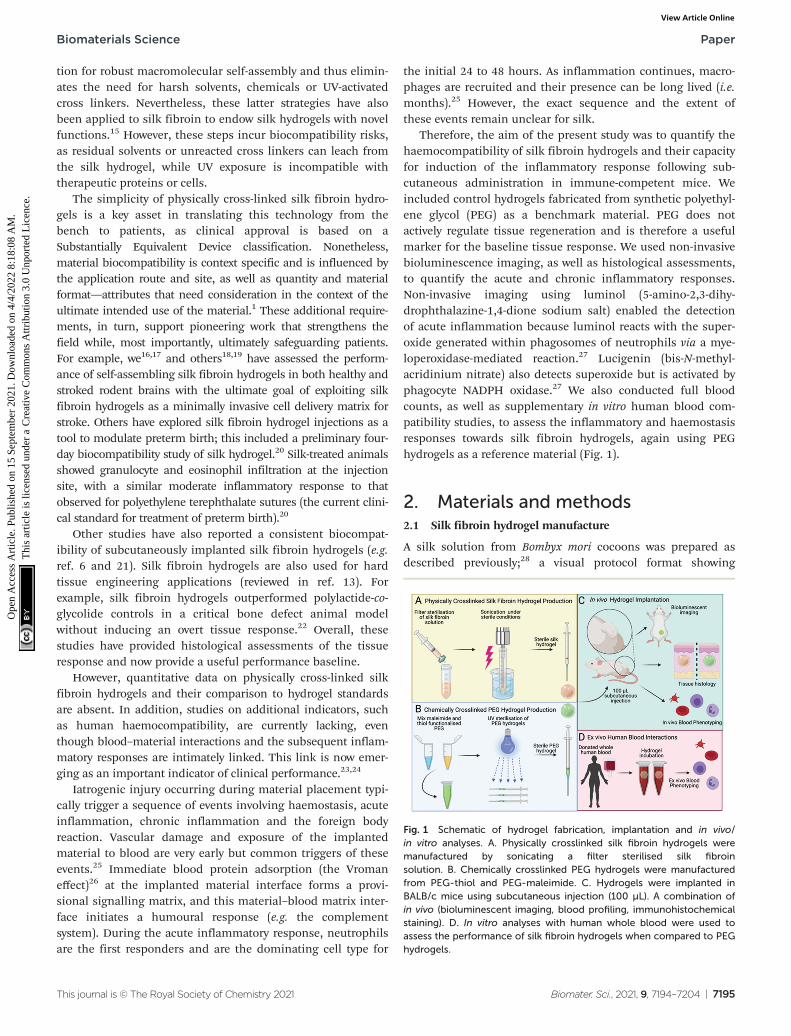
A silk solution from Bombyx mori cocoons was prepared asdescribed previously;28 a visual protocol format showing
Fig. 1 Schematic of hydrogel fabrication, implantation and in vivo/in vitro analyses. A. Physically crosslinked silk fibroin hydrogels weremanufactured by sonicating a filter sterilised silk fibroinsolution. B. Chemically crosslinked PEG hydrogels were manufacturedfrom PEG-thiol and PEG-maleimide. C. Hydrogels were implanted inBALB/c mice using subcutaneous injection (100 µL). A combination ofin vivo (bioluminescent imaging, blood profiling, immunohistochemicalstaining). D. In vitro analyses with human whole blood were used toassess the performance of silk fibroin hydrogels when compared to PEGhydrogels.
Biomaterials Science Paper
This journal is © The Royal Society of Chemistry 2021 Biomater. Sci., 2021, 9, 7194–7204 | 7195
Ope
n A
cces
s A
rtic
le. P
ublis
hed
on 1
5 Se
ptem
ber
2021
. Dow
nloa
ded
on 4
/4/2
022
8:18
:08
AM
. T
his
artic
le is
lice
nsed
und
er a
Cre
ativ
e C
omm
ons
Attr
ibut
ion
3.0
Unp
orte
d L
icen
ce.
View Article Online
reverse engineering of silk cocoons is available.29 Briefly, cutcocoons were boiled for 60 min in 20 mM Na2CO3 and thenrinsed in ddH2O to remove sericin proteins. The extracted silkfibroin was air dried, dissolved in 9.3 M LiBr at 60 °C and dia-lyzed (molecular weight cut-off 3500 g mol−1) against ddH2Oto remove the LiBr salt. Next, sufficient 10× phosphatebuffered saline (PBS) was added to the silk fibroin solution toobtain physiological osmolarity of the final preparation. Theresulting 4% w/v silk fibroin solution was filter sterilised(33 mm Millex-GP syringe filter fitted with a polyether sulfonemembrane containing 0.22 μm pores). The solution-gel tran-sition was induced by sonicating the silk fibroin solution witha Branson Digital Sonifier probe sonicator (HD 2070,Bandelin, Berlin, Germany) fitted with a 23 cm long sonicationtip (0.3 cm diameter tip and tapered over 8 cm) at 15% ampli-tude for 15 to 45 seconds. Silk fibroin undergoing the solu-tion-gel transition was aseptically filled into 1 mL syringes,hermetically sealed and stored at 4 °C until in vivo use. Forin vitro human blood studies, 150 μl of silk fibroin (3, 4 and5% w/v) undergoing the solution-gel transition were trans-ferred to the caps of 2 mL Eppendorf tubes.
2.2. Polyethylene glycol (PEG) hydrogel manufacture
The polymers were purchased from JenKem Technology USAInc. (Plano, TX, USA). The PEG hydrogel synthesis and charac-terisation has been reported previously.30,31 Briefly, four-armed PEG maleimide (10 700 g mol−1, polydispersity 1.03)was added to four-armed methoxy PEG thiol (10 600 g mol−1,polydispersity 1.03) at an equimolecular 1.5 μmol μl−1 ratio toPBS pH 5.5. For in vivo studies, the polymerising solution wastransferred to 1 mL syringes. The PEG hydrogel was bufferedto physiological pH by adding an equivalent volume of PBS pH8.5 to the syringe, followed by UV sterilisation. For in vitrohuman blood studies, 150 μl was transferred to the caps of2 mL Eppendorf tubes and buffered with PBS.
2.3 Human whole blood analysis
All human studies were approved by the ethics board and com-plied with institutional and international guidelines (reviewboard of Sächsische Landesärztekammer, ref. EK-BR-24/18-1).All blood donors provided written informed consent. Weensured that donors had not taken any nonsteroidal anti-inflammatory drugs over the past 10 days and were not on anyother medications that could interfere with coagulation orinflammatory responses. A non-pathological blood cell countand C reactive protein below 10 μg mL−1 (Diagnostik NordGmbH, Schwerin, Germany) served as further entrancecriteria.
Blood was drawn from two AB0-compatible healthy maleand female volunteers, immediately anticoagulated withheparin (1.5 IU mL−1) and pooled. Next, 1800 μL of the pooledblood was filled into 2 mL Eppendorf tubes using triplicatesample sets. The tubes were closed with caps containing therespective hydrogel, and the samples were overhead rotated at12 revolutions per min at 37 °C, 5% CO2 for 2 h. After incu-bation, the blood was processed for analysis as described
before.32–34 The study was repeated once, using two differentblood donors. For scanning electron microscopy, analysis capscontaining hydrogels were dehydrated in increasing ethanolconcentrations, critical point dried, gold sputtered andinspected by scanning electron microscopy as detailedbefore.34
2.4 In vivo experimental design
All procedures complied with the UK Animals (ScientificProcedures) Act 1986 Amendment Regulations 2012, followedthe ARRIVE guidelines and were subjected to review by theInstitutional Animal Care and Use Committee at the Universityof Strathclyde. All in vivo studies were approved by the HomeOffice of the United Kingdom (Project Licence Number PPL70/8801).
Female nulliparous Balb/c mice, 8 weeks of age, were pur-chased from Charles River UK Limited. At the time of surgery,the mice were anaesthetised using isoflurane and randomlyassigned to receive either a PEG hydrogel or a self-assemblingsilk fibroin hydrogel or neither. The injection site at the centreof the lower dorsum was shaved and cleaned, and a single100 µl subcutaneous hydrogel injection was administered(Fig. 1C). The experimenter was blinded to the experimentalgroups. At 3 h post injection, the first non-invasive bio-luminescence imaging was performed. Either luminol (100 mgkg−1; Sigma-Aldrich Company Ltd, Gillingham, UK) or10-methyl-9-(10-methylacridin-10-ium-9-yl)acridin-10-ium dini-trate (lucigenin [25 mg kg−1]; Abcam, Cambridge, UK) wereinjected intraperitonially to monitor the acute and delayedinflammatory response, respectively.27 Luminol and lucigeninsubstrate stock solutions were prepared in normal saline,filter-sterilised and stored at −20 °C.
Mice were imaged at the maximum signal intensity (at the15 min post-injection time point, F/stop = 1 and binning = 16)using a Xenogen IVIS 200 imaging system controlled by LivingImage Software 4.3.1 (Caliper Life Sciences). The IVIS ImagingSystems software package version 4.2 (Caliper Life Sciences,Xenogen Corp. 2007) was used to calculate the peak total bio-luminescent signal by applying standardised regions of inter-est. Data were presented as total flux in photons per secondper region of interest. Macroscopic images were captured witha Samsung Galaxy Neo camera, CMOS 16.0 MP resolution,with f/1.9 aperture.
2.5 Mouse whole blood analysis
Animals were randomly selected at the indicated time points(post-implantation days 2 and 35) from the silk fibroin hydro-gel group, the PEG hydrogel group, and the untreated controlgroup. All animals were dosed with sodium pentobarbital(60 mg kg−1 i.p.) and maintained under isoflurane for arterialblood sampling via cardiac puncture. Whole blood (approx.400 μL per animal) was collected with a 22-gauge needle,drawn into a 1 mL heparinised syringe and then transferredinto EDTA collection tubes (Kabe Labortechnik GmbHNümbrecht, Germany). Blood samples were subjected to fullblood counts on the day of collection at the Veterinary
Paper Biomaterials Science
7196 | Biomater. Sci., 2021, 9, 7194–7204 This journal is © The Royal Society of Chemistry 2021
Ope
n A
cces
s A
rtic
le. P
ublis
hed
on 1
5 Se
ptem
ber
2021
. Dow
nloa
ded
on 4
/4/2
022
8:18
:08
AM
. T
his
artic
le is
lice
nsed
und
er a
Cre
ativ
e C
omm
ons
Attr
ibut
ion
3.0
Unp
orte
d L
icen
ce.
View Article Online
Diagnostic Services, University of Glasgow. Samples were ana-lysed using a Siemens Advia 120 automated haematologyanalyser (Erlangen, Germany). For each sample, a bloodsmear was also examined after staining with Romanowskyand May-Grünwald Giemsa stains, and a manual whiteblood cell differential count was conducted based on 200leukocytes.
2.6 Mouse histology and immunofluorescence
At the indicated time points, the animals were euthanised bycervical dislocation and samples were collected and fixed in4% w/v paraformaldehyde for 24 h. The samples were thenimmersed in cryoprotective solution (30% w/v sucrose in PBSwith 0.05% w/v sodium azide) for 72 h, followed by rapidfreezing on dry ice. Coronal cryostat sections were cut(20 μm thickness) throughout the implant territory. Thetissue samples were stained with haematoxylin and eosin(H&E), and collagen deposition was visualised by trichromestaining.
For immunohistochemistry, sections were incubated in10% (v/v) blocking serum in PBS containing 0.3% v/v TritonX-100 for 40 min prior to overnight incubation with theprimary CD11b antibody (1 : 200, ab1211, Abcam, Cambridge,UK) at 4 °C to detect macrophages. After incubation with theprimary antibody, the sections were washed three times for5 min in PBS and then incubated for 2 h with an Alexa-488 sec-ondary antibody (1 : 500 dilution, Invitrogen, ThermoFisherScientific, Waltham, MA, USA). The sections were rinsed threetimes for 5 min in PBS before application of Vectashield anti-fade mounting medium with DAPI (Vector Laboratories Ltd,Peterborough, UK). Images were captured and analysed usingWinFluor V3.9.1 (Nikon Eclipse E600).
2.7 Statistical analyses and data files
Data were plotted and analysed as detailed previously.35
Briefly, sample pairs were analysed using Welch’s independentt-test while multiple groups were analysed by one-way ANOVAwith a Bonferroni post-hoc test (Prism 9.2.0; GraphPad SoftwareInc., San Diego, CA, USA). Asterisks were used to denote statisti-cal significance as follows: *P < 0.05, **P < 0.01, ***P < 0.001. Alldata were presented as mean values ± standard deviation (SD).The number of independent experiments (n) is noted in eachfigure legend. All data created during this research are openlyavailable from the University of Strathclyde Pure, at https://dx.doi.org/10.15129/09ab2ef9-03e0-470e-a7aa-496cab1a8f60.
3. Results
The performance of the silk fibroin hydrogel was assessedin vivo in mice and in vitro using human whole blood (Fig. 2).The in vitro human whole blood analysis showed no signs ofhaemolysis in any of the samples. The prothrombin F1 + 2fragment served as a biomarker for coagulation activation, andits levels were low across all samples and similar to the blankcontrol. The control and 5% w/v hydrogel treatments levels
were similar, and PEG and 3 & 4% w/v silk hydrogels showedslightly higher activation states (Fig. 2A). An increasing solidcontent of the silk fibroin hydrogels (i.e. from 3 to 5% w/v)further decreased coagulation activation (Fig. 2). Platelet acti-vation was monitored by quantifying platelet decay and leuko-cyte–platelet conjugate formation; both these markers werelow for all hydrogel samples and for the control group (Fig. 2Band C).
The inflammatory response was monitored using C5a as acomplement activation marker. The complement fragmentC5a concentration was always higher for silk fibroin hydrogelsthan for the PEG hydrogel; however, both hydrogel typesinduced higher C5a levels than were seen for the blank refer-ence control (Fig. 2D). The C5a pattern was similar to theCD11b expression pattern (i.e. a marker for granulocyte andmonocyte activation) (Fig. 2E). Both hydrogel types induced asimilar extent of granulocyte decay (approximately 30%) andleukocyte activation (approximately 60%) (Fig. 2E and F).Though the scanning electron microscopy assessment waschallenging because the hydrogel thickness compromisedimage quality, we were able to observe that all silk fibroinhydrogels were densely covered with granulocytes, whereas thePEG hydrogel showed mainly dehydration and crystallisationartefacts (Fig. 2G).
The PEG and silk fibroin hydrogels injected subcutaneouslyinto mice were both readily visible by visual inspection (datanot shown). The PEG hydrogels retained their shape over thecourse of the study, while the silk fibroin hydrogels showedprogressive shape loss from day 7 onwards. The systemic bio-compatibility of self-assembling silk fibroin hydrogels wasassessed by determining full blood counts (Fig. 3). For bothhydrogels, the performance across all 17 measured parametersshowed very similar trends, with no statistically significantdifferences between them. Comparison of these blood valueswith the untreated control animals showed a similar trendacross these parameters (Fig. 3). However, platelets were sig-nificantly raised at day 2 for PEG hydrogels and at day 35 forboth silk fibroin and PEG when compared to untreated controlmice.
The inflammatory response of hydrogels was monitoredlongitudinally in the mice. We used non-invasive bio-luminescence imaging to assess both acute and chronicinflammation using the luminol and lucigenin substrates,respectively.27 The acute inflammatory signal for the silkfibroin group was low and remained low throughout the study(Fig. 4A). By contrast, the PEG hydrogel group showed a signifi-cantly higher inflammatory response, especially at 3 hours.However, over the first two days of the study, this acute inflam-matory response subsided and reached the low levels observedfor the silk group.
The chronic inflammatory response for both hydrogel typeswas similar, although this varied among the hydrogel samples(Fig. 4B). For example, silk hydrogels showed a significantlyhigher signal at days 2, 7 and 14 than the PEG hydrogel. At day36 the chronic inflammatory signal for both hydrogel typesshowed no statistically significant differences. Overall, the
Biomaterials Science Paper
This journal is © The Royal Society of Chemistry 2021 Biomater. Sci., 2021, 9, 7194–7204 | 7197
Ope
n A
cces
s A
rtic
le. P
ublis
hed
on 1
5 Se
ptem
ber
2021
. Dow
nloa
ded
on 4
/4/2
022
8:18
:08
AM
. T
his
artic
le is
lice
nsed
und
er a
Cre
ativ
e C
omm
ons
Attr
ibut
ion
3.0
Unp
orte
d L
icen
ce.
View Article Online
Fig. 2 In vitro whole human blood interactions with silk and PEG hydrogels. PEG or 3–5% silk fibroin hydrogels were incubated for 2 h at 37 °C withwhole human blood and subsequently analysed for markers of haemostasis (A–C) or inflammation (D–F). G. Representative scanning electronmicroscopy images of PEG or 3–5% silk fibroin hydrogels following blood incubation. Scale bars: 20 µm. Triplicate measurements were averagedfrom two independent blood donors.
Fig. 3 In vivowhole blood analysis following hydrogel implantation. Blood counts conducted on samples drawn from BALB/c (A) control mice and (B) mice2 days and 35 days post implantation of silk or PEG hydrogels (n ≥ 3 per hydrogel group except PEG day 35 n = 2). Platelets (PLT); platelet distribution width(PDW); mean platelet volume (MPV); white blood cell (WBC), red cell distribution width (RDW); mean corpuscular hemoglobin concentration (MCHC); meancorpuscular hemoglobin (MCH); mean corpuscular volume (MCV); hematocrit (HCT); haemoglobin (Hb); red blood cell (RBC), procalcitonin (PCT).
Paper Biomaterials Science
7198 | Biomater. Sci., 2021, 9, 7194–7204 This journal is © The Royal Society of Chemistry 2021
Ope
n A
cces
s A
rtic
le. P
ublis
hed
on 1
5 Se
ptem
ber
2021
. Dow
nloa
ded
on 4
/4/2
022
8:18
:08
AM
. T
his
artic
le is
lice
nsed
und
er a
Cre
ativ
e C
omm
ons
Attr
ibut
ion
3.0
Unp
orte
d L
icen
ce.
View Article Online
chronic inflammatory signal for both samples was low over thestudy course.
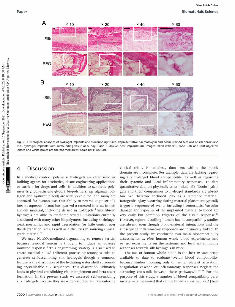
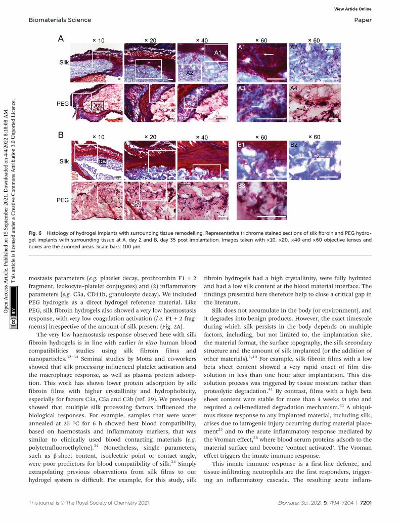
We included histological assessment to corroborate the bio-luminescence data sets. At day 2, both hydrogels were intact(Fig. 5A) and were in close proximity to the adjacent tissues. Atthese contact sites, intermittent but substantial cell infiltrationwas evident, with little difference between the hydrogel types(Fig. 5A). Most of the infiltrating cells advanced up to 200 μminto the hydrogel, although some were present in the core ofthe hydrogel. At day 35, cells were found throughout the hydro-gel, with no apparent differences between silk fibroin andPEG. Both hydrogels showed a uniform cell infiltration,coupled with a close association between the surroundingtissues and the hydrogel (Fig. 5B). However, less cells at thetissue–hydrogel interface appeared lower at day 35 than at day2. Collagen staining was included to assess the contribution ofthe hydrogel to extracellular matrix remodelling (Fig. 6). For
the silk fibroin hydrogels, some collagen staining was present atboth 2 and 35 days (Fig. 6A2 and B1). Active collagen depositionwas clearly evident at the tissue–PEG hydrogel interface alreadyon day 2 and individual collagen fibres could be seen (Fig. 6A3).At day 35, PEG hydrogels showed collagen deposition.
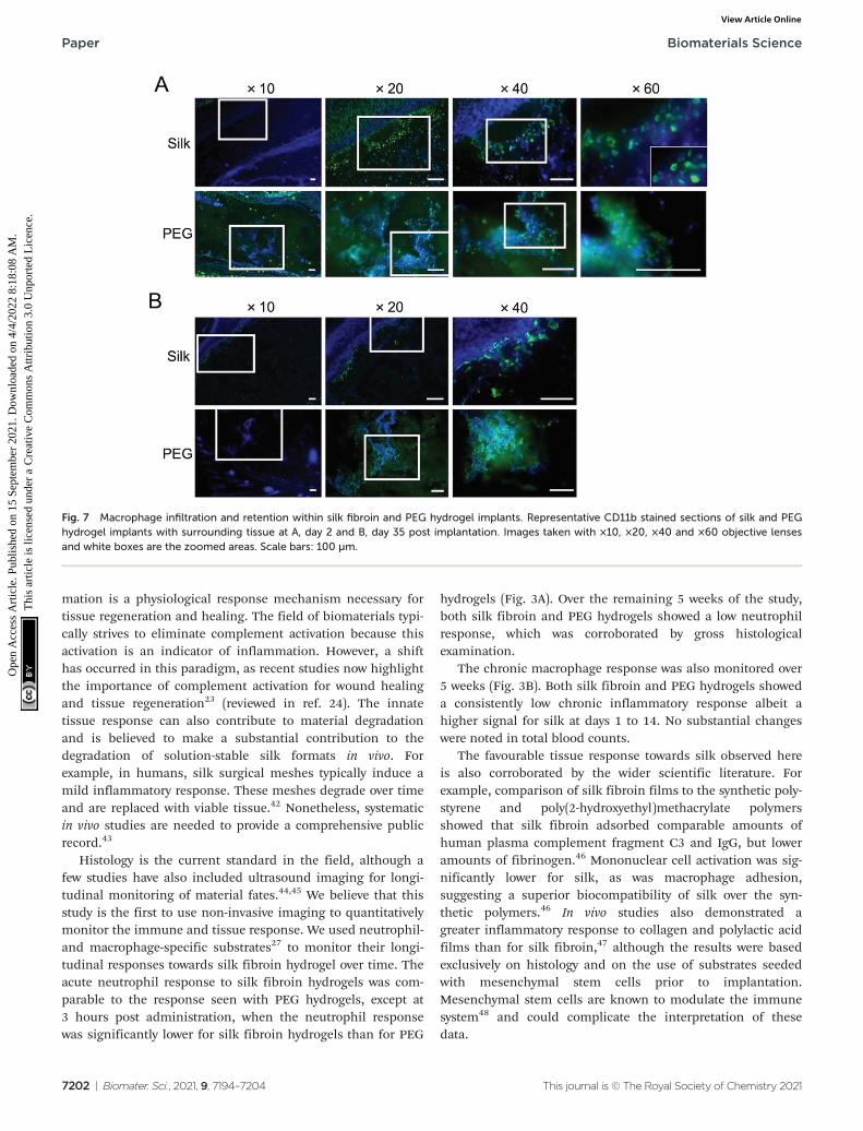
The involvement of macrophages, as part of the acute anddelayed tissue response, was also assessed by CD11b immuno-fluorescent staining (Fig. 7). At day 2, numerous macrophageswere present at the tissue–hydrogel interface for both hydro-gels (Fig. 7A). At day 35, the PEG hydrogels continued toharbour macrophages in the material core while some werealso visible within the silk fibroin hydrogel (Fig. 7B). For silkfibroin, macrophages were still present at the tissue–hydrogelinterface, albeit in substantially less than day 2. Overall, forboth materials, reduced numbers of macrophages wereobserved at the tissue–hydrogel interface and within the hydro-gel after chronic exposure (i.e. day 35).
Fig. 4 Non-invasive imaging of the acute and chronic inflammatory response towards implanted silk fibroin and PEG hydrogels. A. Acute response(i.e. luminol bioluminescence) in BALB/c mice measured at three hours (day 0) and later time points. Silk: day 0 (n = 8), 1 (n = 10), 2 (n = 10), 7 (n = 5),14 (n = 5), 35 (n = 5). PEG: day 0 (n = 5), 1 (n = 5), 2 (n = 5), 7 (n = 5), 14 (n = 5), 35 (n = 5). B. Chronic response (i.e. lucigenin bioluminescence) inBALB/c mice measured at three hours (day 0) and later time points. Silk: day 0 (n = 10), 1 (n = 10), 2 (n = 10), 7 (n = 9), 14 (n = 5), 36 (n = 5). PEG: day0 (n = 5), 1 (n = 5), 2 (n = 5), 8 (n = 5), 15 (n = 5), 36 (n = 5). Error bars are hidden in the plot symbol when not visible.
Biomaterials Science Paper
This journal is © The Royal Society of Chemistry 2021 Biomater. Sci., 2021, 9, 7194–7204 | 7199
Ope
n A
cces
s A
rtic
le. P
ublis
hed
on 1
5 Se
ptem
ber
2021
. Dow
nloa
ded
on 4
/4/2
022
8:18
:08
AM
. T
his
artic
le is
lice
nsed
und
er a
Cre
ativ
e C
omm
ons
Attr
ibut
ion
3.0
Unp
orte
d L
icen
ce.
View Article Online
4. Discussion
In a medical context, polymeric hydrogels are often used asbulking agents for aesthetics, tissue engineering applicationsor carriers for drugs and cells. In addition to synthetic poly-mers (e.g. polyethylene glycol), biopolymers (e.g. alginate, col-lagen and hyaluronic acid) are widely exploited, and many areapproved for human use. Our ability to reverse engineer silkinto its aqueous format has sparked a renewed interest in thisancient material, including its use in hydrogels.3 Silk fibroinhydrogels are able to overcome several limitations currentlyassociated with many other biopolymers, including shrinkage,weak mechanics and rapid degradation (or little control overthe degradation rate), as well as difficulties in sourcing clinicalgrade material.6
We used Na2CO3-mediated degumming to remove sericinbecause residual sericin is thought to induce an adverseimmune response.1 This degumming strategy is also used tocreate medical silks.1 Different processing strategies exist togenerate self-assembling silk hydrogels though a commonfeature is the disruption of the hydrating water shell surround-ing crystallisable silk sequences. This disruption ultimatelyleads to physical crosslinking via entanglement and beta sheetformation. In the present study we assessed self-assemblingsilk hydrogels because they are widely studied and are entering
clinical trials. Nonetheless, data sets within the publicdomain are incomplete. For example, data are lacking regard-ing silk hydrogel blood compatibility, as well as regardingtheir systemic and local inflammatory responses. To datequantitative data on physically cross-linked silk fibroin hydro-gels and their comparison to hydrogel standards are absenttoo. We therefore included PEG as a reference material.Iatrogenic injury occurring during material placement typicallytrigger a sequence of events including haemostasis. Vasculardamage and exposure of the implanted material to blood arevery early but common triggers of the tissue response.25
However, reports detailing human haemocompatibility studiesare absent, even though blood–material interactions and thesubsequent inflammatory responses are intimately linked. Inthe present study, we conducted two main biocompatibilityassessments: in vitro human whole blood experiments andin vivo experiments on the systemic and local inflammatoryresponses towards silk hydrogels in mice.
The use of human whole blood is the best in vitro setupavailable to date to evaluate overall blood compatibility,because studies focusing only on either platelet activation,coagulation cascade or inflammatory responses neglect theactivating cross-talk between these pathways.32,36–38 For thepurpose of this study, a number of blood compatibility para-meters were measured that can be broadly classified as (1) hae-
Fig. 5 Histological analyses of hydrogel implants and surrounding tissue. Representative haematoxylin and eosin-stained sections of silk fibroin andPEG hydrogel implants with surrounding tissue at A, day 2 and B, day 35 post implantation. Images taken with ×10, ×20, ×40 and ×60 objectivelenses and white boxes are the zoomed areas. Scale bars: 100 µm.
Paper Biomaterials Science
7200 | Biomater. Sci., 2021, 9, 7194–7204 This journal is © The Royal Society of Chemistry 2021
Ope
n A
cces
s A
rtic
le. P
ublis
hed
on 1
5 Se
ptem
ber
2021
. Dow
nloa
ded
on 4
/4/2
022
8:18
:08
AM
. T
his
artic
le is
lice
nsed
und
er a
Cre
ativ
e C
omm
ons
Attr
ibut
ion
3.0
Unp
orte
d L
icen
ce.
View Article Online
mostasis parameters (e.g. platelet decay, prothrombin F1 + 2fragment, leukocyte–platelet conjugates) and (2) inflammatoryparameters (e.g. C5a, CD11b, granulocyte decay). We includedPEG hydrogels as a direct hydrogel reference material. LikePEG, silk fibroin hydrogels also showed a very low haemostasisresponse, with very low coagulation activation (i.e. F1 + 2 frag-ments) irrespective of the amount of silk present (Fig. 2A).
The very low haemostasis response observed here with silkfibroin hydrogels is in line with earlier in vitro human bloodcompatibilities studies using silk fibroin films andnanoparticles.32–34 Seminal studies by Motta and co-workersshowed that silk processing influenced platelet activation andthe macrophage response, as well as plasma protein adsorp-tion. This work has shown lower protein adsorption by silkfibroin films with higher crystallinity and hydrophobicity,especially for factors C3a, C5a and C3b (ref. 39). We previouslyshowed that multiple silk processing factors influenced thebiological responses. For example, samples that were waterannealed at 25 °C for 6 h showed best blood compatibility,based on haemostasis and inflammatory markers, that wassimilar to clinically used blood contacting materials (e.g.polytetrafluoroethylene).34 Nonetheless, single parameters,such as β-sheet content, isoelectric point or contact angle,were poor predictors for blood compatibility of silk.34 Simplyextrapolating previous observations from silk films to ourhydrogel system is difficult. For example, for this study, silk
fibroin hydrogels had a high crystallinity, were fully hydratedand had a low silk content at the blood material interface. Thefindings presented here therefore help to close a critical gap inthe literature.
Silk does not accumulate in the body (or environment), andit degrades into benign products. However, the exact timescaleduring which silk persists in the body depends on multiplefactors, including, but not limited to, the implantation site,the material format, the surface topography, the silk secondarystructure and the amount of silk implanted (or the addition ofother materials).1,40 For example, silk fibroin films with a lowbeta sheet content showed a very rapid onset of film dis-solution in less than one hour after implantation. This dis-solution process was triggered by tissue moisture rather thanproteolytic degradation.41 By contrast, films with a high betasheet content were stable for more than 4 weeks in vivo andrequired a cell-mediated degradation mechanism.41 A ubiqui-tous tissue response to any implanted material, including silk,arises due to iatrogenic injury occurring during material place-ment25 and to the acute inflammatory response mediated bythe Vroman effect,26 where blood serum proteins adsorb to thematerial surface and become ‘contact activated’. The Vromaneffect triggers the innate immune response.
This innate immune response is a first-line defence, andtissue-infiltrating neutrophils are the first responders, trigger-ing an inflammatory cascade. The resulting acute inflam-
Fig. 6 Histology of hydrogel implants with surrounding tissue remodelling. Representative trichrome stained sections of silk fibroin and PEG hydro-gel implants with surrounding tissue at A, day 2 and B, day 35 post implantation. Images taken with ×10, ×20, ×40 and ×60 objective lenses andboxes are the zoomed areas. Scale bars: 100 µm.
Biomaterials Science Paper
This journal is © The Royal Society of Chemistry 2021 Biomater. Sci., 2021, 9, 7194–7204 | 7201
Ope
n A
cces
s A
rtic
le. P
ublis
hed
on 1
5 Se
ptem
ber
2021
. Dow
nloa
ded
on 4
/4/2
022
8:18
:08
AM
. T
his
artic
le is
lice
nsed
und
er a
Cre
ativ
e C
omm
ons
Attr
ibut
ion
3.0
Unp
orte
d L
icen
ce.
View Article Online
mation is a physiological response mechanism necessary fortissue regeneration and healing. The field of biomaterials typi-cally strives to eliminate complement activation because thisactivation is an indicator of inflammation. However, a shifthas occurred in this paradigm, as recent studies now highlightthe importance of complement activation for wound healingand tissue regeneration23 (reviewed in ref. 24). The innatetissue response can also contribute to material degradationand is believed to make a substantial contribution to thedegradation of solution-stable silk formats in vivo. Forexample, in humans, silk surgical meshes typically induce amild inflammatory response. These meshes degrade over timeand are replaced with viable tissue.42 Nonetheless, systematicin vivo studies are needed to provide a comprehensive publicrecord.43
Histology is the current standard in the field, although afew studies have also included ultrasound imaging for longi-tudinal monitoring of material fates.44,45 We believe that thisstudy is the first to use non-invasive imaging to quantitativelymonitor the immune and tissue response. We used neutrophil-and macrophage-specific substrates27 to monitor their longi-tudinal responses towards silk fibroin hydrogel over time. Theacute neutrophil response to silk fibroin hydrogels was com-parable to the response seen with PEG hydrogels, except at3 hours post administration, when the neutrophil responsewas significantly lower for silk fibroin hydrogels than for PEG
hydrogels (Fig. 3A). Over the remaining 5 weeks of the study,both silk fibroin and PEG hydrogels showed a low neutrophilresponse, which was corroborated by gross histologicalexamination.
The chronic macrophage response was also monitored over5 weeks (Fig. 3B). Both silk fibroin and PEG hydrogels showeda consistently low chronic inflammatory response albeit ahigher signal for silk at days 1 to 14. No substantial changeswere noted in total blood counts.
The favourable tissue response towards silk observed hereis also corroborated by the wider scientific literature. Forexample, comparison of silk fibroin films to the synthetic poly-styrene and poly(2-hydroxyethyl)methacrylate polymersshowed that silk fibroin adsorbed comparable amounts ofhuman plasma complement fragment C3 and IgG, but loweramounts of fibrinogen.46 Mononuclear cell activation was sig-nificantly lower for silk, as was macrophage adhesion,suggesting a superior biocompatibility of silk over the syn-thetic polymers.46 In vivo studies also demonstrated agreater inflammatory response to collagen and polylactic acidfilms than for silk fibroin,47 although the results were basedexclusively on histology and on the use of substrates seededwith mesenchymal stem cells prior to implantation.Mesenchymal stem cells are known to modulate the immunesystem48 and could complicate the interpretation of thesedata.
Fig. 7 Macrophage infiltration and retention within silk fibroin and PEG hydrogel implants. Representative CD11b stained sections of silk and PEGhydrogel implants with surrounding tissue at A, day 2 and B, day 35 post implantation. Images taken with ×10, ×20, ×40 and ×60 objective lensesand white boxes are the zoomed areas. Scale bars: 100 µm.
Paper Biomaterials Science
7202 | Biomater. Sci., 2021, 9, 7194–7204 This journal is © The Royal Society of Chemistry 2021
Ope
n A
cces
s A
rtic
le. P
ublis
hed
on 1
5 Se
ptem
ber
2021
. Dow
nloa
ded
on 4
/4/2
022
8:18
:08
AM
. T
his
artic
le is
lice
nsed
und
er a
Cre
ativ
e C
omm
ons
Attr
ibut
ion
3.0
Unp
orte
d L
icen
ce.
View Article Online
Others have implanted silk fibroin hydrogels subcu-taneously in immune compromised athymic mice (i.e. Swissnude mice). Subsequent histological assessment showed tran-sient eosinophil, neutrophil and macrophage recruitment,resulting in inflammation at 7 and 14 days post implantation.The inflammation subsided by 4 weeks and cleared within3 months.21 However, the use of athymic mice complicates theinterpretation of the immune response towards silk.
Physically cross-linked silk hydrogels rich in beta sheetspersist for several weeks in vivo, though the exact timescalesare context specific.6 The importance of silk fibroin hydrogelremodelling, cell infiltration49 and silk hydrogel degradation50
are key attributes that impart therapeutic value to silk hydro-gels. For example, the use of silk fibroin injections as a treat-ment for cervical insufficiency in pregnant rabbits augmentedthe cervix and initiated only a low inflammatory response thatwas comparable to cerclage treatment. Histology showed a lossof 70% of the silk hydrogel volume over 6 weeks, accompaniedby a mild tissue response by macrophages and a small numberof neutrophils and eosinophils at days 28 and 42.51
Non-invasive imaging has emerged as a complementarytool to histology for monitoring hydrogel degradation.44,45
However, differentiating material shrinkage from immune cell-mediated degradation is difficult. We therefore believe that theuse of neutrophil-specific and macrophage-specific non-inva-sive bioluminescence imaging is a useful tool for monitoringthe in vivo responses towards silk. Here, we provide a firstexample using silk fibroin hydrogels.
One challenge that arises when comparing our work toprior studies is created by the differences in material formats,amounts of implanted silk fibroin and silk processing (e.g.degumming time, dissolution medium etc.). Emerging evi-dence suggests that the material format, and not just its com-position, impacts the innate immune response.52 For example,primary human monocytes released more IL-1β and IL-6 inresponse to three-dimensional silk scaffolds than to two-dimensional silk films when the surface areas were main-tained constant. However, no adaptive immune response wasobserved in peripheral blood T cells from healthy donors.52
5. Conclusions
This work demonstrated that silk fibroin hydrogels inducedonly slight blood coagulation and platelet activation but clearlyelevated the inflammatory response of human whole blood.However, this blood response did not reflect a systemic inflam-matory response. In vivo bioluminescence imaging of neutro-phils and macrophages showed an acute, but mild, localinflammatory response. The macrophage response towardssilk fibroin hydrogels was mild, peaked at days 1 to 7 and thendeclined to levels equivalent to those of PEG hydrogels at day36. This time-dependent immune response was corroboratedby histology and immunofluorescence evaluations. We specu-late that this tissue response not only contributes to silk hydro-gel degradation but also stimulates new tissue formation.
Overall, this study confirms that silk fibroin hydrogels elicitsimilar immune responses to those seen with PEG hydrogels.The findings also demonstrate the power of non-invasive bio-luminescence imaging for monitoring tissue immuneresponses.
Author contributions
N. G. performed all in vivo experiments, analysed and inter-preted the data. G. B. synthesised PEGhydrogels. M. F. M. performed human blood studies with tech-nical support from Stefanie Hänsel, analysed and interpretedthe data. J. D. T. analysed data, discussed the results and gen-erated figures. All authors discussed the results and edited themanuscript. F. P. S. conceived the research study, supervisedthe work and wrote the manuscript.
Conflicts of interest
There are no conflicts to declare.
Acknowledgements
The authors extend their appreciation to the Deputyship forResearch & Innovation, Ministry of Education in Saudi Arabiafor funding this research work through the project number(862). The authors would like to thank Anne Goudie for techni-cal assistance.
References
1 C. Holland, K. Numata, J. Rnjak-Kovacina and F. P. Seib,Adv. Healthcare Mater., 2019, 8, e1800465.
2 G. Janani, M. Kumar, D. Chouhan, J. C. Moses,A. Gangrade, S. Bhattacharjee and B. B. Mandal, ACS Appl.Bio Mater., 2019, 2, 5460–5491.
3 D. N. Rockwood, R. C. Preda, T. Yucel, X. Wang,M. L. Lovett and D. L. Kaplan, Nat. Protoc., 2011, 6, 1612–1631.
4 F. P. Seib, Materials, 2021, 14, 1160.5 S. Phuagkhaopong, L. Mendes, K. Muller, M. Wobus,
M. Bornhauser, H. V. O. Carswell, I. F. Duarte andF. P. Seib, ACS Appl. Mater. Interfaces, 2021, 13, 30420–30433.
6 F. P. Seib, Ther. Delivery, 2018, 9, 469–487.7 S. H. Tran, C. G. Wilson and F. P. Seib, Pharm. Res., 2018,
35, 248.8 F. P. Seib, E. M. Pritchard and D. L. Kaplan, Adv. Funct.
Mater., 2013, 23, 58–65.9 N. Ashari, H. W. Pang, T. Simon, Y. Xiong, J. M. Coburn,
J. S. Bromberg, D. L. Kaplan, J. McLenithan andM. J. Fontaine, Cell. Immunol., 2018, 329, 10–16.
Biomaterials Science Paper
This journal is © The Royal Society of Chemistry 2021 Biomater. Sci., 2021, 9, 7194–7204 | 7203
Ope
n A
cces
s A
rtic
le. P
ublis
hed
on 1
5 Se
ptem
ber
2021
. Dow
nloa
ded
on 4
/4/2
022
8:18
:08
AM
. T
his
artic
le is
lice
nsed
und
er a
Cre
ativ
e C
omm
ons
Attr
ibut
ion
3.0
Unp
orte
d L
icen
ce.
View Article Online
10 N. E. Davis, L. N. Beenken-Rothkopf, A. Mirsoian, N. Kojic,D. L. Kaplan, A. E. Barron and M. J. Fontaine, Biomaterials,2012, 33, 6691–6697.
11 E. Bellas, B. J. Panilaitis, D. L. Glettig, C. A. Kirker-Head,J. J. Yoo, K. G. Marra, J. P. Rubin and D. L. Kaplan,Biomaterials, 2013, 34, 2960–2968.
12 C. S. Murphy, L. Liaw and M. R. Reagan, BMC Biomed. Eng.,2019, 1, 27.
13 V. J. Neubauer, A. Döbl and T. Scheibel, Materials, 2021, 14,674.
14 A. Matsumoto, J. Chen, A. L. Collette, U. J. Kim,G. H. Altman, P. Cebe and D. L. Kaplan, J. Phys. Chem. B,2006, 110, 21630–21638.
15 X. Mu, V. Fitzpatrick and D. L. Kaplan, Adv. HealthcareMater., 2020, 9, e1901552.
16 N. Gorenkova, I. Osama, F. P. Seib and H. V. O. Carswell,ACS Biomater. Sci. Eng., 2019, 5, 859–869.
17 I. Osama, N. Gorenkova, C. M. McKittrick,T. Wongpinyochit, A. Goudie, F. P. Seib andH. V. O. Carswell, Sci. Rep., 2018, 8, 13655.
18 L. Fernandez-Garcia, N. Mari-Buye, J. A. Barios,R. Madurga, M. Elices, J. Perez-Rigueiro, M. Ramos,G. V. Guinea and D. Gonzalez-Nieto, Acta Biomater., 2016,45, 262–275.
19 L. Fernandez-Garcia, J. Perez-Rigueiro, R. Martinez-Murillo,F. Panetsos, M. Ramos, G. V. Guinea and D. Gonzalez-Nieto, Front. Cell. Neurosci., 2018, 12, 296.
20 A. S. Critchfield, R. McCabe, N. Klebanov, L. Richey,S. Socrate, E. R. Norwitz, D. L. Kaplan and M. House,Reprod. Sci., 2014, 21, 1266–1273.
21 O. Etienne, A. Schneider, J. A. Kluge, C. Bellemin-Laponnaz, C. Polidori, G. G. Leisk, D. L. Kaplan,J. A. Garlick and C. Egles, J. Periodontol., 2009, 80, 1852–1858.
22 M. Fini, A. Motta, P. Torricelli, G. Giavaresi, N. NicoliAldini, M. Tschon, R. Giardino and C. Migliaresi,Biomaterials, 2005, 26, 3527–3536.
23 M. Bergmann, C. Jeanneau, T. Giraud, G. Richard andI. About, Clin. Oral Investig., 2020, 24, 4185–4196.
24 Y. Mödinger, G. Q. Teixeira, C. Neidlinger-Wilke andA. Ignatius, Int. J. Mol. Sci., 2018, 19, 3367.
25 B. Corradetti, The immune response to implanted materialsand devices, Springer, 2017.
26 L. Vroman, A. L. Adams, G. C. Fischer and P. C. Munoz,Blood, 1980, 55, 156–159.
27 J. C. Tseng and A. L. Kung, Chem. Biol., 2012, 19, 1199–1209.
28 S. A. L. Matthew, J. D. Totten, S. Phuagkhaopong, G. Egan,K. Witte, Y. Perrie and F. P. Seib, ACS Biomater. Sci. Eng.,2020, 6, 6748–6759.
29 T. Wongpinyochit, B. F. Johnston and F. P. Seib,J. Visualized Exp., 2016, 116, 54669.
30 Y. D. P. Limasale, P. Atallah, C. Werner, U. Freudenbergand R. Zimmermann, Adv. Funct. Mater., 2020, 30, 2000068.
31 P. Atallah, Y. D. P. Limasale, U. Freudenberg andC. Werner, Faraday Discuss., 2019, 219, 224–251.
32 M. F. Maitz, C. Sperling, T. Wongpinyochit, M. Herklotz,C. Werner and F. P. Seib, Nanomedicine, 2017, 13, 2633–2642.
33 F. P. Seib, M. Herklotz, K. A. Burke, M. F. Maitz, C. Wernerand D. L. Kaplan, Biomaterials, 2014, 35, 83–91.
34 F. P. Seib, M. F. Maitz, X. Hu, C. Werner and D. L. Kaplan,Biomaterials, 2012, 33, 1017–1023.
35 J. D. Totten, T. Wongpinyochit and F. P. Seib, J. DrugTargeting, 2017, 25, 865–872.
36 M. Gorbet, C. Sperling, M. F. Maitz, C. A. Siedlecki,C. Werner and M. V. Sefton, Acta Biomater., 2019, 94, 25–32.
37 C. Sperling, M. Fischer, M. F. Maitz and C. Werner,Biomaterials, 2009, 30, 4447–4456.
38 M. Weber, H. Steinle, S. Golombek, L. Hann, C. Schlensak,H. P. Wendel and M. Avci-Adali, Front. Bioeng. Biotechnol.,2018, 6, 99.
39 A. Motta, D. Maniglio, C. Migliaresi, H. J. Kim, X. Wan,X. Hu and D. L. Kaplan, J. Biomater. Sci., Polym. Ed., 2009,20, 1875–1897.
40 A. E. Thurber, F. G. Omenetto and D. L. Kaplan,Biomaterials, 2015, 71, 145–157.
41 F. P. Seib and D. L. Kaplan, Biomaterials, 2012, 33, 8442–8450.
42 N. A. Fine, M. Lehfeldt, J. E. Gross, S. Downey, G. M. Kind,G. Duda, D. Kulber, R. Horan, J. Ippolito and M. Jewell,Plast. Reconstr. Surg., 2015, 135, 339–351.
43 C. Guo, C. Li and D. L. Kaplan, Biomacromolecules, 2020,21, 1678–1686.
44 X. Leng, B. Liu, B. Su, M. Liang, L. Shi, S. Li,S. Qu, X. Fu, Y. Liu, M. Yao, D. L. Kaplan, Y. Wang andX. Wang, J. Tissue Eng. Regener. Med., 2017, 11, 822–830.
45 S. Li, D. Yu, H. Ji, B. Zhao, L. Ji and X. Leng, Biomed. Eng.Online, 2018, 17, 87.
46 M. Santin, A. Motta, G. Freddi and M. Cannas, J. Biomed.Mater. Res., 1999, 46, 382–389.
47 L. Meinel, S. Hofmann, V. Karageorgiou, C. Kirker-Head,J. McCool, G. Gronowicz, L. Zichner, R. Langer, G. Vunjak-Novakovic and D. L. Kaplan, Biomaterials, 2005, 26, 147–155.
48 A. R. R. Weiss and M. H. Dahlke, Front. Immunol., 2019, 10,1191.
49 Y. Kambe, A. Murakoshi, H. Urakawa, Y. Kimura andT. Yamaoka, J. Mater. Chem. B, 2017, 5, 7557–7571.
50 Y. Kambe and T. Yamaoka, Biomater. Sci., 2019, 7, 4153–4165.
51 Y. Zhang, N. Raia, A. Peterson, D. L. Kaplan and M. House,Tissue Eng., Part A, 2020, 26, 379–386.
52 M. Bhattacharjee, E. Schultz-Thater, E. Trella, S. Miot,S. Das, M. Loparic, A. R. Ray, I. Martin, G. C. Spagnoli andS. Ghosh, Biomaterials, 2013, 34, 8161–8171.
Paper Biomaterials Science
7204 | Biomater. Sci., 2021, 9, 7194–7204 This journal is © The Royal Society of Chemistry 2021
Ope
n A
cces
s A
rtic
le. P
ublis
hed
on 1
5 Se
ptem
ber
2021
. Dow
nloa
ded
on 4
/4/2
022
8:18
:08
AM
. T
his
artic
le is
lice
nsed
und
er a
Cre
ativ
e C
omm
ons
Attr
ibut
ion
3.0
Unp
orte
d L
icen
ce.
View Article Online


![PHYSICAL PROPERTIES OF SILK FIBROIN AND CELLULOSE ...€¦ · material for nanocomposite applications [2]. On the other hand, silk fibroin (SF) is a fibrous protein isolated from](https://img.pdfslide.us/doc/110x75/608ee0d07e325b2195270555/physical-properties-of-silk-fibroin-and-cellulose-material-for-nanocomposite.jpg)