Embed Size (px)
Citation preview
Zhang et al. Journal of Translational Medicine 2012, 10:117http://www.translational-medicine.com/content/10/1/117
RESEARCH Open Access
Preparation and characterization of silk fibroin asa biomaterial with potential for drug deliveryHao Zhang1†, Ling-ling Li2†, Fang-yin Dai3, Hao-hao Zhang1, Bing Ni1, Wei Zhou1, Xia Yang1* and Yu-zhang Wu1*
Abstract
Background: Degummed silk fibroin from Bombyx mori (silkworm) has potential carrier capabilities for drugdelivery in humans; however, the processing methods have yet to be comparatively analyzed to determine thedifferential effects on the silk protein properties, including crystalline structure and activity.
Methods: In this study, we treated degummed silk with four kinds of calcium-alcohol solutions, and performedsecondary structure measurements and enzyme activity test to distinguish the differences between the regeneratedfibroins and degummed silk fibroin.
Results: Gel electrophoresis analysis revealed that Ca(NO3)2-methanol, Ca(NO3)2-ethanol, or CaCl2-methanoltreatments produced more lower molecular weights of silk fibroin than CaCl2-ethanol. X-ray diffraction andFourier-transform infrared spectroscopy showed that CaCl2-ethanol produced a crystalline structure with more silk I(α-form, type II β-turn), while the other treatments produced more silk II (β-form, anti-parallel β-pleated sheet).Solid-State 13C cross polarization and magic angle spinning-nuclear magnetic resonance measurements suggestedthat regenerated fibroins from CaCl2-ethanol were nearly identical to degummed silk fibroin, while the othertreatments produced fibroins with significantly different chemical shifts. Finally, enzyme activity test indicated thatsilk fibroins from CaCl2-ethanol had higher activity when linked to a known chemotherapeutic drug, L-asparaginase,than the fibroins from other treatments.
Conclusions: Collectively, these results suggest that the CaCl2-ethanol processing method produces silk fibroinwith biomaterial properties that are appropriate for drug delivery.
Keywords: Silk fibroin, Calcium-alcohol solutions, Crystalline structure, Drug delivery, Biomaterial
BackgroundSilk fibers produced by silkworms are widely used in ourdaily life. While they have occupied an important nichein the textile industry for thousands of years, their po-tential as biomaterials has been recognized and devel-oped only over the past decade [1]. Being non-toxic,non-immunogenic, and biocompatible with a broadrange of animal species has allowed for the adherentproperties of silk fibroin and silk-like proteins to beexploited for biomedical purposes. To date, silk fibroinshave mainly been applied to wound healing, successfullyperforming as man-made blood-vessels [2], surgicalsutures [3], and repair materials [4]. New processing
* Correspondence: [email protected]; [email protected]†Equal contributors1Institute of Immunology Third Military Medical University, Chongqing400038, Peoples Republic of ChinaFull list of author information is available at the end of the article
© 2012 Zhang et al.; licensee BioMed CentralCommons Attribution License (http://creativecreproduction in any medium, provided the or
strategies for silk fibers and proteins have expanded thebiomedical utility of these molecules. For example, thegel spun silk-based matrix derived from silk fibroin wassuccessfully applied for bladder augmentation in a mur-ine model [5]. More recently, scientists determined thatthe cocoons from Bombyx mori harbor antioxidant andhypolipidemic properties and that the crude silk extractshave bioactivity against hypercholesterolemia and ath-erosclerosis [6].Recently, the regenerated silk fibroin has been proved
as an attractive candidate of a carrier for drug or thera-peutic proteins delivery and is the focus of much on-going research. Attachment of bioactive molecules ortherapeutic proteins to silk fibroin has many benefits toenhance the properties of bioactive molecules in solu-tion for delivery both in vitro and in vivo, including thetherapeutic efficacy in the body, thermal stability, storagestability, and lengthens the circulatory half-life and
Ltd. This is an Open Access article distributed under the terms of the Creativeommons.org/licenses/by/2.0), which permits unrestricted use, distribution, andiginal work is properly cited.
Zhang et al. Journal of Translational Medicine 2012, 10:117 Page 2 of 9http://www.translational-medicine.com/content/10/1/117
decreases immunogenicity and antigenicity [3]. For in-stance, bioconjugations of insulin, glucose oxidase, L-asparaginase (L-ASNase), lipase and phenylalanineammonia-lyase with the regenerated silk fibroin greatlyimproved their biological stability, reduced the immuno-genicity and toxicity of the drug [7-11]. Moreover, TheSELP (silk-elastinlike protein polymer)-controlled genedelivery approach could potentially improve activity ofadenoviral-mediated gene therapy of head and neck can-cer and limit viral spread to normal organs at the sametime [12].It has been known that the properties of silk-matrix
are controlled by a combination of the chemistry andthe spinning process, which directly affect the activityand stability of the enzymes attached. Spinning condi-tions, such as temperature, drawing rate, time, and spe-cific type of silkworm, can modulate biomaterialfeatures. In addition, chemistry, such as ion concentra-tion, type of ion, and solution pH, can also affect themechanical properties of silk fibroins [1]. In previousstudies, degummed fibroin has generally been treatedwith aqueous solutions of hexafluoro-isopropanol (HFIP)[13], methanol [8], CaCl2-ethanol [7,9], or Ca(NO3)2-methanol [14]. Lu et al. has reported glucose oxidaseattached to the regenerated silk fibroin film withouttreated with methanol remain more activity but lowerstability than that treated with methanol [8]. After cross-linking L-ASNase with regenerated silk fibroin preparedwith concentrated CaCl2 mixture solution with ethanoland water (1:2:8, mol), the immunogenicity and toxicityof the drug significantly reduced, and its circulatoryhalf-life lengthened in vitro [9].However, these studies have used only one treatment
per experiment and, up to now, the systematic compara-tive analysis to distinguish the difference of those treat-ments has not yet been reported, thus we do not knowwhich one is the best choice for future potential applica-tion. Here, we describe our systematic comparative ana-lysis of silk fibroins prepared with four of the commonlyused preparative solutions, Ca(NO3)2-methanol, Ca(NO3)2-ethanol, CaCl2-methanol, and CaCl2-ethanol. The resultscould help to reveal the mechanisms of properties of silk-derived matrix under different treating conditions and pro-vide evidence to choose right solution to prepare silk fibroinsfor potential drug delivery applications.
Materials and methodsMaterialsL-asparaginase (L-ASNase) from E. coli (10,000 IU) waspurchased from Changzhou Qianhong Bio-Pherma Co.,Ltd. (Jiangsu Province, China). L-asparagines’ (anhyd-rous) was purchased from Sangon Biotech (Shanghai)Co., Ltd. (Shanghai, China). Trichloroactic acid (TCA)was purchased from Sinopharm Chemical Reagent Co.,
Ltd. (Beijing, China). Methanol, ethanol, calcium nitratetetrahydrate (Ca(NO3)2�4H2O), calcium chloride (CaCl2),and HgI2, all analytical reagent grade, were purchasedfrom Chengdu Kelong Chemical Reagent Factory(Sichuan Province, China).
Preparation of degummed silk fibroinCocoons from B. mori were degummed by incubating ina mixture of sodium dodecyl sulfate (SDS; 0.25%,w/v)and sodium carbonate (0.25%,w/v) at 98°C for 30 min.The samples were then cooled to room temperature,rinsed three times with deionized water, and dried at65°C overnight. The ratio of cocoons and solution was1:100 (w/v). The degummed silk fibroins were isolated,along with another silk protein, sericin.
Calcium-alcohol solvents treatment of silk fibersThe isolated fibroin fibers were separately dissolved inconcentrated CaCl2 solution mixed with ethanol ormethanol and water (1:2:8 mol), and separately dissolvedin concentrated Ca(NO3)2�4H2O solution mixed withethanol or methanol (1:2 mol) at 65°C in a water bathfor 1 h. The ratio of the silk fibers and solution was 1:20(m/v). The aqueous solution of silk fibroin was obtainedby dialyzing against flowing water. After that, the result-ing dialyzed solutions were lyophilized. The dry silkpowder (fibroins treated with CaCl2-ethanol solution) orpieces (from the other three solutions) were stored at4°C until use.
SEMThe silk fibroins were vacuum-coated with a 20 nm layerof gold. The surface morphology of each silk fibroin wasobserved with a scanning electron microscope (S-3400NSEM; Hitachi, Japan) and photographed at a voltage of15 kV and room temperature.
SDS-polyacrylamide gel electrophoresis (PAGE)The silk fibroins separately treated with Ca(NO3)2-methanol, Ca(NO3)2-ethanol, CaCl2-methanol, andCaCl2-ethanol solution were analyzed by SDS-PAGE todetermine the corresponding molecular weights of theprotein. Samples were resolved on 12% acrylamide geland 4% condensing gel, and protein bands were visua-lized by staining with 0.25% Coomassie Brilliant BlueR-250 (Sigma-Aldrich, St. Louis, MO, USA).
FTIR spectroscopyThe infrared spectra of each fibroin produced with Ca(NO3)2-methanol, Ca(NO3)2-ethanol, CaCl2-methanol,and CaCl2-ethanol solution, and degummed fibroins(as control), were measured on a FTIR spectrometerusing KBr pellets (Tensor 27 FTIR; Bruker, Ettlingen,Germany). Spectra, with a resolution of 4 cm-1, were
Zhang et al. Journal of Translational Medicine 2012, 10:117 Page 3 of 9http://www.translational-medicine.com/content/10/1/117
recorded and subtracted from the sample readings. Allsamples were measured in reflection mode; for this,the silk fibroin powder treated with CaCl2-ethanol so-lution had been transformed into tablet form. Theresults are presented as the average of 64 repeated4000 ~ 400 cm-1 scans.
WAXDThe crystalline structure of the silk fibroins producedwith Ca(NO3)2-methanol, Ca(NO3)2-ethanol, CaCl2-methanol, and CaCl2-ethanol solution, and ofdegummed fibroins, were determined by WAXD using aSiemens type F X-ray diffractometer (Siemens, Munich,Germany) with Ni-filtered Cu Kα radiation. The voltageand current of the X-ray source were 30KV and 20 mA,respectively. The wavelength, λ, was 0.15406 nm. Thesamples were mounted on aluminum frames andscanned from 5° to 40° (2θ) at a speed of 2°/min. The D-spacing was calculated by the following equation: D= λ/(2 × sin(θ)), i.e. D = 0.0752/(sin θ) nm. For example, if thescanning angel was 2θ= 20°, then D= 0.0752/(sin 10°)nm, and the D-space was 0.43 nm.
Solid-state 13C CP/MAS-NMR spectra measurementSolid state 13C CP/MAS-NMR has been successfullyused to analyze the secondary structure of proteins [15],and was similarly applied in our study. The 13C CP/MAS-NMR spectra were recorded on a Bruker AVANCEIII 400 WB spectrometer equipped with a 4 mm standardbore CP/MAS probe head, whose X channel was tunedto 100.62 MHz for 13C and the other channel was tunedto 400.18 MHz for broad band 1H decoupling. A mag-netic field of 9.39T at 297 K was used. The dried andfinely powdered samples were packed in a ZrO2 rotorthat was sealed with an Kel-F cap and spun at 12 kHzrate. The experiments were conducted at a contact timeof 2 ms. A total of 3000 scans were recorded with 6 s re-cycle delay for each sample. All 13C CP/MAS chemicalshifts were referenced to the resonances of the adaman-tane (C10H16) standard (δCH2= 38.5).
Enzyme cross-linking and activity testL-ASNase immobilization was performed according tothe method previously described by Zhang et al. [16]with minor modifications. An aliquot (50 mg) of each fi-broin produced with Ca(NO3)2-methanol, Ca(NO3)2-ethanol, CaCl2-methanol, and CaCl2-ethanol solution,and degummed fibroins (as control), were placed intoplastic centrifuge tubes and mixed with 2mL L-ASNasein phosphate buffered saline (PBS) solution (2 mg/mL)and 1 mL L-asparagine PBS solution (5 mg/L). The L-asparagine acted as the enzyme activity center protector.After gentle shaking, glutaraldehyde (0.05%) and PBS(pH 7.4) were added to bring the final volume to 5 mL,
and the solutions were incubated at 4°C overnight to fa-cilitate the cross-linking reaction. The next day, the re-action was stopped by adding 100 mg glycine to eachtube. The fibroin and L-ASNase bioconjugates were thenwashed with Tris–HCl buffer (pH 8.6) and purified bycentrifugation in a J2-MI refrigerated centrifuge (Beck-man-Coulter, Brea, CA, USA) at 10000 rpm for 10 minat 4°C; this purification process was repeated three timeswith intervening Tris–HCl washes. After the final spin,the pellets were each resuspended in 1 mL Tris–HCland incubated in a water bath at 37°C for 10 min. Twomilliliters of 5 mg/mL L-asparagine in Tris–HCl wasadded to each tube, and incubation continued at 37°Cfor another 10 min. The enzymatic reaction was termi-nated by adding 100 mg TCA to each reaction. Thetubes were centrifuged at 3000 rpm for 5 min, and 0.5mL of the supernatant from each was transferred tofresh centrifuge tubes containing 1 mL Nessler’s reagent.After the reaction processed at room temperature for agiven time, 50 μL of each mixture and 150 μL of Tris–HCl buffer were transferred in triplicate to 96 wellmicroplates. The same experiment was repeated threetimes. The activities of the enzymes attached to thefibroins were calculated by determining the change inoptical density at 450 nm as measured on a microplatereader (Paradigm; Beckman-Coulter). Data are presentedas mean ± SD and evaluated using the Student’s t-test(SPSS 13.0, SPSS Inc.). P< 0.05 was considered to bestatistically significant. The mobilization and activity de-tection of glucose oxidase were performed by referringto the published literature [8].
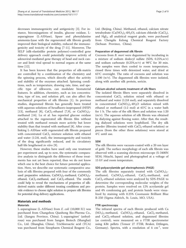
Results and discussionMorphology of silk fibroinsThe silk fibroins treated with Ca(NO3)2�4H2O-methanol,Ca(NO3)2�4H2O-ethanol, CaCl2-methanol-H2O, and CaCl2-ethanol solution were separately dissolved. After lyophi-lized, the surface morphology of degummed silkfibroins and regenerated silk fibroins was observed withSEM (Figure 1). The size and shape of the degummedsilk fibroins were normal, with diameters of 6–8 μm(Figure 1A). In contrast, the regenerated silk fibroinswere spherical or irregular shapes. This shape may haveresulted from the merger of smaller micelles that occurredin the aqueous solutions of Ca(NO3)2�4H2O-methanol(Figure 1B), Ca(NO3)2�4H2O-ethanol (Figure 1C), andCaCl2-methanol-H2O (Figure 1D), and CaCl2-ethanol(Figure 1E).
Molecular weight ranges of silk fibroinsThe silkworm’s cocoon is composed of two kinds of silkprotein, the silk sericin, which makes up the membrane,and the silk fibroin, which makes up the inner portion.The silk sericin is a glue-like mixture of glycoproteins
(B) (C)
(D) (E)
(A)
Figure 1 SEM photographs of B. mori silk fibroin prepared with various solutions. (A) Degummed silk fibroin. (B) Silk fibroin prepared fromCa(NO3)2�4H2O-methanol solution. (C) Silk fibroin prepared from Ca(NO3)2�4H2O-ethanol solution. (D) Silk fibroin prepared from CaCl2-methanol-H2O solution. (E) Silk fibroin prepared from CaCl2-ethanol-H2O solution.
M A B C D E
170130
95
72
55
43
34
26
10
17
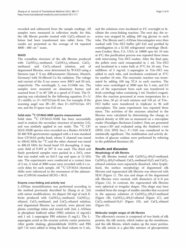
Figure 2 SDS-PAGE analysis of B. mori silk fibroins preparedwith various solutions. Silk fibroins were prepared from fourdifferent calcium-alcohol solutions (as described below) thendissolved in hot water. The range of molecular weight of theproteins produced by each solution was determined by SDS-PAGEwith 12% acrylamide gel and 4% condensing gel, which was stainedwith 0.25% Coomassie Brilliant Blue R-250. Lanes: M, marker. (A) silkfibroin prepared from Ca(NO3)2�4H2O-methanol solution. (B) silkfibroin prepared from Ca(NO3)2�4H2O-ethanol solution. (C) silk fibroinprepared from CaCl2-methanol-H2O solution. (D) silk fibroin preparedfrom CaCl2-ethanol-H2O solution. (E) Degummed silk fibroin.
Zhang et al. Journal of Translational Medicine 2012, 10:117 Page 4 of 9http://www.translational-medicine.com/content/10/1/117
with varying molecular mass, and is removed by thedegumming and rinsing steps. The silk fibroin protein ofB. mori is rich in alanine, glycine and serine residues[17], and is ~400 kDa, with 300 kDa making up a heavychain (H-chain), 26 kDa making up a light chain (L-chain), L-chain and H-chain linked by disulfide bond(s)and about 30 kDa making up a P25 glycoprotein thatassociates with the H-L complex primarily by hydropho-bic interactions [18].The silk fibroins produced with Ca(NO3)2-methanol,
Ca(NO3)2-ethanol, CaCl2-methanol, and CaCl2-ethanolsolutions were dissolved, and the molecular weightswere measured by SDS-PAGE. As shown in Figure 2, theregenerated silk fibroins treated with Ca(NO3)2-metha-nol had a molecular weight from about 95 KDa to over170 kDa, but Ca(NO3)2-ethanol from about 100 KDa toover 170 kDa. The CaCl2-methanol solution fibroinsranged from about 140 to over 170 kDa, while theCaCl2-ethanol fibroins ranged from about 100 to nearly300 kDa. Two low molecular weight bands, ~17 and ~26kDa, were obviously present in these regenerated silkfibroins, but the silk fibroins produced with CaCl2-etha-nol showed relatively faint low molecular weight bandsat these positions. In addition, the degummed silkfibroins are poorly soluble, except in the chemistry solu-tion and organic solvents, we could not observe obviousbands in the gel.This phenomenon suggested that some of the disulfide
linkages and hydrophobic bonds, between silk fibroin
1900 1800 1700 1600 1500 1400 1300 1200 1100 1000 900 800
Wavenumber(cm-1)
%T
ran
smit
tan
ce
A
B
C
D
E
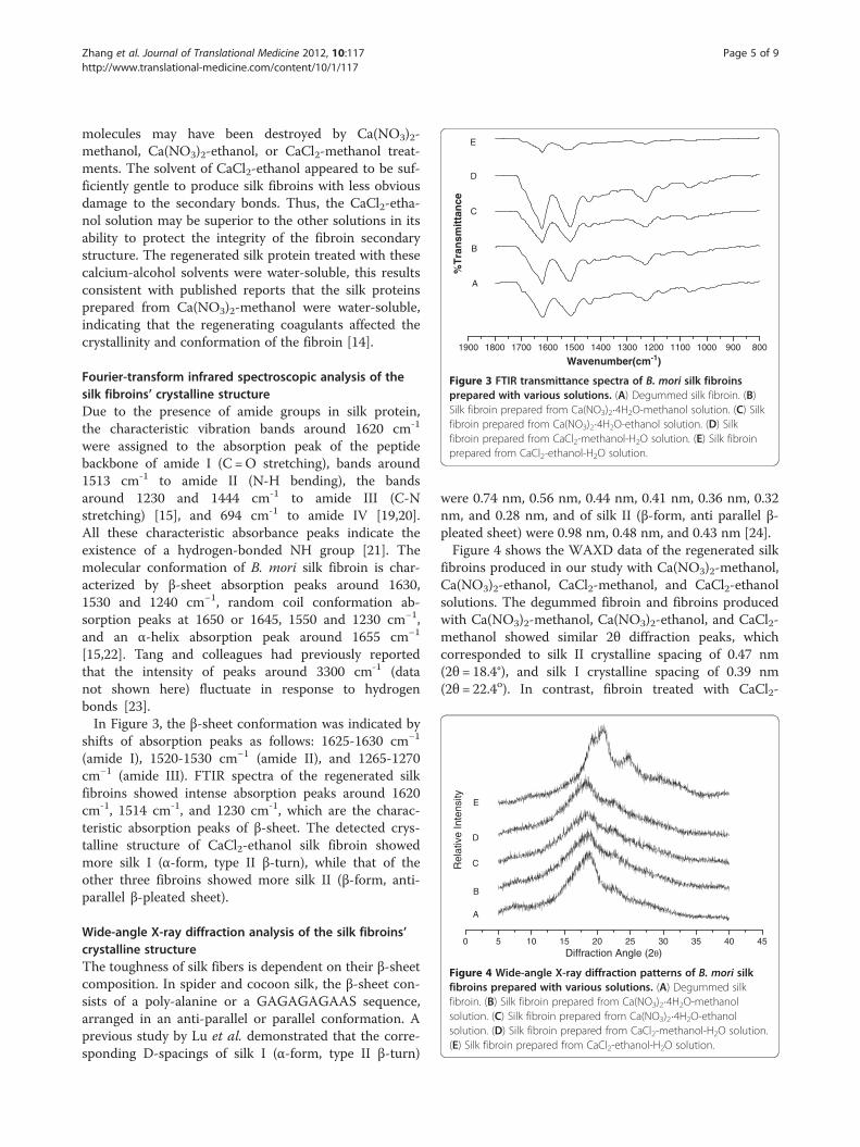
Figure 3 FTIR transmittance spectra of B. mori silk fibroinsprepared with various solutions. (A) Degummed silk fibroin. (B)Silk fibroin prepared from Ca(NO3)2�4H2O-methanol solution. (C) Silkfibroin prepared from Ca(NO3)2�4H2O-ethanol solution. (D) Silkfibroin prepared from CaCl2-methanol-H2O solution. (E) Silk fibroinprepared from CaCl2-ethanol-H2O solution.
0 5 10 15 20 25 30 35 40 45
A
B
C
D
E
Diffraction Angle (2θ)
Rel
ativ
e In
tens
ity
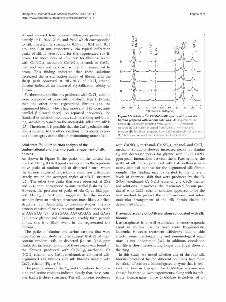
Figure 4 Wide-angle X-ray diffraction patterns of B. mori silkfibroins prepared with various solutions. (A) Degummed silkfibroin. (B) Silk fibroin prepared from Ca(NO3)2�4H2O-methanolsolution. (C) Silk fibroin prepared from Ca(NO3)2�4H2O-ethanolsolution. (D) Silk fibroin prepared from CaCl2-methanol-H2O solution.(E) Silk fibroin prepared from CaCl2-ethanol-H2O solution.
Zhang et al. Journal of Translational Medicine 2012, 10:117 Page 5 of 9http://www.translational-medicine.com/content/10/1/117
molecules may have been destroyed by Ca(NO3)2-methanol, Ca(NO3)2-ethanol, or CaCl2-methanol treat-ments. The solvent of CaCl2-ethanol appeared to be suf-ficiently gentle to produce silk fibroins with less obviousdamage to the secondary bonds. Thus, the CaCl2-etha-nol solution may be superior to the other solutions in itsability to protect the integrity of the fibroin secondarystructure. The regenerated silk protein treated with thesecalcium-alcohol solvents were water-soluble, this resultsconsistent with published reports that the silk proteinsprepared from Ca(NO3)2-methanol were water-soluble,indicating that the regenerating coagulants affected thecrystallinity and conformation of the fibroin [14].
Fourier-transform infrared spectroscopic analysis of thesilk fibroins’ crystalline structureDue to the presence of amide groups in silk protein,the characteristic vibration bands around 1620 cm-1
were assigned to the absorption peak of the peptidebackbone of amide I (C =O stretching), bands around1513 cm-1 to amide II (N-H bending), the bandsaround 1230 and 1444 cm-1 to amide III (C-Nstretching) [15], and 694 cm-1 to amide IV [19,20].All these characteristic absorbance peaks indicate theexistence of a hydrogen-bonded NH group [21]. Themolecular conformation of B. mori silk fibroin is char-acterized by β-sheet absorption peaks around 1630,1530 and 1240 cm−1, random coil conformation ab-sorption peaks at 1650 or 1645, 1550 and 1230 cm−1,and an α-helix absorption peak around 1655 cm−1
[15,22]. Tang and colleagues had previously reportedthat the intensity of peaks around 3300 cm-1 (datanot shown here) fluctuate in response to hydrogenbonds [23].In Figure 3, the β-sheet conformation was indicated by
shifts of absorption peaks as follows: 1625-1630 cm−1
(amide I), 1520-1530 cm−1 (amide II), and 1265-1270cm−1 (amide III). FTIR spectra of the regenerated silkfibroins showed intense absorption peaks around 1620cm-1, 1514 cm-1, and 1230 cm-1, which are the charac-teristic absorption peaks of β-sheet. The detected crys-talline structure of CaCl2-ethanol silk fibroin showedmore silk I (α-form, type II β-turn), while that of theother three fibroins showed more silk II (β-form, anti-parallel β-pleated sheet).
Wide-angle X-ray diffraction analysis of the silk fibroins’crystalline structureThe toughness of silk fibers is dependent on their β-sheetcomposition. In spider and cocoon silk, the β-sheet con-sists of a poly-alanine or a GAGAGAGAAS sequence,arranged in an anti-parallel or parallel conformation. Aprevious study by Lu et al. demonstrated that the corre-sponding D-spacings of silk I (α-form, type II β-turn)
were 0.74 nm, 0.56 nm, 0.44 nm, 0.41 nm, 0.36 nm, 0.32nm, and 0.28 nm, and of silk II (β-form, anti parallel β-pleated sheet) were 0.98 nm, 0.48 nm, and 0.43 nm [24].Figure 4 shows the WAXD data of the regenerated silk
fibroins produced in our study with Ca(NO3)2-methanol,Ca(NO3)2-ethanol, CaCl2-methanol, and CaCl2-ethanolsolutions. The degummed fibroin and fibroins producedwith Ca(NO3)2-methanol, Ca(NO3)2-ethanol, and CaCl2-methanol showed similar 2θ diffraction peaks, whichcorresponded to silk II crystalline spacing of 0.47 nm(2θ= 18.4°), and silk I crystalline spacing of 0.39 nm(2θ= 22.4o). In contrast, fibroin treated with CaCl2-
200 180 160 140 120 100 80 60 40 20 0
A
B
C
D
E
Ala
Cß
Gly
Ca
Ala
Ca
Ser
Ca
Ser
CßG
ly C
=O
Ala
C=
O
13C chemical shift [ppm]
Tyr
Cε
Tyr
Cδ Χ
γ
Tyr
Cξ
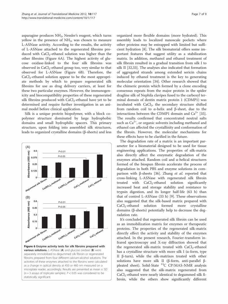
Figure 5 Solid-state 13C CP/MAS-NMR spectra of B. mori silkfibroins prepared with various solutions. (A) Degummed silkfibroin. (B) Silk fibroin prepared from Ca(NO3)2�4H2O-methanolsolution. (C) Silk fibroin prepared from Ca(NO3)2�4H2O-ethanolsolution. (D) Silk fibroin prepared from CaCl2-methanol-H2O solution.(E) Silk fibroin prepared from CaCl2-ethanol-H2O solution.
Zhang et al. Journal of Translational Medicine 2012, 10:117 Page 6 of 9http://www.translational-medicine.com/content/10/1/117
ethanol showed four obvious diffraction peaks at 2θ,namely 19.4°, 20.3°, 24.6°, and 29.3°, which correspondedto silk I crystalline spacing of 0.44 nm, 0.41 nm, 0.35nm, and 0.30 nm, respectively. No typical diffractionpeaks of silk II were found for this regenerated silk fi-broin. The mean peak at 2θ= 18.4° for fibroins treatedwith Ca(NO3)2-methanol, Ca(NO3)2-ethanol, or CaCl2-methanol was not as sharp as that for degummed fi-broin. This finding indicated that these solutionsdecreased the crystallization ability of fibroin, and thesharp peak observed at 2θ= 20.3° of CaCl2-ethanolfibroins indicated an increased crystallization ability offibroin.Furthermore, the fibroins produced with CaCl2-ethanol
were composed of more silk I (α-form, type II β-turn)than the other three regenerated fibroins and thedegummed fibroin, which had more silk II (β-form, anti-parallel β-pleated sheet). As reported previously, thestandard orientation methods, such as rolling and draw-ing, are able to transform the metastable silk I into silk II[25]. Therefore, it is possible that the CaCl2-ethanol solu-tion is superior to the other solutions in its ability to pro-tect the integrity of the fibroin, maintaining more silk I.
Solid-state 13C CP/MAS-NMR analysis of theconformational and inter-molecular arragement of silkfibroinsAs shown in Figure 5, the peaks on the dotted linemarked Ala Cβ II (16.8 ppm) correspond to the represen-tative peaks of random coil or distorted β-turn, wherethe torsion angles of a backbone chain are distributedlargely around the averaged angles of silk II structure[26]. The other two peaks that were observed, at 20.2and 22.6 ppm, correspond to anti-parallel β-sheets [27].However, the presence of peaks of Ala Cβ at 15.2 ppmand Ala Cα at 52.4 ppm suggested that the residuesstrongly favor an ordered structure, most likely a helicalstructure [28]. According to previous studies, the silkprotein consists of many repeated motif sequences, suchas AGSGAG [29], AGYGAG, AGVGYGAG and GAAS[30]; since glycine and alanine can readily form peptidebonds, this is a likely event in the regenerated silkfibroins.The peaks of alanine and serine carbons that were
observed in our study samples suggest that all of themcontain random coils or distorted β-turns (16.8 ppmpeak). An increased amount of these peaks was found inthe fibroins produced with Ca(NO3)2-methanol, Ca(NO3)2-ethanol, and CaCl2-methanol, as compared withdegummed silk fibroins and silk fibroins treated withCaCl2-ethanol (Figure 5).The peak position of the Cα and Cβ carbons from ala-
nine and serine residues indicate clearly that these sam-ples had a β-sheet structure. The silk fibroins produced
with Ca(NO3)2-methanol, Ca(NO3)2-ethanol, and CaCl2-methanol solutions showed increased peaks for alanineCβ and decreased peaks for glycine with C=O (169.1ppm peak) interactions between them. Furthermore, thepeaks of silk fibroin produced with CaCl2-ethanol werenearly identical to those for the degummed silk fibroinsample. This finding may be related to the differentlevels of chemical shift that were produced by the Ca(NO3)2-methanol, Ca(NO3)2-ethanol, and CaCl2-metha-nol solutions. Regardless, the regenerated fibroin pro-duced with CaCl2-ethanol solution appeared to be thebest method to protect the conformational and inter-molecular arrangement of the silk fibroin chains ofdegummed fibroin.
Enzymatic activity of L-ASNase when conjugated with silkfibroinsL-asparaginase is a well-established chemotherapeuticagent in routine use to treat acute lymphoblasticleukemia. However, treatment withdrawal due to sideeffects, some life-threatening and immunological reac-tions is not uncommon [31]. In addition, circulationhalf-life is short, necessitating longer and larger doses ofthe drug.In this study, we tested whether any of the four silk
fibroins produced by the different solutions had morebeneficial effects on a bioconjugated enzyme that is rele-vant for human therapy. The L-ASNase enzyme waschosen for these in vitro experiments, along with its sub-strate L-asparagine. Since L-ASNase hydrolysis of L-
Zhang et al. Journal of Translational Medicine 2012, 10:117 Page 7 of 9http://www.translational-medicine.com/content/10/1/117
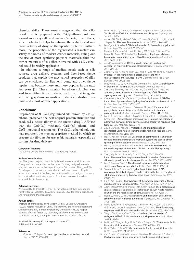
asparagine produces NH3, Nessler’s reagent, which turnsyellow in the presence of NH3, was chosen to measureL-ASNase activity. According to the results, the activityof L-ASNase attached to the regenerated fibroins pro-duced with CaCl2-ethanol solution was higher than theother fibroins (Figure 6A). The highest activity of glu-cose oxidase-linked to the four silk fibroins wasobserved in CaCl2-ethanol group too, very similar to thatobserved for L-ASNase (Figure 6B). Therefore, theCaCl2-ethanol solution appear to be the most appropri-ate methods by which to prepare regenerated silkfibroins for use as drug delivery carriers, at least forthese two particular enzymes. However, the immunogen-icity and biocompatibility properties of these regeneratedsilk fibroins produced with CaCl2-ethanol have yet to bedetermined and require further investigation in an ani-mal model before clinical application.Silk is a unique protein biopolymer, with a block co-
polymer structure dominated by large hydrophobicdomains and small hydrophilic spacers. This primarystructure, upon folding into assembled silk structures,leads to organized crystalline domains (β-sheets) and less
0
0.15
0.3
0.45
0.6
Ab
sorb
ance
(46
0 n
m)
(B)
P<0.05
P<0.01
0
0.125
0.25
0.375
0.5
Ab
sorb
ance
(45
0 n
m)
(A)
P<0.05
P<0.01
Figure 6 Enzyme activity tests for silk fibroins prepared withvarious solutions. L-ASNase (A) and glucose oxidase (B) wereseparately immobilized to degummed silk fibroin or regeneratedfibroins prepared from four different calcium-alcohol solutions. Theactivities of these enzymes attached to the fibroins were calculatedas a change in optical density at 450 or 460 nm measured on amicroplate reader, accordingly. Results are presented as mean ± SD(n = 3 assays of triplicate samples). P< 0.05 was considered to bestatistically significant.
organized more flexible domains (more hydrated). Thisassembly leads to localized nanoscale pockets whereother proteins may be entrapped with limited but suffi-cient hydration [8]. The silk biomaterial offers some im-portant features that suggest utility as a stabilizationmatrix. In addition, methanol and ethanol treatment ofsilk fibroin resulted in a gradual transition from silk I tosilk II [32,33]. The analyses also indicated that formationof aggregated strands among extended sericin chainsinduced by ethanol treatment is the key to generatingmolecular orientation [34]. Other research showed thatthe chimeric protein which formed by a clone encodingconsensus repeats from the major protein in the spiderdragline silk of Nephila clavipes fused to the carboxyl ter-minal domain of dentin matrix protein 1 (CDMP1) wasincubated with CaCl2, the secondary structure shiftedfrom random coil to α-helix and β-sheet, due to theinteractions between the CDMP1 domain and Ca2+ [35].The results confirmed that concentrated neutral saltssuch as Ca2+, or organic solvents including methanol andethanol can affected the crystallinity and conformation ofthe fibroin. However, the molecular mechanisms forthese effects have to be clarified in the future.The degradation rate of a matrix is an important par-
ameter for a biomaterial designed to be used for tissueengineering applications. The properties of silk-matrixalso directly affect the enzymatic degradation of theenzymes attached. Random coil and α-helical structuresformed of the biospun fibroin accelerate the process ofdegradation in both PBS and enzyme solutions in com-parison with β-sheets [36]. Zhang et al. reported thatcross-linking L-ASNase with regenerated silk fibrointreated with CaCl2-ethanol solution significantlyincreased heat and storage stability and resistance totrypsin digestion, and its longer half-life (63 h) thanthat of control L-ASNase (33 h) [9]. These observationsalso suggested that the silk-based matrix prepared withCaCl2-ethanol solution formed more crystallinedomains (β-sheets) potentially help to decrease the deg-radation rate.It’s concluded that regenerated silk fibroin can be used
as an immobilization matrix for enzymes or therapeuticproteins. The properties of the regenerated silk-matrixdirectly effect the activity and stability of the enzymesattached. In the present research, Fourier-transform in-frared spectroscopy and X-ray diffraction showed thatthe regenerated silk-matrix treated with CaCl2-ethanolhas a crystalline structure with more silk I (α-form, typeII β-turn), while the silk-matrixes treated with othersolutions have more silk II (β-form, anti-parallel β-pleated sheet). Solid-State 13C CP/MAS-NMR analysisalso suggested that the silk-matrix regenerated fromCaCl2-ethanol were nearly identical to degummed silk fi-broin, while the others show significantly different
Zhang et al. Journal of Translational Medicine 2012, 10:117 Page 8 of 9http://www.translational-medicine.com/content/10/1/117
chemical shifts. These results suggested that the silk-based matrix prepared with CaCl2-ethanol solutionformed more crystalline domains (β-sheets) than others,which potentially helps to enhance the stability and im-prove activity of drug or therapeutic proteins. Further-more, the properties of the regenerated silk-matrix cansatisfy the needs of modern carrier materials, ruling outthe use of most synthetic polymer materials, thus thecarrier materials of silk fibroin treated with CaCl2-etha-nol could be widely applicable.In addition, a range of medical needs such as silk
sutures, drug delivery systems, and fiber-based tissueproducts that exploit the mechanical properties of silkscan be envisioned for ligament, bone, and other tissuerepairs may become more and more popular in the nextfew years [1]. These materials based on silk fiber canlead to multifunctional material platforms that integratewith living systems for medical materials, industrial ma-terial and a host of other applications.
ConclusionsPreparation of B. mori degummed silk fibroin by CaCl2-ethanol preserved the best original protein structure andproduced a better affinity to the enzyme drug L-ASNasethan the Ca(NO3)2-methanol, Ca(NO3)2-ethanol andCaCl2-methanol treatments. The CaCl2-ethanol solutionmay represent the most appropriate method by which toprepare silk fibroins for use as biomaterials, especially ascarriers for drug delivery.
Competing interestsThe authors declare that they have no competing interests.
Authors’ contributionsHao Zhang and Ling-ling Li mainly performed research; in addition, HaoZhang analyzed data and wrote the paper. Xia Yang designed research,analyzed data and wrote the paper. Fang-yin Dai, Hao-hao Zhang and WeiZhou helped to prepare the materials and performed research. Bing Nirevised the manuscript. Yu-zhang Wu participated in the design of the studyand provided administrative support. All authors have contributed andapproved the final manuscript.
AcknowledgementsWe would like to thank Dr. Jennifer C. van Velkinburgh (van VelkinburghInitiative for Collaboratory BioMedical Research, USA) for helpful discussionsand for polishing this manuscript.
Author details1Institute of Immunology Third Military Medical University, Chongqing400038, Peoples Republic of China. 2Biochemistry engineering department,Chongqing Industry & Trade Polytechnic, Chongqing 408000, PeoplesRepublic of China. 3State Key Laboratory of Silkworm Genome Biology,Southwest University, Chongqing 400715, Peoples Republic of China.
Received: 29 January 2012 Accepted: 21 May 2012Published: 7 June 2012
References1. Omenetto FG, Kaplan DL: New opportunities for an ancient material.
Science 2010, 329:528–531.
2. Lovett M, Eng G, Kluge JA, Cannizzaro C, Vunjak-Novakovic G, Kaplan DL:Tubular silk scaffolds for small diameter vascular grafts. Organogenesis2010, 6:217–224.
3. Altman GH, Diaz F, Jakuba C, Calabro T, Horan RL, Chen J, Lu H, RichmondJ, Kaplan DL: Silk-based biomaterials. Biomaterials 2003, 24:401–416.
4. Leal-Egana A, Scheibel T: Silk-based materials for biomedical applications.Biotechnol Appl Biochem 2010, 55:155–167.
5. Mauney JR, Cannon GM, Lovett ML, Gong EM, Di Vizio D, Gomez P 3rd,Kaplan DL, Adam RM, Estrada CR Jr: Evaluation of gel spun silk-basedbiomaterials in a murine model of bladder augmentation. Biomaterials2011, 32:808–818.
6. Ali MM, Arumugam SB: Effect of crude extract of Bombyx moricoccoons in hyperlipidemia and atherosclerosis. J Ayurveda Integr Med2011, 2:72–78.
7. Zhang YQ, Ma Y, Xia YY, Shen WD, Mao JP, Zha XM, Shirai K, Kiguchi K:Synthesis of silk fibroin-insulin bioconjugates and theircharacterization and activities in vivo. J Biomed Mater Res B ApplBiomater 2006, 79:275–283.
8. Lu S, Wang X, Lu Q, Hu X, Uppal N, Omenetto FG, Kaplan DL: Stabilizationof enzymes in silk films. Biomacromolecules 2009, 10:1032–1042.
9. Zhang YQ, Zhou WL, Shen WD, Chen YH, Zha XM, Shirai K, Kiguchi K:Synthesis, characterization and immunogenicity of silk fibroin-L-asparaginase bioconjugates. J Biotechnol 2005, 120:315–326.
10. Chatterjee S, Barbora L, Cameotra SS, Mahanta P, Goswami P: Silk-fiberimmobilized lipase-catalyzed hydrolysis of emulsified sunflower oil. ApplBiochem Biotechnol 2009, 157:593–600.
11. Inoue S, Matsunaga Y, Iwane H, Sotomura M, Nose T: Entrapment ofphenylalanine ammonia-lyase in silk fibroin for protection fromproteolytic attack. Biochem Biophys Res Commun 1986, 141:165–170.
12. Greish K, Frandsen J, Scharff S, Gustafson J, Cappello J, Li D, O'Malley BW Jr,Ghandehari H: Silk-elastinlike protein polymers improve the efficacy ofadenovirus thymidine kinase enzyme prodrug therapy of head and necktumors. J Gene Med 2010, 12:572–579.
13. Zhu ZH, Ohgo K, Asakura T: Preparation and characterization ofregenerated Bombyx mori silk fibroin fiber with high strength. ExpressPolymer Letters 2008, 2:885–889.
14. Ha SW, Park YH, Hudson SM: Dissolution of Bombyx mori silk fibroin inthe calcium nitrate tetrahydrate-methanol system and aspects of wetspinning of fibroin solution. Biomacromolecules 2003, 4:488–496.
15. Ha SW, Tonelli AE, Hudson SM: Structural studies of Bombyx mori silkfibroin during regeneration from solutions and wet fiber spinning.Biomacromolecules 2005, 6:1722–1731.
16. Zhang YQ, Tao ML, Shen WD, Zhou YZ, Ding Y, Ma Y, Zhou WL:Immobilization of L-asparaginase on the microparticles of the naturalsilk sericin protein and its characters. Biomaterials 2004, 25:3751–3759.
17. Lotz B, Colonna Cesari F: The chemical structure and the crystallinestructures of Bombyx mori silk fibroin. Biochimie 1979, 61:205–214.
18. Tanaka K, Inoue S, Mizuno S: Hydrophobic interaction of P25,containing Asn-linked oligosaccharide chains, with the H-L complex ofsilk fibroin produced by Bombyx mori. Insect Biochem Mol Biol 1999,29:269–276.
19. Chuan Xin Liang KH: Improvements of the physical properties of fibroinmembranes with sodium alginate. J Appl Polym Sci 1992, 45:1937–1943.
20. Anshu Bagga Mathur AT, Thomas Rathke, Sam Hudson: The dissolution andcharacterization of Bombyx mori silk fibroin in calcium nitrate-methanolsolution and the regeneration of films. Biopolymers 1997, 42:61–74.
21. Freddi G, Pessina G, Tsukada M: Swelling and dissolution of silk fibroin(Bombyx mori) in N-methyl morpholine N-oxide. Int J Biol Macromol 1999,24:251–263.
22. Meinel L, Hofmann S, Karageorgiou V, Kirker-Head C, McCool J, GronowiczG, Zichner L, Langer R, Vunjak-Novakovic G, Kaplan DL: The inflammatoryresponses to silk films in vitro and in vivo. Biomaterials 2005, 26:147–155.
23. Tang Y, Cao C, Ma X, Chen C, Zhu H: Study on the preparation ofcollagen-modified silk fibroin films and their properties. Biomed Mater2006, 1:242–246.
24. Lu Q, Hu X, Wang X, Kluge JA, Lu S, Cebe P, Kaplan DL: Water-insoluble silkfilms with silk I structure. Acta Biomater 2010, 6:1380–1387.
25. He SJ, Valluzzi R, Gido SP: Silk I structure in Bombyx mori silk foams. Int JBiol Macromol 1999, 24:187–195.
26. Zhu Z, Kikuchi Y, Kojima K, Tamura T, Kuwabara N, Nakamura T, Asakura T:Mechanical properties of regenerated Bombyx mori silk fibers and
Zhang et al. Journal of Translational Medicine 2012, 10:117 Page 9 of 9http://www.translational-medicine.com/content/10/1/117
recombinant silk fibers produced by transgenic silkworms. J Biomater SciPolym Ed 2010, 21:395–411.
27. Yao J, Asakura T: Synthesis and structural characterization of silk-likematerials incorporated with an elastic motif. J Biochem 2003, 133:147–154.
28. Zhao C, Yao J, Masuda H, Kishore R, Asakura T: Structural characterizationand artificial fiber formation of Bombyx mori silk fibroin in hexafluoro-iso-propanol solvent system. Biopolymers 2003, 69:253–259.
29. Fraser RD, MacRae TP, Stewart FH: Poly-l-alanylglycyl-l-alanylglycyl-l-serylglycine: a model for the crystalline regions of silk fibroin. J Mol Biol1966, 19:580–582.
30. Laemmli UK: Cleavage of structural proteins during the assembly of thehead of bacteriophage T4. Nature 1970, 227:680–685.
31. Killander D, Dohlwitz A, Engstedt L, Franzen S, Gahrton G, Gullbring B, HolmG, Holmgren A, Hoglund S, Killander A, et al: Hypersensitive reactions andantibody formation during L-asparaginase treatment of children andadults with acute leukemia. Cancer 1976, 37:220–228.
32. Wilson D, Valluzzi R, Kaplan D: Conformational transitions in model silkpeptides. Biophys J 2000, 78:2690–2701.
33. Zhang K, Fan L, Yan Z, Yu Q, Mo X: Electrospun Biomimic NanofibrousScaffolds of Silk Fibroin/Hyaluronic Acid for Tissue Engineering. JBiomater Sci Polym Ed 2011, 22:1069–1082.
34. Teramoto H, Miyazawa M: Molecular orientation behavior of silk sericinfilm as revealed by ATR infrared spectroscopy. Biomacromolecules 2005,6:2049–2057.
35. Huang J, Wong C, George A, Kaplan DL: The effect of geneticallyengineered spider silk-dentin matrix protein 1 chimeric protein onhydroxyapatite nucleation. Biomaterials 2007, 28:2358–2367.
36. Mandal BB, Kundu SC: Biospinning by silkworms: silk fiber matrices fortissue engineering applications. Acta Biomater 2010, 6:360–371.
doi:10.1186/1479-5876-10-117Cite this article as: Zhang et al.: Preparation and characterization of silkfibroin as a biomaterial with potential for drug delivery. Journal ofTranslational Medicine 2012 10:117.
Submit your next manuscript to BioMed Centraland take full advantage of:
• Convenient online submission
• Thorough peer review
• No space constraints or color figure charges
• Immediate publication on acceptance
• Inclusion in PubMed, CAS, Scopus and Google Scholar
• Research which is freely available for redistribution
Submit your manuscript at www.biomedcentral.com/submit


![PHYSICAL PROPERTIES OF SILK FIBROIN AND CELLULOSE ...€¦ · material for nanocomposite applications [2]. On the other hand, silk fibroin (SF) is a fibrous protein isolated from](https://img.pdfslide.us/doc/110x75/608ee0d07e325b2195270555/physical-properties-of-silk-fibroin-and-cellulose-material-for-nanocomposite.jpg)