Embed Size (px)
Citation preview
The Influence of Oblique Deposited Angle on Metal Enhanced
Fluorescence Property of Ag and Ag/ZnO Core-Shell Structure
Yanli Ding1,a, Yue Zhao1,b, Linjun Wang1,c
1Shanghai University, People's Republic of China a [email protected],b [email protected],c [email protected]
Key words: oblique deposited angle ,Ag/ZnO core-shell structure, metal enhanced fluorescence
Abstract: In this paper, the Ag nanoparticles were successfully prepared by oblique evaporation
method, and then the ZnO shell layer was coated on surface of silver particles using wet chemical
method. Hereafter, the structure, the morphology and the metal enhanced fluorescence (MEF)
property of the Ag and the Ag/ZnO core-shell particles were studied by XRD,SEM,TEM and
fluorescence (FL) spectra. From the XRD patterns and the SEM,TEM images, the Ag nano column
and the Ag/ZnO composite were successful obtained. Furthermore, the density of Ag nano
columnar structure decreased with the increase of depositing angle. This phenomenon could be
attributed to the surface diffusion function and the shading effect, As the result of the FL spectra,
the MEF performance of the Ag/ZnO composite was more prominent than that of Ag nano column,
due to the quenching effect. Moreover, the MEF performance of the Ag/ZnO composite depended
on the deposited angle, firstly increasing and then decreasing with the tilt angle of Ag/ZnO
core-shell structure. The mechanism of fluorescence enhancement would be due to the surface
plasmon resonance of Ag surface and the change in photonic mode density and/or reduction in
self-quenching of fluorophores for ZnO nano structure.
Introduction
Recently, the novel functionalities of metals/semiconductor had been one of the most active areas
of composites research. These materials were widely used in the field of sensors, catalysis, photonic
crystals, conductive coatings, and antibody detection, such as DNA non-destructive testing [1],
fluorescence resonance energy transfer, single molecule detection, due to its excellent
characteristics of both the nano-materials and composite components.
Some scientists find that the fluorescence can be strongly enhanced by metal surface plasmon [2],
which is caused by surface plasma resonance of metal nano particle [3]. However, the direct contact
between metal particle and dye may be resulted in the fluorescence quenching due to the
non-radiation energy transfer [4].
Among the various materials ,the well-known ZnO and metal Ag can still offer unexplored
opportunities for the realization of novel nanocomposite systems[5]. ZnO is one of the most
investigated oxide materials owing to its important role in applications related to optoelectronic
field[6.7] and Ag nanoparticles are some of the most well-developed materials because they possess
good chemical and physical properties. Nano particles with core-shell structure, such as Au/ZnO [8],
Ag/SiO2 and ZnO/Ag [9], is a type of nanostructure, which can remain core-shell optical-electrical
properties, respectively, and have new excited properties, which may be applied in many field.
However, there is few paper related to Ag/ZnO core-shell structure for MEF application.
Furthermore, the core-shell structure can be prepared by a number of methods, such as
template-confined synthesis routes, high-temperature methods, hydrothermal synthesis et al [10].
Advanced Materials Research Vol. 905 (2014) pp 32-36Online available since 2014/Apr/28 at www.scientific.net© (2014) Trans Tech Publications, Switzerlanddoi:10.4028/www.scientific.net/AMR.905.32
All rights reserved. No part of contents of this paper may be reproduced or transmitted in any form or by any means without the written permission of TTP,www.ttp.net. (ID: 130.15.241.167, Queen's University, Kingston, Canada-20/08/14,16:41:35)
In this paper, the Ag particles were successfully prepared by oblique evaporation method, and the
ZnO was coated on surface of silver particles using wet chemical method. The structure and the
optical properties of these composite particles were studied by XRD, TEM, SEM, UV-VIS and FL.
Experimental section
A vacuum thermal evaporation system with a base pressure of 10-3
Pa was used to deposit Ag
nanoparticles. All Ag nanoparticles were deposited onto tilted glass substrate without heating and
rotation. The depositing angle θ was varied from 10° to 70° with a 20° gradient, and distance from
substrates to sample holder was 20cm.The length of the Ag nano column was fixed at about 100
nm.The Ag nano column were deposited with the source of Ag (99.99% pure).
The wet chemical method was used to coat the ZnO shell on Ag nano particles. The Zn
(NO3) · 6H2O (14.9mg) was dissolved in 10ml de-ionized and then the Triethanolamine (NA) (1ml)
was added into the solution as an assistor. Then slowly put the Ag in the mixed solution. The mixed
solution then was heating at 75oC in the water bath under magnetic stirring about 2h. After the
stirring solution cooled, get the Ag/ZnO core-shell structure composite, then wash three times with
ethanol and three times with distilled water ,then drying at room temperature.
The morphology of Ag/ZnO core-shell structure is observed using a scanning electron
microscope (SEM, PW 6800/70). A transmission electron microscopy (TEM, CM200-FEG)
equipped with energy dispersive spectrometer (EDS) was used to observe the crystal structure and
analysis the elemental distribution of Ag/ZnO core-shell structure, respectively. The optical
absorbance spectra of as-prepared nano particles were characterized by a UV-vis spectrophotometer
(v-570, FASCO) in the visible ranges. Florescence measurements are carried out to study the optical
properties using a He–Cd laser as the excitation source with FL spectrometer (SPEX1403, SPEX).
All of the measurements are carried out at room temperature under ambient atmosphere.
Result and discussion
Fig.1 showed the XRD pattern of the Ag with different depositing angle. The diffraction peaks,
related to the (111), (200), (220), (311) reflections of cubic Ag phase, were observed obviously in
fig1, corresponding to the standard PDF card (JCPDS no. 04-0783). Furthermore, it showed Ag
(111) peak increases as the deposition angle increases, several other peak decreases.Means that the
growth of the Ag orientation is more obvious.This phenomenon could be attributed to the surface
diffusion function and the shading effect.
20 30 40 50 60 70 80
0
2000
4000
6000
8000
Inte
nsity(a
.u)
2θ
Ag(111)
Ag(200) Ag(220) Ag(311)
10
30
50
70
Fig. 1.XRD patterns of Ag particles with different depositing angle
The absorbance spectra of nano Ag particles and the Ag/ZnO core-shell structure in de-ioned
water were presented in Fig.2 (a) (b), all showed that the location of the peak moves to the right as
Advanced Materials Research Vol. 905 33
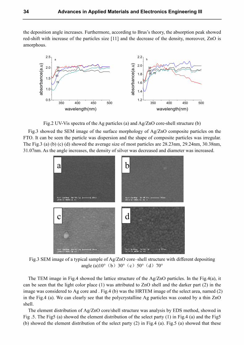
the deposition angle increases. Furthermore, according to Brus’s theory, the absorption peak showed
red-shift with increase of the particles size [11] and the decrease of the density, moreover, ZnO is
amorphous.
350 400 450 5000.5
1.0
1.5
2.0
2.5
ab
so
rba
nce
(a.u
)
wavelength(nm)
a
10
30
50
70
350 400 450 5001.2
1.4
1.6
1.8
2.0
2.2
ab
so
rba
nce
(a.u
)
wavelength(nm)
b
10
30
50 70
Fig.2 UV-Vis spectra of the Ag particles (a) and Ag/ZnO core-shell structure (b)
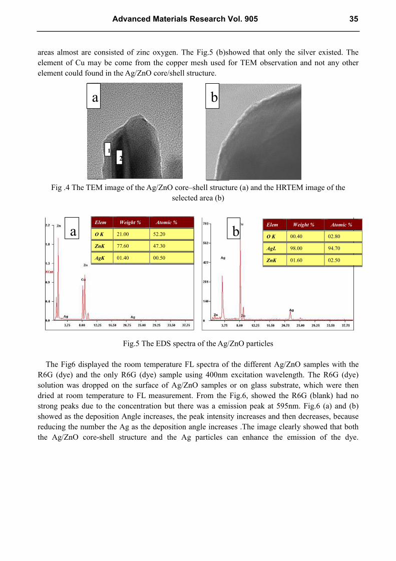
Fig.3 showed the SEM image of the surface morphology of Ag/ZnO composite particles on the
FTO. It can be seen the particle was dispersion and the shape of composite particles was irregular.
The Fig.3 (a) (b) (c) (d) showed the average size of most particles are 28.23nm, 29.24nm, 30.38nm,
31.07nm. As the angle increases, the density of silver was decreased and diameter was increased.
Fig.3 SEM image of a typical sample of Ag/ZnO core–shell structure with different depositing
angle (a)10°(b)30°(c)50°(d)70°
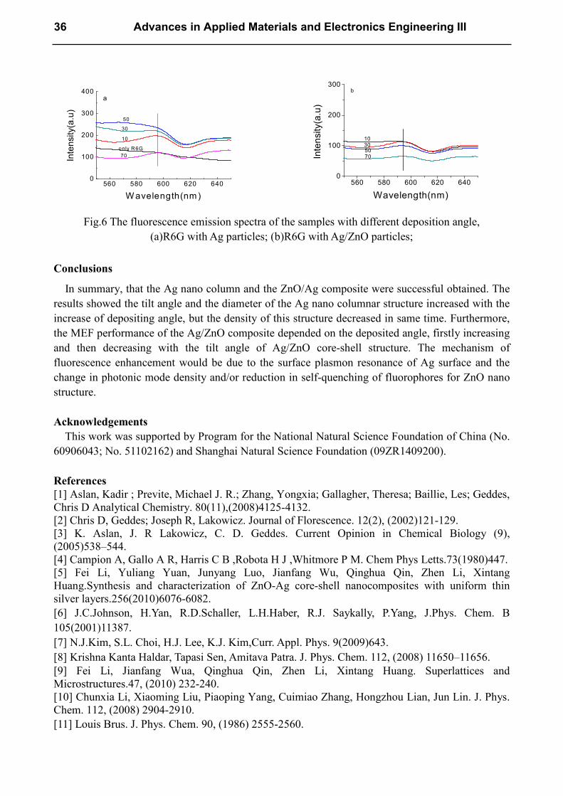
The TEM image in Fig.4 showed the lattice structure of the Ag/ZnO particles. In the Fig.4(a), it
can be seen that the light color place (1) was attributed to ZnO shell and the darker part (2) in the
image was considered to Ag core and . Fig.4 (b) was the HRTEM image of the select area, named (2)
in the Fig.4 (a). We can clearly see that the polycrystalline Ag particles was coated by a thin ZnO
shell.
The element distribution of Ag/ZnO core/shell structure was analysis by EDS method, showed in
Fig .5. The Fig5 (a) showed the element distribution of the select party (1) in Fig.4 (a) and the Fig5
(b) showed the element distribution of the select party (2) in Fig.4 (a). Fig.5 (a) showed that these
a b
c d
34 Advances in Applied Materials and Electronics Engineering III
areas almost are consisted of zinc oxygen. The Fig.5 (b)showed that only the silver existed. The
element of Cu may be come from the copper mesh used for TEM observation and not any other
element could found in the Ag/ZnO core/shell structure.
Fig .4 The TEM image of the Ag/ZnO core–shell structure (a) and the HRTEM image of the
selected area (b)
Fig.5 The EDS spectra of the Ag/ZnO particles
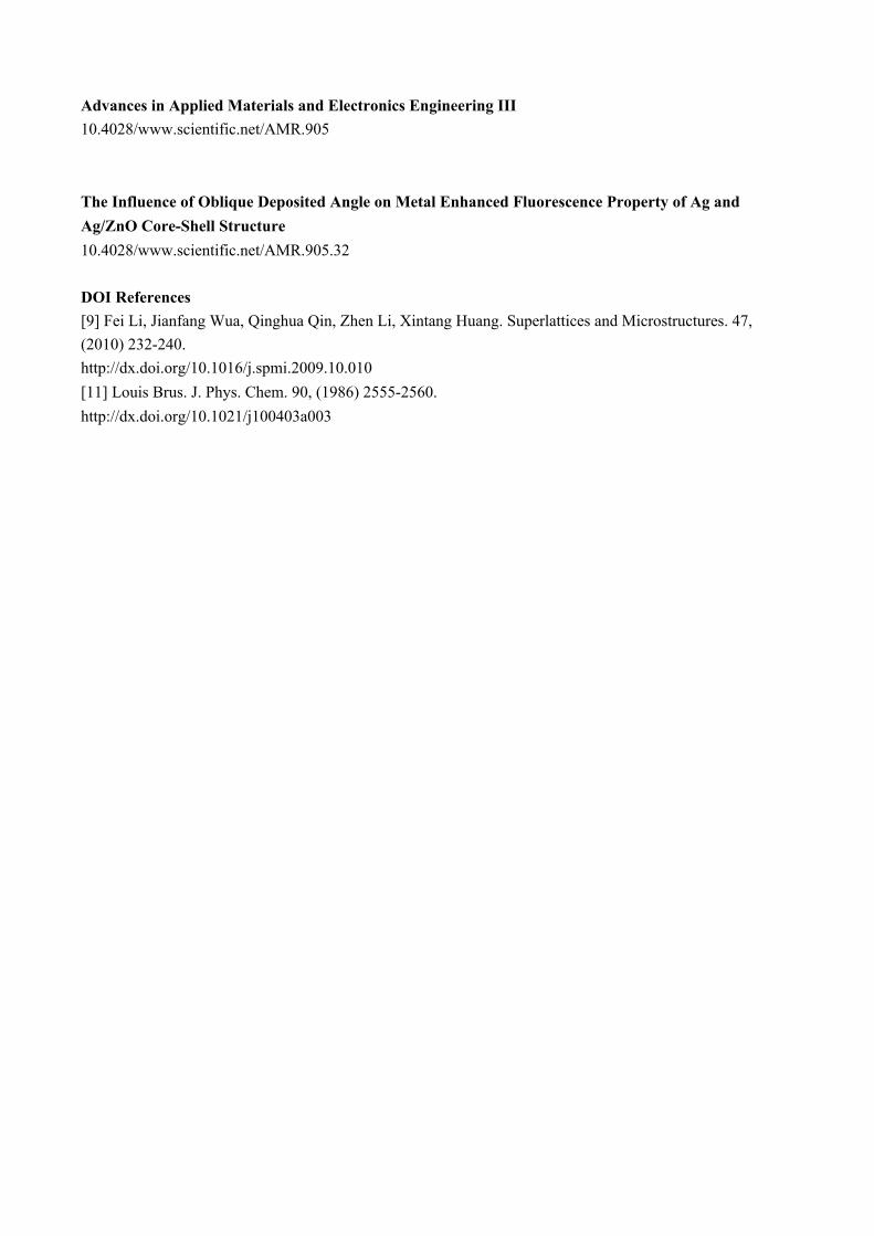
The Fig6 displayed the room temperature FL spectra of the different Ag/ZnO samples with the
R6G (dye) and the only R6G (dye) sample using 400nm excitation wavelength. The R6G (dye)
solution was dropped on the surface of Ag/ZnO samples or on glass substrate, which were then
dried at room temperature to FL measurement. From the Fig.6, showed the R6G (blank) had no
strong peaks due to the concentration but there was a emission peak at 595nm. Fig.6 (a) and (b)
showed as the deposition Angle increases, the peak intensity increases and then decreases, because
reducing the number the Ag as the deposition angle increases .The image clearly showed that both
the Ag/ZnO core-shell structure and the Ag particles can enhance the emission of the dye.
Elem Weight % Atomic %
O K 21.00 52.20
ZnK 77.60 47.30
AgK 01.40 00.50
Elem Weight % Atomic %
O K 00.40 02.80
AgL 98.00 94.70
ZnK 01.60 02.50
1
2
a b
a b
Advanced Materials Research Vol. 905 35
560 580 600 620 6400
100
200
300
400
Inte
nsity(a
.u)
W avelength(nm)
50
30
10
only R6G
70
a
560 580 600 620 6400
100
200
300
Inte
nsity(a
.u)
Wavelength(nm)
b
10305070
Fig.6 The fluorescence emission spectra of the samples with different deposition angle,
(a)R6G with Ag particles; (b)R6G with Ag/ZnO particles;
Conclusions
In summary, that the Ag nano column and the ZnO/Ag composite were successful obtained. The
results showed the tilt angle and the diameter of the Ag nano columnar structure increased with the
increase of depositing angle, but the density of this structure decreased in same time. Furthermore,
the MEF performance of the Ag/ZnO composite depended on the deposited angle, firstly increasing
and then decreasing with the tilt angle of Ag/ZnO core-shell structure. The mechanism of
fluorescence enhancement would be due to the surface plasmon resonance of Ag surface and the
change in photonic mode density and/or reduction in self-quenching of fluorophores for ZnO nano
structure.
Acknowledgements
This work was supported by Program for the National Natural Science Foundation of China (No.
60906043; No. 51102162) and Shanghai Natural Science Foundation (09ZR1409200).
References
[1] Aslan, Kadir ; Previte, Michael J. R.; Zhang, Yongxia; Gallagher, Theresa; Baillie, Les; Geddes,
Chris D Analytical Chemistry. 80(11),(2008)4125-4132.
[2] Chris D, Geddes; Joseph R, Lakowicz. Journal of Florescence. 12(2), (2002)121-129.
[3] K. Aslan, J. R Lakowicz, C. D. Geddes. Current Opinion in Chemical Biology (9),
(2005)538–544.
[4] Campion A, Gallo A R, Harris C B ,Robota H J ,Whitmore P M. Chem Phys Letts.73(1980)447.
[5] Fei Li, Yuliang Yuan, Junyang Luo, Jianfang Wu, Qinghua Qin, Zhen Li, Xintang
Huang.Synthesis and characterization of ZnO-Ag core-shell nanocomposites with uniform thin
silver layers.256(2010)6076-6082.
[6] J.C.Johnson, H.Yan, R.D.Schaller, L.H.Haber, R.J. Saykally, P.Yang, J.Phys. Chem. B
105(2001)11387.
[7] N.J.Kim, S.L. Choi, H.J. Lee, K.J. Kim,Curr. Appl. Phys. 9(2009)643.
[8] Krishna Kanta Haldar, Tapasi Sen, Amitava Patra. J. Phys. Chem. 112, (2008) 11650–11656.
[9] Fei Li, Jianfang Wua, Qinghua Qin, Zhen Li, Xintang Huang. Superlattices and
Microstructures.47, (2010) 232-240.
[10] Chunxia Li, Xiaoming Liu, Piaoping Yang, Cuimiao Zhang, Hongzhou Lian, Jun Lin. J. Phys.
Chem. 112, (2008) 2904-2910.
[11] Louis Brus. J. Phys. Chem. 90, (1986) 2555-2560.
36 Advances in Applied Materials and Electronics Engineering III
Advances in Applied Materials and Electronics Engineering III 10.4028/www.scientific.net/AMR.905 The Influence of Oblique Deposited Angle on Metal Enhanced Fluorescence Property of Ag and
Ag/ZnO Core-Shell Structure 10.4028/www.scientific.net/AMR.905.32
DOI References
[9] Fei Li, Jianfang Wua, Qinghua Qin, Zhen Li, Xintang Huang. Superlattices and Microstructures. 47,
(2010) 232-240.
http://dx.doi.org/10.1016/j.spmi.2009.10.010 [11] Louis Brus. J. Phys. Chem. 90, (1986) 2555-2560.
http://dx.doi.org/10.1021/j100403a003