Embed Size (px)
Citation preview
cancers
Review
The Impact on Survival and Morbidity ofPortal–Mesenteric Resection DuringPancreaticoduodenectomy for Pancreatic HeadAdenocarcinoma: A Systematic Review andMeta-Analysis of Comparative Studies
Alessandro Fancellu 1,*, Niccolò Petrucciani 2, Alberto Porcu 1, Giulia Deiana 1, Valeria Sanna 3,Chiara Ninniri 1, Teresa Perra 1 , Valentina Celoria 1 and Giuseppe Nigri 2
1 Unit of General Surgery 2—Clinica Chirurgica, Department of Medical Surgical and Experimental Sciences,University of Sassari, V. le San Pietro 43, 07100 Sassari, Italy; [email protected] (A.P.);[email protected] (G.D.); [email protected] (C.N.); [email protected] (T.P.);[email protected] (V.C.)
2 Department of Medical and Surgical Sciences and Translational Medicine, St. Andrea University Hospital,Sapienza University of Rome, Via di Grottarossa 1037, 00189 Rome, Italy; [email protected] (N.P.);[email protected] (G.N.)
3 Unit of Medical Oncology, AOU Sassari, Via E. De Nicola, 07100 Sassari, Italy; [email protected]* Correspondence: [email protected]; Tel.: +39-079-22-8432
Received: 13 May 2020; Accepted: 14 July 2020; Published: 20 July 2020�����������������
Abstract: Background: The literature is conflicting regarding oncological outcome and morbidityassociated to portal–mesenteric resection during pancreaticoduodenectomy (PD) in patients withpancreatic head adenocarcinoma (PHAC). Methods: A meta-analysis of studies comparing PD plusvenous resection (PD+VR) and standard PD exclusively in patients with adenocarcinoma of thepancreatic head was conducted. Results: Twenty-three cohort studies were identified, which included6037 patients, of which 28.6% underwent PD+VR and 71.4% underwent standard PD. Patients whoreceived PD+VR had lower 1-year overall survival (OS) (odds radio OR 0.79, 95% CI 0.67–0.92,p = 0.003), 3-year OS (OR 0.72, 95% CI 0.59–0.87, p = 0.0006), and 5-year OS (OR 0.57, 95% CI 0.39–0.83,p = 0.003). Patients in the PD+VR group were more likely to have a larger tumor size (MD 3.87,95% CI 1.75 to 5.99, p = 0.0003), positive lymph nodes (OR 1.24, 95% CI 1.06–1.45, p = 0.007), and R1resection (OR 1.74, 95% CI 1.37–2.20, p < 0.0001). Thirty-day mortality was higher in the PD+VRgroup (OR 1.93, 95% CI 1.28–2.91, p = 0.002), while no differences between groups were observed inrates of total complications (OR 1.07, 95% CI, 0.81–1.41, p = 0.65). Conclusions: Although PD+VR hassignificantly increased the resection rate in patients with PHAC, it has inferior survival outcomes andhigher 30-day mortality when compared with standard PD, whereas postoperative morbidity ratesare similar. Further research is needed to evaluate the role of PD+VR in the context of multimodalitytreatment of PHAC.
Keywords: pancreaticoduodenectomy; portal-mesenteric resection; survival; complications
1. Introduction
Pancreatic head adenocarcinoma (PHAC) accounts for 80–90% of tumors of the exocrine pancreasand remains associated with a dismal long-term prognosis [1–3]. Pancreaticoduodenectomy (PD) withadjuvant chemotherapy offers potential cure for patients with PHAC, although even when surgicalresection is considered curative, the five-year survival rate barely reaches 25% [4–6].
Cancers 2020, 12, 1976; doi:10.3390/cancers12071976 www.mdpi.com/journal/cancers
Cancers 2020, 12, 1976 2 of 21
Involvement of the portal–mesenteric axis is quite frequent at the time of diagnosis of PHAC,being encountered in 25–30% of patients undergoing PD [7,8]. In particular, solid tumor contact withthe portal vein or superior mesenteric vein of >180◦, or contact of ≤180◦ with contour irregularityor thrombosis of the vein are patterns included in the category of borderline resectable tumors [9].Since venous infiltration has not longer been considered a contraindication to resection, in the lastdecade, there has been a surge in the surgical treatment of borderline resectable PHAC, with the aim ofobtaining R0 resection. This trend is also fueled by the encouraging results of systemic preoperativeand postoperative treatments [10–14]. The use of PD with concurrent portal–mesenteric resection hasin fact shifted the field of operable disease, and it is nowadays carried out in up to 20–30% of PD athigh-volume pancreatic surgery centers [15,16].
The resection or replacement of the portal–mesenteric axis during PD represents a challengingand skill-demanding procedure leading to potential complications, in spite of debatable advantagesin survival outcomes. Previous reviews investigating the impact of portal–mesenteric resectionon the oncological outcomes following PD are limited and conflicting, since the results havepredominantly been reported together with those of patients receiving distal or total pancreatectomy [6].Other meta-analyses were limited by heterogeneity, because they also included tumor histotypesdifferent from pancreatic adenocarcinoma, evaluated studies in whom arterial resection was performedalong with venous resection [5], or did not evaluate survival outcomes [5,7,17].
Furthermore, none of previous reviews reported on preoperative and postoperative systemictreatment. Thus, from the present literature, it is difficult to extrapolate valuable conclusions onpatients receiving PD with portal–mesenteric resection specifically for PHAC. In addition, newer andlarger comparative studies have been added to the literature.
To overcome these limitations, we performed a systematic review and a meta-analysis limitedto studies comparing PD with venous portal–mesenteric resection (PD+VR) versus standard PDin patients with PHAC only, in order to critically evaluate the effects on survival outcomes andpostoperative complications.
2. Methods
2.1. Study Selection and Data Extraction
A systematic literature search using the PubMed, Web of Sciences (WOS), and Scopus databaseswas performed in December 2019 to identify studies reporting surgical outcomes of portal–superiormesenteric resection in patients undergoing PD for PHAC. The following keywords were usedand combined for the search: ‘pancreaticoduodenectomy’, ‘duodenopancreatectomy’, ‘Whippleor Kausch–Whipple operation’, ‘venous resection’, ‘portal resection’, ‘mesenteric vein resection’,portal–mesenteric or porto-mesenteric resection’, ‘pancreatic carcinoma’, and ‘pancreatic headadenocarcinoma’. The ‘related articles’ function was used to broaden the search, and all abstracts,citations, and studies scanned as well as the references of relevant articles were reviewed (Table S1).No language restrictions were made. Potentially relevant articles were examined by three independentinvestigators (A.F., G.D, V.C.) who extracted the following data: first author; year of publication; studydesign; number of subjects; patient and tumor characteristics; intraoperative outcomes; postoperativeoutcomes; survival outcomes.
2.2. Inclusion Criteria and Assessment of Study Quality
To be included in this meta-analysis, studies had to meet the following criteria: (1) comparingsurgical outcomes and survival outcomes of PD with portal–mesenteric vein resection (PD+VR) andstandard PD in patients with PHAC, (2) containing a previously unreported patient group. If a patientcohort was reported more than once by the same institution, the most informative and/or recentarticle was included in our analysis. Quality of the included studies was assessed according to theNewcastle–Ottawa Quality Assessment Scale (NOS) for cohort studies [18] (Table S2).
Cancers 2020, 12, 1976 3 of 21
2.3. Exclusion Criteria
We excluded from our meta-analysis studies comparing PD+VR and PD for the following:(a) tumors other than pancreatic adenocarcinoma (such as ampullary carcinoma, cholangiocarcinoma,primitive duodenal cancer, and malignant neuroendocrine tumors); (b) pancreatic cancer treated withtotal pancreatectomy, or distal pancreatectomy; (c) patients in whom also arterial resection was carriedout along with portal–superior mesenteric resection. All those studies were evaluated and excludedonly when it was impossible to extract the data specifically related to patients receiving PD+VR andPD for PHAC. We also excluded studies in which the outcomes of interest (specified below) were notreported or impossible to calculate for both PD+VR and PD groups (Table S1).
2.4. Outcomes of Interest and Definitions
All the studies were abstracted for the following relevant data:
• Patients baseline characteristics: age, gender, American Society of Anesthesiologists Classification(ASA Class), number of patients receiving neoadjuvant chemotherapy, number of patientsundergoing preoperative biliary drainage for obstructive jaundice.
• Tumor characteristics: stage according to AJCC, T stage according to the TNM, tumor size, gradeof differentiation (well, moderate, and poor), and presence of lymphovascular invasion (LVI).
• Operation-related outcomes: type of vein resection/reconstruction performed, operative time,blood loss, rates of transfusion, status of resection margins (positive versus negative), rates of R0and R1 resections, number of patients having venous infiltration at final histologic examination.Definition of resection margins varied among the included studies. Additionally, the definitionof postoperative morbidity varied, although the main part of the complications were definedaccording to the International Study Group of Pancreatic Surgery [19–21].
• Duration of postoperative hospital stay.• 30-day mortality, overall and specific postoperative morbidity: pancreatic fistula, bile leak, delayed
gastric emptying (DGE), hemorrhage, rates of blood transfusions, rates of reoperations, numberof patients receiving adjuvant chemotherapy.
• Duration of follow-up and survival outcomes including 1-, 3-, and 5- year overall survival (OS) inboth the PD+VR and PD groups. Survival was defined as the number of months between the dateof surgery and death.
2.5. Statistical Analysis
Meta-analysis was performed in line with recommendations from the Preferred Reporting Itemsfor Systematic Reviews and MetaAnalyses (PRISMA) statement [22]. RevMan software version 5.3(The Cochrane Collaboration, Software Update, Oxford) was used to perform the meta-analysis.Variables were pooled only if evaluated by three or more studies. For dichotomous variables, oddsratios (ORs) were used as summary measures of efficacy, corresponding to the odds of an eventoccurring in the treatment group (PD+VR) compared to the reference group (PD).
An odd ratio of more than 1 indicates the probability that an outcome is more likely to occur inthe first group, and it is considered statistically significant when P < 0.05 and when the 95% confidenceinterval (CI) does not include the value 1. The Mantel Haenszel method was used to combine theORs for outcomes of interest. A random effect model, which is more robust in terms of anticipatedheterogeneity, was used. The random effect-weighted mean difference (MD) between groups was usedas the summary statistic for continuous variables; 95% confidence intervals were reported. For studiesreporting medians and interquartile ranges instead of means and standard deviations, the methoddescribed by Hozo et al. was used to impute means and standard deviations. Statistical heterogeneitywas evaluated using the I2 statistic. I2 values of 0 to 25%, 26% to 50%, and >51% were considered to beindicative of homogeneity, moderate heterogeneity, and high heterogeneity, respectively. All statisticaldata were considered significant if p < 0.05.
Cancers 2020, 12, 1976 4 of 21
3. Results
3.1. Included Studies
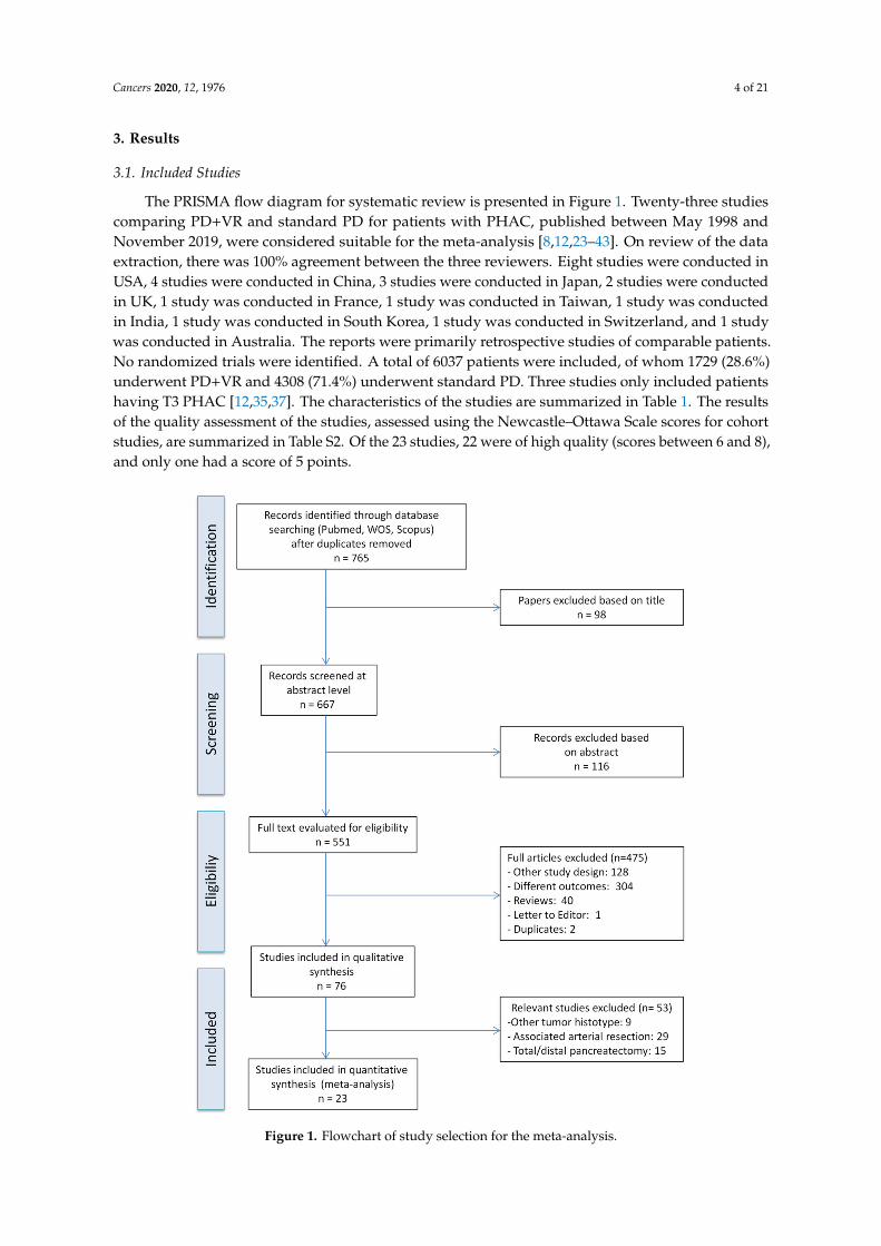
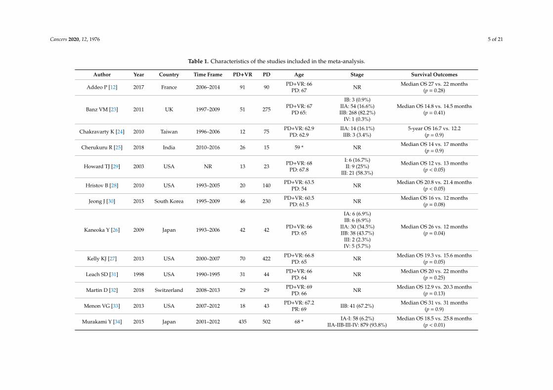
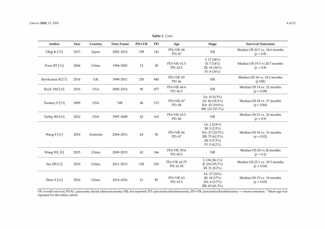
The PRISMA flow diagram for systematic review is presented in Figure 1. Twenty-three studiescomparing PD+VR and standard PD for patients with PHAC, published between May 1998 andNovember 2019, were considered suitable for the meta-analysis [8,12,23–43]. On review of the dataextraction, there was 100% agreement between the three reviewers. Eight studies were conducted inUSA, 4 studies were conducted in China, 3 studies were conducted in Japan, 2 studies were conductedin UK, 1 study was conducted in France, 1 study was conducted in Taiwan, 1 study was conductedin India, 1 study was conducted in South Korea, 1 study was conducted in Switzerland, and 1 studywas conducted in Australia. The reports were primarily retrospective studies of comparable patients.No randomized trials were identified. A total of 6037 patients were included, of whom 1729 (28.6%)underwent PD+VR and 4308 (71.4%) underwent standard PD. Three studies only included patientshaving T3 PHAC [12,35,37]. The characteristics of the studies are summarized in Table 1. The resultsof the quality assessment of the studies, assessed using the Newcastle–Ottawa Scale scores for cohortstudies, are summarized in Table S2. Of the 23 studies, 22 were of high quality (scores between 6 and 8),and only one had a score of 5 points.
Figure 1. Flowchart of study selection for the meta-analysis.
Cancers 2020, 12, 1976 5 of 21
Table 1. Characteristics of the studies included in the meta-analysis.
Author Year Country Time Frame PD+VR PD Age Stage Survival Outcomes
Addeo P [12] 2017 France 2006–2014 91 90 PD+VR: 66PD: 67 NR Median OS 27 vs. 22 months
(p = 0.28)
Banz VM [23] 2011 UK 1997–2009 51 275 PD+VR: 67PD 65:
IB: 3 (0.9%)IIA: 54 (16.6%)IIB: 268 (82.2%)
IV: 1 (0.3%)
Median OS 14.8 vs. 14.5 months(p = 0.41)
Chakravarty K [24] 2010 Taiwan 1996–2006 12 75 PD+VR: 62.9PD: 62.9
IIA: 14 (16.1%)IIB: 3 (3.4%)
5-year OS 16.7 vs. 12.2(p = 0.9)
Cherukuru R [25] 2018 India 2010–2016 26 15 59 * NR Median OS 14 vs. 17 months(p = 0.9)
Howard TJ [29] 2003 USA NR 13 23 PD+VR: 68PD: 67.8
I: 6 (16.7%)II: 9 (25%)
III: 21 (58.3%)
Median OS 12 vs. 13 months(p < 0.05)
Hristov B [28] 2010 USA 1993–2005 20 140 PD+VR: 63.5PD: 54 NR Median OS 20.8 vs. 21.4 months
(p < 0.05)
Jeong J [30] 2015 South Korea 1995–2009 46 230 PD+VR: 60.5PD: 61.5 NR Median OS 16 vs. 12 months
(p = 0.08)
Kaneoka Y [26] 2009 Japan 1993–2006 42 42 PD+VR: 66PD: 65
IA: 6 (6.9%)IB: 6 (6.9%)
IIA: 30 (34.5%)IIB: 38 (43.7%)
III: 2 (2.3%)IV: 5 (5.7%)
Median OS 26 vs. 12 months(p = 0.04)
Kelly KJ [27] 2013 USA 2000–2007 70 422 PD+VR: 66.8PD: 65 NR Median OS 19.3 vs. 15.6 months
(p = 0.05)
Leach SD [31] 1998 USA 1990–1995 31 44 PD+VR: 66PD: 64 NR Median OS 20 vs. 22 months
(p = 0.25)
Martin D [32] 2018 Switzerland 2008–2013 29 29 PD+VR: 69PD: 66 NR Median OS 12.9 vs. 20.3 months
(p = 0.13)
Menon VG [33] 2013 USA 2007–2012 18 43 PD+VR: 67.2PR: 69 IIB: 41 (67.2%) Median OS 31 vs. 31 months
(p = 0.9)
Murakami Y [34] 2015 Japan 2001–2012 435 502 68 * IA-I: 58 (6.2%)IIA-IIB-III-IV: 879 (93.8%)
Median OS 18.5 vs. 25.8 months(p < 0.01)
Cancers 2020, 12, 1976 6 of 21
Table 1. Cont.
Author Year Country Time Frame PD+VR PD Age Stage Survival Outcomes
Ohgi K [35] 2017 Japan 2002–2014 158 141 PD+VR: 68PD: 67 NR Median OS 20.7 vs. 18.6 months
(p = 0.8)
Poon RT [36] 2004 China 1998–2002 12 38 PD+VR: 61.5PD: 62.5
I: 17 (34%)II: 7 (14%)
III: 18 (36%)IV: 8 (16%)
Median OS 19.5 vs 20.7 months(p = 0.8)
Ravikumar R [37] 2014 UK 1998–2011 230 840 PD+VR: 65PD: 66 NR Median OS 18 vs. 18.2 months
(p NR)
Roch AM [38] 2016 USA 2000–2014 90 477 PD+VR: 66.4PD: 66.3 NR Median OS 14 vs. 21 months
(p = 0.08)
Toomey P [39] 2009 USA NR 48 172 PD+VR: 67PD: 68
IA: 10 (4.5%)IA: 40 (18.2%)IIA: 43 (19.6%)IIB: 127 (57.7%)
Median OS 18 vs. 17 months(p = 0.84)
Turley RS [40] 2012 USA 1997–2008 42 162 PD+VR: 63.5PD: 66 NR Median OS 21 vs. 20 months
(p = 0.9)
Wang F [41] 2014 Australia 2004–2012 64 58 PD+VR: 66PD: 67
IA: 1 (0.8%)IB: 3 (2.5%)
IIA: 27 (22.5%)IIB: 75 (62.5%)
III: 9 (7.5%)IV: 5 (4.2%)
Median OS 18 vs. 31 months(p = 0.02)
Wang WL [8] 2015 China 2009–2013 42 166 PD+VR: 59.4PD: 60.5 NR Median OS 20 vs 26 months
(p = 0.2)
Xie ZB [42] 2019 China 2011–2013 138 239 PD+VR: 62.75PD: 61.54
I: 136 (36.1%)II: 210 (55.7%)III: 31 (8.2%)
Median OS 25.1 vs. 29.3 months(p = 0.04)
Zhao X [43] 2016 China 2014–2016 21 85 PD+VR: 63PD: 63.5
IA: 17 (16%)IB: 18 (17%)IIA: 6 (5.7%)
IIB: 65 (61.3%)
Median OS 15 vs. 19 months(p < 0.05)
OS: overall survival; PDAC: pancreatic ductal adenocarcinoma; NR: not reported; PD: pancreaticoduodenectomy; PD+VR: pancreaticoduodenectomy + venous resection. * Mean age wasreported for the entire cohort.
Cancers 2020, 12, 1976 7 of 21
3.2. Patient and Tumor Characteristics
Patients in the two groups (PD+VR and PD) were similar with respect to age (mean difference[MD] 0.89, 95% CI -0.11 to 1.88, p = 0.08 ), ASA score (ASA I: OR 1.27, 95% CI 0.81 to 2.0, p = 0.30; ASA II:OR 0.75, 95% CI 0.53 to 1.05, p = 0.10; ASA III: OR 1.33, 95% CI 0.70 to 2.52, p = 0.38), and the need forpreoperative biliary drainage (OR 0.91, 95% CI 0.61 to 1.35, p = 0.63), while rates of male gender werehigher in the PD+VR group (OR 0.79, 95% CI 0.67 to 0.92, p = 0.003). Tumor size was significantlylarger in the PD+VR group (MD 3.87, 95% CI 1.75 to 5.99, p = 0.0003), while the distribution of T1and T2 categories did not differ (T1: OR 0.41, 95% CI 0.16 to 1.05, p = 0.06; T2: OR 0.30, 95% CI 0.05to 1.67, p = 0.17). T3 tumors were more represented in the group PD+VR (OR 1.24, 95% CI 1.06to 1.45, p = 0.007). Groups were similar with respect to tumor differentiation (well differentiated:OR 0.77, 95% CI 0.52 to 1.16, p = 0.22; moderately differentiated: OR 1.03, 95% CI 0.64 to 1.68, p = 0.90;poorly differentiated: OR 1.43, 95% CI 0.97 to 2.10, p = 0.07). However, when considering tumorstage, there were significantly more patients in the PD group in Stage 1 (OR 0.29, 95% CI 0.18 to 0.47,p < 0.00001), while there were significantly more patients in the PD+VR group in Stage 2 (OR 2.33,95% CI 1.56 to 3.48, p < 0.0001). Lymphovascular invasion was slightly higher in the tumors of thePD+VR group, but not in a significant manner (OR 1.33, 95% CI 0.98 to 1.80, p = 0.07). Lymph nodepositivity was more frequently observed in the PD+VR group (OR 1.24, 95% CI 1.06 to 1.45, p = 0.007)(Figure S1). Nine studies reported on neoadjuvant chemotherapy. It was administrated in only in 5 ofthem [26–28,31,33–35,40,41]. In total, 473 patients received preoperative treatment: 266 in the PD+VRgroup and 207 in the PD group. Patients of the PD+VR group were more likely to receive neoadjuvantchemotherapy (p = 0.01) (Figure S2).
The type of venous resection/reconstruction performed was resumed in Table S3.
3.3. Operative Outcomes
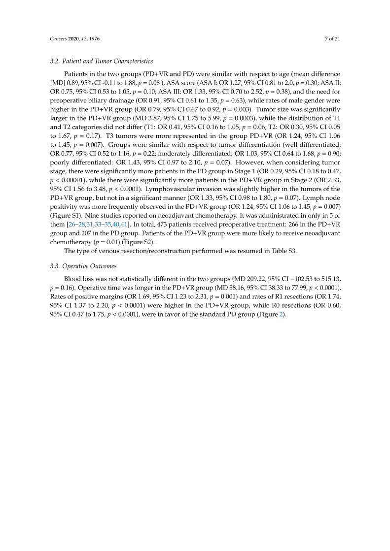
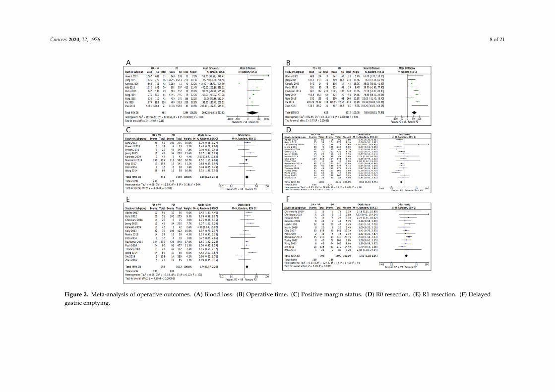
Blood loss was not statistically different in the two groups (MD 209.22, 95% CI −102.53 to 515.13,p = 0.16). Operative time was longer in the PD+VR group (MD 58.16, 95% CI 38.33 to 77.99, p < 0.0001).Rates of positive margins (OR 1.69, 95% CI 1.23 to 2.31, p = 0.001) and rates of R1 resections (OR 1.74,95% CI 1.37 to 2.20, p < 0.0001) were higher in the PD+VR group, while R0 resections (OR 0.60,95% CI 0.47 to 1.75, p < 0.0001), were in favor of the standard PD group (Figure 2).
Cancers 2020, 12, 1976 8 of 21
Figure 2. Meta-analysis of operative outcomes. (A) Blood loss. (B) Operative time. (C) Positive margin status. (D) R0 resection. (E) R1 resection. (F) Delayedgastric emptying.
Cancers 2020, 12, 1976 9 of 21
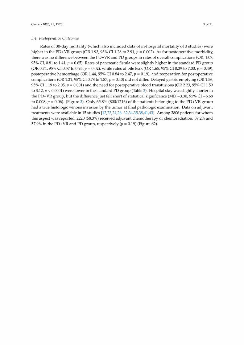
3.4. Postoperative Outcomes
Rates of 30-day mortality (which also included data of in-hospital mortality of 3 studies) werehigher in the PD+VR group (OR 1.93, 95% CI 1.28 to 2.91, p = 0.002). As for postoperative morbidity,there was no difference between the PD+VR and PD groups in rates of overall complications (OR, 1.07,95% CI, 0.81 to 1.41, p = 0.65). Rates of pancreatic fistula were slightly higher in the standard PD group(OR 0.74, 95% CI 0.57 to 0.95, p = 0.02), while rates of bile leak (OR 1.65, 95% CI 0.39 to 7.00, p = 0.49),postoperative hemorrhage (OR 1.44, 95% CI 0.84 to 2.47, p = 0.19), and reoperation for postoperativecomplications (OR 1.21, 95% CI 0.78 to 1.87, p = 0.40) did not differ. Delayed gastric emptying (OR 1.56,95% CI 1.19 to 2.05, p = 0.001) and the need for postoperative blood transfusions (OR 2.23, 95% CI 1.59to 3.12, p < 0.0001) were lower in the standard PD group (Table 2). Hospital stay was slightly shorter inthe PD+VR group, but the difference just fell short of statistical significance (MD −3.30, 95% CI −6.68to 0.008, p = 0.06). (Figure 3). Only 65.8% (800/1216) of the patients belonging to the PD+VR grouphad a true histologic venous invasion by the tumor at final pathologic examination. Data on adjuvanttreatments were available in 15 studies [12,23,24,26–32,34,35,38,41,43]. Among 3806 patients for whomthis aspect was reported, 2220 (58.3%) received adjuvant chemotherapy or chemoradiation: 59.2% and57.9% in the PD+VR and PD group, respectively (p = 0.19) (Figure S2).
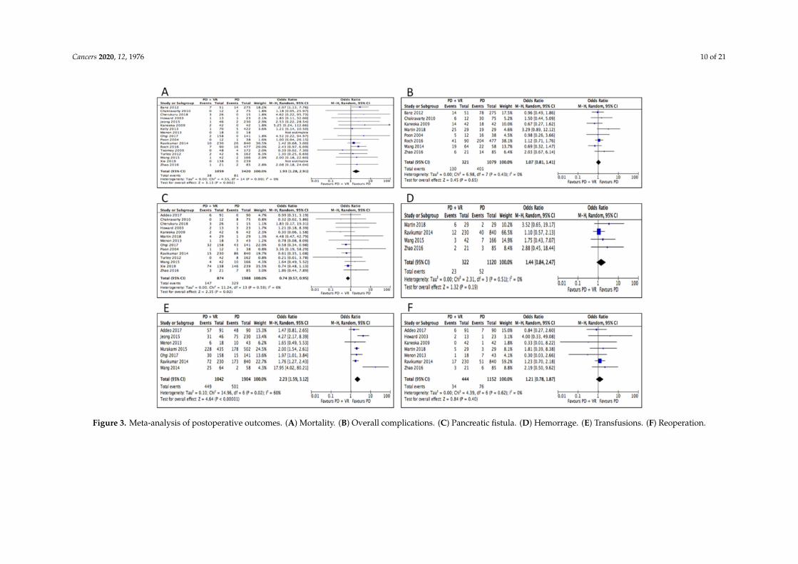
Cancers 2020, 12, 1976 10 of 21
Figure 3. Meta-analysis of postoperative outcomes. (A) Mortality. (B) Overall complications. (C) Pancreatic fistula. (D) Hemorrage. (E) Transfusions. (F) Reoperation.
Cancers 2020, 12, 1976 11 of 21
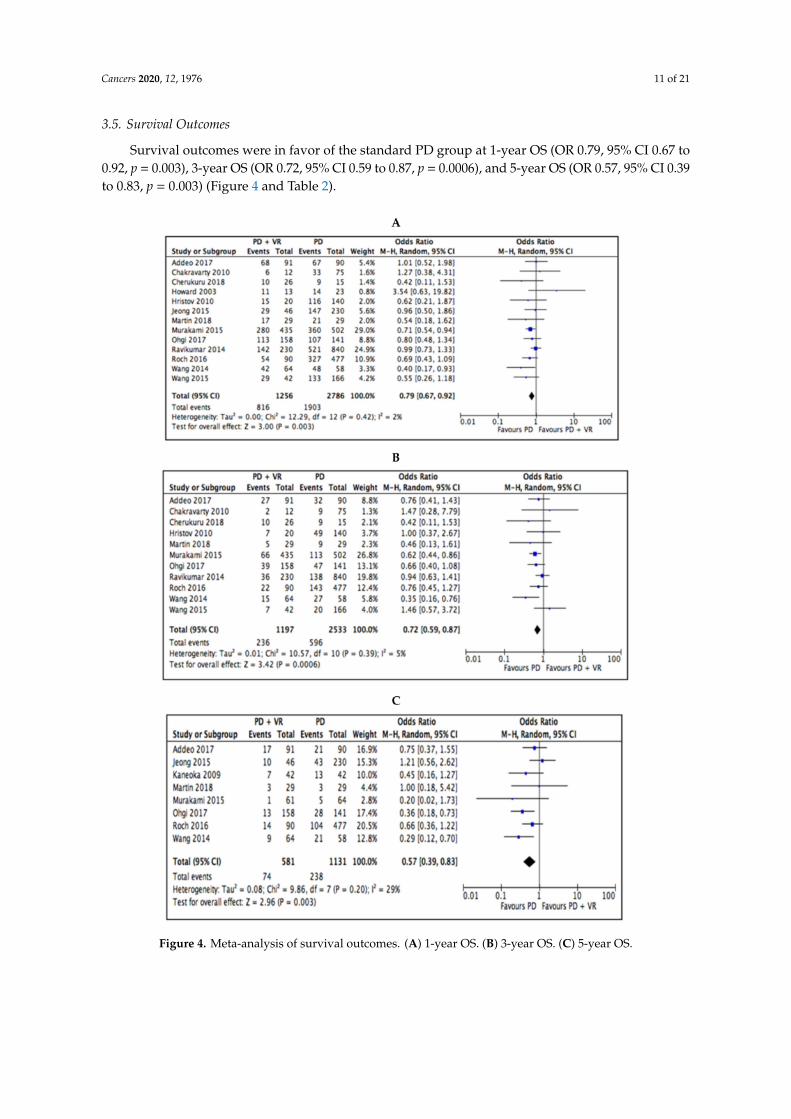
3.5. Survival Outcomes
Survival outcomes were in favor of the standard PD group at 1-year OS (OR 0.79, 95% CI 0.67 to0.92, p = 0.003), 3-year OS (OR 0.72, 95% CI 0.59 to 0.87, p = 0.0006), and 5-year OS (OR 0.57, 95% CI 0.39to 0.83, p = 0.003) (Figure 4 and Table 2).
Figure 4. Meta-analysis of survival outcomes. (A) 1-year OS. (B) 3-year OS. (C) 5-year OS.
Cancers 2020, 12, 1976 12 of 21
Table 2. Comparisons of postoperative outcomes and survival outcomes. LLCI: Lower Level ConfidenceInterval; ULCI: Upper Level Confidence Interval.
Variables No. of PooledStudies
MeanPD+VR
MeanPD
Differenceor Odds Ratio
95%LLCI
95%ULCI
Blood loss (ml) 9 977.8 743.31 209.22 −84.18 502.62
Operative time (min) 9 421.1 361.4 58.16 38.3 77.9
Positive margin status 9 0.27 0.22 1.69 1.23 2.31
R0 resection 17 0.58 0.68 0.6 0.47 0.75
Delayed gastric emptying 13 0.17 0.09 1.56 1.19 2.05
Mortality 17 0.03 0.02 1.93 1.28 2.91
Overall complications 8 0.4 0.4 1.07 0.81 1.41
Pancreatic fistula 14 0.2 0.2 0.74 0.57 0.95
Postoperative Hemorrage 4 0.1 0.04 1.44 0.84 2.47
Reoperations 7 0.1 0.1 1.21 0.78 1.87
1-yr OS 13 0.6 0.7 0.79 0.67 0.92
3-yr OS 11 0.2 0.2 0.72 0.59 0.87
5-yr OS 8 0.1 0.2 0.57 0.39 0.83
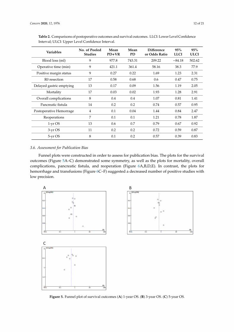
3.6. Assessment for Publication Bias
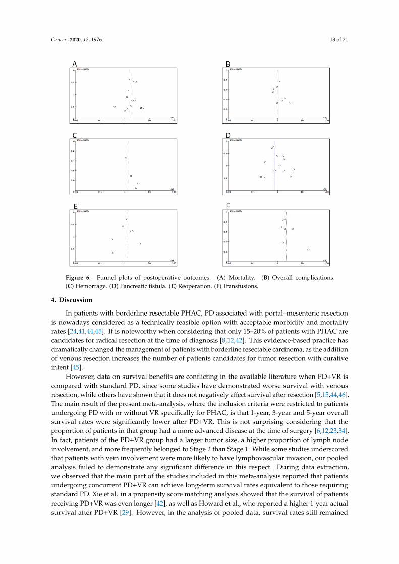
Funnel plots were constructed in order to assess for publication bias. The plots for the survivaloutcomes (Figure 5A–C) demonstrated some symmetry, as well as the plots for mortality, overallcomplications, pancreatic fistula, and reoperation (Figure 6A,B,D,E). In contrast, the plots forhemorrhage and transfusions (Figure 6C–F) suggested a decreased number of positive studies withlow precision.
Figure 5. Funnel plot of survival outcomes (A) 1-year OS. (B) 3-year OS. (C) 5-year OS.
Cancers 2020, 12, 1976 13 of 21
Figure 6. Funnel plots of postoperative outcomes. (A) Mortality. (B) Overall complications.(C) Hemorrage. (D) Pancreatic fistula. (E) Reoperation. (F) Transfusions.
4. Discussion
In patients with borderline resectable PHAC, PD associated with portal–mesenteric resectionis nowadays considered as a technically feasible option with acceptable morbidity and mortalityrates [24,41,44,45]. It is noteworthy when considering that only 15–20% of patients with PHAC arecandidates for radical resection at the time of diagnosis [8,12,42]. This evidence-based practice hasdramatically changed the management of patients with borderline resectable carcinoma, as the additionof venous resection increases the number of patients candidates for tumor resection with curativeintent [45].
However, data on survival benefits are conflicting in the available literature when PD+VR iscompared with standard PD, since some studies have demonstrated worse survival with venousresection, while others have shown that it does not negatively affect survival after resection [5,15,44,46].The main result of the present meta-analysis, where the inclusion criteria were restricted to patientsundergoing PD with or without VR specifically for PHAC, is that 1-year, 3-year and 5-year overallsurvival rates were significantly lower after PD+VR. This is not surprising considering that theproportion of patients in that group had a more advanced disease at the time of surgery [6,12,23,34].In fact, patients of the PD+VR group had a larger tumor size, a higher proportion of lymph nodeinvolvement, and more frequently belonged to Stage 2 than Stage 1. While some studies underscoredthat patients with vein involvement were more likely to have lymphovascular invasion, our pooledanalysis failed to demonstrate any significant difference in this respect. During data extraction,we observed that the main part of the studies included in this meta-analysis reported that patientsundergoing concurrent PD+VR can achieve long-term survival rates equivalent to those requiringstandard PD. Xie et al. in a propensity score matching analysis showed that the survival of patientsreceiving PD+VR was even longer [42], as well as Howard et al., who reported a higher 1-year actualsurvival after PD+VR [29]. However, in the analysis of pooled data, survival rates still remained
Cancers 2020, 12, 1976 14 of 21
in favor of standard PD. When looking to other single-institution studies with the largest criteria,the results of the current meta-analysis are in line with them. For example, a large French multicentricpopulation-based study demonstrated a poorer survival for those undergoing portal–venous resection,as did the currently largest single-center study from Japan [47,48]. In addition, previous systematicreviews and meta-analysis reporting in general results of pancreatic resections for cancer with “en bloc”venous resection showed worse 5-year survival rates in that subgroup [5,7].
Other than tumor characteristics, survival after surgical resection of PHAC is related to surgicalmargin status. We observed that rates of patients with infiltrate margins were significantly higher inpatients undergoing PD+VR, although the definition of resection margin in PD was not homogeneous.The objective of PD+VR is to achieve a R0 resection, which ranges between 40% and 70% in allPD+VR cases described in the literature [8]. The differences can be partially explained by the technicallevel of the centers and the criteria of defining an R0 resection. Several studies have demonstratedthat R0 resection is associated with survival benefit over R1 resection in patients with pancreaticadenocarcinoma [7,39,49,50]. In the present meta-analysis, rates of R1 resection were significantlyhigher in the PD+VR group, while those of R0 were higher in the PD group. One can argue thatthose findings may partially explain the difference in survival outcomes, although the definition of anR0 margin was not identical across the studies due to the fact that histopathologic reporting is notuniversally standardized. Recently, the International Study Group of Pancreatic Surgery suggested thatvenous resection should be attempted if R0 resection is feasible [51]. Since too few studies reported ondifferences in survival between patients obtaining an R0 resection, was not possible to make a pooledanalysis on this item in the current meta-analysis.
To note, data on the use of preoperative chemotherapy or chemoradiation were available onlyin 5 out of the 23 studies included in our meta-analysis [31,33,34,38,40]. This low utilization ofpreoperative therapy is likely correlated to the fact that the main part of the studies was conductedbefore the year 2015, when upfront surgical resection was recommended in the presence of suspiciousportal–mesenteric invasion. The role of preoperative chemotherapy in patients with borderlineresectable PHAC has been gaining importance, as the potential advantages of obtaining a downstagingof the tumor and achieving free margins following PD cannot be overemphasized [7]. Moreover,the response to neoadjuvant therapy may help in identifying patients having tumors with moreaggressive biological behavior and worse outcomes. Although the heterogeneity of the availabletrials reduces the strength of any conclusion, many reports demonstrated improved R0 resectionsand higher overall survival in patients receiving preoperative chemotherapy for resectable pancreaticcancer [52,53], including a recent randomized, multicenter phase 2/3 trial [54]. As a consequence,nowadays, there is a growing trend in the using of chemotherapy as upfront treatment in patients withborderline resectable tumors, although it is still unclear about whether this translates into increasedcure rates [45,55]. For example, in the Dutch PREOPANC trial, preoperative chemoradiotherapyfor resectable or borderline resectable pancreatic cancer did not show a significant overall survivalbenefit [56]. Various treatment regimens have been used, including FOLFIRINOX, gemcitabine-basedinduction chemotherapy followed by 5-FU, nab-paclitaxel plus gemcitabine, and capecitabine-basedchemoradiation [9,35,53]. According to the NCCN guidelines, while there is limited evidence torecommend specific neoadjuvant regimens, most member institutions prefer neoadjuvant therapy andencourage participation in clinical trials at or coordinated through a high-volume center [9]. In addition,ESMO guidelines suggest that patients with borderline resectable tumors should be included in clinicaltrials. If this would not be the case, the best option might be upfront chemotherapy followed bychemoradiation and then surgery (recommendation of type IV,B) [53].
Another remarkable point is that only 65.8% of the patients had a histology-demonstrated invasionof the portal–mesenteric vein wall by tumor cells at final pathologic examination, among the 14 studiesreporting on this parameter. Although in a recent multicentric cohort study, Ravikumar et al. [57]found that 26.5% of patients undergoing portal vein resection had histological evidence of veininvolvement, the proportion of actual vein invasion in the literature ranges from 40% to 100% [58].
Cancers 2020, 12, 1976 15 of 21
Ideally, only patients with true venous invasion should undergo PD+VR, even though the real impactof histologic venous invasion on survival rates remains a matter of debate [12,44]. Studies have shownthat invasion of the wall of the veins, rather than adhesion, is associated with reduced survival [6,7,12].Venous invasion might indicate aggressive tumor biology, but some authors did not find any statisticallysignificant impact on life expectancy and median survival between patients who did and did not havehistopathologic evidence of vein invasion [59,60].
In an era where the decision-making process in borderline resectable pancreatic tumors isexperiencing profound changes, the issue of true venous invasion calls attention to the role ofpre-therapeutic work up. Accurate staging with imaging is of the utmost importance becausepatients need to be stratified into potentially curative upfront surgery versus neaodjuvant treatments.The decision is based on subtle differences regarding the contour irregularity of the vein and/or itscontact with the tumor seen on cross-sectional CT and MRI images [15,61,62]. Preoperative predictionof pathologic invasion of the portal–mesenteric wall is still limited, even with advanced imaging tools.Some recent studies have underscored the need for enhancement of pre-treatment investigations inpatients with suspicious portal–mesenteric invasion and, generally speaking, with borderline resectablepancreatic cancer [12,31].
The available literature suggests that venous resection in combination with multimodality therapymay significantly improve long-term survival for patients having borderline pancreatic cancer [12,34,44].Other than a low use of neoadjuvant therapy, we also noticed a suboptimal number of patients whounderwent adjuvant treatment among the studies. In fact, only 58.3% received chemotherapy orchemoradiation following either PD or PD+VR. In this regard, we can speculate that many patientswere unfit for postoperative chemotherapy, which is a common problem in the setting of patients whoundergo surgery for pancreatic cancer.
Although in many of the comparative studies included in this meta-analysis, the overall mortalitywas not altered by venous resection [16,34,37,38], we found that 30-day mortality rates were significantlyhigher in the PD+VR group. Similar findings were also reported from Peng et al. [17], as well asfrom Giovinazzo et al, who reported that patients undergoing portal–mesenteric venous resection hadincreased mortality rates in a large meta-analysis evaluating all types of pancreatectomy (PD, totalpancreatectomy, and distal pancreatectomy) [6]. These data could be explained by the presence ofa more advanced disease and with the major technical difficulty of venous resection/reconstruction;in fact, the PD+VR group also required a longer operation time, although intraoperative bloodloss did not differ in the current meta-analysis. In this regard, we also found that two potentialvariables affecting mortality rates, such as age and ASA classification were similar in the two groups.Concomitant morbidities might have had a role in the observed mortality rates, but unfortunately, thataspect was not reported in the studies included.
In general, the incidence of postoperative morbidity in patients receiving PD with or withoutvenous resection remains relatively high with rates ranging between 20% and 50% [14,24]. To note,in the current study, postoperative complication rates, excluding mortality, did not statistically differbetween the two groups, although they were slightly higher in the PD+VR group. This finding is in linewith other single institutional studies and reviews on the topic, although the literature is conflicting.Two reports based on the American College of Surgeons National Surgical Quality ImprovementProgram (ACS-NSQIP) and National Inpatient Sample demonstrated a significant increase in overallmorbidity in patients with vascular tumor involvement [63,64]. In the present meta-analysis, we foundthat rates of pancreatic fistula, which remains one of the most dreaded complication followingPD [65–67], were less frequent in the PD+VR group. This finding has also been reported by otherauthors. A possible explanation would be that in patients with portal invasion, a fibrotic texture of thepancreatic remnant, and high incidence of obstructive pancreatitis with pancreatic duct dilatation dueto the large size of the primary tumor, frequently occur. Those findings might decrease the formationof postoperative pancreatic fistula [5,68]. The need for postoperative blood transfusion was morefrequently encountered in the PD+VR group, although the amount of intraoperative blood loss and the
Cancers 2020, 12, 1976 16 of 21
occurrence of postoperative hemorrhage were not different between the two groups. Delayed gastricemptying was more frequent in the PD+VR group. It is not simple to understand the causes behindthis phenomenon, but perhaps the fact that not all the studies specified the proportion of patientsreceiving a pylorus-preserving PD may have influenced the pooled analysis.
With respect to postoperative morbidity, the two main results of our meta-analysis are that PD+VRis associated to higher rates of 30-day mortality and similar rates of total postoperative complicationswhen compared with standard PD. These data are in agreement with those from another recentmeta-analysis evaluating the safety and efficacy of PD+VR in comparison to PD for tumors locatedin the pancreatic head [17]. In general, association between low postoperative morbidity and highhospital volume suggests that PD+VR should be performed by trained surgical team with skills invascular anastomosis practice, or in collaboration with a transplant or vascular surgeon [36].
The results of the present meta-analysis, along with the review of the current literature, allow us tomake some considerations about portal–mesenteric resection in patients with PHAC. Long-term survivaloutcome after PD is related to several factors that remain to be better established; among them, the localextension of the primary tumor, lymph node infiltration status, tumor differentiation, and involvementof surgical margins seem to play a prominent role [15,16]. To date, it is not clear whether portal–venousinfiltration is a consequence of local tumor progression or if it can be considered as an expression ofaggressive tumor biology [15,23,25]. However, the current meta-analysis demonstrates that PD+VRdoes not improve patients’ long-term prognosis when compared with standard PD. Bell et al. [7] in ameta-analysis based on 16 studies published up to 2015 found that portal–superior mesenteric veinresection during PD was associated with a higher R1 rate, lower 5-year survival, and concluded thatsuch an approach was not cost-effective. It is our view that the survival outcomes of patients receivingPD+VR should not only be compared with those of patients receiving standard PD, but rather withthose who receive nonoperative or palliative treatments.
As in any meta-analysis, our study is limited by the quality of studies included, none of whichhad a randomized design. In addition, despite measures taken to standardize surgical population anddefinitions, variations in inclusion criteria and operative technique between studies might have ledto differences in outcomes. In particular, the techniques of vascular reconstruction included directanastomosis, venorrhaphy/patch, and interposition graft, and to date, a consensus on the ideal methodof vascular reconstruction is lacking [57]; these differences might have affected some of the results.Despite these acknowledged limitations, the present work represents a large comparative analysis ofsurgical and oncological outcomes in patients receiving venous resection for PHAC. In particular, thisis the first systematic review and meta-analysis specifically addressing the effects of PD+VR only inpatients having PHAC, which ensured the homogeneity of the research population and reduced bias.Furthermore, it attempts to critically evaluate the PD+VR procedure in the context of a multimodalitytreatment of pancreatic adenocarcinoma.
5. Conclusions
In conclusion, while the prognostic importance of venous invasion in patients undergoing PD+VRstill needs to be established, the technique has significantly increased the resection rate in patients withPHAC. However, it should be taken into account that PD+VR, when compared with standard PD,has worse survival outcomes and higher 30-day mortality, whereas postoperative morbidity rates aresimilar. Further research studies are needed to evaluate the role of portal–mesenteric resection in thecontext of multimodality treatment of PHAC.
Supplementary Materials: The following are available online at http://www.mdpi.com/2072-6694/12/7/1976/s1,Figure S1: Meta-analysis of patients and tumor characteristics. Figure S2: Meta-analysis of use of neoadjuvantchemotherapy. Table S1: Full electronic search strategy used for online databases and data extraction, inclusionand exclusion criteria. Table S2: Critical appraisal of included studies using Newcastle-Ottawa scale. Table S3:Type of venous resection and reconstruction performed in the patients of the PD+VR group.
Cancers 2020, 12, 1976 17 of 21
Author Contributions: A.F.: idea, study concept and design; analysis and interpretation of data; drafting of themanuscript. N.P.: statistical analysis and interpretation of data. A.P.: interpretation of data, study supervision.G.D.: acquisition and interpretation of data. V.S.: interpretation of data, tables and figures preparation. C.N.:acquisition and interpretation of data. T.P.: interpretation of data, tables and figures preparation. V.C.: acquisitionand interpretation of data. G.N.: interpretation of data; critical revision of the manuscript for important intellectualcontent. All authors have read and agreed to the published version of the manuscript.
Funding: This research received no external funding
Conflicts of Interest: The authors declare no conflict of interest.
References
1. Bray, F.; Ferlay, J.; Soerjomataram, I.; Siegel, R.L.; Torre, L.A.; Jemal, A. Global cancer statistics 2018:GLOBOCAN estimates of incidence and mortality worldwide for 36 cancers in 185 countries. CA CancerJ. Clin. 2018, 68, 394–424. [CrossRef] [PubMed]
2. Rawla, P.; Sunkara, T.; Gaduputi, V. Epidemiology of Pancreatic Cancer: Global Trends, Etiology and RiskFactors. World J. Oncol. 2019, 10, 10–27. [CrossRef]
3. Pusceddu, C.; Melis, L.; Sotgia, B.; Fancellu, A.; Meloni, G.B. Computed Tomography-Guided Cryoablation ofLocal Recurrence after Primary Resection of Pancreatic Adenocarcinoma. Clin. Pract. 2015, 5, 741. [CrossRef][PubMed]
4. Perysinakis, I.; Avlonitis, S.; Georgiadou, D.; Tsipras, H.; Margaris, I. Five-year actual survival afterpancreatoduodenectomy for pancreatic head cancer. ANZ J. Surg. 2015, 85, 183–186. [CrossRef] [PubMed]
5. Yu, X.Z.; Li, J.; Fu, D.L.; Di, Y.; Yang, F.; Hao, S.J.; Jin, C. Benefit from synchronous portal-superior mesentericvein resection during pancreaticoduodenectomy for cancer: A meta-analysis. Eur. J. Surg. Oncol. 2014, 40,371–378. [CrossRef] [PubMed]
6. Giovinazzo, F.; Turri, G.; Katz, M.H.; Heaton, N.; Ahmed, I. Meta-analysis of benefits of portal-superiormesenteric vein resection in pancreatic resection for ducal adenocarcinoma. Br. J. Surg. 2016, 103, 179–191.[CrossRef] [PubMed]
7. Bell, R.; Ao, B.T.; Ironside, N.; Bartlett, A.; Windsor, J.A.; Pandanaboyana, S. Meta-analysis and cost effectiveanalysis of portal-superior mesenteric vein resection during pancreatoduodenectomy: Impact on marginstatus and survival. Surg. Oncol. 2017, 26, 53–62. [CrossRef]
8. Wang, W.L.; Ye, S.; Yan, S.; Shen, Y.; Zhang, M.; Wu, J.; Zheng, S.S. Pancreaticoduodenectomy withportal vein/superior mesenteric vein resection for patients with pancreatic cancer with venous invasion.Hepatobiliary Pancreat. Dis. Int. 2015, 14, 429–435. [CrossRef]
9. NCCN Guidelines Version 1.2020 Pancreatic Adenocarcinoma. Available online: https://www.nccn.org/store/
login/login.aspx?Returnurl=https://www.nccn.org/professionals/physician_gls/pdf/pancreatic.pdf (accessedon 15 April 2020).
10. Nigri, G.; Petrucciani, N.; Pinna, A.D.; Ravaioli, M.; Jovine, E.; Minni, F.; Grazi, G.L.; Chirletti, P.; Balzano, G.;Ferla, F.; et al. Evolution of pancreatectomy with en bloc venous resection for pancreatic cancer in Italy.Retrospective cohort study on 425 cases in 10 pancreatic referral units. Int. J. Surg. 2018, 55, 103–109.[CrossRef]
11. Ramacciato, G.; Nigri, G.; Petrucciani, N.; Pinna, A.D.; Ravaioli, M.; Jovine, E.; Minni, F.; Grazi, G.L.;Chirletti, P.; Tisone, G.; et al. Pancreatectomy with Mesenteric and Portal Vein Resection for BorderlineResectable Pancreatic Cancer: Multicenter Study of 406 Patients. Ann. Surg. Oncol. 2016, 23, 2028–2037.[CrossRef]
12. Addeo, P.; Velten, M.; Averous, G.; Faitot, F.; Nguimpi-Tambou, M.; Nappo, G.; Felli, E.; Fuchshuber, P.;Bachellier, P. Prognostic value of venous invasion in resected T3 pancreatic adenocarcinoma: Depth ofinvasion matters. Surgery 2017, 162, 264–274. [CrossRef] [PubMed]
13. Gilbert, J.W.; Wolpin, B.; Clancy, T.; Wang, J.; Mamon, H.; Shinagare, A.B.; Jagannathan, J.; Rosenthal, M.Borderline resectable pancreatic cancer: Conceptual evolution and current approach to image-basedclassification. Ann. Oncol. 2017, 28, 2067–2076. [CrossRef] [PubMed]
14. Gong, Y.; Zhang, L.; He, T.; Ding, J.; Zhang, H.; Chen, G.; Zhang, D.; Wu, Z.; Chen, Q.; Fan, H.; et al.Pancreaticoduodenectomy combined with vascular resection and reconstruction for patients with locallyadvanced pancreatic cancer: A multicenter; retrospective analysis. PLoS ONE 2013, 8, e70340. [CrossRef][PubMed]
Cancers 2020, 12, 1976 18 of 21
15. Lapshyn, H.; Bronsert, P.; Bolm, L.; Werner, M.; Hopt, U.T.; Makowiec, F.; Wittel, U.A.; Keck, T.; Wellner, U.F.;Bausch, D. Prognostic factors after pancreatoduodenectomy with en bloc portal venous resection forpancreatic cancer. Langenbecks Arch. Surg. 2016, 401, 63–69. [CrossRef]
16. Flis, V.; Potrc, S.; Kobilica, N.; Ivanecz, A. Pancreaticoduodenectomy for ducal adenocarcinoma of thepancreatic head with venous resection. Radiol. Oncol. 2016, 50, 321–328. [CrossRef]
17. Peng, C.; Zhou, D.; Meng, L.; Cao, Y.; Zhang, H.; Pan, Z.; Lin, C. The value of combined vein resectionin pancreaticoduodenectomy for pancreatic head carcinoma: A meta-analysis. BMC Surg. 2019, 19, 84.[CrossRef]
18. Wells, G.A.; Shea, B.; O’connell, D.; Peterson, J.; Welch, V.; Losos, M.; Tugwell, P. The Newcastle-OttawaScale (NOS) for assessing the quality of nonrandomised studies in meta-analyses. Available online:http://www.ohri.ca/programs/clinical_epidemiology/oxford.asp (accessed on 28 February 2020).
19. Besselink, M.G.; van Rijssen, L.B.; Bassi, C.; Dervenis, C.; Montorsi, M.; Adham, M.; Asbun, H.J.; Bockhorn, M.;Strobel, O.; Büchler, M.W.; et al. Definition and classification of chyle leak after pancreatic operation:A consensus statement by the International Study Group on pancreatic surgery. Surgery 2017, 161, 365–372.[CrossRef]
20. Wente, M.N.; Bassi, C.; Dervenis, C.; Fingerhut, A.; Gouma, D.J.; Izbicki, J.R.; Neoptolemos, J.P.; Padbury, R.T.;Sarr, M.G.; Traverso, L.W.; et al. Delayed gastric emptying (DGE) after pancreatic surgery: A suggesteddefinition by the International Study Group of Pancreatic Surgery (ISGPS). Surgery 2007, 142, 761–768.[CrossRef]
21. Bassi, C.; Marchegiani, G.; Dervenis, C.; Sarr, M.; Abu Hilal, M.; Adham, M.; Allen, P.; Andersson, R.;Asbun, H.J.; Besselink, M.G.; et al. The 2016 update of the International Study Group (ISGPS) definition andgrading of postoperative pancreatic fistula: 11 years after. Surgery 2017, 161, 584–591. [CrossRef]
22. Moher, D.; Liberati, A.; Tetzlaff, J.; Altman, D.G. PRISMA Group. Preferred reporting items for systematicreviews and meta-analyses: The PRISMA statement. BMJ 2009, 339, b2535. [CrossRef]
23. Banz, V.M.; Croagh, D.; Coldham, C.; Tanière, P.; Buckels, J.; Isaac, J.; Mayer, D.; Muiesan, P.; Bramhall, S.;Mirza, D.F. Factors influencing outcome in patients undergoing portal vein resection for adenocarcinoma ofthe pancreas. Eur. J. Surg. Oncol. 2012, 38, 72–79. [CrossRef] [PubMed]
24. Chakravarty, K.D.; Hsu, J.T.; Liu, K.H.; Yeh, C.N.; Yeh, T.S.; Hwang, T.L.; Jan, Y.Y.; Chen, M.F. Prognosis andfeasibility of en-bloc vascular resection in stage II pancreatic adenocarcinoma. World J. Gastroenterol. 2010, 16,997–1002. [CrossRef] [PubMed]
25. Cherukuru, R.; Govil, S.; Vij, M.; Rela, M. Vein resection in patients with adenocarcinoma of the headof pancreas adherent to the portomesenteric venous axis is beneficial despite a high rate of R1 resection.Ann. Hepatobiliary Pancreat. Surg. 2018, 22, 261–268. [CrossRef] [PubMed]
26. Kaneoka, Y.; Yamaguchi, A.; Isogai, M. Portal or superior mesenteric vein resection for pancreatic headadenocarcinoma: Prognostic value of the length of venous resection. Surgery 2009, 145, 417–425. [CrossRef]
27. Kelly, K.J.; Winslow, E.; Kooby, D.; Lad, N.L.; Parikh, A.A.; Scoggins, C.R.; Ahmad, S.; Martin, R.C.;Maithel, S.K.; Kim, H.J.; et al. Vein involvement during pancreaticoduodenectomy: Is there a need forredefinition of "borderline resectable disease"? J. Gastrointest. Surg. 2013, 17, 1209–1217. [CrossRef]
28. Hristov, B.; Reddy, S.; Lin, S.H.; Cameron, J.L.; Pawlik, T.M.; Hruban, R.H.; Swartz, M.J.; Edil, B.H.;Kemp, C.; Wolfgang, C.L.; et al. Outcomes of adjuvant chemoradiation after pancreaticoduodenectomy withmesenterico-portal vein resection for adenocarcinoma of the pancreas. Int. J. Radiat. Oncol. Biol. Phys. 2010,76, 176–180. [CrossRef]
29. Howard, T.J.; Villanustre, N.; Moore, S.A.; DeWitt, J.; LeBlanc, J.; Maglinte, D.; McHenry, L. Efficacy ofvenous reconstruction in patients with adenocarcinoma of the pancreatic head. J. Gastrointest. Surg. 2003, 7,1089–1095. [CrossRef]
30. Jeong, J.; Choi, D.W.; Choi, S.H.; Heo, J.S.; Jang, K.T. Long-term outcome of portomesenteric vein invasionand prognostic factors in pancreas head adenocarcinoma. ANZ J. Surg. 2015, 85, 264–269. [CrossRef]
31. Leach, S.D.; Lee, J.E.; Charnsangavej, C.; Cleary, K.R.; Lowy, A.M.; Fenoglio, C.J.; Pisters, P.W.; Evans, D.B.Survival following pancreaticoduodenectomy with resection of the superior mesenteric-portal vein confluencefor adenocarcinoma of the pancreatic head. Br. J. Surg. 1998, 85, 611–617. [CrossRef]
32. Martin, D.; Petermann, D.; Fontanella, S.; Pu, Y.; Halkic, N.; Demartines, N.; Schäfer, M. Pancreaticadenocarcinoma with histologically proven portal vein infiltration: What is the outcome? Eur. J.Gastroenterol. Hepatol. 2018, 30, 1507–1513. [CrossRef]
Cancers 2020, 12, 1976 19 of 21
33. Menon, V.G.; Puri, V.C.; Annamalai, A.A.; Tuli, R.; Nissen, N.N. Outcomes of vascular resection inpancreaticoduodenectomy: Single-surgeon experience. Am. Surg. 2013, 79, 1064–1067. [CrossRef] [PubMed]
34. Murakami, Y.; Satoi, S.; Motoi, F.; Sho, M.; Kawai, M.; Matsumoto, I.; Honda, G.; Multicentre Study Group ofPancreatobiliary Surgery (MSG-PBS). Portal or superior mesenteric vein resection in pancreatoduodenectomyfor pancreatic head carcinoma. Br. J. Surg. 2015, 102, 837–846. [CrossRef] [PubMed]
35. Ohgi, K.; Yamamoto, Y.; Sugiura, T.; Okamura, Y.; Ito, T.; Ashida, R.; Aramaki, T.; Uesaka, K. Is PancreaticHead Cancer with Portal Venous Involvement Really Borderline Resectable? Appraisal of an Upfront SurgerySeries. Ann. Surg. Oncol. 2017, 24, 2752–2761. [CrossRef] [PubMed]
36. Poon, R.T.; Fan, S.T.; Lo, C.M.; Liu, C.L.; Lam, C.M.; Yuen, W.K.; Yeung, C.; Wong, J. Pancreaticoduodenectomywith en bloc portal vein resection for pancreatic carcinoma with suspected portal vein involvement.World J. Surg. 2004, 28, 602–608. [CrossRef]
37. Ravikumar, R.; Sabin, C.; Abu Hilal, M.; Bramhall, S.; White, S.; Wigmore, S.; Imber, C.J.; Fusai, G.; UK VascularResection in Pancreatic Cancer Study Group. Portal vein resection in borderline resectable pancreatic cancer:A United Kingdom multicenter study. J. Am. Coll. Surg. 2014, 218, 401–411.
38. Roch, A.M.; House, M.G.; Cioffi, J.; Ceppa, E.P.; Zyromski, N.J.; Nakeeb, A.; Schmidt, C.M. Significance ofPortal Vein Invasion and Extent of Invasion in Patients Undergoing Pancreatoduodenectomy for PancreaticAdenocarcinoma. J. Gastrointest. Surg. 2016, 20, 479–487. [CrossRef]
39. Toomey, P.; Hernandez, J.; Morton, C.; Duce, L.; Farrior, T.; Villadolid, D.; Ross, S.; Rosemurgy, A. Resectionof portovenous structures to obtain microscopically negative margins during pancreaticoduodenectomy forpancreatic adenocarcinoma is worthwhile. Am. Surg. 2009, 75, 804–809.
40. Turley, R.S.; Peterson, K.; Barbas, A.S.; Ceppa, E.P.; Paulson, E.K.; Blazer, D.G., 3rd; Clary, B.M.; Pappas, T.N.;Tyler, D.S.; McCann, R.L.; et al. Vascular surgery collaboration during pancreaticoduodenectomy withvascular reconstruction. Ann. Vasc. Surg. 2012, 26, 685–692. [CrossRef]
41. Wang, F.; Gill, A.J.; Neale, M.; Puttaswamy, V.; Gananadha, S.; Pavlakis, N.; Clarke, S.; Hugh, T.J.; Samra, J.S.Adverse tumor biology associated with mesenterico-portal vein resection influences survival in patientswith pancreatic ductal adenocarcinoma. Ann. Surg. Oncol. 2014, 21, 1937–1947. [CrossRef]
42. Xie, Z.B.; Li, J.; Gu, J.C.; Jin, C.; Zou, C.F.; Fu, D.L. Pancreatoduodenectomy with portal vein resection favorsthe survival time of patients with pancreatic ductal adenocarcinoma: A propensity score matching analysis.Oncol. Lett. 2019, 18, 4563–4572. [CrossRef]
43. Zhao, X.; Li, L.X.; Fan, H.; Kou, J.T.; Li, X.L.; Lang, R.; He, Q. Segmental portal/superior mesenteric veinresection and reconstruction with the iliac vein after pancreatoduodenectomy. J. Int. Med. Res. 2016, 44,1339–1348. [CrossRef] [PubMed]
44. Zettervall, S.L.; Ju, T.; Holzmacher, J.L.; Huysman, B.; Werba, G.; Sidawy, A.; Lin, P.; Vaziri, K. Arterial,but Not Venous; Reconstruction Increases 30-Day Morbidity and Mortality in Pancreaticoduodenectomy.J. Gastrointest. Surg. 2020, 24, 578–584. [CrossRef] [PubMed]
45. Kasumova, G.G.; Conway, W.C.; Tseng, J.F. The Role of Venous and Arterial Resection in Pancreatic CancerSurgery. Ann. Surg. Oncol. 2018, 25, 51–58. [CrossRef] [PubMed]
46. Siriwardana, H.P.P.; Siriwardena, A.K. Systematic review of outcome of synchronous portal-superiormesenteric vein resection during pancreatectomy for cancer. Br. J. Surg. 2006, 93, 662–673. [CrossRef]
47. Delpero, J.R.; Boher, J.M.; Sauvanet, A.; Le Treut, Y.P.; Sa-Cunha, A.; Mabrut, J.Y.; Chiche, L.; Turrini, O.;Bachellier, P.; Paye, F. Pancreatic adenocarcinoma with venous involvement: Is up-front synchronousportal-superior mesenteric vein resection still justified? A survey of the Association Française de Chirurgie.Ann. Surg. Oncol. 2015, 22, 1874–1883. [CrossRef]
48. Nakao, A.; Kanzaki, A.; Fujii, T.; Kodera, Y.; Yamada, S.; Sugimoto, H.; Nomoto, S.; Nakamura, S.; Morita, S.;Takeda, S. Correlation between radiographic classification and pathological grade of portal vein wall invasionin pancreatic head cancer. Ann. Surg. 2012, 255, 103–108. [CrossRef]
49. Tummala, P.; Howard, T.; Agarwal, B. Dramatic Survival Benefit Related to R0 Resection ofPancreatic Adenocarcinoma in Patients With Tumor ≤25 mm in Size and ≤1 Involved Lymph Nodes.Clin. Transl. Gastroenterol. 2013, 4, e33. [CrossRef]
50. Konstantinidis, I.T.; Warshaw, A.L.; Allen, J.N.; Blaszkowsky, L.S.; Castillo, C.F.; Deshpande, V.; Hong, T.S.;Kwak, E.L.; Lauwers, G.Y.; Ryan, D.P.; et al. Pancreatic ductal adenocarcinoma: Is there a survival differencefor R1 resections versus locally advanced unresectable tumors? What is a "true" R0 resection? Ann. Surg.2013, 257, 731–736. [CrossRef]
Cancers 2020, 12, 1976 20 of 21
51. Bockhorn, M.; Uzunoglu, F.G.; Adham, M.; Imrie, C.; Milicevic, M.; Sandberg, A.A.; Asbun, H.J.; Bassi, C.;Büchler, M.; Charnley, R.M.; et al. Borderline resectable pancreatic cancer: A consensus statement by theInternational Study Group of Pancreatic Surgery (ISGPS). Surgery 2014, 155, 977–988. [CrossRef]
52. Artinyan, A.; Anaya, D.A.; McKenzie, S.; Ellenhorn, J.D.; Kim, J. Neoadjuvant therapy is associated withimproved survival in resectable pancreatic adenocarcinoma. Cancer 2010, 117, 2044–2049. [CrossRef]
53. Ducreux, M.; Cuhna, A.S.; Caramella, C.; Hollebecque, A.; Burtin, P.; Goéré, D.; Seufferlein, T.;Haustermans, K.; Van Laethem, J.L.; Conroy, T.; et al. Cancer of the pancreas: ESMO Clinical PracticeGuidelines for diagnosis; treatment and follow-up. Ann. Oncol. 2015, 26(Suppl. 5), v56–v68. [CrossRef]
54. Jang, J.Y.; Han, Y.; Lee, H.; Kim, S.W.; Kwon, W.; Lee, K.H.; Oh, D.Y.; Chie, E.K.; Lee, J.M.; Heo, J.S.; et al.Oncological Benefits of Neoadjuvant Chemoradiation With Gemcitabine Versus Upfront Surgery in PatientsWith Borderline Resectable Pancreatic Cancer: A Prospective, Randomized, Open-label, Multicenter Phase2/3 Trial. Ann. Surg. 2018, 268, 215–222. [CrossRef]
55. D’Angelo, F.; Antolino, L.; Farcomeni, A.; Sirimarco, D.; Kazemi Nava, A.; De Siena, M.; Petrucciani, N.;Nigri, G.; Valabrega, S.; Aurello, P.; et al. Neoadjuvant treatment in pancreatic cancer: Evidence-basedmedicine? A systematic review and meta-analysis. Med. Oncol. 2017, 34, 85. [CrossRef] [PubMed]
56. Versteijne, E.; Suker, M.; Groothuis, K.; Akkermans-Vogelaar, J.M.; Besselink, M.G.; Bonsing, B.A.; Buijsen, J.;Busch, O.R.; Creemers, G.M.; van Dam, R.M.; et al. Preoperative Chemoradiotherapy Versus ImmediateSurgery for Resectable and Borderline Resectable Pancreatic Cancer: Results of the Dutch Randomized PhaseIII PREOPANC Trial. J. Clin. Oncol. 2020, 38, 1763–1773. [CrossRef] [PubMed]
57. Ravikumar, R.; Sabin, C.; Abu Hilal, M.; Al-Hilli, A.; Aroori, S.; Bond-Smith, G.; Bramhall, S.; Coldham, C.;Hammond, J.; Hutchins, R.; et al. Impact of portal vein infiltration and type of venous reconstruction insurgery for borderline resectable pancreatic cancer. Br. J. Surg. 2017, 104, 1539–1548. [CrossRef] [PubMed]
58. Malleo, G.; Maggino, L.; Marchegiani, G.; Feriani, G.; Esposito, A.; Landoni, L.; Casetti, L.; Paiella, S.; Baggio, E.;Lipari, G.; et al. Pancreatectomy with venous resection for pT3 head adenocarcinoma: Perioperative outcomes;recurrence pattern and prognostic implications of histologically confirmed vascular infiltration. Pancreatology2017, 17, 847–857. [CrossRef] [PubMed]
59. Tseng, J.F.; Raut, C.P.; Lee, J.E.; Pisters, P.W.; Vauthey, J.N.; Abdalla, E.K.; Gomez, H.F.; Sun, C.C.; Crane, C.H.;Wolff, R.A.; et al. Pancreaticoduodenectomy with vascular resection: Margin status and survival duration.J. Gastrointest. Surg. 2004, 8, 935–950. [CrossRef]
60. Yekebas, E.F.; Bogoevski, D.; Cataldegirmen, G.; Kunze, C.; Marx, A.; Vashist, Y.K.; Schurr, P.G.; Liebl, L.;Thieltges, S.; Gawad, K.A.; et al. En bloc vascular resection for locally advanced pancreatic malignanciesinfiltrating major blood vessels: Perioperative outcome and long-term survival in 136 patients. Ann. Surg.2008, 247, 300–309. [CrossRef]
61. Kaissis, G.A.; Lohöfer, F.K.; Ziegelmayer, S.; Danner, J.; Jäger, C.; Schirren, R.; Ankerst, D.; Ceyhan, G.O.;Friess, H.; Rummeny, E.J.; et al. Borderline-resectable pancreatic adenocarcinoma: Contour irregularityof the venous confluence in pre-operative computed tomography predicts histopathological infiltration.PLoS ONE 2019, 14, e0208717. [CrossRef]
62. Fang, J.Z.; Lu, C.D.; Wu, S.D.; Huang, J.; Zhou, J. Portal vein/superior mesenteric vein resection in pancreaticcancer treatment in the elderly. Medicine 2017, 96, e7335. [CrossRef]
63. Castleberry, A.W.; White, R.R.; De La Fuente, S.G.; Clary, B.M.; Blazer, D.G., 3rd; McCann, R.L.; Pappas, T.N.;Tyler, D.S.; Scarborough, J.E. The impact of vascular resection on early postoperative outcomes afterpancreaticoduodenectomy: An analysis of the American College of Surgeons National Surgical QualityImprovement Program database. Ann. Surg. Oncol. 2012, 19, 4068–4077. [CrossRef] [PubMed]
64. Worni, M.; Castleberry, A.W.; Clary, B.M.; Gloor, B.; Carvalho, E.; Jacobs, D.O.; Pietrobon, R.; Scarborough, J.E.;White, R.R. Concomitant vascular reconstruction during pancreatectomy for malignant disease: A propensityscore-adjusted; population-based trend analysis involving10;206 patients. JAMA Surg. 2013, 148, 331–338.[CrossRef] [PubMed]
65. Ramacciato, G.; Mercantini, P.; Petrucciani, N.; Nigri, G.R.; Kazemi, A.; Muroni, M.; Del Gaudio, M.;Balesh, A.; Cescon, M.; Cucchetti, A.; et al. Risk factors of pancreatic fistula after pancreaticoduodenectomy:A collective review. Am. Surg. 2011, 77, 257–269.
66. Nigri, G.R.; Rosman, A.S.; Petrucciani, N.; Fancellu, A.; Pisano, M.; Zorcolo, L.; Ramacciato, G.; Melis, M.Metaanalysis of trials comparing minimally invasive and open distal pancreatectomies. Surg. Endosc. 2011,25, 1642–1651. [CrossRef] [PubMed]

Cancers 2020, 12, 1976 21 of 21
67. Fancellu, A.; Ginesu, G.C.; Feo, C.F.; Cossu, M.L.; Puledda, M.; Pinna, A.; Porcu, A. Pancreatic headexcavation for tissue diagnosis may reduce unnecessary pancreaticoduodenectomies in the setting of chronicpancreatitis. Hepatobiliary Pancreat. Dis. Int. 2017, 16, 315–322. [CrossRef]
68. Carrère, N.; Sauvanet, A.; Goere, D.; Kianmanesh, R.; Vullierme, M.P.; Couvelard, A.; Ruszniewski, P.;Belghiti, J. Pancreaticoduodenectomy with mesenteric portal vein resection for adenocarcinoma of thepancreatic head. World J. Surg. 2006, 30, 1526–1535. [CrossRef]
© 2020 by the authors. Licensee MDPI, Basel, Switzerland. This article is an open accessarticle distributed under the terms and conditions of the Creative Commons Attribution(CC BY) license (http://creativecommons.org/licenses/by/4.0/).