Embed Size (px)
Citation preview
Zurich Open Repository andArchiveUniversity of ZurichMain LibraryStrickhofstrasse 39CH-8057 Zurichwww.zora.uzh.ch
Year: 2011
The impact of root dentine conditioning on sealing ability and push-outbond strength of an epoxy resin root canal sealer
Neelakantan, P ; Subbarao, C ; Subbarao, C V ; De-Deus, G ; Zehnder, Matthias
Abstract: AH Plus appears to bond to the organic phase of dentine. This bond influences its sealingability.
DOI: https://doi.org/10.1111/j.1365-2591.2010.01848.x
Posted at the Zurich Open Repository and Archive, University of ZurichZORA URL: https://doi.org/10.5167/uzh-50248Journal ArticleAccepted Version
Originally published at:Neelakantan, P; Subbarao, C; Subbarao, C V; De-Deus, G; Zehnder, Matthias (2011). The impact ofroot dentine conditioning on sealing ability and push-out bond strength of an epoxy resin root canalsealer. International Endodontic Journal, 44(6):491-8.DOI: https://doi.org/10.1111/j.1365-2591.2010.01848.x
The impact of root dentine conditioning on sealing ability and push-
out bond strength of an epoxy resin root canal sealer
P. Neelakantan1, X. Y, W. L., G. De-Deus2, M. Zehnder3
1Your Institution
2Universidade Federal Fluminense, Rio de Janeiro, Brazil
3Division of Endodontology, Department of Preventive Dentistry, Periodontology,
and Cariology, University of Zürich Center for Dental Medicine, Zürich, Switzerland.
Key words: fluid transport, leakage, bond strength, AH Plus
Running title: Dentine conditioning
Correspondence: Dr. Prasanna Neelakantan
(Address etc.)
1
Abstract
Aim To investigate the impact of dentine conditioning on sealing ability and dentine bond
strength of an epoxy resin sealer.
Methodology Root canals in 90 single-rooted teeth were instrumented using a rotary Ni-Ti
system. Fifty canals were irrigated with water during instrumentation, 40 with 3% NaOCl. A
final flush was performed in the water-irrigated specimens with water (negative control), 3%
NaOCl, 17% EDTA, 7% maleic acid (MA), or 2% chlorhexidine. The hypochlorite irrigated
specimens received a final flush with a decalcifying agent (EDTA or MA) and then 3%
NaOCl or 3% NaOCl and then the decalcifying agent (n = 10, each). Canals were all filled
with AH Plus. Fluid transport was measured on day 3 and 30. Roots were then sectioned, and
push-out tests were performed in coronal, middle, and apical root thirds. Results were
analyzed using analysis of variance (ANOVA) with Bonferroni’s adjustment. Spearman’s
rank correlation was computed between fluid transport and push-out bond strength.
Results Leakage decreased over time (P < 0.05). Push-out bond strength was highest in
coronal and lowest in apical root thirds. Irrigating protocols with final application of a
decalcifying agent resulted in a fold decrease in leakage and increase push-out bond strength
values. If NaOCl was applied last, the effect of the decalcifying agent was abolished.
Chlorhexidine had no impact on the outcomes. Fluid transport and push-out bond strength
correlated strongly (rho = -0-83).
Conclusions AH Plus appears to bond to the organic phase of dentine. This bond influences
its sealing ability.
2
Introduction
Adhesion is a clinically desirable property of root canal sealers. Ideally, these
materials should not shrink and bond effectively to both the surrounding substrates: the root
canal walls and the core root-filling material (Grossman 1976). Upon introduction of
adhesive root-filling materials, it was claimed that methacrylate-based sealers, which are
rooted in adhesive technology developed for restorative dentistry and thus aimed to adhere to
coronal dentine, could minimize leakage by increasing the seal between the core root-filling
material and the root canal walls (Schwartz 2006). However, it has been shown that epoxy-
resin cements such as AH Plus (DeTrey-Dentsply, Konstanz, Germany) have higher bond
strength to root dentine than methacrylate sealers (Gesi et al. 2005, De-Deus et al. 2009).
Furthermore, epoxy resin sealers have higher bond strength to the core filling material than
other types of sealers (Lee et al. 2002). Methacrylate-based root canal sealers undergo
significant volumetric shrinkage during the polymerization process (Souza et al. 2009).
Epoxy resin sealers, in contrast, do not shrink when they cure (Orstavik et al. 2001, Souza et
al. 2009).
Interestingly, there appears to be an impact of irrigating protocols on the adhesion of
sealers to root dentine (De-Deus et al. 2008, Nunes et al. 2008, Pinna et al. 2009). It has been
theorized that the adhesiveness of AH Plus to root dentine is related to covalent bonds
between epoxide rings and the exposed amino groups in the collagen network (Fisher et al.
2007). Consequently, it could be so that the collagen network needs to be exposed and
minimally preserved to improve bond strength (Nunes et al. 2008). Root dentine is
differentially affected by calcium chelating agents and the proteolytic sodium hypochlorite
(Mai et al. 2010, Zhang et al. 2010). The type of decalcifying agent has a significant impact
on the root dentine wall: EDTA will cause a complete demineralization of the exposed wall,
whilst organic acids cause a mineral gradient (Lottanti et al. 2009). The latter dentine
3
condition could yield itself better to resin infiltration (Prati et al. 1990). Furthermore, it has
been speculated that chlorhexidine could have a positive effect on dentine bonding because of
its inhibitory effect on matrix metalloproteinases (Hebling et al. 2005). However, hitherto,
little information is available regarding the effect of chemical root dentine conditioning on
sealer adhesion. In addition, it remains unknown whether the adhesion of root canal sealers to
dentine or to the core root filling material is related to any other outcome variable, such as
sealing ability of a given root filling system.
The present study was designed to examine the effect of dentine chemical pre-
treatment using 3% NaOCl, 17% EDTA, 7% maleic acid, and 2% chlorhexidene on the
sealing ability and push-out bond strength of an epoxy resin root canal sealer to root dentine.
Moreover, the effect of the sequential usage of NaOCl and decalcifying agents was studied.
The null hypotheses tested were: (1) the irrigation protocols had no influence on the sealing
ability measured by fluid transport, (2) these protocols had no influence on the sealer-dentine
bond strength and, (3) there was no significant correlation between sealer-dentine push-out
bond strength and sealing ability of the root canal sealer.
Materials and Methods
Human single-rooted maxillary canines (N= 90) were collected and thoroughly
cleaned by removing the hard deposits using curettes and the soft deposits by soaking in
5.25% NaOCl for 10 minutes. The teeth were decoronated at the cemento-enamel junction
using a diamond disc, under water-cooling. The root lengths were standardised to 15 mm.
The teeth were radiographed (DSX 730, Owandy Dental Imaging, France; Kodak 2100 X ray
unit, Kodak Dental Systems, GA, USA) in different angulations to confirm the presence of a
single canal. Working length was established using size 10 K-file (Mani Inc, Tochigi, Japan)
to the root canal terminus and subtracting 0.5 mm from this measurement. The roots were
4
randomly divided into nine groups (n = 10) with the aid of a computer algorithm
(http://www.random.org).
The root canals were instrumented using Mtwo nickel titanium rotary instruments
(VDW GmbH, Munich, Germany) up to size # 35, 0.06 taper. Irrigation was performed using
a 5 mL disposable plastic syringe (Ultradent Products Inc., South Jordan, UT, USA) with a
polypropylene capillary tip (Ultradent) placed passively into the canal, up to 2 mm from the
apical foramen without binding. During instrumentation, irrigation was performed with
distilled water in fifty roots. These samples were divided randomly into five groups (n = 10)
based on the final irrigant (5 mL) used: Group 1, distilled water; group 2, 3% NaOCl (Prime
Dental Products, Mumbai, India); group 3, 17% EDTA (Pulpdent, MA, USA); group 4, 7%
maleic acid (Sigma Aldrich, St.Louis, MO, USA); group 5, 2% chlorhexidine (Asep RC,
Stedman Pharmaceuticals, Chennai, India). All final irrigants were allowed to remain in the
canal for 2 min.
In the remaining forty roots, irrigation was performed applying 5 mL of 3% sodium
hypochlorite during instrumentation. The samples were then divided into four groups (n =
10) according to the final irrigation regimen, during which 5 mL of the following irrigants,
each, were used: group 6, 17% EDTA followed by 3% NaOCl ; group 7, 3% NaOCl followed
by 17% EDTA; group 8, 7% maleic acid followed by 3% NaOCl and group 9, 3% NaOCl
followed by 7% maleic acid. Each irrigant was allowed to remain in the canal for 2 min.
After test and control irrigating procedures, canals were rinsed with 5 mL of distilled
water, dried using paper points (Dentsply Maillefer, Ballaigues, Switzerland) and filled with
an epoxy resin root canal sealer (AH Plus, Dentsply DeTrey, GmbH, Germany) using a
lentulo spiral (Dentsply Maillefer). The teeth were radiographed (DSX 730, Owandy Dental
Imaging, Champs sur Marne, France) at different angulations to verify the quality of filling
procedure. The specimens were coated with nail varnish except for the root canal orifices and
5
apical foramina. The roots were coded and placed in 100% humidity for 48 hours to ensure
complete setting of the sealer.
Fluid transport model
The sealing qualities of the test materials were recorded following the progress of a
tiny air bubble travelling within a 25-µL glass capillary tube (Microcaps, Fisher Scientific,
Philadelphia, PA, USA). The methodology and set-up for the fluid transport model have been
described elsewhere (Kececi et al. 2010). To estimate the fluid movement through the root
fillings, 0.2 bar water pressure was applied to the roots for 24 h during the third day after the
root filling procedure, and fluid flow was measured continuously. At the end of the 24 h,
specimens were stored in a humid atmosphere for 27 days at 37°C. During storage, the
specimens were stored in hermetically sealed jars containing 0.2% sodium azide, to prevent
any dehydration and contamination. Thirty days after root filling, the specimens were
immersed in distilled water for 6 h before recording fluid-flow rates for 2 h. Fluid movement
during the fluid penetration test was observed under loop magnification to differentiate the
0.5 mm intervals on the scale, in µL/h. The fluid movement was recorded as 0 in the samples
where no air bubble movement was observed.
Push-out bond strength
Each root was embedded in epoxy resin in a custom-made split-ring copper mould.
After setting of the epoxy resin, twelve slices (1 mm thick) were obtained from each root
(four per root third) using a water-cooled precision saw (Ernst-Leitz, Wetzlar, Germany). The
first slice of each third was selected for the push-out test. Each specimen was marked on its
coronal surface with an indelible marker, and the exact thickness of each slice was measured
using a digital caliper to 0.04 mm accuracy (Mitutoyo, Tokyo, Japan).
6
Each section was coded and measured for the apical and coronal diameters of the
obturated area using an Olympus Camedia C-5060 digital camera (Tokyo, Japan) attached to
a Zeiss stereomicroscope. Each root section was then subjected to a compressive load via a
universal testing machine (LIoyd LRX-plus; LIyod Instruments Ltd, Fareham, UK) at a
crosshead speed of 1 mm/min using a 0.8-mm diameter stainless steel cylindrical plunger.
The plunger tip was positioned so that it only contacted the filling material. The push-out
force was applied in an apico-coronal direction until bond failure occurred, which was
manifested by extrusion of the epoxy resin obturation material and a sudden drop along the
load deflection. The force was recorded by using Nexygen data analysis software (Lloyd
Instruments Ltd). The maximum failure load was recorded in Newtons and was used to
calculate the push-out bond strength in megapascals (MPa) according to the following
formula (Nagas et al. 2007):
Push-out bond strength (MPa) = N / A ; where, N = Maximum load (N), A = Adhesion area
of root canal filling (mm2).
The adhesion (bonding) surface area of each section was calculated as: (πr1 + πr2) x L, where
L = √ (r1 - r2)2 + h2 ;where π is the constant 3.14, r1 and r2 are the smaller and larger radii
respectively and h is the thickness of the section in mm.
Data Presentation and analysis
The two main outcome variables in this study were fluid movement (in µL/h) and
push-out bond strength (in MPa). Data was evenly distributed (D'Agostino & Pearson
omnibus normality test), except when the grouping variable was disregarded (see below).
Consequently, parametric statistical tests were applied.
To assess the impact of time after root filling (3 days vs. 30 days) and group (i.e.
treatment protocols) on fluid movement through filled root canals, repeated measures analysis
7
of variance (ANOVA) was performed. Subsequently, one-way ANOVA was applied to
compare values between groups at each time point. Regarding push-out bond strength, two-
way ANOVA was performed to weigh the impact of root third and group, i.e. irrigating
protocol, and root thirds as the two independent variables on the outcome (dependent
variable). To compare the impact of the different irrigating protocols on push-out bond
strength within each root third, one-way ANOVA was applied. Bonferroni’s correction for
multiple testing was used in one-way analysis of variance.
The correlation between the average push-out bond strength values per root and
average leakage (averaged between 3 days and 30 days for each root) was viewed on a
bivariate scatter plot (Fig. 1). When the grouping variable (irrigation protocol) was
disregarded, the two data sets were skewed. Consequently, non-parametric statistics were
applied to test for correlation (Spearman’s rank correlation).
The alpha-type error was set at 0.05 for all statistical analyses.
Results
Fluid transport
All groups showed some degree of leakage in the fluid transport assay. Time had a
significant impact (P < 0.05) on fluid flow rate. Leakage was higher on day 3 than on day 30
in all treatment groups (Table 1). This difference was more pronounced if one of the two
decalcifying agents (EDTA or maleic acid) were applied last. At both time points, it was
apparent that groups treated with a decalcifying agent last performed significantly (P < 0.05)
better than the other groups. Furthermore, irrigation with sodium hypochlorite during and
after root canal instrumentation reduced fluid transport values in groups with a final
application of a decalcifying agent. However, when a sodium hypochlorite flush was applied
after the decalcifying agent, this effect was abolished, and leakage values were statistically
8
similar (P > 0.05) to the water control. Chlorhexidine had no impact on fluid transport; values
were essentially identical to those obtained with mere water application. After 30 days, the
effects observed immediately after setting of the sealer became more pronounced (Table 1).
The protocols involving NaOCl during and after instrumentation followed by a final
application of a decalcifying agent (groups 7 & 9) now allowed less than one third of the
fluid transport observed with the water control group. Chlorhexidine still had no effect.
Push-out bond strength
Two-way ANOVA revealed that both, the type of treatment protocol and the root third
had a significant (P < 0.05) impact on push-out bond strength values. Push-out bond strength
was highest in the coronal and lowest in the apical third (Table 2). Irrigation with sodium
hypochlorite during instrumentation and a final rinse with maleic acid resulted in the highest
bond strengths in all root thirds, significantly (P < 0.05) different from all other groups. A
similar treatment using EDTA as a final irrigant resulted in the second highest values (Table
2). Following the same pattern of the leakage assessment, a final rinse with 3% NaOCl
abolished the desired effects of the decalcifying agents, and push-out bond strength became
similar to those observed with the water control. Sodium hypochlorite per se lowered the
push-out bond strength values compared to those of water significantly (P < 0.05) in coronal
and middle root thirds. Chlorhexidine had no effect on push-out bond strength.
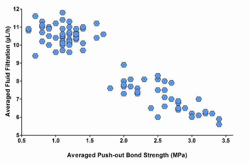
Correlation between outcomes
Analysis of correlation between the averaged leakage values between the two
observation times and averaged push-out bond strength values per root showed very high
negative correlation (Spearman’s rank correlation coefficient rho = -0.83, P < 0.001, Fig. 1).
This means that treatments that resulted in low leakage (Table 1) also caused the highest
9
bond strength to dentine (Table 2).
Discussion
The current study showed a significant impact on the chemicals contained in irrigating
solutions on both, sealing ability assessed by the fluid transport method and bond strength to
dentine in a push-out test of an epoxy resin sealer. Furthermore, sealing ability and bond
strength strongly correlated with each other. Consequently, the three null hypotheses tested
were rejected.
To the best of our knowledge, this study would be the first to correlate sealing ability
with bonding to dentine of a root canal sealer. Hitherto, this correlation has been established
for adhesive cements used for fiber-post bonding (Zicari et al. 2008). As indicated above, it
still remains questionable whether either of these two outcome variables, whilst being
connected to each other for the epoxy resin material under investigation, is related to clinical
outcomes. The endodontic treatment goal remains to prevent oral pathogens from colonizing
and re-infecting the root and periapical tissues and to thereby maintain long-term periapical
health. To this end, it is questionable whether bonding to dentine is truly necessary for a root
filling material. Silicone-based materials, for instance, have good sealing ability (De-Deus et
al. 2007), but no bonding capacity. However, at least from a clinical standpoint, some
adhesion of the root filling is desirable. It is a commonly observed problem with silicone-
based sealers that the whole root filling is removed during the attempt to drill a post space,
for instance.
The current study produced some further results that merit mentioning. First and
foremost, application of a decalcifying agent improved bond strength and sealing ability of
the epoxy resin root canal sealer under investigation – AH Plus. There is but one logical
explanation for this result: AH Plus is able to bond to the organic phase of the root dentine,
10
most likely in the collagen network. However, this effect was abolished when sodium
hypochlorite was applied subsequently as the final irrigating solution. The point is that a final
NaOCl flush after EDTA has been recommended based on the appearance of root canal walls
after cleaning and shaping and final irrigation (Yamada et al. 1983, Baumgartner & Mader
1987). However, according to the present observations, this irrigation regimen should be
thought over, at least if an epoxy resin-based sealer is used. A final sodium hypochlorite flush
has two effects, which may be responsible for the decrease in epoxy resin bond strength to
dentine and the thus-resulting increase in leakage. First, sodium hypochlorite will remove
organic material from the exposed dentine surface (Marending et al. 2007). Second, sodium
hypochlorite breaks down to sodium chloride and oxygen. Oxygen causes strong inhibition of
the interfacial polymerization of methacrylate resins (Munksgaard et al. 1985). The
polyaddition reaction during the curing of epoxy resins, however, is not affected by oxygen.
On the other hand, the generation of oxygen bubbles at the resin-dentine interface may
directly interfere with resin infiltration into the tubules and inter-tubular dentine (Rueggeberg
& Margeson 1990). Nonetheless, it should be mentioned that using NaOCl during
instrumentation and as a flush before application of a decalcifying agent actually had a
desired effect on sealing ability (Table 1) and push-out bond strength (Table 2) of AH Plus.
The reason for this positive effect may be that NaOCl can remove loose organic remnants
from the canal system, which may interfere with sealer bonding to the canal wall.
Maleic acid, an organic acid contained in dentine bonding agents such as Syntac
Classic (Vivadent, Schaan, Liechtenstein), appears to provide higher bond strength than
EDTA. Based on observations of trans-sectioned roots after different irrigation protocols, it
would appear that the complete demineralization of the exposed dentine wall caused by
EDTA is less ideal for bonding than the structure created by a final application of maleic
acid. Maleic acid is a weak acid, and it is likely that it will affect the dentine similarly to
11
other organic acids such as acetic or lactic acid: it may cause a mineral gradient in the
exposed dentine rather than the complete surface demineralization observed with strong acids
such as phosphoric acid or strong chelators such as EDTA (Kawasaki et al. 2000). However,
this needs to be investigated further. A recent study showed that maleic acid reduced the
microhardness of root dentin similar to EDTA but was able to increase dentine surface
roughness markedly more than EDTA (Ballal et al. 2010). Thus, the potential relationship
between surface roughness, bond-strength and sealing ability deserves further investigation.
Chlorhexidine in a concentration of 2% has been recommended as a final
antimicrobial rinse for canal disinfection because of its sustained antimicrobial activity
(White et al. 1999), and the fact that chlorhexidine can enhance the durability of resin-dentine
bonds (Carrilho et al. 2007). Whilst we did not find any positive effect of chlorhexidine on
the bond of the epoxy resin material to dentine, there was no negative effect either.
Chlorhexidine resulted in leakage and push-out bond strength values that were almost
identical to those obtained with the water control treatment.
Another notable finding regarding bond strength was that this value decreased in a
coronal to apical direction. The explanation for this may be that apical dentine contains less
patent tubules than coronal dentine (Paque et al. 2006, Lottanti et al. 2009). The more
complex structure of tubular dentine apparently yields itself better to infiltration with epoxy
resin compared to the sclerotic apical counterpart. However, this does not necessarily mean
that sclerotic dentine cannot be sealed as well as tubular dentine. Most likely, the opposite is
the case (unpublished observations). What the current results show is that overall (averaged)
bonding to dentine had an effect on fluid transport. It was not differentiated between different
root thirds and dentine structure in this regard.
A recent study found similar effects of irrigating protocols on AH Plus push-out bond
strength in root canals (Hashem et al. 2009). However, in contrast to the current study,
12
individual chemicals were not singled out in that study. Furthermore, these authors filled
canals with gutta-percha and AH Plus. Consequently, it remains unclear whether the failure
modes in their push-out test were adhesive, i.e. between sealer and dentine, or cohesive, i.e.
between the sealer and the core material. In the current study, only AH Plus sealer was used
to fill the entire root canal space. This does not introduce bias, because AH Plus does not
shrink. In fact, AH Plus expands over time in a humid environment (Orstavik et al. 2001).
This can explain why leakage values decreased over time. Filling the canals just with the
epoxy resin material had the advantage that all the failures were adhesive, and thus only the
sealer/dentine interface was studied rather than anything else. Clinically, however, filling
with mere AH Plus is not advisable, because the material sets hard as a rock, and thus makes
retreating almost impossible. The current results are in accordance with a published report on
push-out bond strength of sealers to dentine without a core material in that the bond strength
of AH Plus to dentine treated with NaOCl followed by EDTA were higher than specimens
treated with NaOCl or distilled water (Nunes et al. 2008).
Future studies should look at the effect of alternative irrigating protocols on leakage
and dentine bond strength in conjunction with different root filling systems. From a
microbiological point of view, it would make sense to flush a strong disinfectant such as
NaOCl in the canal system prior to root filling. However, extended exposure to NaOCl may
deproteinize dentine to a point that cannot be reversed by the subsequent application of a
decalcifying agent.
Conclusions
• Chemical treatment of dentine with commonly used irrigants had a significant impact
on sealing ability and dentine bond strength of AH Plus;
13
• a final flush with a decalcifying agent appears advisable, whilst a final flush with
NaOCl caused untoward effects;
• conditioning of canal walls by maleic acid, which is a weak acid, resulted in superior
sealing ability and higher epoxy resin bond strength compared to EDTA, which is a
strong calcium-complexing agent;
• the two outcomes investigated here, fluid transport and dentine bond strength, were
strongly negatively correlated to each other.
References:
Ballal NV, Mala K, Bhat KS (2010) Evaluation of the effect of maleic acid and
ethylenediaminetetraacetic Acid on the microhardness and surface roughness of human
root canal dentin. Journal of Endodontics 36, 1385-8.
Baumgartner JC, Mader CL (1987) A scanning electron microscopic evaluation of four root
canal irrigation regimens. Journal of Endodontics 13, 147-57.
Carrilho MR, Carvalho RM, de Goes MF, di Hipolito V, Geraldeli S, Tay FR, Pashley DH,
Tjaderhane L (2007) Chlorhexidine preserves dentin bond in vitro. Journal of Dental
Research 86, 90-4.
De-Deus G, Brandao MC, Fidel RA, Fidel SR (2007) The sealing ability of GuttaFlow in
oval-shaped canals: an ex vivo study using a polymicrobial leakage model.
International Endodontic Journal 40, 794-9.
De-Deus G, Di Giorgi K, Fidel S, Fidel RA, Paciornik S (2009) Push-out bond strength of
Resilon/Epiphany and Resilon/Epiphany self-etch to root dentin. Journal of
Endodontics 35, 1048-50.
14
De-Deus G, Namen F, Galan JJ, Zehnder M (2008) Soft chelating irrigation protocol
optimizes bonding quality of Resilon/Epiphany root fillings. Journal of Endodontics 34,
703-5.
Fisher MA, Berzins DW, Bahcall JK (2007) An in vitro comparison of bond strength of
various obturation materials to root canal dentin using a push-out test design. Journal of
Endodontics 33, 856-8.
Gesi A, Raffaelli O, Goracci C, Pashley DH, Tay FR, Ferrari M (2005) Interfacial strength of
Resilon and gutta-percha to intraradicular dentin. Journal of Endodontics 31, 809-13.
Grossman LI (1976) Physical properties of root canal cements. Journal of Endodontics 2,
166-75.
Hashem AA, Ghoneim AG, Lutfy RA, Fouda MY (2009) The effect of different irrigating
solutions on bond strength of two root canal-filling systems. Journal of Endodontics 35,
537-40.
Hebling J, Pashley DH, Tjaderhane L, Tay FR (2005) Chlorhexidine arrests subclinical
degradation of dentin hybrid layers in vivo. Journal of Dental Research 84, 741-6.
Kawasaki K, Ruben J, Tsuda H, Huysmans MC, Takagi O (2000) Relationship between
mineral distributions in dentine lesions and subsequent remineralization in vitro. Caries
Research 34, 395-403.
Kececi AD, Kaya BU, Belli S (2010) Corono-apical leakage of various root filling materials
using two different penetration models--a 3-month study. Journal of Biomedical
Materials Research B: Applied Biomaterials 92, 261-7.
Lee KW, Williams MC, Camps JJ, Pashley DH (2002) Adhesion of endodontic sealers to
dentin and gutta-percha. Journal of Endodontics 28, 684-8.
15
Lottanti S, Gautschi H, Sener B, Zehnder M (2009) Effects of ethylenediaminetetraacetic,
etidronic and peracetic acid irrigation on human root dentine and the smear layer.
International Endodontic Journal 42, 335-43.
Mai S, Kim YK, Arola DD, Gu LS, Kim JR, Pashley DH, Tay FR (2010) Differential
aggressiveness of ethylenediamine tetraacetic acid in causing canal wall erosion in the
presence of sodium hypochlorite. Journal of Dentistry 38, 201-6.
Marending M, Luder HU, Brunner TJ, Knecht S, Stark WJ, Zehnder M (2007) Effect of
sodium hypochlorite on human root dentine--mechanical, chemical and structural
evaluation. International Endodontic Journal 40, 786-93.
Munksgaard EC, Irie M, Asmussen E (1985) Dentin-polymer bond promoted by Gluma and
various resins. Journal of Dental Research 64, 1409-11.
Nagas E, Cehreli ZC, Durmaz V, Vallittu PK, Lassila LV (2007) Regional push-out bond
strength and coronal microleakage of Resilon after different light-curing methods.
Journal of Endodontics 33, 1464-8.
Nunes VH, Silva RG, Alfredo E, Sousa-Neto MD, Silva-Sousa YT (2008) Adhesion of
Epiphany and AH Plus sealers to human root dentin treated with different solutions.
Brazilian Dental Journal 19, 46-50.
Orstavik D, Nordahl I, Tibballs JE (2001) Dimensional change following setting of root canal
sealer materials. Dental Materials 17, 512-9.
Paque F, Luder HU, Sener B, Zehnder M (2006) Tubular sclerosis rather than the smear layer
impedes dye penetration into the dentine of endodontically instrumented root canals.
International Endodontic Journal 39, 18-25.
Pinna L, Loushine RJ, Bishop FDJ, Cotti E, Weller RN, Pashley DH, Tay FR (2009) Hybrid
Root SEAL (MetaSEAL) creates hybrid layers in radicular dentin only when EDTA is
used as the final rinse. American Journal of Dentistry 22, 299-303.
16
Prati C, Biagini G, Nucci C, Castaldini C, Zucchini C (1990) Effects of chemical
pretreatments on dentin bonding. American Journal of Dentistry 3, 199-206.
Rueggeberg FA, Margeson DH (1990) The effect of oxygen inhibition on an unfilled/filled
composite system. Journal of Dental Research 69, 1652-8.
Schwartz RS (2006) Adhesive dentistry and endodontics. Part 2: bonding in the root canal
system-the promise and the problems: a review. Journal of Endodontics 32, 1125-34.
Souza SF, Bombana AC, Francci C, Goncalves F, Castellan C, Braga RR (2009)
Polymerization stress, flow and dentine bond strength of two resin-based root canal
sealers. International Endodontic Journal 42, 867-73.
White RR, Janer LR, Hays GL (1999) Residual antimicrobial activity associated with a
chlorhexidine endodontic irrigant used with sodium hypochlorite. American Journal of
Dentistry 12, 148-50.
Yamada RS, Armas A, Goldman M, Lin PS (1983) A scanning electron microscopic
comparison of a high volume final flush with several irrigating solutions: Part 3.
Journal of Endodontics 9, 137-42.
Zhang K, Kim YK, Cadenaro M, Bryan TE, Sidow SJ, Loushine RJ, Ling JQ, Pashley DH,
Tay FR (2010) Effects of different exposure times and concentrations of sodium
hypochlorite/ethylenediaminetetraacetic acid on the structural integrity of mineralized
dentin. Journal of Endodontics 36, 105-9.
Zicari F, Couthino E, De Munck J, Poitevin A, Scotti R, Naert I, Van Meerbeek B (2008)
Bonding effectiveness and sealing ability of fiber-post bonding. Dental Materials 24,
967-77.
17
Figure caption
Figure 1 Bivariate scatter plot depicting the correlation between fluid transport (averaged
between day 3 and 30) and push-out bond strength (averaged per tooth). There was a high
negative rank correlation between the two measurements (Spearman’s rho = -0.83).
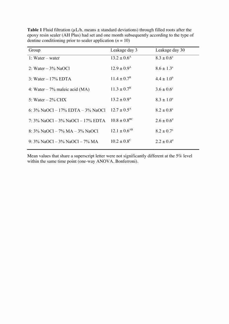
Table 1 Fluid filtration (µL/h, means ± standard deviations) through filled roots after the epoxy resin sealer (AH Plus) had set and one month subsequently according to the type of dentine conditioning prior to sealer application (n = 10)
Group Leakage day 3 Leakage day 30
1: Water – water 13.2 ± 0.6A 8.3 ± 0.6a
2: Water – 3% NaOCl 12.9 ± 0.9A 8.6 ± 1.3a
3: Water – 17% EDTA 11.4 ± 0.7B 4.4 ± 1.0b
4: Water – 7% maleic acid (MA) 11.3 ± 0.7B 3.6 ± 0.6c
5: Water – 2% CHX 13.2 ± 0.9A 8.3 ± 1.0a
6: 3% NaOCl – 17% EDTA – 3% NaOCl 12.7 ± 0.5A 8.2 ± 0.8a
7: 3% NaOCl – 3% NaOCl – 17% EDTA 10.8 ± 0.8BC 2.6 ± 0.6d
8: 3% NaOCl – 7% MA – 3% NaOCl 12.1 ± 0.6AB 8.2 ± 0.7a
9: 3% NaOCl – 3% NaOCl – 7% MA 10.2 ± 0.8C 2.2 ± 0.4d
Mean values that share a superscript letter were not significantly different at the 5% level within the same time point (one-way ANOVA, Bonferroni).
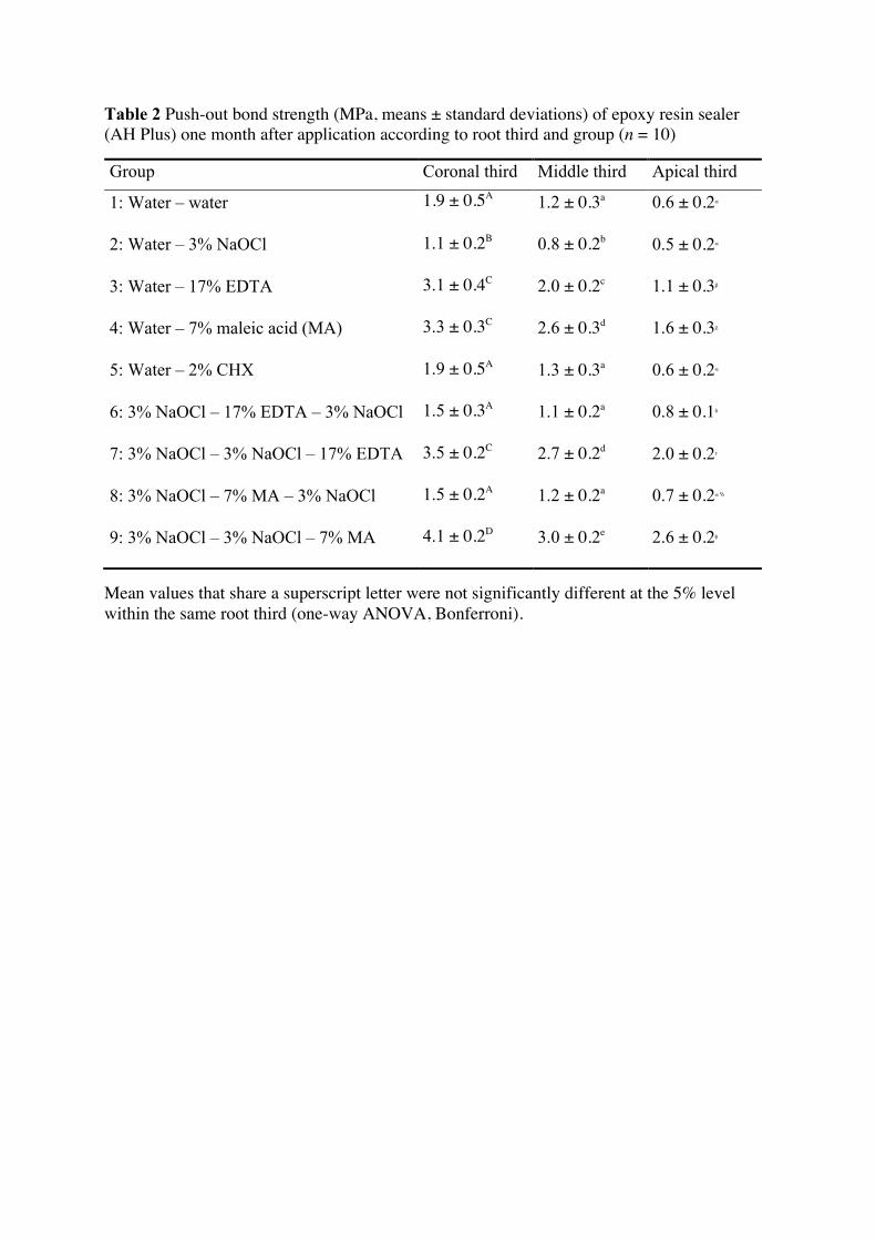
Table 2 Push-out bond strength (MPa, means ± standard deviations) of epoxy resin sealer (AH Plus) one month after application according to root third and group (n = 10)
Group Coronal third Middle third Apical third
1: Water – water 1.9 ± 0.5A 1.2 ± 0.3a 0.6 ± 0.2α
2: Water – 3% NaOCl 1.1 ± 0.2B 0.8 ± 0.2b 0.5 ± 0.2α
3: Water – 17% EDTA 3.1 ± 0.4C 2.0 ± 0.2c 1.1 ± 0.3β
4: Water – 7% maleic acid (MA) 3.3 ± 0.3C 2.6 ± 0.3d 1.6 ± 0.3χ
5: Water – 2% CHX 1.9 ± 0.5A 1.3 ± 0.3a 0.6 ± 0.2α
6: 3% NaOCl – 17% EDTA – 3% NaOCl 1.5 ± 0.3A 1.1 ± 0.2a 0.8 ± 0.1δ
7: 3% NaOCl – 3% NaOCl – 17% EDTA 3.5 ± 0.2C 2.7 ± 0.2d 2.0 ± 0.2ε
8: 3% NaOCl – 7% MA – 3% NaOCl 1.5 ± 0.2A 1.2 ± 0.2a 0.7 ± 0.2α,δ
9: 3% NaOCl – 3% NaOCl – 7% MA 4.1 ± 0.2D 3.0 ± 0.2e 2.6 ± 0.2φ
Mean values that share a superscript letter were not significantly different at the 5% level within the same root third (one-way ANOVA, Bonferroni).


![[ 189 ] A NOTE ON THE INNERVATION OF HUMAN DENTINE](https://img.pdfslide.us/doc/110x75/586cd50a1a28ab3f528b8b42/-189-a-note-on-the-innervation-of-human-dentine.jpg)