Embed Size (px)
Citation preview
SW
ED
ISH
DE
NTA
L JO
UR
NA
L, S
UP
PL
EM
EN
T 2
33
, 20
14
. DO
CT
OR
AL
DIS
SE
RTA
TIO
N IN
OD
ON
TO
LOG
Y
AN
DE
RS
HE
DE
NB
JÖR
K LA
GE
R
MA
LMÖ
UN
IVE
RS
ITY
20
14
MALMÖ UNIVERSITY
205 06 MALMÖ, SWEDEN
WWW.MAH.SE
DENTINE CARIES: ACID-TOLERANT MICRO-ORGANISMS AND ASPECTS ON COLLAGEN DEGRADATION
isbn 978-91-7104-398-6 (print)
isbn 978-91-7104-397-9 (pdf)
issn 0348-6672
DE
NTIN
E C
AR
IES
: AC
ID-TO
LER
AN
T MIC
RO
OR
GA
NIS
MS
AN
D A
SP
EC
TS O
N C
OLLA
GE
N D
EG
RA
DA
TION
Swedish Dental Journal, Supplement 233, 2014

D E N T I N E C A R I E S : A C I D - T O L E R A N T M I C R O O R G A N I S M S A N D A S P E C T S O N C O L L A G E N D E G R A D A T I O N
Swedish Dental Journal Supplement 233, 2014
© Anders Hedenbjörk Lager 2014
All previously published papers were reproduced with permission from the publishers:
S. Karger AG (Caries Research, I and III) and Quintessence Publishing (II)
Cover photo: Anders Hedenbjörk Lager
ISBN 978-91-7104-398-6 (print)
ISBN 978-91-7104-397-9 (PDF)
ISSN 0348-6672
Holmbergs, Malmö 2014
ANDERS HEDENBJÖRK LAGER DENTINE CARIES: ACID-TOLERANT MICRO-ORGANISMS AND ASPECTS ON COLLAGEN DEGRADATION
Malmö University, 2014Department of Cariology
Faculty of OdontologyMalmö, Sweden

The summary of this publication can be downloaded from:www.mah.se/muep

I dedicate this book to my family, and especially to my father,
who passed away way too early.

CONTENTS
ABSTRACT .................................................................... 9
POPULÄRVETENSKAPLIG SAMMANFATTNING .................. 12 (Summary in Swedish)
PREFACE ..................................................................... 15
ABBREVIATIONS AND DEFINITIONS ............................... 17
INTRODUCTION .......................................................... 18Dental caries ........................................................................18Dentine caries ......................................................................20Physiological and chemical aspects of the dentine caries lesion ...24Enzymes in dentine caries......................................................26Final remark ........................................................................33
AIMS ......................................................................... 34
HYPOTHESES .............................................................. 35
MATERIALS AND METHODS ........................................... 36Microorganisms in dentine caries ...........................................36Clinical procedures ...............................................................36Proteolytic enzyme activity in dentine caries .............................41MMPs in saliva (Paper III) ......................................................41MMPs in dentine (Paper IV) ...................................................43
RESULTS ..................................................................... 49Paper I ................................................................................49Paper II ...............................................................................51Paper III ..............................................................................55Paper IV ..............................................................................59
DISCUSSION ............................................................... 63Bacteria in dentine caries ......................................................63MMPs and dentine caries ......................................................69
CONCLUSIONS AND DIRECTIONS FOR FUTURE WORK ..... 77
ACKNOWLEDGEMENTS ............................................... 80
REFERENCES ............................................................... 83
PAPERS I – IV ............................................................... 95
9
ABSTRACT
Dental caries is a common disease all over the world, despite the fact that it can be both effectively prevented and treated. It is driven by acids produced by oral microorganisms as a consequence of their metabolism of dietary carbohydrates. Given enough acid challenge, eventually the tooth enamel barrier will be broken down, and the carious lesion will extend into underlying hard tissue, forming a macroscopic cavity in the dentine. In comparison to biofilm on enamel, a dentine carious lesion provides a vastly different environment for the residing microorganisms. The environment influences the types and numbers of microorganisms that can colonize the dentine caries lesion.
The overall aims for this thesis are to enumerate and further study microorganisms found in established dentine caries lesions and also to illuminate how host-derived proteolytic enzymes might contribute to this degradation, not only to better understand the caries process in dentine but also to find incitements for new methods to influence the natural progression of caries lesions.
In Paper I, the numbers of remaining viable microorganisms after completed excavation using two excavation methods were investigated. Samples of carious dentine tissue were collected before and after excavation and cultivated on different agar media in different atmospheres. Analysis was performed by counting the number of colony-forming units (CFUs).
10
Key findings: The number of remaining microorganisms after excavation was low for both methods, but some microorganisms always remained in the cavity floors even when the cavities were judged as caries free using normal clinical criteria.
In Paper II, the acid tolerant microbiota in established dentine caries lesions was investigated. Samples were taken as in Paper I, but on three levels (superficial, center of lesion, floor of lesion after completed excavation). The samples were cultivated in anaerobic conditions on solid pH-selective agar media of different acidity.
Key findings: Each investigated lesion harbored a unique microbiota in terms of both species composition and numbers of microorganisms. This indicates that various combinations of aciduric microorganisms can colonize, survive in and probably also propagate dentine carious lesions. We also found that solid pH-selective agars can be used successfully to select acid-tolerant microorganisms in caries lesions. This would preserve their phenotypic traits for further study.
In Paper III, the relation between salivary levels of matrix metalloproteinase-8 (MMP-8), salivary levels of tissue inhibitor of MMP (TIMP-1), and the presence of manifest caries lesions in a large number of subjects was investigated. Saliva samples were collected and analyzed for concentrations of MMP-8, TIMP-1 and total protein using immunofluorometric assays, enzyme linked immunosorbent assays and Bradford assays, respectively.
Key findings: Subjects with manifest caries lesions had significantly elevated levels of salivary MMP-8 compared to subjects without caries lesions. TIMP-1 was not significant in any case.
In Paper IV, a new method for generating bioactive demineralized dentine matrix substrate (DDM) was developed using a dialysis system and two different demineralization approaches (acetic acid or EDTA). The generated DDM was subsequently analyzed for the presence of type 1 collagen, active MMP-8 and hydroxyproline (HYP) levels using SDS-PAGE, ELISA or immunofluorescence assay.
11
Key findings: Both demineralization methods produced a substrate rich in collagen and with preserved MMP-8 activity.
This report presents new knowledge on the composition of the acid tolerant dentine caries microbiota from three levels in dentine carious lesions and on the efficacy of operative caries removal on the numbers of viable microorganisms in the caries free cavity using two operative methods. Moreover, the basic mechanisms behind collagen degradation in the dentine caries process are studied from both a clinical and laboratory perspective.
The report also provides a reference for further studies on dentine caries microbiology and dentine caries collagen degradation mechanisms, both of which are known only in part.
12
POPULÄRVETENSKAPLIG SAMMANFATTNING (Summary in Swedish)
Karies är en stor folksjukdom, trots att den både kan förebyggas och behandlas effektivt. Om man inte ingriper preventivt utan låter det naturliga förloppet råda, kommer kariesangreppet till slut att bryta igenom emaljen och involvera även den underliggande dentinvävnaden.
Kariessjukdomen orsakas av orala mikroorganismer, som en konsekvens av dessas nedbrytning av kostrelaterade kolhydrater. Som en biprodukt bildas då syror som löser upp (demineraliserar) tandvävnaden, så att synliga hål till slut bildas.
Syran kan lösa upp mineralfasen i tanden, men dentin består även till stor del av kollagen, vilket inte kan lösas upp av enbart syror. Man hänförde länge detta till proteinnedbrytande bakterier, men det har visat sig att munhålebakterierna inte har förmågan att lösa upp kollagen, och man tror nu att denna nedbrytning sker med hjälp av kroppsegna enzymer, bland annat matrix metalloproteinaser (MMP). De biologiska mekanismerna bakom kollagennedbrytning vid dentinkaries är emellertid dåligt undersökta, och delar av denna rapport (Studie III och IV) inriktar sig därför på detta område.
Syran som bakterierna bildar skapar också en sur närmiljö för dem själva, vilket gör det svårt för dem att överleva, särskilt i ett begränsat utrymme som ett kariesangrepp. Man har länge ansett
13
att endast vissa specifika bakterier har förmågan att leva och trivas i sura miljöer, men nya studier har ifrågasatt detta. I denna rapport (Studie II) undersöks även förekomsten av syratåliga bakterier på olika nivåer i dentinkariesangrepp med hjälp av en ny metod.
Målet vid avlägsnande av karies är att ta bort fullständigt förstörd tandvävnad, men också att försöka spara så mycket som möjligt av den delvis skadade vävnaden, vilken kan återställas. Detta har också aktualiserats då nya operativa principer och material lanserats under den senaste tioårsperioden. Det är emellertid svårt att avgöra var gränsen går kliniskt, och ett sätt att mäta dentinets ”friskhet” kan vara att mäta antalet bakterier i vävnaden. I den första rapporten (Studie I) undersöks den kvarvarande bakterieförekomsten efter kariesborttagning med två olika operativa metoder.
Avhandlingen söker svar på följande frågeställningar:
Studie I. Finns det några skillnader vad gäller antal kvarvarande bakterier efter dentinkariesavlägsnande med mekanisk (vanligt borr) eller kemo-mekanisk (Carisolv) metod?
Studie II. Hur ser sammansättningen av den syratåliga bakteriefloran ut på tre olika nivåer i olika dentinkariesangrepp?
Studie III. Finns det något samband mellan förekomsten av etablerade kariesangrepp och nivåerna av enzymet MMP-8 och dess nedreglerande protein TIMP-1 i saliv?
Studie IV. Kan man framställa demineraliserat dentinmatrix med bibehållen biologisk aktivitet inför framtida studier av mekanismerna bakom nedbrytning av kollagen vid dentinkaries? Vad händer spontant med detta demineraliserade dentinmatrix över tid?
Huvudfynden i studierna är:
1. Båda metoderna för att avlägsna dentinkaries minskade bakterieantalet radikalt. Det fanns dock alltid kvar små mängder av bakterier i botten av kaviteten.
2. Alla de undersökta kaviteterna hade en unik sammansättning av syratåliga bakterier, både till typ och till antal, vilket indikerar att ett flertal olika bakteriearter har förmågan att anpassa sig till sura
14
miljöer och potentiellt bidra till kariesutvecklingen. Vidare fungerade de pH-specifika odlingsmedierna väl för att få fram de syratåliga bakterierna, något som är svårt med konventionella metoder.
3. Försökspersoner med etablerade dentinkariesangrepp uppvisade mycket högre förekomst av MMP-8 i saliven i jämförelse med kariesfria försökspersoner. Det MMP-8 hämmande proteinet TIMP-1 uppvisade inget liknande samband.
4. Dentinmatrix framställt med båda testmetoderna uppvisade förekomst av intakt kollagen samt aktivt MMP-8. Vidare så uppvisades en spontan nedbrytning av kollagen över tid, vilket tolkades som ett resultat i huvudsak beroende på det aktiva MMP-8 enzymet.
De nyvunna grundkunskaperna bildar underlag för nya studier inom forskningsområdet, samt för nya behandlingsmetoder, framför allt sådana som skulle kunna moderera eller förhindra dentinkariesprogression.
15
PREFACE
This thesis is based on the following papers, which are referred to in the text by their roman numerals:
I. Lager A, Thornqvist E, Ericson D. Cultivatable bacteria in dentine after caries excavation using rose-bur or Carisolv. Caries Research 2003;37:206-211.
II. Hedenbjörk-Lager A, Ericson D. Aciduric bacterial communities at three levels in dentine caries. Oral Health & Preventive Dentistry 2013;11(4):359-67. doi: 10.3290/j.ohpd.a30483.
III. Hedenbjörk-Lager A, Bjørndal L, Gustafsson A, Sorsa T, Tjäderhane L, Åkerman S, Ericson D. Caries correlates strongly to salivary levels of MMP-8. Caries Research 2015;49:1-8. doi: 10.1159/0000360625. E-pub ahead of print.
IV. Hedenbjörk-Lager A, Hamberg K, Pääkkönen V, Tjäderhane L, Ericson D. Collagen degradation in human dentine matrix after demineralization using EDTA or acetic acid. 2014. Manuscript.
All previously published papers were reproduced with permission from the publishers: S. Karger AG (Papers I and III) and Quintessence Publishing (Paper II).
16
THES
IS A
T A
GLA
NC
E ! Stu
dy
Obje
ctives
Meth
ods
Main
fin
din
gs/C
onclu
sio
ns
(I)
Cu
ltiv
ata
ble
bacte
ria
in
den
tin
e
aft
er c
arie
s e
xcavati
on
usin
g
ro
se-b
ur o
r C
aris
olv
.
To
dete
rm
ine t
he n
um
ber o
f resid
ual
via
ble
bacte
ria
in
den
tin
e a
fter
ex
cavati
on
usin
g e
ith
er c
on
ven
tio
nal
ro
se-b
ur o
r c
hem
o-m
ech
an
ical
ex
cavati
on
.
Den
tin
e s
am
ple
s (
22
lesio
ns)
fro
m b
efo
re a
nd
aft
er e
xcavati
on
were a
naly
sed
fo
r c
fu u
sin
g
dif
feren
t agar m
ed
ia a
nd
cu
ltiv
ati
on
meth
od
s.
Ch
em
o-m
ech
an
ical
ex
cavati
on
was o
n a
par w
ith
co
nven
tio
nal
dril
lin
g f
or r
ed
ucin
g n
um
bers o
f cfu
in
cario
us d
en
tin
e.
So
me c
fu w
ill
alw
ays r
em
ain
in
cli
nic
all
y c
arie
s-f
ree d
en
tin
e.
(II)
Acid
uric
bacte
ria
l co
mm
un
itie
s a
t
three l
evels
in
den
tin
e c
arie
s.
To
assess t
he a
cid
to
leran
t m
icro
bio
ta
at
dif
feren
t le
vels
in
esta
bli
sh
ed
den
tin
e c
arie
s l
esio
ns u
sin
g s
oli
d p
H-
sele
cti
ve m
ed
ia.
Prim
ary d
en
tin
e c
arie
s l
esio
ns (
10
su
bje
cts
)
were s
am
ple
d a
t 3
levels
. Sam
ple
s w
ere
incu
bate
d o
n n
eu
tral
an
d p
H-s
ele
cti
ve a
gars.
Cfu
co
un
ts w
ere d
ete
rm
ined
an
d c
olo
nie
s
ch
aracte
ris
ed
, is
ola
ted
an
d f
urth
er a
naly
sed
.
Each
den
tin
e c
arie
s l
esio
n h
arb
ou
red
a u
niq
ue
mic
ro
bia
l fl
ora.
So
lid
pH
-sele
cti
ve a
gars c
an
be u
sed
to s
ele
ct
acid
-to
leran
t m
icro
organ
ism
s i
n d
en
tin
e
carie
s l
esio
ns.
(III
)
Carie
s c
orrela
tes s
tro
ngly
to
sali
vary l
evels
of
MM
P-8
.
To
rela
te s
ali
vary M
MP
-8 a
nd
TIM
P-
1 l
evels
wit
h m
an
ifest
carie
s i
n a
large
nu
mb
er o
f su
bje
cts
.
Sali
vary a
nd
cli
nic
al
data
was c
oll
ecte
d f
ro
m
45
1 r
an
do
m a
du
lts,
an
d a
naly
sed
fo
r M
MP
-8,
TIM
P-1
an
d t
ota
l p
ro
tein
usin
g
imm
un
ofl
uo
ro
metr
ic a
ssay,
en
zym
e l
ink
ed
imm
un
oso
rb
en
t assay a
nd
Brad
ford
assay.
Su
bje
cts
wit
h m
an
ifest
carie
s l
esio
ns p
resen
ted
sig
nif
ican
tly h
igh
er l
evels
of
sali
vary M
MP
rela
tive
to s
ub
ject
wit
h n
o c
arie
s l
esio
ns.
TIM
P-1
did
no
t
dem
on
str
ate
an
y s
imil
ar p
att
ern
.
(IV
)
Co
llagen
degrad
ati
on
in
hu
man
den
tin
e m
atr
ix a
fter
dem
inerali
zati
on
usin
g E
DT
A o
r
aceti
c a
cid
.
To
develo
p a
meth
od
fo
r g
en
erati
ng
dem
inerali
zed
den
tin
e m
atr
ix
su
bstr
ate
, w
ith
ou
t lo
sin
g o
r d
estr
oyin
g
the b
ioacti
ve p
ro
tein
s,
or i
nte
rfe
ren
ce
wit
h t
he a
ssays u
sed
fo
r a
ssessm
en
t o
f
MM
P-8
an
d H
YP
.
Po
ole
d h
um
an
den
tin
e p
ow
der w
as
dem
inerali
zed
in
a d
ialy
sis
syste
m u
sin
g
ED
TA
or a
ceti
c a
cid
. T
he d
em
inerali
zed
den
tin
e m
atr
ix w
as t
hen
an
aly
zed
fo
r M
MP
-8
acti
vit
y a
nd
co
llagen
degrad
ati
on
(H
YP
) aft
er
su
bseq
uen
t b
uff
er t
reatm
en
t.
It i
s p
ossib
le t
o d
em
inerali
ze r
ela
tively
large a
mo
un
ts
of
den
tin
e p
ow
der w
ith
th
e m
eth
od
s u
sed
, w
hil
e
keep
ing M
MP
-8 b
ioacti
vit
y.
If E
DT
A
dem
inerali
zati
on
is u
sed
, T
ESC
A b
uff
er t
reatm
en
t
seem
s t
o b
e b
en
efi
cia
l fo
r M
MP
-8 a
cti
vit
y.
!
17
ABBREVIATIONS AND DEFINITIONS
16S rRNA 16S ribosomal nucleic acidAA acetic acidBA brain heart infusion agarBOP bleeding on probingCFU colony forming unitDDM demineralized dentine matrixDL detection limitDMFT decayed/missing/filled teethDNA deoxyribonucleic acidECM extracellular matrixEDJ enamel-dentine junctionEDTA ethylenediaminetetraacetic acidGCF gingival crevicular fluidMCL manifest caries lesionMMP/-8 matrix metalloproteinase/-8MS mutans streptococciMSA mitis salivarius agarPCR polymerase chain reactionPPD periodontal probing depthproMMP precursor of MMPRTF reduced transport fluidSIP stable isotope probingSL selective lactobacilli agarTIMP-1 tissue inhibitor of matrix metalloproteinase-1TVC total viable count
18
INTRODUCTION
Dental cariesDespite the fact that dental caries can be effectively prevented and treated, the disease is a very common, if not the most common bacterial disease. On a worldwide basis, 60–90% of school children and close to 100% of the adult population suffer from carious cavities (WHO, 2012). The disease also contributes to high economic demands on society. In 2011, the annual cost of dental treatment within the EU was estimated at €79 billion, of which a large part can be expected to be due to dental caries and its sequelae (Rugg-Gunn, 2013).
Dental cavities are formed as a consequence of the release of organic acids from fermentative bacteria normally occupying the oral cavity. This is a result of a disruptive imbalance in the oral ecosystem, often caused by excessive dietary intake of fermentable carbohydrates (Marsh, 2003; Takahashi and Nyvad, 2011).
As a result of the bacterial metabolism of fermentable carbohydrates, organic acids like lactic, acetic and propionic acid are released, causing the pH to drop (Hojo et al., 1991; Takahashi and Nyvad, 2011). As most the bacteria are attached to the teeth, forming the biofilm, these acids accumulate near or on the tooth surfaces and subsequently cause a demineralization of the mineralized part of the tooth substance (Takahashi and Nyvad, 2008). Given enough time, a macroscopic cavity will form. If measures are not taken, the cavity will eventually involve the dentine.
19
Consequences of lowered oral pHIncreased acid production due to an imbalance in the oral ecosystem creates an acidic oral environment, which is also found in dentine caries lesions. Early studies revealed low pH in the deep layers of carious dentine (MacGregor, 1961; Dirksen et al., 1962) and later studies have confirmed a pH of 4.9 to 5.5 in active dentine carious lesions (Kitasako et al., 2002; Kuribayashi et al., 2012). The critical pH of dentine has been estimated at around 6.7 (Hoppenbrouwers et al., 1987), and a newer study on root dentine estimates an even lower critical pH of 5.2–5.7 (Shellis, 2010). Such a low pH has consequences not only for the oral cavity as a whole but also for the microecological environment within the dentine carious lesion. First, the low pH induces a demineralization of the hard tissues, due to a shift in the chemical equilibrium between mineral hydroxyapatite in the teeth and the respective ions in saliva:
Ca10(PO4)6(OH)2+8H+ ⇌ 10Ca2++6HPO2+2H2O
In simple terms, the low pH shifts the equilibrium from left to right, making the ions in the hydroxyapatite disassociate and dissolve in the saliva or plaque fluid. Conversely, increasing the pH reverses the chemical equilibrium, making the ions in saliva or plaque fluid precipitate, a process called remineralization.
Second, it has been suggested that the lowered pH induces a change in the composition of the microbial community, favoring acid tolerant and acid producing microorganisms (Marsh, 2003; Takahashi and Nyvad, 2011). These bacteria are better equipped to deal with low-pH environmental conditions, thus enabling them to expand and occupy a larger part of the microbiota than in a balanced oral ecosystem. The low pH can also cause significant changes in the phenotypic expression of the oral microbiota as the bacteria adapt their activity to the demands of the environment. Very low intraoral pH or prolonged periods of low intraoral pH may also trigger genotypic changes in the oral microbiota (Takahashi and Nyvad, 2011).
20
Third, it has been suggested that the low pH activates endogenous proteolytic enzymes, thus enabling destruction of the otherwise very resilient type 1 collagen fibers in dentine (Tjäderhane et al., 1998).
In this thesis, the dentine caries microbiota found in the lesion, especially those bacteria that can be considered acid-tolerant are studied. Furthermore, the role of endogenous proteolytic enzymes, especially the matrix metalloproteinases, in the dentine caries process is discussed, all with the aim of enabling the eventual discovery of novel ways to influence the natural course of the dentine caries process.
Dentine cariesBy volume, dentine is composed of 50% mineral, 30% organic matrix and 20% water. About 90% of the organic matrix is type 1 collagen that acts as a scaffolding and reinforcement for the mineral (Chaussain-Miller et al., 2006). The caries process in dentine can be thought of as a two-step process involving acid dissolution of the mineral phase of the dental tissue followed by degradation of the dentine collagenous matrix by the action of proteolytic enzymes.
Bacteria in dentine cariesOver the years, several studies have addressed the infecting dentine caries microbiota. Most of them have been based on culturing dentine bacteria on various media. In these studies, bacteria of the genera Streptococcus, Actinomyces, Lactobacillus, Bifidobacterium, Rothia, Arachnia, Eubacterium, Propionibacterium, Veillonella and Prevotella have been isolated (Edwardsson, 1987; Hahn et al., 1991; van Houte, 1994).
More recent culture-independent studies have revealed an even more complex array of bacteria in dentine caries. Using molecular techniques, such as clonal analysis of the 16S rRNA bacterial gene, they have confirmed earlier results and added S. mutans, non-mutans streptococci, Actinomyces, Lactobacillus, Bifidobacterium, Propionibacterium, Veillonella, Selenomonas and Atopobium to the list (Becker et al., 2002; Chhour et al., 2005; Munson et al., 2004; Aas et al., 2008). Later studies have confirmed these results and
21
further confirmed a substantial dominance of lactobacilli in deep dentine caries (Gross et al., 2010; Gross et al., 2012). However, a drawback of 16S rRNA sequencing and similar molecular techniques is that they cannot reveal phenotypic information on the detected taxa (Nyvad et al., 2013).
Dentine caries as a microbial habitatFrom a microecological point of view, the dentine carious lesion forms a complex environment. With limited contact surfaces with the outside world, a carious lesion forms a fairly self-contained ecological habitat. Proposed key microecological determinant factors (or stress factors) include pH, access to nutrition and oxygen concentration (Marquis, 1995; Bowden and Hamilton, 1998; Marsh, 2003; Takahashi and Nyvad, 2011).
Microbial ecological determinantspHThe available knowledge on intralesion pH is limited, but the existing studies indicate a pH of 4.9 to 5.5 in active dentine carious lesions (Kitasako et al., 2002; Kuribayashi et al., 2012). It has been suggested that the major acid production takes place in the surface plaque overlying the lesion (Kidd, 2000; Kidd, 2004). However, nothing is known about how intralesion pH fluctuates, or whether there is a pH gradient going from the superficial layers to the advancing caries front. The quite robust effect of the dentine buffer capacity on this process is also unknown (Haapasalo et al., 2007).
Nevertheless, it is reasonable to hypothesize that the pH would fluctuate analogously to what is seen in plaque biofilm (Bowden and Hamilton, 1998; Kidd, 2004; Chaussain-Miller et al., 2006), as a result of bacterial acid production in response to fermentation of dietary carbohydrate, and that acid tolerance ought to be an important ecological determinant for the dentine caries microbiota (Marchant et al., 2001).
NutrientsThe availability of nutrients in the near vicinity may also be crucial for dentine caries bacteria because they depend on continuous access
22
to sustenance to uphold the metabolic activity (Marsh, 2003). In the superficial layers of the lesion the source of these presumptive nutrients could be saliva (proteins and glycoproteins) or ingested food components.
Even though dietary nutrients might not be able to diffuse to the deeper parts of the lesion bacteria do survive there, as well as under fillings for prolonged periods (Weerheijm and Groen, 1999; Maltz et al., 2012), indicating that there must be alternative sources of nutrients. These sources could plausibly be components from dentinal fluid, degraded dentine or dead bacterial cells. Both saliva and dentine fluid contain many glycosylated proteins (Wiig et al., 2000; Larmas, 2003; Helmerhorst and Oppenheim, 2007), which could be degraded and used by the microbiota. Due to the large variability in glycosylation in host proteins, this would require an extensive collection of complementary enzymes, created by bacterial cooperation, which has been demonstrated in bacteria from dental plaque (Bradshaw et al., 1994; Wickström et al., 2009).
OxygenThe knowledge on the oxygen tension conditions in carious dentine is very limited. However, as dental plaque has for long been considered anaerobic (Marquis, 1995; Bowden and Hamilton, 1998), it stands to reason that this might also be the case in dentine caries. It is also feasible that the oxygen tension increases closer to the cavity surface, but to our knowledge, this has not been demonstrated. Low or fluctuating pH, varying access to nutrients and variation in oxygen tension, coupled with complex interactions with other microorganisms in the vicinity make the dentine caries microenvironment quite challenging for the microbiota. The bacteria can utilize various coping strategies to compensate for these environmental stress factors. However, this coping capacity varies significantly between different taxa (Marquis, 1995; Marsh, 2003). Moreover, it has been shown that organisms lacking a specific coping mechanism can interact or collaborate with other bacteria, which enables them to survive stresses they could not have managed themselves (Marquis, 1995). To our knowledge, the coping strategies of the dentine caries microbial community have not yet been studied.
23
Aciduric bacteria in dentine cariesIt seems reasonable to assume that a pH of around 5 in the immediate environment in the dentine caries lesion would influence the resident microbiota in some way. In plaque, it has been suggested that a low pH results in a shift to a more acid producing as well as acid tolerant microflora, which would push the balance toward demineralization (Takahashi and Nyvad, 2008; Takahashi and Nyvad, 2011). It has previously been reported that non-mutans streptococci are more aciduric when isolated from carious lesions than from caries-free subjects (Sansone et al., 1993; van Houte et al., 1996) and that S. oralis displays several different phenotypes simultaneously within the same plaque sample populations.
In a chemostat model using a few model species, it has also been demonstrated that a pH drop will induce a shift in the microbiota in direct proportion to the level of acidity, toward more acid producing and acid tolerant species (Bradshaw and Marsh, 1998). A more recent study employing stable isotope probing (SIP) on supragingival plaque from children revealed that the number of genera able to metabolize lactate from glucose under acidic conditions (pH 5.5) corresponded very well with those at neutral pH (7.0) but that the diversity of active genera dropped rapidly at pH 4.5, with domination of Lactobacillus spp. and Propionibacterium spp. (McLean et al., 2012).
There are two mechanisms behind this adaptation to acidic conditions in the microbiota. Change of phenotype describes the process by which microorganisms change their activity (i.e. metabolism, protein synthesis, enzymatic profile etc.), or adapt in order to survive changing environmental demands. Change of genotype refers to the process of microbial selection, where certain bacteria prevail over others due to innate genetic traits, in this case acid tolerance or even a preference for acidic environments (Paddick et al., 2005; review by Takahashi and Nyvad, 2011). It should be noted, however, that these acid tolerance studies have been performed on plaque microorganisms, while similar studies using dentine caries bacteria are missing.
24
Physiological and chemical aspects of the dentine caries lesionNew developments in operative dentistry have reactualized the question of how much carious dentine must be removed before restoration (Kidd, 2004; review by Schwendicke et al., 2013). Traditionally, removal of all soft and discolored dentine has been advocated, but this concept has been challenged, and several alternative operative methodologies are currently being studied (review by Ricketts et al., 2013).
Pioneer work by Johansen and Parks (1961) using electron microscopy to study carious dentine concluded that most of the carious dentine retained the same structure even after being demineralized, but that following a “second wave” of destruction the collagenous matrix was also destroyed and replaced by a biomass of bacteria. Later research has shown that the dentine caries lesion can be roughly divided into two distinct zones, the outer and inner carious layer (Kuboki et al., 1977; Fusayama, 1979; Dung and Liu, 1999). The outer layer is completely demineralized, with denatured collagen fibers and large amounts of invading microorganisms; the inner layer is only partly demineralized, with reversibly denatured collagen and very few microorganisms (Ogawa et al., 1983). When excavating a carious lesion, it is only the outer carious layer that needs to be removed. In contrast, the inner layer should be preserved to the largest extent because it can remineralize as well as reorganize, and new collagen cross-links can be established (Shimizu et al., 1981; Beeley et al., 2000).
Operative proceduresThe traditional way to remove carious dentine has been to use rotary instruments: high-speed diamonds to get access to the dentine caries lesion, then spherical burs in the low-speed handpiece to remove the necrotic dentine. Over the last decade, several optional methods have become available, such as chemo-mechanical, laser and air abrasion excavation, (see reviews by Banerjee et al., 2000; Li et al., 2014). The interest in the development of alternative dentine excavation methods has largely been a reaction to the renewed discourse on
25
how much carious dentine needs to be removed or alternatively, how much carious tissue can safely be left after excavation (Kidd, 2004; review by Schwendicke et al., 2013). The foundation for removing all soft and discolored dentine has also been questioned because it is based on rather crude clinical criteria and not in scientific fact (Banerjee et al., 2000; Kidd, 2000). It also heavily emphasizes the operator’s subjective judgment of the status of the carious dentine. Furthermore, it is unclear how these clinically observable parameters relate to the histologic patterns found in dentine caries, and evidence now indicates that removal of all caries-affected dentine is not needed as long as the final restoration provides a sufficient seal toward the oral cavity (Thompson et al., 2008; Maltz et al., 2012; Ricketts et al., 2013). However, more long-term clinical studies are needed to confirm these concepts.
Diagnostics in dentine excavationVarious methods have been proposed to supply the clinician with more objective ways to determine how much of the carious dentine needs to be removed and when to stop excavating, including self-limiting methods such as chemo-mechanical excavation or the use of ceramic burs (Neves et al., 2011; Almahdy et al., 2012). Unfortunately, there is no consensus today on how to clinically determine the endpoint of excavation or even where that endpoint should be (Kidd, 2010), and most clinicians still use visual and sensory input as their main assessment techniques.
Bacteria in dentine as a diagnostic toolOne way to determine the excavation endpoint – or rather, to determine how common clinical criteria such as color, wetness and hardness might be related to level of tissue destruction – could be to assess the bacterial content in the carious dentine after excavation (Kidd et al., 1993; Bjørndal et al., 1997; Lula et al., 2009; Orhan et al., 2008). Studies have shown that even with vigorous excavation, microorganisms will always persist in the clinically caries free cavity floor (Maltz et al., 2011). In Paper I we wanted to ascertain whether the (at the time new) chemo-mechanical excavation method Carisolv was on a par with conventional drilling in removing carious tissue, using bacterial numbers as our measure point.
26
Chemo-mechanical excavationSince Carisolv contains chloramines (0.5% NaOCl), which have an antibacterial action (Kneist and Heinrich-Weltzien, 2001), we wanted to assess whether this would yield any additive effect on the microbiota in dentine caries excavation (Paper I).
Enzymes in dentine cariesDentine has been described as a biologic composite made up of hydroxyapatite and collagen, where the collagen acts as scaffolding for the mineral phase (Tjäderhane et al., 2012). The dentine caries process involves dissolution of the mineral as well as the organic matrix, where demineralization is caused by acids produced by oral microorganisms as a byproduct of their metabolism of fermentable carbohydrates (Takahashi and Nyvad, 2011).
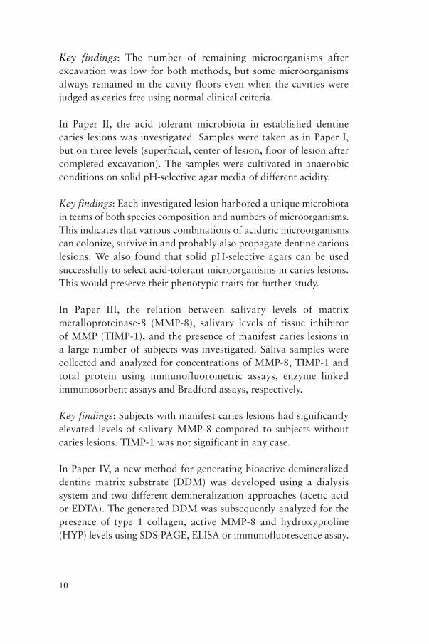
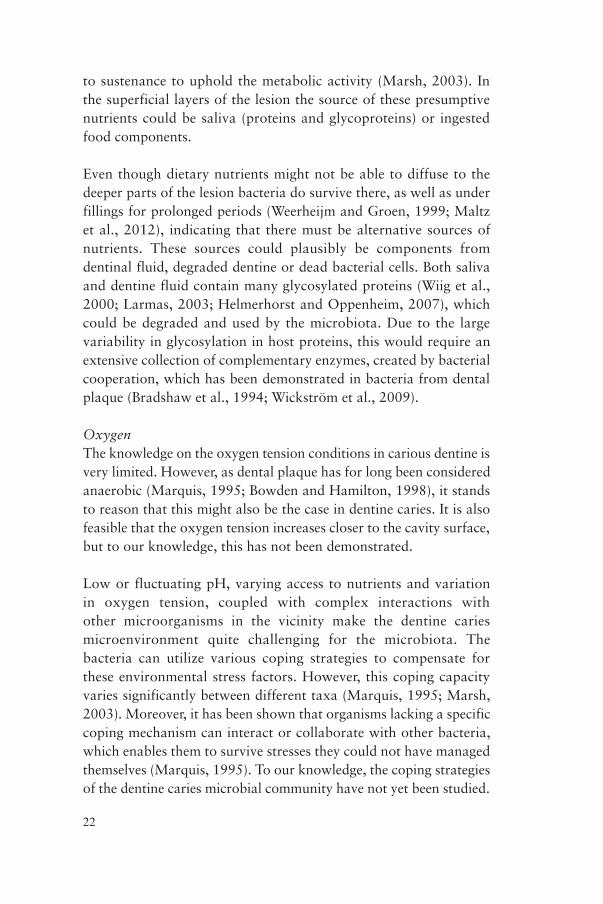
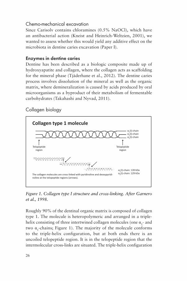
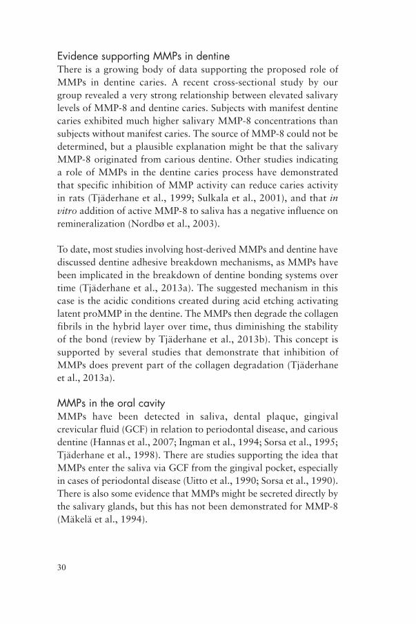
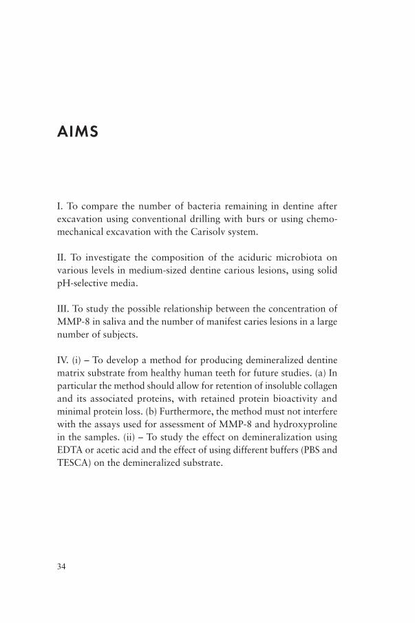
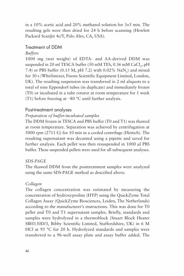
Collagen biology
Collagen type 1 molecule1(I)-chain
1(I)-chain2(I)-chain
Telopeptide region
Telopeptide region
The collagen molecules are cross-linked with pyridinoline and deoxypyridi-1(I)-chain: 139 kDa2(I)-chain: 129 kDa
Figure 1. Collagen type I structure and cross-linking. After Garnero et al., 1998.
Roughly 90% of the dentinal organic matrix is composed of collagen type 1. The molecule is heteropolymeric and arranged in a triple-helix consisting of three intertwined collagen molecules (one α2- and two α1-chains; Figure 1). The majority of the molecule conforms to the triple-helix configuration, but at both ends there is an uncoiled telopeptide region. It is in the telopeptide region that the intermolecular cross-links are situated. The triple-helix configuration
27
makes the molecule highly resistant to general proteolysis, and specific proteases are required to degrade it, including MMPs and cathepsins (Tersariol et al., 2010).
Collagen degradationThe mechanisms behind dentine collagen degradation in dentine caries are still unclear. They were for a long time attributed to the action of acids or unspecific bacterial proteases originating from the dentine caries microbiota. However, acids alone cannot degrade collagen (Katz et al., 1987), and other in vitro studies have demonstrated that the oral microbiota lacks the enzymatic competence to degrade intact collagen (van Strijp et al., 1994; van Strijp et al., 1997). So the question remains: How can collagen degradation in dentine caries be explained?
MMPs in generalMore recent research has focused on the role of host-derived matrix metalloproteinases (MMPs) in connection with collagen degradation in dentine caries (Tjäderhane et al., 1998), and today these enzymes together with cysteine cathepsins are considered the main players in the degradation of collagen in dentine caries (Nascimento et al., 2011; review by Chaussain et al., 2013).
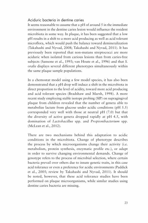
MMPs are a group of genetically distinct but structurally related endopeptidase enzymes, produced by connective tissue cells (fibro-blasts, osteoblasts and odontoblasts) as well as polymorphonuclear leukocytes (PMN cells) and other inflammatory cells (Hannas et al., 2007). Considered as a group, they are able to degrade most extracellular molecules (Visse and Nagase, 2003). In addition, MMPs have other functions in tissue remodeling and tissue development (for reviews, see Mazzoni et al., 2009; Hannas et al., 2007).
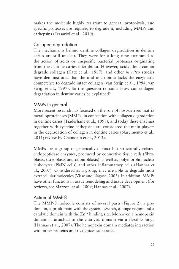
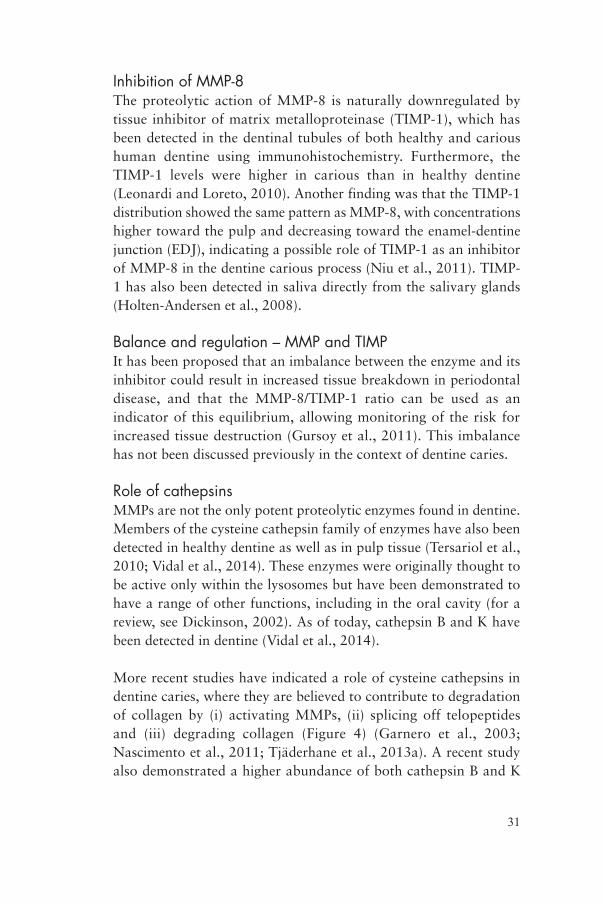
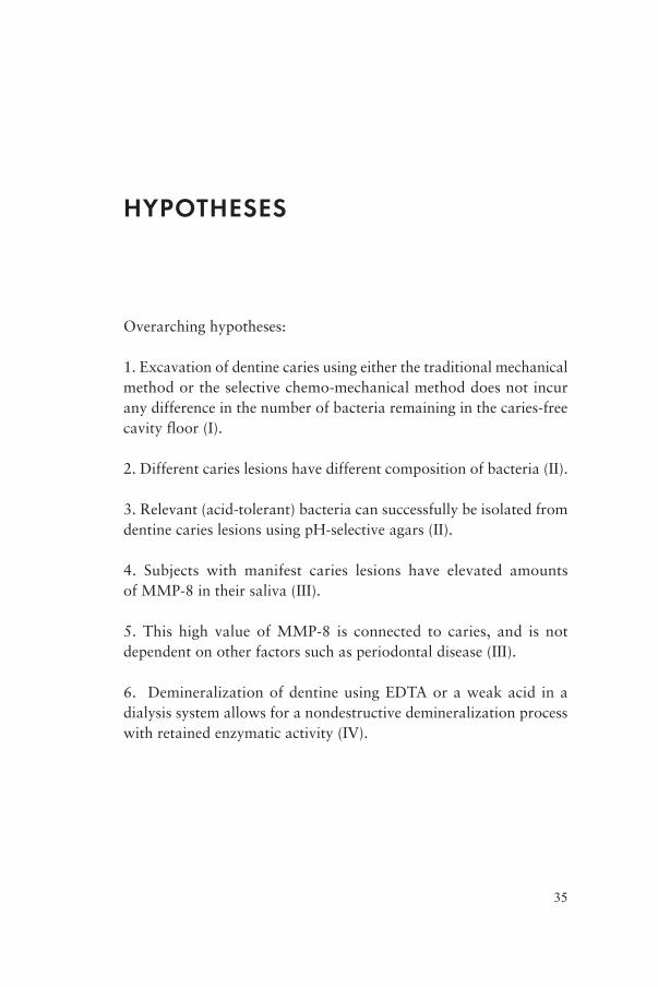
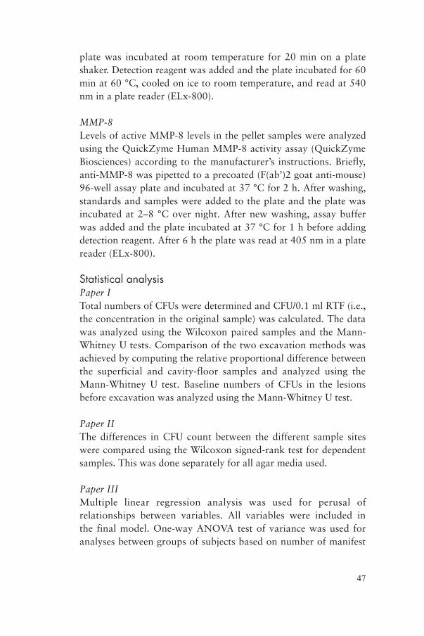
Action of MMP-8The MMP-8 molecule consists of several parts (Figure 2): a pre-domain, a prodomain with the cysteine switch, a hinge region and a catalytic domain with the Zn2+ binding site. Moreover, a hemopexin domain is attached to the catalytic domain via a flexible hinge (Hannas et al., 2007). The hemopexin domain mediates interaction with other proteins and recognizes substrates.
28
MMP-8 molecule
Predomain
Prodomain
Hinge region
Flexible hinge region
Hemopexin domain
C Zn2+
C Zn2+
Figure 2. MMP-8 molecular structure. After Hannas et al., 2007.
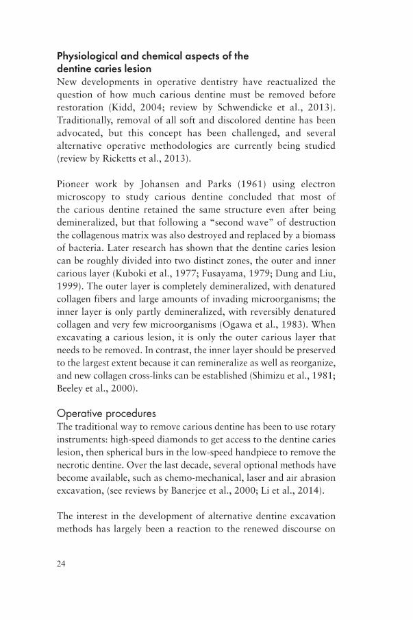
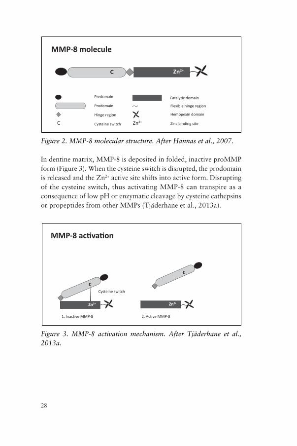
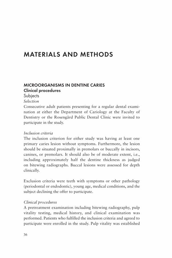
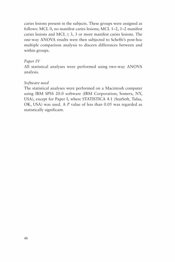
In dentine matrix, MMP-8 is deposited in folded, inactive proMMP form (Figure 3). When the cysteine switch is disrupted, the prodomain is released and the Zn2+ active site shifts into active form. Disrupting of the cysteine switch, thus activating MMP-8 can transpire as a consequence of low pH or enzymatic cleavage by cysteine cathepsins or propeptides from other MMPs (Tjäderhane et al., 2013a).
Cysteine switch
C
2+
C2+
2+
Figure 3. MMP-8 activation mechanism. After Tjäderhane et al., 2013a.
29
MMPs in dentineIn dentine, MMP-2, -3, -8, -9, -14 and -20 have been consistently detected (Martin-De Las Heras et al., 2000; Mazzoni et al., 2007; Mazzoni et al., 2011; Boushell et al., 2011). Of these, MMP-8 is considered the main collagenolytic species in dentine (Sorsa et al., 2004; Sorsa et al., 2006). The dentinal MMP-8 probably originates from the dental pulp, where odontoblasts and other pulpal cells express it (Palosaari et al., 2003). The enzyme is secreted into the predentine matrix in inactive zymogen form (proMMP) at dentinogenesis and becomes trapped within the organic phase of dentine when mineralization takes place (Martin-De Las Heras et al., 2000; Tjäderhane et al., 2001; Sulkala et al., 2007; Mazzoni et al., 2007). It could also be that MMPs, including MMP-8, are distributed from the dentinal fluid via the dentinal tubules (Sulkala et al., 2002; Zehnder et al., 2011; Zehnder et al., 2014; Chibinski et al., 2014). Studies on the localization of MMP-2 and -9 in dentine have revealed higher concentrations in the dentine closer to the pulp, with diminishing amounts in the more superficial layers, which could support the latter hypothesis (Niu et al., 2011). It has also been demonstrated that MMP-2 secretion is upregulated in odontoblasts related to caries-affected dentinal tubules (Boushell et al., 2011).
MMP mechanismThe mechanism by which MMP-8 contributes to collagen degradation in dentine caries has been suggested to be a stepwise process: (i) – Hydroxyapatite is dissolved by bacterial acidic action, thereby revealing the previously mineral-protected collagen molecules as well as other extracellular (ECM) components, such as proMMPs. (ii) – The acid induced pH drop to between 4 and 5 also activates proMMP-8 (Tjäderhane et al., 1998). MMPs are neutral proteases, and are not active at such a low pH, but they are also very robust and can withstand long periods of low pH without becoming denatured, thus losing their bioactive properties (Tezvergil-Mutluay et al., 2013). (iii) – When the pH increases again (due to the pH fluctuation cycles) (Chaussain-Miller et al., 2006), the now activated MMP-8 might exert its catalytic properties and degrade collagen. (iv) – The fragmented collagen – gelatin – can then be further degraded by the gelatinases MMP-2 and -9 or other nonspecific proteases of either bacterial or endogenous origin.
30
Evidence supporting MMPs in dentineThere is a growing body of data supporting the proposed role of MMPs in dentine caries. A recent cross-sectional study by our group revealed a very strong relationship between elevated salivary levels of MMP-8 and dentine caries. Subjects with manifest dentine caries exhibited much higher salivary MMP-8 concentrations than subjects without manifest caries. The source of MMP-8 could not be determined, but a plausible explanation might be that the salivary MMP-8 originated from carious dentine. Other studies indicating a role of MMPs in the dentine caries process have demonstrated that specific inhibition of MMP activity can reduce caries activity in rats (Tjäderhane et al., 1999; Sulkala et al., 2001), and that in vitro addition of active MMP-8 to saliva has a negative influence on remineralization (Nordbø et al., 2003).
To date, most studies involving host-derived MMPs and dentine have discussed dentine adhesive breakdown mechanisms, as MMPs have been implicated in the breakdown of dentine bonding systems over time (Tjäderhane et al., 2013a). The suggested mechanism in this case is the acidic conditions created during acid etching activating latent proMMP in the dentine. The MMPs then degrade the collagen fibrils in the hybrid layer over time, thus diminishing the stability of the bond (review by Tjäderhane et al., 2013b). This concept is supported by several studies that demonstrate that inhibition of MMPs does prevent part of the collagen degradation (Tjäderhane et al., 2013a).
MMPs in the oral cavityMMPs have been detected in saliva, dental plaque, gingival crevicular fluid (GCF) in relation to periodontal disease, and carious dentine (Hannas et al., 2007; Ingman et al., 1994; Sorsa et al., 1995; Tjäderhane et al., 1998). There are studies supporting the idea that MMPs enter the saliva via GCF from the gingival pocket, especially in cases of periodontal disease (Uitto et al., 1990; Sorsa et al., 1990). There is also some evidence that MMPs might be secreted directly by the salivary glands, but this has not been demonstrated for MMP-8 (Mäkelä et al., 1994).
31
Inhibition of MMP-8The proteolytic action of MMP-8 is naturally downregulated by tissue inhibitor of matrix metalloproteinase (TIMP-1), which has been detected in the dentinal tubules of both healthy and carious human dentine using immunohistochemistry. Furthermore, the TIMP-1 levels were higher in carious than in healthy dentine (Leonardi and Loreto, 2010). Another finding was that the TIMP-1 distribution showed the same pattern as MMP-8, with concentrations higher toward the pulp and decreasing toward the enamel-dentine junction (EDJ), indicating a possible role of TIMP-1 as an inhibitor of MMP-8 in the dentine carious process (Niu et al., 2011). TIMP-1 has also been detected in saliva directly from the salivary glands (Holten-Andersen et al., 2008).
Balance and regulation – MMP and TIMPIt has been proposed that an imbalance between the enzyme and its inhibitor could result in increased tissue breakdown in periodontal disease, and that the MMP-8/TIMP-1 ratio can be used as an indicator of this equilibrium, allowing monitoring of the risk for increased tissue destruction (Gursoy et al., 2011). This imbalance has not been discussed previously in the context of dentine caries.
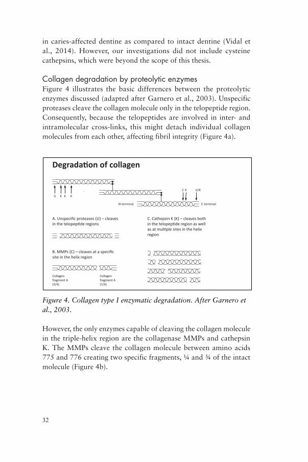
Role of cathepsinsMMPs are not the only potent proteolytic enzymes found in dentine. Members of the cysteine cathepsin family of enzymes have also been detected in healthy dentine as well as in pulp tissue (Tersariol et al., 2010; Vidal et al., 2014). These enzymes were originally thought to be active only within the lysosomes but have been demonstrated to have a range of other functions, including in the oral cavity (for a review, see Dickinson, 2002). As of today, cathepsin B and K have been detected in dentine (Vidal et al., 2014).
More recent studies have indicated a role of cysteine cathepsins in dentine caries, where they are believed to contribute to degradation of collagen by (i) activating MMPs, (ii) splicing off telopeptides and (iii) degrading collagen (Figure 4) (Garnero et al., 2003; Nascimento et al., 2011; Tjäderhane et al., 2013a). A recent study also demonstrated a higher abundance of both cathepsin B and K
32
in caries-affected dentine as compared to intact dentine (Vidal et al., 2014). However, our investigations did not include cysteine cathepsins, which were beyond the scope of this thesis.
Collagen degradation by proteolytic enzymesFigure 4 illustrates the basic differences between the proteolytic enzymes discussed (adapted after Garnero et al., 2003). Unspecific proteases cleave the collagen molecule only in the telopeptide region. Consequently, because the telopeptides are involved in inter- and intramolecular cross-links, this might detach individual collagen molecules from each other, affecting fibril integrity (Figure 4a).
U
Figure 4. Collagen type I enzymatic degradation. After Garnero et al., 2003.
However, the only enzymes capable of cleaving the collagen molecule in the triple-helix region are the collagenase MMPs and cathepsin K. The MMPs cleave the collagen molecule between amino acids 775 and 776 creating two specific fragments, ¼ and ¾ of the intact molecule (Figure 4b).
33
In contrast, cathepsin K can cleave the collagen molecule at multiple sites, both in the C-terminal telopeptide region as well as at four sites along the triple helix region (between amino acids 9 and 10, 21 and 22, 96 and 97 [α1], 99 and 100 [α2], 810 and 811 [α1] and 814 and 815 [α2]; Figure 4c).
Final remarkIn order to enhance our understanding of the dentine caries process, it is of paramount importance that we learn more about the involved microbiota and the host factors. And even more important is to understand the dynamics between these factors. Hopefully, the new insights gained in this thesis, added to the current knowledge base, will assist in finding new possibilities for prevention and treatment of caries.
34
AIMS
I. To compare the number of bacteria remaining in dentine after excavation using conventional drilling with burs or using chemo-mechanical excavation with the Carisolv system.
II. To investigate the composition of the aciduric microbiota on various levels in medium-sized dentine carious lesions, using solid pH-selective media.
III. To study the possible relationship between the concentration of MMP-8 in saliva and the number of manifest caries lesions in a large number of subjects.
IV. (i) – To develop a method for producing demineralized dentine matrix substrate from healthy human teeth for future studies. (a) In particular the method should allow for retention of insoluble collagen and its associated proteins, with retained protein bioactivity and minimal protein loss. (b) Furthermore, the method must not interfere with the assays used for assessment of MMP-8 and hydroxyproline in the samples. (ii) – To study the effect on demineralization using EDTA or acetic acid and the effect of using different buffers (PBS and TESCA) on the demineralized substrate.
35
HYPOTHESES
Overarching hypotheses:
1. Excavation of dentine caries using either the traditional mechanical method or the selective chemo-mechanical method does not incur any difference in the number of bacteria remaining in the caries-free cavity floor (I).
2. Different caries lesions have different composition of bacteria (II).
3. Relevant (acid-tolerant) bacteria can successfully be isolated from dentine caries lesions using pH-selective agars (II).
4. Subjects with manifest caries lesions have elevated amounts of MMP-8 in their saliva (III).
5. This high value of MMP-8 is connected to caries, and is not dependent on other factors such as periodontal disease (III).
6. Demineralization of dentine using EDTA or a weak acid in a dialysis system allows for a nondestructive demineralization process with retained enzymatic activity (IV).
36
MATERIALS AND METHODS
MICROORGANISMS IN DENTINE CARIESClinical proceduresSubjectsSelectionConsecutive adult patients presenting for a regular dental exami-nation at either the Department of Cariology at the Faculty of Dentistry or the Rosengård Public Dental Clinic were invited to participate in the study.
Inclusion criteriaThe inclusion criterion for either study was having at least one primary caries lesion without symptoms. Furthermore, the lesion should be situated proximally in premolars or buccally in incisors, canines, or premolars. It should also be of moderate extent, i.e., including approximately half the dentine thickness as judged on bitewing radiographs. Buccal lesions were assessed for depth clinically.
Exclusion criteria were teeth with symptoms or other pathology (periodontal or endodontic), young age, medical conditions, and the subject declining the offer to participate.
Clinical proceduresA pretreatment examination including bitewing radiography, pulp vitality testing, medical history, and clinical examination was performed. Patients who fulfilled the inclusion criteria and agreed to participate were enrolled in the study. Pulp vitality was established
37
using both thermal and electronic vitality testing (Model 2006 Vitality Scanner, Analytic Technology, Redmond, USA). The extent of the carious lesion was determined clinically for buccal lesions (incisors, canines, and premolars) and radiographically for proximal lesions in premolars. Bitewing radiographs were taken of all teeth with proximal caries lesions. After sampling and excavation, the cavity was restored using convention methods and materials.
Carious lesionsClinical dataTooth number, sample surface, consistency and color of the carious dentine, as well as lesion morphology (closed or open cavity) was recorded (Nyvad and Fejerskov, 1986) using a modified protocol by Bjørndal et al. (1997).
RandomizationPaper I included 22 subjects (6 females, 16 males; age 20–68 years, median 36 years). Twelve teeth were excavated using chemo-mechanical method (CMM; Carisolv, MediTeam; Sävedalen, Sweden) and ten with rotating instruments (conventional drilling with low-speed burs).
Of the original 22 subjects, a subpopulation of 10 subjects was selected for further microbiological analysis of the acid-tolerant microbiota (Paper II). These subjects were all recruited from the Department of Cariology at the Faculty of Dentistry (2 females, 8 males; aged 25-68 years, mean 42 years, median 38 years). In this group five lesions were excavated using the chemo-mechanical method and five using conventional drilling.
Ethical aspectsThe study was approved by the Ethical Research Committee at Lund University (Dnr: LU 273-99).
Dentin sample collection and excavation procedureExcavation procedureIn cases where the dentine lesion was covered by enamel, access was obtained by using a small spherical diamond in the high-speed handpiece. A rubber dam was placed and the outer layer of plaque
38
debris, as well as the topmost layer of carious dentine, gently removed using a sterile spoon excavator. The cavity was rinsed meticulously using sterile saline (CCS, Sweden) and dried with a cotton pellet. Antibacterial compounds were not used for presampling cleaning of the rubber dam and operation area due to the risk of influencing the microbiota residing in the carious dentine.
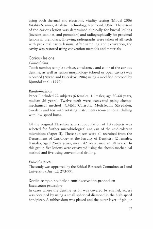
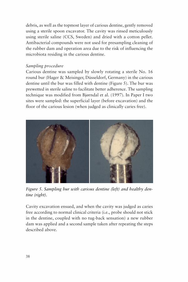
Sampling procedureCarious dentine was sampled by slowly rotating a sterile No. 16 round bur (Hager & Meisinger, Düsseldorf, Germany) in the carious dentine until the bur was filled with dentine (Figure 5). The bur was prewetted in sterile saline to facilitate better adherence. The sampling technique was modified from Bjørndal et al. (1997). In Paper I two sites were sampled: the superficial layer (before excavation) and the floor of the carious lesion (when judged as clinically caries free).
Figure 5. Sampling bur with carious dentine (left) and healthy den-tine (right).
Cavity excavation ensued, and when the cavity was judged as caries free according to normal clinical criteria (i.e., probe should not stick in the dentine, coupled with no tug-back sensation) a new rubber dam was applied and a second sample taken after repeating the steps described above.
39
In Paper II three sites were sampled: the superficial layer (before excavation), the approximate center (after partial excavation, approximately 1.5–2 mm from the superficial sample), and the floor of the carious lesion (after completed excavation).
Chemo-mechanical methodThe sampling procedure in the cases excavated using the chemo-mechanical method was almost identical to above. The difference was that all excavation steps were performed using the Carisolv system (gel and special hand instruments). After access to the carious lesion had been established and the first sample had been taken, the carious dentine was covered with a viscous droplet of Carisolv gel. After 30 s the carious dentine was gently scraped using the specially designed hand instruments in order to remove the softened carious tissue. This was repeated until the dentine surface was hard (determined as for drilling above). Residual Carisolv gel was removed with a cotton pellet soaked in saline and the cavity thoroughly rinsed. Samples were taken as for drilling, including rubber dam application and rinsing of the cavity.
Aseptic proceduresAll excavation and sampling was performed under rubber dam using aseptic technique, including washing of the operation field with copious amounts of sterile saline prior to every sampling step, use of separate sterile burs and handpieces for each sample and operator hand wash and new sterile gloves for each sample.
ReproducibilityTo assess the accuracy and reproducibility of the sampling method, ten extracted teeth with and ten without dentine caries were sampled as described above (in vitro). The sampled dentine was scraped off the burs after 2 min and immediately weighed on a laboratory grade scale (Sartorius, model #2400, Göttingen, Germany). Results were analyzed statistically.
Microbiological proceduresThe sampling burs were immediately placed in sterile vials containing 3 ml of prereduced transport fluid (RTF) (Syed and Loesche, 1972)
40
and glass beads (Paper I: 2 mm ∅, 0.6 g; Paper II: 1 mm ∅, 0.3 g). The dentine samples were dislodged from the bur by vortexing the vials for 30 s (Whirlimixer, Fisons Scientific Apparatus, Leicestershire, England), and the bur was subsequently removed from the vial using sterile tweezers in order to minimize ion leakage from the metal.
Paper IThe dentine samples were diluted tenfold and 100 µl of diluted sample was plated on different agar plates in duplicate: blood agar (BHI agar, Difco, Detroit, MI, USA, supplemented with 5% human blood) for total microbial counts, Rogosa Lactobacilli selective agar (SL, Merck, Darmstadt, Germany) for the isolation of lactobacilli (Rogosa et al., 1951), and mitis salivarius agar (MSA, Difco) for the isolation of oral streptococci. All agars were incubated aerobically at 37° C. Incubation times were 2–4 days for blood agar, 4 days for SL agar, and 2 days for MSA. Duplicates of the blood agar plates were also incubated anaerobically at 37° C for 7 days.
Paper IIIsolation of the acid-tolerant microbiotaThe dentine samples were diluted ten- and hundredfold in prereduced phosphate buffered saline at pH 7.2. pH agar (Todd-Hewitt broth [Difco Lab; Detroit, MI, USA] supplemented with Bacto agar, glucose and citrate-phosphate buffer) buffered to pH 5.5, 5.0, 4.5, or 4.0 was used for selection of aciduric microorganisms (Svensäter et al., 2003). Blood agar (BA, BHI agar [Difco Lab] supplemented with 5% human blood) (Holdeman et al., 1977) was used for total counts. The pH agars and the BA were inoculated and incubated anaerobically for 7 days at 37° C in a 95% N2 and 5% CO2 atmosphere. The number of colony-forming units (CFUs) per sample (3 ml RTF) was determined using a light microscope at 40 × magnification (Leica GZ 6; Buffalo, NY, USA). pH agars with 30 to 300 individual colonies were selected and individual morphological colony types recorded. One colony of each morphological colony type was recultured on BA and incubated in an atmosphere of 9% H2, 6% CO2 and 85% N2 for 7 days. Isolates were kept frozen (–18° C) in skim milk until identification.
41
Identification of microbial groupsThe frozen isolates were thawed, incubated anaerobically for 24 h on BA, and subjected to Gram staining. Gram-positive cocci were tested for catalase on brain heart infusion agar (Difco Lab). Gram-positive cocci were inoculated in Bacto Todd-Hewitt broth (Difco Lab) for fermentation and enzymatic testing (Beighton et al., 1991b; Beighton et al., 1991a; Whiley and Beighton, 1998). The minimal criteria for identification of various streptococci were as described by Chavez de Paz et al. (2005). Gram-negative cocci were identified as Veillonella spp. based on cell size, obligate anaerobic growth, and increased growth in the presence of lactate but not glucose (Holdeman et al., 1977). Gram-positive, catalase-negative rods growing in palisade formation on Gram-stained smears were considered to be Lactobacillus spp. confirmed by cultivation on Rogosa SL-agar. Large Gram-positive oval cocci (4–8 μm) showing budding and hyphae were regarded as yeasts. Gram-positive, catalase-negative pleomorphic rods with obligate anaerobic growth with occasional bifid shapes were preliminarily grouped as Bifidobacterium spp. Gram-positive, catalase positive short pleomorphic rods with club-shaped or rudimentary branched forms were considered to be Propionibacterium spp. (Holt et al., 1993).
PROTEOLYTIC ENZYME ACTIVITY IN DENTINE CARIESMMPs in saliva (Paper III)The methodological descriptions below regarding participants, clinical and radiographic examination, saliva sampling, and biomarker analysis have been described previously (Lundegren et al., 2012; Rathnayake et al., 2013b).
ParticipantsA sample of adults (age 18–87 years; mean 48.6 ± 16.9 years) from the south of Sweden were randomly selected and invited to partake in a clinical study on oral health. Of this sample, 966 persons were accessible and thus formed the initial sample. Clinical examination was performed on 451 of these individuals (232 women and 219 men) (Lundegren et al., 2011). Of these, 441 contributed with complete saliva samples (Rathnayake et al., 2013a).
42
Clinical examinationFour calibrated dentists at the Faculty of Odontology, Malmö University, performed 90% of the clinical examinations. A standard clinical and radiographic examination was performed, including caries parameters such as DMFT, number of manifest caries lesions (D3 lesions clearly involving dentine, as seen on bitewing radiographs and evident cavitated lesions on other surfaces; MCL), plaque index (PI), saliva sampling, and bleeding on probing (BOP).
The study was approved by the Research Ethical Committee at Lund University (Dnr: 513/2006).
Saliva sampling and chair-side testsStimulated saliva was collected in graded test tubes and secretion rate was determined (ml/min). Salivary mutans streptococci, lactobacilli and buffer capacity were determined using Dentocult® SM–Strip mutans and Dentocult® LB, respectively (Orion Diagnostica, Espoo, Finland) according to the manufacturer’s instructions. Remaining saliva was frozen at –20°C until further processed. Individual saliva samples were centrifuged and the supernatant transferred to 1.5 ml Eppendorf tubes and subsequently frozen at −80°C.
Analysis of MMP-8 and TIMP-1MMP-8 was analyzed by a time-resolved immunofluorometric assay as described by Rathnayake and coworkers (2013b). Monoclonal MMP-8- antibodies 8708 and 8706 (Medix Biochemica, Kauniainen, Finland) were used as capture and tracer antibodies respectively. The tracer antibody (8706) was labeled using europium chelate and the assay buffer contained 20 mM Tris-HCl (pH 7.5), 0.5 M NaCl, 5 mM CaCl2, 50 µM ZnCl2, 0.5% bovine serum albumin, 0.05% sodium azide and 20 mg/l diethylenetriaminepentaacetic acid. Saliva samples were diluted in assay buffer and incubated for 1 h, followed by incubation for a further 1 h with tracer antibody. Enhancement solution was added, and after 5 min, fluorescence was measured using a 1234 Delfia Research Fluorometer (Wallac, Turku, Finland), (Hanemaaijer et al., 1997; Gursoy et al., 2010). MMP-8 had a detection limit of 0.08 ng/ml and a coefficient of variation of 7.3%. TIMP-1 (Amersham Biotrak, GE Healthcare, Buckinghamshire, UK)
43
was analyzed by enzyme-linked immunosorbent assay kits according to the manufacturer’s instructions. The interassay coefficient of variation for TIMP-1 was 8.2%, and the detection limit for the assay was 1.25 ng/ml. Total protein concentration was determined using the Bradford assay (Bradford, 1976).
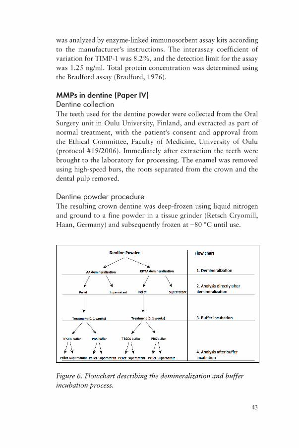
MMPs in dentine (Paper IV)Dentine collectionThe teeth used for the dentine powder were collected from the Oral Surgery unit in Oulu University, Finland, and extracted as part of normal treatment, with the patient’s consent and approval from the Ethical Committee, Faculty of Medicine, University of Oulu (protocol #19/2006). Immediately after extraction the teeth were brought to the laboratory for processing. The enamel was removed using high-speed burs, the roots separated from the crown and the dental pulp removed.
Dentine powder procedureThe resulting crown dentine was deep-frozen using liquid nitrogen and ground to a fine powder in a tissue grinder (Retsch Cryomill, Haan, Germany) and subsequently frozen at −80 °C until use.
Figure 6. Flowchart describing the demineralization and buffer incubation process.
44
Demineralization processEDTA10 g of thawed dentine powder was aliquoted into three equal portions and placed in a dialysis tube (Pur-A-Lyzer 10 ml 12-14 kDa; Sigma-Aldrich, St. Louis, MO, USA) with 8 ml of 0.5 M EDTA (pH 7.4). The tubes were shaken thoroughly to suspend the powder in the liquid and placed in a large beaker containing 350 ml of 0.5 M EDTA under constant mixing using a magnetic stirrer (300 rpm). To prevent excessive sedimentation of the powder, the tubes were removed from the beaker and agitated twice per day. The EDTA in the beaker was replaced every 48 h and the procedure repeated until the powder had dissolved entirely. This process demanded four times 48 h of EDTA treatment (total EDTA treatment 192 h). To wash away the EDTA within the dialysis tubes, the liquid in the beaker was changed to laboratory grade water (350 ml) for 24 h before the water was changed, and was agitated as described above. The water wash procedure was repeated four times (total water wash 192 h). The resulting demineralized dentine matrix (DDM; suspended in water) was subsequently frozen at −80 °C until use.
Acetic Acid6 g of thawed dentine powder was aliquoted into three equal portions. Each portion was placed in a dialysis tube as above and 8 ml 1 M (pH 2.4) acetic acid (AA) was added. The rest of the demineralization process was performed as described above except that EDTA was substituted with AA.
DDM retrieval processEDTA-derived pelletAfter the demineralization procedure, the DDM was suspended in remaining analysis water (from the EDTA washout process). Separation of insoluble DDM was achieved by centrifugation at 5000 rpm (3913 G) for 10 min in a cooled (4 °C) centrifuge (Hettich Universal 320R; Hettich 1431 rotor, Tuttlingen, Germany). The resulting supernatant was decanted using a pipette. To retrieve as much DDM as possible, the supernatant was recentrifuged, and the resulting pellet added to the pellet from the first centrifugation. The resulting pellets from the three dialysis tubes and from the re-
45
centrifuged supernatant were pooled before treatment. Total final pellet wet weight was 4.30 g (from 10 g dry dentine powder). The resulting supernatants were also pooled.
Acetic acid-derived pelletThe same protocol as for the EDTA-derived pellet above was used. The resulting pellets from the three dialysis tubes and from the recentrifuged supernatant were pooled before treatment, as were the supernatants. Total final pellet wet weight was 2.8 g (from 6 g dry dentine powder).
Analyses of DDM directly after demineralizationSoluble proteinSoluble protein concentration in the DDM directly after demineralization (pellet and supernatant) was estimated in order to determine suitable dilutions for SDS-PAGE analysis, using Bio-Rad Protein Assay with the standard protocol for microtiter plates (Bio-Rad, Richmond, CA, USA). The samples were undiluted, except for pellet samples, which were diluted in PBS to 100 mg/ml and shaken in a vortex mixer. 10 µl of blank, standard, and sample respectively were put in duplicates in the wells of a microtiter plate, and 200 µl Dye Reagent 1:5 was added to each well. The plate was gently shaken (PMS-1000, Grant-Bio, Cambridge, UK) for 10 min to mix the contents in the wells. Reading of the plate was performed at 595 nm using a plate reader (ELx-800, Bio-Tek, VT, USA).
SDS-PAGE Uncentrifuged DDM samples (10 µl/sample) were diluted in 10 µl Laemmli SDS sample buffer together with 5% 2-mercaptoethanol, and heated to 100 °C for 5 min. From this mixture, 5 µl was transferred to SDS-PAGE gel in duplicate. Electrophoresis separation of the demineralized dentine proteins was carried out in a 7.5% SDS-PAGE Criterion TRIS-HCl precast gel (Bio-Rad Laboratories, Hercules, CA, USA) for 50 min at 200 V. As collagen control rat-tail collagen was used (Sigma-C8897, Sigma-Aldrich). The gels were stained with 0.1% Coomassie blue on an Orbital shaker (Bellco Glass Inc., NJ, USA) for 30 min and fixed in a 10% acetic acid and 40% methanol solution for 30 min, after which they were de-stained
46
in a 10% acetic acid and 20% methanol solution for 3×5 min. The resulting gels were then dried for 24 h before scanning (Hewlett Packard ScanJet 4c/T, Palo Alto, CA, USA).
Treatment of DDMBuffers1000 mg (wet weight) of EDTA- and AA-derived DDM was suspended in 20 ml TESCA buffer (50 mM TES, 0.36 mM CaCl2, pH 7.4) or PBS buffer (0.15 M, pH 7.2) with 0.02% NaN3) and mixed for 30 s (Whirlimixer, Fisons Scientific Equipment Limited, London, UK). The resulting suspension was transferred in 2 ml aliquots to a total of nine Eppendorf tubes (in duplicate) and immediately frozen (T0) or incubated in a tube rotator at room temperature for 1 week (T1) before freezing at −80 °C until further analysis.
Post-treatment analysesPreparation of buffer-incubated samplesThe DDM frozen in TESCA and PBS buffer (T0 and T1) was thawed at room temperature. Separation was achieved by centrifugation at 5000 rpm (2711 G) for 10 min in a cooled centrifuge (Hettich). The resulting supernatant was decanted using a pipette and saved for further analysis. Each pellet was then resuspended in 1000 µl PBS buffer. These suspended pellets were used for all subsequent analyses.
SDS-PAGEThe thawed DDM from the posttreatment samples were analyzed using the same SDS-PAGE method as described above.
CollagenThe collagen concentration was estimated by measuring the concentration of hydroxyproline (HYP) using the QuickZyme Total Collagen Assay (QuickZyme Biosciences, Leiden, The Netherlands) according to the manufacturer’s instructions. This was done for T0 pellet and T0 and T1 supernatant samples. Briefly, standards and samples were hydrolyzed in a thermoblock (Stuart Block Heater SBH130D/3, Bibby Scientific Limited, Staffordshire, UK) in 6 M HCl at 95 °C for 20 h. Hydrolyzed standards and samples were transferred to a 96-well assay plate and assay buffer added. The
47
plate was incubated at room temperature for 20 min on a plate shaker. Detection reagent was added and the plate incubated for 60 min at 60 °C, cooled on ice to room temperature, and read at 540 nm in a plate reader (ELx-800).
MMP-8Levels of active MMP-8 levels in the pellet samples were analyzed using the QuickZyme Human MMP-8 activity assay (QuickZyme Biosciences) according to the manufacturer’s instructions. Briefly, anti-MMP-8 was pipetted to a precoated (F(ab’)2 goat anti-mouse) 96-well assay plate and incubated at 37 °C for 2 h. After washing, standards and samples were added to the plate and the plate was incubated at 2–8 °C over night. After new washing, assay buffer was added and the plate incubated at 37 °C for 1 h before adding detection reagent. After 6 h the plate was read at 405 nm in a plate reader (ELx-800).
Statistical analysisPaper I Total numbers of CFUs were determined and CFU/0.1 ml RTF (i.e., the concentration in the original sample) was calculated. The data was analyzed using the Wilcoxon paired samples and the Mann-Whitney U tests. Comparison of the two excavation methods was achieved by computing the relative proportional difference between the superficial and cavity-floor samples and analyzed using the Mann-Whitney U test. Baseline numbers of CFUs in the lesions before excavation was analyzed using the Mann-Whitney U test.
Paper IIThe differences in CFU count between the different sample sites were compared using the Wilcoxon signed-rank test for dependent samples. This was done separately for all agar media used.
Paper IIIMultiple linear regression analysis was used for perusal of relationships between variables. All variables were included in the final model. One-way ANOVA test of variance was used for analyses between groups of subjects based on number of manifest
48
caries lesions present in the subjects. These groups were assigned as follows: MCL 0, no manifest caries lesions; MCL 1–2, 1–2 manifest caries lesions and MCL ≥ 3, 3 or more manifest caries lesions. The one-way ANOVA results were then subjected to Scheffe’s post-hoc multiple comparison analysis to discern differences between and within groups.
Paper IVAll statistical analyses were performed using two-way ANOVA analysis.
Software usedThe statistical analyses were performed on a Macintosh computer using IBM SPSS 20.0 software (IBM Corporation; Somers, NY, USA), except for Paper I, where STATISTICA 4.1 (StatSoft, Tulsa, OK, USA) was used. A P value of less than 0.05 was regarded as statistically significant.
49
RESULTS
Paper IReproducibilityMean sample weight for carious dentine was 0.30 ± 0.05 mg (mean ± SD) and 0.38 ± 0.09 mg for caries-free dentine. The difference was statistically significant (two-tailed t-test; P = 0.02). Volume-wise, the samples were roughly equal, as determined by ocular inspection in a microscope.
CFUs in lesions before excavationThere were no statistically significant differences in CFU count between the excavation groups before excavation, regardless of agar medium or incubation atmosphere (BA aerobic P = 0.099; BA anaerobic P = 0.509; SL P = 0.380; MSA P = 0.355).
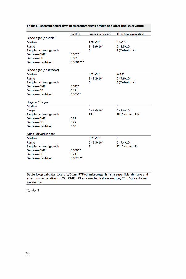
CFUs in lesions after excavationThe numbers of CFUs decreased significantly after final excavation except for bacterial growth on SL agar, where the decrease was not significant. Table 1 shows the medians and ranges of the CFU counts before and after final excavation, as well as the P values for the decrease. Figure 7 gives a graphical description of numbers of CFUs before and after final excavation for the different agar media used.
Comparison of excavation methodsThere were no statistically significant differences in proportional CFU count between the excavation methods except for BA aerobic, where the chemo-mechanical method was significantly more effective (P = 0.033).
50
Table 1.
51
b d
a c
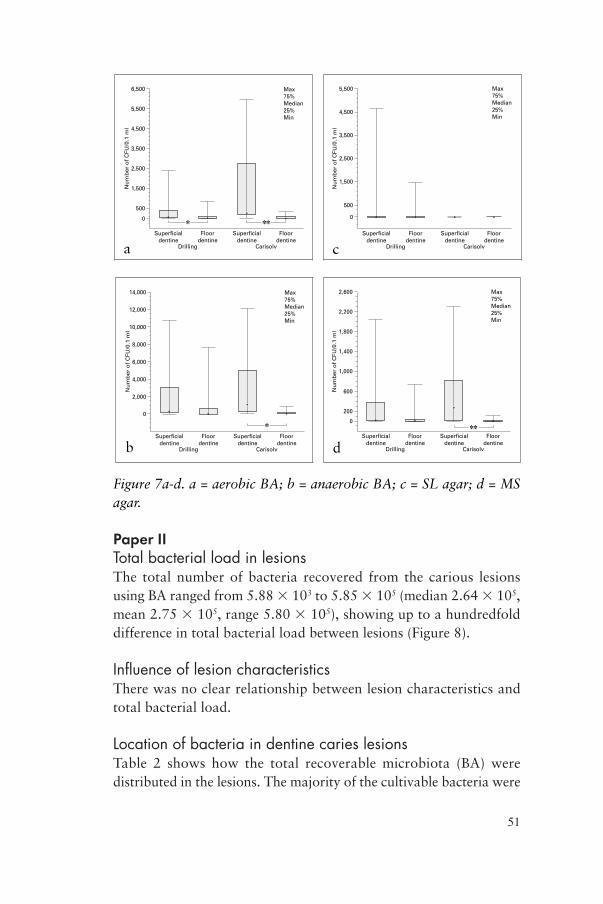
Figure 7a-d. a = aerobic BA; b = anaerobic BA; c = SL agar; d = MS agar.
Paper IITotal bacterial load in lesionsThe total number of bacteria recovered from the carious lesions using BA ranged from 5.88 103 to 5.85 105 (median 2.64 105, mean 2.75 105, range 5.80 105), showing up to a hundredfold difference in total bacterial load between lesions (Figure 8).
Influence of lesion characteristicsThere was no clear relationship between lesion characteristics and total bacterial load.
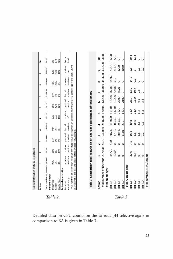
Location of bacteria in dentine caries lesionsTable 2 shows how the total recoverable microbiota (BA) were distributed in the lesions. The majority of the cultivable bacteria were
52
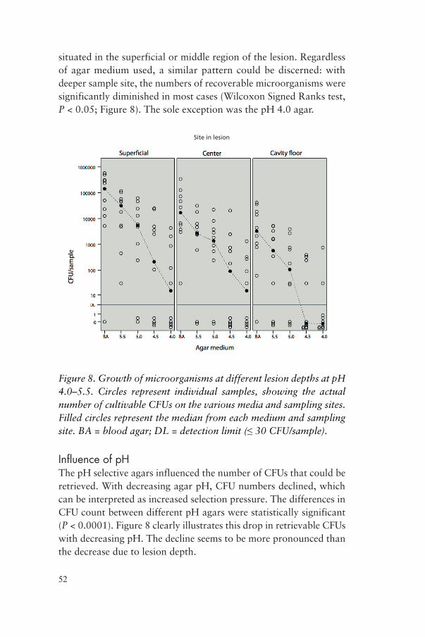
situated in the superficial or middle region of the lesion. Regardless of agar medium used, a similar pattern could be discerned: with deeper sample site, the numbers of recoverable microorganisms were significantly diminished in most cases (Wilcoxon Signed Ranks test, P < 0.05; Figure 8). The sole exception was the pH 4.0 agar.
Figure 8. Growth of microorganisms at different lesion depths at pH 4.0–5.5. Circles represent individual samples, showing the actual number of cultivable CFUs on the various media and sampling sites. Filled circles represent the median from each medium and sampling site. BA = blood agar; DL = detection limit (≤ 30 CFU/sample).
Influence of pHThe pH selective agars influenced the number of CFUs that could be retrieved. With decreasing agar pH, CFU numbers declined, which can be interpreted as increased selection pressure. The differences in CFU count between different pH agars were statistically significant (P < 0.0001). Figure 8 clearly illustrates this drop in retrievable CFUs with decreasing pH. The decline seems to be more pronounced than the decrease due to lesion depth.
Site in lesion
53
Table&2&Distrib
ution&of&cfu&by&lesion
&levels&
Lesion
12
34
56
78
910
Total&num
ber&o
f&bacteria
2370
0059
7054
8880
2916
6012
0300
6420
058
5810
4566
0043
0500
5880
Percen
t&of&total
Supe
rficial
99%
86%
91%
98%
10%
36%
99%
68%
12%
0%Ce
nter
1%1%
8%2%
60%
42%
1%28
%79
%64
%Cavity&floo
r0%
13%
1%0%
30%
22%
0%4%
10%
36%
Lesion
&cha
racteristics
Locatio
nproxim
alproxim
alproxim
albu
ccal
proxim
alproxim
albu
ccal
proxim
alproxim
albu
ccal
Consisten
cymed
ium
med
ium
med
ium
med
ium
soft
med
ium
med
ium
med
ium
hard
hard
Total&num
bers&of&cfu&in&each&lesio
n&and&the&respectiv
e&distrib
ution&on
&differen
t&lesion&levels&as&a&percentage&of&th
e&total.&Lesio
n&characteristics&a
re&also
&includ
ed.&Total&num
bers&=&cfu/sam
ple.
Table 2. Table 3.
Detailed data on CFU counts on the various pH selective agars in comparison to BA is given in Table 3.
Table 3. Com
parison
total growth on pH
agars as a
percentage of to
tal on BA
Lesion
12
34
56
78
910
Total num
ber o
f bacteria
2370
0059
7054
8880
2916
6012
0300
6420
058
5810
4566
0043
0500
5880
Total on pH
agar
pH 5.5
4872
045
088
740
1188
9016
110
1521
076
080
6426
023
670
1200
pH 5.0
1050
047
010
4803
013
740
1029
062
580
510
2157
072
0pH
4.5
00
2064
025
530
7620
2700
3570
012
900
pH 4.0
00
1110
210
6270
2100
00
1050
0To
tal %
on pH
agar
pH 5.5
20.6
7.5
16.2
40.8
13.4
23.7
13.0
14.1
5.5
20.4
pH 5.0
0.4
08.6
16.5
11.4
16.0
10.7
0.1
512
.2pH
4.5
00
3.8
8.8
6.3
4.2
0.6
00.3
0pH
4.0
00
0.2
0.1
5.2
3.3
00
0.2
0To
tal num
bers = cfu/sam
ple
54
Composition of acid-tolerant floraA few examples of the composition of the acid-tolerant microflora subpopulation in comparison with the corresponding total recoverable microflora (BA) are given in Figure 9 and 10.
This pattern of diversity was found in most of the cases, and both the size and composition of the aciduric subpopulation differed significantly between the carious lesions.
Blood agar78%
pH 5.022%
Streptococci82%
Others14%
G+rods4%
Figure 9. (Subject 11, pH 5.0, center) Total counts on pH 5.0 were 22% of the corresponding count on BA. The aciduric subpopulation was composed of 82 % streptococci, 4 % Gram-positive rods and 14% other microorganisms.
Blood agar98%
pH 5.02% Streptococci
90.7%
Lactobacilli8.9%
G+rods0.4%
Figure 10. (Subject 16, pH 5.0, center) Total counts on pH 5.0 were 2% of the corresponding count on BA. The aciduric subpopulation was composed of 90.7 % streptococci, 8.9 % lactobacilli and 0.4 % Gram-positive rods.
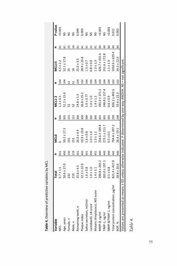
55
Table&4.
!Ove
rvie
w!o
f!pre
dictive
!variable
s!by!M
CL
Varia
ble
Total
NMCL0
NMCL132
NMCL≥3
NP3value
MCL
0.7
!±!1
.5451
0306
1.3
!±!0
.5104
4.5
!±!2
.241
<0.0
01
Age
,!ye
ars
50.6
!±!1
7.0
450
50.2
!±!1
7.3
305
51.2
!±!1
5.6
104
52.1
!±!1
7.8
41
NS
Fem
ale
,!n
232
232
155
155
52
52
25
25
NS
Male
,!n
219
219
151
151
52
52
16
16
NS
Rem
ain
ing!te
eth
,!n
25.6
!±!4
.5451
26.0
!±!4
.4306
24.8
!±!5
.3104
25.0
!±!3
.541
0.0
49
Pla
que!in
dex
22.3
!±!2
2.0
443
19.9
!±!1
9.8
300
26.8
!±!2
5.2
102
28.9
!±!2
5.8
41
0.0
03
Saliv
a!secr
etion,!m
l/m
in1.6
!±!0
.8451
1.6
!±!0
.8306
1.6
!±!0
.77
104
1.3
!±!0
.741
NS
Lact
obacilli,!LB
!sco
re1.0
!±!1
.0450
1.0
!±!1
.0305
1.0
!±!1
.0104
0.8
!±!0
.941
NS
Muta
ns!st
repto
cocc
i,!M
S!sc
ore
1.4
!±!1
.1451
1.3
!±!1
.1306
1.4
!±!1
.2104
1.3
!±!1
.341
NS
MM
PS8
,!ng/
ml
290.8
!±!2
65.2
449
254.6
!±!1
89.8
305
345.0
!±!3
71.3
104
426.7
!±!3
55.4
40
<0.0
01
TIM
PS1
,!ng/
ml
265.0
!±!1
97.1
449
273.1
!±!2
11.7
305
246.8
!±!1
57.4
104
251.6
!±!1
72.8
40
NS
MM
PS8
/TIM
PS1
,!ng/
ml
0.5
!±!0
.8449
0.5
!±!0
.5305
0.6
!±!0
.9104
1.1
!±!1
.940
<0.0
01
Tota
l!pro
tein
!conce
ntration,!µg/
ml
821.4
!±!4
27.9
441
794.4
!±!3
97.2
299
826.2
!±!4
03.5
102
1010.6
!±!6
29.4
40
0.0
11
BOP,!%
28.8
!±!2
0.6
440
26.4
!±!1
9.1
298
33.6
!±!2
3.3
102
34.3
!±!2
2.0
40
0.0
02
Valu
es!are
!pre
sente
d!a
s!m
eans!±!SD!u
nle
ss!o
therw
ise!in
dicate
d.!P!d
ete
rmin
ed!b
y!oneSw
ay!ANOVA.!NS!=!n
ot!sign
ificant.
Tab
le 4
.
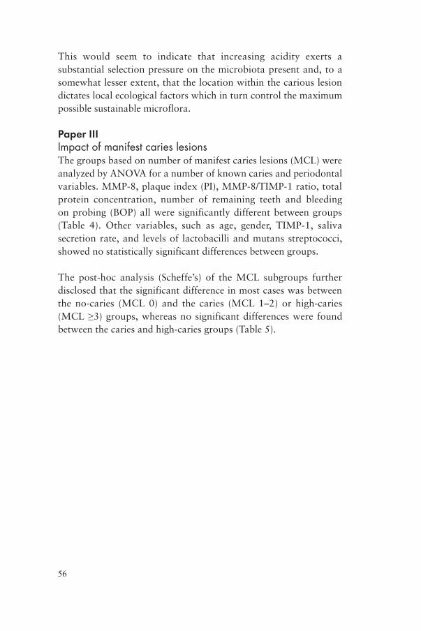
56
This would seem to indicate that increasing acidity exerts a substantial selection pressure on the microbiota present and, to a somewhat lesser extent, that the location within the carious lesion dictates local ecological factors which in turn control the maximum possible sustainable microflora.
Paper IIIImpact of manifest caries lesionsThe groups based on number of manifest caries lesions (MCL) were analyzed by ANOVA for a number of known caries and periodontal variables. MMP-8, plaque index (PI), MMP-8/TIMP-1 ratio, total protein concentration, number of remaining teeth and bleeding on probing (BOP) all were significantly different between groups (Table 4). Other variables, such as age, gender, TIMP-1, saliva secretion rate, and levels of lactobacilli and mutans streptococci, showed no statistically significant differences between groups.
The post-hoc analysis (Scheffe’s) of the MCL subgroups further disclosed that the significant difference in most cases was between the no-caries (MCL 0) and the caries (MCL 1–2) or high-caries (MCL ≥3) groups, whereas no significant differences were found between the caries and high-caries groups (Table 5).
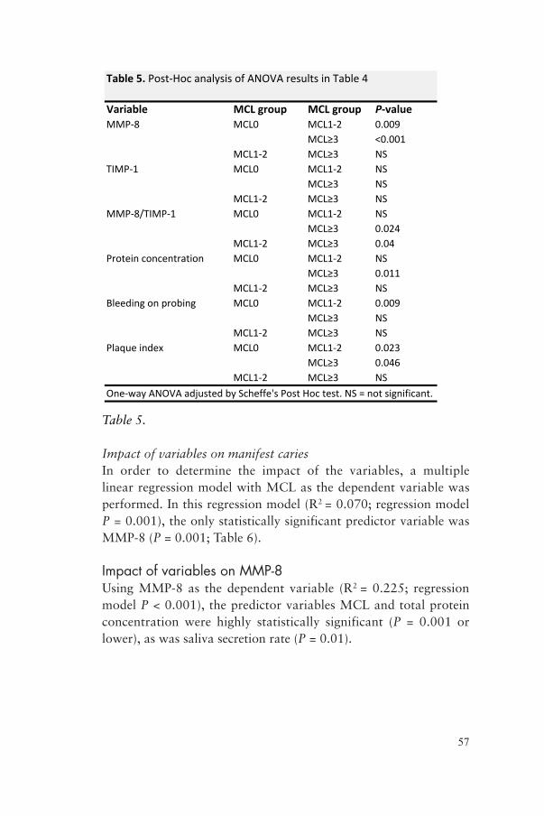
57
Table 5.
Impact of variables on manifest cariesIn order to determine the impact of the variables, a multiple linear regression model with MCL as the dependent variable was performed. In this regression model (R2 = 0.070; regression model P = 0.001), the only statistically significant predictor variable was MMP-8 (P = 0.001; Table 6).
Impact of variables on MMP-8Using MMP-8 as the dependent variable (R2 = 0.225; regression model P < 0.001), the predictor variables MCL and total protein concentration were highly statistically significant (P = 0.001 or lower), as was saliva secretion rate (P = 0.01).
Table&5.&Post%Hoc(analysis(of(ANOVA(results(in(Table(4
Variable MCL&group MCL&group P3valueMMP%8 MCL0 MCL1%2 0.009
MCL≥3 <0.001MCL1%2 MCL≥3 NS
TIMP%1 MCL0 MCL1%2 NSMCL≥3 NS
MCL1%2 MCL≥3 NSMMP%8/TIMP%1 MCL0 MCL1%2 NS
MCL≥3 0.024MCL1%2 MCL≥3 0.04
Protein(concentration MCL0 MCL1%2 NSMCL≥3 0.011
MCL1%2 MCL≥3 NSBleeding(on(probing MCL0 MCL1%2 0.009
MCL≥3 NSMCL1%2 MCL≥3 NS
Plaque(index MCL0 MCL1%2 0.023MCL≥3 0.046
MCL1%2 MCL≥3 NS
One%way(ANOVA(adjusted(by(Scheffe's(Post(Hoc(test.(NS(=(not(significant.
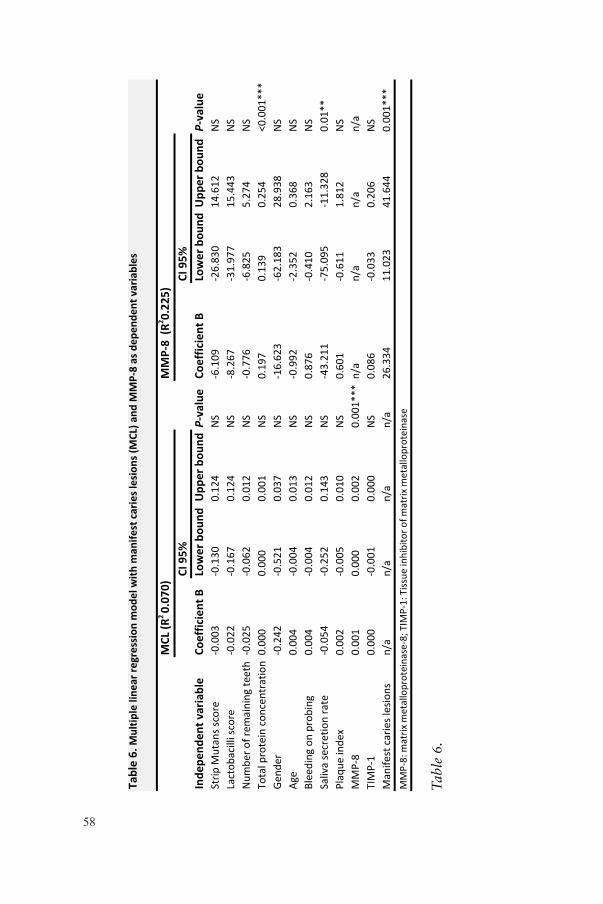
58
Tab
le 6
.
Tabl
e 6.
Mul
tiple
line
ar re
gres
sion
mod
el w
ith m
anife
st c
arie
s les
ions
(MCL
) and
MM
P-‐8
as d
epen
dent
var
iabl
es
MCL
(R2 0.
070)
MM
P-‐8
(R2 0.
225)
CI 9
5%CI
95%
Inde
pend
ent v
aria
ble
Coef
ficie
nt B
Low
er b
ound
Upp
er b
ound
P-‐va
lue
Coef
ficie
nt B
Low
er b
ound
Upp
er b
ound
P-‐va
lue
Strip
Mutan
s sco
re-‐0.003
-‐0.130
0.12
4NS
-‐6.109
-‐26.83
014
.612
NS
Lactob
acilli sco
re-‐0.022
-‐0.167
0.12
4NS
-‐8.267
-‐31.97
7 15
.443
NS
Num
ber o
f rem
aining
teeth
-‐0.025
-‐0.062
0.01
2NS
-‐0.776
-‐6.825
5.27
4NS
Total p
rotein con
centratio
n0.00
00.00
00.00
1NS
0.19
70.13
90.25
4<0
.001
***
Gend
er-‐0.242
-‐0.521
0.03
7NS
-‐16.62
3-‐62.18
3 28
.938
NS
Age
0.00
4-‐0.004
0.01
3NS
-‐0.992
-‐2.352
0.36
8NS
Blee
ding
on pr
obing
0.00
4-‐0.004
0.01
2NS
0.87
6-‐0.410
2.16
3NS
Saliv
a se
cretion rate
-‐0.054
-‐0.252
0.14
3NS
-‐43.21
1-‐75.09
5-‐11.32
80.01
**Plaq
ue in
dex
0.00
2-‐0.005
0.01
0NS
0.60
1-‐0.611
1.81
2NS
MMP-‐8
0.00
10.00
00.00
20.00
1***
n/a
n/a
n/a
n/a
TIMP-‐1
0.00
0-‐0.001
0.00
0NS
0.08
6-‐0.033
0.20
6NS
Man
ifest caries les
ions
n/a
n/a
n/a
n/a
26.334
11.023
41.644
0.00
1***
MMP-‐8: m
atrix
metallopr
oteina
se-‐8; T
IMP-‐1: Tiss
ue in
hibitor o
f matrix
metallopr
oteina
se
59
Paper IVAnalysis of DDM directly after demineralizationSoluble protein assay of original DDMDirectly after demineralization, soluble protein levels of the DDM and supernatant were determined. With EDTA-demineralized samples, the supernatant contained 245.2 g/ml and pellet 55.0 g/ml soluble proteins. AA-demineralized samples demonstrated completely different profiles: the supernatant contained 42.7 g/ml soluble proteins and the pellet 198.4 g/ml.
SDS-PAGE of original DDMThe SDS-PAGE gel from directly after demineralization exhibited distinct collagen type 1 bands in all samples. The characteristic 130 kDa and 115 kDa collagen type 1 bands corresponding to the α1- and α2-chains from human type 1 collagen, respectively, were present in all instances (Figure 11). This demonstrated the presence of collagen type 1 in all fractions of the original DDM, although in different concentrations.
Analysis of DDM after buffer treatmentSDS-PAGE at start and after buffer incubation (T0 and T1)The SDS-PAGE gel from after the buffer treatment displayed the same distinct collagen type 1 bands as the uncentrifuged DDM in all instances, regardless of buffer used (PBS or TESCA) or treatment time (no incubation or 1 week incubation), indicating that collagen type 1 α1- and α2-chains were present in all samples (data not shown).
Collagen measurements after incubation in buffer (T0)HYP levels in pellet for the different demineralization methods, buffer solutions and fractions are given in Table 7. AA demineralization yielded approximately twice the concentration of HYP compared to EDTA demineralization. The difference due to demineralization method was highly statistically significant (P < 0.001, Table 7).
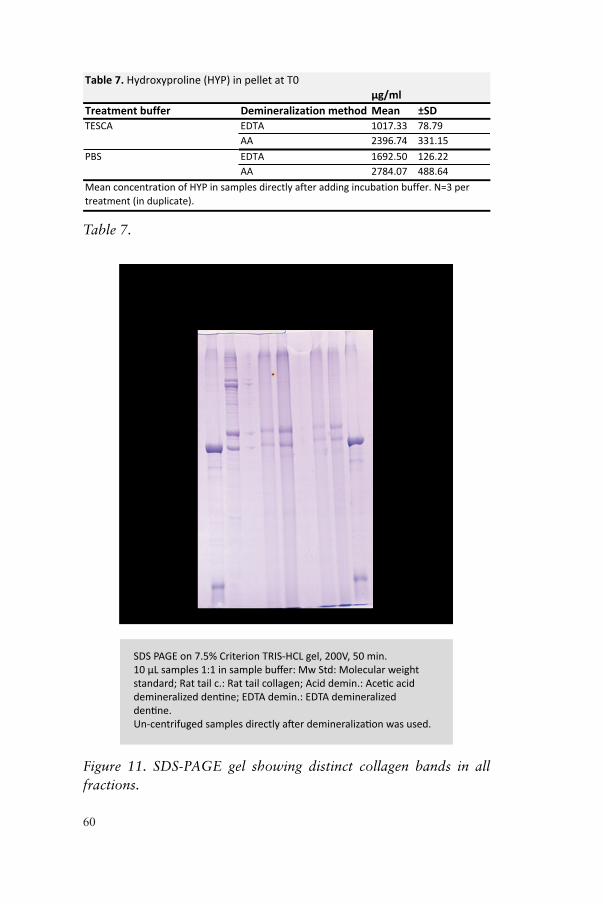
60
Table&7.&Hydroxyproline,(HYP),in,pellet,at,T0
Treatment&buffer Demineralization&method Mean ±SDTESCA EDTA 1017.33 78.79
AA 2396.74 331.15PBS EDTA 1692.50 126.22
AA 2784.07 488.64
µg/ml
Mean,concentration,of,HYP,in,samples,directly,after,adding,incubation,buffer.,N=3,per,treatment,(in,duplicate).
Table 7.
SDS PAGE on 7.5% Criterion TRIS-HCL gel, 200V, 50 min. 10 µL samples 1:1 in sample buffer: Mw Std: Molecular weight standard; Rat tail c.: Rat tail collagen; Acid demin.: Acetic acid demineralized dentine; EDTA demin.: EDTA demineralized dentine.Un-centrifuged samples directly after demineralization was used.
116 kDa
Mw
Std
Rat t
ail c
.
(Em
pty)
EDTA
dem
in.
AA d
emin
.AA
dem
in.
EDTA
dem
in
Mw
Std
(Em
pty)
α-chainβ-chain
Figure 11. SDS-PAGE gel showing distinct collagen bands in all fractions.
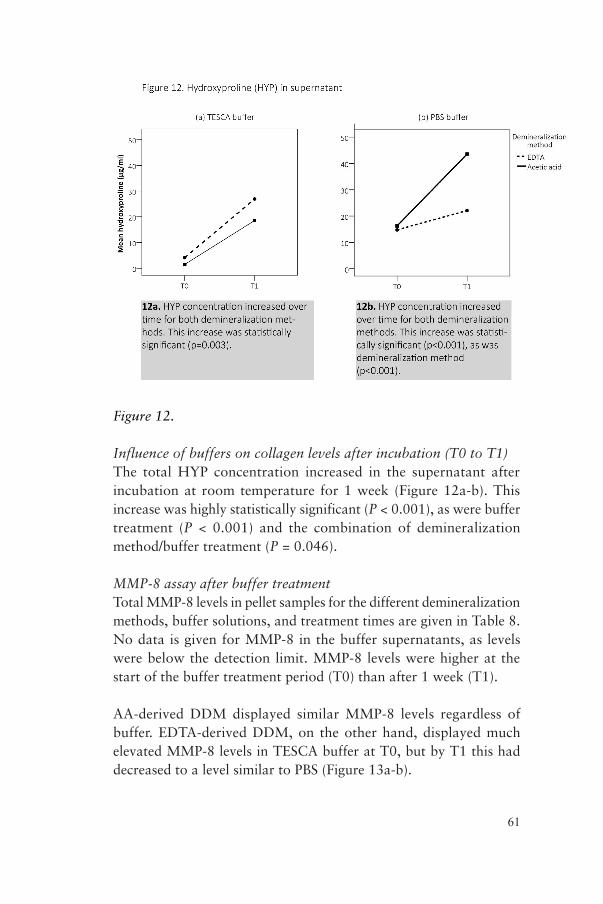
61
Figure 12.
Influence of buffers on collagen levels after incubation (T0 to T1)The total HYP concentration increased in the supernatant after incubation at room temperature for 1 week (Figure 12a-b). This increase was highly statistically significant (P < 0.001), as were buffer treatment (P < 0.001) and the combination of demineralization method/buffer treatment (P = 0.046).
MMP-8 assay after buffer treatmentTotal MMP-8 levels in pellet samples for the different demineralization methods, buffer solutions, and treatment times are given in Table 8. No data is given for MMP-8 in the buffer supernatants, as levels were below the detection limit. MMP-8 levels were higher at the start of the buffer treatment period (T0) than after 1 week (T1).
AA-derived DDM displayed similar MMP-8 levels regardless of buffer. EDTA-derived DDM, on the other hand, displayed much elevated MMP-8 levels in TESCA buffer at T0, but by T1 this had decreased to a level similar to PBS (Figure 13a-b).
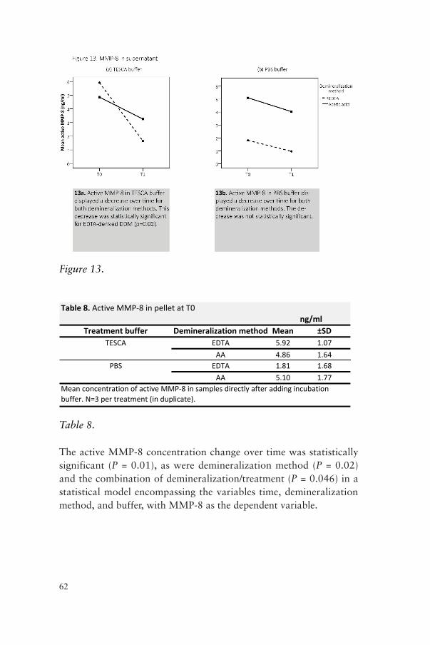
62
Figure 13.
Table&8.&Active'MMP*8'in'pellet'at'T0
Treatment&buffer Demineralization&method Mean ±SDTESCA EDTA 5.92 1.07
AA 4.86 1.64PBS EDTA 1.81 1.68
AA 5.10 1.77
ng/ml
Mean'concentration'of'active'MMP*8'in'samples'directly'after'adding'incubation'buffer.'N=3'per'treatment'(in'duplicate).
Table 8.
The active MMP-8 concentration change over time was statistically significant (P = 0.01), as were demineralization method (P = 0.02) and the combination of demineralization/treatment (P = 0.046) in a statistical model encompassing the variables time, demineralization method, and buffer, with MMP-8 as the dependent variable.
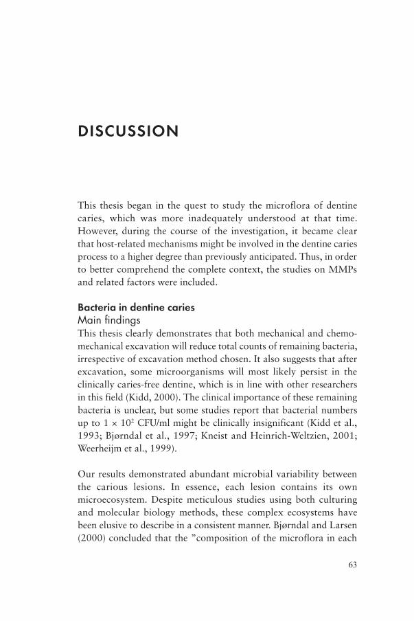
63
DISCUSSION
This thesis began in the quest to study the microflora of dentine caries, which was more inadequately understood at that time. However, during the course of the investigation, it became clear that host-related mechanisms might be involved in the dentine caries process to a higher degree than previously anticipated. Thus, in order to better comprehend the complete context, the studies on MMPs and related factors were included.
Bacteria in dentine cariesMain findingsThis thesis clearly demonstrates that both mechanical and chemo-mechanical excavation will reduce total counts of remaining bacteria, irrespective of excavation method chosen. It also suggests that after excavation, some microorganisms will most likely persist in the clinically caries-free dentine, which is in line with other researchers in this field (Kidd, 2000). The clinical importance of these remaining bacteria is unclear, but some studies report that bacterial numbers up to 1 × 10² CFU/ml might be clinically insignificant (Kidd et al., 1993; Bjørndal et al., 1997; Kneist and Heinrich-Weltzien, 2001; Weerheijm et al., 1999).
Our results demonstrated abundant microbial variability between the carious lesions. In essence, each lesion contains its own microecosystem. Despite meticulous studies using both culturing and molecular biology methods, these complex ecosystems have been elusive to describe in a consistent manner. Bjørndal and Larsen (2000) concluded that the ”composition of the microflora in each
64
lesion is a reflection of the local environment, including the clinical characteristics of that particular lesion,” a declaration that is fully supported by our findings.
The ability to cope with low and fluctuating pH is a crucial trait for a microorganism if it is to succeed within the dentine carious lesion. To study this acid-tolerant subgroup of the total microbiota, we utilized pH-selective agars and found that about 6 to 41% of the total recoverable microbiota was able to grow on pH 5.5 agar, while 0 to 16.5% was able to grow on pH 5.0 agar. This demonstrates that although a large proportion of the total recoverable microbiota is acid-tolerant, the proportion of acid-tolerant microorganisms varies between different carious lesions.
The considerable range of different bacterial species found in this and other reports, implies that a greater variety of microorganisms than previously thought might have the potential to form and contribute to viable dentine caries ecosystems. Studies based on 16S rRNA (PCR) and reverse-capture checkerboard techniques have implicated an ecological niche in dentine caries for several new species (Becker et al., 2002; Munson et al., 2004; Chhour et al., 2005; Aas et al., 2008; Gross et al., 2010; Kianoush et al., 2014). All of these microorganisms could be said to contribute to dentine caries progression, as it is the local microbiota as a whole that needs to be considered, not individual caries-promoting species (Gross et al., 2012).
The solid pH-selective agar method was successful in selecting the acid-tolerant fraction of the dentine caries microbiota, and can be considered a useful tool to study this subgroup of the total lesion flora.
LimitationsMany earlier studies on dentine caries microbiology do not specify sample size, instead using terms like ”a full excavator”. In our studies, sample size was clearly defined as a bur-full of dentin (Figure 5), which was confirmed clinically by ocular means. The robustness of the method was confirmed in a pilot study on dentine weight
65
before actual sampling of the subjects. The dentine sampling method used a modified protocol based on Bjørndal and coworkers (1997) and yielded results on a par with that study. The hard caries-free samples displayed a significant tendency to weigh more despite sample volumes being kept constant. Bjørndal, on the other hand, reported slightly higher (but not significant) yields from carious dentine in a study comparing soft carious dentine with inactive carious dentine. These differences might be due to different sample volumes or varying dentine-drying time. In a recent study using a similar sampling technique, Maltz and coworkers (2012) confirmed that the sampling method we used has a high reproducibility.
When taking samples from different layers of carious dentine there is always the possibility that more superficial microorganisms could be mechanically transferred to a deeper sampling site, thus confusing the results. In order to minimize the risk of cross contamination between layers during the sampling procedure, we used new sterile burs for each sample and washed the cavity with sterile saline prior to each step of sampling and excavation respectively.
The use of a low-concentration hypochlorite compound (0.5% NaOCl) could in theory have influenced the number of recoverable microorganisms in the chemo-mechanical excavation group. However, no statistical difference was found between the different excavation methods (P = 0.995). A study on storing dentine samples in more concentrated chloramine solutions before bacterial incubation experiments also concluded that it is safe to use chloramines, and that the antibacterial effect will diminish to negligible levels after washing, especially in the presence of organic matter (Rolland et al., 2007). Furthermore, the low concentration, low dose, and short contact time of hypochlorite, coupled with the fact that the microbiota sampled was shielded by overlying dentine, makes hypochlorite killing highly unlikely.
Why is acid tolerance important?In this thesis, we were particularly interested in the acid-tolerant microbiota of carious dentin due to its central role in demineralization of dentine and progression of caries lesions. The pH in active dentine
66
carious lesions can drop as low as 4.9 (Kitasako et al., 2002), and consequently acid tolerance would be mandatory for causing demineralization in infected dentine. To study the naturally adapted acid-tolerant sub-microflora of the dentine microbiome, we used pH-selective agars. The results demonstrated that a large proportion of the total recoverable microbiota indeed was acid-tolerant but also that the proportion of acid tolerant microorganisms tended to vary between different carious lesions and, likewise, between different sample layers. Kianoush et al. also (2014) demonstrated this in a recent paper. As above, this could be explained by the variation in ecological determinants in each lesion.
Several sample sites within the carious lesionPrior studies have described the entire carious lesion as a unit, thus not taking the ecological differences within different layers of the lesion into account. Only two previous studies and one very recent one have addressed comparable intralesion levels. Lima et al. (2010) described the dentine caries microbiota from three layers of deep dentine-caries lesions using a reverse-capture checkerboard technique, whereas Banerjee et al. (2002) investigated dentine caries bacteria from four different levels but instead used the FISH (fluorescence in situ hybridization) technique to enumerate the bacteria. Neither of these methods reveals phenotypic characteristics such as acid tolerance of the microorganisms isolated. Moreover, studies on moderately sized dentine carious lesions are rare in the literature; preceding studies have tended to address carious lesions in more advanced states. In a recent study on 25 teeth extracted due to dentine caries and irreversible pulpitis, samples of carious dentine were taken from four to five sites within each lesion (superficial to sound dentine). At the same time, each sample site was also analyzed for pH in order to be able to relate the microbiota to the pH at the site it was sampled from. The dentine samples were subsequently subjected to 454 sequencing in order to examine the microbiome. The researchers found a clear difference between bacterial communities from more-neutral pH regions as compared to those from more-acidic ones, but they also found that a large part of the microbiota (about 60% of the taxa) was present regardless of pH, suggesting a core microbial community in dentine caries (Kianoush et al., 2014).
67
Distribution of bacteria within the carious lesionsUsing pH-neutral media, the major part of the microorganisms was found in the superficial layer or in the center of the carious lesions, never in the caries-free cavity floor. This suggests a distinct distribution of microorganisms in the dentine carious lesion; the advancing front of the lesion seems to harbor rather minute amounts of bacteria.
Interestingly, the numbers of bacteria recovered from the various sample sites were quite high even in the deeper layers (Figure 1). The only statistically significant differences in numbers of bacteria were obtained when the superficial and cavity-floor sample sites in the lesions were compared (P = 0.036), but since the numbers of samples were small, this conclusion must be confirmed by studies including more lesions. Banerjee et al. (2002) also reported this finding.
The impact of acidity in the environmentEarly studies demonstrated that approximately 29% of dentine sample bacteria were able to grow on pH 5.2 agar, as opposed to 11% of the plaque bacteria overlying the carious lesion, indicating an acidic environment in dentine caries lesions (Loesche and Syed, 1973). Our results did confirm this, but the variation was quite sizeable (exemplified by Figure 9 and 10). Furthermore, with decreasing agar plate pH, the number of recoverable microorganisms decreased from max 40.8% at pH 5.5 to max 5.2% at pH 4.0.
Number of different taxa in dentine lesionsThe pH of the agar plates did not affect only the number of CFUs but also the number of recoverable bacterial groups. With lower pH, fewer groups were recovered. Kianoush and coworkers (2014) observed a similar decrease in bacterial diversity when comparing active and nonactive dentine carious lesions. This suggests that an acidic environment has a strong influence on the number of CFUs as well as the composition of the dentine caries microbiota.
On the other hand, the mean number of bacterial groups recovered from the various sample depths (at pH 5.0) was 6.0 (± 2.45) superficially, 5.67 (± 1.70) in the center, and 3.33 (± 2.05) at the
68
cavity floor, suggesting a fairly consistent number of taxa irrespective of sample site. This is consistent with the findings of Lima et al. (2010), who found no significant difference in prevalence of taxa in three distinct layers in carious dentine using the reverse-capture checkerboard technique, and is further confirmed by Banerjee et al. (2002), who also found greater numbers of bacteria through the lesion than previously reported.
Together, these results suggest that the pH of the dentine environment exerts a strong ecological pressure on the resident microbiota whereas the relative depth of the lesion seems to be of lesser importance.
Clinical implicationsTo be able to interfere with the caries process, it is important to understand the environmental factors creating an ecosystem that propagates decay. Methods for noninvasive caries management and for management of caries deliberately left remaining under fillings should be explored because the need for complete caries removal has been questioned (Kidd, 2004) and clinical methods for placing restorations over active dentine caries are now being advocated (Maltz et al., 2007; Alves et al., 2010). Although it is also known that substantial amounts of bacteria remain in the dentine after both complete and incomplete caries removal (Mertz-Fairhurst et al., 1998; Maltz et al., 2002; Lager et al., 2003), it appears that the subsequent reduction of nutrient availability from the oral cavity arrests further lesion progression (Maltz et al., 2007; Ricketts and Pitts, 2009). Nonetheless, a significant amount of these bacteria remain viable, even after prolonged periods under a restoration, and this may be a clinical problem if the restoration fails and the seal toward the oral cavity is broken (Thompson et al., 2008).
RemarksIn the beginning of the work on this thesis, studies on the dentine caries microbiota was scarce and the base knowledge of the biologic mechanisms involved in dentine destruction due to caries was sporadic at best. During this time, several excellent research papers emerged, pointing to a possible combined host-microbial etiology behind the dentine carious process in the form of endogenous
69
enzymes taking part in the collagen degradation process. To further explore that concept, we decided to study these enzymes in relation to dentine caries.
MMPs and dentine cariesMMPs in saliva (III)Main findingsThe main finding (III) was that salivary MMP-8 levels were elevated in subjects with manifest caries lesions, compared with subjects with no manifest caries lesions, regardless of age or gender. The MMP-8/TIMP-1 ratio was also elevated but to a lesser degree. TIMP-1 alone did not exhibit any significant difference between subjects with and without manifest caries lesions. Our results also indicate that the main difference in MMP-8 levels manifested between subjects with and subjects without caries, with no statistical difference between subjects with few or many carious lesions.
LimitationsThe cross-sectional design of the study did not allow for an association between caries activity and salivary MMP-8 levels. However, the clear association between manifest carious lesions and MMP-8 levels suggests a role of this collagenase in the dentine caries process, which needs to be explored further.
The findings correlate well with a recent study where MMP activity was found to be significantly higher in relation to active than chronic dentine lesions (Nascimento et al., 2011). It is conceivable that MMPs leak out into the saliva from the open caries lesions, but it is impossible to establish a cause-effect correlation between salivary MMP-8 and MCL from this study, because the source of MMP-8 is unknown. The MMP-8 may possibly originate from the carious lesions, the GCF, or even the salivary glands.
Source of MMPsThere is evidence suggesting that MMP-8 is released during dentine demineralization, at least partially (Sulkala et al., 2007; Santos et al., 2009). It has also been demonstrated that some of the dentinal MMP-8 remains in the demineralized dentine, where it is believed
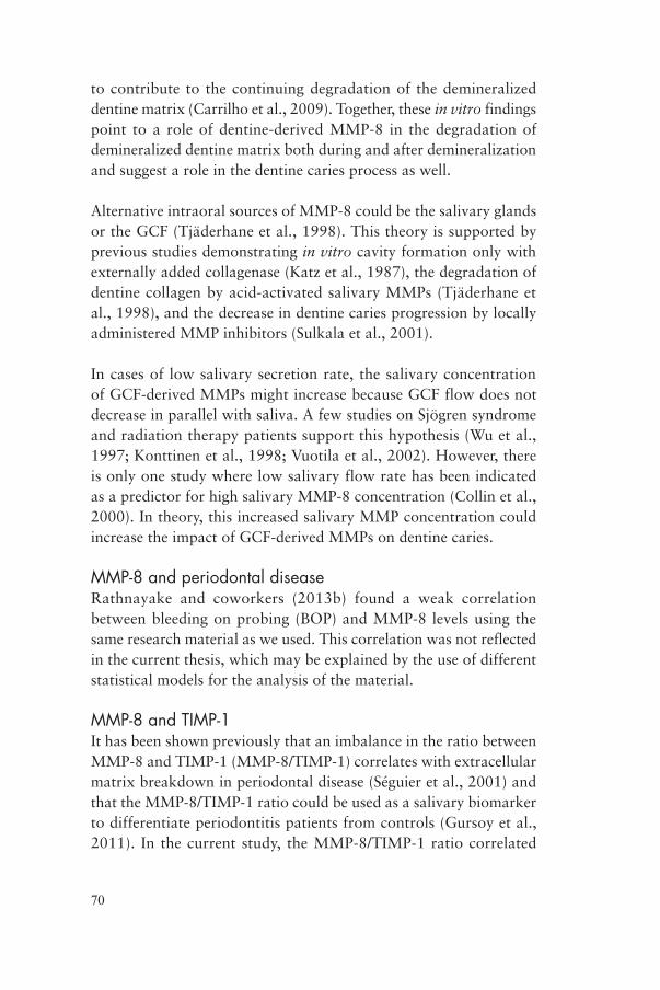
70
to contribute to the continuing degradation of the demineralized dentine matrix (Carrilho et al., 2009). Together, these in vitro findings point to a role of dentine-derived MMP-8 in the degradation of demineralized dentine matrix both during and after demineralization and suggest a role in the dentine caries process as well.
Alternative intraoral sources of MMP-8 could be the salivary glands or the GCF (Tjäderhane et al., 1998). This theory is supported by previous studies demonstrating in vitro cavity formation only with externally added collagenase (Katz et al., 1987), the degradation of dentine collagen by acid-activated salivary MMPs (Tjäderhane et al., 1998), and the decrease in dentine caries progression by locally administered MMP inhibitors (Sulkala et al., 2001).
In cases of low salivary secretion rate, the salivary concentration of GCF-derived MMPs might increase because GCF flow does not decrease in parallel with saliva. A few studies on Sjögren syndrome and radiation therapy patients support this hypothesis (Wu et al., 1997; Konttinen et al., 1998; Vuotila et al., 2002). However, there is only one study where low salivary flow rate has been indicated as a predictor for high salivary MMP-8 concentration (Collin et al., 2000). In theory, this increased salivary MMP concentration could increase the impact of GCF-derived MMPs on dentine caries.
MMP-8 and periodontal diseaseRathnayake and coworkers (2013b) found a weak correlation between bleeding on probing (BOP) and MMP-8 levels using the same research material as we used. This correlation was not reflected in the current thesis, which may be explained by the use of different statistical models for the analysis of the material.
MMP-8 and TIMP-1It has been shown previously that an imbalance in the ratio between MMP-8 and TIMP-1 (MMP-8/TIMP-1) correlates with extracellular matrix breakdown in periodontal disease (Séguier et al., 2001) and that the MMP-8/TIMP-1 ratio could be used as a salivary biomarker to differentiate periodontitis patients from controls (Gursoy et al., 2011). In the current study, the MMP-8/TIMP-1 ratio correlated
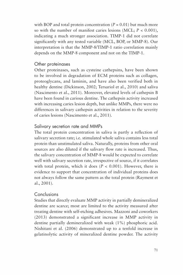
71
with BOP and total protein concentration (P = 0.01) but much more so with the number of manifest caries lesions (MCL; P < 0.001), indicating a much stronger association. TIMP-1 did not correlate significantly with any tested variable (MCL, BOP, or MMP-8). Our interpretation is that the MMP-8/TIMP-1 ratio correlation mainly depends on the MMP-8 component and not on the TIMP-1.
Other proteinasesOther proteinases, such as cysteine cathepsins, have been shown to be involved in degradation of ECM proteins such as collagen, proteoglycans, and laminin, and have also been verified both in healthy dentine (Dickinson, 2002; Tersariol et al., 2010) and saliva (Nascimento et al., 2011). Moreover, elevated levels of cathepsin B have been found in carious dentine. The cathepsin activity increased with increasing caries lesion depth, but unlike MMPs, there were no differences in salivary cathepsin activities in relation to the severity of caries lesions (Nascimento et al., 2011).
Salivary secretion rate and MMPsThe total protein concentration in saliva is partly a reflection of salivary secretion rate; i.e. stimulated whole saliva contains less total protein than unstimulated saliva. Naturally, proteins from other oral sources are also diluted if the salivary flow rate is increased. Thus, the salivary concentration of MMP-8 would be expected to correlate well with salivary secretion rate, irrespective of source, if it correlates with total protein, which it does (P < 0.001). However, there is evidence to support that concentration of individual proteins does not always follow the same pattern as the total protein (Rayment et al., 2001).
ConclusionsStudies that directly evaluate MMP activity in partially demineralized dentine are scarce; most are limited to the activity measured after treating dentine with self-etching adhesives. Mazzoni and coworkers (2013) demonstrated a significant increase in MMP activity in dentine partially demineralized with weak (1%) phosphoric acid. Nishitani et al. (2006) demonstrated up to a tenfold increase in gelatinolytic activity of mineralized dentine powder. The activity
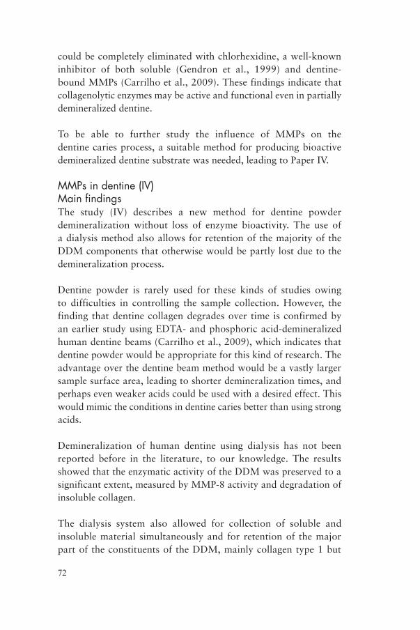
72
could be completely eliminated with chlorhexidine, a well-known inhibitor of both soluble (Gendron et al., 1999) and dentine-bound MMPs (Carrilho et al., 2009). These findings indicate that collagenolytic enzymes may be active and functional even in partially demineralized dentine.
To be able to further study the influence of MMPs on the dentine caries process, a suitable method for producing bioactive demineralized dentine substrate was needed, leading to Paper IV.
MMPs in dentine (IV)Main findingsThe study (IV) describes a new method for dentine powder demineralization without loss of enzyme bioactivity. The use of a dialysis method also allows for retention of the majority of the DDM components that otherwise would be partly lost due to the demineralization process.
Dentine powder is rarely used for these kinds of studies owing to difficulties in controlling the sample collection. However, the finding that dentine collagen degrades over time is confirmed by an earlier study using EDTA- and phosphoric acid-demineralized human dentine beams (Carrilho et al., 2009), which indicates that dentine powder would be appropriate for this kind of research. The advantage over the dentine beam method would be a vastly larger sample surface area, leading to shorter demineralization times, and perhaps even weaker acids could be used with a desired effect. This would mimic the conditions in dentine caries better than using strong acids.
Demineralization of human dentine using dialysis has not been reported before in the literature, to our knowledge. The results showed that the enzymatic activity of the DDM was preserved to a significant extent, measured by MMP-8 activity and degradation of insoluble collagen.
The dialysis system also allowed for collection of soluble and insoluble material simultaneously and for retention of the major part of the constituents of the DDM, mainly collagen type 1 but
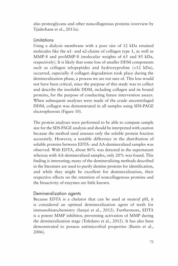
73
also proteoglycans and other noncollagenous proteins (overview by Tjäderhane et al., 2013a).
LimitationsUsing a dialysis membrane with a pore size of 12 kDa retained molecules like the α1- and α2-chains of collagen type 1, as well as MMP-8 and proMMP-8 (molecular weights of 65 and 85 kDa, respectively). It is likely that some loss of smaller DDM components such as collagen telopeptides and hydroxyproline (<12 kDa), occurred, especially if collagen degradation took place during the demineralization phase, a process we are not sure of. This loss would not have been critical, since the purpose of this study was to collect and describe the insoluble DDM, including collagen and its bound proteins, for the purpose of conducting future intervention assays. When subsequent analyses were made of the crude uncentrifuged DDM, collagen was demonstrated in all samples using SDS-PAGE electrophoresis (Figure 10).
The protein analyses were performed to be able to compute sample size for the SDS-PAGE analysis and should be interpreted with caution because the method used assesses only the soluble protein fraction accurately. However, a notable difference in the distribution of soluble proteins between EDTA- and AA-demineralized samples was observed. With EDTA, about 80% was detected in the supernatant whereas with AA-demineralized samples, only 20% was found. This finding is interesting; many of the demineralizing methods described in the literature are used to purify dentine proteins for identification, and while they might be excellent for demineralization, their respective effects on the retention of noncollagenous proteins and the bioactivity of enzymes are little known.
Demineralization agentsBecause EDTA is a chelator that can be used at neutral pH, it is considered an optimal demineralization agent of teeth for immunohistochemistry (Sanjai et al., 2012). Furthermore, EDTA is a potent MMP inhibitor, preventing activation of MMP during the demineralization stage (Toledano et al., 2012). It has also been demonstrated to possess antimicrobial properties (Banin et al., 2006).
74
Comparative studies between different decalcifying methods in terms of preservation or loss of dentinal proteins are lacking. Earlier dentine demineralization studies have often utilized HCl in combination with guanidine, which is a powerful protein-denaturing agent and, in our case would harm the enzymes. AA was chosen for this study based on its weaker and presumably less denaturing properties. Theoretically, AA probably activates MMPs due to its low pH (2.4), but since MMPs have a pH optimum of 7, no or very low activity would take place during the demineralization stage (Okada et al., 1995). The activity could, however, increase during dialysis with water in the washout stage of the dialysis. Total collagen assessmentThe total collagen assessments were performed using HYP as an indicator of collagen levels after complete hydrolysis of the samples. As can be seen in Table 7, the AA-derived DDM pellet levels of total collagen were twice as high as the corresponding EDTA-derived DDM. An explanation for this could be that the resulting DDM from the diverse demineralization methods has differing water retention capacity, which would influence the wet pellet weight. We did not perform complete water removal by lyophilization, as the process might harm bioactive molecules, and especially enzymes (Roy and Gupta, 2004). In addition, repeated freezing and thawing can activate MMPs (Nishitani et al., 2006).
Collagen degradationThe HYP levels increased in the supernatants of all samples (Figure 12a-b), which indicates that collagen is solubilized over time. For AA-derived DDM samples a difference in PBS buffer was demonstrated at T1 (Figure 12a-b). The reason for this could be that the AA demineralization sequence had initiated hydrolysis of the molecules, making them more prone to further degradation. However, the most important finding regarding HYP is that solubilization of DDM collagen can be demonstrated in dentine powder samples, similar to dentine beams (Carrilho et al., 2009).
MMP-8Table 8 presents the endogenously activated MMP-8 data from the buffer-treated pellets. Corresponding supernatant data is not
75
presented as the values were below detection level. For AA-derived DDM the buffer did not affect the MMP-8 values, but in the EDTA-derived DDM group, the MMP-8 values from PBS-treated samples were much lower, indicating that EDTA demineralization could effectively inhibit MMP activity and that Ca2+ from the TESCA buffer was needed to reactivate MMP-8. In contrast, AA demineralization did not seem to inhibit MMP-8 as much.
This is supported by a previous study where dentine powder was demineralized using four different agents, and AA yielded about the double the amount of active MMPs (MMP-2 and -9) after demineralization (Mazzoni et al., 2007). One explanation for higher MMP-8 activity in AA-derived DDM could be acid activation (Tjäderhane et al., 1998) or trace amounts of Ca2+ being present. This was mirrored by the fact that in EDTA-derived DDM, the use of TESCA buffer enhanced MMP-8 activity to a great extent.
Figure 13 reveal that MMP-8 activity decreases with time, in particular for EDTA-derived DDM in TESCA buffer. This decrease in MMP activity was statistically significant (Figure 13a) and could be attributed to depletion of necessary reagents or enzyme activity regulation by feedback inhibition mediated by degradation products. These enzyme activity-limiting processes could differ between the buffers used.
Cysteine cathepsinsThe presence of active MMP-8 and simultaneous degradation of collagen in this experiment suggests that MMP-8 can contribute to collagen breakdown. However, the action of other endogenous enzymes was not assessed. Cysteine cathepsins have been implicated in both activation of MMPs and collagen degradation (Tjäderhane et al., 2013a; Nascimento et al., 2011), but inclusion of these enzymes was beyond the scope of this study.
BuffersTESCA buffer seemed to enhance collagen degradation over time more than PBS buffer (Figure 13a). TESCA buffer also increased MMP-8 activity in EDTA-demineralized DDM compared to PBS.
76
This can be explained by the fact that TESCA buffer contains Ca2+, which is needed for MMP-8 to be able to exert its catabolic action.
ConclusionsThe presented demineralization methods both provided insoluble DDM substrates suitable for further intervention studies. However, it was found that the substrates differed depending on the demineralization method and buffers used. This needs to be studied further in order to find an optimal technique to generate DDM with retained proteins and bioactivity.
77
CONCLUSIONS AND DIRECTIONS FOR FUTURE WORK
I. The numbers of remaining microorganisms after excavation were low for both methods used in the study. However, some microorganisms always remained in the cavity floors even when the cavities were judged as caries free using normal clinical criteria. The consequences of these remaining bacteria are uncertain, but several studies have demonstrated that they probably cannot do any harm as long as the overlying restoration is intact. Future research should be directed at clarifying the role of bacteria remaining under restorations.
II. The microbiota from the manifest carious lesions studied showed a marked diversity, both in numbers of microorganisms and in species composition, which in turn adds to the conceptual idea that each dentine carious lesion can be described as its own microecological system. In turn, this fits very well with the ecological microbial hypothesis, which states that changes in the oral environment force the oral microbiota to adapt and that these events eventually lead to an unbalanced ecosystem that promotes disease. Moreover, the diversity of the found acid-tolerant microorganisms indicates that a larger group of bacteria than was previously thought have the ability to adapt to, survive in, and conceivably propagate dentine caries lesions. The pH-selective agars used were able to select the already acid-tolerant parts of the total dentine caries microflora, thus making the study of them possible.
78
III. Subjects with manifest caries lesions had significantly elevated levels of salivary MMP-8 compared to subjects without caries lesions, even when periodontal variables were included in the statistical model. The level of TIMP-1 was not significant in any case. However, the study could not reveal the source of the salivary MMP-8. This is a project for future research.
IV. In this paper we demonstrate that it is possible to demineralize relatively large amounts of dentine powder without losing MMP-8 bioactivity and that demineralized dentine powder can be used successfully as a substrate for laboratory studies on the mechanisms behind dentine collagen degradation. However, the results also indicate that different hard tissue demineralization methods may influence the bioactivity of the resulting substrate as well as the relative loss of proteins during the demineralization process. It would be interesting to study these differences further in order to pinpoint which demineralization method is appropriate depending on what parameters are being studied.
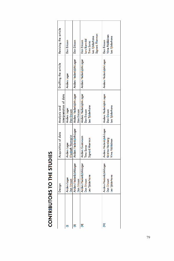
79
80
ACKNOWLEDGEMENTS
This book could not have been written without a lot of support! Thus, I would like to thank all the wonderful people that have contributed to the research and encouraged me throughout the work on my thesis, and especially:
The research subjects who contributed the material for studies I, II, III, and IV. Thanks for sharing your bacteria, saliva, and teeth with me.
Dan Ericson, my supervisor, coauthor and mentor, for your patience and 24-7 support, especially when times were tough, and for generously sharing your considerable knowledge as well as your word jokes. Thank you, sir – I’m most obliged!
Lars Bjørndal, my cosupervisor and coauthor from Copenhagen University, for critical feedback, inspiration, and guidance through the studies and also the CAP project. Thank you Lars – I look forward to more cooperative research projects.
Leo Tjäderhane, my coauthor from Oulu University, for brilliant research feedback, ideas, and profound MMP expertise. And also for letting me visit your lab in Oulu. It has been a privilege to work with you, Leo!
Virve Pääkkönen, my coauthor from Oulu University, for providing excellent guidance in the lab during my stay in Oulu and for making me feel at home there. Also, thanks for providing the dentine powder for Paper IV, as well as teaching me how to weld.
81
Kristina Hamberg and Elisabeth Thornqvist, my coauthors at the Cariology lab, and Birgitta Lindberg at the Oral Biology lab for endless patience and lots of laboratory work on my behalf. Gracious thanks!
All my coauthors that were not mentioned above: Anders Gustafsson from the Karolinska Institute, Timo Sorsa from Helsinki University and Sigvard Åkerman from the Department of Oral Diagnostics at Malmö University. I am indebted to you and look forward to future common research projects.
Eva Larsson, the best research dental nurse in the world, for helping me with all the practicalities of clinical research.
Per-Olof Isberg from Lund University for invaluable assistance and solid statistical expertise. There will always be more questions!
Gunnel Svensäter and Claes Wickström at the Department of Oral Biology for valuable guidance on microbiological matters during the work with Paper II. Thank you.
Anders Michael Nielsen for the quick and precise language review of the thesis.
And all the staff at the Department of Cariology for being fun colleagues and awesome in general. You guys are the department! I would also like to especially thank Anders Hoszek and Peter Carlsson for letting me pick your brains on research ideas and statistics.
Peter Olsson and Roger Larsson at the Centre of Oral Sciences print shop for excellent help in all printing matters, and for always getting those posters ready in time despite short deadlines.
My fellow PhD candidates, past and present, and the PhD Candidate Club for being awesome and good friends. You know who you are! I would especially like to thank Helena Fransson and Mikael Sonesson for good advice regarding the practicalities around writing a thesis.
82
My mother and sister and all our families for love and encouragement over the years. And my father, who passed away in 2002 and never got to see any of this. This book is for you, dad.
And last, but not least, my lovely wife, Marie-Therése and my children, Astrid and Martin, for always being there and for cheering me on and believing in me. You’re the best, and I love you all very much!
Research grants were received from:The Faculty of Odontology, Malmö UniversityThe Swedish Dental Society (multiple grants)The Foundation Swedish Patent Revenue FundTe-Pe Munhygienprodukter AB, Malmö
83
REFERENCES
1. Aas JA, Griffen AL, Dardis SR, Lee AM, Olsen I, Dewhirst FE, Leys EJ, Paster BJ. Bacteria of dental caries in primary and permanent teeth in children and young adults. J Clin Microbiol 2008;46:1407-1417.
2. Almahdy A, Downey FC, Sauro S, Cook RJ, Sherriff M, Richards D, Watson TF, Banerjee A, Festy F. Microbiochemical analysis of carious dentine using raman and fluorescence spectroscopy. Caries Res 2012;46:432-440.
3. Alves LS, Fontanella V, Damo AC, Ferreira de Oliveira E, Maltz M. Qualitative and quantitative radiographic assessment of sealed carious dentin: a 10-year prospective study. Oral Surg Oral Med Oral Pathol Oral Radiol Endod 2010;109:135-141.
4. Banerjee A, Watson TF, Kidd EA. Dentine caries excavation: a review of current clinical techniques. Br Dent J 2000;188:476-482.
5. Banerjee A, Yasseri M, Munson M. A method for the detection and quantification of bacteria in human carious dentine using fluorescent in situ hybridisation. J Dent 2002;30:359-363.
6. Banin E, Brady KM, Greenberg EP. Chelator-induced dispersal and killing of Pseudomonas aeruginosa cells in a biofilm. Appl Environ Microbiol 2006;72:2064-2069.
7. Becker MR, Paster BJ, Leys EJ, Moeschberger ML, Kenyon SG, Galvin JL, Boches SK, Dewhirst FE, Griffen AL. Molecular analysis of bacterial species associated with childhood caries. J Clin Microbiol 2002;40:1001-1009.
8. Beeley JA, Yip HK, Stevenson AG. Chemochemical caries removal: a review of the techniques and latest developments. Br Dent J 2000;188:427-430.
9. Beighton D, Hardie JM, Whiley RA. A scheme for the identification of viridans streptococci. J Med Microbiol 1991a;35:367-372.
84
10. Beighton D, Russell RR, Whiley RA. A simple biochemical scheme for the differentiation of Streptococcus mutans and Streptococcus sobrinus. Caries Res 1991b;25:174-178.
11. Bjørndal L, Larsen T, Thylstrup A. A clinical and microbiological study of deep carious lesions during stepwise excavation using long treatment intervals. Caries Res 1997;31:411-417.
12. Bjørndal L, Larsen T. Changes in the cultivable flora in deep carious lesions following a stepwise excavation procedure. Caries Res 2000;34:502-508.
13. Boushell LW, Nagaoka H, Nagaoka H, Yamauchi M. Increased matrix metalloproteinase-2 and bone sialoprotein response to human coronal caries. Caries Res 2011;45:453-459.
14. Bowden GH, Hamilton IR. Survival of oral bacteria. Crit Rev Oral Biol Med 1998;9:54-85.
15. Bradford MM. A rapid and sensitive method for the quantitation of microgram quantities of protein utilizing the principle of protein-dye binding. Anal Biochem 1976;72:248-254.
16. Bradshaw DJ, Homer KA, Marsh PD, Beighton D. Metabolic cooperation in oral microbial communities during growth on mucin. Microbiology 1994;140:3407-3412.
17. Bradshaw DJ, Marsh PD. Analysis of pH-driven disruption of oral microbial communities in vitro. Caries Res 1998;32:456-462.
18. Carrilho MR, Tay FR, Donnelly AM, Agee KA, Tjäderhane L, Mazzoni A, Breschi L, Foulger S, Pashley DH. Host-derived loss of dentin matrix stiffness associated with solubilization of collagen. J Biomed Mater Res B Appl Biomater 2009;90:373-380.
19. Chaussain C, Boukpessi T, Khaddam M, Tjaderhane L, George A, Menashi S. Dentin matrix degradation by host matrix metalloproteinases: inhibition and clinical perspectives toward regeneration. Front Physiol 2013;4:308.
20. Chaussain-Miller C, Fioretti F, Goldberg M, Menashi S. The role of matrix metalloproteinases (MMPs) in human caries. J Dent Res 2006;85:22-32.
21. Chavez de Paz L, Svensater G, Dahlen G, Bergenholtz G. Streptococci from root canals in teeth with apical periodontitis receiving endodontic treatment. Oral Surgery, Oral Medicine, Oral Pathology, Oral Radiology, and Endodontology 2005;100:232-241.
22. Chhour KL, Nadkarni MA, Byun R, Martin FE, Jacques NA, Hunter N. Molecular analysis of microbial diversity in advanced caries. J Clin Microbiol 2005;43:843-849.
85
23. Chibinski AC, Gomes JR, Camargo K, Reis A, Wambier DS. Bone sialoprotein, matrix metalloproteinases and type I collagen expression after sealing infected caries dentin in primary teeth. Caries Res 2014;48:312-319.
24. Collin HL, Sorsa T, Meurman JH, Niskanen L, Salo T, Rönkä H, Konttinen YT, Koivisto AM, Uusitupa M. Salivary matrix metalloproteinase (MMP-8) levels and gelatinase (MMP-9) activities in patients with type 2 diabetes mellitus. J Periodontal Res 2000;35: 259-265.
25. Dickinson DP. Cysteine peptidases of mammals: their biological roles and potential effects in the oral cavity and other tissues in health and disease. Crit Rev Oral Biol Med 2002;13:238-275.
26. Dirksen TR, Little MF, Bibby BG, Crump SL. The pH of carious cavities. I. The effect of glucose and phosphate buffer on cavity pH. Arch Oral Biol 1962;7:49-57.
27. Dung TZ, Liu AH. Molecular pathogenesis of root dentin caries. Oral Dis 1999;5:92-99.
28. Edwardsson S. Bacteriology of dentin caries. In: Thylstrup A, Leach S, Qvist V (eds). Dentine and dentine reactions in the oral cavity. Oxford: IRL Press Ltd. 1987;95-102.
29. Fusayama T. Two layers of carious dentin; diagnosis and treatment. Oper Dent 1979;4:63-70.
30. Garnero P, Ferreras M, Karsdal MA, Nicamhlaoibh R, Risteli J, Borel O, Qvist P, Delmas PD, Foged NT, Delaissé JM. The type I collagen fragments ICTP and CTX reveal distinct enzymatic pathways of bone collagen degradation. J Bone Miner Res 2003;18:859-867.
31. Gendron R, Grenier D, Sorsa T, Mayrand D. Inhibition of the activities of matrix metalloproteinases 2, 8, and 9 by chlorhexidine. Clin Diagn Lab Immunol 1999;6:437-439.
32. Gross EL, Leys EJ, Gasparovich SR, Firestone ND, Schwartzbaum JA, Janies DA, Asnani K, Griffen AL. Bacterial 16S sequence analysis of severe caries in young permanent teeth. J Clin Microbiol 2010;48: 4121-4128.
33. Gross EL, Beall CJ, Kutsch SR, Firestone ND, Leys EJ, Griffen AL. Beyond Streptococcus mutans: dental caries onset linked to multiple species by 16S rRNA community analysis. PLoS One 2012;7:e47722.
34. Gursoy UK, Könönen E, Pradhan-Palikhe P, Tervahartiala T, Pussinen PJ, Suominen-Taipale L, Sorsa T. Salivary MMP-8, TIMP-1, and ICTP as markers of advanced periodontitis. J Clin Periodontol 2010;37:487-493.
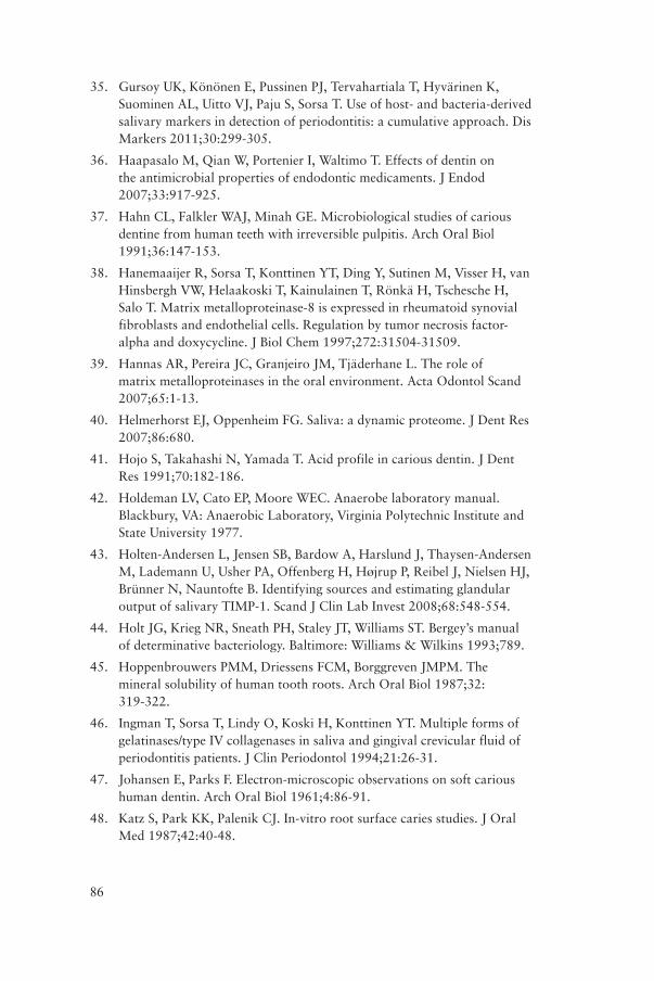
86
35. Gursoy UK, Könönen E, Pussinen PJ, Tervahartiala T, Hyvärinen K, Suominen AL, Uitto VJ, Paju S, Sorsa T. Use of host- and bacteria-derived salivary markers in detection of periodontitis: a cumulative approach. Dis Markers 2011;30:299-305.
36. Haapasalo M, Qian W, Portenier I, Waltimo T. Effects of dentin on the antimicrobial properties of endodontic medicaments. J Endod 2007;33:917-925.
37. Hahn CL, Falkler WAJ, Minah GE. Microbiological studies of carious dentine from human teeth with irreversible pulpitis. Arch Oral Biol 1991;36:147-153.
38. Hanemaaijer R, Sorsa T, Konttinen YT, Ding Y, Sutinen M, Visser H, van Hinsbergh VW, Helaakoski T, Kainulainen T, Rönkä H, Tschesche H, Salo T. Matrix metalloproteinase-8 is expressed in rheumatoid synovial fibroblasts and endothelial cells. Regulation by tumor necrosis factor-alpha and doxycycline. J Biol Chem 1997;272:31504-31509.
39. Hannas AR, Pereira JC, Granjeiro JM, Tjäderhane L. The role of matrix metalloproteinases in the oral environment. Acta Odontol Scand 2007;65:1-13.
40. Helmerhorst EJ, Oppenheim FG. Saliva: a dynamic proteome. J Dent Res 2007;86:680.
41. Hojo S, Takahashi N, Yamada T. Acid profile in carious dentin. J Dent Res 1991;70:182-186.
42. Holdeman LV, Cato EP, Moore WEC. Anaerobe laboratory manual. Blackbury, VA: Anaerobic Laboratory, Virginia Polytechnic Institute and State University 1977.
43. Holten-Andersen L, Jensen SB, Bardow A, Harslund J, Thaysen-Andersen M, Lademann U, Usher PA, Offenberg H, Højrup P, Reibel J, Nielsen HJ, Brünner N, Nauntofte B. Identifying sources and estimating glandular output of salivary TIMP-1. Scand J Clin Lab Invest 2008;68:548-554.
44. Holt JG, Krieg NR, Sneath PH, Staley JT, Williams ST. Bergey’s manual of determinative bacteriology. Baltimore: Williams & Wilkins 1993;789.
45. Hoppenbrouwers PMM, Driessens FCM, Borggreven JMPM. The mineral solubility of human tooth roots. Arch Oral Biol 1987;32: 319-322.
46. Ingman T, Sorsa T, Lindy O, Koski H, Konttinen YT. Multiple forms of gelatinases/type IV collagenases in saliva and gingival crevicular fluid of periodontitis patients. J Clin Periodontol 1994;21:26-31.
47. Johansen E, Parks F. Electron-microscopic observations on soft carious human dentin. Arch Oral Biol 1961;4:86-91.
48. Katz S, Park KK, Palenik CJ. In-vitro root surface caries studies. J Oral Med 1987;42:40-48.
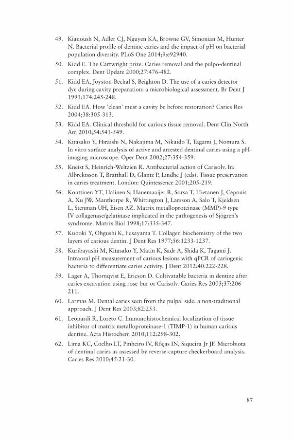
87
49. Kianoush N, Adler CJ, Nguyen KA, Browne GV, Simonian M, Hunter N. Bacterial profile of dentine caries and the impact of pH on bacterial population diversity. PLoS One 2014;9:e92940.
50. Kidd E. The Cartwright prize. Caries removal and the pulpo-dentinal complex. Dent Update 2000;27:476-482.
51. Kidd EA, Joyston-Bechal S, Beighton D. The use of a caries detector dye during cavity preparation: a microbiological assessment. Br Dent J 1993;174:245-248.
52. Kidd EA. How ’clean’ must a cavity be before restoration? Caries Res 2004;38:305-313.
53. Kidd EA. Clinical threshold for carious tissue removal. Dent Clin North Am 2010;54:541-549.
54. Kitasako Y, Hiraishi N, Nakajima M, Nikaido T, Tagami J, Nomura S. In vitro surface analysis of active and arrested dentinal caries using a pH-imaging microscope. Oper Dent 2002;27:354-359.
55. Kneist S, Heinrich-Weltzien R. Antibacterial action of Carisolv. In: Albrektsson T, Bratthall D, Glantz P, Lindhe J (eds). Tissue preservation in caries treatment. London: Quintessence 2001;205-219.
56. Konttinen YT, Halinen S, Hanemaaijer R, Sorsa T, Hietanen J, Ceponis A, Xu JW, Manthorpe R, Whittington J, Larsson A, Salo T, Kjeldsen L, Stenman UH, Eisen AZ. Matrix metalloproteinase (MMP)-9 type IV collagenase/gelatinase implicated in the pathogenesis of Sjögren’s syndrome. Matrix Biol 1998;17:335-347.
57. Kuboki Y, Ohgushi K, Fusayama T. Collagen biochemistry of the two layers of carious dentin. J Dent Res 1977;56:1233-1237.
58. Kuribayashi M, Kitasako Y, Matin K, Sadr A, Shida K, Tagami J. Intraoral pH measurement of carious lesions with qPCR of cariogenic bacteria to differentiate caries activity. J Dent 2012;40:222-228.
59. Lager A, Thornqvist E, Ericson D. Cultivatable bacteria in dentine after caries excavation using rose-bur or Carisolv. Caries Res 2003;37:206-211.
60. Larmas M. Dental caries seen from the pulpal side: a non-traditional approach. J Dent Res 2003;82:253.
61. Leonardi R, Loreto C. Immunohistochemical localization of tissue inhibitor of matrix metalloproteinase-1 (TIMP-1) in human carious dentine. Acta Histochem 2010;112:298-302.
62. Lima KC, Coelho LT, Pinheiro IV, Rôças IN, Siqueira Jr JF. Microbiota of dentinal caries as assessed by reverse-capture checkerboard analysis. Caries Res 2010;45:21-30.
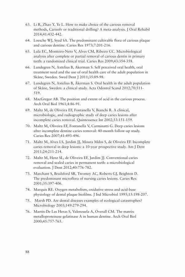
88
63. Li R, Zhao Y, Ye L. How to make choice of the carious removal methods, Carisolv or traditional drilling? A meta-analysis. J Oral Rehabil 2014;41:432-442.
64. Loesche WJ, Syed SA. The predominant cultivable flora of carious plaque and carious dentine. Caries Res 1973;7:201-216.
65. Lula EC, Monteiro-Neto V, Alves CM, Ribeiro CC. Microbiological analysis after complete or partial removal of carious dentin in primary teeth: a randomized clinical trial. Caries Res 2009;43:354-358.
66. Lundegren N, Axtelius B, Akerman S. Self perceived oral health, oral treatment need and the use of oral health care of the adult population in Skåne, Sweden. Swed Dent J 2011;35:89-98.
67. Lundegren N, Axtelius B, Åkerman S. Oral health in the adult population of Skåne, Sweden: a clinical study. Acta Odontol Scand 2012;70:511-519.
68. MacGregor AB. The position and extent of acid in the carious process. Arch Oral Biol 1961;4:86-91.
69. Maltz M, de Oliveira EF, Fontanella V, Bianchi R. A clinical, microbiologic, and radiographic study of deep caries lesions after incomplete caries removal. Quintessence Int 2002;33:151-159.
70. Maltz M, Oliveira EF, Fontanella V, Carminatti G. Deep caries lesions after incomplete dentine caries removal: 40-month follow-up study. Caries Res 2007;41:493-496.
71. Maltz M, Alves LS, Jardim JJ, Moura Mdos S, de Oliveira EF. Incomplete caries removal in deep lesions: a 10-year prospective study. Am J Dent 2011;24:211-214.
72. Maltz M, Henz SL, de Oliveira EF, Jardim JJ. Conventional caries removal and sealed caries in permanent teeth: a microbiological evaluation. J Dent 2012;40:776-782.
73. Marchant S, Brailsford SR, Twomey AC, Roberts GJ, Beighton D. The predominant microflora of nursing caries lesions. Caries Res 2001;35:397-406.
74. Marquis RE. Oxygen metabolism, oxidative stress and acid-base physiology of dental plaque biofilms. J Ind Microbiol 1995;15:198-207.
75. Marsh PD. Are dental diseases examples of ecological catastrophes? Microbiology 2003;149:279-294.
76. Martin-De Las Heras S, Valenzuela A, Overall CM. The matrix metalloproteinase gelatinase A in human dentine. Arch Oral Biol 2000;45:757-765.
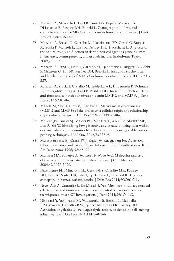
89
77. Mazzoni A, Mannello F, Tay FR, Tonti GA, Papa S, Mazzotti G, Di Lenarda R, Pashley DH, Breschi L. Zymographic analysis and characterization of MMP-2 and -9 forms in human sound dentin. J Dent Res 2007;86:436-440.
78. Mazzoni A, Breschi L, Carrilho M, Nascimento FD, Orsini G, Ruggeri A, Gobbi P, Manzoli L, Tay FR, Pashley DH, Tjäderhane L. A review of the nature, role, and function of dentin non-collagenous proteins. Part II: enzymes, serum proteins, and growth factors. Endodontic Topics 2009;21:19-40.
79. Mazzoni A, Papa V, Nato F, Carrilho M, Tjäderhane L, Ruggeri A, Gobbi P, Mazzotti G, Tay FR, Pashley DH, Breschi L. Immunohistochemical and biochemical assay of MMP-3 in human dentine. J Dent 2011;39:231-237.
80. Mazzoni A, Scaffa P, Carrilho M, Tjäderhane L, Di Lenarda R, Polimeni A, Tezvergil-Mutluay A, Tay FR, Pashley DH, Breschi L. Effects of etch-and-rinse and self-etch adhesives on dentin MMP-2 and MMP-9. J Dent Res 2013;92:82-86.
81. Mäkelä M, Salo T, Uitto VJ, Larjava H. Matrix metalloproteinases (MMP-2 and MMP-9) of the oral cavity: cellular origin and relationship to periodontal status. J Dent Res 1994;73:1397-1406.
82. McLean JS, Fansler SJ, Majors PD, McAteer K, Allen LZ, Shirtliff ME, Lux R, Shi W. Identifying low pH active and lactate-utilizing taxa within oral microbiome communities from healthy children using stable isotope probing techniques. PLoS One 2012;7:e32219.
83. Mertz-Fairhurst EJ, Curtis JWJ, Ergle JW, Rueggeberg FA, Adair SM. Ultraconservative and cariostatic sealed restorations: results at year 10. J Am Dent Assoc 1998;129:55-66.
84. Munson MA, Banerjee A, Watson TF, Wade WG. Molecular analysis of the microflora associated with dental caries. J Clin Microbiol 2004;42:3023-3029.
85. Nascimento FD, Minciotti CL, Geraldeli S, Carrilho MR, Pashley DH, Tay FR, Nader HB, Salo T, Tjäderhane L, Tersariol IL. Cysteine cathepsins in human carious dentin. J Dent Res 2011;90:506-511.
86. Neves Ade A, Coutinho E, De Munck J, Van Meerbeek B. Caries-removal effectiveness and minimal-invasiveness potential of caries-excavation techniques: a micro-CT investigation. J Dent 2011;39:154-162.
87. Nishitani Y, Yoshiyama M, Wadgaonkar B, Breschi L, Mannello F, Mazzoni A, Carvalho RM, Tjäderhane L, Tay FR, Pashley DH. Activation of gelatinolytic/collagenolytic activity in dentin by self-etching adhesives. Eur J Oral Sci 2006;114:160-166.
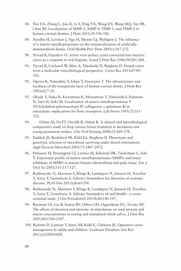
90
88. Niu LN, Zhang L, Jiao K, Li F, Ding YX, Wang DY, Wang MQ, Tay FR, Chen JH. Localization of MMP-2, MMP-9, TIMP-1, and TIMP-2 in human coronal dentine. J Dent 2011;39:536-542.
89. Nordbø H, Leirskar J, Ngo H, Mount GJ, Wahlgren J. The influence of a matrix metalloproteinase on the remineralization of artificially demineralized dentin. Oral Health Prev Dent 2003;1:267-272.
90. Nyvad B, Fejerskov O. Active root surface caries converted into inactive caries as a response to oral hygiene. Scand J Dent Res 1986;94:281-284.
91. Nyvad B, Crielaard W, Mira A, Takahashi N, Beighton D. Dental caries from a molecular microbiological perspective. Caries Res 2013;47:89-102.
92. Ogawa K, Yamashita Y, Ichijo T, Fusayama T. The ultrastructure and hardness of the transparent layer of human carious dentin. J Dent Res 1983;62:7-10.
93. Okada Y, Naka K, Kawamura K, Matsumoto T, Nakanishi I, Fujimoto N, Sato H, Seiki M. Localization of matrix metalloproteinase 9 (92-kilodalton gelatinase/type IV collagenase = gelatinase B) in osteoclasts: implications for bone resorption. Lab Invest 1995;72:311-322.
94. Orhan AI, Oz FT, Ozcelik B, Orhan K. A clinical and microbiological comparative study of deep carious lesion treatment in deciduous and young permanent molars. Clin Oral Investig 2008;12:369-378.
95. Paddick JS, Brailsford SR, Kidd EA, Beighton D. Phenotypic and genotypic selection of microbiota surviving under dental restorations. Appl Environ Microbiol 2005;71:2467-2472.
96. Palosaari H, Pennington CJ, Larmas M, Edwards DR, Tjäderhane L, Salo T. Expression profile of matrix metalloproteinases (MMPs) and tissue inhibitors of MMPs in mature human odontoblasts and pulp tissue. Eur J Oral Sci 2003;111:117-127.
97. Rathnayake N, Akerman S, Klinge B, Lundegren N, Jansson H, Tryselius Y, Sorsa T, Gustafsson A. Salivary biomarkers for detection of systemic diseases. PLoS One 2013a;8:e61356.
98. Rathnayake N, Åkerman S, Klinge B, Lundegren N, Jansson H, Tryselius Y, Sorsa T, Gustafsson A. Salivary biomarkers of oral health – a cross-sectional study. J Clin Periodontol 2013b;40:140-147.
99. Rayment SA, Liu B, Soares RV, Offner GD, Oppenheim FG, Troxler RF. The effects of duration and intensity of stimulation on total protein and mucin concentrations in resting and stimulated whole saliva. J Dent Res 2001;80:1584-1587.
100. Ricketts D, Lamont T, Innes NP, Kidd E, Clarkson JE. Operative caries management in adults and children. Cochrane Database Syst Rev 2013;3:CD003808.
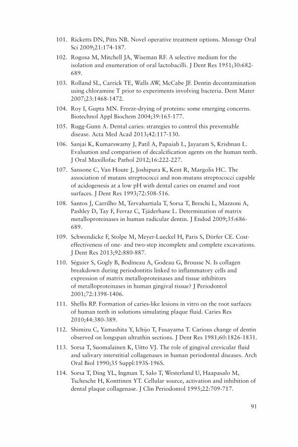
91
101. Ricketts DN, Pitts NB. Novel operative treatment options. Monogr Oral Sci 2009;21:174-187.
102. Rogosa M, Mitchell JA, Wiseman RF. A selective medium for the isolation and enumeration of oral lactobacilli. J Dent Res 1951;30:682-689.
103. Rolland SL, Carrick TE, Walls AW, McCabe JF. Dentin decontamination using chloramine T prior to experiments involving bacteria. Dent Mater 2007;23:1468-1472.
104. Roy I, Gupta MN. Freeze-drying of proteins: some emerging concerns. Biotechnol Appl Biochem 2004;39:165-177.
105. Rugg-Gunn A. Dental caries: strategies to control this preventable disease. Acta Med Acad 2013;42:117-130.
106. Sanjai K, Kumarswamy J, Patil A, Papaiah L, Jayaram S, Krishnan L. Evaluation and comparison of decalcification agents on the human teeth. J Oral Maxillofac Pathol 2012;16:222-227.
107. Sansone C, Van Houte J, Joshipura K, Kent R, Margolis HC. The association of mutans streptococci and non-mutans streptococci capable of acidogenesis at a low pH with dental caries on enamel and root surfaces. J Dent Res 1993;72:508-516.
108. Santos J, Carrilho M, Tervahartiala T, Sorsa T, Breschi L, Mazzoni A, Pashley D, Tay F, Ferraz C, Tjäderhane L. Determination of matrix metalloproteinases in human radicular dentin. J Endod 2009;35:686-689.
109. Schwendicke F, Stolpe M, Meyer-Lueckel H, Paris S, Dörfer CE. Cost-effectiveness of one- and two-step incomplete and complete excavations. J Dent Res 2013;92:880-887.
110. Séguier S, Gogly B, Bodineau A, Godeau G, Brousse N. Is collagen breakdown during periodontitis linked to inflammatory cells and expression of matrix metalloproteinases and tissue inhibitors of metalloproteinases in human gingival tissue? J Periodontol 2001;72:1398-1406.
111. Shellis RP. Formation of caries-like lesions in vitro on the root surfaces of human teeth in solutions simulating plaque fluid. Caries Res 2010;44:380-389.
112. Shimizu C, Yamashita Y, Ichijo T, Fusayama T. Carious change of dentin observed on longspan ultrathin sections. J Dent Res 1981;60:1826-1831.
113. Sorsa T, Suomalainen K, Uitto VJ. The role of gingival crevicular fluid and salivary interstitial collagenases in human periodontal diseases. Arch Oral Biol 1990;35 Suppl:193S-196S.
114. Sorsa T, Ding YL, Ingman T, Salo T, Westerlund U, Haapasalo M, Tschesche H, Konttinen YT. Cellular source, activation and inhibition of dental plaque collagenase. J Clin Periodontol 1995;22:709-717.
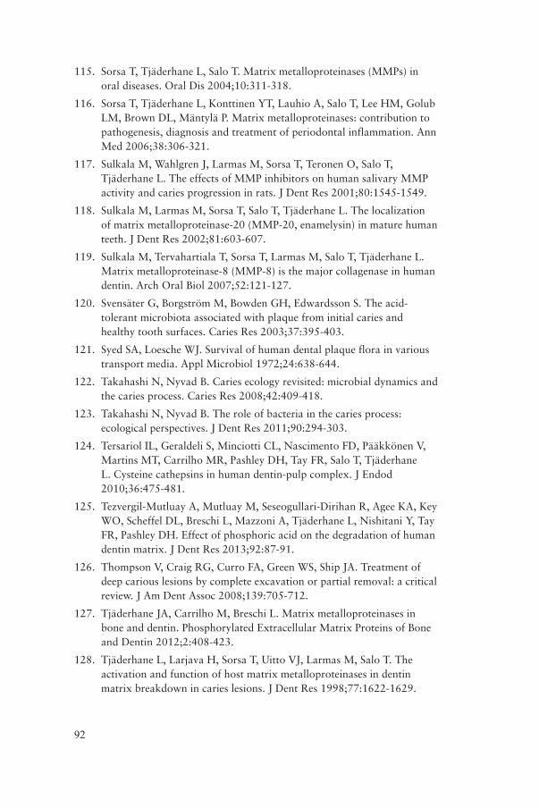
92
115. Sorsa T, Tjäderhane L, Salo T. Matrix metalloproteinases (MMPs) in oral diseases. Oral Dis 2004;10:311-318.
116. Sorsa T, Tjäderhane L, Konttinen YT, Lauhio A, Salo T, Lee HM, Golub LM, Brown DL, Mäntylä P. Matrix metalloproteinases: contribution to pathogenesis, diagnosis and treatment of periodontal inflammation. Ann Med 2006;38:306-321.
117. Sulkala M, Wahlgren J, Larmas M, Sorsa T, Teronen O, Salo T, Tjäderhane L. The effects of MMP inhibitors on human salivary MMP activity and caries progression in rats. J Dent Res 2001;80:1545-1549.
118. Sulkala M, Larmas M, Sorsa T, Salo T, Tjäderhane L. The localization of matrix metalloproteinase-20 (MMP-20, enamelysin) in mature human teeth. J Dent Res 2002;81:603-607.
119. Sulkala M, Tervahartiala T, Sorsa T, Larmas M, Salo T, Tjäderhane L. Matrix metalloproteinase-8 (MMP-8) is the major collagenase in human dentin. Arch Oral Biol 2007;52:121-127.
120. Svensäter G, Borgström M, Bowden GH, Edwardsson S. The acid-tolerant microbiota associated with plaque from initial caries and healthy tooth surfaces. Caries Res 2003;37:395-403.
121. Syed SA, Loesche WJ. Survival of human dental plaque flora in various transport media. Appl Microbiol 1972;24:638-644.
122. Takahashi N, Nyvad B. Caries ecology revisited: microbial dynamics and the caries process. Caries Res 2008;42:409-418.
123. Takahashi N, Nyvad B. The role of bacteria in the caries process: ecological perspectives. J Dent Res 2011;90:294-303.
124. Tersariol IL, Geraldeli S, Minciotti CL, Nascimento FD, Pääkkönen V, Martins MT, Carrilho MR, Pashley DH, Tay FR, Salo T, Tjäderhane L. Cysteine cathepsins in human dentin-pulp complex. J Endod 2010;36:475-481.
125. Tezvergil-Mutluay A, Mutluay M, Seseogullari-Dirihan R, Agee KA, Key WO, Scheffel DL, Breschi L, Mazzoni A, Tjäderhane L, Nishitani Y, Tay FR, Pashley DH. Effect of phosphoric acid on the degradation of human dentin matrix. J Dent Res 2013;92:87-91.
126. Thompson V, Craig RG, Curro FA, Green WS, Ship JA. Treatment of deep carious lesions by complete excavation or partial removal: a critical review. J Am Dent Assoc 2008;139:705-712.
127. Tjäderhane JA, Carrilho M, Breschi L. Matrix metalloproteinases in bone and dentin. Phosphorylated Extracellular Matrix Proteins of Bone and Dentin 2012;2:408-423.
128. Tjäderhane L, Larjava H, Sorsa T, Uitto VJ, Larmas M, Salo T. The activation and function of host matrix metalloproteinases in dentin matrix breakdown in caries lesions. J Dent Res 1998;77:1622-1629.
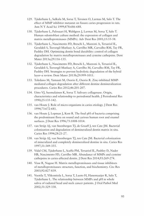
93
129. Tjäderhane L, Sulkala M, Sorsa T, Teronen O, Larmas M, Salo T. The effect of MMP inhibitor metastat on fissure caries progression in rats. Ann N Y Acad Sci 1999;878:686-688.
130. Tjäderhane L, Palosaari H, Wahlgren J, Larmas M, Sorsa T, Salo T. Human odontoblast culture method: the expression of collagen and matrix metalloproteinases (MMPs). Adv Dent Res 2001;15:55-58.
131. Tjäderhane L, Nascimento FD, Breschi L, Mazzoni A, Tersariol IL, Geraldeli S, Tezvergil-Mutluay A, Carrilho MR, Carvalho RM, Tay FR, Pashley DH. Optimizing dentin bond durability: control of collagen degradation by matrix metalloproteinases and cysteine cathepsins. Dent Mater 2013a;29:116-135.
132. Tjäderhane L, Nascimento FD, Breschi L, Mazzoni A, Tersariol IL, Geraldeli S, Tezvergil-Mutluay A, Carrilho M, Carvalho RM, Tay FR, Pashley DH. Strategies to prevent hydrolytic degradation of the hybrid layer–a review. Dent Mater 2013b;29:999-1011.
133. Toledano M, Yamauti M, Osorio E, Osorio R. Zinc-inhibited MMP-mediated collagen degradation after different dentine demineralization procedures. Caries Res 2012;46:201-207.
134. Uitto VJ, Suomalainen K, Sorsa T. Salivary collagenase. Origin, characteristics and relationship to periodontal health. J Periodontal Res 1990;25:135-142.
135. van Houte J. Role of micro-organisms in caries etiology. J Dent Res 1994;73:672-681.
136. van Houte J, Lopman J, Kent R. The final pH of bacteria comprising the predominant flora on sound and carious human root and enamel surfaces. J Dent Res 1996;75:1008-1014.
137. van Strijp AJ, van Steenbergen TJ, de Graaff J, ten Cate JM. Bacterial colonization and degradation of demineralized dentin matrix in situ. Caries Res 1994;28:21-27.
138. van Strijp AJ, van Steenbergen TJ, ten Cate JM. Bacterial colonization of mineralized and completely demineralized dentine in situ. Caries Res 1997;31:349-355.
139. Vidal CM, Tjäderhane L, Scaffa PM, Tersariol IL, Pashley D, Nader HB, Nascimento FD, Carrilho MR. Abundance of MMPs and cysteine cathepsins in caries-affected dentin. J Dent Res 2014;93:269-274.
140. Visse R, Nagase H. Matrix metalloproteinases and tissue inhibitors of metalloproteinases: structure, function, and biochemistry. Circ Res 2003;92:827-839.
141. Vuotila T, Ylikontiola L, Sorsa T, Luoto H, Hanemaaijer R, Salo T, Tjäderhane L. The relationship between MMPs and pH in whole saliva of radiated head and neck cancer patients. J Oral Pathol Med 2002;31:329-338.
94
142. Weerheijm KL, Kreulen CM, de Soet JJ, Groen HJ, van Amerongen WE. Bacterial counts in carious dentine under restorations: 2-year in vivo effects. Caries Res 1999;33:130-134.
143. Weerheijm KL, Groen HJ. The residual caries dilemma. Community Dent Oral Epidemiol 1999;27:436-441.
144. Whiley RA, Beighton D. Current classification of the oral streptococci. Oral Microbiol Immunol 1998;13:195-216.
145. WHO (2012) Fact sheet No318. Oral Health: World Health Organization.
146. Wickström C, Herzberg MC, Beighton D, Svensäter G. Proteolytic degradation of human salivary MUC5B by dental biofilms. Microbiology 2009;155:2866-2872.
147. Wiig H, Reed RK, Tenstad O. Interstitial fluid pressure, composition of interstitium, and interstitial exclusion of albumin in hypothyroid rats. Am J Physiol Heart Circ Physiol 2000;278:H1627-H1639.
148. Wu AJ, Lafrenie RM, Park C, Apinhasmit W, Chen ZJ, Birkedal-Hansen H, Yamada KM, Stetler-Stevenson WG, Baum BJ. Modulation of MMP-2 (gelatinase A) and MMP-9 (gelatinase B) by interferon-gamma in a human salivary gland cell line. J Cell Physiol 1997;171:117-124.
149. Zehnder M, Wegehaupt FJ, Attin T. A first study on the usefulness of matrix metalloproteinase 9 from dentinal fluid to indicate pulp inflammation. J Endod 2011;37:17-20.
150. Zehnder M, Rechenberg DK, Bostanci N, Sisman F, Attin T. Comparison of vehicles to collect dentinal fluid for molecular analysis. J Dent 2014;42:1027-1032.