Embed Size (px)
Citation preview
The impact of focused training onabnormality detection and provision of
accurate preliminary clinical evaluation innewly qualified radiographers
Stevens, BJ and Thompson, JD
http://dx.doi.org/10.1016/j.radi.2017.08.007
Title The impact of focused training on abnormality detection and provision of accurate preliminary clinical evaluation in newly qualified radiographers
Authors Stevens, BJ and Thompson, JD
Type Article
URL This version is available at: http://usir.salford.ac.uk/id/eprint/43815/
Published Date 2018
USIR is a digital collection of the research output of the University of Salford. Where copyright permits, full text material held in the repository is made freely available online and can be read, downloaded and copied for non-commercial private study or research purposes. Please check the manuscript for any further copyright restrictions.
For more information, including our policy and submission procedure, pleasecontact the Repository Team at: [email protected].
1
Introduction 1
2 The Preliminary Clinical Evaluation (PCE) is a commenting scheme designed to improve the 3
specificity of the widely adopted red-dot abnormality detection system; the Society and 4
College of Radiographers(1) are advocates of this system and the Standards for Proficiency 5
outline that radiographers should be able to distinguish abnormal appearances and trauma 6
processes (HCPC 2013). Furthermore, there is an expectation that all radiographers have 7
sufficient knowledge of radiographic anatomy and common abnormalities (Education and 8
Career Framework for the Radiography Workforce document (SOR 2013), which would 9
facilitate effective participation in a PCE system. PCE provides radiographers with an 10
opportunity to have a positive impact on timely patient management. Effective 11
communication of abnormal findings is considered to reduce the time-to-diagnosis, which 12
may also have an impact on the length of hospital stay(2). Despite recognised benefits, there 13
has been minimal publication of large-scale empirical studies confirming the success of PCE. 14
The uptake of PCE has been slow with the suggestion that this may in part be due to the 15
increase of reporting radiographer activity(3). If PCE is to be a worthy successor to the red-16
dot abnormality detection system, radiographers must provide a service that is accurate, 17
and an effective driver of improved patient outcomes. 18
The meta-analysis by Brealey et al(4) suggests radiographers have good accuracy when using 19
a red-dot abnormality detection system, albeit against varying reference standards with 20
associated differential verification biases. Very little exists by way of objective observer 21
studies that assess performance but a few recent studies aptly illustrate the image 22
interpretation abilities of radiographers. 23
24
2
Piper and Paterson(5) undertook an alternative free-response receiver operating 25
characteristic (AFROC) study to assess the effect of training on the ability of 38 participants 26
(radiographers and nurses) to accurately locate an abnormality and to simply state the 27
nature of the abnormality. Improvements were observed after training with radiographers 28
demonstrating post-training increases in figure of merit (0.63 to 0.73), sensitivity (60% to 29
69%), and specificity (73% to 83%), respectively. 30
The FROC study by McEntee and Dunnion(6) indicated that radiographers can accurately 31
detect abnormal wrist images with sensitivity comparable to that of radiologists 32
(radiographers 87.7%, radiologists 88.9%), but specificity is poor (radiographers 64.4%, 33
radiologists 80.5%). McEntee and Dunnion(6) concluded that, although not statistically 34
significant, the number of years of experience could positively affect interpretation skill; 35
they did not however assess the effects of training on performance. Earlier work by Hardy & 36
Culpan(7) has proven that sensitivity and specificity levels do improve following training; 72% 37
to 88% and 50% to 53%%, respectively. 38
It is generally accepted that an increasing number of years of radiographic experience will 39
have a positive impact on the correct interpretation of trauma images. In less experienced 40
staff it is likely that providing training for newly qualified radiographers would expedite 41
accurate contributions in a PCE system. 42
Despite claims of good accuracy, it is thought that PCE has not been widely implemented 43
due to a perceived lack of confidence and inadequate training(2,8) with previous research 44
suggesting that the requirement to provide a written comment caused a reduction in 45
abnormality detection accuracy(7, 9). However, this is not a universal opinion, where it has 46
been suggested that good red-dot performance indicates an ability to provide a written 47
3
comment(10). If training issues do exist, and are not addressed appropriately, then the 48
effectiveness of the PCE could be restricted(7). 49
Much of the previous work discussing the uptake of PCE focuses on the quality of training 50
and the preparedness of radiographers to provide an accurate PCE comment. Graduate 51
radiographers are expected to have sufficient image interpretation ability, despite a lack of 52
certification of competency(9). The aim of this paper is to evaluate the fracture detection 53
performance and PCE accuracy of a small sample of graduate radiographers using an 54
objective observer study to assess detection accuracy, and a scoring system to assess 55
commenting accuracy. Given that questions remain about training and the ability of 56
radiographers to provide a comment, this study will operate a pre- and post-training design 57
to assess the impact of focussed training on a graduate radiographer’s ability to accurately 58
localise and describe a red-dot type abnormality. 59
60
Materials & Methods 61
62
Local Research and Development, and the Health Research Authority(11) decided that the 63
project was suitable as service evaluation. The clinical cases selected were all acquired more 64
than 12-months prior to this study. This reduces the likelihood of new fractures being 65
detected on our review of the cases, since the patient is likely to have presented 66
symptomatically in this time period if an occult fracture had been present. This was 67
important to ensure the correct fracture status in normal and abnormal images. Where 68
follow-up imaging was available, it was reviewed to ensure that no occult fractures were 69
present on cases used in the observer study. All observers provided written consent. 70
4
71
Case Selection 72
A three-month audit of abnormality prevalence for all examinations of trauma to single 73
appendicular parts was undertaken in the study centre revealing a 29.4% incidence of 74
abnormality. We used this data to determine the number of normal/abnormal cases 75
(prevalence) for the observer study, and also the distribution of appendicular examinations 76
that should be included. The range of the subtlety of abnormalities within the selected cases 77
was also consistent with the local workload. One of the authors (BS) compiled the caseload 78
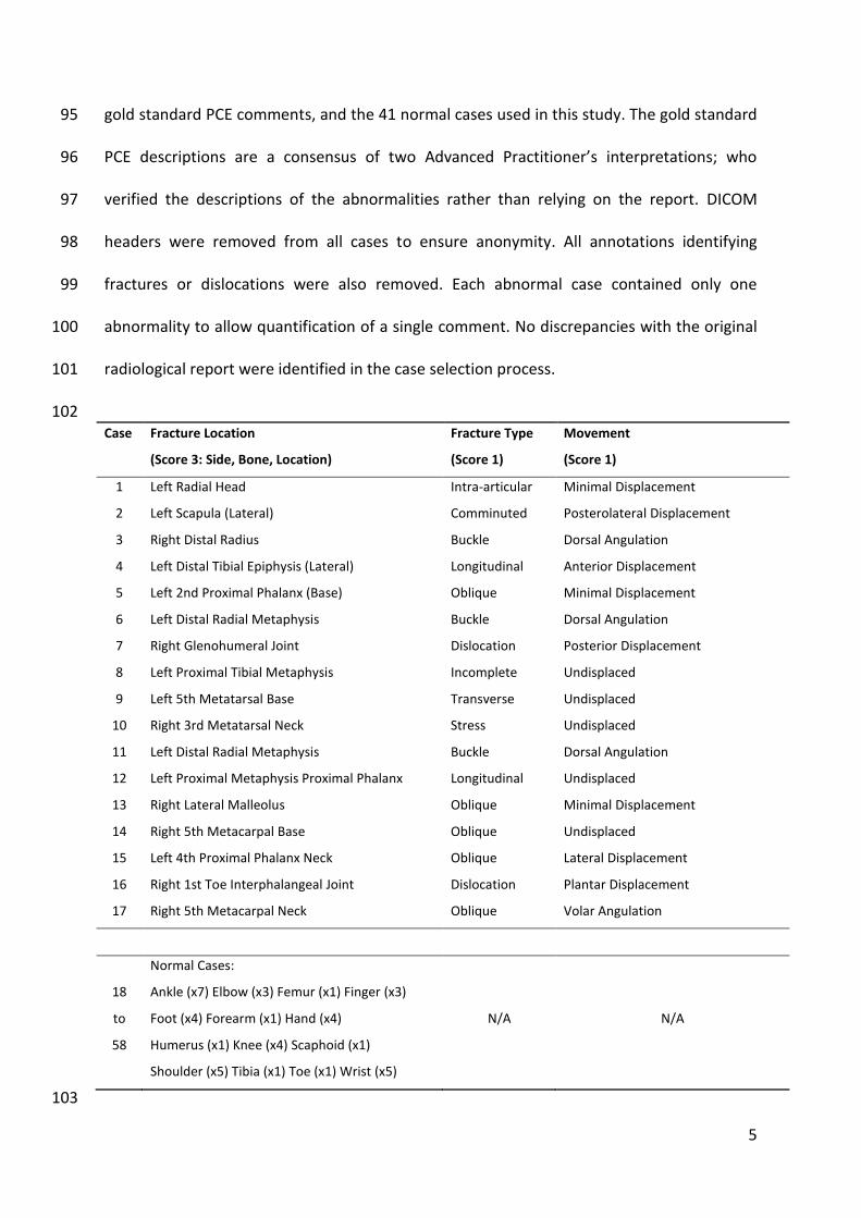
based on the findings of the abnormality prevalence audit. Replicating the local clinical 79
workload provides a comparative assessment of participant interpretation, relative to their 80
clinical practice(12). We performed a sample size calculation to predict the required number 81
of cases, based on six observers completing the study. Obuchowski(13) developed a 82
mathematical model to provide sample size tables for ROC analyses based on the intricate 83
relationships of accuracy, inter-observer variability, patient variability and the correlations 84
in accuracy imposed by the study design. Test alpha was set at 0.05 to control the 85
probability of Type I error, while the power is set at 80%. We estimated that 58 cases would 86
be required for a suitably powered study with a ratio of 4:1 (negative: positive) cases. This 87
ratio was the nearest to the 29.4% prevalence of abnormal cases established from our audit. 88
The image bank of 58 examinations consisted of 17 abnormal appendicular examinations 89
and 41 normal appendicular examinations. Cases containing normal variants were not 90
excluded and were considered as normal. The mean distribution of each appendicular 91
examination over the previous three months was calculated alongside the percentage 92
occurrence. The percentage occurrence was then applied to the sample size to provide the 93
number of each examinations required. Table 1 summarises the 17 abnormal cases and the 94
5
gold standard PCE comments, and the 41 normal cases used in this study. The gold standard 95
PCE descriptions are a consensus of two Advanced Practitioner’s interpretations; who 96
verified the descriptions of the abnormalities rather than relying on the report. DICOM 97
headers were removed from all cases to ensure anonymity. All annotations identifying 98
fractures or dislocations were also removed. Each abnormal case contained only one 99
abnormality to allow quantification of a single comment. No discrepancies with the original 100
radiological report were identified in the case selection process. 101
102 Case Fracture Location
(Score 3: Side, Bone, Location)
Fracture Type
(Score 1)
Movement
(Score 1)
1 Left Radial Head Intra-articular Minimal Displacement
2 Left Scapula (Lateral) Comminuted Posterolateral Displacement
3 Right Distal Radius Buckle Dorsal Angulation
4 Left Distal Tibial Epiphysis (Lateral) Longitudinal Anterior Displacement
5 Left 2nd Proximal Phalanx (Base) Oblique Minimal Displacement
6 Left Distal Radial Metaphysis Buckle Dorsal Angulation
7 Right Glenohumeral Joint Dislocation Posterior Displacement
8 Left Proximal Tibial Metaphysis Incomplete Undisplaced
9 Left 5th Metatarsal Base Transverse Undisplaced
10 Right 3rd Metatarsal Neck Stress Undisplaced
11 Left Distal Radial Metaphysis Buckle Dorsal Angulation
12 Left Proximal Metaphysis Proximal Phalanx Longitudinal Undisplaced
13 Right Lateral Malleolus Oblique Minimal Displacement
14 Right 5th Metacarpal Base Oblique Undisplaced
15 Left 4th Proximal Phalanx Neck Oblique Lateral Displacement
16 Right 1st Toe Interphalangeal Joint Dislocation Plantar Displacement
17 Right 5th Metacarpal Neck Oblique Volar Angulation
18
to
58
Normal Cases:
Ankle (x7) Elbow (x3) Femur (x1) Finger (x3)
Foot (x4) Forearm (x1) Hand (x4)
Humerus (x1) Knee (x4) Scaphoid (x1)
Shoulder (x5) Tibia (x1) Toe (x1) Wrist (x5)
N/A N/A
103
6
Table 1: Breakdown of the image case mix used showing the gold standard PCE comment for each of the 104
abnormal images. 105
106
107
Observer Performance Study & PCE Scoring 108
Four observers evaluated the 58 cases on two occasions: (i) pre-training and (ii) post-109
training. All observers were in a preceptorship period; eight weeks of training elapsed 110
between the two evaluations. We based our sample size calculation on 6 observers, but only 111
4 were able to complete the study. For one of the observers it transpired that they did not 112
fulfil the inclusion criteria (newly-qualified radiographer, first-appointment), and for another 113
there was an unavoidable delay in commencing their employment, therefore they were 114
excluded from the study. An eight-week training schedule, separating the pre- and post-115
training evaluations, consisted of intensive educational sessions designed to deliver 116
information relative to abnormality detection. The sessions were designed and delivered by 117
one of the authors (BS), Advanced Practitioner (skeletal reporting). The introductory session 118
covered basic terminology and concepts, which familiarised participants to a systematic 119
approach of detecting a fracture, forces and fracture patterns, established vocabulary, and a 120
model of forming a comment. All appendicular body parts were covered; each session 121
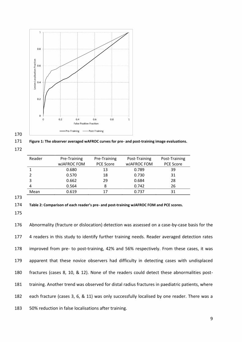
followed the same format, which included radiographic anatomical knowledge, common 122
fractures, assessment lines and measurements, concepts relative to each body part and the 123
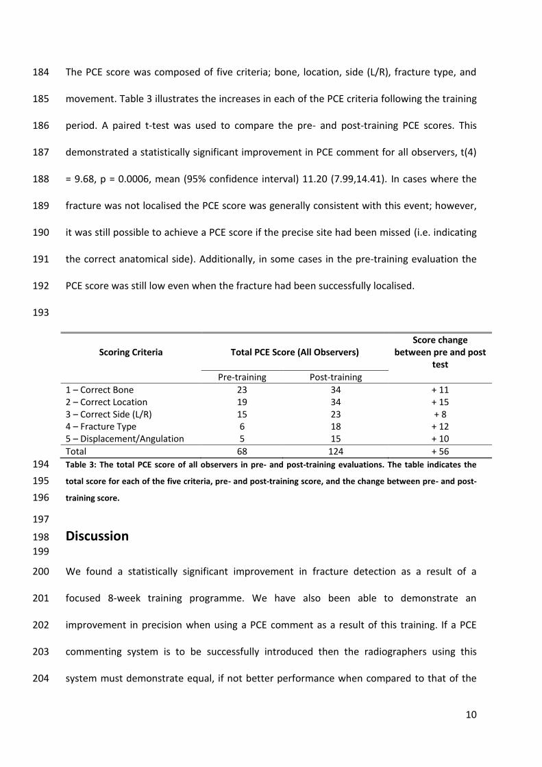
relevant abnormal cases, as well as examples to practice forming a comment. 124
All observers were trained to use the software for the observer study and how to approach 125
the study. They were given a test set of 10 images with which they were asked to localise 126
suspicious areas and provide a PCE comment. This test-set could be repeated until the 127
observer was confident with the data collection method. Each case could include 2-4 128
7
images, depending on the type of examination. Observers were instructed to mark all areas 129
suspicious of fracture/dislocation with a mouse click; this prompted an unmarked slider-bar 130
rating scale to appear with which they could indicate confidence (1-10) in their decision. 131
Moving the slider further to the right indicated increased confidence. Since multiple images 132
were available for localisation (i.e. AP and lateral), it was possible that a fracture could be 133
localised on more than one image. In such cases, we took the highest rating, as only one 134
rating could be used per fracture/dislocation in the analysis. It was not necessary for the 135
observers to mark the fracture on all projections for it to be deemed a successful 136
localisation. An acceptance radius classified observer marks; and a visual assessment 137
confirmed whether mark-rating pairs were true or false. All image evaluations were 138
completed on a 20” LCD flat panel monitor at 60Hz (NEC MultiSync LCD 2090UXI, 600 x 139
1200, NEC Display Solutions, Itasca, Illinois, USA) using ROCView(14) to record observer 140
responses. Each image evaluation was completed in a different randomised order. 141
For each localisation the observers were also asked to provide a PCE comment. Pre-training 142
comments were based on experience from undergraduate education. Post-training they 143
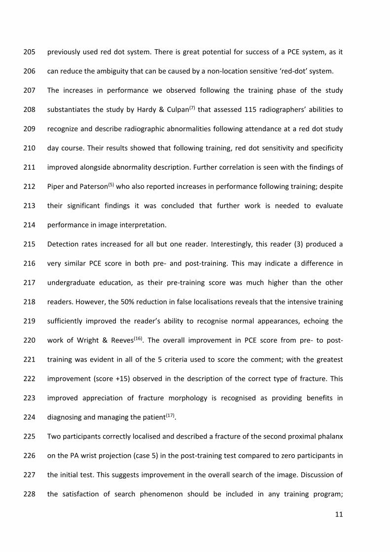
were expected to be familiar with the components of an accurate PCE comment, following 144
the eight week training programme. They were scored on the following components, with 145
each assigned a single point for a maximum score of 5 for each comment: name of bone, 146
location of fracture, anatomical side (L/R), fracture type, and the presence of any 147
movement, such as displacement or angulation. A gold standard comment was agreed by 148
two experienced musculoskeletal reporting advanced practitioners. 149
150
Statistical Analysis 151
8
We are interested in the accuracy of the clinical comment and the precise localisation of 152
abnormalities. The equally weighted jack-knife alternative FROC JAFROC (wJAFROC) figure 153
of merit is sensitive to location information and defines probability that a true abnormality 154
is rated with higher confidence than a false localisation(15). Data was analysed using Rjafroc; 155
an implementation of wJAFROC analysis in the R programming language. A difference in 156
abnormality detection between pre- and post-training was considered significant if the 157
result of the overall F-test was significant and the 95% confidence interval (CI) did not 158
include zero. Test alpha was set at 0.05. 159
160
Results 161
162
A significant difference in fracture detection performance was found between pre- and 163
post-training evaluations for a fixed reader random case analysis (F (1,57) = 10.57, p = 164
0.0019). The reader averaged wJAFROC FOM and 95% CIs for pre- and post-training were 165
0.619 (0.516, 0.737) and 0.703 (0.622, 0.852) respectively. The reader averaged wJAFROC 166
curves are displayed in Figure 1. All readers demonstrated improvement from pre- to post-167
training, as evidenced by the increase in wJAFROC FOM, Table 2. 168
169
9
170
Figure 1: The observer averaged wAFROC curves for pre- and post-training image evaluations. 171
172
Reader Pre-Training wJAFROC FOM
Pre-Training PCE Score
Post-Training wJAFROC FOM
Post-Training PCE Score
1 0.680 13 0.789 39 2 0.570 18 0.730 31 3 0.662 29 0.684 28 4 0.564 8 0.742 26
Mean 0.619 17 0.737 31
173
Table 2: Comparison of each reader’s pre- and post-training wJAFROC FOM and PCE scores. 174
175
Abnormality (fracture or dislocation) detection was assessed on a case-by-case basis for the 176
4 readers in this study to identify further training needs. Reader averaged detection rates 177
improved from pre- to post-training, 42% and 56% respectively. From these cases, it was 178
apparent that these novice observers had difficulty in detecting cases with undisplaced 179
fractures (cases 8, 10, & 12). None of the readers could detect these abnormalities post-180
training. Another trend was observed for distal radius fractures in paediatric patients, where 181
each fracture (cases 3, 6, & 11) was only successfully localised by one reader. There was a 182
50% reduction in false localisations after training. 183
10
The PCE score was composed of five criteria; bone, location, side (L/R), fracture type, and 184
movement. Table 3 illustrates the increases in each of the PCE criteria following the training 185
period. A paired t-test was used to compare the pre- and post-training PCE scores. This 186
demonstrated a statistically significant improvement in PCE comment for all observers, t(4) 187
= 9.68, p = 0.0006, mean (95% confidence interval) 11.20 (7.99,14.41). In cases where the 188
fracture was not localised the PCE score was generally consistent with this event; however, 189
it was still possible to achieve a PCE score if the precise site had been missed (i.e. indicating 190
the correct anatomical side). Additionally, in some cases in the pre-training evaluation the 191
PCE score was still low even when the fracture had been successfully localised. 192
193
Scoring Criteria
Total PCE Score (All Observers)
Score change between pre and post
test
Pre-training Post-training
1 – Correct Bone 23 34 + 11 2 – Correct Location 19 34 + 15 3 – Correct Side (L/R) 15 23 + 8 4 – Fracture Type 6 18 + 12 5 – Displacement/Angulation 5 15 + 10
Total 68 124 + 56
Table 3: The total PCE score of all observers in pre- and post-training evaluations. The table indicates the 194
total score for each of the five criteria, pre- and post-training score, and the change between pre- and post-195
training score. 196
197
Discussion 198
199
We found a statistically significant improvement in fracture detection as a result of a 200
focused 8-week training programme. We have also been able to demonstrate an 201
improvement in precision when using a PCE comment as a result of this training. If a PCE 202
commenting system is to be successfully introduced then the radiographers using this 203
system must demonstrate equal, if not better performance when compared to that of the 204
11
previously used red dot system. There is great potential for success of a PCE system, as it 205
can reduce the ambiguity that can be caused by a non-location sensitive ‘red-dot’ system. 206
The increases in performance we observed following the training phase of the study 207
substantiates the study by Hardy & Culpan(7) that assessed 115 radiographers’ abilities to 208
recognize and describe radiographic abnormalities following attendance at a red dot study 209
day course. Their results showed that following training, red dot sensitivity and specificity 210
improved alongside abnormality description. Further correlation is seen with the findings of 211
Piper and Paterson(5) who also reported increases in performance following training; despite 212
their significant findings it was concluded that further work is needed to evaluate 213
performance in image interpretation. 214
Detection rates increased for all but one reader. Interestingly, this reader (3) produced a 215
very similar PCE score in both pre- and post-training. This may indicate a difference in 216
undergraduate education, as their pre-training score was much higher than the other 217
readers. However, the 50% reduction in false localisations reveals that the intensive training 218
sufficiently improved the reader’s ability to recognise normal appearances, echoing the 219
work of Wright & Reeves(16). The overall improvement in PCE score from pre- to post-220
training was evident in all of the 5 criteria used to score the comment; with the greatest 221
improvement (score +15) observed in the description of the correct type of fracture. This 222
improved appreciation of fracture morphology is recognised as providing benefits in 223
diagnosing and managing the patient(17). 224
Two participants correctly localised and described a fracture of the second proximal phalanx 225
on the PA wrist projection (case 5) in the post-training test compared to zero participants in 226
the initial test. This suggests improvement in the overall search of the image. Discussion of 227
the satisfaction of search phenomenon should be included in any training program; 228
12
whereby the detection of one abnormality interferes with detection of another, and is often 229
affected by knowledge of common fractures(18). This level of understanding may not 230
manifest itself in the search strategy of newly qualified radiographers. 231
In this study we have a trend of a failure to detect buckle fractures of the paediatric distal 232
radius, and this correlates with the findings of previous work(19). There were also difficulties 233
in detecting subtle and undisplaced fractures; all of these findings could help direct training 234
for newly qualified radiographers. We recommend that intensive PCE training should be 235
included in the preceptorship program or during the transitional period from graduate to 236
independent practitioner. It must be stressed though that the issue of sustaining any 237
improvements in performance is just as challenging as attaining the desired level. Previous 238
work by Mackay (2006) indicated that the immediate improvements in abnormality 239
detection following training were not demonstrable after 6 months; reinforcing the need for 240
regular CPD sessions to maintain standards, not just for newly qualified radiographers but 241
also those who are more experienced. For the newly qualified radiographer the transition 242
from student to practitioner can be quite daunting. However, the pressure of contributing 243
successfully to a PCE system can be reduced by this comparatively simple, cheap and regular 244
departmental training intervention. 245
This study has demonstrated the effectiveness of the method we proposed; the study 246
should now be repeated with a larger sample size and over a larger number of cases in 247
order to generalise the results to the population of newly qualified radiographers. However, 248
the initial results are encouraging, where we have demonstrated the effectiveness of a 249
focussed training programme to improve fracture detection rates and the accuracy of a PCE 250
comment. Experiential learning, peer support and educational reading cannot be excluded 251
13
as potential influences on the performance increase from pre- to post-training evaluations, 252
but it would not be practical to conduct this study in isolation of any these external factors. 253
As with all observer studies using a test/re-test method there is a risk of memory effects 254
influencing the second evaluation. However, the 8-week period between evaluations, 255
randomisation of image order and the fact that the observers would see a large number of 256
other clinical cases during this time as part of their daily work do limit this effect. Another 257
limitation of this work is the relatively small sample of observers and the fact that the 258
clinical cases, and estimation of fracture prevalence, were drawn from a single centre. 259
However, we believe the methods applied to be robust, but would be strengthened by a 260
multi-centre approach. The sample of observers was reduced from our original calculation; 261
this will have a negative impact on the power of the study. 262
Future work could also assess the impact of the accuracy of a PCE comment on emergency 263
practitioners’ evaluation of the image, and the speed and appropriateness of care delivered 264
to the patient as they return to the emergency department. 265
266 267
Conclusion 268
269
This study found a statistically significant improvement from pre- to post-training fracture 270
detection performance. Post-training PCE scores also showed an overall increase. These 271
results were also consolidated by a 50% reduction in false localisations post-training. A 272
larger, multi-centre study, using a greater number of observers should be conducted to 273
provide a result that can be generalised to the population of UK radiographers. However, on 274
the basis of these findings we recommend an intensive training program would benefit 275
14
newly qualified radiographers in providing the necessary framework for participating in a 276
PCE system. 277
278 279
Conflict of Interest 280
281 No conflicts of interest influenced this work. 282 283 284
References 285
1. SOR. Preliminary Clinical Evaluation and Clinical Reporting by Radiographers : Policy 286 and Practice Guidance. 2013. 287
2. Lancaster A, Hardy M. An investigation into the opportunities and barriers to 288 participation in a radiographer comment scheme, in a multi-centre NHS trust. 289 Radiography [Internet]. Elsevier Ltd; 2012;18(2):105–8. Available from: 290 http://dx.doi.org/10.1016/j.radi.2011.08.003 291
3. Snaith B, Hardy M. Radiographer abnormality detection schemes in the trauma 292 environment-An assessment of current practice. Radiography. 2008;14(4):277–81. 293
4. Brealey S, Scally A, Hahn S, Thomas N, Godfrey C, Crane S. Accuracy of radiographers 294 red dot or triage of accident and emergency radiographs in clinical practice: a 295 systematic review. Clin Radiol. 2006;61(7):604–15. 296
5. Piper KJ, Paterson A. Initial image interpretation of appendicular skeletal radiographs: 297 A comparison between nurses and radiographers. Radiography [Internet]. Elsevier 298 Ltd; 2009;15(1):40–8. Available from: http://dx.doi.org/10.1016/j.radi.2007.10.006 299
6. McEntee MF, Dunnion S. A FROC analysis of radiographers performance in 300 identification of distal radial fractures. Eur J Radiogr Eur J Radiogr. 2009;1(3):90–4. 301
7. Hardy M, Culpan G. Accident and emergency radiography: A comparison of 302 radiographer commenting and ‘red dotting’. Radiography. 2007;13(1):65–71. 303
8. Neep MJ, Steffens T, Owen R, McPhail SM. A survey of radiographers’ confidence and 304 self-perceived accuracy in frontline image interpretation and their continuing 305 educational preferences. J Med Radiat Sci [Internet]. 2014;61(2):69–77. Available 306 from: 307 http://www.pubmedcentral.nih.gov/articlerender.fcgi?artid=4175834&tool=pmcentr308 ez&rendertype=abstract 309
9. Hardy M, Snaith B. Radiographer interpretation of trauma radiographs: Issues for 310 radiography education providers. Radiography [Internet]. Elsevier Ltd; 311 2009;15(2):101–5. Available from: http://dx.doi.org/10.1016/j.radi.2007.10.004 312
10. Coleman L, Piper K. Radiographic interpretation of the appendicular skeleton: A 313 comparison between casualty officers, nurse practitioners and radiographers. 314 Radiography [Internet]. Elsevier Ltd; 2009;15(3):196–202. Available from: 315 http://dx.doi.org/10.1016/j.radi.2007.12.001 316
11. Health Research Authority. Is my study research? [Internet]. 2016 [cited 2016 Jul 3]. 317 Available from: http://www.hra-decisiontools.org.uk/research/ 318
12. Hardy M, Flintham K, Snaith B, Lewis EF. The impact of image test bank construction 319
15
on radiographic interpretation outcomes: A comparison study. Radiography 320 [Internet]. Elsevier Ltd; 2016;22(2):166–70. Available from: 321 http://dx.doi.org/10.1016/j.radi.2015.10.010 322
13. Obuchowski N. Sample size tables for receiver operating characteristic studies. AJR 323 Am J Roentgenol. 2000;175(3):603–8. 324
14. Thompson, J. D., Thompson, S., Hogg, P., Manning, D., and Szcepura K. ROCView : 325 prototype software for data collection in jackknife alternative free-response receiver 326 operating characteristic analysis. 2012;85(September):1320–6. 327
15. Chakraborty D, Berbaum K. Observer studies involving detection and localization: 328 modeling, analysis, and validation. Med Phys. 2004;31(8):2313–30. 329
16. Wright C, Reeves P. Radiography Image interpretation performance : A longitudinal 330 study from novice to professional. Radiography [Internet]. Elsevier Ltd; 2016;6–12. 331 Available from: http://dx.doi.org/10.1016/j.radi.2016.08.006 332
17. Chew FS. Skeletal Radiology : the Bare Bones. Philadelphia: Wolters Kluwer Health; 333 2012. 334
18. Ashman C., Yu J., Wolfman D. Satisfaction of Search in Radiology. 2000;(August):541–335 4. 336
19. Nunn H, Nunn DL. Determination of difficult concepts in the interpretation of 337 musculoskeletal radiographs using a web-based learning/teaching tool. Radiography 338 [Internet]. Elsevier Ltd; 2011;17(4):311–8. Available from: 339 http://dx.doi.org/10.1016/j.radi.2011.06.006 340
341