Embed Size (px)
Citation preview
JOURNAL OF VIROLOGY, Sept. 2009, p. 9329–9338 Vol. 83, No. 180022-538X/09/$08.00�0 doi:10.1128/JVI.00836-09Copyright © 2009, American Society for Microbiology. All Rights Reserved.
The Immune Adaptor Molecule SARM Modulates Tumor NecrosisFactor Alpha Production and Microglia Activation in the Brainstem
and Restricts West Nile Virus Pathogenesis�
Kristy J. Szretter,1 Melanie A. Samuel,2 Susan Gilfillan,3 Anja Fuchs,1Marco Colonna,3 and Michael S. Diamond1,2,3*
Departments of Medicine,1 Molecular Microbiology,2 and Pathology and Immunology,3 Washington University School ofMedicine, St. Louis, Missouri 63110
Received 24 April 2009/Accepted 30 June 2009
Sterile alpha and HEAT/Armadillo motif (SARM) is a highly conserved Toll/interleukin-1 receptor (TIR)-containing adaptor protein that is believed to negatively regulate signaling of the pathogen recognitionreceptors Toll-like receptor 3 (TLR3) and TLR4. To test its physiological function in the context of a microbialinfection, we generated SARM�/� mice and evaluated the impact of this deficiency on the pathogenesis of WestNile virus (WNV), a neurotropic flavivirus that requires TLR signaling to restrict infection. Although SARMwas preferentially expressed in cells of the central nervous system (CNS), studies with primary macrophages,neurons, or astrocytes showed no difference in viral growth kinetics. In contrast, viral replication was increasedspecifically in the brainstem of SARM�/� mice, and this was associated with enhanced mortality afterinoculation with a virulent WNV strain. A deficiency of SARM was also linked to reduced levels of tumornecrosis factor alpha (TNF-�), decreased microglia activation, and increased neuronal death in the brainstemafter WNV infection. Thus, SARM appears to be unique among the TIR adaptor molecules, since it functionsto restrict viral infection and neuronal injury in a brain region-specific manner, possibly by modulating theactivation of resident CNS inflammatory cells.
Understanding the outcome of a host-pathogen encounterrequires identification of the molecular mechanisms by whichmicrobes are recognized and inflammatory signals are gener-ated. Pattern recognition receptors (PRR) detect conservedmicrobial structural elements identified as pathogen-associ-ated molecular patterns. PRR that recognize single- and dou-ble-stranded genomes of RNA viruses include Toll-like recep-tors (TLR) on the cell surface or within endosomes, whichsignal through the adaptor molecules MyD88 and TRIF, andthe RIG-I and MDA5 cytoplasmic helicases, which signalthrough the adaptor protein IPS-1 (also known as MAVS,Cardif, and VISA) (reviewed in references 18 and 41). Recog-nition of viral RNA by these sensors results in the downstreamactivation and nuclear translocation of master transcriptionalregulators, including IRF-3, IRF-7, and NF-�B, which inducegene transcription of proinflammatory cytokines, includingtype I interferon (IFN-� and -�) and tumor necrosis factoralpha (TNF-�) (34). PRR signaling and the immune responsesthat ensue are regulated in part through the expression andlocalization of cell type-specific adaptor molecules.
Sterile alpha and HEAT/Armadillo motif (SARM) is thefifth identified Toll/interleukin-1 receptor (TIR)-containingadaptor protein and is highly conserved across many species(23). Diverse functions have been attributed to SARM. Theresults of in vitro studies have suggested that human SARM
specifically inhibits the function of TRIF, an adaptor that me-diates the MyD88-independent signaling of TLR3 and TLR4(3). When overexpressed in 293T cells, human SARM associ-ates with TRIF via its TIR and sterile-alpha motif (SAM)domains to block the induction of proinflammatory genes (3).In contrast, studies of mouse macrophages lacking SARMhave demonstrated that cytokine production is unaltered afterTLR stimulation (20). In vivo, its function is also uncertain. InCaenorhabditis elegans, SARM protects against infection in aTLR-independent manner by promoting the induction of an-timicrobial peptides (5). In mice, SARM appeared proapop-totic in hippocampal neurons under metabolic stress, suggest-ing that it has tissue-specific signaling functions (20). Thus,SARM likely has diverse molecular targets in animals, and itsrole in modulating immunity remains to be fully elucidated.
West Nile virus (WNV) is a single-stranded, positive-polar-ity RNA virus of the Flaviviridae family. In humans, WNVinfection is commonly asymptomatic, although in a subset ofindividuals it can cause a febrile illness and progress to en-cephalitis (15). The elderly and immunocompromised are atgreatest risk for developing severe neurological disease, sug-gesting a role for the host immune response in the control ofinfection. The mouse model of WNV recapitulates many fea-tures of human disease and results in infection of myeloid cellsand neurons in peripheral and central nervous system (CNS)tissues, respectively (reviewed in reference 31). Studies of micealso have highlighted the importance of the IFN-�/-� andTNF-� signaling pathways in controlling lethal WNV infectionand minimizing cell death of CNS neurons (19, 30, 36). Recentstudies have also demonstrated an essential protective role forTLR3 and IRF-3 in controlling WNV pathogenesis in the brain(6, 7). Given the importance of TLR-mediated immunity in
* Corresponding author. Mailing address: Departments of Medi-cine, Molecular Microbiology, and Pathology and Immunology, Wash-ington University School of Medicine, 660 South Euclid Ave., Box8051, St. Louis, MO 63110. Phone: (314) 362-2842. Fax: (314) 362-9230. E-mail: [email protected].
� Published ahead of print on 8 July 2009.
9329
on July 23, 2018 by guesthttp://jvi.asm
.org/D
ownloaded from
modulating WNV pathogenesis, we reasoned that this modelwould be well suited to define the role of SARM in immunityin vivo. To test this hypothesis, we generated SARM�/� miceand evaluated the impact of this deficiency on WNV patho-genesis. Little if any direct antiviral effect was observed onWNV replication ex vivo in several cell types. However, adeficiency of SARM enhanced WNV pathogenesis specificallyin the brainstem and was associated with altered TNF-� pro-duction, microglia activation, and neuronal cell death.
MATERIALS AND METHODS
Generation of SARM�/� mice. A targeting vector was designed to replace a7.3-kb fragment containing exons 1 and 2 of the SARM gene (encoding the startsite and first 363 amino acids) with an MC1-neor gene flanked by loxP sites (Fig.1A). The construct was electroporated into SSC#10 (129X1/SvJ) embryonicstem cells by the Siteman core ES facility (Washington University School ofMedicine), and 225 clones were expanded and screened. One targeted clone wasidentified and confirmed by Southern blotting using multiple restriction enzymedigests and external and internal probes. The clone was injected into C57BL/6blastocysts, and the resulting chimeras were bred to C57BL/6 mice expressing aCre transgene under the cytomegalovirus promoter to delete the MC1-neor gene.Germ line transmission was assessed by coat color, and the presence of theSARM mutation by PCR. The SARM deletion was subsequently backcrossedonto C57BL/6 mice, which was facilitated by genome-wide screening of poly-morphic microsatellite markers at 10-centimorgan intervals at each generation(performed by the Rheumatology Speed Congenics Core Laboratory at Wash-ington University School of Medicine).
RNA and RT-PCR. Total RNA was prepared from cells and tissues by using anRNeasy kit (Qiagen). cDNA was synthesized by using oligo(dT), a 650-bp frag-ment of SARM amplified with 5�-AGGTTCTTTAGGGAGCTCACAGAG-3�and 5�-TTGATGCCGTTGAAGGTGAGTACAGCCTG-3�, and a 519-bp frag-ment of actin amplified with 5�-AGCCATGTACGTAGCCATCCA-3� and 5�-GTGGTACCACCAGACAACACT-3�. The PCR conditions were 94°C for 4 min,35 cycles of 94°C for 30 s, 63°C for 30 s, and 72°C for 30 s, with a final 5 minat 72°C.
Virus propagation and titration. The lineage 1 New York WNV strain (WNV-NY) (3000.0259) was isolated in 2000 (10) and passaged once in C6/36 Aedesalbopictus cells to generate a stock that was used in most experiments. Thelineage 2 Madagascar WNV strain (DakAnMg798) (WNV-MAD) was isolatedin 1978 (1) and amplified once in Vero cells. BHK21-15 and Vero cells were usedto measure the viral titer of infected cells or tissues by plaque assay (9). Viralburden was also measured by analyzing viral RNA levels using quantitativereal-time PCR (RT-PCR) (9).
Mouse experiments and tissue preparation. Eight- to 12-week-old SARM�/�
or age-matched wild-type control mice were bred in parallel and used for all invivo studies. Peripheral infection was performed by footpad inoculation of 102
PFU of WNV-NY diluted in 50 �l of Hanks balanced salt solution (HBSS) with1% heat-inactivated fetal bovine serum (FBS). Intracranial inoculation was per-formed using 101 PFU of WNV-MAD diluted in 10 �l of HBSS with 1%heat-inactivated FBS. On specified days, mice were euthanized and perfused with20 ml of iced phosphate-buffered saline (PBS), and organs were removed,weighed, and stored at �80°C until further processing. Alternatively, groups ofmice were monitored for survival for 21 days postinfection.
Serological analysis. WNV-specific immunoglobulin M (IgM) and IgG levelswere determined by enzyme-linked immunosorbent assay (ELISA) as previouslydescribed (9). Soluble WNV envelope (E) protein (24) was absorbed to Maxi-Sorp microtiter plates (Nunc) overnight at 4°C, followed by blocking at 37°C for
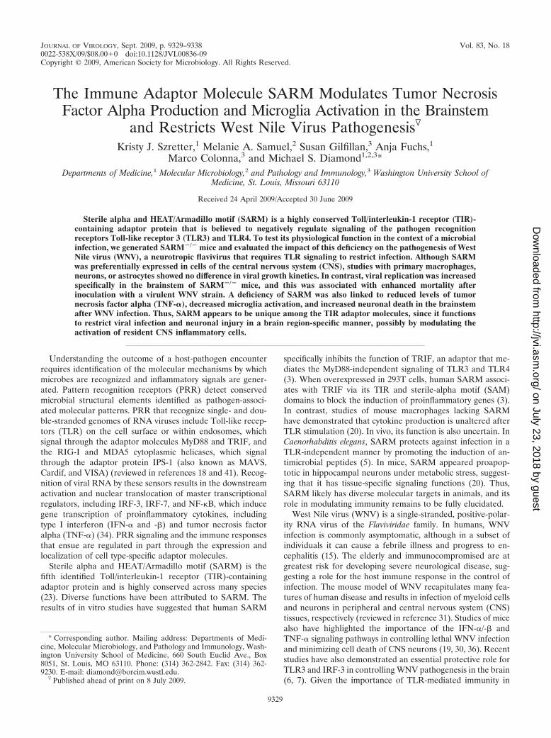
FIG. 1. Expression and generation of SARM knockout mice. (A) Schematic of the SARM locus and targeting cassette. Exons 1 through 6 ofSARM are represented by gray boxes, and the fragments included in the targeting construct by a heavy black line. The locations of external probesare shown, as are the restriction sites used for screening. S, SphI. (B) A representative image of the RT-PCR analysis of SARM mRNA from wholeorgans, specific brain regions, and primary cells derived from wild-type C57BL/6 mice is shown. The SARM transcript, at approximately 650 bp,is indicated; a 519-bp fragment of �-actin was amplified in parallel as a control. BM Mac, bone marrow-derived macrophages. (C) A representativeimage of the RT-PCR analysis of SARM�/� and wild-type mouse mRNA is shown. SARM and �-actin mRNA were amplified from equivalentamounts of mRNA derived from wild-type, SARM�/�, and SARM�/� astrocytes. (D) A representative image of RT-PCR analysis of SARMmRNA in macrophages activated with LPS and TNF-� or infected with WNV is shown. Cerebral cortex mRNA was used as a positive control.M, molecular size ladder.
9330 SZRETTER ET AL. J. VIROL.
on July 23, 2018 by guesthttp://jvi.asm
.org/D
ownloaded from
1 h with PBS containing 1% bovine serum albumin, 3% horse serum, 0.05%Tween 20, and 0.025% NaN3. Fourfold serial dilutions of heat-inactivated serumsamples were added to the plates and incubated overnight at 4°C. Plates werewashed, incubated with biotin-conjugated goat anti-mouse IgM or IgG (SouthernBiotech) followed by streptavidin-horseradish peroxidase (Invitrogen), and de-veloped with tetramethylbenzidine substrate (Dako). Antibody titers representthe serum dilution yielding a value for the optical density at 450 nm equivalentto three standard deviations above the background of the assay.
Immunohistochemistry of brain tissues. Tissues were prepared essentially asdescribed previously (32). Briefly, following extensive perfusion with chilled PBS,brains were harvested, fixed in 4% paraformaldehyde overnight at 4°C, and thencryoprotected in 30% sucrose solutions over three days at 4°C. Brain tissues wereembedded in optimal cutting temperature (O.C.T.) compound (Tissue Tek) andflash frozen prior to sectioning on a cryostat. Terminal deoxynucleotidyltrans-ferase-mediated dUTP-biotin nick end labeling (TUNEL) staining was per-formed by using a NeuroTACS II in situ apoptosis detection kit (Trevigen) or anin situ cell death detection kit (Roche) according to the manufacturer’s proto-cols, with some modifications (32). For fluorescence staining and confocal mi-croscopic imaging, frozen sections were rehydrated with PBS and permeabilizedand blocked with 0.1% Triton X-100 and 10% normal goat serum (Sigma) for 1 hat 37°C in a humidified chamber. Sections were incubated overnight at 4°C with1 �g/ml anti-CD11b (BD Biosciences) or anti-microtubule-associated protein 2(anti-MAP-2; Chemicon) antibody, followed by detection with 5 �g/ml AlexaFluor 488-conjugated secondary antibody (Molecular Probes) for 1 h at roomtemperature. Nuclei were counterstained with ToPro-3 (Molecular Probes).Slides were mounted with ProLong Gold antifade reagent (Molecular Probes).Sections were visualized on a Zeiss LSM 510 META confocal laser scanningmicroscope.
Quantification of brain leukocytes. Whole brains were harvested from wild-type and SARM�/� mice on day 8 after subcutaneous infection with WNV-NYor intracranial infection with WNV-MAD. Following perfusion, brains werehomogenized through a 70-�m cell strainer, digested with a collagenase solution(500 �g/ml collagenase D, 0.1 �g/ml N�-p-tosyl-L-lysine chloromethyl ketone[TLCK] trypsin inhibitor, 10 �g/ml DNase I, 10 mM HEPES in HBSS) for 1 h atroom temperature. Cells were separated by centrifugation on a discontinuous70-to-37-to-30% Percoll gradient. For detection of resident microglia and infil-trating macrophages, cells were incubated with fluorescein isothiocyanate-con-jugated anti-major histocompatibility complex class II, phycoerythrin-conjugatedanti-CD45, and allophycocyanin-conjugated anti-CD11b antibodies (BD Bio-sciences). For detection of WNV-specific CD8� T cells, leukocytes were restim-ulated ex vivo with a Db-restricted immunodominant NS4B WNV peptide (27)and incubated with fluorescein isothiocyanate-conjugated anti-CD3 or allophy-cocyanin-conjugated anti-CD8 (BD Biosciences) at 4°C for 30 min. Subse-quently, cells were washed, fixed in 1% paraformaldehyde, permeabilized, andincubated additionally with phycoerythrin-conjugated anti-IFN-� or -TNF-�prior to analysis by flow cytometry.
Primary cell cultures. Cortical and hippocampal neurons were generated asdescribed previously (21). All experiments with neuron cultures were performedon cells propagated for 4 days. Bone marrow-derived macrophages and myeloiddendritic cells were isolated and maintained as described previously (33). Murineembryonic fibroblasts were generated from embryonic-day-14 wild-type orSARM�/� congenic mice according to established protocols and were main-tained in Dulbecco’s modified Eagle’s medium (DMEM) containing 10% FBS.Astrocytes were prepared essentially as described previously for rats, with someadaptations (47). Briefly, forebrains were dissected from 1- to 2-day-old mice,gently dissociated by trituration in DMEM containing 10% FBS, and filteredthrough a 70-�m cell strainer. Dissociated cells were seeded into poly-D-lysine–laminin-coated 75-cm2 flasks and cultured for 8 to 10 days until confluent. Flaskswere shaken at 150 RPM for 1 h to remove nonadherent microglia, and freshDMEM containing 3% FBS was added. This procedure was repeated for threeconsecutive days, and on the last day, astrocyte monolayers were recovered aftertrypsin treatment and seeded into poly-D-lysine–laminin-coated culture plates.
Quantification of cytokines and chemokines. Cell culture supernatants andclarified tissue homogenates were analyzed by using an ELISA kit (R&D Sys-tems) and Bioplex cytokine bead array (Bio-Rad) according to the manufactur-ers’ protocols. Quantitative RT-PCR was used to measure levels of gene expres-sion as previously described (21, 37). Briefly, RNA was isolated by using anRNeasy kit (Qiagen), followed by DNase I treatment (Invitrogen) and cDNAgeneration using a high-capacity cDNA reverse transcription kit (Applied Bio-systems). Gene expression was measured by using Sybr green PCR master mix(Applied Biosystems) and primers for TNF-� and glyceraldehyde-3-phosphatedehydrogenase (GAPDH) (21); the latter was used for normalization of cyclethreshold values to account for the amount of input tissue.
Statistical analysis. All data were analyzed with Prism software (GraphpadSoftware, Inc.). For survival analysis, Kaplan-Meier survival curves were ana-lyzed by using the log-rank test. For viral burden analysis, significance wasdetermined by using the Mann-Whitney test. An unpaired Student’s t test wasused to determine significant differences in cytokine production and TUNELassay results.
RESULTS
SARM expression and generation of null mice. Recent stud-ies with human cells and transgenic mice have suggested thatSARM is a negative regulator of TRIF-dependent TLR3 sig-naling and a mediator of stress-induced toxicity in hippocam-pal neurons (3, 20). However, it remains uncertain how SARMcontributes to the immune response during a pathogen chal-lenge, when multiple PRR are stimulated concurrently. Theresults of previous experiments had suggested that SARM wasdistinct from other TLR adaptors in its preferential expressionin neurons (20). To confirm this, we examined the expressionof murine SARM in various tissues by using RT-PCR. Rela-tively high levels of SARM mRNA were observed in the brain,whereas lower levels were observed in other tissues (Fig. 1B).Consistent with this, SARM mRNA was detected in primaryneurons, microglia, and astrocytes but was undetectable or atvery low levels in primary lymphocytes and bone marrow-de-rived macrophages (Fig. 1B and data not shown). Activatedmacrophages that were pretreated with lipopolysaccharide(LPS) or TNF-� or infected with WNV also expressed little, ifany, SARM mRNA (Fig. 1D). We were unable to confirmprotein expression of SARM in tissue sections, as only oneanti-mouse SARM antibody is available commercially and itfails to recognize SARM reliably in fixed or frozen tissue sec-tions (K. Szretter and M. Diamond, unpublished observations).
To elucidate the function of SARM in the innate immuneresponse, we generated SARM�/� mice by a homologous re-combination strategy (Fig. 1A). Homozygous SARM�/�, het-erozygotic, and wild-type mice were born at expected Mende-lian ratios and confirmed by Southern blotting and RT-PCR(Fig. 1C and data not shown). Flow cytometric analyses ofSARM�/� mice revealed normal hematopoietic cell develop-ment, as well as typical B- and T-cell maturation profiles (datanot shown).
SARM restricts WNV pathogenesis. Given that SARM isprimarily expressed in the CNS, we evaluated its function invivo in the context of infection by WNV, a neurotropic RNAflavivirus. We used WNV as the model pathogen for two rea-sons: (i) it infects and injures neurons in diverse brain regions,including the hippocampus, brainstem, and cerebral cortex(35); and (ii) TLR3 restricts WNV infection in neurons in vitroand in vivo (7). As SARM is reported to control infection insome model systems and negatively regulate TLR3 signaling inothers, we anticipated that challenging SARM-deficient micemight result in a phenotype that would clarify its function.SARM�/� and wild-type mice were infected subcutaneouslywith 102 PFU of a pathogenic North American WNV strain(WNV-NY) and monitored for disease. SARM�/� mice weremore vulnerable to WNV-NY infection (36% survival rate)than age-matched wild-type mice (63% survival rate) (Fig. 2A)(P 0.03).
To better understand how a deficiency of SARM causedexcess mortality following WNV infection, wild-type and
VOL. 83, 2009 SARM RESTRICTS PATHOGENESIS OF WEST NILE VIRUS 9331
on July 23, 2018 by guesthttp://jvi.asm
.org/D
ownloaded from
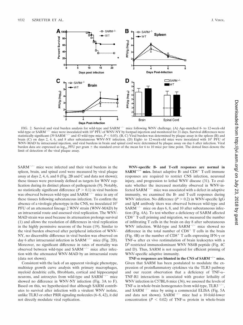
SARM�/� mice were infected and their viral burdens in thespleen, brain, and spinal cord were measured by viral plaqueassay at days 2, 4, 6, and 8 (Fig. 2B and C and data not shown);these tissues were previously defined as targets for WNV rep-lication during its distinct phases of pathogenesis (9). Notably,no statistically significant difference (P 0.1) in viral burdenswas observed between wild-type and SARM�/� mice in any ofthese tissues following subcutaneous infection. To confirm theabsence of a virologic phenotype in the CNS, we inoculated 101
PFU of an attenuated lineage 2 WNV strain (WNV-MAD) byan intracranial route and assessed viral replication. The WNV-MAD strain was used because its attenuation prolongs survival(1) and allows the resolution of differences in viral replicationin the highly permissive neurons of the brain (19). Similar tothe viral burden observed after peripheral infection of WNV-NY, no discernible difference in viral burden was observed onday 6 after intracranial infection in SARM�/� mice (Fig. 2D).Moreover, no significant difference in rates of mortality wasobserved between wild-type and SARM�/� mice after infec-tion with the attenuated WNV-MAD by an intracranial route(data not shown).
Consistent with the lack of an apparent virologic phenotype,multistep growth curve analysis with primary macrophages,myeloid dendritic cells, fibroblasts, cortical and hippocampalneurons, and astrocytes from wild-type and SARM�/� miceshowed no difference in WNV-NY infection (Fig. 3A to F).Based on this, we hypothesized that although SARM contrib-utes to survival after infection with a virulent WNV isolate,unlike TLR3 or other PRR signaling molecules (6–8, 42), it didnot directly modulate viral replication.
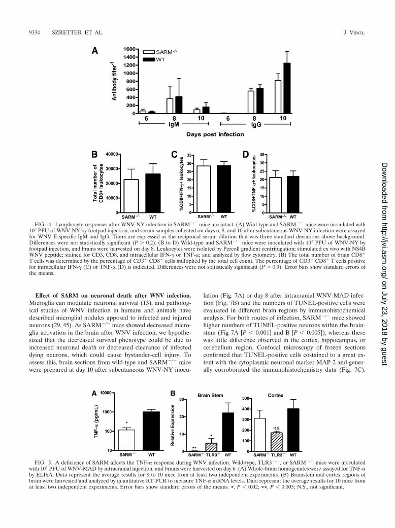
WNV-specific B- and T-cell responses are normal inSARM�/� mice. Intact adaptive B- and CD8� T-cell immuneresponses are required to restrict CNS infection, neuronalinjury, and progression to lethal WNV disease (31). To eval-uate whether the increased mortality observed in WNV-in-fected SARM�/� mice was associated with a defect in adaptiveimmunity, we examined the B- and T-cell responses duringWNV infection. No difference (P 0.2) in WNV-specific IgGand IgM antibody titers was observed between wild-type andSARM�/� mice on days 6, 8, and 10 after subcutaneous infec-tion (Fig. 4A). To test whether a deficiency of SARM affectedCD8� T-cell priming and migration, we measured the numberof infiltrating T cells in the brain on day 8 after subcutaneousWNV infection. Wild-type and SARM�/� mice showed nodifference in the total number of CD8� T cells in the brain(Fig. 4B) or the number of CD8� T cells expressing IFN-� orTNF-� after ex vivo restimulation of brain leukocytes with aDb-restricted immunodominant WNV NS4B peptide (Fig. 4Cand D). Thus, SARM is not required for efficient priming ofWNV-specific adaptive immunity.
TNF-� responses are blunted in the CNS of SARM�/� mice.Given that SARM has been postulated to modulate the ex-pression of proinflammatory cytokines via the TLR3 pathwayand our recent observation that a deficiency of TNF-�–TNF-R1 interactions is associated with greater lethality ofWNV infection in C57BL/6 mice (36), we assessed the levels ofTNF-� in whole-brain homogenates from wild-type, TLR3�/�,and SARM�/� mice by using a commercial ELISA (Fig. 5Aand data not shown). SARM�/� mice had a 10-fold-lowerconcentration (P 0.02) of TNF-� protein in whole-brain
FIG. 2. Survival and viral burden analysis for wild-type and SARM�/� mice following WNV challenge. (A) Age-matched 8- to 12-week-oldwild-type or SARM�/� mice were inoculated with 102 PFU of WNV-NY by footpad injection and monitored for 21 days. Survival differences werestatistically significant (39 SARM�/� and 43 wild-type mice, P 0.03). (B, C) Viral burden was determined by plaque assay in the spleen (B) andbrain (C) on days 2, 4, 6, and 8 after subcutaneous WNV-NY infection. (D) Eight- to 12-week-old mice were inoculated with 101 PFU ofWNV-MAD by intracranial injection, and viral burdens in brain and spinal cord were determined by plaque assay on day 6 after infection. Viralburden data are expressed as log10 PFU per gram � the standard error of the mean for 6 to 10 mice per time point. The dotted lines denote thelimit of detection of the viral plaque assay.
9332 SZRETTER ET AL. J. VIROL.
on July 23, 2018 by guesthttp://jvi.asm
.org/D
ownloaded from

homogenates at day 6; although TLR3�/� mice showed a sim-ilar trend (5-fold) toward reduced TNF-� levels compared tothe levels in wild-type mice, this did not attain statistical sig-nificance (P 0.2). However, based on analysis of TNF-�mRNA levels in distinct brain regions, the reductions inSARM�/� and TLR3�/� mice correlated primarily with dif-ferences in gene expression in the brainstem (Fig. 5B) (P �0.02) and were not observed in the cortex (Fig. 5C) (P � 0.4).In contrast, other inflammatory cytokine (e.g., interleukin-6)and chemokine (e.g., CXCL10) mRNA levels that are elevatedin CNS cells after stimulation with the TLR3 synthetic ligandpoly(I:C) (43) did not differ significantly between SARM�/�,TLR3�/�, and wild-type mice regardless of the brain region(data not shown).
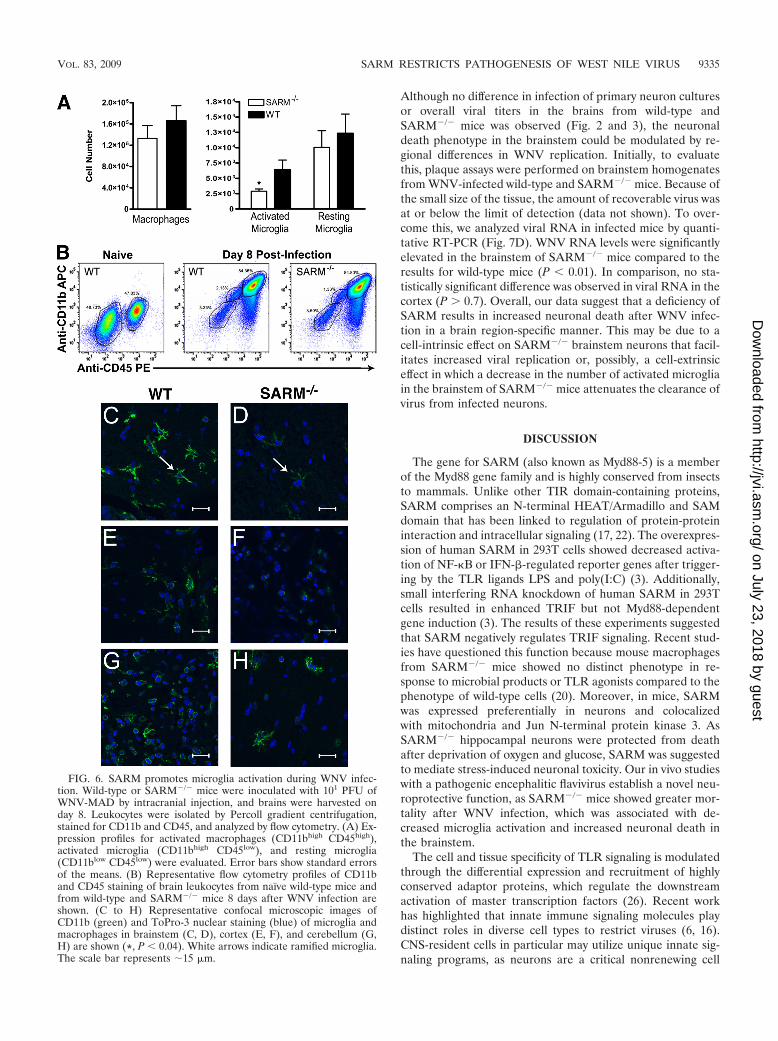
Effect of SARM on myeloid cell accumulation and activationin the CNS. As prior experiments with TNF-R1�/� andTLR3�/� mice showed defects in the accumulation and acti-vation of macrophages and microglia in the brain after WNVinfection (36) or poly(I:C) injection (43), respectively, we eval-
uated the numbers and phenotypes of CD45� CD11b� cellpopulations in wild-type and SARM�/� mice on day 8 afterinfection. Based on the classification of CNS myeloid cells ofFord et al. (11), SARM�/� mice had significantly fewerCD11bhigh CD45low, activated microglia (P 0.04), whereasthe numbers of CD11bhigh CD45high, activated macrophagesand CD11blow CD45low, resting microglia were not substan-tially different from those in WNV-infected wild-type mice(Fig. 6A and B). Immunohistochemistry confirmed these find-ings, as fewer ramified, activated microglia cells were observedby confocal microscopy, especially in the brainstem (Fig. 6Cand D) and, to a lesser degree, in the cerebral cortex (Fig. 6Eand F) of SARM�/� mice. In comparison, the activation stateof myeloid cells in the cerebellum in WNV-infected wild-typeand SARM�/� mice was similar (Fig. 6G and H). Given thatmyeloid cells express little, if any, SARM mRNA (Fig. 1B), wespeculate that the differential activation of brain myeloid cellsin SARM�/� mice may result from altered cell-extrinsic sig-nals, such as the blunted levels of TNF-� in the brain.
FIG. 3. SARM does not have a direct antiviral effect against WNV in primary cells. Primary cells derived from SARM�/� and wild-type mice(described in Materials and Methods) were infected with a multiplicity of infection of 0.001, and virus replication was measured at 6, 24, 48, and72 h postinfection by viral plaque assay. Multistep growth curves showed no difference in viral yields between SARM�/� and wild-type(A) macrophages, (B) dendritic cells, (C) fibroblasts, (D) hippocampal neurons, (E) cortical neurons, or (F) astrocytes. Viral burden data areexpressed as log10 PFU per ml � the standard error of the mean for triplicate samples from two independent experiments.
VOL. 83, 2009 SARM RESTRICTS PATHOGENESIS OF WEST NILE VIRUS 9333
on July 23, 2018 by guesthttp://jvi.asm
.org/D
ownloaded from
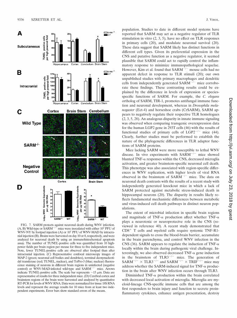
Effect of SARM on neuronal death after WNV infection.Microglia can modulate neuronal survival (13), and patholog-ical studies of WNV infection in humans and animals havedescribed microglial nodules apposed to infected and injuredneurons (29, 45). As SARM�/� mice showed decreased micro-glia activation in the brain after WNV infection, we hypothe-sized that the decreased survival phenotype could be due toincreased neuronal death or decreased clearance of infecteddying neurons, which could cause bystander-cell injury. Toassess this, brain sections from wild-type and SARM�/� micewere prepared at day 10 after subcutaneous WNV-NY inocu-
lation (Fig. 7A) or day 8 after intracranial WNV-MAD infec-tion (Fig. 7B) and the numbers of TUNEL-positive cells wereevaluated in different brain regions by immunohistochemicalanalysis. For both routes of infection, SARM�/� mice showedhigher numbers of TUNEL-positive neurons within the brain-stem (Fig 7A [P 0.001] and B [P 0.005]), whereas therewas little difference observed in the cortex, hippocampus, orcerebellum region. Confocal microscopy of frozen sectionsconfirmed that TUNEL-positive cells costained to a great ex-tent with the cytoplasmic neuronal marker MAP-2 and gener-ally corroborated the immunohistochemistry data (Fig. 7C).
FIG. 4. Lymphocyte responses after WNV-NY infection in SARM�/� mice are intact. (A) Wild-type and SARM�/� mice were inoculated with102 PFU of WNV-NY by footpad injection, and serum samples collected on days 6, 8, and 10 after subcutaneous WNV-NY infection were assayedfor WNV E-specific IgM and IgG. Titers are expressed as the reciprocal serum dilution that was three standard deviations above background.Differences were not statistically significant (P 0.2). (B to D) Wild-type and SARM�/� mice were inoculated with 102 PFU of WNV-NY byfootpad injection, and brains were harvested on day 8. Leukocytes were isolated by Percoll gradient centrifugation; stimulated ex vivo with NS4BWNV peptide; stained for CD3, CD8, and intracellular IFN-� or TNF-�; and analyzed by flow cytometry. (B) The total number of brain CD8�
T cells was determined by the percentage of CD3� CD8� cells multiplied by the total cell count. The percentage of CD3� CD8� T cells positivefor intracellular IFN-� (C) or TNF-� (D) is indicated. Differences were not statistically significant (P 0.9). Error bars show standard errors ofthe means.
FIG. 5. A deficiency of SARM affects the TNF-� response during WNV infection. Wild-type, TLR3�/�, or SARM�/� mice were inoculatedwith 101 PFU of WNV-MAD by intracranial injection, and brains were harvested on day 6. (A) Whole-brain homogenates were assayed for TNF-�by ELISA. Data represent the average results for 8 to 10 mice from at least two independent experiments. (B) Brainstem and cortex regions ofbrain were harvested and analyzed by quantitative RT-PCR to measure TNF-� mRNA levels. Data represent the average results for 10 mice fromat least two independent experiments. Error bars show standard errors of the means. *, P 0.02; **, P 0.005; N.S., not significant.
9334 SZRETTER ET AL. J. VIROL.
on July 23, 2018 by guesthttp://jvi.asm
.org/D
ownloaded from
Although no difference in infection of primary neuron culturesor overall viral titers in the brains from wild-type andSARM�/� mice was observed (Fig. 2 and 3), the neuronaldeath phenotype in the brainstem could be modulated by re-gional differences in WNV replication. Initially, to evaluatethis, plaque assays were performed on brainstem homogenatesfrom WNV-infected wild-type and SARM�/� mice. Because ofthe small size of the tissue, the amount of recoverable virus wasat or below the limit of detection (data not shown). To over-come this, we analyzed viral RNA in infected mice by quanti-tative RT-PCR (Fig. 7D). WNV RNA levels were significantlyelevated in the brainstem of SARM�/� mice compared to theresults for wild-type mice (P 0.01). In comparison, no sta-tistically significant difference was observed in viral RNA in thecortex (P 0.7). Overall, our data suggest that a deficiency ofSARM results in increased neuronal death after WNV infec-tion in a brain region-specific manner. This may be due to acell-intrinsic effect on SARM�/� brainstem neurons that facil-itates increased viral replication or, possibly, a cell-extrinsiceffect in which a decrease in the number of activated microgliain the brainstem of SARM�/� mice attenuates the clearance ofvirus from infected neurons.
DISCUSSION
The gene for SARM (also known as Myd88-5) is a memberof the Myd88 gene family and is highly conserved from insectsto mammals. Unlike other TIR domain-containing proteins,SARM comprises an N-terminal HEAT/Armadillo and SAMdomain that has been linked to regulation of protein-proteininteraction and intracellular signaling (17, 22). The overexpres-sion of human SARM in 293T cells showed decreased activa-tion of NF-�B or IFN-�-regulated reporter genes after trigger-ing by the TLR ligands LPS and poly(I:C) (3). Additionally,small interfering RNA knockdown of human SARM in 293Tcells resulted in enhanced TRIF but not Myd88-dependentgene induction (3). The results of these experiments suggestedthat SARM negatively regulates TRIF signaling. Recent stud-ies have questioned this function because mouse macrophagesfrom SARM�/� mice showed no distinct phenotype in re-sponse to microbial products or TLR agonists compared to thephenotype of wild-type cells (20). Moreover, in mice, SARMwas expressed preferentially in neurons and colocalizedwith mitochondria and Jun N-terminal protein kinase 3. AsSARM�/� hippocampal neurons were protected from deathafter deprivation of oxygen and glucose, SARM was suggestedto mediate stress-induced neuronal toxicity. Our in vivo studieswith a pathogenic encephalitic flavivirus establish a novel neu-roprotective function, as SARM�/� mice showed greater mor-tality after WNV infection, which was associated with de-creased microglia activation and increased neuronal death inthe brainstem.
The cell and tissue specificity of TLR signaling is modulatedthrough the differential expression and recruitment of highlyconserved adaptor proteins, which regulate the downstreamactivation of master transcription factors (26). Recent workhas highlighted that innate immune signaling molecules playdistinct roles in diverse cell types to restrict viruses (6, 16).CNS-resident cells in particular may utilize unique innate sig-naling programs, as neurons are a critical nonrenewing cell
FIG. 6. SARM promotes microglia activation during WNV infec-tion. Wild-type or SARM�/� mice were inoculated with 101 PFU ofWNV-MAD by intracranial injection, and brains were harvested onday 8. Leukocytes were isolated by Percoll gradient centrifugation,stained for CD11b and CD45, and analyzed by flow cytometry. (A) Ex-pression profiles for activated macrophages (CD11bhigh CD45high),activated microglia (CD11bhigh CD45low), and resting microglia(CD11blow CD45low) were evaluated. Error bars show standard errorsof the means. (B) Representative flow cytometry profiles of CD11band CD45 staining of brain leukocytes from naïve wild-type mice andfrom wild-type and SARM�/� mice 8 days after WNV infection areshown. (C to H) Representative confocal microscopic images ofCD11b (green) and ToPro-3 nuclear staining (blue) of microglia andmacrophages in brainstem (C, D), cortex (E, F), and cerebellum (G,H) are shown (*, P 0.04). White arrows indicate ramified microglia.The scale bar represents �15 �m.
VOL. 83, 2009 SARM RESTRICTS PATHOGENESIS OF WEST NILE VIRUS 9335
on July 23, 2018 by guesthttp://jvi.asm
.org/D
ownloaded from
population. Studies to date in different model systems havereported that SARM may act as a negative regulator of TLRstimulation in vitro (2, 3, 5), have no effect on TLR responsesin primary cells (20), and modulate neuronal survival (20).These data suggest that SARM likely has distinct functions indifferent cell types. Given its preferential expression in theCNS and putative function as a negative regulator, it seemedplausible that SARM could act to rapidly control the inflam-matory response to minimize immunopathological sequelae.However, Kim et al. found that SARM�/� mouse cells had noapparent defect in response to TLR stimuli (20); our ownunpublished studies with primary macrophages and dendriticcells from independently generated SARM�/� mice corrobo-rate these findings. These contrasting results could be ex-plained by the difference in levels of expression or species-specific functions of SARM. For example, the C. elegansortholog of SARM, TIR-1, promotes antifungal immune func-tion and neuronal development, whereas in Drosophila mela-nogaster (Ect-4) and horseshoe crabs (CrSARM), SARM ap-pears to negatively regulate their respective TLR homologues(2, 3, 5, 28). An analogous disparity in innate immune signalingwas observed when comparing transgenic overexpression datafor the human LGP2 gene in 293T cells (46) with the results offunctional studies of primary cells of LGP2�/� mice (44).Clearly, further studies must be performed to establish thenature of the phylogenetic differences in TLR adaptor func-tions of SARM proteins.
Mice lacking SARM were more susceptible to lethal WNVdisease. In vivo experiments with SARM�/� mice showedblunted TNF-� responses within the CNS, decreased microgliaactivation, and greater brainstem-specific neuronal cell death.This phenotype was also associated with region-specific differ-ences in WNV replication, with higher levels of viral RNAobserved in the brainstem of SARM�/� mice. The data onneuronal death contrasts with the results of a recent study withindependently generated knockout mice in which a lack ofSARM protected against metabolic stress-induced death inhippocampal neurons (20). The disparity in results likely re-flects fundamental mechanistic differences between metabolicand virus-induced cell death pathways in distinct neuron pop-ulations.
The extent of microbial infection in specific brain regionsand magnitude of TNF-� production affect whether TNF-�plays a neurotoxic or neuroprotectve role in the CNS (re-viewed in reference 40). A recent study demonstrated thatCD8� T cells and myeloid cells require systemic TNF-R1-dependent signals to cross the blood-brain barrier, accumulatein the brain parenchyma, and control WNV infection in theCNS (36). SARM appears to regulate the induction of TNF-�locally within the brain during pathogenic viral challenge. In-terestingly, we also observed decreased TNF-� gene inductionin the brainstem of TLR3�/� mice. The generation ofSARM�/� TLR3�/� and SARM�/� TRIF�/� mice mayaddress whether the SARM-induced signal for TNF-� produc-tion in the brain after WNV infection occurs through TLR3.
Diminished TNF-� production within the brain correlatedwith decreased local activation of microglia. Microglia are my-eloid-lineage CNS-specific immune cells that are among thefirst responders to brain injury and function to secrete proin-flammatory cytokines, enhance antigen presentation, destroy
FIG. 7. SARM protects against neuronal death during WNV infection.(A, B) Wild-type or SARM�/� mice were inoculated with either 102 PFU ofWNV-NY by footpad injection (A) or 101 PFU of WNV-MAD by intracra-nial injection (B). Brains were harvested on day 10 or 8, respectively, and wereanalyzed for neuronal death by using an immunohistochemical apoptosisassay. The number of TUNEL-positive cells was quantified from 10 high-power fields per brain region per mouse for three to five independent mice.Note, fewer TUNEL-positive cells are observed after footpad than afterintracranial injection. (C) Representative confocal microscopy images ofMAP-2 (green; neuronal cell bodies and dendrites), terminal deoxynucleoti-dyl transferase (red; TUNEL, nuclear), and ToPro-3 (blue; nuclear) fluores-cence staining of neurons in different brain regions in uninfected (negativecontrol) or WNV-MAD-infected wild-type and SARM�/� mice. Arrowsindicate TUNEL-positive cells. The scale bar represents �15 �m. Data arerepresentative of results for three independent mice. (D) Cerebral cortex andbrainstem regions of the brain were harvested and analyzed by quantitativeRT-PCR for levels of WNV RNA. Data were normalized for tissue 18S RNAlevels and represent the average results for 10 mice from at least two inde-pendent experiments. Error bars show standard errors of the means.
9336 SZRETTER ET AL. J. VIROL.
on July 23, 2018 by guesthttp://jvi.asm
.org/D
ownloaded from
pathogens before they injure neurons, and clear dying cells(14). Activated microglia become juxtaposed to WNV-infectedneurons in human and animal brain tissue pathological speci-mens, where they may have protective or pathogenic roles (4,25, 43). Interestingly, a recent study using an attenuated lin-eage 2 Sarafend WNV strain suggested (36) that increases inthe total number of activated microglia during infection mayhave a pathogenic role (12). In contrast, using a more-virulentlineage 1 WNV-NY strain, we observed a localized decrease ofactivated microglia in the brainstem of SARM�/� mice thatwas associated with increased neuronal death and mortality.
Though several factors contribute to microglial stimulation,TNF-� directly regulates activation. Mice lacking TNF-� re-ceptors showed decreased microglia activation and corre-spondingly higher levels of neuronal injury in the hippocampusafter treatment with a dopaminergic neurotoxin (38, 39). Ourdata are thus consistent with a model in which SARM regu-lates local production of TNF-�, which in turn modulates mi-croglia activation, viral replication, and neuronal survival. Al-though SARM modulates microglia activation regionally, thecellular basis of this phenotype also remains uncertain. It couldreflect a cell-autonomous regulatory effect within microglia or,given the expression pattern of SARM in the CNS, a complexcircuit in which SARM regulates the production of cytokines(e.g., TNF-�) in neurons or astrocytes, resulting in cell-extrin-sic effects on microglia. As an alternative model, region-spe-cific differences in local TNF-� production could attenuate theflux of nonresident inflammatory monocyte-derived microgliaacross the blood-brain barrier (12); indeed, our prior studieshave shown that TNF-� modulates the trafficking of monocytesinto the brain, possibly by affecting early adhesion steps (36).Clearly, the specific signaling pathways that SARM uses toregulate microglia activation in the brainstem remain uncer-tain and an avenue for future studies.
In summary, the results of our experiments suggest thatSARM modulates the host response to WNV infection in atissue- and cell-specific manner. Thus, in contrast to its role inhuman cells, murine SARM in the CNS appears to positivelyregulate TNF-� induction, which affects microglia activationand accumulation and protects critical neuron subsets fromvirus-induced pathology. These studies illuminate the host andtissue-specific complexity of TLR adaptor molecules and en-hance our understanding of the balance between an effectiveinnate response and immunopathology and injury to neuronsin the CNS.
ACKNOWLEDGMENTS
This work was supported by the NIH (U54 AI057160 [MidwestRegional Center of Excellence for Biodefense and Emerging Infec-tious Diseases Research]), a predoctoral fellowship from the HowardHughes Medical Institute (M.A.S.), a W. M. Keck Postdoctoral Fel-lowship in Molecular Medicine, and a Ruth L. Kirschstein Postdoc-toral NRSA (K.J.S.).
We thank Michael White for blastocyst injections and the SpeedCongenics Laboratory at Washington University School of Medicinefor high-density microsatellite mapping.
REFERENCES
1. Beasley, D. W., C. T. Davis, M. Whiteman, B. Granwehr, R. M. Kinney, andA. D. Barrett. 2004. Molecular determinants of virulence of West Nile virusin North America. Arch. Virol. Suppl. 2004:35–41.
2. Belinda, L. W., W. X. Wei, B. T. Hanh, L. X. Lei, H. Bow, and D. J. Ling.
2008. SARM: a novel Toll-like receptor adaptor, is functionally conservedfrom arthropod to human. Mol. Immunol. 45:1732–1742.
3. Carty, M., R. Goodbody, M. Schroder, J. Stack, P. N. Moynagh, and A. G.Bowie. 2006. The human adaptor SARM negatively regulates adaptor pro-tein TRIF-dependent Toll-like receptor signaling. Nat. Immunol. 7:1074–1081.
4. Cheeran, M. C., S. Hu, W. S. Sheng, A. Rashid, P. K. Peterson, and J. R.Lokensgard. 2005. Differential responses of human brain cells to West Nilevirus infection. J. Neurovirol. 11:512–524.
5. Couillault, C., N. Pujol, J. Reboul, L. Sabatier, J. F. Guichou, Y. Kohara,and J. J. Ewbank. 2004. TLR-independent control of innate immunity inCaenorhabditis elegans by the TIR domain adaptor protein TIR-1, an or-tholog of human SARM. Nat. Immunol. 5:488–494.
6. Daffis, S., M. A. Samuel, B. C. Keller, M. Gale, Jr., and M. S. Diamond. 2007.Cell-specific IRF-3 responses protect against West Nile virus infection byinterferon-dependent and -independent mechanisms. PLoS Pathog. 3:e106.
7. Daffis, S., M. A. Samuel, M. S. Suthar, M. Gale, Jr., and M. S. Diamond.2008. Toll-like receptor 3 has a protective role against West Nile virusinfection. J. Virol. 82:10349–10358.
8. Daffis, S., M. A. Samuel, M. S. Suthar, B. C. Keller, M. Gale, Jr., and M. S.Diamond. 2008. Interferon regulatory factor IRF-7 induces the antiviralalpha interferon response and protects against lethal West Nile virus infec-tion. J. Virol. 82:8465–8475.
9. Diamond, M. S., B. Shrestha, A. Marri, D. Mahan, and M. Engle. 2003. Bcells and antibody play critical roles in the immediate defense of dissemi-nated infection by West Nile encephalitis virus. J. Virol. 77:2578–2586.
10. Ebel, G. D., J. Carricaburu, D. Young, K. A. Bernard, and L. D. Kramer.2004. Genetic and phenotypic variation of West Nile virus in New York,2000–2003. Am. J. Trop. Med. Hyg. 71:493–500.
11. Ford, A. L., E. Foulcher, F. A. Lemckert, and J. D. Sedgwick. 1996. Microgliainduce CD4 T lymphocyte final effector function and death. J. Exp. Med.184:1737–1745.
12. Getts, D. R., R. L. Terry, M. T. Getts, M. Muller, S. Rana, B. Shrestha, J.Radford, N. Van Rooijen, I. L. Campbell, and N. J. King. 2008. Ly6c�“inflammatory monocytes” are microglial precursors recruited in a patho-genic manner in West Nile virus encephalitis. J. Exp. Med. 205:2319–2337.
13. Glezer, I., A. R. Simard, and S. Rivest. 2007. Neuroprotective role of theinnate immune system by microglia. Neuroscience 147:867–883.
14. Gonzalez-Scarano, F., and G. Baltuch. 1999. Microglia as mediators ofinflammatory and degenerative diseases. Annu. Rev. Neurosci. 22:219–240.
15. Hayes, E. B., J. J. Sejvar, S. R. Zaki, R. S. Lanciotti, A. V. Bode, and G. L.Campbell. 2005. Virology, pathology, and clinical manifestations of WestNile virus disease. Emerg. Infect. Dis. 11:1174–1179.
16. Ishii, K. J., S. Koyama, A. Nakagawa, C. Coban, and S. Akira. 2008. Hostinnate immune receptors and beyond: making sense of microbial infections.Cell Host Microbe 3:352–363.
17. Jault, C., L. Pichon, and J. Chluba. 2004. Toll-like receptor gene family andTIR-domain adapters in Danio rerio. Mol. Immunol. 40:759–771.
18. Kawai, T., and S. Akira. 2006. Innate immune recognition of viral infection.Nat. Immunol. 7:131–137.
19. Keller, B. C., B. L. Fredericksen, M. A. Samuel, R. E. Mock, P. W. Mason,M. S. Diamond, and M. Gale, Jr. 2006. Resistance to alpha/beta interferonis a determinant of West Nile virus replication fitness and virulence. J. Virol.80:9424–9434.
20. Kim, Y., P. Zhou, L. Qian, J. Z. Chuang, J. Lee, C. Li, C. Iadecola, C.Nathan, and A. Ding. 2007. MyD88-5 links mitochondria, microtubules, andJNK3 in neurons and regulates neuronal survival. J. Exp. Med. 204:2063–2074.
21. Klein, R. S., E. Lin, B. Zhang, A. D. Luster, J. Tollett, M. A. Samuel, M.Engle, and M. S. Diamond. 2005. Neuronal CXCL10 directs CD8� T-cellrecruitment and control of West Nile virus encephalitis. J. Virol. 79:11457–11466.
22. Mink, M., and K. Csiszar. 2005. SARM1: a candidate gene in the onset ofhereditary infectious/inflammatory diseases. Clin. Immunol. 115:333–334.
23. Mink, M., B. Fogelgren, K. Olszewski, P. Maroy, and K. Csiszar. 2001. Anovel human gene (SARM) at chromosome 17q11 encodes a protein with aSAM motif and structural similarity to Armadillo/beta-catenin that is con-served in mouse, Drosophila, and Caenorhabditis elegans. Genomics 74:234–244.
24. Oliphant, T., M. Engle, G. E. Nybakken, C. Doane, S. Johnson, L. Huang, S.Gorlatov, E. Mehlhop, A. Marri, K. M. Chung, G. D. Ebel, L. D. Kramer,D. H. Fremont, and M. S. Diamond. 2005. Development of a humanizedmonoclonal antibody with therapeutic potential against West Nile virus. Nat.Med. 11:522–530.
25. Omalu, B. I., A. A. Shakir, G. Wang, W. I. Lipkin, and C. A. Wiley. 2003.Fatal fulminant pan-meningo-polioencephalitis due to West Nile virus. BrainPathol. 13:465–472.
26. O’Neill, L. A., and A. G. Bowie. 2007. The family of five: TIR-domain-containing adaptors in Toll-like receptor signalling. Nat. Rev. Immunol.7:353–364.
27. Purtha, W. E., N. Myers, V. Mitaksov, E. Sitati, J. Connolly, D. H. Fremont,T. H. Hansen, and M. S. Diamond. 2007. Antigen-specific cytotoxic T lym-
VOL. 83, 2009 SARM RESTRICTS PATHOGENESIS OF WEST NILE VIRUS 9337
on July 23, 2018 by guesthttp://jvi.asm
.org/D
ownloaded from
phocytes protect against lethal West Nile virus encephalitis. Eur. J. Immunol.37:1845–1854.
28. Raha, D., Q. D. Nguyen, and A. Garen. 1990. Molecular and developmentalanalyses of the protein encoded by the Drosophila gene ectodermal. Dev.Genet. 11:310–317.
29. Sampson, B. A., and V. Armbrustmacher. 2001. West Nile encephalitis: theneuropathology of four fatalities. Ann. N. Y. Acad. Sci. 951:172–178.
30. Samuel, M. A., and M. S. Diamond. 2005. Alpha/beta interferon protectsagainst lethal West Nile virus infection by restricting cellular tropism andenhancing neuronal survival. J. Virol. 79:13350–13361.
31. Samuel, M. A., and M. S. Diamond. 2006. Pathogenesis of West Nile Virusinfection: a balance between virulence, innate and adaptive immunity, andviral evasion. J. Virol. 80:9349–9360.
32. Samuel, M. A., J. D. Morrey, and M. S. Diamond. 2007. Caspase 3-depen-dent cell death of neurons contributes to the pathogenesis of West Nile virusencephalitis. J. Virol. 81:2614–2623.
33. Samuel, M. A., K. Whitby, B. C. Keller, A. Marri, W. Barchet, B. R. Wil-liams, R. H. Silverman, M. Gale, Jr., and M. S. Diamond. 2006. PKR andRNase L contribute to protection against lethal West Nile Virus infection bycontrolling early viral spread in the periphery and replication in neurons.J. Virol. 80:7009–7019.
34. Sato, M., H. Suemori, N. Hata, M. Asagiri, K. Ogasawara, K. Nakao, T.Nakaya, M. Katsuki, S. Noguchi, N. Tanaka, and T. Taniguchi. 2000. Dis-tinct and essential roles of transcription factors IRF-3 and IRF-7 in responseto viruses for IFN-alpha/beta gene induction. Immunity 13:539–548.
35. Shrestha, B., D. Gottlieb, and M. S. Diamond. 2003. Infection and injury ofneurons by West Nile encephalitis virus. J. Virol. 77:13203–13213.
36. Shrestha, B., B. Zhang, W. E. Purtha, R. S. Klein, and M. S. Diamond. 2008.Tumor necrosis factor alpha protects against lethal West Nile virus infectionby promoting trafficking of mononuclear leukocytes into the central nervoussystem. J. Virol. 82:8956–8964.
37. Sitati, E., E. E. McCandless, R. S. Klein, and M. S. Diamond. 2007. CD40-CD40 ligand interactions promote trafficking of CD8� T cells into the brainand protection against West Nile virus encephalitis. J. Virol. 81:9801–9811.
38. Sriram, K., J. M. Matheson, S. A. Benkovic, D. B. Miller, M. I. Luster, andJ. P. O’Callaghan. 2006. Deficiency of TNF receptors suppresses microglialactivation and alters the susceptibility of brain regions to MPTP-inducedneurotoxicity: role of TNF-alpha. FASEB J. 20:670–682.
39. Sriram, K., J. M. Matheson, S. A. Benkovic, D. B. Miller, M. I. Luster, andJ. P. O’Callaghan. 2002. Mice deficient in TNF receptors are protectedagainst dopaminergic neurotoxicity: implications for Parkinson’s disease.FASEB J. 16:1474–1476.
40. Sriram, K., and J. P. O’Callaghan. 2007. Divergent roles for tumor necrosisfactor-alpha in the brain. J. Neuroimmune Pharmacol. 2:140–153.
41. Thompson, A. J., and S. A. Locarnini. 2007. Toll-like receptors, RIG-I-likeRNA helicases and the antiviral innate immune response. Immunol. CellBiol. 85:435–445.
42. Town, T., F. Bai, T. Wang, A. T. Kaplan, F. Qian, R. R. Montgomery, J. F.Anderson, R. A. Flavell, and E. Fikrig. 2009. Toll-like receptor 7 mitigateslethal West Nile encephalitis via interleukin 23-dependent immune cell in-filtration and homing. Immunity 30:242–253.
43. Town, T., D. Jeng, L. Alexopoulou, J. Tan, and R. A. Flavell. 2006. Microgliarecognize double-stranded RNA via TLR3. J. Immunol. 176:3804–3812.
44. Venkataraman, T., M. Valdes, R. Elsby, S. Kakuta, G. Caceres, S. Saijo, Y.Iwakura, and G. N. Barber. 2007. Loss of DExD/H box RNA helicase LGP2manifests disparate antiviral responses. J. Immunol. 178:6444–6455.
45. Xiao, S. Y., H. Guzman, H. Zhang, A. P. Travassos da Rosa, and R. B. Tesh.2001. West Nile virus infection in the golden hamster (Mesocricetus aura-tus): a model for West Nile encephalitis. Emerg. Infect. Dis. 7:714–721.
46. Yoneyama, M., M. Kikuchi, K. Matsumoto, T. Imaizumi, M. Miyagishi, K.Taira, E. Foy, Y. M. Loo, M. Gale, Jr., S. Akira, S. Yonehara, A. Kato, andT. Fujita. 2005. Shared and unique functions of the DExD/H-box helicasesRIG-I, MDA5, and LGP2 in antiviral innate immunity. J. Immunol. 175:2851–2858.
47. Zhang, B., L. Yang, Y. Konishi, N. Maeda, M. Sakanaka, and J. Tanaka.2002. Suppressive effects of phosphodiesterase type IV inhibitors on ratcultured microglial cells: comparison with other types of cAMP-elevatingagents. Neuropharmacology 42:262–269.
9338 SZRETTER ET AL. J. VIROL.
on July 23, 2018 by guesthttp://jvi.asm
.org/D
ownloaded from