Embed Size (px)
Citation preview
of July 23, 2018.This information is current as
PromoterCcl5Polymerase II to the of Transcription Factors and RNAMacrophages by Promoting the Recruitment SARM Regulates CCL5 Production in
G. BowieAndrewSchattgen, Aihao Ding, Katherine A. Fitzgerald and
Claudia Gürtler, Michael Carty, Jay Kearney, Stefan A.
http://www.jimmunol.org/content/192/10/4821doi: 10.4049/jimmunol.1302980April 2014;
2014; 192:4821-4832; Prepublished online 7J Immunol
MaterialSupplementary
0.DCSupplementalhttp://www.jimmunol.org/content/suppl/2014/04/05/jimmunol.130298
Referenceshttp://www.jimmunol.org/content/192/10/4821.full#ref-list-1
, 18 of which you can access for free at: cites 46 articlesThis article
average*
4 weeks from acceptance to publicationFast Publication! •
Every submission reviewed by practicing scientistsNo Triage! •
from submission to initial decisionRapid Reviews! 30 days* •
Submit online. ?The JIWhy
Subscriptionhttp://jimmunol.org/subscription
is online at: The Journal of ImmunologyInformation about subscribing to
Permissionshttp://www.aai.org/About/Publications/JI/copyright.htmlSubmit copyright permission requests at:
Email Alertshttp://jimmunol.org/alertsReceive free email-alerts when new articles cite this article. Sign up at:
Print ISSN: 0022-1767 Online ISSN: 1550-6606. Immunologists, Inc. All rights reserved.Copyright © 2014 by The American Association of1451 Rockville Pike, Suite 650, Rockville, MD 20852The American Association of Immunologists, Inc.,
is published twice each month byThe Journal of Immunology
by guest on July 23, 2018http://w
ww
.jimm
unol.org/D
ownloaded from
by guest on July 23, 2018
http://ww
w.jim
munol.org/
Dow
nloaded from
The Journal of Immunology
SARM Regulates CCL5 Production in Macrophages byPromoting the Recruitment of Transcription Factors andRNA Polymerase II to the Ccl5 Promoter
Claudia G€urtler,* Michael Carty,* Jay Kearney,* Stefan A. Schattgen,† Aihao Ding,‡
Katherine A. Fitzgerald,† and Andrew G. Bowie*
The four Toll/IL-1R domain–containing adaptor proteins MyD88, MAL, TRIF, and TRAM are well established as essential
mediators of TLR signaling and gene induction following microbial detection. In contrast, the function of the fifth, most evolu-
tionarily conserved Toll/IL-1R adaptor, sterile a and HEAT/Armadillo motif-containing protein (SARM), has remained more
elusive. Recent studies of Sarm2/2 mice have highlighted a role for SARM in stress-induced neuronal cell death and immune
responses in the CNS. However, whether SARM has a role in immune responses in peripheral myeloid immune cells is less clear.
Thus, we characterized TLR-induced cytokine responses in SARM-deficient murine macrophages and discovered a requirement
for SARM in CCL5 production, whereas gene induction of TNF, IL-1b, CCL2, and CXCL10 were SARM-independent. SARM
was not required for TLR-induced activation of MAPKs or of transcription factors implicated in CCL5 induction, namely NF-kB
and IFN regulatory factors, nor for Ccl5 mRNA stability or splicing. However, SARM was critical for the recruitment of
transcription factors and of RNA polymerase II to the Ccl5 promoter. Strikingly, the requirement of SARM for CCL5 induction
was not restricted to TLR pathways, as it was also apparent in cytosolic RNA and DNA responses. Thus, this study identifies a new
role for SARM in CCL5 expression in macrophages. The Journal of Immunology, 2014, 192: 4821–4832.
Innate immune cells such as macrophages are key players ininitiating an immune response following the detection ofinvading pathogens via pattern recognition receptors (PRRs).
Engagement of a PRR with its microbial ligand initiates signalingcascades leading to activation of MAPKs and transcription factors,such as NF-kB and IFN regulatory factors (IRFs) (1). Thesetranscription factors subsequently translocate to the nucleus andbind to specific promoter elements of proinflammatory cytokines(e.g., TNF, IL-1b), chemokines (e.g., CCL5, CCL2, CXCL10), andtype I IFNs (IFN-a and IFN-b) to upregulate gene expression.One major family of PRRs are the membrane-bound TLRs,
which signal through homotypic interactions via distinct Toll/IL-1R (TIR) domain–containing adaptor proteins, namely MyD88,MyD88-adaptor–like protein (MAL), TIR domain-containing
adaptor inducing IFN-b (TRIF), and TRIF-related adaptor mole-cule (TRAM) (2). TLR4 is located on the plasma membrane,where it detects LPS. TLR4 utilizes all four adaptor proteins forsignaling, activating a MAL/MyD88-dependent pathway from theplasma membrane, followed by TLR4 internalization and TRAM/TRIF signaling from endolysosomes. In contrast, the microbialnucleic acid–sensing TLRs, namely TLR3, TLR7, and TLR9,are expressed on endosomes, where they sense viral dsRNA,ssRNA, or CpG DNA, respectively (1). A single TIR adaptor isthen recruited to mediate downstream signaling, which is TRIFfor TLR3 and MyD88 for TLR7 and TLR9. Apart from the TLRs,ubiquitously expressed RNA and DNA sensors are found in thecytosol of many different cell types to detect intracellular viruses(3). Such cytosolic PRRs include the retinoic acid–inducible geneI–like receptors retinoic acid–inducible gene I and melanomadifferentiation-associated gene 5, which sense viral RNA andsignal via the adaptor mitochondrial antiviral signaling, and cy-tosolic DNA sensors such as IFN-inducible protein 16 and cyclicGMP-AMP synthase, which signal via the endoplasmic reticu-lum–resident protein stimulator of IFN genes (STING) (3).Sterile a and HEAT/Armadillo motif–containing protein
(SARM) has a C-terminal TIR domain and was therefore expectedto function in the TLR pathway (4). Notably, SARM is highlyconserved between mammals, Drosophila, and worms and is infact the most evolutionary conserved member of the cytosolicTIR-containing proteins (5). Unlike the other four TIR adaptorproteins, early studies showed that overexpressed SARM did notlead to NF-kB and IRF3 activation (6, 7). Instead, SARM wasfound to negatively regulate TRIF-dependent TLR3 and TLR4signaling in human cells through direct interaction with TRIF (7).However, studies of Sarm2/2 mice have mainly revealed neuronalfunctions for SARM. In an initial study by Kim et al. (8) SARMwas discovered to trigger stress-induced neuronal cell death in theCNS after oxygen and glucose deprivation, whereas SARM wasrecently also implicated in mediating neuronal cell death during
*School of Biochemistry and Immunology, Trinity Biomedical Sciences Institute,Trinity College Dublin, Dublin 2, Ireland; †Division of Infectious Disease and Im-munology, Department of Medicine, University of Massachusetts Medical School,Worcester, MA 01605; and ‡Department of Microbiology and Immunology, WeillMedical College of Cornell University, New York, NY 10021
Received for publication November 6, 2013. Accepted for publication March 12,2014.
This work was supported by Science Foundation Ireland Grant 11/PI/1056 (to A.G.B.)and National Institutes of Health Grants AI067497 (to K.A.F.) and T32 AI095213.
Address correspondence and reprint requests to Prof. Andrew G. Bowie, School ofBiochemistry and Immunology, Trinity Biomedical Sciences Institute, Trinity Col-lege Dublin, Dublin 2, Ireland. E-mail address: [email protected]
The online version of this article contains supplemental material.
Abbreviations used in this article: BMDM, bone marrow–derived macrophage; ChIP,chromatin immunoprecipitation; CHX, cycloheximide; IRF, IFN regulatory factor;ISG, IFN-stimulated gene; MAL, MyD88-adaptor–like protein; Pol II, RNA poly-merase II; poly(I:C), polyinosinic-polycytidylic acid; pre-mRNA, precursor mRNA;PRG, primary response gene; PRR, pattern recognition receptor; SARM, sterile a andHEAT/Armadillo motif–containing protein; SRG, secondary response gene; STING,stimulator of IFN genes; TIR, Toll/IL-1R; TRAM, TIR domain–containing adaptorinducing IFN-b–related adaptor molecule; TRIF, TIR domain–containing adaptorinducing IFN-b; VACV, vaccinia virus; WNV, West Nile virus; WT, wild-type.
Copyright� 2014 by The American Association of Immunologists, Inc. 0022-1767/14/$16.00
www.jimmunol.org/cgi/doi/10.4049/jimmunol.1302980
by guest on July 23, 2018http://w
ww
.jimm
unol.org/D
ownloaded from
infection with neurotropic La Crosse virus (9). In both cases,SARM was found to trigger neuronal cell death via apoptosis,whereas SARM was also discovered to be required for Walleriandegeneration, whereby injured neurons die via a nonapoptotic celldeath pathway (10, 11). Furthermore, SARM was found to regu-late neuronal morphology through controlling dendritic aboriza-tion and outgrowth (12). From an evolutionary perspective, thesenewly discovered functions of mammalian SARM are consistentwith the role of the SARM Caenorhabditis elegans ortholog TIR-1in neuronal development and anoxic death (13, 14). TIR-1 func-tions upstream of an MAPK cascade in the worm, and it has alsobeen shown to have a role in worm immunity by controlling theinduction of antimicrobial peptides (6, 15). SARM was alsodemonstrated to have a role in mammalian immunity in the CNS,for example Szretter et al. (16) reported that Sarm2/2 miceinfected with neurotropic West Nile virus (WNV) displayed a de-fect in viral clearance and reduced TNF production in the brain-stem. However, whether SARM has any role in peripheral innateimmunity in the mouse has remained unclear.Given the importance of macrophages in peripheral innate im-
mune defense, we investigated a potential role for mouse SARMin cytokine responses in bone marrow–derived macrophages(BMDMs). We found that SARM was required for optimal CCL5production in response to TLR4 and TLR7 stimulation, whereasthe induction of TNF and of some other proinflammatory cyto-kines and chemokines was SARM-independent. Surprisinglytherefore, the key transcription factors implicated in CCL5 in-duction, namely NF-kB, IRF3, and IRF1, were activated normallyin the absence of SARM, as were the MAPKs, p38, JNK, andERK. Rather, chromatin immunoprecipitation (ChIP) analysis ofSarm2/2 BMDMs revealed a requirement of SARM for the re-cruitment of transcription factors and RNA polymerase II (Pol II)to the Ccl5 promoter, a crucial step in the transcriptional inductionof Ccl5 (17). Accordingly, non-TLR pathways to CCL5 inductionwere also dependent on SARM. Thus, SARM has an essential rolein CCL5 chemokine induction in mouse BMDMs.
Materials and MethodsMice and cell culture
Generation of the Sarm2/2 mice on the C57BL/6 background has beenpreviously described (8). Femurs and tibias of wild-type (WT) and Sarm2/2
mice were obtained from mice bred and maintained at the University ofMassachusetts Medical School in accordance with the guidelines set forthby the University of Massachusetts Medical School Department of AnimalMedicine and the Institutional Animal Care and Use Committee. PrimaryBMDMs were differentiated from bone marrow cells for 7 d in completeDMEM (DMEM plus GlutaMAX, 10% (v/v) FCS, 10 mg/ml ciprofloxacin)supplemented with 15–20% (v/v) L929 supernatant as a source of M-CSF.On day 7, cells were trypsinized and seeded for experiments in completeDMEM. Immortalized WT and Sarm2/2 BMDMs were generated with J2recombinant retrovirus carrying v-myc and v-raf/mil oncogenes as previ-ously described (18, 19). Thioglycollate-elicited peritoneal macrophageswere isolated from mice 4 d after i.p. injection of sterile 3% thioglycollate(Remel, Lenexa, KS). NIH3T3 cells and HEK293T cells were purchasedfrom Sigma-Aldrich and the European Cell Culture Collection, respectively,and maintained in complete DMEM.
Receptor agonists and cell transfection
Ultrapure LPS from Escherichia coli, serotype EH100 (.99.9% pure inrespect to contaminating protein and DNAwith agonistic TLR activity) wasfrom Alexis Biochemicals, CLO75 was from InvivoGen, mouse rIFN-awas from Miltenyi Biotec, polyinosinic-polycytidylic acid (poly(I:C))was from Sigma-Aldrich. The vaccinia virus (VACV) 70-bp dsDNA oli-gonucleotide (dsVACV 70mer) was synthesized by MWG Biotech aspreviously described (20), and complementary strands were annealed byheating the mixture to 99˚C and slow cooling to room temperature. Cellswere transfected with poly(I:C) or dsVACV 70mer using Lipofectamine2000 (Invitrogen).
Antibodies
Primary Abs used for immunoblotting were anti–b-actin (AC-74) and anti-Flag(M2) from Sigma-Aldrich, anti-IкBa (a gift from Prof. R. Hay, Universityof Dundee, Dundee, U.K.), anti–phospho-JNK (Thr183/Tyr185, 44-682G)from BioSource International, and anti-p38 (no. 9212), anti–phospho-p38(Thr180/Tyr182, no. 9211), anti-JNK (no. 9252), anti–phospho-ERK1/2(Thr202/Tyr204, no. 4377), and anti–phospho-STAT1 (Tyr701, no. 9171)from Cell Signaling Technology. The secondary Abs for immunoblottingwere IRDye 680LT anti-mouse, IRDye 800CW anti-rabbit, and IRDye680LT anti-goat (LI-COR Biosciences). Primary Abs for confocal mi-croscopy were anti-p65 (F-6, sc-8008) from Santa Cruz Biotechnologyand anti-IRF3 (no. 51-3200) from Invitrogen, whereas secondary Abswere Alexa Fluor 647 anti-mouse or Alexa Fluor 488 anti-rabbit (Invi-trogen). Abs used for ChIP were anti–Pol II (N-20, sc-899X), anti-p65(C-20, sc-372X), anti-IRF1 (M-20, sc-640X), and anti-IRF8 (C-19,sc-6058X) from Santa Cruz Biotechnology, anti-IRF3 (no. 51-3200) fromInvitrogen, and isotype control rabbit IgG (Sigma-Aldrich). The neutralizingIFNAR1 Ab (MAR1-5A3, no. 16-5945) was purchased from eBioscience.
Quantitative RT-PCR
For mRNA expression analysis, cells were seeded at 4 3 105 cells/ml in24-well plates and stimulated as indicated the next day. Total RNA wasextracted using the High Pure RNA Isolation Kit (Roche) and reversetranscribed with random hexamers (MWG Biotech) using Moloney murineleukemia virus reverse transcriptase (Promega) according to the manu-facturer’s instructions. cDNA was analyzed by quantitative RT-PCR usingthe SYBR Green quantitative PCR Master Mix GoTaq (Promega) or KAPASYBR FAST Universal (Kapa Biosystems) and gene-specific primer pairs(Table I). Relative mRNA expression was calculated using the comparativeCT method, normalizing the gene of interest to the housekeeping geneb-actin, and comparing it to an untreated sample as calibrator.
ELISA
Cell culture supernatants were assayed for CCL5 or TNF protein usingELISA kits (R&D Systems) according to the manufacturer’s instructions.
Immunoblotting
Cells were seeded at 4 3 105 cells/ml in six-well plates and stimulatedas indicated the next day. Cells were washed with ice-cold PBS, freeze-thawed once at 280˚C, and then scraped into 80 ml lysis buffer (50 mMTris [pH 7.4], 150 mM NaCl, 30 mM NaF, 5 mM EDTA, 10% [v/v]glycerol, 40 mM b-glycerophosphate, 1% [v/v] Triton X-100, containingthe inhibitors 1 mM Na3VO4, 1 mM PMSF, and 1% [v/v] aprotinin) andleft on ice for 45 min. Cleared lysates were mixed with 33 sample buffer(187.5 mM Tris [pH 6.8], 6% [w/v] SDS, 30% [v/v] glycerol, 0.3% [w/v]bromophenol blue, 150 mM DTT) and boiled for 5 min at 99˚C. Twenty-microliter lysates were resolved on 10% SDS-PAGE, transferred to poly-vinylidene difluoride membrane (Millipore), blocked for 1 h in 3% (w/v)BSA in PBS, and probed overnight with primary Ab (1:1000 dilution inblocking solution). The next day, membranes were incubated with sec-ondary Abs (1:8000 dilution in blocking solution) and blots were visual-ized using the Odyssey imaging system (LI-COR Biosciences).
Confocal microscopy
Cells were seeded at 3 3 105 cells/ml on glass coverslips in 24-well platesand stimulated the next day as indicated. Cells were fixed for 12 min in4% (w/v) paraformaldehyde and permeabilized for 15 min with 0.5% (v/v)Triton X-100 in PBS. Coverslips were blocked for 1 h in 5% (w/v) BSA/0.05% (v/v) Tween 20 in PBS and stained overnight with primary Abs(1:200 dilution in blocking solution). The following day, coverslips wereincubated for 3 h with secondary Abs (1:500 dilution in blocking solution)and mounted in Mowiol 4-88 (Calbiochem) containing 1 mg/ml DAPI.Images were obtained with an Olympus FV1000 confocal microscopeusing a 360 oil-immersion objective.
Reporter gene assay
The 724-aa murine SARM (SARM_724) was subcloned from pGW1-SARM1 (a gift from Y.P. Hsueh, Academia Sinica, Taipei, Taiwan), andthe 764-aa murine SARM (SARM_764) was subcloned from pUNO-mSARM1B (InvivoGen). Both isoforms were cloned with a C-terminalFlag-tag attached into the mammalian expression vector pEF-BOS.The 2190 to +57 Ccl5 gene promoter region (transcriptional start sitedesignated +1) was cloned from genomic DNA of primary WT BMDMsinto the pGL3-control vector (Promega), replacing the SV40 promoter, andgenerating a pGL3-Ccl5 promoter firefly luciferase reporter gene construct.
4822 SARM REGULATES CCL5 PRODUCTION IN MACROPHAGES
by guest on July 23, 2018http://w
ww
.jimm
unol.org/D
ownloaded from
The murine (21260) pGL3-Tnf promoter luciferase reporter gene con-struct was a gift from D.V. Kuprash (Russian Academy of Sciences,Moscow, Russia). For reporter gene assays, NIH3T3 cells were seeded at0.8 3 105 cells/ml in 96-well plates and transfected 16 h later with 150 ngSARM_724, SARM_764, or pEF-BOS empty vector control, 60 ng pGL3-Ccl5 or pGL3-Tnf promoter reporter, and 20 ng pGL3-Renilla luciferaseusing GeneJuice transfection reagent (Novagen). Forty-eight hours aftertransfection, cells were lysed in passive lysis buffer (Promega) and ana-lyzed for luciferase activity. Firefly luciferase activity was normalized toRenilla luciferase activity to control for transfection efficiency.
Retroviral transduction
SARM_724 was subcloned from pGW1-SARM1, and SARM_764 wassubcloned from pUNO-mSARM1B, with a C-terminal Flag-tag attachedinto the retroviral expression vector pMSCV-neo. Retroviral particles wereproduced in HEK293T cells: cells were seeded at 23 105 cells/ml in 10-cmdishes and transfected the next day with 3 mg pMSCV-SARM_724,pMSCV-SARM_764, or pMSCV empty vector control, together with1 mg VSV-G and 1 mg Gag-Pol plasmids using 15 ml GeneJuice accordingto the manufacturer’s instructions. Medium was then replaced 8 h later.Retroviral supernatants were harvested 48 and 72 h after transfection,centrifuged, and filtered through a 0.45-mm filter. NIH3T3 cells wereseeded at 1 3 105 cells/ml in 10-cm dishes and transduced the next daywith viral supernatant mixed 1:1 with fresh medium and 6 mg/ml poly-brene (Sigma-Aldrich). The transduction was repeated 24 h later, and aftera further 48 h cells were selected for 10 d in medium containing 1 mg/mlG418 (BD Biosciences). For transduction of immortalized BMDMs, cellswere seeded at 3 3 105 cells/ml in six-well plates and spinoculated thenext day with viral supernatant containing 8 mg/ml polybrene at 2000 3 gfor 1 h at 20˚C. Spinoculation was repeated 24 h later, and cells werecultured as described for NIH3T3 cells. Expression of exogenous proteinwas confirmed by immunoblotting.
ChIP
The protocol was adapted from Nelson et al. (21). Cells (1.5 3 107) wereseeded into 15-cm dishes and stimulated as indicated the next day prior toharvesting. Formaldehyde was added to the medium to a final concentra-tion of 1% (v/v) to fix the cells for 10 min at room temperature, and thenthe reaction was quenched for 5 min with 125 mM glycine (from 1 M stocksolution). Cell monolayers were washed with ice-cold PBS and scrapedinto 10 ml PBS. Cell pellets were washed again with 1 ml PBS before lysisin 1 ml ChIP buffer (50 mM Tris-HCl [pH 7.5], 150 mM NaCl, 5 mMEDTA, 0.5% [v/v] Nonidet P-40, and 1% [v/v] Triton X-100, containingthe inhibitors 1 mM Na3VO4, 1 mM PMSF, and 1% [v/v] aprotinin].Lysates were immediately centrifuged, and nuclear pellets were washedwith 1 ml ChIP buffer. The pellet was resuspended in 1 ml ChIP buffer andsonicated using a Branson Sonifier 250 with a microtip attached. ForBMDMs, eight cycles of 183 1-s pulses (power output 3, 90% duty cycle),with 1 min rest on ice between each cycle, was found to generate optimalDNA fragment sizes ranging from 200 to 1000 bp. Sheared chromatin wascleared by centrifugation. For each immunoprecipitate, sheared chromatinequivalent to 2 3 106 cells was made up to 300 ml with ChIP buffer and
incubated overnight at 4˚C with 2 mg specific Ab or an isotype control(IgG) while rotating. The following day, protein A– or protein A/G–Sepharose beads (Sigma-Aldrich) were blocked for 45 min with 100 mgsalmon sperm DNA (Invitrogen) and 0.5 mg BSA per 1 ml beads (50%slurry in ChIP buffer), then washed once in ChIP buffer. Blocked beads (40ml; 50% slurry) were incubated with cleared chromatin immunocomplexesfor 1 h at 4˚C while rotating. Then beads were washed five times with ChIPbuffer (without inhibitors) before 100 ml Chelex (10% [w/v] slurry in H2O;Bio-Rad) was added to the beads. Samples were vortexed and boiled for10 min at 99˚C. After cooling, 1 ml proteinase K (20 mg/ml; Qiagen) wasadded and incubated for 40 min at 55˚C while shaking. Samples wereboiled again, centrifuged, and 70 ml supernatant (containing DNA) wascollected. In a different protocol, 250 ml 1% (w/v) SDS/0.1 M NaHCO3
was added to the washed bead pellets instead of Chelex, and complexeswere eluted for 2 h at 65˚C while shaking. Eluates were collected andincubated overnight at 65˚C to reverse cross-links. The next day, an equalvolume of TE buffer and 1 ml RNase (100 mg/ml; Qiagen) were added andincubated for 1 h at 37˚C. Then, samples were incubated with 3 ml pro-teinase K for 2 h at 55˚C, followed by boiling. DNA fragments were pu-rified with Wizard SV Gel and PCR Clean-Up System (Promega) andeluted in 40 ml H2O. DNA (2 ml) was applied to quantitative RT-PCR usingSYBR Green KAPA Universal and primer pairs specific to the proximalregion of the gene promoter (Table I).
Statistical analysis
All data were analyzed with Prism (GraphPad Software) and statisticalsignificance was determined using the two-tailed unpaired Student t test.
ResultsSARM is required for optimal TLR-induced CCL5 expression inmacrophages
To investigate a potential function of SARM in murine macro-phages, we generated BMDMs from WT and Sarm2/2 mice. Thegenotype of the cells was confirmed by PCR analysis of the ge-nomic Sarm locus (Supplemental Fig. 1A). As previously re-ported, SARM protein expression was difficult to assess inperipheral cells with currently available Abs (data not shown) (8,16). However, analysis of Sarm mRNA revealed clear expressionof SARM in WT BMDMs (Supplemental Fig. 1B). Two mouseSARM isoforms have been reported, of 724 and 764 aa in length(here termed SARM_724 and SARM_764, respectively), with thelatter being a splice variant with an extended region between thesecond sterile a motif and the TIR domain. Further PCR analysisdetermined that BMDMs predominantly expressed SARM_724,whereas SARM_764 was barely detectable (Supplemental Fig.1C), which was in agreement with the expression profile of SARMin murine T cells (22).
Table I. PCR primer sequences
Target Gene Forward Primer (59-39) Reverse Primer (59-39)
b-actin mRNA TCCAGCCTTCCTTCTTGGGT GCACTGTGTTGGCATAGAGGTCCcl5 mRNA CTCACCATATGGCTCGGACA ACAAACACGACTGCAAGATTGGTnf mRNA TCCCCAAAGGGATGAGAAGTT GTTTGCTACGACGTGGGCTACIl1b mRNA GTGAAATGCCACCTTTTGACAGTGATGAG CTGCTGCGAGATTTGAAGCTGGATGCcl2 mRNA AACTGCATCTGCCCT ACGGGTCAACTTCACCxcl10 mRNA TCTGAGTGGGACTCAAGGGAT TCGTGGCAATGATCTCAACACGIfit2 mRNA GACTTAGAGGTGCTGCACAG CTCGTTGTACTCATGACTGCTGIrf7 mRNA TTGGATCTACTGTGGGCCCA CTTGCCAGAAATGATCCTGGGCcl5 pre-mRNAa GTGTTGACCTTCCTCTCTCC CCTCTATCCTAGCTCATCTCCCcl5 mRNAa CTTGCAGTCGTGTTTGTCACTC CCTCTATCCTAGCTCATCTCCCcl5 promoter (ChIP) GCAGTTAGAGGCAGAGTCATAC CCAGGGTAGCAGAGGAAGTGTnf promoter (ChIP) GATTCCTTGATGCCTGGGTG GCTCTCATTCAACCCTCGGATotal Sarm mRNA CGCTGCCCTGTACTGGAGG CTTCAGGAGGCTGGCCAGCTSarm_724/_764 mRNA CCTTCGCCAGCTACGCTACTTG CTTCAGGAGGCTGGCCAGCTWT Sarm locus (genotyping) ACGCCTGGTTTCTTACTCTACGA GCTGGGGCCTCCTTACCTCTTMutated Sarm locus (genotyping) CAGGTAGCCGGATCAAGCGTATGC CCTGTCCGGTGCCCTGAATGAACT
aTo distinguish unspliced transcripts (pre-mRNA) and spliced transcripts (mRNA), the forward primer was either designed to bind within an intron (unspliced) or intronspanning (spliced), while the reverse primer targeted an exon.
The Journal of Immunology 4823
by guest on July 23, 2018http://w
ww
.jimm
unol.org/D
ownloaded from
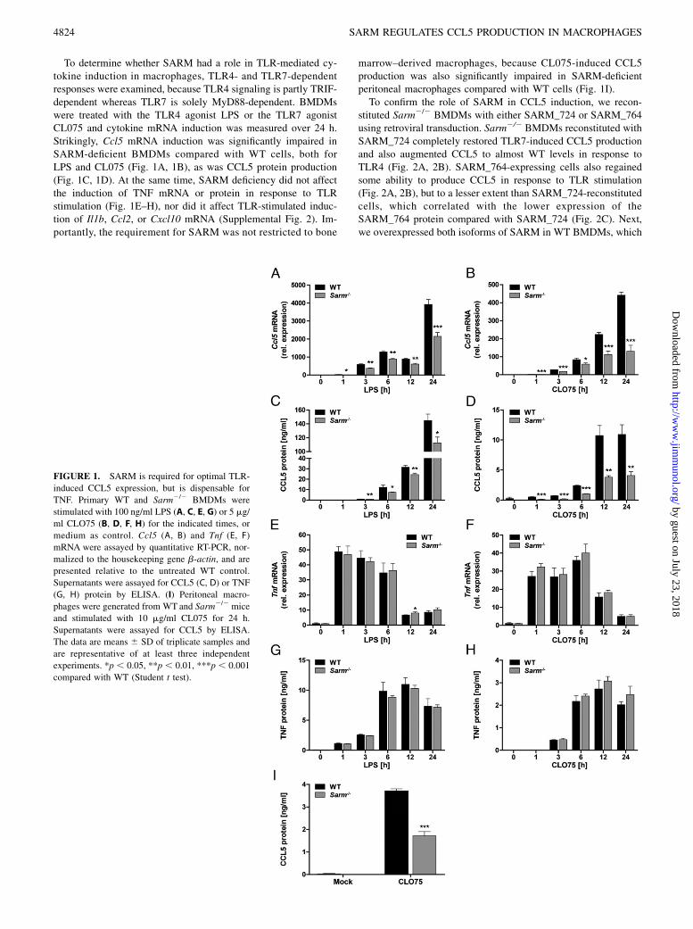
To determine whether SARM had a role in TLR-mediated cy-tokine induction in macrophages, TLR4- and TLR7-dependentresponses were examined, because TLR4 signaling is partly TRIF-dependent whereas TLR7 is solely MyD88-dependent. BMDMswere treated with the TLR4 agonist LPS or the TLR7 agonistCL075 and cytokine mRNA induction was measured over 24 h.Strikingly, Ccl5 mRNA induction was significantly impaired inSARM-deficient BMDMs compared with WT cells, both forLPS and CL075 (Fig. 1A, 1B), as was CCL5 protein production(Fig. 1C, 1D). At the same time, SARM deficiency did not affectthe induction of TNF mRNA or protein in response to TLRstimulation (Fig. 1E–H), nor did it affect TLR-stimulated induc-tion of Il1b, Ccl2, or Cxcl10 mRNA (Supplemental Fig. 2). Im-portantly, the requirement for SARM was not restricted to bone
marrow–derived macrophages, because CL075-induced CCL5production was also significantly impaired in SARM-deficientperitoneal macrophages compared with WT cells (Fig. 1I).To confirm the role of SARM in CCL5 induction, we recon-
stituted Sarm2/2 BMDMs with either SARM_724 or SARM_764using retroviral transduction. Sarm2/2 BMDMs reconstituted withSARM_724 completely restored TLR7-induced CCL5 productionand also augmented CCL5 to almost WT levels in response toTLR4 (Fig. 2A, 2B). SARM_764-expressing cells also regainedsome ability to produce CCL5 in response to TLR stimulation(Fig. 2A, 2B), but to a lesser extent than SARM_724-reconstitutedcells, which correlated with the lower expression of theSARM_764 protein compared with SARM_724 (Fig. 2C). Next,we overexpressed both isoforms of SARM in WT BMDMs, which
FIGURE 1. SARM is required for optimal TLR-
induced CCL5 expression, but is dispensable for
TNF. Primary WT and Sarm2/2 BMDMs were
stimulated with 100 ng/ml LPS (A, C, E, G) or 5 mg/
ml CLO75 (B, D, F, H) for the indicated times, or
medium as control. Ccl5 (A, B) and Tnf (E, F)
mRNA were assayed by quantitative RT-PCR, nor-
malized to the housekeeping gene b-actin, and are
presented relative to the untreated WT control.
Supernatants were assayed for CCL5 (C, D) or TNF
(G, H) protein by ELISA. (I) Peritoneal macro-
phages were generated from WT and Sarm2/2 mice
and stimulated with 10 mg/ml CL075 for 24 h.
Supernatants were assayed for CCL5 by ELISA.
The data are means 6 SD of triplicate samples and
are representative of at least three independent
experiments. *p , 0.05, **p , 0.01, ***p , 0.001
compared with WT (Student t test).
4824 SARM REGULATES CCL5 PRODUCTION IN MACROPHAGES
by guest on July 23, 2018http://w
ww
.jimm
unol.org/D
ownloaded from
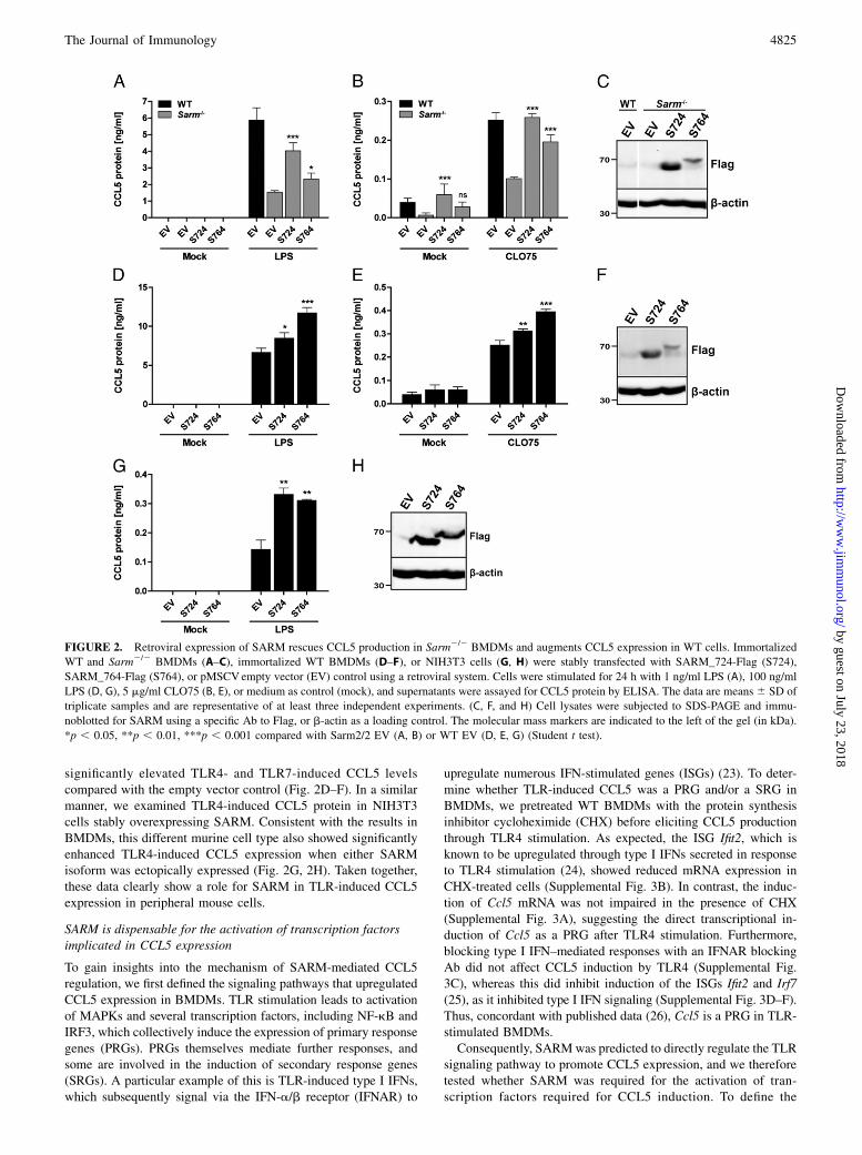
significantly elevated TLR4- and TLR7-induced CCL5 levelscompared with the empty vector control (Fig. 2D–F). In a similarmanner, we examined TLR4-induced CCL5 protein in NIH3T3cells stably overexpressing SARM. Consistent with the results inBMDMs, this different murine cell type also showed significantlyenhanced TLR4-induced CCL5 expression when either SARMisoform was ectopically expressed (Fig. 2G, 2H). Taken together,these data clearly show a role for SARM in TLR-induced CCL5expression in peripheral mouse cells.
SARM is dispensable for the activation of transcription factorsimplicated in CCL5 expression
To gain insights into the mechanism of SARM-mediated CCL5regulation, we first defined the signaling pathways that upregulatedCCL5 expression in BMDMs. TLR stimulation leads to activationof MAPKs and several transcription factors, including NF-kB andIRF3, which collectively induce the expression of primary responsegenes (PRGs). PRGs themselves mediate further responses, andsome are involved in the induction of secondary response genes(SRGs). A particular example of this is TLR-induced type I IFNs,which subsequently signal via the IFN-a/b receptor (IFNAR) to
upregulate numerous IFN-stimulated genes (ISGs) (23). To deter-mine whether TLR-induced CCL5 was a PRG and/or a SRG inBMDMs, we pretreated WT BMDMs with the protein synthesisinhibitor cycloheximide (CHX) before eliciting CCL5 productionthrough TLR4 stimulation. As expected, the ISG Ifit2, which isknown to be upregulated through type I IFNs secreted in responseto TLR4 stimulation (24), showed reduced mRNA expression inCHX-treated cells (Supplemental Fig. 3B). In contrast, the induc-tion of Ccl5 mRNA was not impaired in the presence of CHX(Supplemental Fig. 3A), suggesting the direct transcriptional in-duction of Ccl5 as a PRG after TLR4 stimulation. Furthermore,blocking type I IFN–mediated responses with an IFNAR blockingAb did not affect CCL5 induction by TLR4 (Supplemental Fig.3C), whereas this did inhibit induction of the ISGs Ifit2 and Irf7(25), as it inhibited type I IFN signaling (Supplemental Fig. 3D–F).Thus, concordant with published data (26), Ccl5 is a PRG in TLR-stimulated BMDMs.Consequently, SARMwas predicted to directly regulate the TLR
signaling pathway to promote CCL5 expression, and we thereforetested whether SARM was required for the activation of tran-scription factors required for CCL5 induction. To define the
FIGURE 2. Retroviral expression of SARM rescues CCL5 production in Sarm2/2 BMDMs and augments CCL5 expression in WT cells. Immortalized
WT and Sarm2/2 BMDMs (A–C), immortalized WT BMDMs (D–F), or NIH3T3 cells (G, H) were stably transfected with SARM_724-Flag (S724),
SARM_764-Flag (S764), or pMSCVempty vector (EV) control using a retroviral system. Cells were stimulated for 24 h with 1 ng/ml LPS (A), 100 ng/ml
LPS (D, G), 5 mg/ml CLO75 (B, E), or medium as control (mock), and supernatants were assayed for CCL5 protein by ELISA. The data are means6 SD of
triplicate samples and are representative of at least three independent experiments. (C, F, and H) Cell lysates were subjected to SDS-PAGE and immu-
noblotted for SARM using a specific Ab to Flag, or b-actin as a loading control. The molecular mass markers are indicated to the left of the gel (in kDa).
*p , 0.05, **p , 0.01, ***p , 0.001 compared with Sarm2/2 EV (A, B) or WT EV (D, E, G) (Student t test).
The Journal of Immunology 4825
by guest on July 23, 2018http://w
ww
.jimm
unol.org/D
ownloaded from
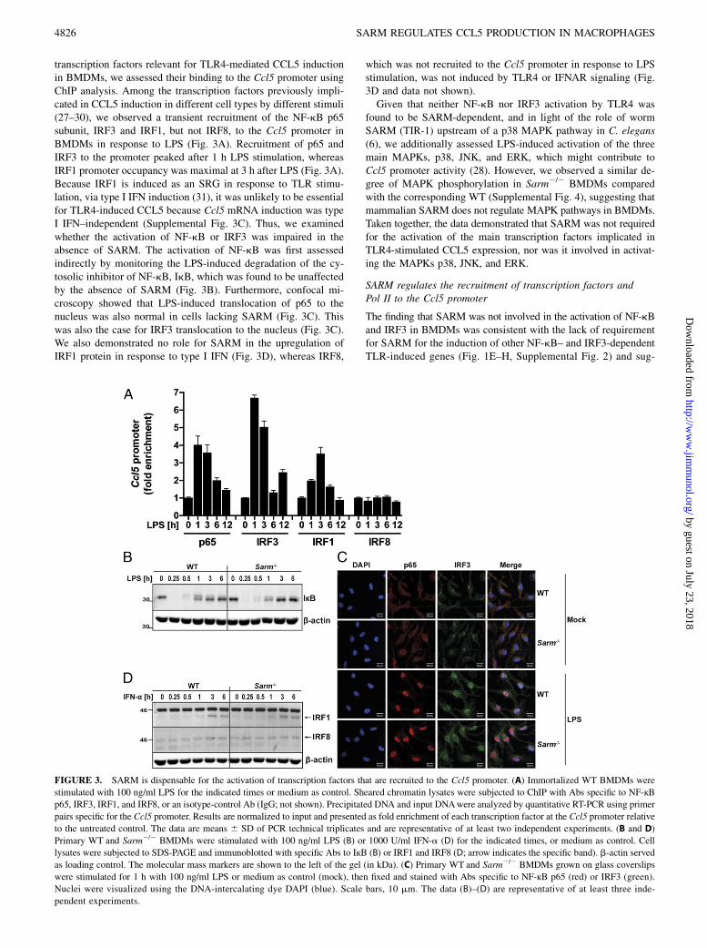
transcription factors relevant for TLR4-mediated CCL5 inductionin BMDMs, we assessed their binding to the Ccl5 promoter usingChIP analysis. Among the transcription factors previously impli-cated in CCL5 induction in different cell types by different stimuli(27–30), we observed a transient recruitment of the NF-kB p65subunit, IRF3 and IRF1, but not IRF8, to the Ccl5 promoter inBMDMs in response to LPS (Fig. 3A). Recruitment of p65 andIRF3 to the promoter peaked after 1 h LPS stimulation, whereasIRF1 promoter occupancy was maximal at 3 h after LPS (Fig. 3A).Because IRF1 is induced as an SRG in response to TLR stimu-lation, via type I IFN induction (31), it was unlikely to be essentialfor TLR4-induced CCL5 because Ccl5 mRNA induction was typeI IFN–independent (Supplemental Fig. 3C). Thus, we examinedwhether the activation of NF-kB or IRF3 was impaired in theabsence of SARM. The activation of NF-kB was first assessedindirectly by monitoring the LPS-induced degradation of the cy-tosolic inhibitor of NF-kB, IkB, which was found to be unaffectedby the absence of SARM (Fig. 3B). Furthermore, confocal mi-croscopy showed that LPS-induced translocation of p65 to thenucleus was also normal in cells lacking SARM (Fig. 3C). Thiswas also the case for IRF3 translocation to the nucleus (Fig. 3C).We also demonstrated no role for SARM in the upregulation ofIRF1 protein in response to type I IFN (Fig. 3D), whereas IRF8,
which was not recruited to the Ccl5 promoter in response to LPSstimulation, was not induced by TLR4 or IFNAR signaling (Fig.3D and data not shown).Given that neither NF-kB nor IRF3 activation by TLR4 was
found to be SARM-dependent, and in light of the role of wormSARM (TIR-1) upstream of a p38 MAPK pathway in C. elegans(6), we additionally assessed LPS-induced activation of the threemain MAPKs, p38, JNK, and ERK, which might contribute toCcl5 promoter activity (28). However, we observed a similar de-gree of MAPK phosphorylation in Sarm2/2 BMDMs comparedwith the corresponding WT (Supplemental Fig. 4), suggesting thatmammalian SARM does not regulate MAPK pathways in BMDMs.Taken together, the data demonstrated that SARM was not requiredfor the activation of the main transcription factors implicated inTLR4-stimulated CCL5 expression, nor was it involved in activat-ing the MAPKs p38, JNK, and ERK.
SARM regulates the recruitment of transcription factors andPol II to the Ccl5 promoter
The finding that SARM was not involved in the activation of NF-kBand IRF3 in BMDMs was consistent with the lack of requirementfor SARM for the induction of other NF-kB– and IRF3-dependentTLR-induced genes (Fig. 1E–H, Supplemental Fig. 2) and sug-
FIGURE 3. SARM is dispensable for the activation of transcription factors that are recruited to the Ccl5 promoter. (A) Immortalized WT BMDMs were
stimulated with 100 ng/ml LPS for the indicated times or medium as control. Sheared chromatin lysates were subjected to ChIP with Abs specific to NF-кBp65, IRF3, IRF1, and IRF8, or an isotype-control Ab (IgG; not shown). Precipitated DNA and input DNAwere analyzed by quantitative RT-PCR using primer
pairs specific for the Ccl5 promoter. Results are normalized to input and presented as fold enrichment of each transcription factor at the Ccl5 promoter relative
to the untreated control. The data are means 6 SD of PCR technical triplicates and are representative of at least two independent experiments. (B and D)
Primary WT and Sarm2/2 BMDMs were stimulated with 100 ng/ml LPS (B) or 1000 U/ml IFN-a (D) for the indicated times, or medium as control. Cell
lysates were subjected to SDS-PAGE and immunoblotted with specific Abs to IкB (B) or IRF1 and IRF8 (D; arrow indicates the specific band). b-actin served
as loading control. The molecular mass markers are shown to the left of the gel (in kDa). (C) Primary WT and Sarm2/2 BMDMs grown on glass coverslips
were stimulated for 1 h with 100 ng/ml LPS or medium as control (mock), then fixed and stained with Abs specific to NF-кB p65 (red) or IRF3 (green).
Nuclei were visualized using the DNA-intercalating dye DAPI (blue). Scale bars, 10 mm. The data (B)–(D) are representative of at least three inde-
pendent experiments.
4826 SARM REGULATES CCL5 PRODUCTION IN MACROPHAGES
by guest on July 23, 2018http://w
ww
.jimm
unol.org/D
ownloaded from
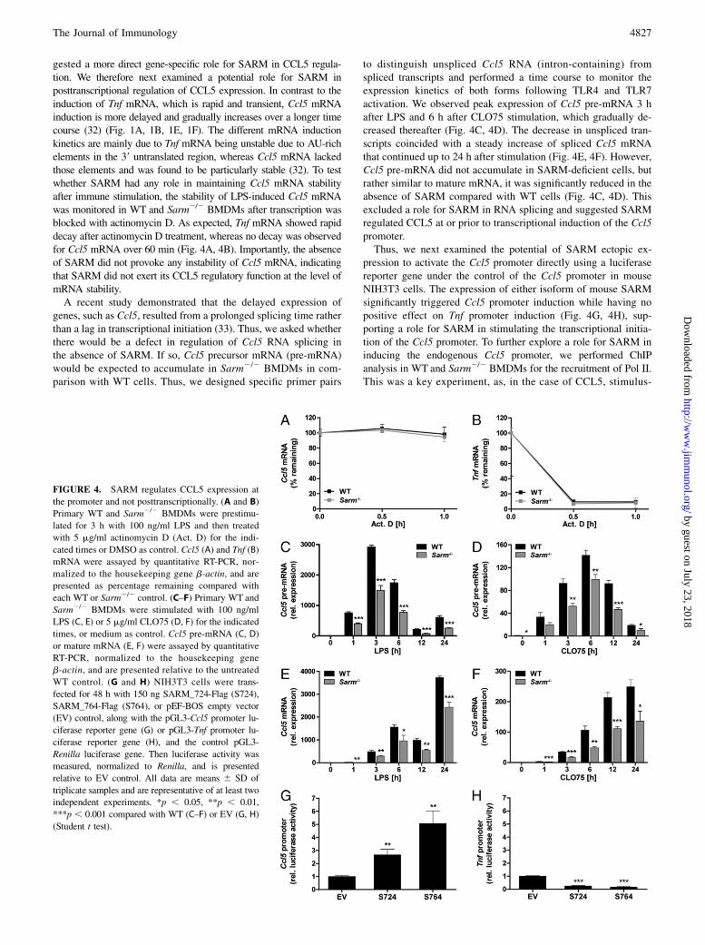
gested a more direct gene-specific role for SARM in CCL5 regula-tion. We therefore next examined a potential role for SARM inposttranscriptional regulation of CCL5 expression. In contrast to theinduction of Tnf mRNA, which is rapid and transient, Ccl5 mRNAinduction is more delayed and gradually increases over a longer timecourse (32) (Fig. 1A, 1B, 1E, 1F). The different mRNA inductionkinetics are mainly due to Tnf mRNA being unstable due to AU-richelements in the 39 untranslated region, whereas Ccl5 mRNA lackedthose elements and was found to be particularly stable (32). To testwhether SARM had any role in maintaining Ccl5 mRNA stabilityafter immune stimulation, the stability of LPS-induced Ccl5 mRNAwas monitored in WT and Sarm2/2 BMDMs after transcription wasblocked with actinomycin D. As expected, Tnf mRNA showed rapiddecay after actinomycin D treatment, whereas no decay was observedfor Ccl5 mRNA over 60 min (Fig. 4A, 4B). Importantly, the absenceof SARM did not provoke any instability of Ccl5 mRNA, indicatingthat SARM did not exert its CCL5 regulatory function at the level ofmRNA stability.A recent study demonstrated that the delayed expression of
genes, such as Ccl5, resulted from a prolonged splicing time ratherthan a lag in transcriptional initiation (33). Thus, we asked whetherthere would be a defect in regulation of Ccl5 RNA splicing inthe absence of SARM. If so, Ccl5 precursor mRNA (pre-mRNA)would be expected to accumulate in Sarm2/2 BMDMs in com-parison with WT cells. Thus, we designed specific primer pairs
to distinguish unspliced Ccl5 RNA (intron-containing) fromspliced transcripts and performed a time course to monitor theexpression kinetics of both forms following TLR4 and TLR7activation. We observed peak expression of Ccl5 pre-mRNA 3 hafter LPS and 6 h after CLO75 stimulation, which gradually de-creased thereafter (Fig. 4C, 4D). The decrease in unspliced tran-scripts coincided with a steady increase of spliced Ccl5 mRNAthat continued up to 24 h after stimulation (Fig. 4E, 4F). However,Ccl5 pre-mRNA did not accumulate in SARM-deficient cells, butrather similar to mature mRNA, it was significantly reduced in theabsence of SARM compared with WT cells (Fig. 4C, 4D). Thisexcluded a role for SARM in RNA splicing and suggested SARMregulated CCL5 at or prior to transcriptional induction of the Ccl5promoter.Thus, we next examined the potential of SARM ectopic ex-
pression to activate the Ccl5 promoter directly using a luciferasereporter gene under the control of the Ccl5 promoter in mouseNIH3T3 cells. The expression of either isoform of mouse SARMsignificantly triggered Ccl5 promoter induction while having nopositive effect on Tnf promoter induction (Fig. 4G, 4H), sup-porting a role for SARM in stimulating the transcriptional initia-tion of the Ccl5 promoter. To further explore a role for SARM ininducing the endogenous Ccl5 promoter, we performed ChIPanalysis in WT and Sarm2/2 BMDMs for the recruitment of Pol II.This was a key experiment, as, in the case of CCL5, stimulus-
FIGURE 4. SARM regulates CCL5 expression at
the promoter and not posttranscriptionally. (A and B)
Primary WT and Sarm2/2 BMDMs were prestimu-
lated for 3 h with 100 ng/ml LPS and then treated
with 5 mg/ml actinomycin D (Act. D) for the indi-
cated times or DMSO as control. Ccl5 (A) and Tnf (B)
mRNA were assayed by quantitative RT-PCR, nor-
malized to the housekeeping gene b-actin, and are
presented as percentage remaining compared with
each WT or Sarm2/2 control. (C–F) Primary WT and
Sarm2/2 BMDMs were stimulated with 100 ng/ml
LPS (C, E) or 5 mg/ml CLO75 (D, F) for the indicated
times, or medium as control. Ccl5 pre-mRNA (C, D)
or mature mRNA (E, F) were assayed by quantitative
RT-PCR, normalized to the housekeeping gene
b-actin, and are presented relative to the untreated
WT control. (G and H) NIH3T3 cells were trans-
fected for 48 h with 150 ng SARM_724-Flag (S724),
SARM_764-Flag (S764), or pEF-BOS empty vector
(EV) control, along with the pGL3-Ccl5 promoter lu-
ciferase reporter gene (G) or pGL3-Tnf promoter lu-
ciferase reporter gene (H), and the control pGL3-
Renilla luciferase gene. Then luciferase activity was
measured, normalized to Renilla, and is presented
relative to EV control. All data are means 6 SD of
triplicate samples and are representative of at least two
independent experiments. *p , 0.05, **p , 0.01,
***p , 0.001 compared with WT (C–F) or EV (G, H)
(Student t test).
The Journal of Immunology 4827
by guest on July 23, 2018http://w
ww
.jimm
unol.org/D
ownloaded from
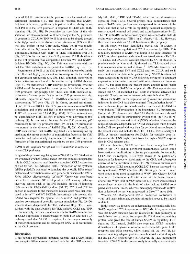
induced Pol II recruitment to the promoter is a hallmark of tran-scriptional induction (17). The analysis revealed that SARM-deficient cells were significantly impaired in their ability to re-cruit Pol II to the Ccl5 promoter in response to TLR4 and TLR7signaling (Fig. 5A, 5B). To determine the specificity of this ob-servation, we also examined Pol II occupancy at the Tnf promoter.In contrast to CCL5, for TNF Pol II is known to be already presentat the Tnf promoter in the absence of cell stimulation (17), and thiswas also evident in our ChIP study, where Pol II was readilydetectable at the Tnf promoter in unstimulated cells and did notsignificantly increase with TLR4 or TLR7 stimulation over 3 h(Fig. 5C, 5D). In contrast to the Ccl5 promoter, Pol II abundanceat the Tnf promoter was comparable between WT and SARM-deficient BMDMs (Fig. 5C, 5D). This was consistent with thefact that TNF induction is independent of SARM (Fig. 1E, 1F).Recruitment of Pol II to the Ccl5 promoter is known to be tightly
controlled and highly dependent on transcription factor bindingand chromatin remodeling (34, 35). Thus, although transcriptionfactor activation was found to be unaffected by SARM ablation(Fig. 3), we performed ChIP analysis to investigate whetherSARM would be required for transcription factor binding to theCcl5 promoter. Intriguingly, both TLR4- and TLR7-mediated re-cruitment of transcription factors to the Ccl5 promoter was sig-nificantly impaired in Sarm2/2 BMDMs compared with thecorresponding WT cells (Fig. 5E–I). Hence, optimal recruitmentof p65, IRF3, and IRF1 to the Ccl5 promoter in response to TLR4stimulation, and of p65 and IRF1 to the promoter in response toTLR7, all depended on SARM (Fig. 5E–I). IRF3 recruitment wasnot examined for TLR7, as IRF3 is generally not activated by thispathway (1). In contrast to the case for the Ccl5 promoter, p65recruitment to the Tnf promoter after LPS or CL075 stimulationwas normal in the absence of SARM (Fig. 5J, 5K). Overall, theChIP data showed that SARM regulated Ccl5 transcription bymediating the proper assembly of transcription factors at the Ccl5promoter and subsequently recruitment of Pol II, and thus theformation of the transcriptional machinery on the Ccl5 promoter.
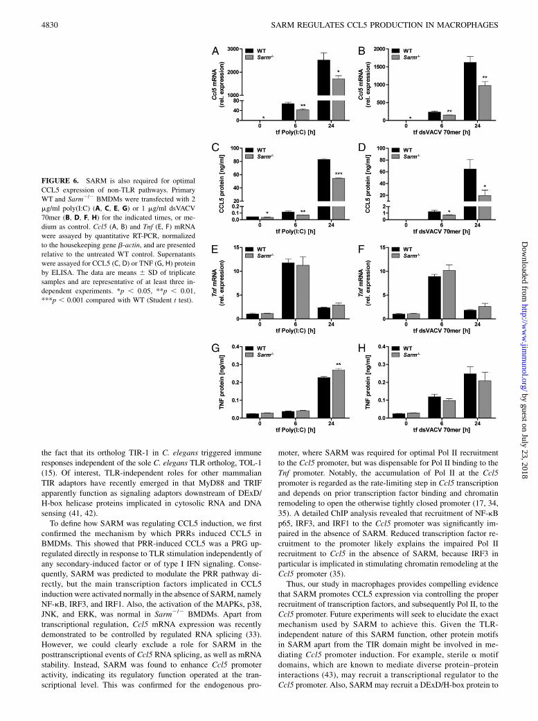
SARM is also required for optimal CCL5 induction in responseto non-TLR pathways
In light of the promoter regulatory role of SARM for CCL5 induction,we wondered whether SARM had an intrinsic stimulus-independentrole in CCL5 induction and therefore examined CCL5 expressionelicited by non-TLR cytosolic PRRs. Transfection of the syntheticdsRNA poly(I:C) was used to stimulate the cytosolic RNA sensormelanoma differentiation-associated gene 5 (3), whereas the VACV70-bp dsDNA oligonucleotide (dsVACV 70mer) was transfectedinto cells to stimulate STING-dependent DNA sensing pathwaysinvolving sensors such as the IFN-inducible protein 16 homologp204 and cyclic GMP-AMP synthase (20, 36). CCL5 and TNF in-duction in response to the transfected nucleic acids was then com-pared in Sarm2/2 and WT BMDMs. Interestingly, this revealed thatSARM was required for optimal CCL5 mRNA and protein ex-pression downstream of cytosolic receptor stimulation (Fig. 6A–D),whereas it was dispensable for TNF induction (Fig. 6E–H), con-cordant with the data obtained for TLR-induced CCL5 and TNF.Cumulatively, the data reveal a new role for SARM in induction
of CCL5 expression in macrophages by both TLR and non-TLRpathways, and that SARM is required for the proper assemblyof transcription factors and for subsequent RNA Pol II recruitmentat the Ccl5 promoter.
DiscussionIt has become increasingly apparent recently that SARM mightexecute quite different roles compared with the other TIR adaptors,
MyD88, MAL, TRIF, and TRAM, which initiate downstreamsignaling from TLRs. Several groups have demonstrated thatmouse SARM was predominantly expressed in neurons of thebrain, and that it had a role in regulating neuronal morphology,stress-induced neuronal cell death, and axon degeneration (8–12).The role of SARM in the nervous system was concordant with itsevolutionary counterpart TIR-1 in C. elegans (13, 14). However,there are fewer data on SARM functions in the periphery.In this study, we have identified a crucial role for SARM in
macrophages in the regulation of CCL5 expression by PRRs. Thisregulatory function of SARM was found to be quite selective forCCL5, as other cytokines and chemokines tested, namely TNF, IL-1b, CCL2, and CXCL10, were not affected by SARM ablation. Aprevious study by Kim et al. (8) showed that TLR-induced cyto-kine responses were normal in Sarm2/2 BMDMs; however, thatstudy only examined TNF and CCL2 and not CCL5, and so isconsistent with our data in the present study. SARM function hadbeen suggested to be likely CNS-restricted owing to its abundantexpression in the brain compared with other tissues and immunecells (8), but apart from the present study, another recent papershowed a role for SARM in peripheral cells. That report demon-strated that SARM-mediated T cell death in immune-activated andexpanded T cells to restore homeostasis after infection (22).Roles for SARM in the regulation of cytokine and chemokine
induction in the CNS have also emerged. Thus, infecting Sarm2/2
mice with neurotropic WNV indicated a requirement of SARM forvirus-induced TNF production in the brainstem (16). Furthermore,Hou et al. (37) demonstrated that SARM-deficient mice showeda significant defect in upregulating cytokines in the CNS in re-sponse to vesicular stomatitis virus (VSV) infection. However, therange of cytokines dependent on SARM seems to be much broaderin the CNS compared with our data for peripheral macrophages inthe present study and includes IL-6, TNF, CCL2, CCL5, and type IIFNs. A broader requirement for SARM for cytokine gene in-duction in the CNS might result from the higher SARM expres-sion in CNS-resident cells.Of note, therefore, SARM has been found to regulate CCL5
both in the CNS and in peripheral macrophages, which couldhave particular implications in disease pathogenesis. For example,CCL5 and its receptor CCR5 were shown to be particularlyimportant for leukocyte recruitment to the CNS, and subsequentcontrol of WNV infection in mice (38, 39), whereas humans witha homozygous CCR5 mutation (CCR5D32) have an increased riskfor symptomatic WNV infection (40). Strikingly, Sarm2/2 micewere shown to be more susceptible to WNV (16). Clearly SARMis required for immune cell infiltration into the brain, becauseafter either WNV (16) or VSV infection (37) there were reducedmacrophage numbers in the brain of mice lacking SARM com-pared with normal mice, whereas macrophage/monocyte infiltra-tion of lesioned nerves was suppressed in Sarm2/2 mice (10).Whether SARM-dependent CCL5 production contributes to
virus- and insult-stimulated cellular infiltration needs to be studiedin detail.In this study, we focused on understanding mechanistically how
SARM regulated CCL5 expression in BMDMs. A key observationwas that SARM function was not restricted to TLR pathways, aswould have been expected for a cytosolic TIR domain–containingprotein, and given the role of human SARM in regulating TRIFsignaling (7). Instead, SARM also regulated CCL5 inductiondownstream of cytosolic retinoic acid–inducible gene I–likereceptors and DNA sensors, which signal via the non-TIR do-main–containing adaptor proteins mitochondrial antiviral signal-ing and STING, respectively (1). However, the TLR-independentfunction of SARM in the present study is actually consistent with
4828 SARM REGULATES CCL5 PRODUCTION IN MACROPHAGES
by guest on July 23, 2018http://w
ww
.jimm
unol.org/D
ownloaded from
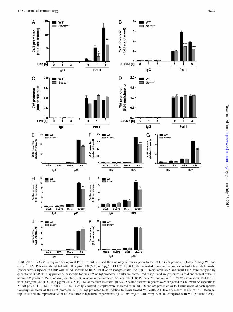
FIGURE 5. SARM is required for optimal Pol II recruitment and the assembly of transcription factors at the Ccl5 promoter. (A–D) Primary WT and
Sarm2/2 BMDMs were stimulated with 100 ng/ml LPS (A, C) or 5 mg/ml CLO75 (B, D) for the indicated times, or medium as control. Sheared chromatin
lysates were subjected to ChIP with an Ab specific to RNA Pol II or an isotype-control Ab (IgG). Precipitated DNA and input DNA were analyzed by
quantitative RT-PCR using primer pairs specific for the Ccl5 or Tnf promoter. Results are normalized to input and are presented as fold enrichment of Pol II
at the Ccl5 promoter (A, B) or Tnf promoter (C, D) relative to the untreated WT control. (E–K) Primary WT and Sarm2/2 BMDMs were stimulated for 1 h
with 100ng/ml LPS (E–G, J), 5 mg/ml CLO75 (H, I, K), or medium as control (mock). Sheared chromatin lysates were subjected to ChIP with Abs specific to
NF-кB p65 (E, H, J, K), IRF3 (F), IRF1 (G, I), or IgG control. Samples were analyzed as in (A)–(D) and are presented as fold enrichment of each specific
transcription factor at the Ccl5 promoter (E–I) or Tnf promoter (J, K) relative to mock-treated WT cells. All data are means 6 SD of PCR technical
triplicates and are representative of at least three independent experiments. *p , 0.05, **p , 0.01, ***p , 0.001 compared with WT (Student t test).
The Journal of Immunology 4829
by guest on July 23, 2018http://w
ww
.jimm
unol.org/D
ownloaded from
the fact that its ortholog TIR-1 in C. elegans triggered immuneresponses independent of the sole C. elegans TLR ortholog, TOL-1(15). Of interest, TLR-independent roles for other mammalianTIR adaptors have recently emerged in that MyD88 and TRIFapparently function as signaling adaptors downstream of DExD/H-box helicase proteins implicated in cytosolic RNA and DNAsensing (41, 42).To define how SARM was regulating CCL5 induction, we first
confirmed the mechanism by which PRRs induced CCL5 inBMDMs. This showed that PRR-induced CCL5 was a PRG up-regulated directly in response to TLR stimulation independently ofany secondary-induced factor or of type I IFN signaling. Conse-quently, SARM was predicted to modulate the PRR pathway di-rectly, but the main transcription factors implicated in CCL5induction were activated normally in the absence of SARM, namelyNF-kB, IRF3, and IRF1. Also, the activation of the MAPKs, p38,JNK, and ERK, was normal in Sarm2/2 BMDMs. Apart fromtranscriptional regulation, Ccl5 mRNA expression was recentlydemonstrated to be controlled by regulated RNA splicing (33).However, we could clearly exclude a role for SARM in theposttranscriptional events of Ccl5 RNA splicing, as well as mRNAstability. Instead, SARM was found to enhance Ccl5 promoteractivity, indicating its regulatory function operated at the tran-scriptional level. This was confirmed for the endogenous pro-
moter, where SARM was required for optimal Pol II recruitmentto the Ccl5 promoter, but was dispensable for Pol II binding to theTnf promoter. Notably, the accumulation of Pol II at the Ccl5promoter is regarded as the rate-limiting step in Ccl5 transcriptionand depends on prior transcription factor binding and chromatinremodeling to open the otherwise tightly closed promoter (17, 34,35). A detailed ChIP analysis revealed that recruitment of NF-kBp65, IRF3, and IRF1 to the Ccl5 promoter was significantly im-paired in the absence of SARM. Reduced transcription factor re-cruitment to the promoter likely explains the impaired Pol IIrecruitment to Ccl5 in the absence of SARM, because IRF3 inparticular is implicated in stimulating chromatin remodeling at theCcl5 promoter (35).Thus, our study in macrophages provides compelling evidence
that SARM promotes CCL5 expression via controlling the properrecruitment of transcription factors, and subsequently Pol II, to theCcl5 promoter. Future experiments will seek to elucidate the exactmechanism used by SARM to achieve this. Given the TLR-independent nature of this SARM function, other protein motifsin SARM apart from the TIR domain might be involved in me-diating Ccl5 promoter induction. For example, sterile a motifdomains, which are known to mediate diverse protein–proteininteractions (43), may recruit a transcriptional regulator to theCcl5 promoter. Also, SARM may recruit a DExD/H-box protein to
FIGURE 6. SARM is also required for optimal
CCL5 expression of non-TLR pathways. Primary
WT and Sarm2/2 BMDMs were transfected with 2
mg/ml poly(I:C) (A, C, E, G) or 1 mg/ml dsVACV
70mer (B, D, F, H) for the indicated times, or me-
dium as control. Ccl5 (A, B) and Tnf (E, F) mRNA
were assayed by quantitative RT-PCR, normalized
to the housekeeping gene b-actin, and are presented
relative to the untreated WT control. Supernatants
were assayed for CCL5 (C, D) or TNF (G, H) protein
by ELISA. The data are means 6 SD of triplicate
samples and are representative of at least three in-
dependent experiments. *p , 0.05, **p , 0.01,
***p , 0.001 compared with WT (Student t test).
4830 SARM REGULATES CCL5 PRODUCTION IN MACROPHAGES
by guest on July 23, 2018http://w
ww
.jimm
unol.org/D
ownloaded from
the Ccl5 promoter, because such helicases can interact with TIRadaptors (41, 42) and are also know to regulate gene promoters(44). Furthermore, it will be interesting to define whether SARMacts in close proximity to the Ccl5 promoter, or whether it func-tions in a more upstream role. In support of the latter model is thefact that SARM is reported to be expressed in the cytosol andmostly located at the mitochondria (8, 45). However, we andothers (46) have also detected SARM in the nucleus, althoughwe have no evidence to date of PRR-stimulated relocalization ofSARM (data not shown). Apart from elucidating the exactmechanism of Ccl5 transcriptional regulation by SARM in mac-rophages, it will also be of interest to determine whether SARM-dependent cytokine induction in the CNS (37) is also due toregulation of recruitment of transcription factors and Pol II toselected gene promoters.Overall, this study has identified a novel function of mouse
SARM in selectively promoting PRR-induced CCL5 expressionin macrophages. Accordingly, the data contribute to establishingSARM as a positive regulator of cytokine responses, concordantwith previous murine in vivo studies. Importantly, the present studyhighlights the TLR-independent role of another mammalian TIRadaptor protein.
AcknowledgmentsWe thank Dmitry Kuprash for the gift of the Tnf promoter luciferase
reporter gene.
DisclosuresThe authors have no financial conflicts of interest.
References1. Takeuchi, O., and S. Akira. 2010. Pattern recognition receptors and inflamma-
tion. Cell 140: 805–820.2. O’Neill, L. A., and A. G. Bowie. 2007. The family of five: TIR-domain-containing
adaptors in Toll-like receptor signalling. Nat. Rev. Immunol. 7: 353–364.3. G€urtler, C., and A. G. Bowie. 2013. Innate immune detection of microbial
nucleic acids. Trends Microbiol. 21: 413–420.4. O’Neill, L. A., K. A. Fitzgerald, and A. G. Bowie. 2003. The Toll-IL-1 receptor
adaptor family grows to five members. Trends Immunol. 24: 286–290.5. Mink, M., B. Fogelgren, K. Olszewski, P. Maroy, and K. Csiszar. 2001. A novel
human gene (SARM) at chromosome 17q11 encodes a protein with a SAM motifand structural similarity to Armadillo/b-catenin that is conserved in mouse,Drosophila, and Caenorhabditis elegans. Genomics 74: 234–244.
6. Liberati, N. T., K. A. Fitzgerald, D. H. Kim, R. Feinbaum, D. T. Golenbock, andF. M. Ausubel. 2004. Requirement for a conserved Toll/interleukin-1 resistancedomain protein in the Caenorhabditis elegans immune response. Proc. Natl.Acad. Sci. USA 101: 6593–6598.
7. Carty, M., R. Goodbody, M. Schroder, J. Stack, P. N. Moynagh, and A. G. Bowie.2006. The human adaptor SARM negatively regulates adaptor protein TRIF-dependent Toll-like receptor signaling. Nat. Immunol. 7: 1074–1081.
8. Kim, Y., P. Zhou, L. Qian, J. Z. Chuang, J. Lee, C. Li, C. Iadecola, C. Nathan,and A. Ding. 2007. MyD88-5 links mitochondria, microtubules, and JNK3 inneurons and regulates neuronal survival. J. Exp. Med. 204: 2063–2074.
9. Mukherjee, P., T. A. Woods, R. A. Moore, and K. E. Peterson. 2013. Activationof the innate signaling molecule MAVS by bunyavirus infection upregulates theadaptor protein SARM1, leading to neuronal death. Immunity 38: 705–716.
10. Osterloh, J. M., J. Yang, T. M. Rooney, A. N. Fox, R. Adalbert, E. H. Powell,A. E. Sheehan, M. A. Avery, R. Hackett, M. A. Logan, et al. 2012. dSarm/Sarm1is required for activation of an injury-induced axon death pathway. Science 337:481–484.
11. Gerdts, J., D. W. Summers, Y. Sasaki, A. DiAntonio, and J. Milbrandt. 2013.Sarm1-mediated axon degeneration requires both SAM and TIR interactions. J.Neurosci. 33: 13569–13580.
12. Chen, C. Y., C. W. Lin, C. Y. Chang, S. T. Jiang, and Y. P. Hsueh. 2011. Sarm1,a negative regulator of innate immunity, interacts with syndecan-2 and regulatesneuronal morphology. J. Cell Biol. 193: 769–784.
13. Chuang, C. F., and C. I. Bargmann. 2005. A Toll-interleukin 1 repeat protein atthe synapse specifies asymmetric odorant receptor expression via ASK1MAPKKK signaling. Genes Dev. 19: 270–281.
14. Hayakawa, T., K. Kato, R. Hayakawa, N. Hisamoto, K. Matsumoto, K. Takeda,and H. Ichijo. 2011. Regulation of anoxic death in Caenorhabditis elegans bymammalian apoptosis signal-regulating kinase (ASK) family proteins. Genetics187: 785–792.
15. Couillault, C., N. Pujol, J. Reboul, L. Sabatier, J. F. Guichou, Y. Kohara, andJ. J. Ewbank. 2004. TLR-independent control of innate immunity in Caeno-rhabditis elegans by the TIR domain adaptor protein TIR-1, an ortholog ofhuman SARM. Nat. Immunol. 5: 488–494.
16. Szretter, K. J., M. A. Samuel, S. Gilfillan, A. Fuchs, M. Colonna, andM. S. Diamond. 2009. The immune adaptor molecule SARM modulates tumornecrosis factor a production and microglia activation in the brainstem andrestricts West Nile virus pathogenesis. J. Virol. 83: 9329–9338.
17. Adelman, K., M. A. Kennedy, S. Nechaev, D. A. Gilchrist, G. W. Muse,Y. Chinenov, and I. Rogatsky. 2009. Immediate mediators of the inflammatoryresponse are poised for gene activation through RNA polymerase II stalling.Proc. Natl. Acad. Sci. USA 106: 18207–18212.
18. Roberson, S. M., and W. S. Walker. 1988. Immortalization of cloned mousesplenic macrophages with a retrovirus containing the v-raf/mil and v-myconcogenes. Cell. Immunol. 116: 341–351.
19. Hornung, V., F. Bauernfeind, A. Halle, E. O. Samstad, H. Kono, K. L. Rock,K. A. Fitzgerald, and E. Latz. 2008. Silica crystals and aluminum salts activatethe NALP3 inflammasome through phagosomal destabilization. Nat. Immunol. 9:847–856.
20. Unterholzner, L., S. E. Keating, M. Baran, K. A. Horan, S. B. Jensen, S. Sharma,C. M. Sirois, T. Jin, E. Latz, T. S. Xiao, et al. 2010. IFI16 is an innate immunesensor for intracellular DNA. Nat. Immunol. 11: 997–1004.
21. Nelson, J. D., O. Denisenko, and K. Bomsztyk. 2006. Protocol for the fastchromatin immunoprecipitation (ChIP) method. Nat. Protoc. 1: 179–185.
22. Panneerselvam, P., L. P. Singh, V. Selvarajan, W. J. Chng, S. B. Ng, N. S. Tan,B. Ho, J. Chen, and J. L. Ding. 2013. T-cell death following immune activation ismediated by mitochondria-localized SARM. Cell Death Differ. 20: 478–489.
23. Noppert, S. J., K. A. Fitzgerald, and P. J. Hertzog. 2007. The role of type Iinterferons in TLR responses. Immunol. Cell Biol. 85: 446–457.
24. Thomas, K. E., C. L. Galligan, R. D. Newman, E. N. Fish, and S. N. Vogel. 2006.Contribution of interferon-beta to the murine macrophage response to the Toll-like receptor 4 agonist, lipopolysaccharide. J. Biol. Chem. 281: 31119–31130.
25. Sato, M., N. Hata, M. Asagiri, T. Nakaya, T. Taniguchi, and N. Tanaka. 1998.Positive feedback regulation of type I IFN genes by the IFN-inducible tran-scription factor IRF-7. FEBS Lett. 441: 106–110.
26. Doyle, S., S. Vaidya, R. O’Connell, H. Dadgostar, P. Dempsey, T. Wu, G. Rao,R. Sun, M. Haberland, R. Modlin, and G. Cheng. 2002. IRF3 mediates a TLR3/TLR4-specific antiviral gene program. Immunity 17: 251–263.
27. Genin, P., M. Algarte, P. Roof, R. Lin, and J. Hiscott. 2000. Regulation ofRANTES chemokine gene expression requires cooperativity between NF-kB andIFN-regulatory factor transcription factors. J. Immunol. 164: 5352–5361.
28. Casola, A., R. P. Garofalo, H. Haeberle, T. F. Elliott, R. Lin, M. Jamaluddin, andA. R. Brasier. 2001. Multiple cis regulatory elements control RANTES promoteractivity in alveolar epithelial cells infected with respiratory syncytial virus. J.Virol. 75: 6428–6439.
29. Liu, J., X. Guan, and X. Ma. 2005. Interferon regulatory factor 1 is an essentialand direct transcriptional activator for interferon g-induced RANTES/CCl5 ex-pression in macrophages. J. Biol. Chem. 280: 24347–24355.
30. Liu, J., and X. Ma. 2006. Interferon regulatory factor 8 regulates RANTES genetranscription in cooperation with interferon regulatory factor-1, NF-kB, andPU.1. J. Biol. Chem. 281: 19188–19195.
31. Tamura, T., H. Yanai, D. Savitsky, and T. Taniguchi. 2008. The IRF familytranscription factors in immunity and oncogenesis. Annu. Rev. Immunol. 26:535–584.
32. Hao, S., and D. Baltimore. 2009. The stability of mRNA influences the temporalorder of the induction of genes encoding inflammatory molecules. Nat. Immunol.10: 281–288.
33. Hao, S., and D. Baltimore. 2013. RNA splicing regulates the temporal order ofTNF-induced gene expression. Proc. Natl. Acad. Sci. USA 110: 11934–11939.
34. Hargreaves, D. C., T. Horng, and R. Medzhitov. 2009. Control of inducible geneexpression by signal-dependent transcriptional elongation. Cell 138: 129–145.
35. Ramirez-Carrozzi, V. R., D. Braas, D. M. Bhatt, C. S. Cheng, C. Hong,K. R. Doty, J. C. Black, A. Hoffmann, M. Carey, and S. T. Smale. 2009. Aunifying model for the selective regulation of inducible transcription by CpGislands and nucleosome remodeling. Cell 138: 114–128.
36. Xiao, T. S., and K. A. Fitzgerald. 2013. The cGAS-STING pathway for DNAsensing. Mol. Cell 51: 135–139.
37. Hou, Y. J., R. Banerjee, B. Thomas, C. Nathan, A. Garcıa-Sastre, A. Ding, andM. B. Uccellini. 2013. SARM is required for neuronal injury and cytokineproduction in response to central nervous system viral infection. J. Immunol.191: 875–883.
38. Hosking, M. P., and T. E. Lane. 2010. The role of chemokines during viral in-fection of the CNS. PLoS Pathog. 6: e1000937.
39. Lim, J. K., and P. M. Murphy. 2011. Chemokine control of West Nile virusinfection. Exp. Cell Res. 317: 569–574.
40. Lim, J. K., C. Y. Louie, C. Glaser, C. Jean, B. Johnson, H. Johnson,D. H. McDermott, and P. M. Murphy. 2008. Genetic deficiency of chemokinereceptor CCR5 is a strong risk factor for symptomatic West Nile virus infection:a meta-analysis of 4 cohorts in the US epidemic. J. Infect. Dis. 197: 262–265.
41. Kim, T., S. Pazhoor, M. Bao, Z. Zhang, S. Hanabuchi, V. Facchinetti, L. Bover,J. Plumas, L. Chaperot, J. Qin, and Y. J. Liu. 2010. Aspartate-glutamate-alanine-histidine box motif (DEAH)/RNA helicase A helicases sense microbial DNA inhuman plasmacytoid dendritic cells. Proc. Natl. Acad. Sci. USA 107: 15181–15186.
42. Zhang, Z., T. Kim, M. Bao, V. Facchinetti, S. Y. Jung, A. A. Ghaffari, J. Qin,G. Cheng, and Y. J. Liu. 2011. DDX1, DDX21, and DHX36 helicases form
The Journal of Immunology 4831
by guest on July 23, 2018http://w
ww
.jimm
unol.org/D
ownloaded from
a complex with the adaptor molecule TRIF to sense dsRNA in dendritic cells.Immunity 34: 866–878.
43. Kim, C. A., and J. U. Bowie. 2003. SAM domains: uniform structure, diversityof function. Trends Biochem. Sci. 28: 625–628.
44. Fuller-Pace, F. V. 2006. DExD/H box RNA helicases: multifunctional proteinswith important roles in transcriptional regulation. Nucleic Acids Res. 34: 4206–4215.
45. Panneerselvam, P., L. P. Singh, B. Ho, J. Chen, and J. L. Ding. 2012. Tar-geting of pro-apoptotic TLR adaptor SARM to mitochondria: definition ofthe critical region and residues in the signal sequence. Biochem. J. 442: 263–271.
46. Sethman, C. R., and J. Hawiger. 2013. The innate immunity adaptor SARMtranslocates to the nucleus to stabilize lamins and prevent DNA fragmentation inresponse to pro-apoptotic signaling. PLoS ONE 8: e70994.
4832 SARM REGULATES CCL5 PRODUCTION IN MACROPHAGES
by guest on July 23, 2018http://w
ww
.jimm
unol.org/D
ownloaded from