Embed Size (px)
Citation preview

The History of the Light Microscope

The more closely a phenomenon is observed, the more complex it is seen to be.
Heinrich Weisskopf

Starting with use of a simple lens in ancient times, to the first compound microscope around 1590, and up to the microscopes you are using in 7th grade life science, the microscope has allowed scientists to make discoveries about the “invisible world.”
The microscope has become one of the most recognizable symbols of science.

Some of the very first magnifiers used water. There are records from 1000 BC of people using blown glass globes filled with water to magnify things.
In ancient times, people noticed that tiny objects and writing could be seen enlarged and seen more clearly through a round glass container filled with water.
Magnifiers are made of curved, clear pieces of glass, or plastic called lenses. Lenses make objects look bigger by bending light rays. All light rays passing through the lens bend toward a point called the focus. After passing through the focus, the light rays form a clear, sharp image (likeness) of the object.

A series of convex lenses bend light rays. In the process, the image is flipped upside down (inverted) and reversed!

The use of rounded pieces of glass to enlarge small objects didn’t become important until the reinvention* of spectacles around 1280-1285 in Florence, Italy. The inventor is unknown but the use of corrective lenses spread to the rest of the world in just a few years. Considering the large number of people with visual problems, it’s not difficult to understand why people were enthusiastic about spectacles.
*Spectacles were used in China many years earlier as fashion accessories but not to improve eyesight.

With the lenses of spectacles and their obvious magnification properties being used by many people, it was just a matter of time before someone put two lenses together to make the first compound microscope. At this time, Dutch spectacle makers were experimenting with multiple lenses. Telescopes were invented in this way. By reversing a telescope the microscope was discovered!
Credit for the first microscope is usually given to Zacharias Jansen in Middleburg,
Holland around 1595.

The Jansen microscope was composed of 3 sliding tubes, measuring 18 inches long when fully extended and 2 inches in diameter.
It contained 2 lenses and diaphragms between the tubes to cut down on the glare from the crude lenses. It was said to have a magnification of 3X when fully closed and 9X when fully extended.
After the Jansen invention, word traveled rapidly throughout the world. Within a few years, there were many microscope makers in Europe, and learned men such as Galileo were using them.

The first technical advancement of the microscope after Jansen was a change from a two-lens system to a three-lens system.
In the above diagram, B is the eye-lens, D is the field lens, and F is the objective lens. Robert Hooke may have been the first to use this system. The three-lens system is the standard for light microscopes today.

In the middle of the 17th century two important discoveries were made with the use of the microscope.
Marcello Malpighi was one of the first great microscopists.
He used the microscope to see capillaries, the microscopically thin blood vessels which connect arteries and veins.
17th Century Italian microscope
Marcello Malpighi

Robert Hooke, an English scientist, looked at a thin slice of cork under a microscope. It became clear to him why cork was very light and could float on water. He could see that cork was mostly air, with pieces of material making up a mesh-work of supporting structure around the tiny air pockets.
Hooke named these pockets of air “cells” after the small monastery rooms used by monks.Hooke’s cells Robert Hooke

Robert Hooke was a mechanical genius. His book, Micrographia, was an important milestone in proving the importance of microscopy. While he didn’t at the time understand the what “cells” were, his name for them remains.
Hooke’s microscope was a very large instrument - nearly 2 feet tall! The very large body tube was attached to the stand by a screw, and so, by rotation, an object could be brought into focus.
The object was placed on a pin on the lower stage, and light illuminated the object from above.
The illumination came from an oil flame and a globe (rounded glass container) and a lens to focus the light.

Robert Hooke’s Micrographia, printed in 1665 was an important milestone in proving the importance of microscopy.
A detailed drawing of a fly by Robert Hooke

At about the same time that Robert Hooke was making discoveries with a microscope in England, a Dutch amateur scientist named Antony van Leeuwenhoek was making incredible discoveries with a tiny single-lens microscope of his own design. He made the first observations of single-celled organisms such as protists and bacteria. He called his discoveries animalicules.
Antony van Leeuwenhoek
Leeuwenhoek’s simple microscope

Leeuwenhoek experimented with different metals and made hundreds of simple microscopes. His lenses were much better that those in more advanced scopes.
A specimen is placed on a pin in front of the lens which is held in place by two metal plates.

By 1690, the two leading microscope makers were John Yarwell and John Marshall. The body tubes could be rotated and a small glass stage could hold a specimen. The light was below the stage.
Soon other improvements were made on the Marshall scope. The body tube could be slid up and down and the stage could be raised and lowered.

The next style of microscope to become popular was the Cuff scope. This microscope had a much smaller body tube than earlier styles which allowed more stability.
Henry Baker published a book entitled The Microscope Made Easy in 1742. The book devoted a whole chapter to John Cuff’s microscopes. This provided opportunities for many people, not just scientists, to use a microscope.

The 19th Century was a great time for the microscope. Microscope makers were finally working on the quality of the optical image. The optical problems of the past were corrected through the work of Lister and Dolland. Large clear images were being produced instead of large, blurry ones. By the end of the 19th Century, microscopes were being massed produced in high volume for low cost.

Typical Classroom Microscope: External & Cut-away views

From a water drop to glasses . . . . from telescope to microscope . . .
magnification allows us to discover hidden worlds!
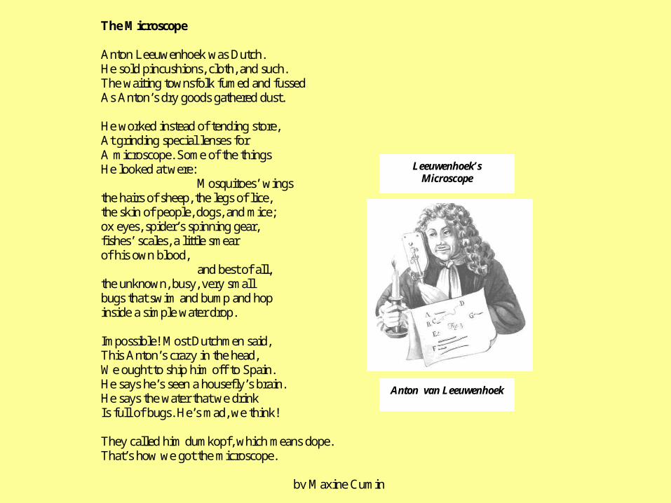
The Microscope
Anton Leeuwenhoek was Dutch.He sold pincushions, cloth, and such.The waiting townsfolk fumed and fussedAs Anton’s dry goods gathered dust.
He worked instead of tending store,At grinding special lenses forA microscope. Some of the thingsHe looked at were:
Mosquitoes’ wingsthe hairs of sheep, the legs of lice,the skin of people, dogs, and mice;ox eyes, spider’s spinning gear,fishes’ scales, a little smearof his own blood,
and best of all,the unknown, busy, very smallbugs that swim and bump and hopinside a simple water drop.
Impossible! Most Dutchmen said,This Anton’s crazy in the head,We ought to ship him off to Spain.He says he’s seen a housefly’s brain.He says the water that we drinkIs full of bugs. He’s mad, we think!
They called him dumkopf, which means dope.That’s how we got the microscope.
by Maxine Cumin
Leeuwenhoek’sMicroscope
Anton van Leeuwenhoek

http://www.microscopeworld.com/MSWorld/104_Diagram.pdf


![Mathematical Theory of the Wigner-Weisskopf Atom · Mathematical Theory of the Wigner-Weisskopf Atom 3 [Ja, Si1]. The theory discussed in this section naturally applies to the cases](https://img.pdfslide.us/doc/110x75/6013ab3aed750d623a0c02c0/mathematical-theory-of-the-wigner-weisskopf-atom-mathematical-theory-of-the-wigner-weisskopf.jpg)