Embed Size (px)
Citation preview

The history of lymphoma classification
and the 2016 WHO revision
A journey from morphology to a
multidisciplinary view
Elias Campo
Hospital Clinic, University of Barcelona
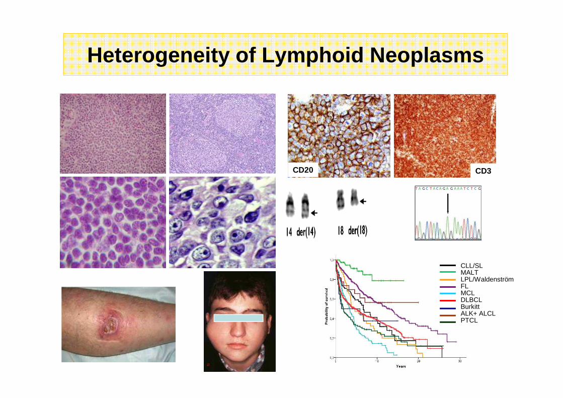
Heterogeneity of Lymphoid Neoplasms
CD20 CD3
CLL/SLMALTLPL/WaldenströmFLMCLDLBCLBurkittALK+ ALCLPTCL
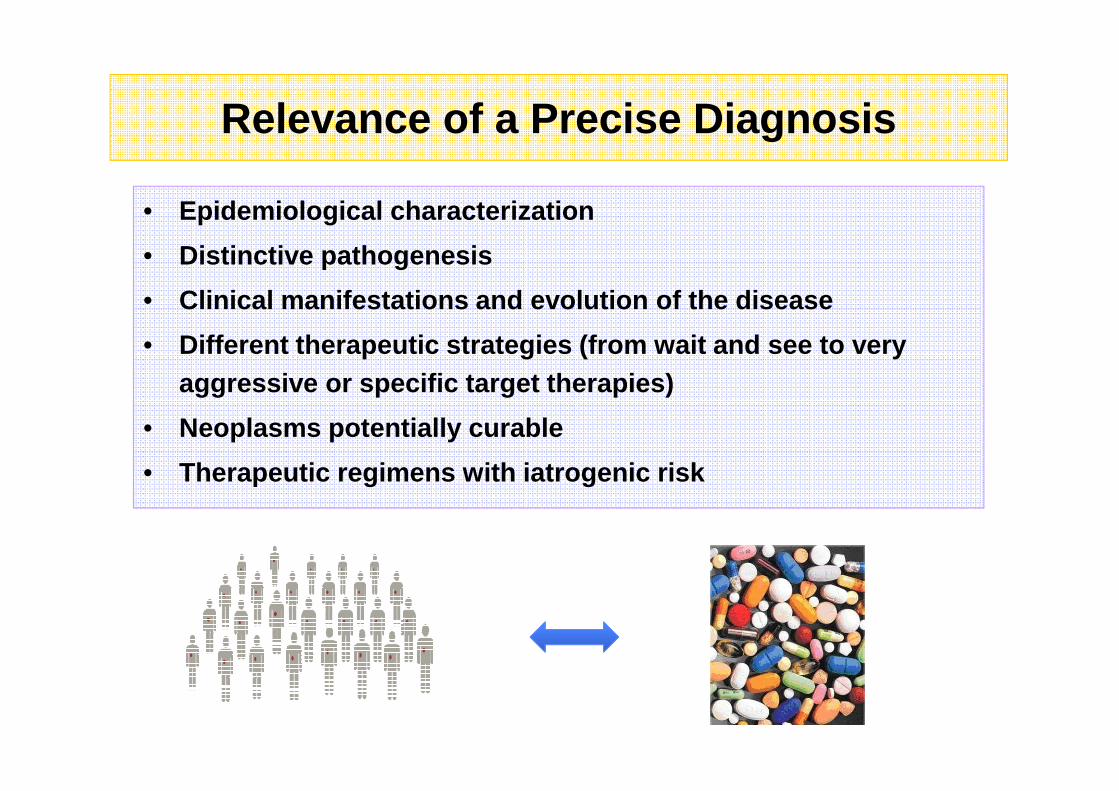
Relevance of a Precise Diagnosis
• Epidemiological characterization
• Distinctive pathogenesis
• Clinical manifestations and evolution of the diseas e
• Different therapeutic strategies (from wait and see to very aggressive or specific target therapies)
• Neoplasms potentially curable
• Therapeutic regimens with iatrogenic risk
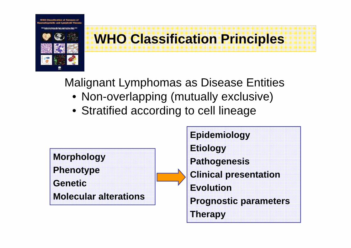
WHO Classification Principles
Morphology Phenotype GeneticMolecular alterations
EpidemiologyEtiologyPathogenesisClinical presentationEvolutionPrognostic parameters Therapy
Malignant Lymphomas as Disease Entities• Non-overlapping (mutually exclusive)• Stratified according to cell lineage
• Classification is the “language” of medicine
– Diseases must be described and defined before they
can be diagnosed and treated
• Disease entities should be clearly defined and
clinically distinctive
• Consensus on terminology and definitions
– Essential for both clinical practice and research
Why Classify?
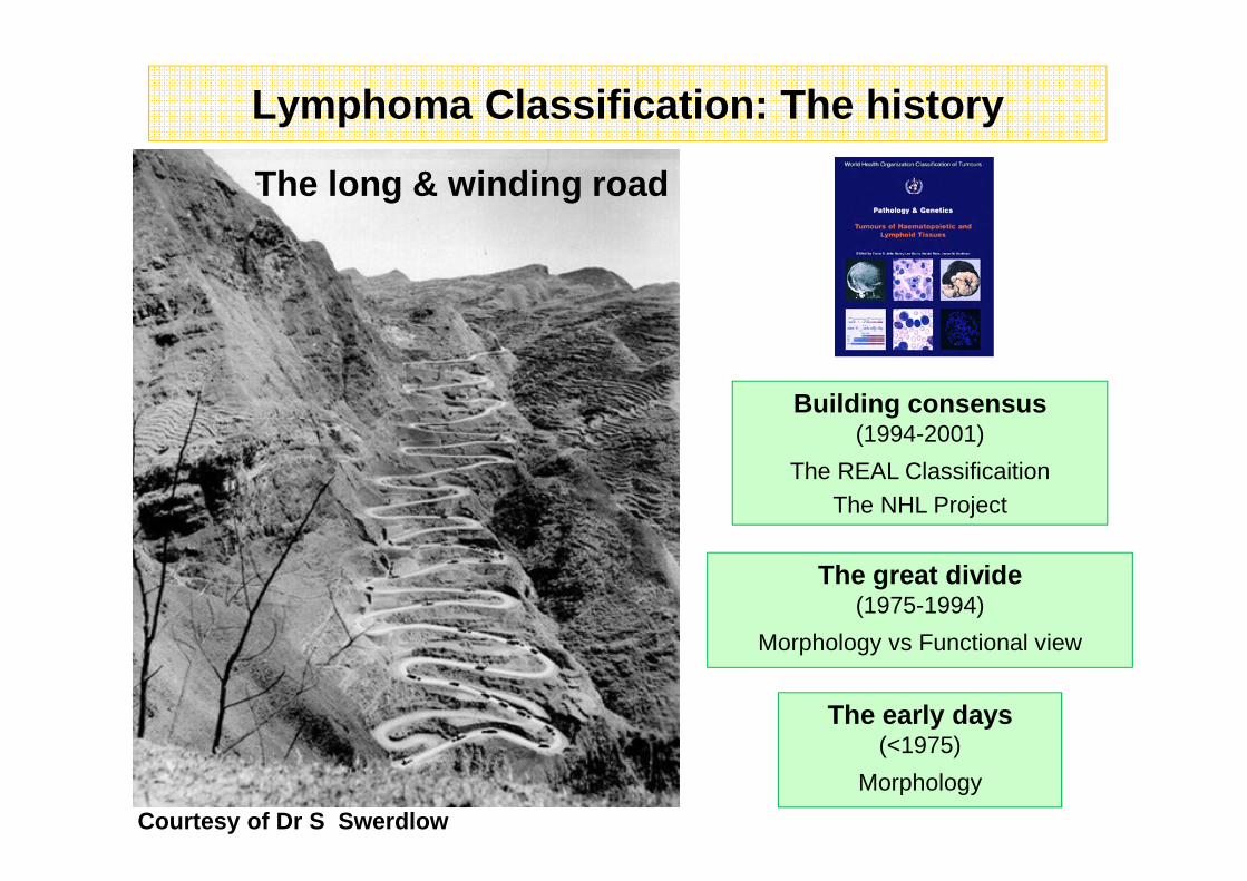
Lymphoma Classification : The history
The long & winding road
The early days(<1975)
Morphology
The great divide(1975-1994)
Morphology vs Functional view
Building consensus(1994-2001)
The REAL ClassificaitionThe NHL Project
Courtesy of Dr S Swerdlow
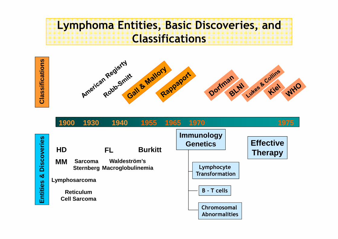
Lymphoma Entities, Basic Discoveries, and
ClassificationsC
lass
ifica
tions
Ent
ities
& D
isco
verie
s
1900 1930 1940 1955 1965 1970 1975
HD FL Burkitt
SarcomaSternberg
Waldeström’sMacroglobulinemia
MM
ImmunologyGenetics
Lymphocyte
Transformation
B - T cells
Chromosomal
Abnormalities
EffectiveTherapy
Lymphosarcoma
ReticulumCell Sarcoma
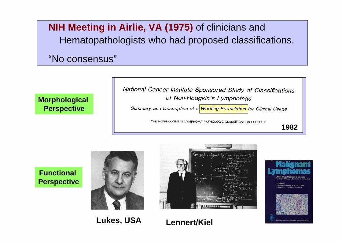
NIH Meeting in Airlie, VA (1975) of clinicians and Hematopathologists who had proposed classifications.
“No consensus”
Lennert/KielLukes, USA
MorphologicalPerspective
FunctionalPerspective
1982

Poznan, Poland
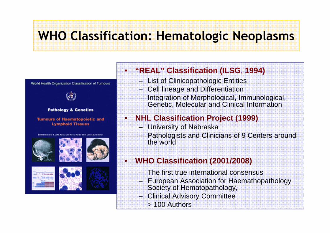
WHO Classification: Hematologic Neoplasms
• “REAL” Classification (ILSG , 1994)– List of Clinicopathologic Entities– Cell lineage and Differentiation– Integration of Morphological, Immunological,
Genetic, Molecular and Clinical Information
• NHL Classification Project (1999)– University of Nebraska– Pathologists and Clinicians of 9 Centers around
the world
• WHO Classification (2001/2008)– The first true international consensus – European Association for Haemathopathology
Society of Hematopathology, – Clinical Advisory Committee– > 100 Authors
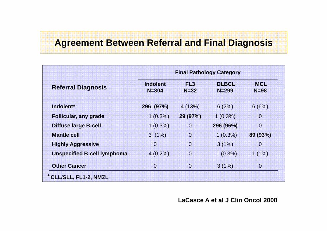
LaCasce A et al J Clin Oncol 2008
Final Pathology Category
Referral Diagnosis IndolentN=304
FL3N=32
DLBCLN=299
MCLN=98
Indolent* 296 (97%) 4 (13%) 6 (2%) 6 (6%)
Follicular, any grade 1 (0.3%) 29 (97%) 1 (0.3%) 0
Diffuse large B-cell 1 (0.3%) 0 296 (96%) 0
Mantle cell 3 (1%) 0 1 (0.3%) 89 (93%)
Highly Aggressive 0 0 3 (1%) 0
Unspecified B-cell lymphoma 4 (0.2%) 0 1 (0.3%) 1 (1%)
Other Cancer 0 0 3 (1%) 0
Agreement Between Referral and Final Diagnosis
CLL/SLL, FL1-2, NMZL*
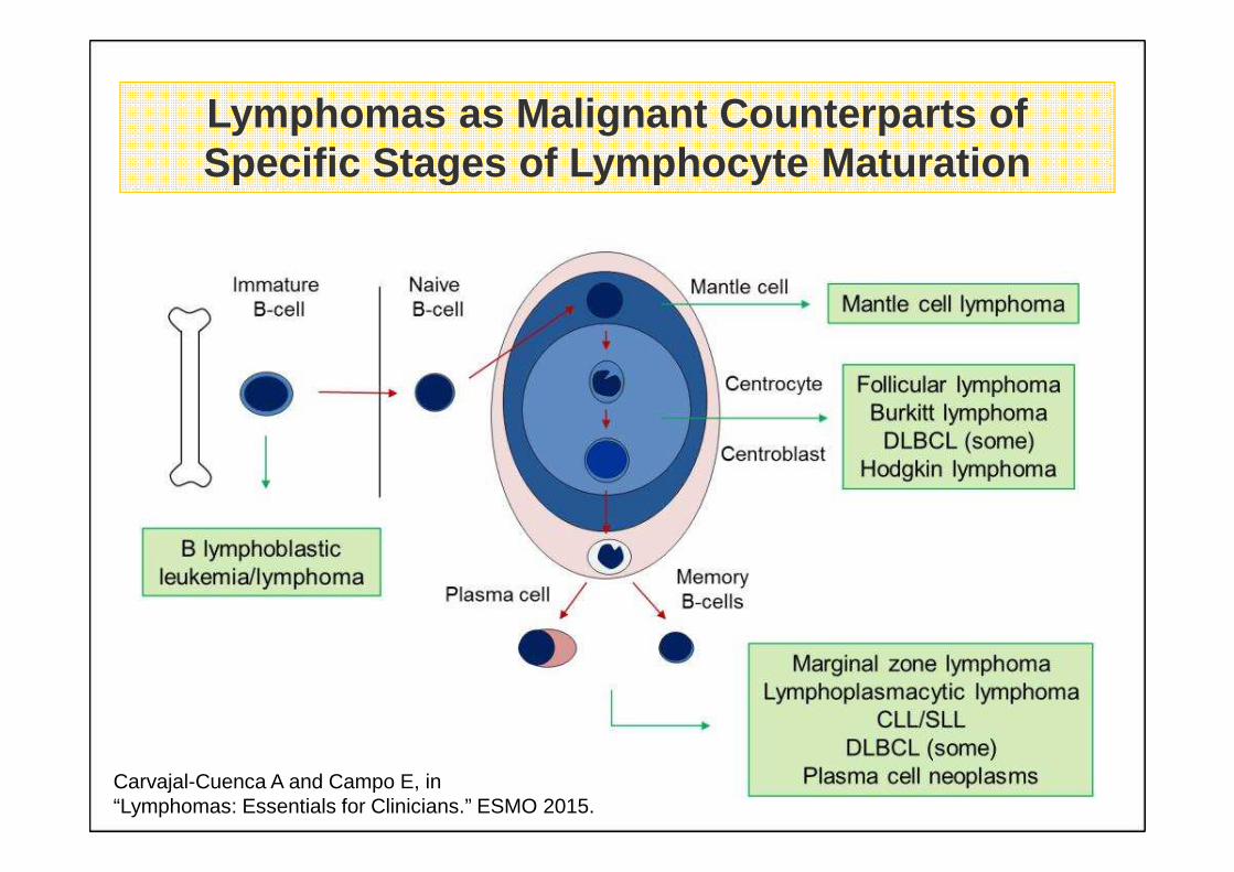
Carvajal-Cuenca A and Campo E, in “Lymphomas: Essentials for Clinicians.” ESMO 2015.
Lymphomas as Malignant Counterparts of Specific Stages of Lymphocyte Maturation
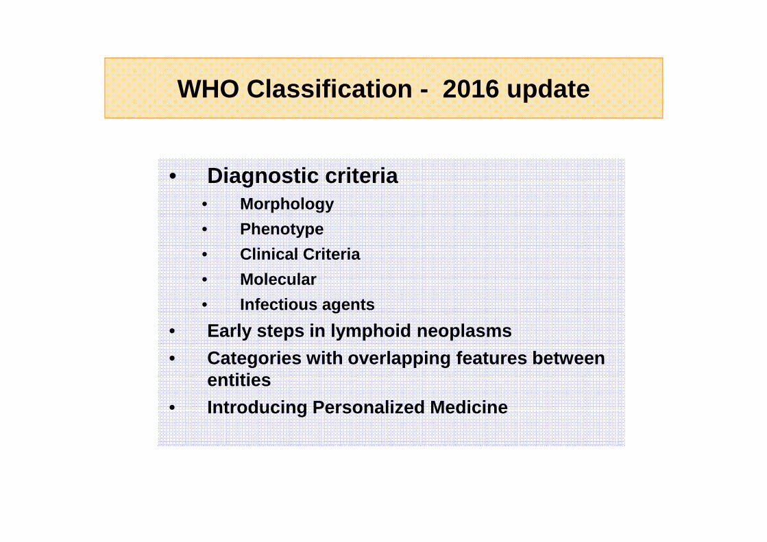
WHO Classification - 2016 update
• Diagnostic criteria• Morphology
• Phenotype
• Clinical Criteria
• Molecular
• Infectious agents
• Early steps in lymphoid neoplasms
• Categories with overlapping features between entities
• Introducing Personalized Medicine
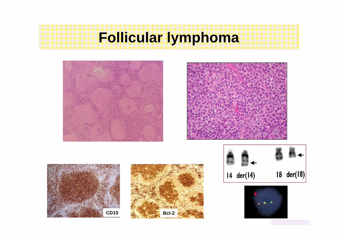
Bcl-2CD10
Follicular lymphoma
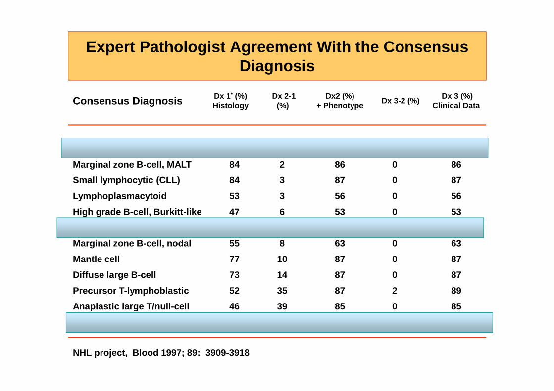
Expert Pathologist Agreement With the Consensus Diagnosis
Consensus Diagnosis Dx 1* (%) Histology
Dx 2-1 (%)
Dx2 (%)+ Phenotype
Dx 3-2 (%) Dx 3 (%)
Clinical Data
Follicular, any grade 93 1 94 0 94
Marginal zone B-cell, MALT 84 2 86 0 86
Small lymphocytic (CLL) 84 3 87 0 87
Lymphoplasmacytoid 53 3 56 0 56
High grade B-cell, Burkitt-like 47 6 53 0 53
Primary mediastinal large B-cell 51 7 58 37 85
Marginal zone B-cell, nodal 55 8 63 0 63
Mantle cell 77 10 87 0 87
Diffuse large B-cell 73 14 87 0 87
Precursor T-lymphoblastic 52 35 87 2 89
Anaplastic large T/null-cell 46 39 85 0 85
Peripheral T-cell, all types 41 45 86 0 86
NHL project, Blood 1997; 89: 3909-3918
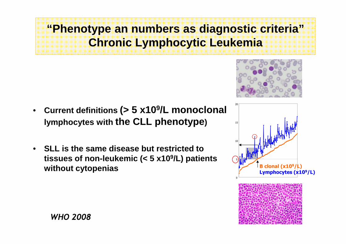
“Phenotype an numbers as diagnostic criteria”Chronic Lymphocytic Leukemia
• Current definitions (> 5 x10⁹/L monoclonal lymphocytes with the CLL phenotype )
• SLL is the same disease but restricted to tissues of non-leukemic (< 5 x10 9/L) patients without cytopenias
WHO 2008
0
5
10
15
20
B clonal (x109/L)Lymphocytes (x109/L)
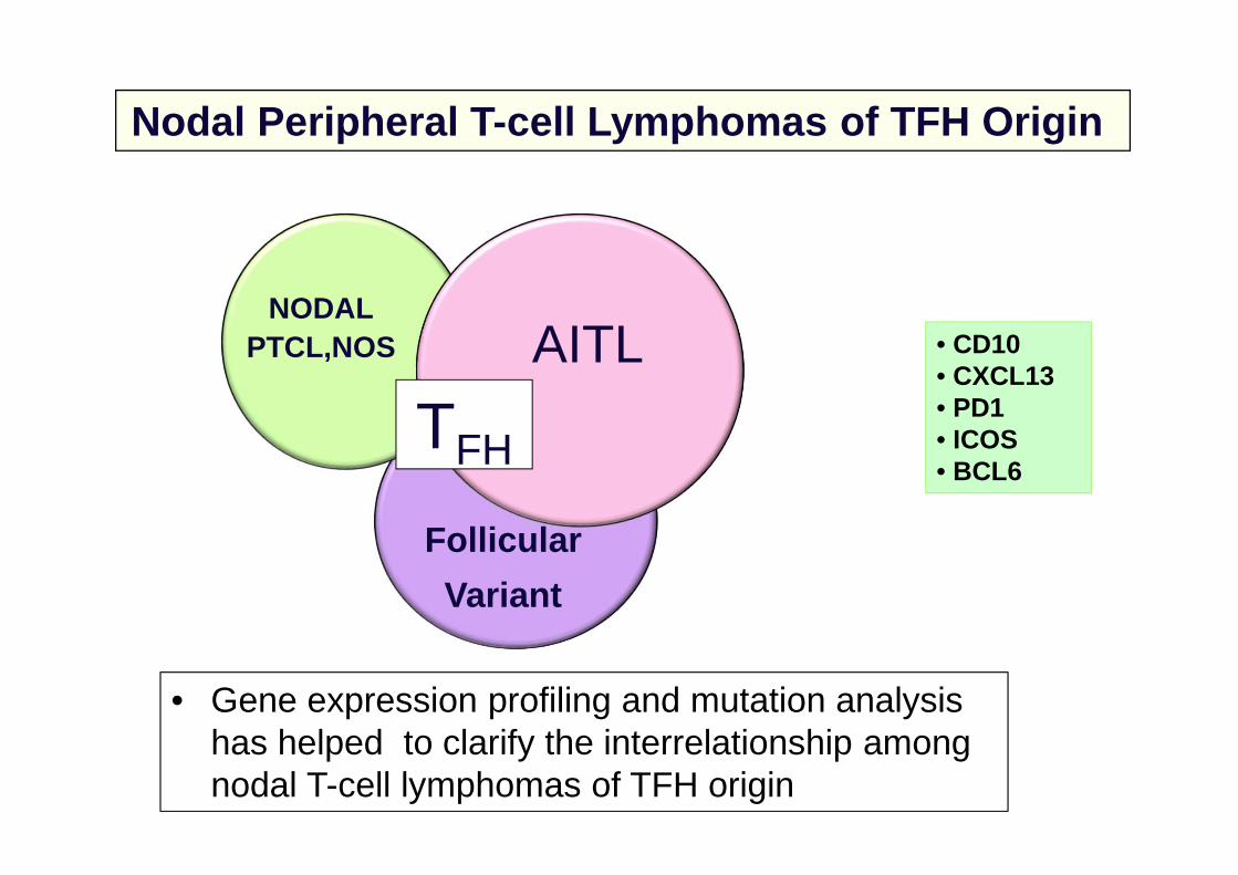
Nodal Peripheral T -cell Lymphomas of TFH Origin
NODALPTCL,NOS
Follicular
Variant
AITL
TFH
• Gene expression profiling and mutation analysis has helped to clarify the interrelationship among nodal T-cell lymphomas of TFH origin
• CD10• CXCL13• PD1• ICOS• BCL6
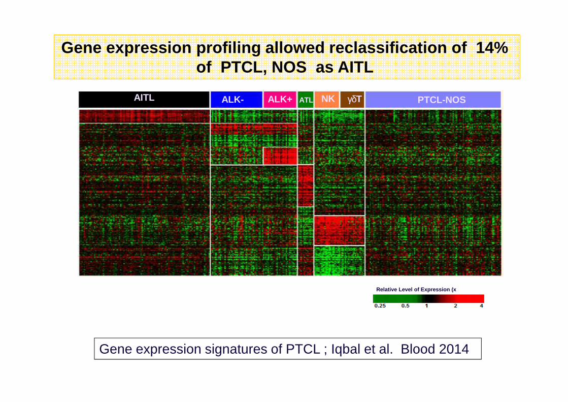
Relative Level of Expression (x median value)
AITL ALK- NKALK+ γδTATL PTCL-NOS
Gene expression profiling allowed reclassification of 14% of PTCL, NOS as AITL
Gene expression signatures of PTCL ; Iqbal et al. Blood 2014
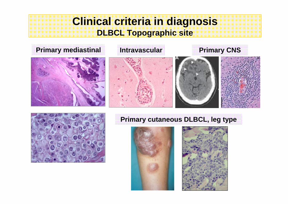
Primary mediastinal Intravascular Primary CNS
Primary cutaneous DLBCL, leg type
Clinical criteria in diagnosisDLBCL Topographic site
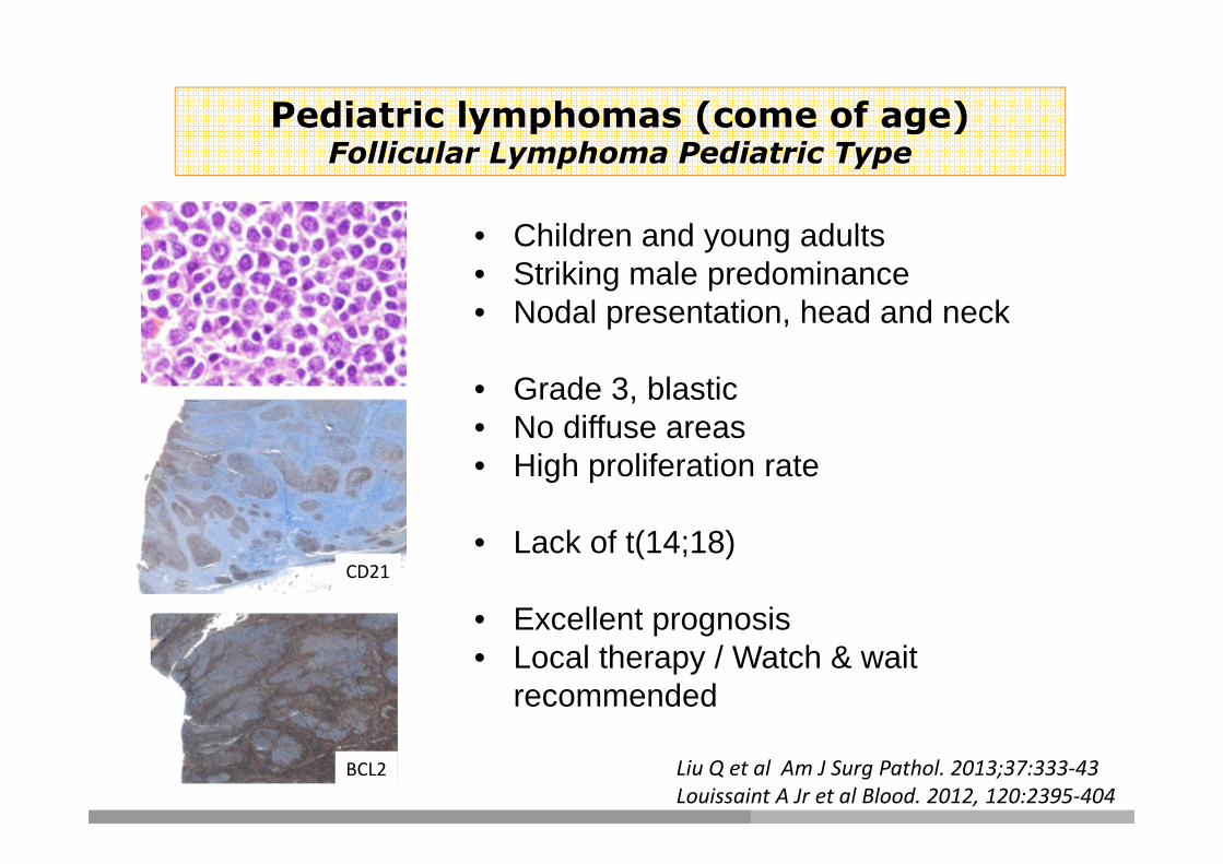
• Children and young adults• Striking male predominance• Nodal presentation, head and neck
• Grade 3, blastic• No diffuse areas• High proliferation rate
• Lack of t(14;18)
• Excellent prognosis• Local therapy / Watch & wait
recommended
Pediatric lymphomas (come of age)Follicular Lymphoma Pediatric Type
BCL2
CD21
Liu Q et al Am J Surg Pathol. 2013;37:333-43
Louissaint A Jr et al Blood. 2012, 120:2395-404

Genetic alterations in Pediatric Type Follicular Ly mphoma
GenesPTFL (n=42
(%) t(14;18)-neg FL
(%)
tt(14;18)-pos FL*
(%) P-value
TNFRSF14 51 36 18-46 Ns
KMT2D 16 36 67-82 Ns
CREBBP 3 45 33-64 0.001
FOXO1 5 27 - Ns
GNA13 11 0 - Ns
EZH2 0 18 7-20 0.0049
Genes PTFL (n=24)
MAP2K1 9 (38%)
MAPK1 2 (8%)
RRAS 1 (4%)
Schmidt J et al Blood 2016
Louissaint A et al Blood 2016
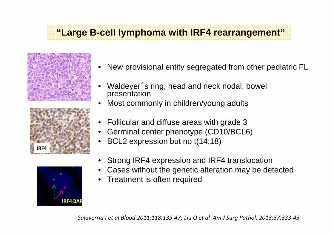
• New provisional entity segregated from other pediatric FL
• Waldeyer´s ring, head and neck nodal, bowel presentation
• Most commonly in children/young adults
• Follicular and diffuse areas with grade 3 • Germinal center phenotype (CD10/BCL6)• BCL2 expression but no t(14;18)
• Strong IRF4 expression and IRF4 translocation • Cases without the genetic alteration may be detected• Treatment is often required
“Large B -cell lymphoma with IRF4 rearrangement”
IRF4 BAP
Salaverria I et al Blood 2011;118:139-47; Liu Q et al Am J Surg Pathol. 2013;37:333-43
IRF4
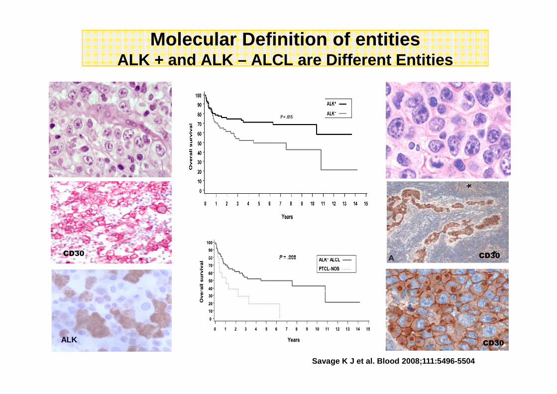
Molecular Definition of entitiesALK + and ALK – ALCL are Different Entities
ALK
Savage K J et al. Blood 2008;111:5496-5504
CD30 CD30
CD30
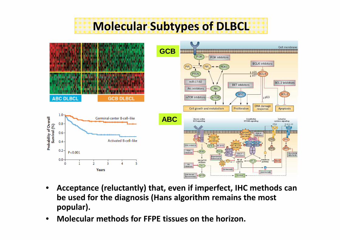
Molecular Subtypes of DLBCL
GCB
ABC
• Acceptance (reluctantly) that, even if imperfect, IHC methods can be used for the diagnosis (Hans algorithm remains the most popular).
• Molecular methods for FFPE tissues on the horizon.
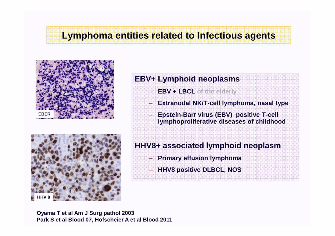
Lymphoma entities related to Infectious agents
EBV+ Lymphoid neoplasms
– EBV + LBCL of the elderly
– Extranodal NK/T-cell lymphoma, nasal type
– Epstein-Barr virus (EBV) positive T-cell lymphoproliferative diseases of childhood
HHV8+ associated lymphoid neoplasm
– Primary effusion lymphoma
– HHV8 positive DLBCL, NOS
Oyama T et al Am J Surg pathol 2003Park S et al Blood 07, Hofscheier A et al Blood 2011
EBER
HHV 8
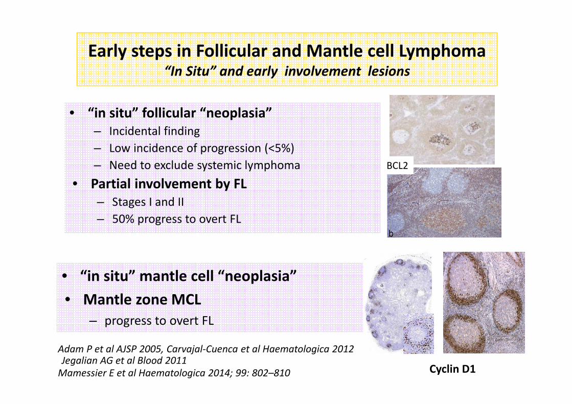
Early steps in Follicular and Mantle cell Lymphoma“In Situ” and early involvement lesions
• “in situ” follicular “neoplasia”
– Incidental finding
– Low incidence of progression (<5%)
– Need to exclude systemic lymphoma
• Partial involvement by FL
– Stages I and II
– 50% progress to overt FL
Adam P et al AJSP 2005, Carvajal-Cuenca et al Haematologica 2012Jegalian AG et al Blood 2011
Mamessier E et al Haematologica 2014; 99: 802–810
• “in situ” mantle cell “neoplasia”
• Mantle zone MCL
– progress to overt FL
BCL2
Cyclin D1
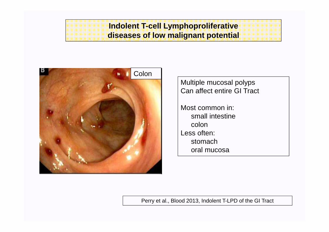
Colon
Indolent T-cell Lymphoproliferative diseases of low malignant potential
Perry et al., Blood 2013, Indolent T-LPD of the GI Tract
Multiple mucosal polypsCan affect entire GI Tract
Most common in: small intestinecolon
Less often: stomachoral mucosa
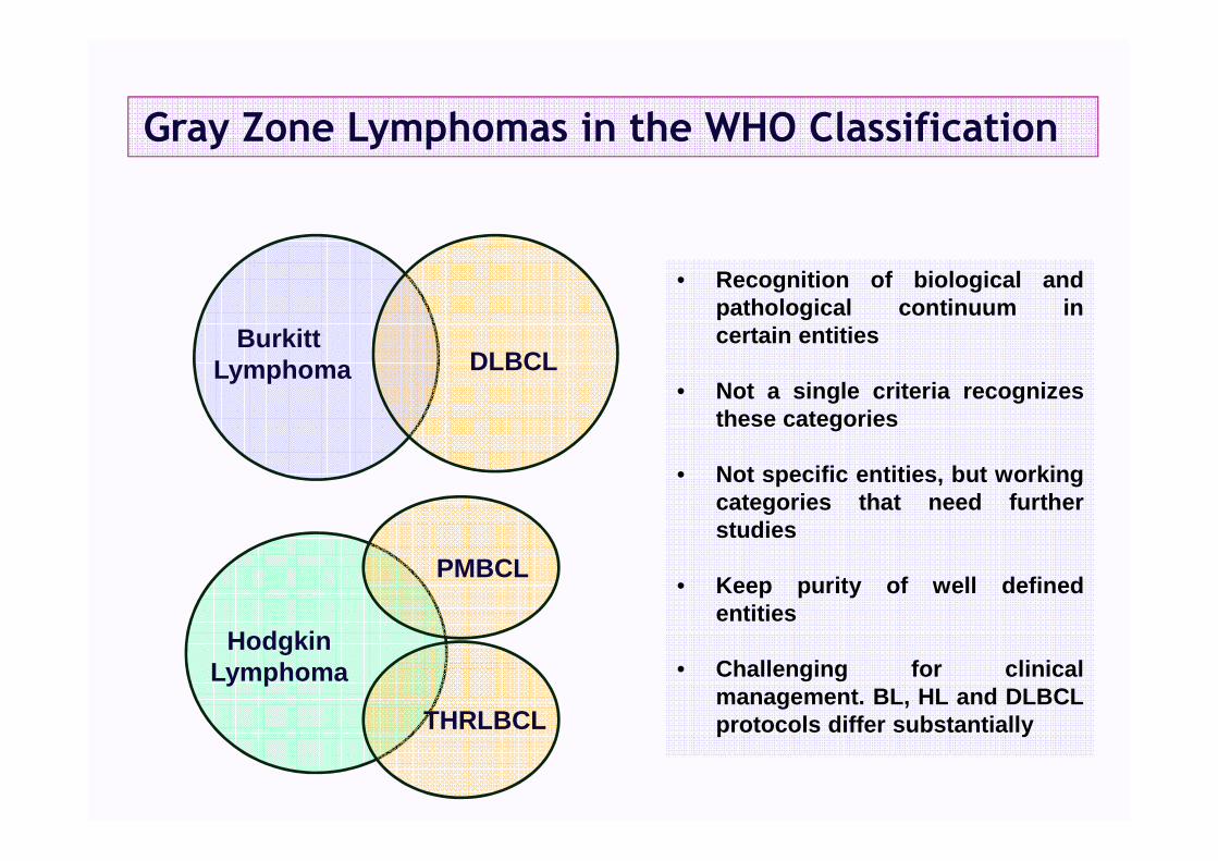
Gray Zone Lymphomas in the WHO Classification
BurkittLymphoma DLBCL
PMBCL
HodgkinLymphoma
• Recognition of biological andpathological continuum incertain entities
• Not a single criteria recognizesthese categories
• Not specific entities, but workingcategories that need furtherstudies
• Keep purity of well definedentities
• Challenging for clinicalmanagement. BL, HL and DLBCLprotocols differ substantiallyTHRLBCL
Human Genome ProjectTowards a personalized medicine
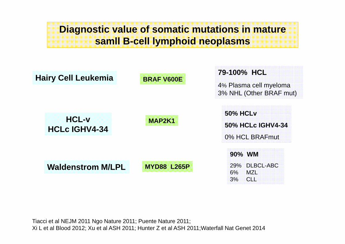
Diagnostic value of somatic mutations in mature samll B-cell lymphoid neoplasms
79-100% HCL
4% Plasma cell myeloma3% NHL (Other BRAF mut)
BRAF V600EHairy Cell Leukemia
Waldenstrom M/LPL
Tiacci et al NEJM 2011 Ngo Nature 2011; Puente Nature 2011; Xi L et al Blood 2012; Xu et al ASH 2011; Hunter Z et al ASH 2011;Waterfall Nat Genet 2014
MYD88 L265P
90% WM
29% DLBCL-ABC6% MZL3% CLL
50% HCLv
50% HCLc IGHV4-34
0% HCL BRAFmut
MAP2K1HCL-vHCLc IGHV4-34

Somatic Mutations and CNA in CLL and MBL
(Whole genome/exome sequencing)
Puente X et al Nature 2015
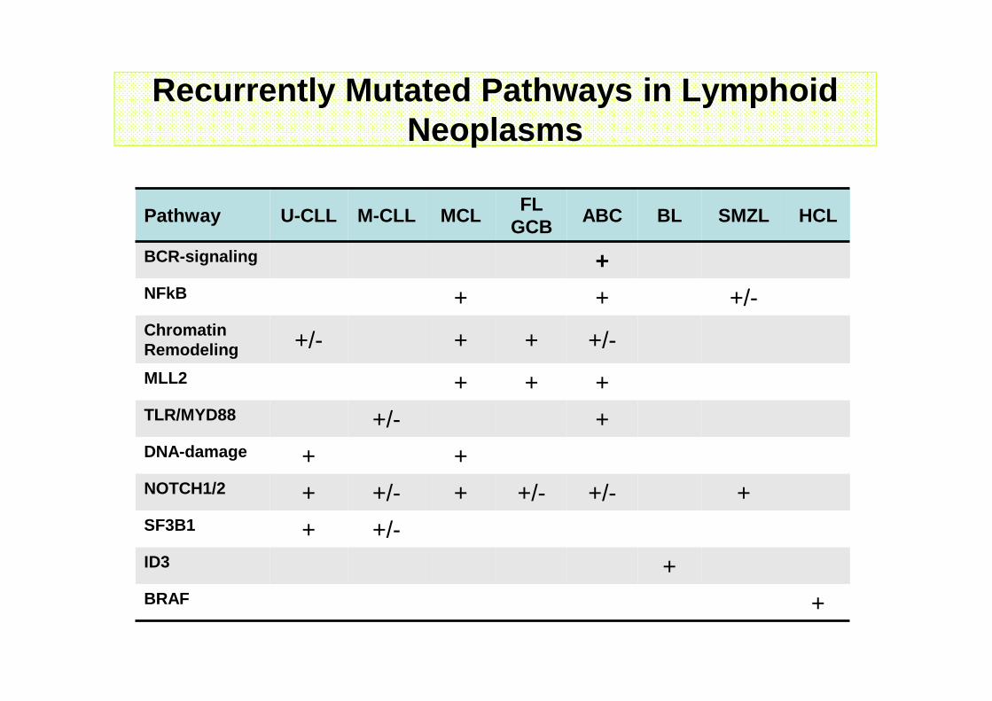
Recurrently Mutated Pathways in Lymphoid Neoplasms
Pathway U-CLL M-CLL MCLFL
GCBABC BL SMZL HCL
BCR-signaling +NFkB + + +/-ChromatinRemodeling +/- + + +/-
MLL2 + + +TLR/MYD88 +/- +DNA-damage + +NOTCH1/2 + +/- + +/- +/- +SF3B1 + +/-ID3 +BRAF +
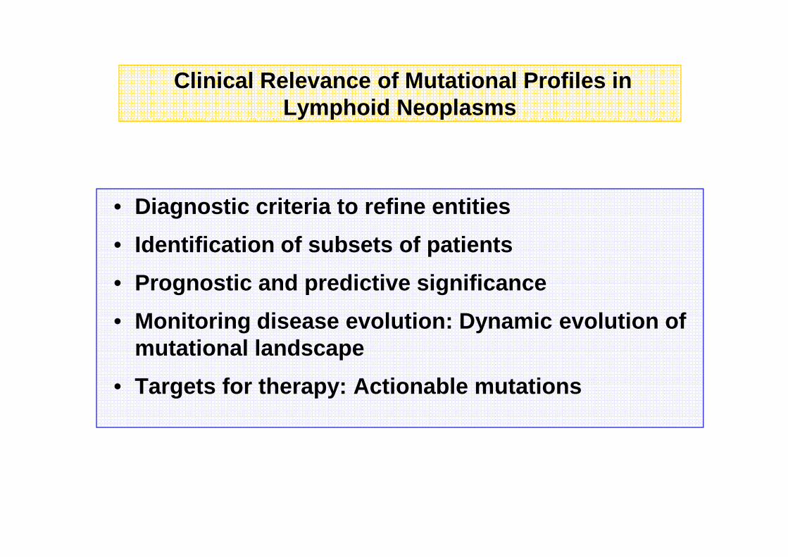
• Diagnostic criteria to refine entities
• Identification of subsets of patients
• Prognostic and predictive significance
• Monitoring disease evolution: Dynamic evolution of mutational landscape
• Targets for therapy: Actionable mutations
Clinical Relevance of Mutational Profiles in Lymphoid Neoplasms