Embed Size (px)
Citation preview
~ ~~
3. small Anim. Pract. Vol. 8, 1967, pp. 639 to 647. Pergamon Press Ltd. Printed in Great Britain
The Histology and Histochemistry of the Circumanal Hepatoid Glands of the Dog* K. P. BAKER University of Dublin School of Veterinary Medicinc, Ballsbridge, Dublin 4
Abstract-The hepatoid circumanal glands are small at birth and continue enlarging throughout life until senility. They develop as buds from the epidermis of hair follicles of the region and in the adult surround the anus in an irregular circle, development being greater in the male. The cyto- plasm of the gland cell contains non-lipid, non-proteinaceous granules and there appears to be no duct system connecting gland acini to hair follicles. I t is suggested that the glands may be endocrine in type.
I N T R O D U C T I O N THERE are considerable differences of opinion in the literature of the nature and function of the glands which surround the anus in the dog. Confusion results because most authors refer collectively to all the glands in the region as circumanal glands, others confining this term to the modified sebaceous glands, the “hepatoid Drusen” of Krolling and Grau (1960). This paper is concerned mainly with a description of these latter glands, which are referred to herein as hepatoid circumanal glands.
Henry and Bory (1936) described three different kinds of gland in the anal region of carnivores: firstly, the glands of the anal sac, secondly, a circular zone of little glands occurring in clusters immediately around the anus, these being circum- scribed at some distance by a third group of glands, the “circumanal glands”, 16-18 mm across. This third group they described as being composed of many sebaceous glands, sweat glands and well developed acinar glands. McClelland (1940) did not differentiate between the glands about the anus, describing them collectively as modified apocrine glands. Trautmann and Fiebiger (1957) citing the thesis of Parks (1950) described the circumanal hepatoid glands as being in two parts, a superficial sebaceous part opening into a hair follicle and a deeper hepatoid part without a patent duct. Parks (1950) was unable to confirm the presence in hepatoid circumanal glands of secretory canaliculi which had been described by Schaffer (1923 a, b) as minute passages between the gland cells, whose purpose was
+This paper forms part of a Ph.D. thesis submitted to and approved by the University of Dublin.
639
640 K . P. BAKER
to transport the secretory products of these cells to the surface. In Parks’ view the superficial sebaceous structures produced secretions by holocrine breakdown which were discharged to the surface ; the deeper hepatoid glands were aborted sebaceous structures without patent ducts, the cells of which elaborated proteinaceous granules.
In a paper on glands of the canine skin Nielsen (1953) stated that glands similar to “perianal glands”, i.e. hepatoid glands, are found in the skin of the prepuce and groin. His findings support those of Parks.
Cotchin (1956) described the hepatoid glands as forming a ring round the anus, being best developed above it, occurring only in canines and being well developed only after puberty.
Krdlling and Grau (1960) cited Schaffer (1923 a) as first calling the circumanal glands “hepatoide Driisen” and gave their function as that of producing an odorifer- ous secretion without cellular breakdown, their products being gathered in canaliculi and passed by short ducts to the hair follicles and thence to the epidermis.
Kral and Schwartzman (1964) described a zonal distribution of glands about the anus. The hepatoid glands formed a ring 5 mm in dia. about the anus and consisted of solid lobules without function. They described the hepatoid circumanal glands as abortive sebaceous glands. Occurring immediately around the anus, they described in addition large holocrine sebaceous glands and merocrine sweat glands.
Schwartzman and Orkin (1 964) referred only to hepatoid glands as surrounding the anus. They consisted of polyhedral cells with eosinophilic granular cytoplasm, about each bundle was present a single row of smaller cells (reserve cells). Well developed ducts were said to open to the surface.
Because of the variable descriptions in the literature of the nature and distribu- tion of the glands surrounding the anus in the dog, it was decided that there was need for considerable further investigation, particular attention being given to the hepatoid circumanal glands.
MATERIAL AND METHODS Circumanal skin was examined from twenty-eight healthy dogs, of which
sixteen were males and tweIve were females. Animals of various age groups were examined to determine developmental changes in early life and in old age. Adults were of similar stature.
To obtain an appreciation of the anatomy of the region a technique was developed in which frozen sections were cut by hand. Circumanal tissue to a radius of 3 cm from the anus was removed immediately after death, gently pressed flat in a petri dish and kept at a temperature of -20°C overnight. I t was then found possible to cut thin radial sections by hand with a new scalpel blade; eight such radial sections were cut at equal spacings around the anal orifice. The sections were then placed in glacial acetic acid for 5 min, washed in tap water and placed between glass slides no other pressure was applied. The various structures of the region were then examined microscopically, measurements taken, and the results plotted on graph paper.
Serial paraffin sections, cut at right angles to the epidermis were stained with Harris’ haematoxylin and eosin and projected consecutively on to tracing paper. From this, the various structures were cut out and used as templates on cardboard
T H E C I R C U M A N A L H E P A T O I D GLANDS O F T H E D O G 64 1
1 in. thick. A three dimensional model was obtained by sticking the corresponding cut-outs together. For histological analysis, paraffin or frozen sections were subjected to various staining methods, as necessary.
1 0
OBSERVATIONS Distribution of the circumanal hepatoid g l a n h
In adult dogs the circumanal hepatoid glands are distributed in an irregular manner about the anus within a circle 5-25 mm in dia.
Contrary to Cotchin's (1956) finding, development does not seem to be greater above the anus. The hepatoid glands are accompanied by relatively large sebaceous glands, and large apocrine glands. Immediately around the anus is a ring of large sebaceous glands, which are fully developed and active at birth. In the young adult, the hepatoid circumanal glands are spaced at intervals; with increasing age the glands enlarge and it is often not possible to separate individual glands. The hepatoid circumanal glands appear to enlarge throughout life until senility, this enlargement occurs in both sexes but is greater in the male. No similar enlargement occurs in the sebaceous glands of the region after puberty.
Histology of the circumanal region The skin immediately around the anus is soft, with few hairs. The epidermis is
thick and rete ridging is present. Arrectores pilorum muscles are absent but much striated muscle is present. Sections cut by hand show the hepatoid circumanal glands as off-white, the sebaceous glands being white. The sebaceous glands are multilobed (up to thirty lobes) and open directly to the surface, each gland having a central collecting duct. Sebaceous glands in the region about the anus range from 600 x 500 p in the 8-week-old animal to 1100 x 1000 p in some adults. The largest sebaceous gland seen measured 1100 p in dia. In middle-aged dogs the orifices of the hair follicles in the region are often occluded by plugs of sebum which project above the surface. Individual hepatoid circumanal glands may extend 9 mm into the dermis and subcutaneously. Their diameter may be 1.5 mm in middle age, but in younger dogs they are smaller. The hepatoid glands are 1500 p in depth x 500 p in width in the 8-week-old dog and increase in size throughout adult life when they may attain 9000 p in depth by 1100 p in width. In senility atrophy occurs.
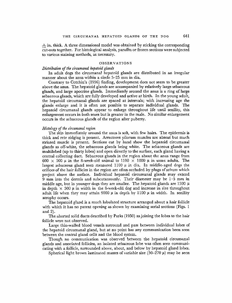
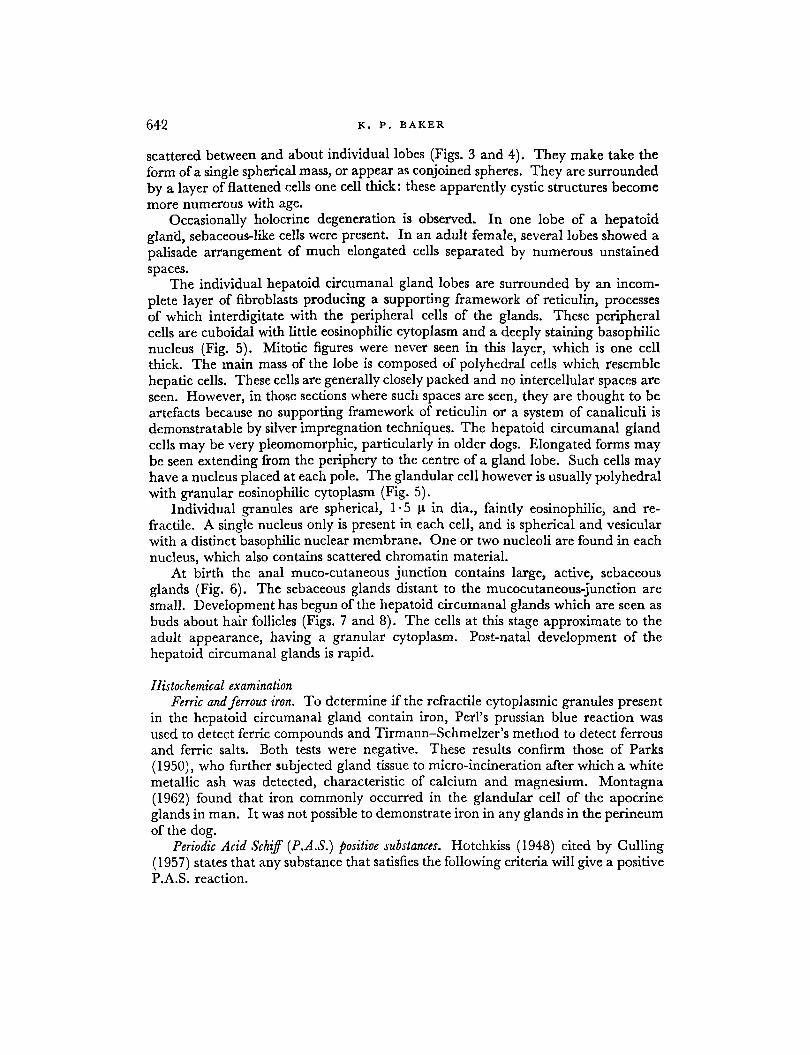
The hepatoid gland is a much lobulated structure arranged about a hair follicle with which it has no patent opening as shown by examining serial sections (Figs. 1 and 2).
The aborted solid ducts described by Parks (1950) as joining the lobes to the hair follicle were not observed.
Large thin-walled blood vessels surround and pass between individual lobes of the hepatoid circumanal gland, but at no point has any communication been seen between the central gland cells and the blood system.
Though no communication was observed between the hepatoid circumanal glands and associated follicles, an isolated sebaceous lobe was often seen communi- cating with a follicle, surrounded above, about, and below by hepatoid gland lobes.
Spherical light brown laminated masses of variable size (50-270 p) may be seen
K . P. B A K E R 642
scattered between and about individual lobes (Figs. 3 and 4). They make take the form of a single spherical mass, or appear as conjoined spheres. They are surrounded by a layer of flattened cells one cell thick: these apparently cystic structures become more numerous with age.
Occasionally holocrine degeneration is observed. In one lobe of a hepatoid gland, sebaceous-like cells were present. In an adult female, several lobes showed a palisade arrangement of much elongated cells separated by numerous unstained spaces.
The individual hepatoid circumanal gland lobes are surrounded by an incom- plete layer of fibroblasts producing a supporting framework of reticulin, processes of which interdigitate with the peripheral cells of the glands. These peripheral cells are cuboidal with little eosinophilic cytoplasm and a deeply staining basophilic nucleus (Fig. 5). Mitotic figures were never seen in this layer, which is one cell thick. The main mass of the lobe is composed of polyhedral cells which resemble hepatic cells. These cells are generally closely packed and no intercellular spaces are seen. However, in those sections where such spaces are seen, they are thought to be artefacts because no supporting framework of reticulin or a system of canaliculi is demonstratable by silver impregnation techniques. The hepatoid circumanal gland cells may be very pleomomorphic, particularly in older dogs. Elongated forms may be seen extending from the periphery to the centre of a gland lobe. Such cells may have a nucleus placed at each pole. The glandular cell however is usually polyhedral with granular eosinophilic cytoplasm (Fig. 5).
Individual granules are spherical, 1 - 5 p in dia., faintly eosinophilic, and re- fractile. A single nucleus only is present in each cell, and is spherical and vesicular with a distinct basophilic nuclear membrane. One or two nucleoli are found in each nucleus, which also contains scattered chromatin material.
At birth the anal muco-cutaneous junction contains large, active, sebaceous glands (Fig. 6). The sebaceous glands distant to the mucocutaneous-junction are small. Development has begun of the hepatoid circumanal glands which are seen as buds about hair follicles (Figs. 7 and 8). The cells at this stage approximate to the adult appearance, having a granular cytoplasm. Post-natal development of the hepatoid circumanal glands is rapid.
Histochemical examination Ferric and ferrous iron. To determine if the refractile cytoplasmic granules present
in the hepatoid circumanal gland contain iron, Perl's prussian blue reaction was used to detect ferric compounds and Tirmann-Schmelzer's method to detect ferrous and ferric salts. Both tests were negative. These results confirm those of Parks (1950), who further subjected gland tissue to micro-incineration after which a white metallic ash was detected, characteristic of calcium and magnesium. Montagna (1962) found that iron commonly occurred in the glandular cell of the apocrine glands in man. I t was not possible to demonstrate iron in any glands in the perineum of the dog.
Periodic Acid Schiff (P.A.S.) positive substances. Hotchkiss (1948) cited by Culling (1957) states that any substance that satisfies the following criteria will give a positive P.A.S. reaction.
FIG. 1. Hepatoid circumanal gland of adult dog. The gland acini surround a
EP .= epidermis Hf = hair follicles Hgl = hepatoid circumanal gland acini H & E x 4 0
transversely cut hair follicle.
( f a c i n g p . 642)
FIG. 2. Hepatoid circurnanal glands of adult dog. The gland extends around a vertically cut hair follicle.
Hf = hair follicle Hgl = hepatoid circumanal gland acini Stm = striated muscle Sudan IV x 40
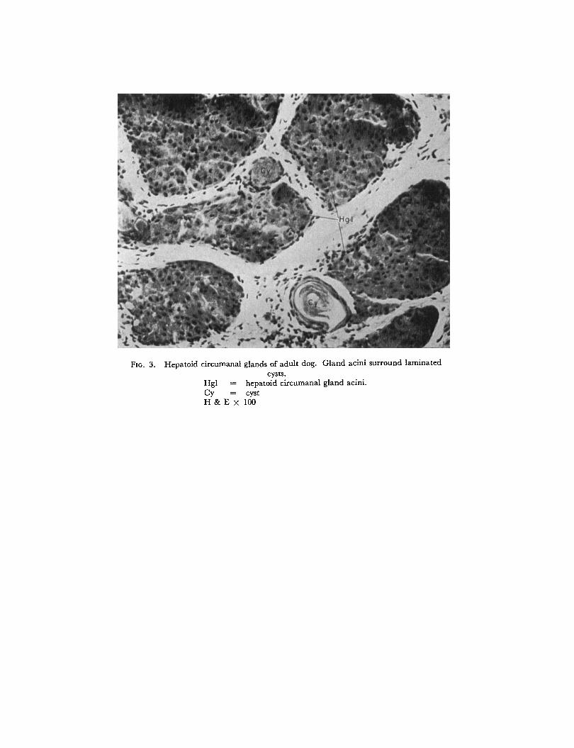
FIG. 3. Hepatoid circumanal glands of adult dog. Gland acini surround laminated cysts.
Hgl = hepatoid circumanal gland acini.
H & E x 100 c y = cyst
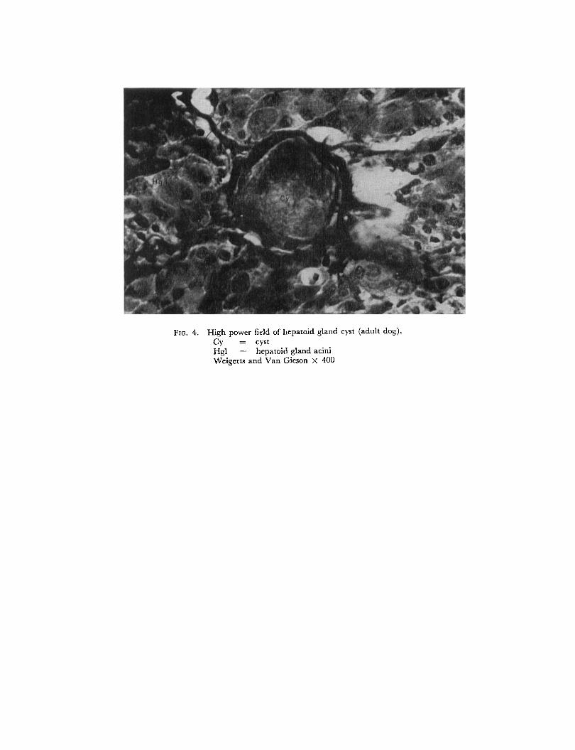
FIG. 4. High power field of hepatoid gland cyst (adult dog). c y = cyst Hgl = hepatoid gland acini Weigerts and Van Gieson x 400
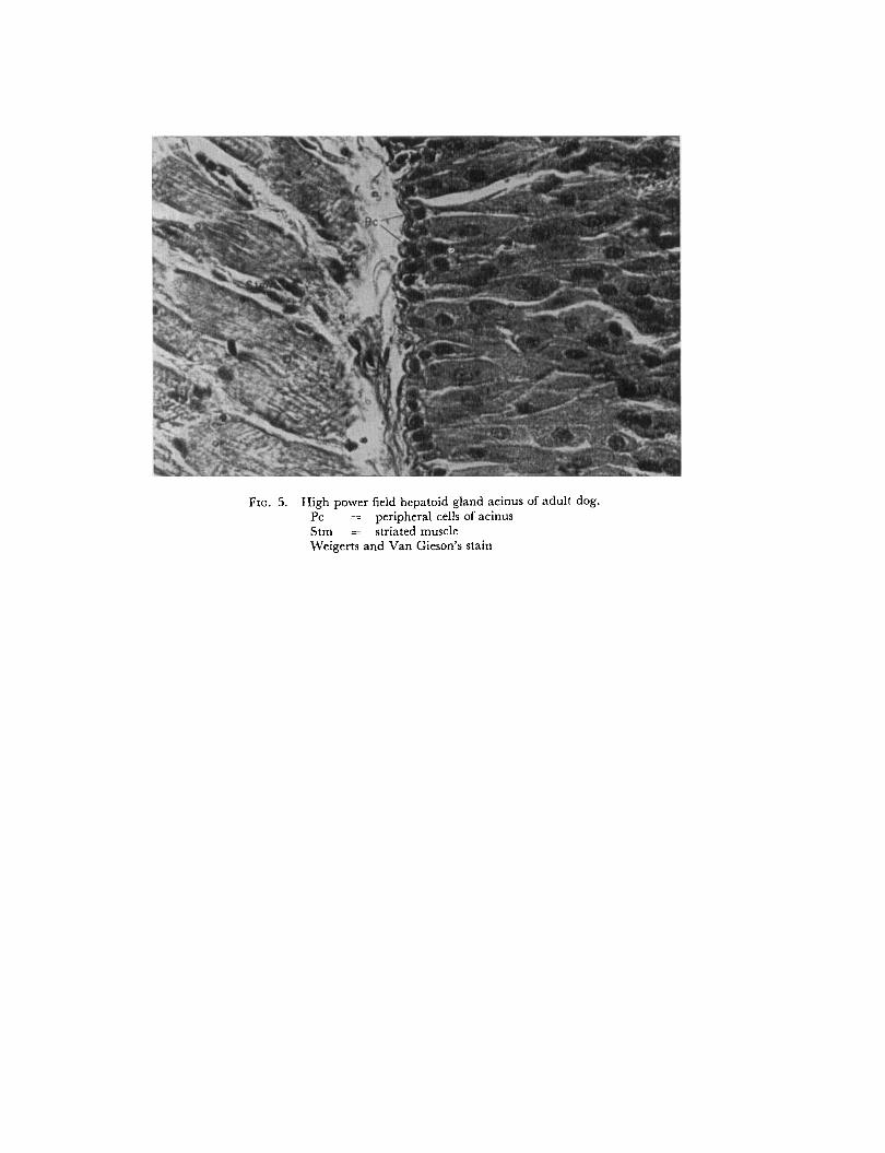
FIG. 5. High power field hepatoid gland acinus of adult dog. Pc =;. peripheral cells of acinus Stm = striated muscle Weigerts and Van Gieson’s stain
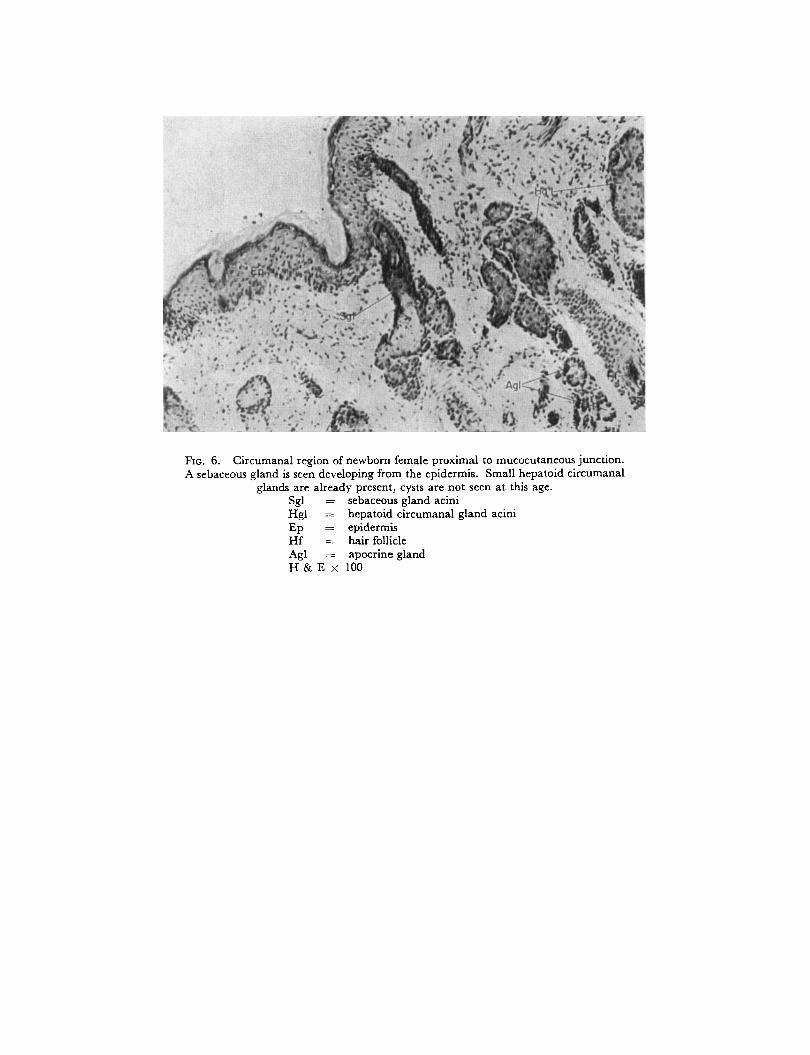
FIG. 6. Circumanal region of newborn female proximal to mucocutaneous junction. A sebaceous gland is seen developing from the epidermis. Small hepatoid circumanal
glands are already present, cysts are not seen at this age. Sgl = sebaceous gland acini Hgl = hepatoid circumanal gland acini Ep = epidermis Hf = hair follicle Agl = apocrine gland H & E x 100
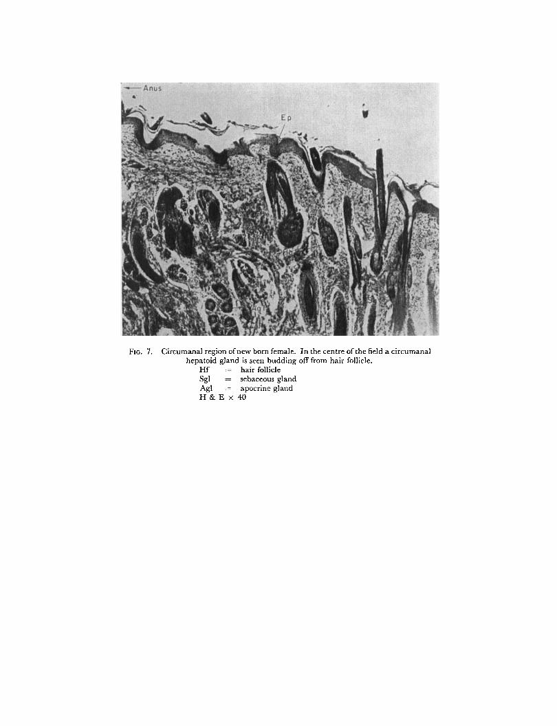
FIG. 7. Circumanal region of new born female. In the centre of the field a circumanal hepatoid gland is seen budding off from hair follicle.
Hf = hair follicle Sgl = sebaceous gland Agl = apocrine gland H & E x 4 0
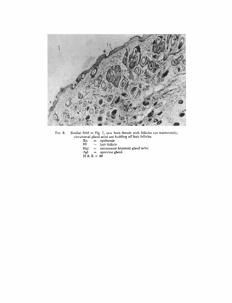
FIG. 8. Similar field to Fig. 7, new born female with follicles cut transversely, circumanal gland acini are budding off hair follicles.
Ep = epidermis Hf - hair follicle Hgl = circumanal hepatoid gland acini Ag1 = apocrine gland H & E x 40
T H E CIRCUMANAL H E P A T O I D G L A N D S O F T H E D O G 643
(1)
(2) (3) (4)
I t contains the 1 * 2 glycol grouping, of the equivalent amino or alkylamino derivative, or the oxidation product CHOC-CO. It does not diffuse away in the course of fixation. It gives an oxidation product which is not diffusable. Sufficient concentration is present to give a detectable final colour.
Among substances which are P.A.S. positive are cerebrosides, compound lipids, glycogen, mucins, phospholipids, starch and zymogen granules of pancreas (Culling, 1957).
No P.A.S. positive material was seen in any part of the hepatoid gland. The apices of apocrine cells immediately around the lumina of apocrine glands were positive. Fernando (1962) showed that glycogen is present in the secretory cells of the apocrine glands in the external auditory meatus of cats and dogs, and in small amounts in the immature sebaceous gland cells in this region in these animals.
Acid rnucopolysaccharides. The mucopolysaccharides are an important group of biological complexes found in the skin, some bacteria and elsewhere. They are made up of units of hexoses and pentoses with either hexosamines, uronic acids, or both (Bell et al., 1961) and are P.A.S. positive substances. Steedman (1950) reported that alcian blue was a selective stain for mucin. Faint traces of alcian blue stained substance occur between cells of the hepatoid glands and more pronounced collec- tions are seen in the lumina of the apocrine glands about the anal sac, in the basal layer of the epidermis of the perineum and in the stratum corneum of this region.
Ribonucleic acid and deoxyr‘bonucleic acids. Bertalanaffy’s acridine orange fluorescent method was used to demonstrate the distribution of these acids. A sharp division occurred. The periphery of each gland has a green fluorescence indicating the presence of deoxyribonucleic acid, while the central mass of cells has an orange red fluorescence indicating ribonucleic acid. The latter nucleo-protein is confined mainly to the cytoplasm of cells, the former to the nucleus. I t will be remembered that the peripheral cells contain little cytoplasm but have large vesicular nuclei, the central cells have considerably more cytoplasm in proportion to nucleus. It was not possible to distinguish the individual cytoplasmic refractile granules using the fluorescent microscope.
Examination for protein. In an effort to verify Parks’ statement that the cytoplas- mic granules found in the “circumanal glands” are proteinaceous, sections were examined with Ehrlich’s indole reaction for tryptophane, Sakaguchi’s test, Millon’s reagent, the tannin ferric method of Salazar (Gomori, 1952) and with eosin light green and chromic acid (a method described by Hrsel, 1957 and cited by Gurr, 1962). The granules failed to stain with each test.
Gomori (1952)) stated that Salazar’s method stains some proteins grey black, and that Millon’s reagent is specific for tyrosine. Hrsel (1957) examined the staining properties of a number of proteins with eosin light green and chromic acid. He found that eosin stained tryptophane-containing proteins, amino groups having an affinity for light green. However, this test is not specific for tryptophane.
Examination f o r lipids. Lipids are a group of fat-like substances which are in- soluble in water but soluble in fat solvents-ether, acetone, chloroform for example. They may be simple (fats and waxes) or compound (phospholipid). Sebaceous glands have a high lipid content (Montagna, 1962). Parks found the cyst-like
644 K. P. B A K E R
structures and the Golgi elements in “circumanal glands” to be positive for lipid. Fernando (1962) found Sudanophilic substances and phospholipids in all parts
of the sebaceous glands in the external meatus of the cat and small amounts of cholesterol esters in the mature cells and sebum of these glands. In the tissue examined here Sudan I11 and IV produce orange staining of frozen sections of the immature and mature cells and the sebum of the sebaceous glands. The superficial layer of the stratum corneum is also stained. No staining of granules of the hepatoid circumanal gland cells occurs but the contents of the cysts do stain. Extraction of frozen sections with acetone for 30 min a t room temperature followed by Sudan IV treatment results in staining of the cyst-like structures only. Neutral lipids alone give this result. Frozen sections treated with Nile Blue sulphate give a positive red reaction in the cysts, sebaceous gland cells and sebum. The contents of the hepatoid gland cysts are also red indicating the presence of neutral lipids. Baker’s test for phospholipids using acid haematoin produces staining of the peripheral cells of the sebaceous glands only. Similarly Schultz’ method (Romieu’s modification) for cholesterol and cholesterol esters is negative for all parts of the circumanal gland but positive for mature sebaceous glands and sebum. These results indicate that hepatoid cysts contain neutral lipids.
Frozen sections subjected to the plasma1 reaction, in which the Schiff reagent is used to show acetal lipids, are negative. I t was not possible to confirm Parks’ statement that the Golgi bodies of the “circumanal gland” cells contain neutral lipids and acid substances; such structures were not demonstratable with De Fano’s method.
Alkaline phosphatare. Montagna ( 1962) noted that alkaline phosphatase activity is most in evidence where there is mitotic activity. In the hepatoid circumanal glands examined by Gomori’s azo dye method there is marked activity in the peripheral cells and little or none elsewhere in the glands.
DISCUSSION Parks (1950) concluded from his studies that the hepatoid circumanal glands were
aborted sebaceous glands, the cells of which elaborated proteinaceous granules in their Golgi apparatus but were without function.
The rapid post-natal growth of the hepatoid circumanal glands indicates a response to growth hormone produced by the anterior pituitary. Continuing growth throughout life might indicate response to sex hormones, the hepatoid glands then being regarded as accessory sex organs. This hypothesis is supported by the fact that castration of the male results in involution of circumanal adenomas (Nielsen and Aftosmis, 1964). The latter authors reported that “circumanal adenomas” rank third in incidence of all tumours of the dog, and that in a series of 300 circum- anal gland tumours 85 per cent were in males, only one of which was a castrated animal. Further, Nielson and Aftosmis found that 6 per cent of their series were in castrated females. Stilboestrol has long been used to produce involution of circum- anal adenomas (Smythe, 1945 ; Mulligan, 1949), presumably by depressing the production of gonadotrophins by the anterior pituitary (Moore and Price, 1930, 1932). The fact that in Nielsen’s and Aftosmis’ series circumanal adenomas occurred in castrated females, and almost as frequently in them as in intact females, might
T H E C I R C U M A N A L H E P A T O I D G L A N D S OF T H E D O G 645
indicate that in females, circumanal gland growth is dependent upon androgens produced in the adrenal cortex. Hepatoid circumanal glands are smaller and less extensive in this sex.
In view of the marked resemblance circumanal adenomas have to the normal gland it might in many instances be more correct to describe them as being merely grossly hyperplastic glands. Carcinomas are rare and metastasis seldom occurs. Because in one circumanal adenoma examined, mitotic figures were confined to the layer of peripheral cuboidal cells, the suggestion by Parks (1950), that the central cells of hepatoid glands were immature because they contained few granules, would seem to be incorrect. Also, the central cells of the normal hepatoid gland show more differentiation than the peripheral cells, pleomorphism being common.
In the author’s opinion the histological configuration of the hepatoid circumanal gland is not that of an aborted and function-less structure. Growth continues throughout life and granules which may be secretory are always present, except in the senile gland. Though cystic degeneration does occur occasionally, atrophy is not seen except in senility. Throughout active life the hepatoid circumanal gland has the histological appearance of a functioning gland. Gradual involution as seen in the thymus gland does not occur with adolescence. Whilst it is not possible to demonstrate communication channels between hepatoid gland cell and blood vessel this does not preclude the existence of communication. I t will be remembered that in the pars distalis and in the Islets of Langerhans no secretory canaliculi exist, the gland structure consisting of cell cords about which are sinusoids to which secretory granules are passed. However, while the hepatoid circumanal gland of the dog may be a target organ for male hormone no conclusive evidence is yet available to suggest that it is an endocrine gland.
The whorled cysts apparently increase in number with age: scattered as they are throughout the glands, it does not seem likely that they represent the termina- tions of aborted ducts. No connection was seen with glands undergoing holocrine breakdown, and though the cyst contents are whorled they are not composed of keratin, for they give an intense reaction for lipid stains. Possibly they represent accumulations of secretory granules between lobules which have undergone de- generation; this might explain increase in number with age. Certainly the lining cells do not have the appearance of those found lining the ducts of sebaceous glands.
The large sebaceous glands in the circumanal region have similar staining properties to sebaceous glands elsewhere on the body surface. Frozen sections stained for fat reveal a thick layer of sebum covering the epidermis of the region. I t is suggested that the function of these glands is protective in an area comparatively hairless and subject to soiling.
SUMMARY The hepatoid circumanal glands of the dog are bipartite as Parks concludes,
composed of a major hepatoid gland element and a minor sebaceous element. However, the rigid separation into a superficial sebaceous part and lower hepatoid circumanal gland made by Parks is not apparent, the sebaceous part when present being surrounded by the hepatoid circumanal gland.
Hepatoid circumanal glands develop as buds from compound hair follicles and enlarge rapidly after birth; growth continues throughout life, and is greater in the
646 K. P. B A K E R
male dog where cords of cells may extend into the subcutis for some ditsance. The hepatoid glands surround the anus in an irregular circle with little or no gap between the mucocutaneous-junction and the glands. From birth the gland cells contain non-proteinaceous non-lipid granules which stain lightly with eosin. I t is suggested, however, that the hepatoid gland is an endocrine gland producing secretory granules which are passed to blood vessels which are extensive in this area, and that initially they are controlled only by the pars distalis, but after puberty, by the effect of this gland on the gonads. Possibly in females androgens produced by the adrenal cortex play a part. No communication could be demonstrated between circumanal gland and follicle.
At birth large sebaceous glands are present at the mucocutaneous-junction, the sebaceous glands in the hepatoid gland zone develop at the same time as the latter, but they do not increase in size after puberty. I t is suggested that the sebaceous glands produce sebum which protects the epidermis. Large coiled aporcine glands intermingle with the hepatoid glands and open into the hair follicles in the region.
Acknotuledgenents-Thanks are due to Professor F. St. G. SLEITH, €or advice and criticism in the preparation of the manuscript, to Mr. E. KILLFEATHER for technical assistance and to Mr. W. DELANEY for processing the photomicrographs.
REFERENCES BELL, G. H., DAVIDSON, J. N. and SCARBOROUGH, H. (1961) Textbook of Physiology and Biochemistry,
5th edn. E. & S. Livingstone, London. COTCHIN, E. (1956) Neoplasms of the Domesticated Mammals. Review Series No. 4, Commonwealth
Bureau of Animal Health, Commonwealth Agricultural Bureau, Farnham Royal, Bucks. CULLING, C. F. A. (1957) Handbook of Histopathological Technique. Reprinted 1964. Butterworth,
London. FERNANDO, S. D. A. (1962) A Sfudy of the Histology, Histochemistry and Histopathobgy of the Glandr o f the
External Auditory Meatus in the Dog and Cat wi th Observations on Ofher Species. Ph.D. Thesis, London University.
GOMORI, G. (1952) Microscopic Histochemistry Princigles and Practice. The University of Chicago Press. GURR, E. (1962) Staining A n i d s Tissues. Leonard Hill (Books), London. HENRY, A. and BORY, A. (1936) Nouuelle Practique D m a t o t o g i e Vol. 7, p. 869. Masson, Paris. HOTCHKISS, R. D. (1948) Arch. Biochem. 16, 13 1. Cited by Culling. HRSEL, L. (1957) Acta Histochem. 4,4744. Cited by Gum. I(RAL, F. and SCHWARTZMAN, R. M. (1964) Veterinary and Comparative Dermatology. J. B. Lippincott,
Philadelphia. KROLLING, 0. and G ~ u , H. (1 960) Lehrbruch der Histologie und vergleichenden mikroskopischen. Anatomic
dcr Haccstiere, 10th edn, Paul Parey, Berlin. MCCLELLAND, R. B. (1940) Cornell Vet. 30,67-72. MONTAGNA, W. (1962) The Structure and Funcfwn of Skin, 2nd edn. Academic Press, New York. MOORE, C. R. and PRICE, D. (1930) Proc. SOC. exp. Biol. Med. 28, 3840. MOORE, C. R. and PRICE, D. (1932) Am. J. Anat. 50, 13-71. MULLIGAN, R. M. (1949) Neoplasms of the Dog, Williams & Wilkins, Baltimore. NIELSEN, S. W. (1953) Am. 3. vet. Rzs. 14, 448-454. NIEISEN, S. W. and ~ O S M I S , J. (1964) J. Am. vet. med. Ass. 144, 127-135. PARKS, H. (1950) Morphological and Cytochemical Observations of the Circumanal GlandF of the Dog. Ph.D.
SCHAFFER, J. (1923a) Typus. A n z . Akad. Wiss . Wien. SCHAFFER, J. (1923b) Neue Dnlsen Typus, Verhandlungen. Anat. Gem. 32. Vers. Heidelburg, pp.
Thesis, Cornell University, Ithaca.
242-2 52.
T H E C I R C U M A N A L H E P A T O I D G L A N D S O F T H E DOG 647
SCHWARTZMAN, R. M. and ORKIN, M. (1964) A Comparative Study of Skin Diseases of Dog and M a n .
SMYTHE, R. H. (1 945) Vet. Rec. 57, 1 15. STEEDMAN, H. F. (1950) Quart. Jl. rnicrosc. Sci. 91, 477. Cited by Culling (1957). TRAUTMANN, A. and FIEBIGER, J. (1957) Fundamentals of the Histology of Domestic Animals. Comstock,
Charles C. Thomas, Illinois.
New York.
RCsrrm&Les glandes circumanales htpatoides sont petites 8 la naissance et continuent 8 s’tlargir durant toute la vie jusqu’8 la veillesse. Elles se dCvCloppent comme des bourgeons de l’tpidenne de follicules du systPme pileux de la rtgion et chez l’adulte entourent l’anus dans un cercle irrtgulier, ce dtvkloppement ttant plus grand chez les miles. La cytoplasme de cellules glandulaires contient des granules sans lipides ou prottines, et il pardt qu’il n’y a pas de systkme de communication, unissant les acini glandulaires am follicules pileux. I1 a ttC suggtrt que les glandes pourraient &re de type endocrine.
Zusammenfassang-Die hepatoiden perianalen Driisen sind bei der Geburt klein. Sie werden wahrend des ganzen Lebens immer grosser bis zum Alter. Sie entwickeln sich als Keime von de Epidermis der HaarfolIikel des Gebietes; beim Envachsenen umgeben sie den Anus in einem un- regelmassigen Kreise, die Entwicklung ist grosser beim mannlichen Tier. Das ZytopIasma der Driisenzelle enthalt Kornchen, die Lipidund Proteinfrei sind. Anscheinend besteht kein Duktus- System, das die Driisen mit den Haarfollikeln verbindet. Man nimmt an, dass die Driisen endokriien Charakter haben.