Embed Size (px)
Citation preview
J. Neurol. Neurosurg. Psychiat., 1968, 31, 589-595
Histochemistry of Rathke pouch tumoursW. R. TIMPERLEY
From the Department of Neuropathology, University of Manchester
A variety of names have been given to solid andcystic epithelial tumours anatomically related to thepituitary gland and its stalk. By ordinary stainingtechniques they closely resemble epidermoid cystselsewhere, but differ from simple epidermoid ordermoid cysts when these occur in the brain, byprovoking an unusual reaction in surrounding glia.Astrocytic cells near the epithelial cells of Rathkepouch tumours show marked eosinophilia of thefibres, which are often expanded to form tadpole-shaped Rosenthal bodies; an epidermoid cyst isenclosed by an unremarkable gliosis. It was felt thatthese differences might reflect different enzymaticactivity in these superficially similar epithelialtumours.
This paper reports a survey of the activity of anumber of enzymes as compared in four cranio-pharyngiomas, an ameloblastoma of the jaw,normal squamous epithelium from six sites, andepithelium from three epidermoid cysts.
CASES I-III
Three craniopharyngiomas, from a woman aged 36, awoman of 18, and a girl of 11. Histologically the tumourswere all very similar, consisting of solid trabeculae ofepithelium with a basal columnar layer and a centralzone of squamous cells forming keratin and pearls inplaces. The basal columnar layer rested upon a basementmembrane supported by a loosely cellular, vascularisedconnective tissue.
CASE IV
A more active craniopharyngioma from a man aged 69who presented with progressive deterioration of visionover a period of three weeks. Histologically this tumouralso consisted of trabeculae of squamous epithelium witha basal columnar layer separated from a loosely cellularconnective tissue stroma by a collagenous basementmembrane. This case differed from the other threecraniopharyngiomas in that in many places the connec-tive tissue stroma was invaded by strands and clumps ofepithelial cells, showing a moderate degree of pleo-morphism and an occasional mitotic figure.
CASE V
An ameloblastic tumour from the mandible in the region
of the 5th, 6th, and 7th teeth of a girl aged 11 years.Histologically the tumour consisted of clumps andtrabeculae of epithelial cells, the outermost layer of cellsresembling those of the enamel epithelium of the deve-loping tooth-bud-that is, the ameloblastic-layer; thecells of this layer are tall columnar cells and are separ-ated from the stromal connective tissue by a collagenousbasement membrane. In places in the tumour long strandsand clumps of epithelial cells had broken through thecollagenous basement membrane and were invading thestroma. These invasive cells were surrounded by aninfiltrate of lymphocytes and plasma cells.
TECHNIQUE
Blocks of fresh tissue 0 5-1-0 cm diameter were frozenonto chucks by immersion in liquid nitrogen. Sectionswere then cut at -20°C in a cryostat, and unfixed sec-tions were used for the demonstration of enzymes. In allcases the sections were washed after completion of thehistochemical reaction, and mounted in glycerin-jelly.The following histochemical techniques were used:
DEHYDROGENASES The technique was that of Pearse(1960) using the following substrates: sodium L-gluta-mate, sodium DL-,B-hydroxybutyrate, glucose-6-phos-phate disodium salt, 6-phosphogluconic acid barium salt,sodium DL-a-glycerophosphate, sodium DL-isocitrate,sodium lactate, sodium succinate, and sodium malate.A fresh solution of 0.1 M triphosphopyridine nucleotidewas used for the pentose-shunt enzymes, no coenzymewas used for succinic dehydrogenase, and 0-1 M di-phosphopyridine nucleotide was used for the rest.Sodium cyanide (0-1 M) was used as a respiratory in-hibitor for all enzymes except the pentose-shunt enzymeswhere 0-1 M sodium azide was used instead. Sectionswere incubated for 45 min at 37°C.
DIAPHORASES Sections were incubated in a mediumcontaining 0.1 M reduced diphosphopyridine nucleotide(DPNH) or reduced triphosphopyridine nucleotide(TPNH) and nitroblue tetrazolium atpH 7-4 for 45 min.
CYTOCHROME OXIDASE The method of Burstone (1960)using p-aminodiphenylamine and 3-amino-9-ethyl-carbazole. Sections were incubated for one hour beforechelation in cobaltous acetate.
ALKALINE PHOSPHATASE A Naphthol AS-TR phosphatemethod was used (Burstone, 1958a). The pH of the in-cubating medium was 8-9 and Red Violet L-B salt wasused as coupling agent.
589
guest. Protected by copyright.
on March 14, 2020 by
http://jnnp.bmj.com
/J N
eurol Neurosurg P
sychiatry: first published as 10.1136/jnnp.31.6.589 on 1 Decem
ber 1968. Dow
nloaded from
590 W. R. Timperley
TABLE IENZYMES FOUND IN DIFFERENT TISSUES EXAMINED
Enzyme Craniopharyngiomas Ameloblastomas Normal squamous Epidermoid cystEpithelium Epithelium
Epithelium: Epithelium:Regular Invasive Regular Invasive
Dehydrogenases-6-phospho-gluconic acid + I + I + +glucose-6-phosphate + I + I + +a-glycero-phosphate + + + + + +lactic acid + I + I + +isocitric acid + + + + + +succinic acid + + + + + +malic acid + + + + + +glutamic acid + + + + + +13-hydroxy-butyric acid + + + + + +
DPNH diaphorase + + + + + +TPNH diaphorase + + + + + +Cytochrome oxidase + + + + +±Acid phosphatase ± T + I + +Non-specific esterase + ± + + + +Leucine amino-peptidase 0 + 0 + 0 0Alkaline phosphatase + + + + 0 0
0 = absent. + = present. t -activity increased.
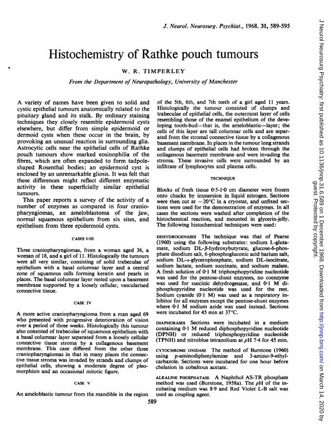
FIG. 1. An acid phosphatase stain on the craniopharyn-gioma from Case 1. There is a strong reaction in the basalcolumnar layer of cells (arrow) and a weaker reaction inthe intermediate zone of cells. x 150.
C.i ' .Sa .....S~~~~~~~~~
X.~~~~~S
I~~~
# :c
*.. .. PA. ' / .~~~~~~....
FIG. 2. An acid phosphatase stain on the ameloblastomashowing a moderate reaction in the basal layer of cells(arrow) and a weak reaction in the remainder of the tumourcells. x 150.
guest. Protected by copyright.
on March 14, 2020 by
http://jnnp.bmj.com
/J N
eurol Neurosurg P
sychiatry: first published as 10.1136/jnnp.31.6.589 on 1 Decem
ber 1968. Dow
nloaded from
Histochemistry of Rathke pouch tumours
-.
I . -
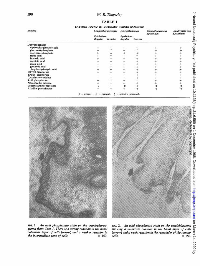
FIG. 3. A lactic acid dehydrogenase preparation of thecraniopharyngioma from Case 1. The basal columnarlayer of cells (arrow) is slightly stronger than the inter-mediate zone. x 120.
ACID PHOSPHATASE A similar method to that used foralkaline phosphatase was used except that the pH of theincubating medium was 5-2.
NON-SPECIFIC ESTERASE The method was based on
Gomori's modification of the technique of Nachlas andSeligman (1949) using a-naphthyl acetate as substrate,and Fast Blue BB salt as coupling agent.
LEUCINE AMINOPEPTIDASE The method was that of Bur-stone and Folk (1956) using L-leucyl-fl naphthylamide as
substrate. Sections were incubated for one hour atpH 7-1.
RESULTS
The Table shows the enzymes found in the differenttissues examined.
All enzymes, with the exception of alkaline phos-phatase and leucine aminopeptidase, were slightlymore active in the basal columnar layer than in theintermediate zone of squamous epithelium in all five
FIG. 4. A lactic acid dehydrogenase stain on the amelo-blastoma. There is a slightly stronger reaction in the basallayer of cells (arrow) than in the intermediate zone. x 150.
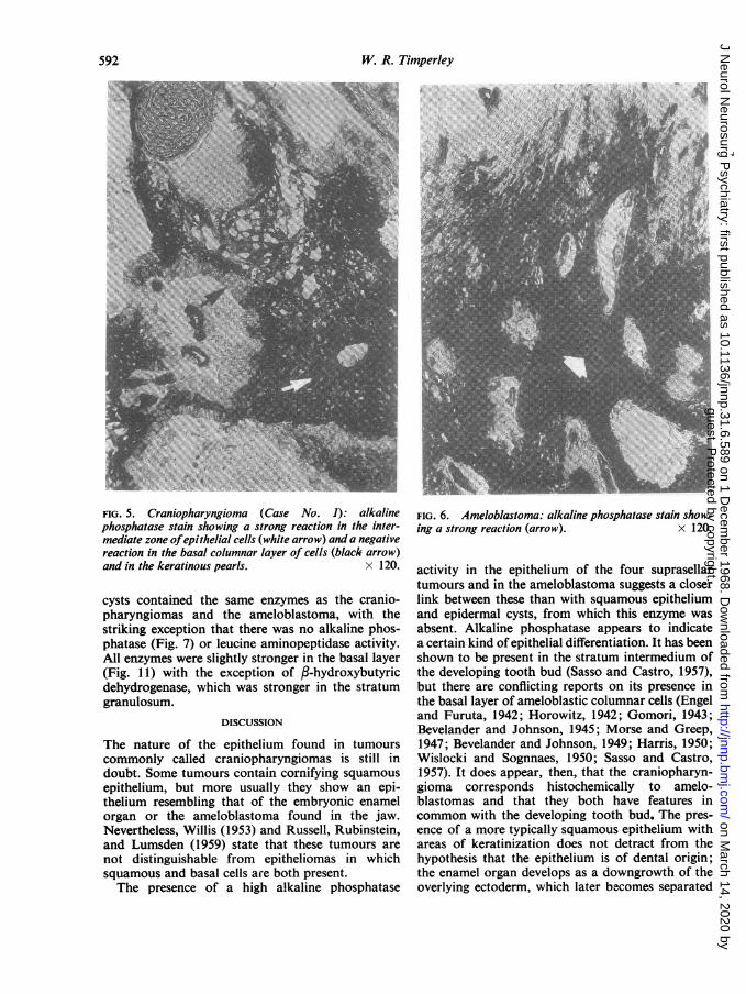
tumours examined (Figs. 1-4). Alkaline phosphatasewas very strong in the intermediate zone, but wasalmost absent in the basal layers of cells adjacent totho connective-tissue stroma (Figs. 5 and 6).The clumps and columns of cells invading the
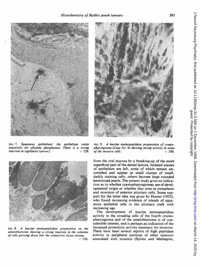
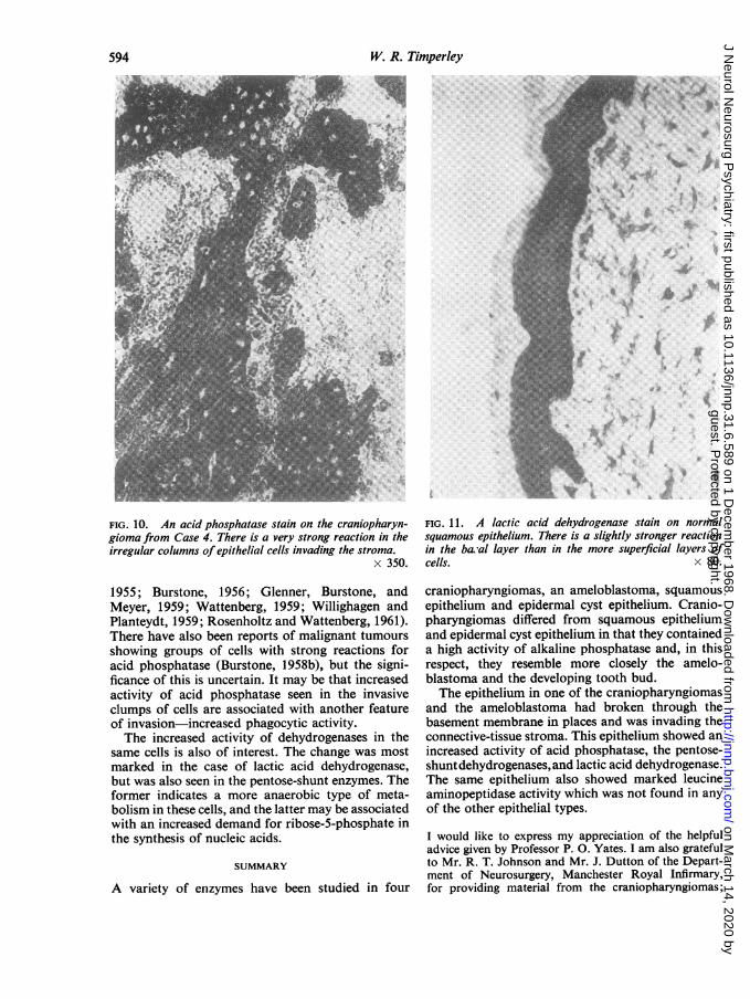
stroma of the active craniopharyngioma from CaseIV and of the ameloblastoma were the only sites toshow a moderate degree of leucine amino-peptidaseactivity (Figs. 8 and 9). This was completely absentfrom the first three craniopharyngiomas, and wasnot found in the differentiated and non-invasive cellsof the other two tumours.The invasive cells of the active craniopharyngioma
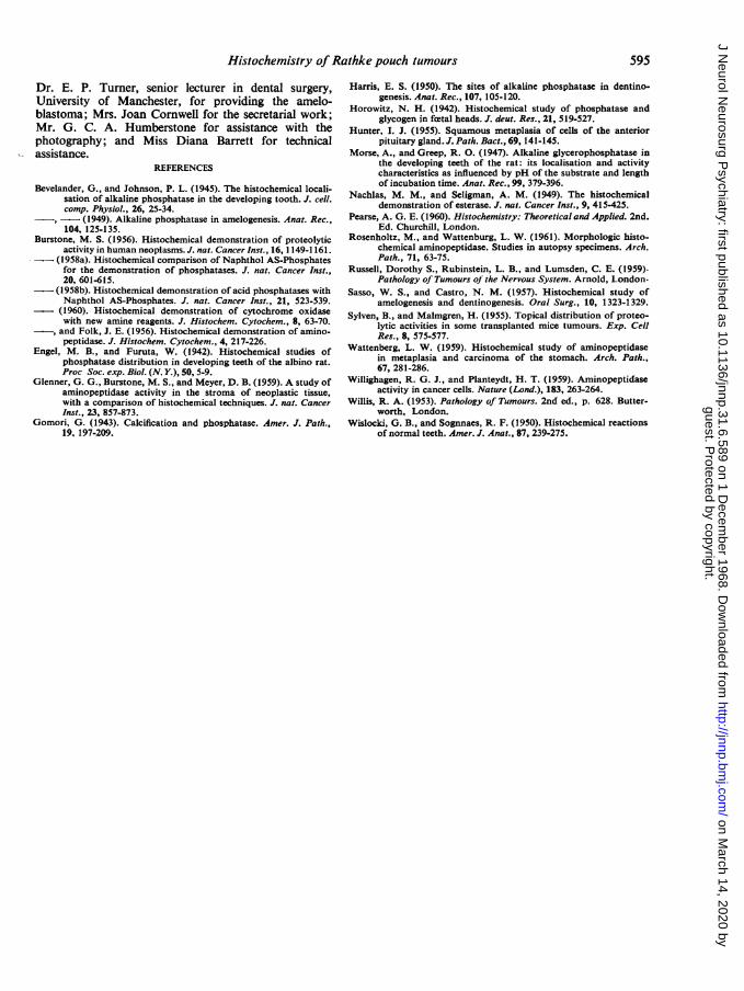
and the ameloblastoma also showed stronger acidphosphatase (Fig. 10) and dehydrogenase activitythan the more differentiated parts of the tumour.This increased activity was most marked in the caseof lactic acid dehydrogenase, and the pentose-shuntenzymes, and was not seen in the citric-acid cycleenzymes.Squamous epithelium and the three epidermal
591
guest. Protected by copyright.
on March 14, 2020 by
http://jnnp.bmj.com
/J N
eurol Neurosurg P
sychiatry: first published as 10.1136/jnnp.31.6.589 on 1 Decem
ber 1968. Dow
nloaded from
W. R. Timperley
FIG. 5. Craniopharyngioma (Case No. 1): alkaline FIG. 6. Ameloblastoma: alkaline phosphatase stain show-phosphatase stain showing a strong reaction in the inter- ing a strong reaction (arrow). x 120.mediate zone ofepithelial cells (white arrow) and a negativereaction in the basal columnar layer of cells (black arrow)~_ *- . I_1A l 1 _ _ ___ana in me keratinous pearls. x I zU.
cysts contained the same enzymes as the cranio-pharyngiomas and the ameloblastoma, with thestriking exception that there was no alkaline phos-phatase (Fig. 7) or leucine aminopeptidase activity.All enzymes were slightly stronger in the basal layer(Fig. 11) with the exception of f-hydroxybutyricdehydrogenase, which was stronger in the stratumgranulosum.
DISCUSSION
The nature of the epithelium found in tumourscommonly called craniopharyngiomas is still indoubt. Some tumours contain cornifying squamousepithelium, but more usually they show an epi-thelium resembling that of the embryonic enamelorgan or the ameloblastoma found in the jaw.Nevertheless, Willis (1953) and Russell, Rubinstein,and Lumsden (1959) state that these tumours arenot distinguishable from epitheliomas in whichsquamous and basal cells are both present.The presence of a high alkaline phosphatase
activity in the epithelium of the four suprasellartumours and in the ameloblastoma suggests a closerlink between these than with squamous epitheliumand epidermal cysts, from which this enzyme wasabsent. Alkaline phosphatase appears to indicatea certain kind of epithelial differentiation. It has beenshown to be present in the stratum intermedium ofthe developing tooth bud (Sasso and Castro, 1957),but there are conflicting reports on its presence inthe basal layer of ameloblastic columnar cells (Engeland Furuta, 1942; Horowitz, 1942; Gomori, 1943;Bevelander and Johnson, 1945; Morse and Greep,1947; Bevelander and Johnson, 1949; Harris, 1950;Wislocki and Sognnaes, 1950; Sasso and Castro,1957). It does appear, then, that the craniopharyn-gioma corresponds histochemically to amelo-blastomas and that they both have features incommon with the developing tooth bud. The pres-ence of a more typically squamous epithelium withareas of keratinization does not detract from thehypothesis that the epithelium is of dental origin;the enamel organ develops as a downgrowth of theoverlying ectoderm, which later becomes separated
592
guest. Protected by copyright.
on March 14, 2020 by
http://jnnp.bmj.com
/J N
eurol Neurosurg P
sychiatry: first published as 10.1136/jnnp.31.6.589 on 1 Decem
ber 1968. Dow
nloaded from
Histochemistry of Rathke pouch tumours 593
4.~~~~~~7T
A~~~~~~~~~~A
,..FIG_| | N | |. 7. Squamous epithelium te e m s F. 9. A e ap.i
<;~~~~~~~~~~~~fothe>^ oramucosa; by abreakinrg-u of th mor
i?'?~~~ ~ ~ ~~~~ ~spefca parofmith deta laia Isolatedmasses
\~~~~~~~~~~~~~eaiie perl. The prset suy g S2 ,t.N=1iveno indica-l
IG.7.quaouspitelim: teeithliumstanstIon as to whethier caninophpiarygimapeareto of develo-negaiveyfrakaliepospatae.Tereis ston opmerntioalorigin Norwhehe theyin ariseg ascmtapit asiaomreactioincapllaries(arrow) x12 andrheiversion oflsanero piutaycll.Smesp
" su~~~poriilprtfor thelteidenalwasmivnabysHunter (1955),wo foellund inreasnlevidncofsml slands of sqma-l_<~mr mousepilytheniag cells; inther pituitaryestalknwit^'S~~~Y~~ _ ^ _ tio~~~ncrasin agwe.hrcaipaygoa rfdvl
74~~~~~~~~~~hdevelg_opmentaofiiorweucinteyaie asmioeptdlasea|t.,,h;2 a ' .._ actiitrvsionthe inadngeirpiutrcells.ohfourt csani-t
sX~~~~~~^. hayniom andortfothe aeolastoma idawsofv coHn-e 15)
siderable interest, and is perhaps an indication of theFIG. 8. A leucine aminopeptidase preparation on the increased proteolytic activity necessary for invasion.ameloblastoma showing a strong reaction in the columns There have been several reports of high peptidaseof cells growing down into the connective tissue stroma. activity in peripheral portions of other tumours
x 120. associated with invasion (Sylven and Malmgren,
guest. Protected by copyright.
on March 14, 2020 by
http://jnnp.bmj.com
/J N
eurol Neurosurg P
sychiatry: first published as 10.1136/jnnp.31.6.589 on 1 Decem
ber 1968. Dow
nloaded from
W. R. Timperley
*t 1}
....
FIG. 10. An acid phosphatase stain on the craniopharyn-gioma from Case 4. There is a very strong reaction in theirregular columns of epithelial cells invading the stroma.
x 350.
1955; Burstone, 1956; Glenner, Burstone, andMeyer, 1959; Wattenberg, 1959; Willighagen andPlanteydt, 1959; Rosenholtz and Wattenberg, 1961).There have also been reports of malignant tumoursshowing groups of cells with strong reactions foracid phosphatase (Burstone, 1958b), but the signi-ficance of this is uncertain. It may be that increasedactivity of acid phosphatase seen in the invasiveclumps of cells are associated with another featureof invasion-increased phagocytic activity.The increased activity of dehydrogenases in the
same cells is also of interest. The change was mostmarked in the case of lactic acid dehydrogenase,but was also seen in the pentose-shunt enzymes. Theformer indicates a more anaerobic type of meta-bolism in these cells, and the latter may be associatedwith an increased demand for ribose-5-phosphate inthe synthesis of nucleic acids.
SUMMARY
A variety of enzymes have been studied in four
FIG. 1L. A lactic acid dehydrogenase stain on normalsquamous epithelium. There is a slightly stronger reactionin the ba:al layer than in the more superficial layers ofcells. x 80.
craniopharyngiomas, an ameloblastoma, squamousepithelium and epidermal cyst epithelium. Cranio-pharyngiomas differed from squamous epitheliumand epidermal cyst epithelium in that they containeda high activity of alkaline phosphatase and, in thisrespect, they resemble more closely the amelo-blastoma and the developing tooth bud.The epithelium in one of the craniopharyngiomas
and the ameloblastoma had broken through thebasement membrane in places and was invading theconnective-tissue stroma. This epithelium showed anincreased activity of acid phosphatase, the pentose-shuntdehydrogenases,and lactic acid dehydrogenase.The same epithelium also showed marked leucineaminopeptidase activity which was not found in anyof the other epithelial types.
I would like to express my appreciation of the helpfuladvice given by Professor P. 0. Yates. I am also gratefulto Mr. R. T. Johnson and Mr. J. Dutton of the Depart-ment of Neurosurgery, Manchester Royal Infirmary,for providing material from the craniopharyngiomas;
594
-r
.4;-.4
i
guest. Protected by copyright.
on March 14, 2020 by
http://jnnp.bmj.com
/J N
eurol Neurosurg P
sychiatry: first published as 10.1136/jnnp.31.6.589 on 1 Decem
ber 1968. Dow
nloaded from
Histochemistry of Rathke pouch tumours
Dr. E. P. Turner, senior lecturer in dental surgery,University of Manchester, for providing the amelo-blastoma; Mrs. Joan Cornwell for the secretarial work;Mr. G. C. A. Humberstone for assistance with thephotography; and Miss Diana Barrett for technicalassistance.
REFERENCES
Bevelander, G., and Johnson, P. L. (1945). The histochemical locali-sation of alkaline phosphatase in the developing tooth. J. cell.comp. Physiol., 26, 25-34.- (1949). Alkaline phosphatase in amelogenesis. Anat. Rec.,104, 125-135.
Burstone, M. S. (1956). Histochemical demonstration of proteolyticactivity in human neoplasms. J. nat. Cancer Inst., 16, 1149-1161.
- (1958a). Histochemical comparison of Naphthol AS-Phosphatesfor the demonstration of phosphatases. J. nat. Cancer Inst.,20, 601-615.
-(1958b). Histochemical demonstration of acid phosphatases withNaphthol AS-Phosphates. J. nat. Cancer Inst., 21, 523-539.
-(1960). Histochemical demonstration of cytochrome oxidasewith new amine reagents. J. Histochem. Cytochem., 8, 63-70.
-, and Folk, J. E. (1956). Histochemical demonstration of amino-peptidase. J. Histochem. Cytochem., 4, 217-226.
Engel, M. B., and Furuta, W. (1942). Histochemical studies ofphosphatase distribution in developing teeth of the albino rat.Proc Soc. exp. Biol. (N. Y.), 50, 5-9.
Glenner, G. G., Burstone, M. S., and Meyer, D. B. (1959). A study ofaminopeptidase activity in the stroma of neoplastic tissue,with a comparison of histochemical techniques. J. nat. CancerInst., 23, 857-873.
Gomori, G. (1943). Calcification and phosphatase. Amer. J. Path.,19. 197-209.
Harris, E. S. (1950). The sites of alkaline phosphatase in dentino-genesis. Anat. Rec., 107, 105-120.
Horowitz, N. H. (1942). Histochemical study of phosphatase andglycogen in fectal heads. J. deut. Res., 21, 519-527.
Hunter, I. J. (1955). Squamous metaplasia of cells of the anteriorpituitary gland. J. Path. Bact., 69, 141-145.
Morse, A., and Greep, R. 0. (1947). Alkaline glycerophosphatase inthe developing teeth of the rat: its localisation and activitycharacteristics as influenced by pH of the substrate and lengthof incubation time. Anat. Rec., 99, 379-396.
Nachlas, M. M., and Seligman, A. M. (1949). The histochemicaldemonstration of esterase. J. nat. Cancer Inst., 9, 415-425.
Pearse, A. G. E. (1960). Histochemistry: Theoretical and Applied. 2nd.Ed. Churchill, London.
Rosenholtz, M., and Wattenburg, L. W. (1961). Morphologic histo-chemical aminopeptidase. Studies in autopsy specimens. Arch.Path., 71, 63-75.
Russell, Dorothy S., Rubinstein, L. B., and Lumsden, C. E. (1959).Pathology of Tumours of the Nervous System. Arnold, London.
Sasso, W. S., and Castro, N. M. (1957). Histochemical study ofamelogenesis and dentinogenesis. Oral Surg., 10, 1323-1329.
Sylven, B., and Malmgren, H. (1955). Topical distribution of proteo-lytic activities in some transplanted mice tumours. Exp. CellRes., 8, 575-577.
Wattenberg, L. W. (1959). Histochemical study of aminopeptidasein metaplasia and carcinoma of the stomach. Arch. Path.,67, 281-286.
Willighagen, R. G. J., and Planteydt, H. T. (1959). Aminopeptidaseactivity in cancer cells. Nature (Lond.), 183, 263-264.
Willis, R. A. (1953). Pathology of Tumours. 2nd ed., p. 628. Butter-worth, London.
Wislocki, G. B., and Sognnaes, R. F. (1950). Histochemical reactionsof normal teeth. Amer. J. Anat., 87, 239-275.
595
guest. Protected by copyright.
on March 14, 2020 by
http://jnnp.bmj.com
/J N
eurol Neurosurg P
sychiatry: first published as 10.1136/jnnp.31.6.589 on 1 Decem
ber 1968. Dow
nloaded from