Embed Size (px)
Citation preview

C h a p t e r
20
The Heart
PowerPoint® Lecture Slides prepared by Jason LaPres
Lone Star College - North Harris
Copyright © 2009 Pearson Education, Inc.,publishing as Pearson Benjamin CummingsCopyright © 2009 Pearson Education, Inc., publishing as Pearson Benjamin Cummings

Introduction to Cardiovascular System
The Pulmonary Circuit
Carries blood to and from gas exchange surfaces of
lungs
The Systemic Circuit
Carries blood to and from the body
Blood alternates between pulmonary circuit and
systemic circuit
Copyright © 2009 Pearson Education, Inc., publishing as Pearson Benjamin Cummings

The Conducting System
Heartbeat
A single contraction of the heart
The entire heart contracts in series
First the atria
Then the ventricles
Copyright © 2009 Pearson Education, Inc., publishing as Pearson Benjamin Cummings
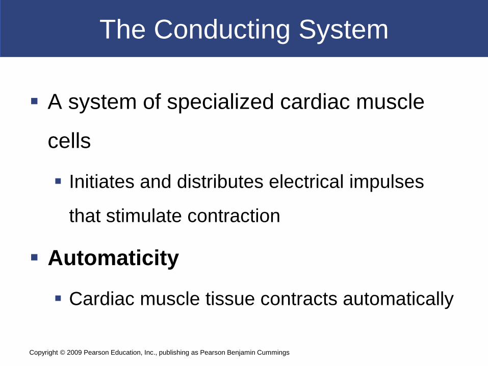
The Conducting System
A system of specialized cardiac muscle
cells
Initiates and distributes electrical impulses
that stimulate contraction
Automaticity
Cardiac muscle tissue contracts automatically
Copyright © 2009 Pearson Education, Inc., publishing as Pearson Benjamin Cummings

The Conducting System
Structures of the Conducting System
Sinoatrial (SA) node - wall of right atrium
Atrioventricular (AV) node - junction between
atria and ventricles
Conducting cells - throughout myocardium
Copyright © 2009 Pearson Education, Inc., publishing as Pearson Benjamin Cummings

The Conducting System
Conducting Cells
Interconnect SA and AV nodes
Distribute stimulus through myocardium
In the atrium
Internodal pathways
In the ventricles
AV bundle and the bundle branches
Copyright © 2009 Pearson Education, Inc., publishing as Pearson Benjamin Cummings

The Conducting System
Heart Rate
SA node generates 80–100 action potentials
per minute
Parasympathetic stimulation slows heart rate
AV node generates 40–60 action potentials
per minute
Copyright © 2009 Pearson Education, Inc., publishing as Pearson Benjamin Cummings
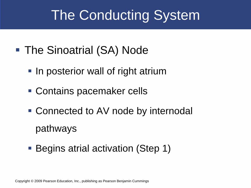
The Conducting System
The Sinoatrial (SA) Node
In posterior wall of right atrium
Contains pacemaker cells
Connected to AV node by internodal
pathways
Begins atrial activation (Step 1)
Copyright © 2009 Pearson Education, Inc., publishing as Pearson Benjamin Cummings

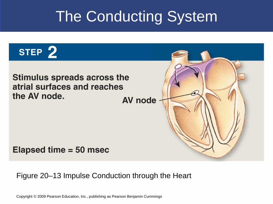
The Conducting System
Figure 20–13 Impulse Conduction through the Heart
Copyright © 2009 Pearson Education, Inc., publishing as Pearson Benjamin Cummings

The Conducting System
The Atrioventricular (AV) Node
In floor of right atrium
Receives impulse from SA node (Step 2)
Delays impulse (Step 3)
Atrial contraction begins
Copyright © 2009 Pearson Education, Inc., publishing as Pearson Benjamin Cummings
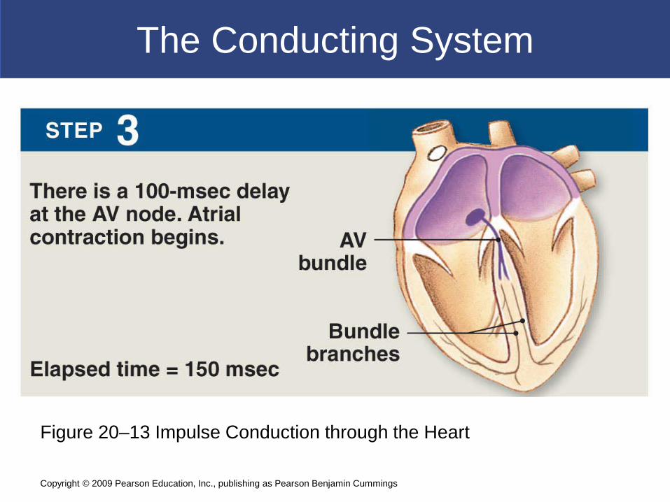
The Conducting System
Figure 20–13 Impulse Conduction through the Heart
Copyright © 2009 Pearson Education, Inc., publishing as Pearson Benjamin Cummings
The Conducting System
Figure 20–13 Impulse Conduction through the Heart
Copyright © 2009 Pearson Education, Inc., publishing as Pearson Benjamin Cummings

The Conducting System
The AV Bundle
In the septum
Carries impulse to left and right bundle
branches
Which conduct to Purkinje fibers (Step 4)
And to the moderator band
Which conducts to papillary muscles
Copyright © 2009 Pearson Education, Inc., publishing as Pearson Benjamin Cummings

The Conducting System
Figure 20–13 Impulse Conduction through the Heart
Copyright © 2009 Pearson Education, Inc., publishing as Pearson Benjamin Cummings

The Conducting System
Purkinje Fibers
Distribute impulse through ventricles (Step 5)
Atrial contraction is completed
Ventricular contraction begins
Copyright © 2009 Pearson Education, Inc., publishing as Pearson Benjamin Cummings

The Conducting System
Figure 20–13 Impulse Conduction through the Heart
Copyright © 2009 Pearson Education, Inc., publishing as Pearson Benjamin Cummings
The Conducting System
Abnormal Pacemaker Function
Bradycardia: abnormally slow heart rate
Tachycardia: abnormally fast heart rate
Ectopic pacemaker
Abnormal cells
Generate high rate of action potentials
Bypass conducting system
Disrupt ventricular contractions
Copyright © 2009 Pearson Education, Inc., publishing as Pearson Benjamin Cummings
The Conducting System
Electrocardiogram (ECG or EKG)
A recording of electrical events in the heart
Obtained by electrodes at specific body
locations
Abnormal patterns diagnose damage
Copyright © 2009 Pearson Education, Inc., publishing as Pearson Benjamin Cummings

The Conducting System
Features of an ECG
P wave
Atria depolarize
QRS complex
Ventricles depolarize
T wave
Ventricles repolarize
Copyright © 2009 Pearson Education, Inc., publishing as Pearson Benjamin Cummings

The Conducting System
Time Intervals Between ECG Waves
P–R interval
From start of atrial depolarization
To start of QRS complex
Q–T interval
From ventricular depolarization
To ventricular repolarization
Copyright © 2009 Pearson Education, Inc., publishing as Pearson Benjamin Cummings

The Conducting System
Contractile Cells
Purkinje fibers distribute the stimulus to the
contractile cells, which make up most of the
muscle cells in the heart
Copyright © 2009 Pearson Education, Inc., publishing as Pearson Benjamin Cummings

The Cardiac Cycle
Cardiac cycle = The period between the
start of one heartbeat and the beginning of
the next
Includes both contraction and relaxation
Copyright © 2009 Pearson Education, Inc., publishing as Pearson Benjamin Cummings

The Conducting System
The Cardiac Cycle
Begins with action potential at SA node
Transmitted through conducting system
Produces action potentials in cardiac muscle cells (contractile
cells)
Copyright © 2009 Pearson Education, Inc., publishing as Pearson Benjamin Cummings
The Cardiac Cycle
Phases of the Cardiac Cycle
Within any one chamber
Systole (contraction)
Diastole (relaxation)
Copyright © 2009 Pearson Education, Inc., publishing as Pearson Benjamin Cummings

The Cardiac Cycle
Cardiac Cycle and Heart Rate
At 75 beats per minute
Cardiac cycle lasts about 800 msecs
When heart rate increases
All phases of cardiac cycle shorten, particularly
diastole
Copyright © 2009 Pearson Education, Inc., publishing as Pearson Benjamin Cummings

The Cardiac Cycle
Eight Steps in the Cardiac Cycle1. Atrial systole
Atrial contraction begins
Right and left AV valves are open
2. Atria eject blood into ventricles Filling ventricles
3. Atrial systole ends AV valves close
Ventricles contain maximum blood volume
Known as end-diastolic volume (EDV)
Copyright © 2009 Pearson Education, Inc., publishing as Pearson Benjamin Cummings

The Cardiac Cycle
Eight Steps in the Cardiac Cycle4. Ventricular systole
Isovolumetric ventricular contraction
Pressure in ventricles rises
AV valves shut
5. Ventricular ejection Semilunar valves open
Blood flows into pulmonary and aortic trunks
Stroke volume (SV) = 60% of end-diastolic volume
Copyright © 2009 Pearson Education, Inc., publishing as Pearson Benjamin Cummings

The Cardiac Cycle
Eight Steps in the Cardiac Cycle6. Ventricular pressure falls
Semilunar valves close
Ventricles contain end-systolic volume (ESV), about 40% of end-diastolic volume
7. Ventricular diastole Ventricular pressure is higher than atrial pressure
All heart valves are closed
Ventricles relax (isovolumetric relaxation)
Copyright © 2009 Pearson Education, Inc., publishing as Pearson Benjamin Cummings

The Cardiac Cycle
Eight Steps in the Cardiac Cycle
8. Atrial pressure is higher than ventricular pressure AV valves open
Passive atrial filling
Passive ventricular filling
Cardiac cycle ends
The Heart: Cardiac Cycle
Copyright © 2009 Pearson Education, Inc., publishing as Pearson Benjamin Cummings
The Cardiac Cycle
Blood Pressure
In any chamber
Rises during systole
Falls during diastole
Blood flows from high to low pressure
Controlled by timing of contractions
Directed by one-way valves
Copyright © 2009 Pearson Education, Inc., publishing as Pearson Benjamin Cummings
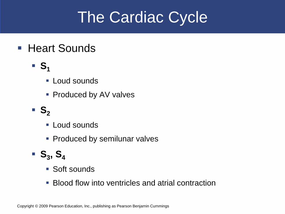
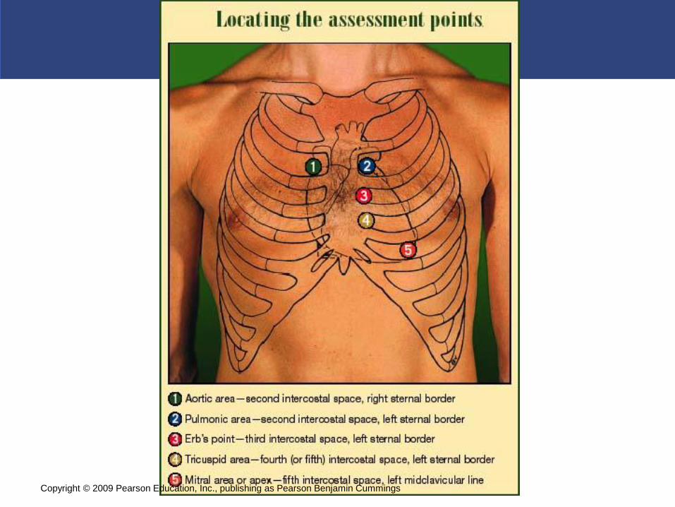
The Cardiac Cycle
Heart Sounds S1
Loud sounds
Produced by AV valves
S2
Loud sounds
Produced by semilunar valves
S3, S4
Soft sounds
Blood flow into ventricles and atrial contraction
Copyright © 2009 Pearson Education, Inc., publishing as Pearson Benjamin Cummings

The Cardiac Cycle
Copyright © 2009 Pearson Education, Inc., publishing as Pearson Benjamin Cummings
The Cardiac Cycle
Copyright © 2009 Pearson Education, Inc., publishing as Pearson Benjamin Cummings

The Cardiac Cycle
Heart Murmur
Sounds produced by regurgitation through
valves
Copyright © 2009 Pearson Education, Inc., publishing as Pearson Benjamin Cummings
Cardiodynamics
The movement and force generated by cardiac contractions End-diastolic volume (EDV) End-systolic volume (ESV) Stroke volume (SV)
SV = EDV – ESV
Cardiac output (CO) The volume pumped by left ventricle in 1 minute
Copyright © 2009 Pearson Education, Inc., publishing as Pearson Benjamin Cummings
Cardiodynamics
Cardiac Output
CO = HR X SV
CO = cardiac output (mL/min)
HR = heart rate (beats/min)
SV = stroke volume (mL/beat)
Copyright © 2009 Pearson Education, Inc., publishing as Pearson Benjamin Cummings

Cardiodynamics
Factors Affecting Cardiac Output
Cardiac output
Adjusted by changes in heart rate or stroke volume
Heart rate
Adjusted by autonomic nervous system or hormones
Stroke volume
Adjusted by changing EDV or ESV
Copyright © 2009 Pearson Education, Inc., publishing as Pearson Benjamin Cummings

Cardiodynamics
Atrial Reflex
Also called Bainbridge reflex
Adjusts heart rate in response to venous
return
Stretch receptors in right atrium
Trigger increase in heart rate
Through increased sympathetic activity
Copyright © 2009 Pearson Education, Inc., publishing as Pearson Benjamin Cummings

Cardiodynamics
Hormonal Effects on Heart Rate
Increase heart rate (by sympathetic
stimulation of SA node)
Epinephrine (E)
Norepinephrine (NE)
Thyroid hormone
Copyright © 2009 Pearson Education, Inc., publishing as Pearson Benjamin Cummings
Cardiodynamics
Factors Affecting the Stroke Volume
The EDV: amount of blood a ventricle contains at the
end of diastole
Filling time:
– duration of ventricular diastole
Venous return:
– rate of blood flow during ventricular diastole
Copyright © 2009 Pearson Education, Inc., publishing as Pearson Benjamin Cummings

Cardiodynamics
The EDV and Stroke Volume
At rest
EDV is low
Myocardium stretches less
Stroke volume is low
With exercise
EDV increases
Myocardium stretches more
Stroke volume increases
Copyright © 2009 Pearson Education, Inc., publishing as Pearson Benjamin Cummings

Cardiodynamics
The Frank–Starling Principle
As EDV increases, stroke volume increases
Physical Limits
Ventricular expansion is limited by
Myocardial connective tissue
The cardiac (fibrous) skeleton
The pericardial sac
Copyright © 2009 Pearson Education, Inc., publishing as Pearson Benjamin Cummings

Cardiodynamics
End-Systolic Volume (ESV)
The amount of blood that remains in the
ventricle at the end of ventricular systole is
the ESV
Copyright © 2009 Pearson Education, Inc., publishing as Pearson Benjamin Cummings

Cardiodynamics
Afterload
Is increased by any factor that restricts arterial
blood flow
As afterload increases, stroke volume
decreases
Copyright © 2009 Pearson Education, Inc., publishing as Pearson Benjamin Cummings

Cardiodynamics
Heart Rate Control Factors
Autonomic nervous system
Sympathetic and parasympathetic
Circulating hormones
Venous return and stretch receptors
Copyright © 2009 Pearson Education, Inc., publishing as Pearson Benjamin Cummings

Cardiodynamics
Stroke Volume Control Factors
EDV
Filling time
Rate of venous return
ESV
Preload
Contractility
Afterload
Copyright © 2009 Pearson Education, Inc., publishing as Pearson Benjamin Cummings
Cardiodynamics
Cardiac Reserve
The difference between resting and maximal
cardiac output
Copyright © 2009 Pearson Education, Inc., publishing as Pearson Benjamin Cummings
Cardiodynamics
The Heart and Cardiovascular System
Cardiovascular regulation
Ensures adequate circulation to body tissues
Cardiovascular centers
Control heart and peripheral blood vessels
Cardiovascular system responds to
Changing activity patterns
Circulatory emergencies
Copyright © 2009 Pearson Education, Inc., publishing as Pearson Benjamin Cummings