Embed Size (px)
Citation preview

The Heart Chapter 18
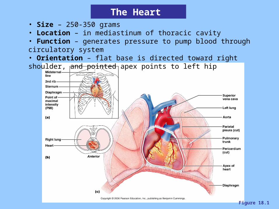
Figure 18.1
The Heart • Size – 250-350 grams• Location – in mediastinum of thoracic cavity• Function – generates pressure to pump blood through circulatory system • Orientation – flat base is directed toward right shoulder, and pointed apex points to left hip

Figure 18.2
Heart Coverings Pericardium – the two layered, membranous sac in which the heart sitsFibrous pericardium – the outer, thick layer composed of dense connective tissue, for protecting, anchoring the heart in position, and preventing overfilling Serous pericardium – the inner thin layer composed of a serous membrane
Visceral layer – membrane clinging to the outer heart surfaceParietal layer – membrane clinging to the inside of the fibrous pericardium
Pericardial cavity – serous fluid-filled space between the visceral and parietal layers
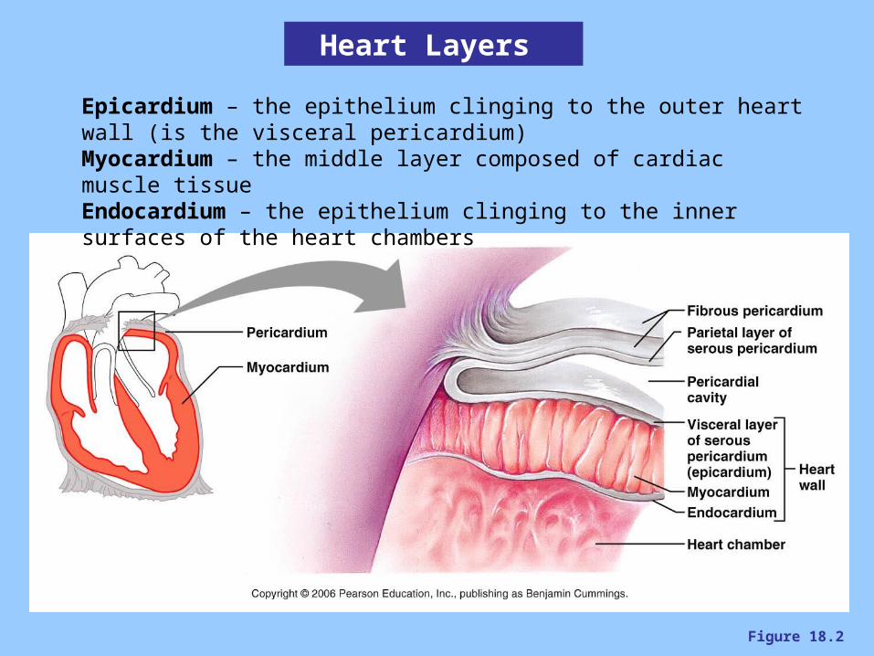
Figure 18.2
Heart Layers
Epicardium – the epithelium clinging to the outer heart wall (is the visceral pericardium)Myocardium – the middle layer composed of cardiac muscle tissueEndocardium – the epithelium clinging to the inner surfaces of the heart chambers

Figure 18.4 a, b
The Heart
• 4 chambers: 2 atria, 2 ventricles
Atria :•the superior chambers• auricles – ear-like extensions of the atria• receiving chambers, limited pumping means thin walls
Ventricles:• the inferior chambers, the majority of the heart volume• pumping chambers, thick walls
Sulci: • The indentations on the outer heart surface, corresponding to borderlines between chambers• contain fat and vessels• Ex – interventricular sulcus
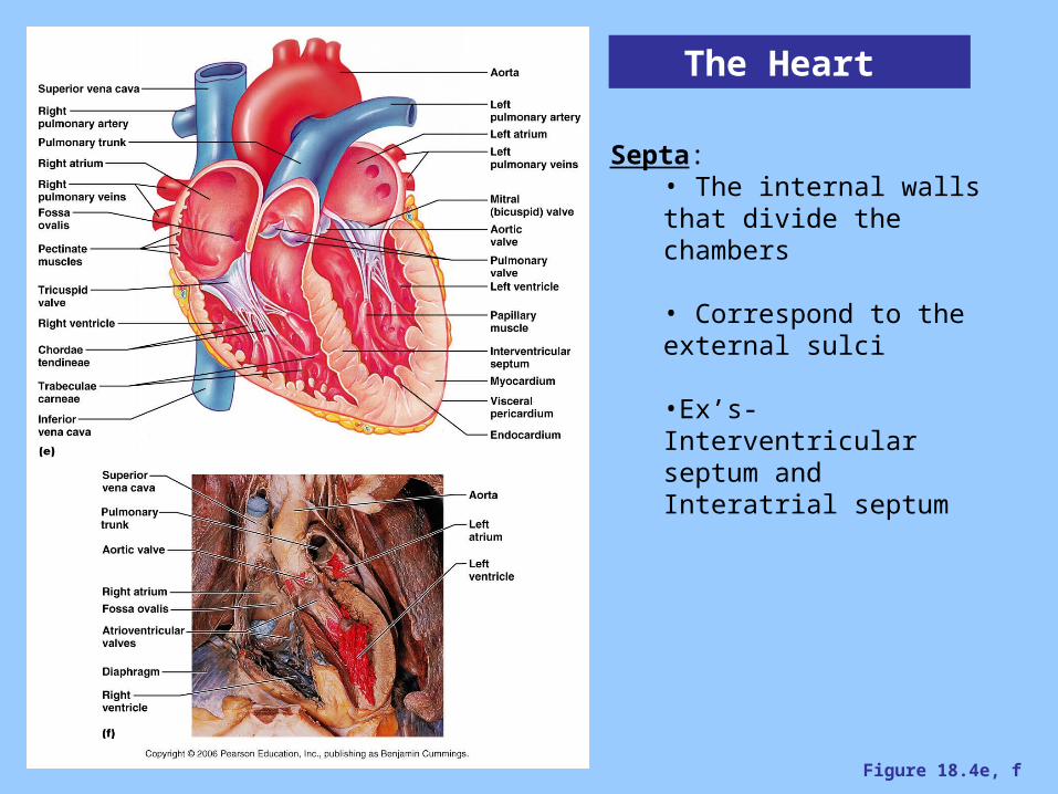
Figure 18.4e, f
The Heart
Septa:• The internal walls that divide the chambers
• Correspond to the external sulci
•Ex’s- Interventricular septum and Interatrial septum
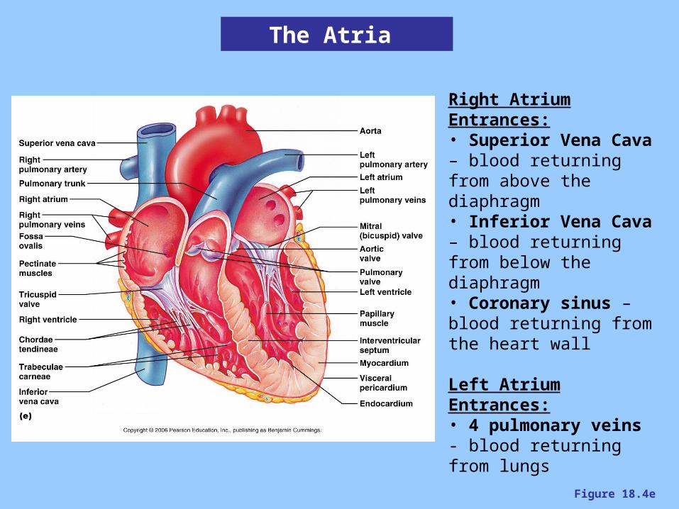
Figure 18.4e
Right Atrium Entrances:• Superior Vena Cava – blood returning from above the diaphragm• Inferior Vena Cava – blood returning from below the diaphragm• Coronary sinus – blood returning from the heart wall
Left Atrium Entrances:• 4 pulmonary veins - blood returning from lungs
The Atria
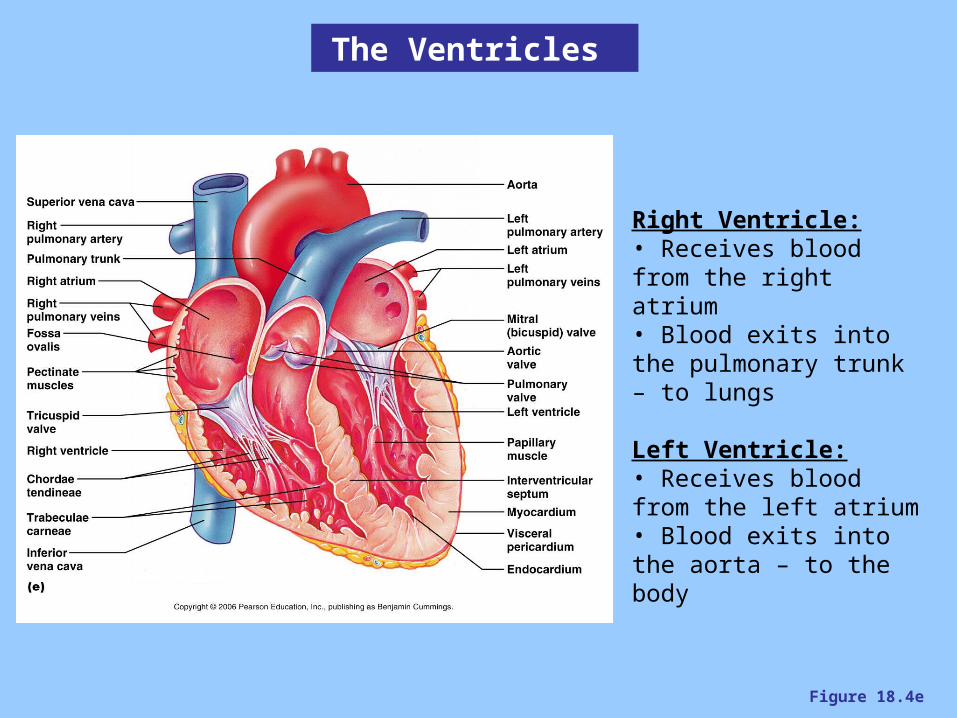
Figure 18.4e
Right Ventricle:• Receives blood from the right atrium• Blood exits into the pulmonary trunk – to lungs
Left Ventricle:• Receives blood from the left atrium• Blood exits into the aorta – to the body
The Ventricles
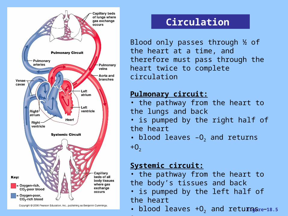
Figure 18.5
Circulation
Blood only passes through ½ of the heart at a time, and therefore must pass through the heart twice to complete circulation
Pulmonary circuit:• the pathway from the heart to the lungs and back• is pumped by the right half of the heart• blood leaves –O2 and returns +O2
Systemic circuit:• the pathway from the heart to the body’s tissues and back• is pumped by the left half of the heart• blood leaves +O2 and returns –O2
NOTE – arteries do NOT always carry oxygenated blood, and veins do NOT always carry deoxygenated blood
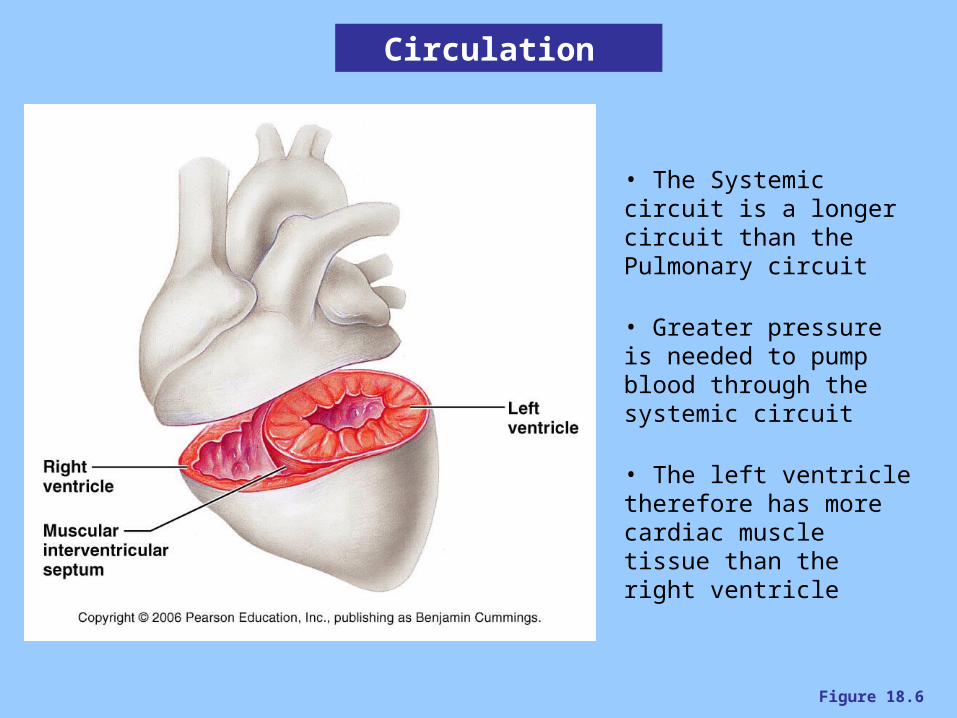
Figure 18.6
Circulation
• The Systemic circuit is a longer circuit than the Pulmonary circuit
• Greater pressure is needed to pump blood through the systemic circuit
• The left ventricle therefore has more cardiac muscle tissue than the right ventricle
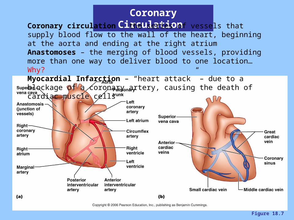
Figure 18.7
Coronary Circulation Coronary circulation – the series of vessels that supply blood flow to the wall of the heart, beginning at the aorta and ending at the right atriumAnastomoses – the merging of blood vessels, providing more than one way to deliver blood to one location…Why?Myocardial Infarction – “heart attack” – due to a blockage of a coronary artery, causing the death of cardiac muscle cells
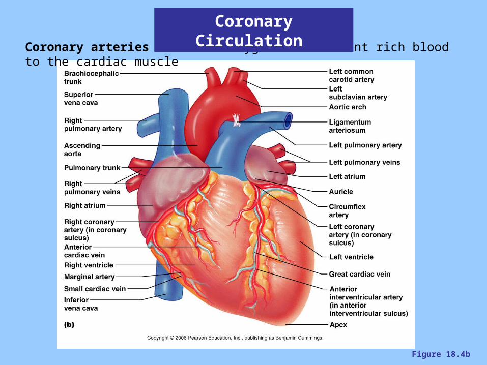
Figure 18.4b
Coronary arteries – deliver oxygen and nutrient rich blood to the cardiac muscle
Coronary Circulation

Figure 18.4d
Coronary Circulation
Cardiac veins – drain the oxygen and nutrient poor blood from the cardiac muscleCoronary sinus – large vein on posterior side that empties into the right atrium
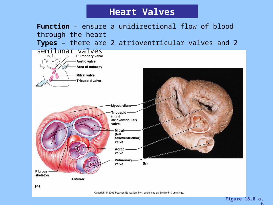
Figure 18.8 a, b
Heart Valves
Function – ensure a unidirectional flow of blood through the heartTypes – there are 2 atrioventricular valves and 2 semilunar valves
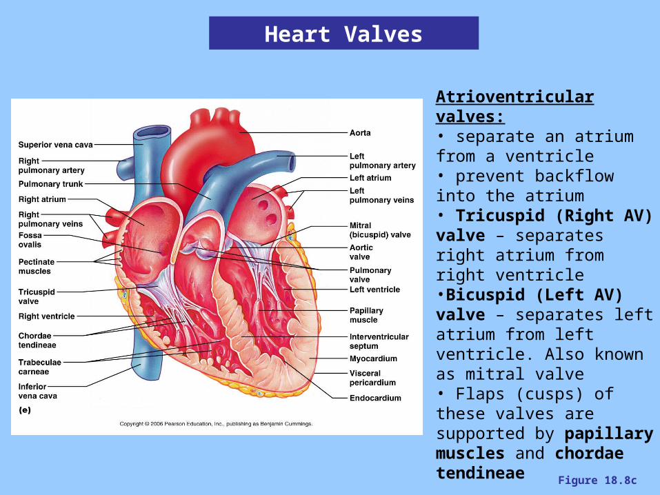
Figure 18.8c
Heart Valves
Atrioventricular valves:• separate an atrium from a ventricle• prevent backflow into the atrium• Tricuspid (Right AV) valve – separates right atrium from right ventricle•Bicuspid (Left AV) valve – separates left atrium from left ventricle. Also known as mitral valve• Flaps (cusps) of these valves are supported by papillary muscles and chordae tendineae
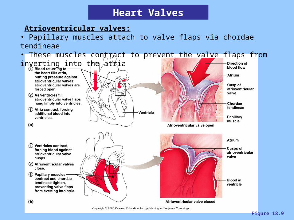
Figure 18.9
Heart Valves
Atrioventricular valves:• Papillary muscles attach to valve flaps via chordae tendineae• These muscles contract to prevent the valve flaps from inverting into the atria

Figure 18.8c
Heart Valves
Semilunar valves:• separate a ventricle from a great vessel• prevent backflow into the ventricle• composed of three cup-like flaps• Pulmonary semilunar valve – regulates movement of blood into pulmonary trunk• Aortic semilunar valve – regulates movement of blood into the aorta• Flaps (cusps) of these valves are NOT supported by papillary muscles and chordae tendineae
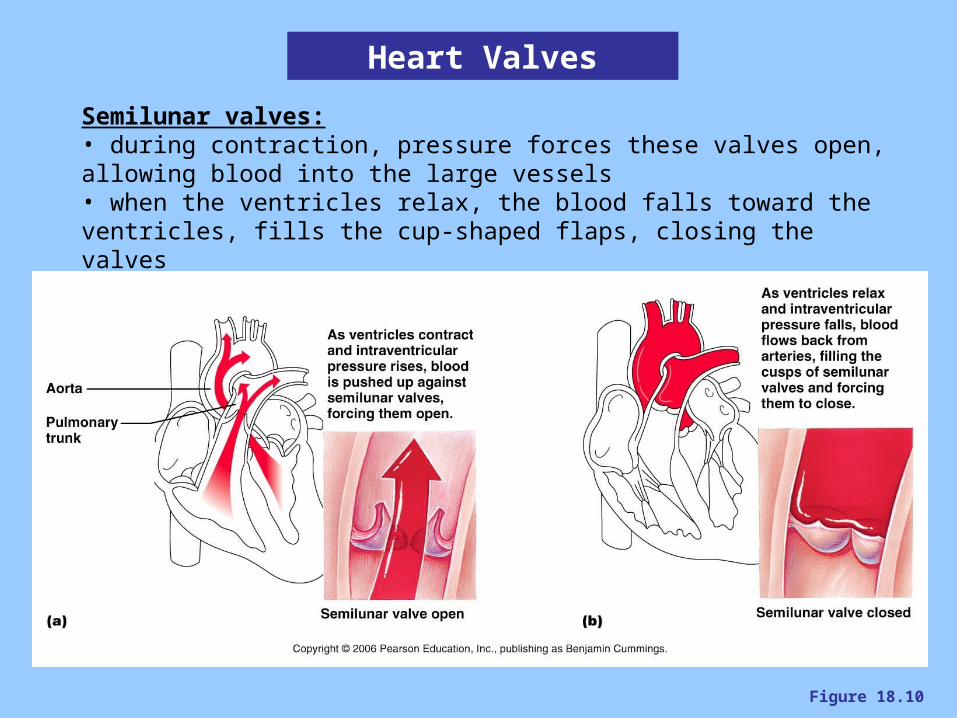
Figure 18.10
Heart Valves
Semilunar valves:• during contraction, pressure forces these valves open, allowing blood into the large vessels• when the ventricles relax, the blood falls toward the ventricles, fills the cup-shaped flaps, closing the valves
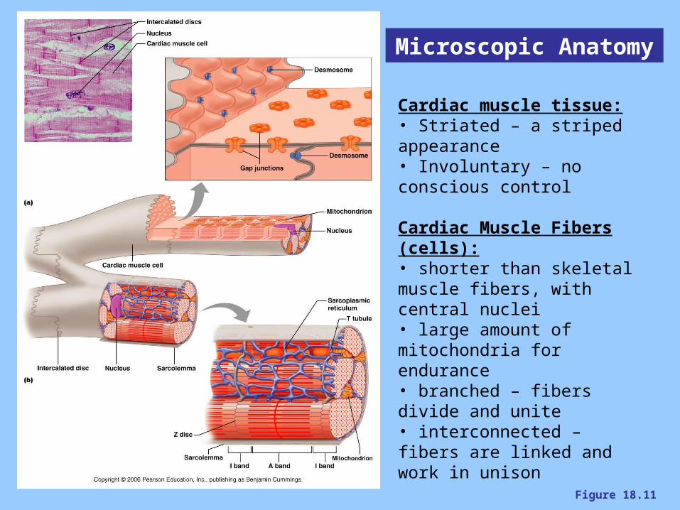
Figure 18.11
Microscopic Anatomy
Cardiac muscle tissue:• Striated – a striped appearance• Involuntary – no conscious control
Cardiac Muscle Fibers (cells):• shorter than skeletal muscle fibers, with central nuclei• large amount of mitochondria for endurance• branched – fibers divide and unite• interconnected – fibers are linked and work in unison
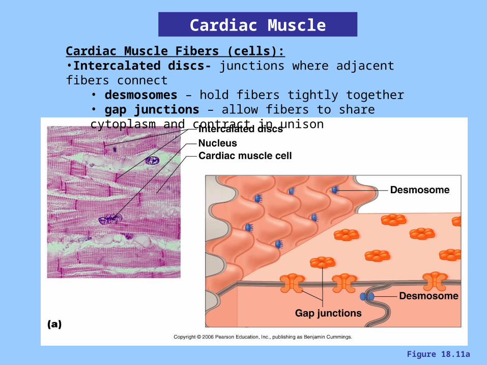
Figure 18.11a
Cardiac Muscle Fibers (cells):•Intercalated discs- junctions where adjacent fibers connect
• desmosomes – hold fibers tightly together• gap junctions – allow fibers to share cytoplasm and contract in unison
Cardiac Muscle
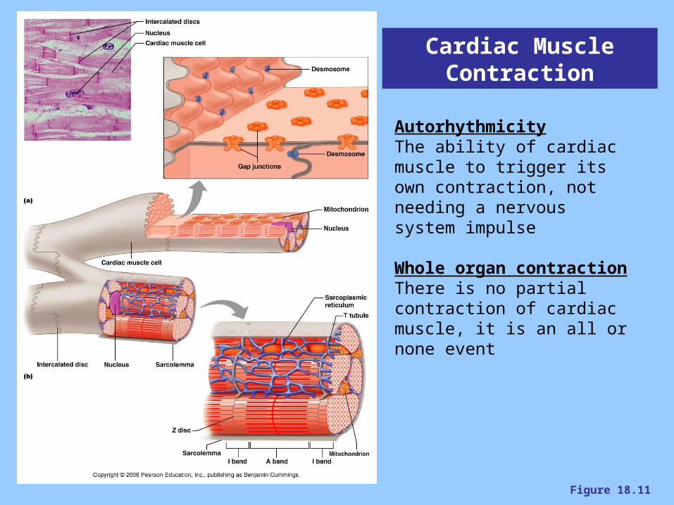
Figure 18.11
Cardiac Muscle Contraction
AutorhythmicityThe ability of cardiac muscle to trigger its own contraction, not needing a nervous system impulse
Whole organ contractionThere is no partial contraction of cardiac muscle, it is an all or none event
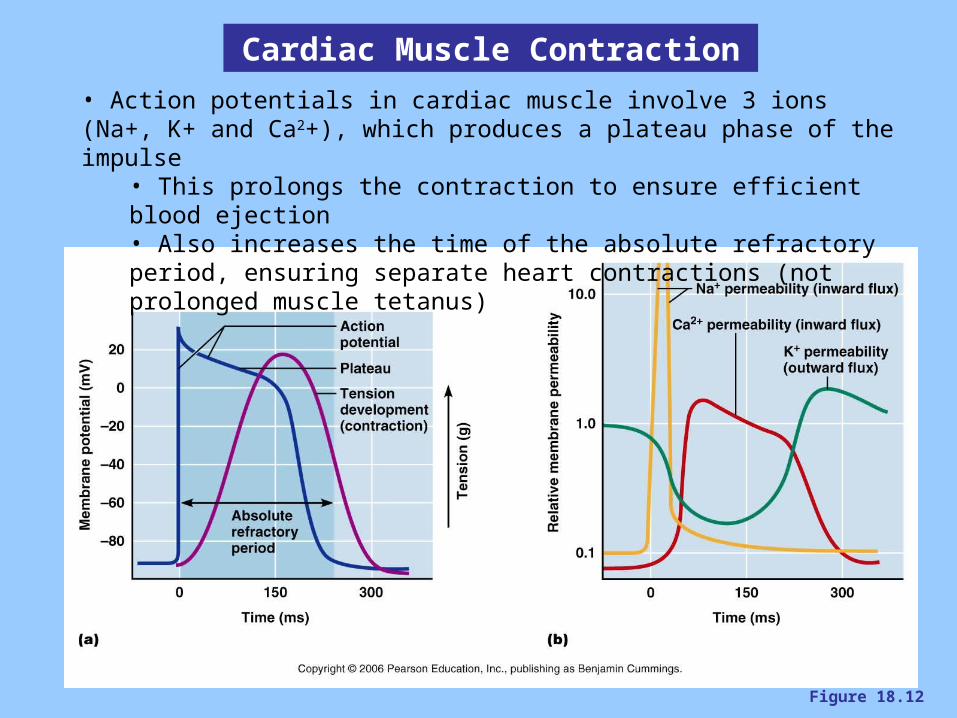
Figure 18.12
Cardiac Muscle Contraction
• Action potentials in cardiac muscle involve 3 ions (Na+, K+ and Ca2+), which produces a plateau phase of the impulse
• This prolongs the contraction to ensure efficient blood ejection• Also increases the time of the absolute refractory period, ensuring separate heart contractions (not prolonged muscle tetanus)
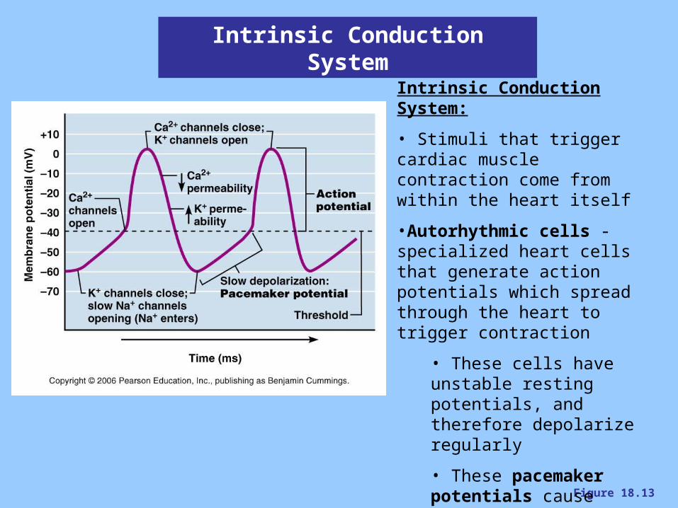
Figure 18.13
Intrinsic Conduction System
Intrinsic Conduction System:
• Stimuli that trigger cardiac muscle contraction come from within the heart itself
•Autorhythmic cells - specialized heart cells that generate action potentials which spread through the heart to trigger contraction
• These cells have unstable resting potentials, and therefore depolarize regularly
• These pacemaker potentials cause action potentials in the cardiac muscle fibers, triggering contraction

Figure 18.14
Intrinsic Conduction System
Sinoatrial node – in the right atrium, the “pacemaker” whose cells generate the sinus rhythm of 75 beats/minAtrioventricular node - causes a delay of .1sec to allow atrial contraction before ventricular contraction. Rhythm is 40-60 beats/minBundle of His – pathway into the interventricular septum
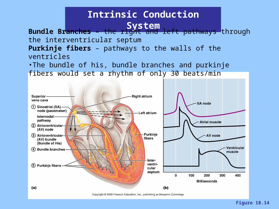
Figure 18.14
Intrinsic Conduction System
Bundle Branches – the right and left pathways through the interventricular septumPurkinje fibers – pathways to the walls of the ventricles•The bundle of his, bundle branches and purkinje fibers would set a rhythm of only 30 beats/min
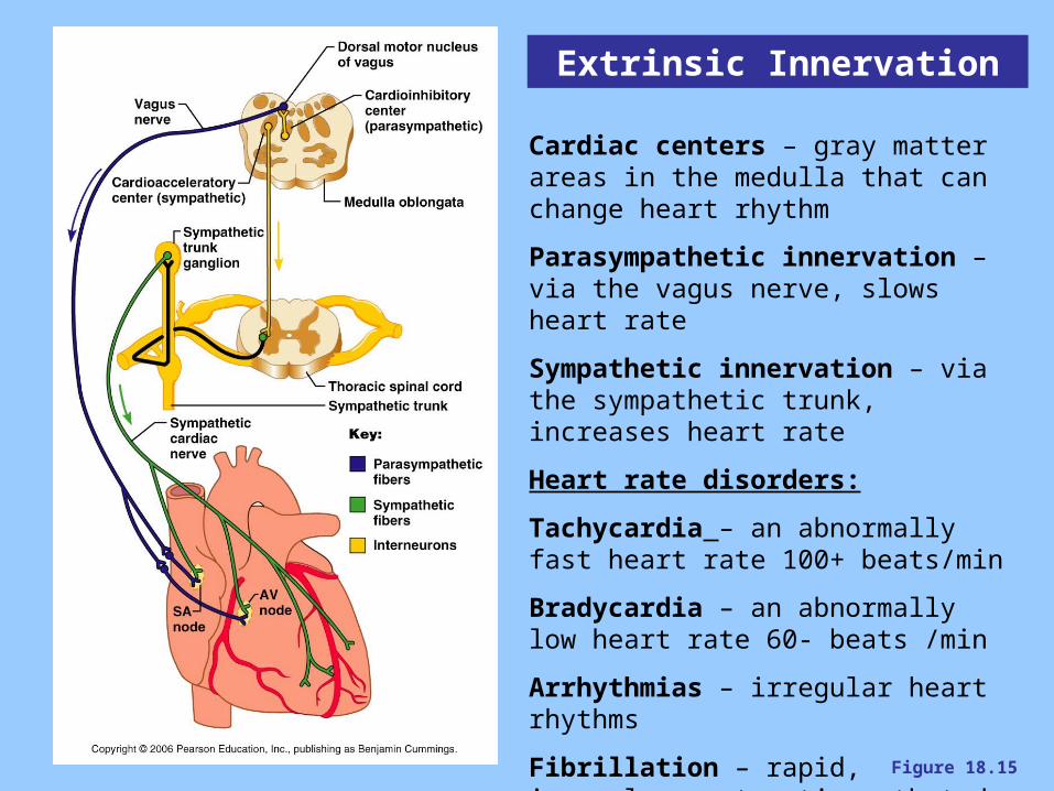
Figure 18.15
Extrinsic Innervation
Cardiac centers – gray matter areas in the medulla that can change heart rhythm
Parasympathetic innervation – via the vagus nerve, slows heart rate
Sympathetic innervation – via the sympathetic trunk, increases heart rate
Heart rate disorders:
Tachycardia – an abnormally fast heart rate 100+ beats/min
Bradycardia – an abnormally low heart rate 60- beats /min
Arrhythmias – irregular heart rhythms
Fibrillation – rapid, irregular contractions that do not function to pump blood
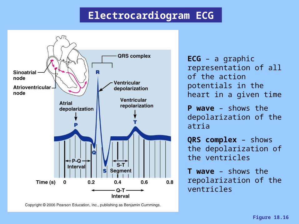
Figure 18.16
Electrocardiogram ECG
ECG – a graphic representation of all of the action potentials in the heart in a given time
P wave – shows the depolarization of the atria
QRS complex – shows the depolarization of the ventricles
T wave – shows the repolarization of the ventricles
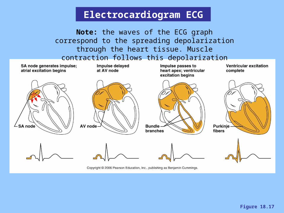
Figure 18.17
Electrocardiogram ECG
Note: the waves of the ECG graph correspond to the spreading depolarization through the heart tissue.
Muscle contraction follows this depolarization

Figure 18.18
Electrocardiogram ECG
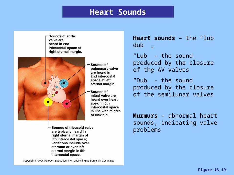
Figure 18.19
Heart Sounds
Heart sounds – the “lub dub”
“Lub” – the sound produced by the closure of the AV valves
“Dub” – the sound produced by the closure of the semilunar valves
Murmurs – abnormal heart sounds, indicating valve problems
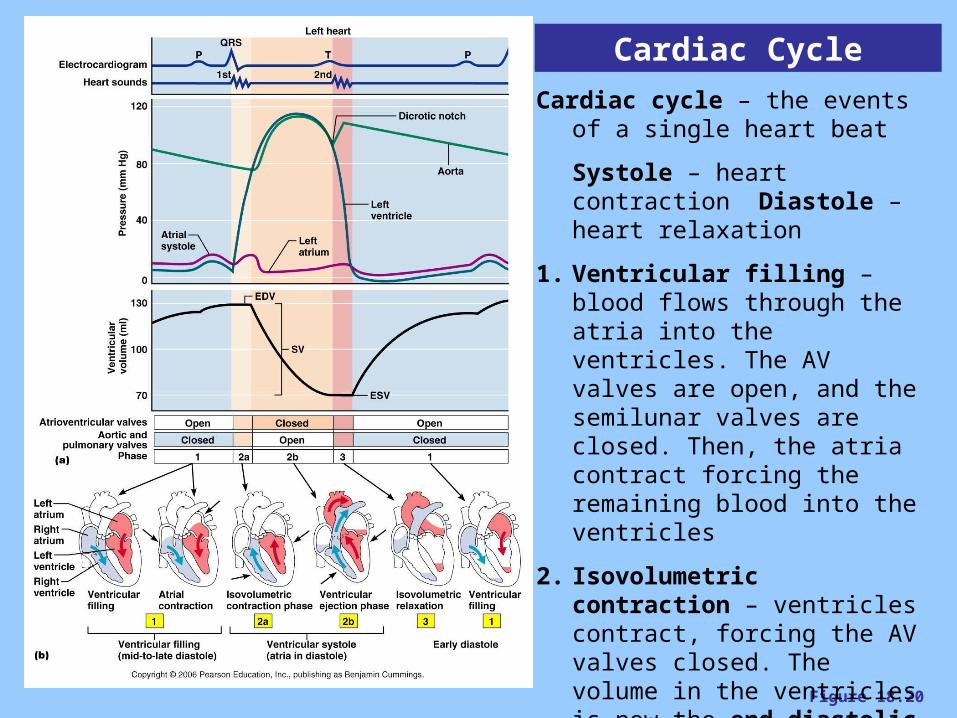
Figure 18.20
Cardiac Cycle
Cardiac cycle – the events of a single heart beat
Systole – heart contraction Diastole – heart relaxation
1. Ventricular filling – blood flows through the atria into the ventricles. The AV valves are open, and the semilunar valves are closed. Then, the atria contract forcing the remaining blood into the ventricles
2. Isovolumetric contraction – ventricles contract, forcing the AV valves closed. The volume in the ventricles is now the end diastolic volume (EDV)
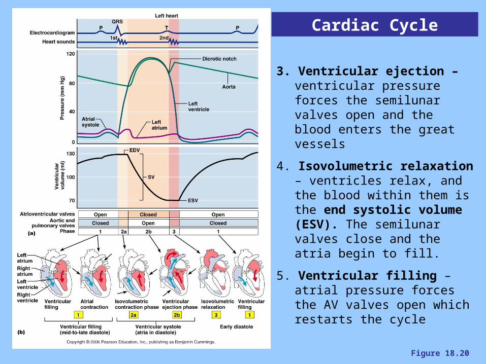
Figure 18.20
Cardiac Cycle
3. Ventricular ejection – ventricular pressure forces the semilunar valves open and the blood enters the great vessels
4. Isovolumetric relaxation – ventricles relax, and the blood within them is the end systolic volume (ESV). The semilunar valves close and the atria begin to fill.
5. Ventricular filling – atrial pressure forces the AV valves open which restarts the cycle
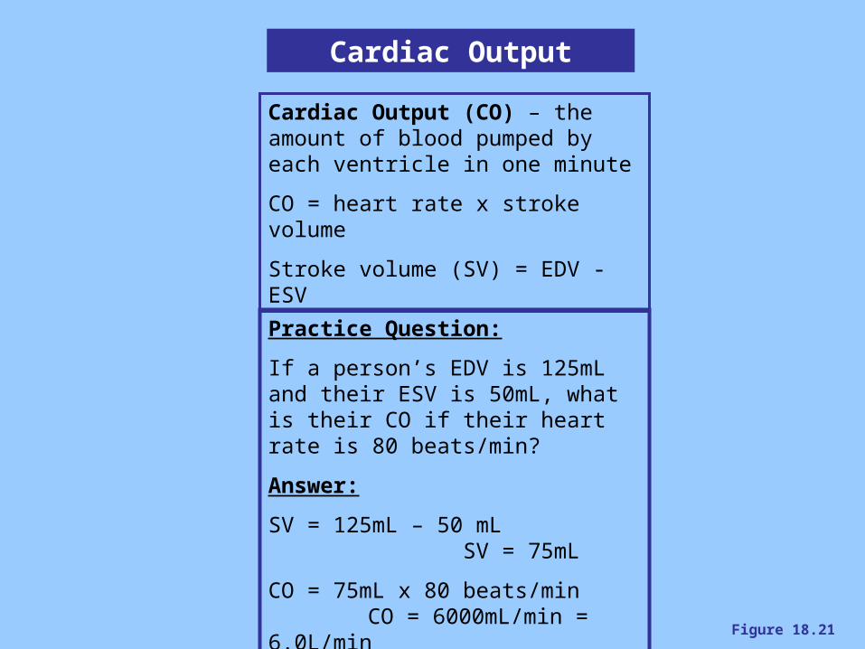
Figure 18.21
Cardiac Output
Cardiac Output (CO) – the amount of blood pumped by each ventricle in one minute
CO = heart rate x stroke volume
Stroke volume (SV) = EDV - ESV
Practice Question:
If a person’s EDV is 125mL and their ESV is 50mL, what is their CO if their heart rate is 80 beats/min?
Answer:
SV = 125mL – 50 mL SV = 75mL
CO = 75mL x 80 beats/min CO = 6000mL/min = 6.0L/min
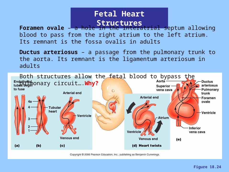
Figure 18.24
Fetal Heart Structures
Foramen ovale – a hole in the interatrial septum allowing blood to pass from the right atrium to the left atrium. Its remnant is the fossa ovalis in adults
Ductus arteriosus – a passage from the pulmonary trunk to the aorta. Its remnant is the ligamentum arteriosum in adults
Both structures allow the fetal blood to bypass the pulmonary circuit….Why?