Embed Size (px)
Citation preview

Lecture Presentation by
Lori Garrett
18The Heart and
Cardiovascular
Function
© 2018 Pearson Education, Inc.

Section 1: Structure of the Heart
Learning Outcomes
18.1 Describe the heart’s location, shape, its four
chambers, and the pulmonary and systemic
circuits.
18.2 Describe the location and general features of
the heart.
18.3 Describe the structure of the pericardium and
explain its functions, identify the layers of the
heart wall, and describe the structures and
functions of cardiac muscle.
18.4 Describe the cardiac chambers and the heart’s
external anatomy.
© 2018 Pearson Education, Inc.

Section 1: Structure of the Heart
Learning Outcomes (continued)
18.5 Describe the major vessels supplying the heart,
and cite their locations.
18.6 Trace blood flow through the heart, identifying
the major blood vessels, chambers, and heart
valves.
18.7 Describe the relationship between the AV and
semilunar valves during a heartbeat.
18.8 Define arteriosclerosis, and explain its
significance to health.
© 2018 Pearson Education, Inc.

Module 18.1: The heart has four chambers that pump and circulate blood through the pulmonary and systemic circuits
Cardiovascular system = heart and blood vessels
transporting blood
Heart—directly behind sternum
Base—superior
• where major vessels are
• ~1.2 cm (0.5 in.) to left
• 3rd costal cartilage
Apex—inferior, pointed tip
• ~12.5 cm (5 in.) from base
• ~7.5 cm (3 in.) to left
• 5th intercostal space © 2018 Pearson Education, Inc.

Borders of the heart
© 2018 Pearson Education, Inc.

Module 18.1: Heart location and chambers
Heart = 2-sided pump with 4 chambers
Right atrium receives blood from systemic circuit
Right ventricle pumps blood into pulmonary circuit
Left atrium receives blood from pulmonary circuit
Left ventricle pumps blood into systemic circuit
© 2018 Pearson Education, Inc.

Module 18.1: Review
A. Describe the location and position of the heart.
B. Compare the base of the heart with the apex.
C. Name the four chambers of the heart.
D. Compare the volume of blood each circuit
receives from contraction of the ventricles.
Learning Outcome: Describe the heart’s location,
shape, its four chambers, and the pulmonary and
systemic circuits.
© 2018 Pearson Education, Inc.

Module 18.2: The heart is located in the mediastinum and is enclosed by the pericardial cavity
Mediastinum = space or region in thorax between
the two pleural cavities (between the lungs)
© 2018 Pearson Education, Inc.

Module 18.2: The pericardium
Pericardium = sac-like structure wrapped around
heart
Fibrous pericardium: Outermost layer; dense fibrous
tissue that extends to sternum and diaphragm
Serous pericardium (2 layers): Outer parietal layer
lines fibrous pericardium; inner serous layer covers
surface of the heart
Pericardial cavity: Space between serous layers;
contains 15–50 mL of pericardial fluid secreted
from serous membranes; lubricates movement of
heart.
© 2018 Pearson Education, Inc.

Module 18.2: The pericardium
Pericardium = sac-like structure wrapped around
heart (continued)
Pericarditis = inflammation of the pericardium
Cardiac tamponade = excess accumulation of
pericardial fluid
© 2018 Pearson Education, Inc.

Module 18.2: The pericardium
Relationship between heart and pericardium
Push fist into partly inflated balloon
Fist = heart
Wrist = base of heart, with great vessels
Inside of balloon = pericardial cavity
© 2018 Pearson Education, Inc.

Module 18.2: The pericardium
Superior view of the thorax, showing the positions of the
pericardium, pericardial cavity, heart, mediastinum, and three of
the great vessels: aorta, superior vena cava, and pulmonary
trunk.
© 2018 Pearson Education, Inc.

Module 18.2: Review
A. Define mediastinum.
B. Describe the heart’s location in the body.
C. Why can cardiac tamponade be a life-
threatening condition?
Learning Outcome: Describe the location and
general features of the heart.
© 2018 Pearson Education, Inc.

Module 18.3: The heart wall contains concentric layers of cardiac muscle tissue
Heart wall has 3 layers:
1. Pericardium—outer fibrous pericardium (dense
fibrous tissue) and 2-layered serous pericardium
(mesothelium and underlying areolar tissue)
• Epicardium = visceral layer of serous pericardium
2. Myocardium—middle layer; concentric layers of
cardiac muscle; supporting blood vessels, nerves
3. Endocardium—(endo = within); innermost layer;
simple squamous epithelium (renamed
endothelium inside heart and vessels) and areolar
tissue; endocardium is continuous with
endothelium in vessels and covers heart valves
© 2018 Pearson Education, Inc.

The layers of the heart wall
© 2018 Pearson Education, Inc.

Module 18.3: The heart wall
Myocardium arrangement:
Atrial musculature wraps around atria in figure-8
pattern
Ventricular musculature
• Superficial layer surrounds
both ventricles
• Deeper layers spiral
around and between
the ventricles
© 2018 Pearson Education, Inc.

Module 18.3: The heart wall—cardiac muscle
Cardiac muscle vs. skeletal muscle:
1. Smaller cell size (avg. 10–20 μm diameter; 50–100 μm length)
2. Single, centrally located nucleus
3. Intercalated discs = branching interconnections between cells
4. Specialized intercellular connections
© 2018 Pearson Education, Inc.

Module 18.3: The heart wall—cardiac muscle
Cardiac muscle tissue
Only in heart
Striated (from organized myofibrils and aligned
sarcomeres)
Almost totally dependent on
aerobic metabolism
(need oxygen) for energy
• Abundant mitochondria
and myoglobin
(stores O2)
• Extensive
capillaries
© 2018 Pearson Education, Inc.

Module 18.3: The heart wall—cardiac muscle
Cardiac muscle tissue (continued)
Intercalated discs
• Intertwined plasma
membranes of
adjacent cardiac
muscle cells
• Attached by desmosomes
and gap junctions
• Gap junctions allow action
potentials to spread cell to cell; allows all
interconnected cells to function together as single
unit = a functional syncytium
© 2018 Pearson Education, Inc.

Module 18.3: Review
A. From superficial to deep, name the layers of the
heart wall.
B. Describe the tissue layers of the pericardium.
C. Why is it important that cardiac tissue contain
many mitochondria and capillaries?
Learning Outcome: Describe the structure of the
pericardium and explain its functions, identify the
layers of the heart wall, and describe the structures
and functions of cardiac muscle.
© 2018 Pearson Education, Inc.

Module 18.4: The boundaries between the four chambers of the heart can be identified on its external surface
Visible on anterior surface
All four chambers
Auricle of each atrium (expandable pouch)
Coronary sulcus—groove separating atria and
ventricles
Anterior interventricular sulcus—groove marking
boundary between the two ventricles
Ligamentum arteriosum—fibrous remnant of fetal
connection between aorta and pulmonary trunk
© 2018 Pearson Education, Inc.

Surface anatomy of the heart (anterior view)
© 2018 Pearson Education, Inc.

Cadaver heart (anterior view)
© 2018 Pearson Education, Inc.

Module 18.4: Heart surface anatomy
Visible on posterior surface:
All four chambers
Pulmonary veins (4) returning blood to left atrium
Superior and inferior venae cavae returning blood to
right atrium
Coronary sinus—returns blood from myocardium to
right atrium
Posterior interventricular sulcus—groove marking
boundary between the two ventricles
© 2018 Pearson Education, Inc.

Surface anatomy of the heart (posterior view)
© 2018 Pearson Education, Inc.

Module 18.4: Review
A. The anterior view of the heart is dominated by
which chambers?
B. Which structures collect blood from the
myocardium, and into which heart chamber
does this blood flow?
C. Name and describe the shallow depressions
and grooves found on the heart’s external
surface.
Learning Outcome: Describe the cardiac chambers
and the heart’s external anatomy.
© 2018 Pearson Education, Inc.

Module 18.5: The heart has an extensive blood supply
Coronary circulation
Continuously supplies cardiac muscle (myocardium)
with oxygen/nutrients
Left and right coronary arteries
arise from ascending aorta;
fill when ventricles are
relaxed (diastole)
Myocardial blood
flow may increase
to 9 times the resting
level during maximal
exertion
© 2018 Pearson Education, Inc.

Module 18.5: Coronary circulation
Right coronary artery
Supplies right atrium, parts of both ventricles, and
parts of cardiac (electrical) conducting system
Follows coronary sulcus (groove between atria and
ventricles)
Main branches:
• Marginal arteries—supply right ventricle
• Posterior interventricular (posterior descending)
artery—runs in posterior interventricular sulcus;
supplies interventricular septum and adjacent parts of
ventricles
© 2018 Pearson Education, Inc.

Module 18.5: Coronary circulation
Left coronary artery
Supplies left ventricle, left atrium, interventricular
septum
Main Branches:
• Anterior interventricular artery (left anterior
descending artery)—follows anterior interventricular
sulcus; supplies interventricular septum and adjacent
parts of ventricles
• Circumflex artery—follows coronary sulcus to the
left; meets branches of right coronary artery
posteriorly; marginal artery off circumflex supplies
posterior of left ventricle
© 2018 Pearson Education, Inc.

Coronary circulation, main arteries (anterior view)
© 2018 Pearson Education, Inc.

Coronary circulation, main arteries (posterior view)
© 2018 Pearson Education, Inc.

Module 18.5: Coronary circulation
Coronary circulation—veins (anterior)
Great cardiac vein—in anterior interventricular
sulcus
• Drains area supplied by anterior interventricular artery
• Empties into coronary
sinus posteriorly
Anterior cardiac veins
• Drain anterior surface
of right ventricle
• Empty directly
into the right
atrium
© 2018 Pearson Education, Inc.

Module 18.5: Coronary circulation
Coronary circulation—veins (posterior)
Coronary sinus—expanded vein that empties into
right atrium
Posterior vein of left ventricle—drains area
supplied by circumflex artery
Middle cardiac vein—drains area supplied by
posterior interventricular artery; empties into
coronary sinus
Small cardiac vein—
drains posterior of right
atrium/ventricle; empties
into coronary sinus
© 2018 Pearson Education, Inc.
Module 18.5: Coronary circulation
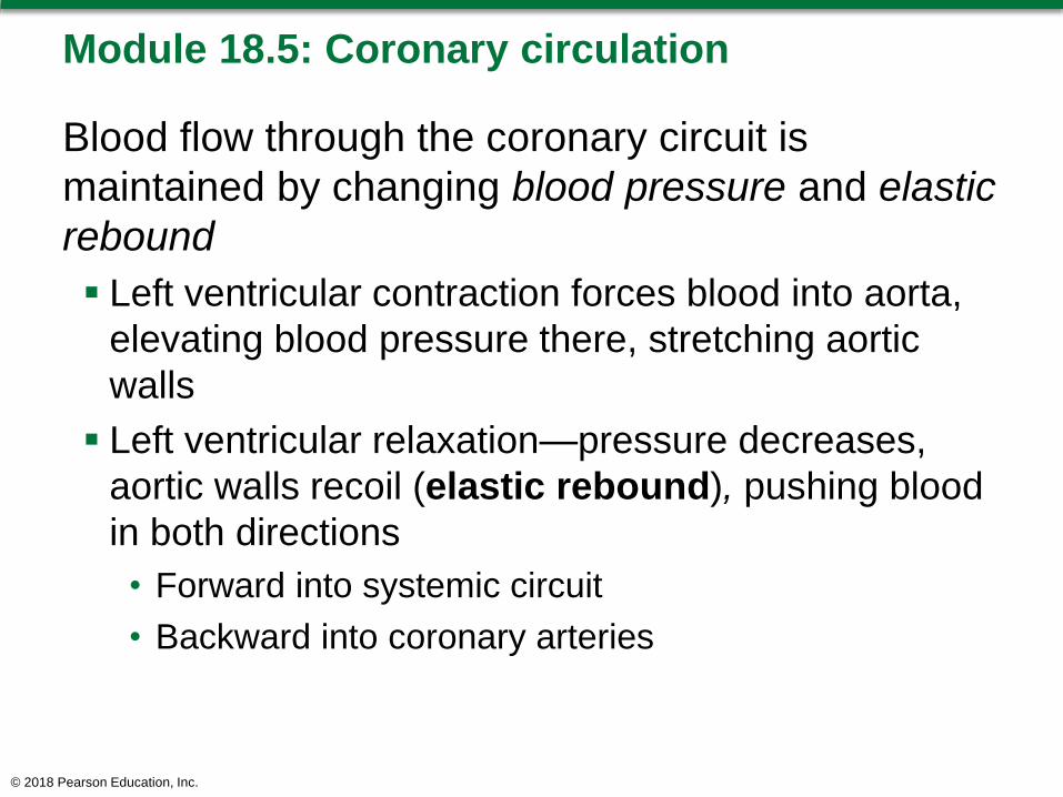
Blood flow through the coronary circuit is
maintained by changing blood pressure and elastic
rebound
Left ventricular contraction forces blood into aorta,
elevating blood pressure there, stretching aortic
walls
Left ventricular relaxation—pressure decreases,
aortic walls recoil (elastic rebound), pushing blood
in both directions
• Forward into systemic circuit
• Backward into coronary arteries
© 2018 Pearson Education, Inc.

Module 18.5: Review
A. Describe the areas of the heart supplied by the
right and left coronary arteries.
B. Compare the anterior cardiac veins to the
posterior vein of the left ventricle.
C. List the arteries and veins of the heart.
D. Describe what happens to blood flow during
elastic rebound.
Learning Outcome: Describe the major vessels
supplying the heart, and cite their locations.
© 2018 Pearson Education, Inc.

Module 18.6: Internal valves control the direction of blood flow between the heart chambers and great vessels
Each side of heart has two chambers: an atrium
(receives blood) sends blood to a ventricle (pumps
blood out of heart)
Right and left atria are separated by the interatrial
septum
Right and left ventricles are separated by
interventricular septum (much thicker)
© 2018 Pearson Education, Inc.

Module 18.6: Internal valves control the direction of blood flow between the heart chambers and great vessels
Atrioventricular (AV) valves—between each
atrium and ventricle
• Allow only one-way blood flow from atrium into
ventricle
Semilunar valves—at exit from each ventricle;
allow only one-way blood flow from ventricle out into
aorta or pulmonary trunk
© 2018 Pearson Education, Inc.

Internal anatomy of the heart, showing chambers and heart valves (coronal section)
© 2018 Pearson Education, Inc.

Module 18.6: Heart valves
Atria
Right atrium receives deoxygenated blood from
superior and inferior venae cavae and coronary
sinus
• Fossa ovalis—remnant of fetal foramen ovale that
allowed fetal blood to pass between atria; closes at
birth
Left atrium receives oxygenated blood from
pulmonary veins
Pectinate muscles—muscular ridges located inside
both atria along the anterior atrial wall and in the
auricles
© 2018 Pearson Education, Inc.

Module 18.6: Heart valves
Ventricles
Right ventricle—receives blood from right atrium
through tricuspid valve (has three cusps or flaps),
also called the right atrioventricular (AV) valve
• With contraction, blood exits through the pulmonary
valve (pulmonary semilunar valve) into the pulmonary
trunk
© 2018 Pearson Education, Inc.
Module 18.6: Heart valves
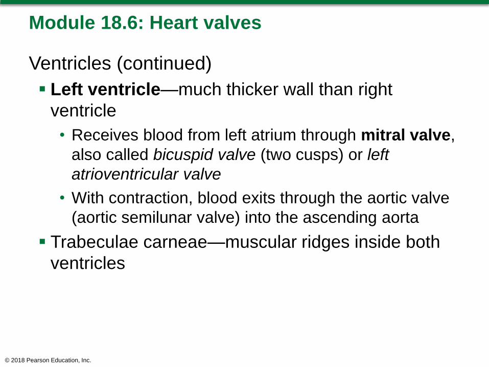
Ventricles (continued)
Left ventricle—much thicker wall than right
ventricle
• Receives blood from left atrium through mitral valve,
also called bicuspid valve (two cusps) or left
atrioventricular valve
• With contraction, blood exits through the aortic valve
(aortic semilunar valve) into the ascending aorta
Trabeculae carneae—muscular ridges inside both
ventricles
© 2018 Pearson Education, Inc.

Module 18.6: Heart valves
AV valve structure (tricuspid and mitral valve)
Each has three (tricuspid) or two (mitral/bicuspid)
cusps
Cusps attach to tendon-like connective tissue bands
= chordae tendineae
Chordae tendineae anchored to thickened cone-
shaped papillary muscles
Moderator band—thickened muscle ridge providing
rapid conduction path; tenses papillary muscles just
before ventricular contraction; prevents slamming or
inversion of AV valve
© 2018 Pearson Education, Inc.

Internal anatomy of the heart (coronal section)
© 2018 Pearson Education, Inc.
Module 18.6: Heart valves
Comparison between chambers
Atria have similar workloads; walls about same
thickness
Ventricles have very different loads
• Right ventricle—thinner wall; sends blood to adjacent
lungs (pulmonary circuit)
– Contraction squeezes against left ventricle, forces
blood out pulmonary trunk efficiently; minimal effort, low
pressure
© 2018 Pearson Education, Inc.

Module 18.6: Heart valves
Comparison between chambers (continued)
Ventricles have very different loads (continued)
• Left ventricle—very thick wall, rounded chamber
– 4–6 times the pressure of right; sends blood through
entire systemic circuit
– Contraction decreases diameter and apex-to-base
distance
– Reduces right ventricular volume, aiding its emptying
© 2018 Pearson Education, Inc.

Module 18.6: Heart valves
© 2018 Pearson Education, Inc.

Module 18.6: Heart valves
© 2018 Pearson Education, Inc.
The wall of the left ventricle is much
thicker than that of the right ventricle
because it must generate tremendous
force.
Note the shape and change
in size of both ventricles
when they contract.
Module 18.6: Review
A. Why is the left ventricle more muscular than the
right ventricle?
B. Damage to the semilunar valve on the right side
of the heart would affect blood flow to which
vessel?
Learning Outcome: Trace blood flow through the
heart, identifying the major blood vessels,
chambers, and heart valves.
© 2018 Pearson Education, Inc.

Module 18.7: When the heart beats, the AV valves close before the semilunar valves open, and the semilunar valves close before the AV valves open
When ventricles are relaxed, they fill
AV valves—open
• Chordae tendineae
are loose
Semilunar valves—
closed
• Blood pressure
from pulmonary
and systemic circuits
keeps them closed
© 2018 Pearson Education, Inc.

Position of heart valves while ventricles are filling (ventricular relaxation)
© 2018 Pearson Education, Inc.

Module 18.7: Valves control direction of flow
When ventricles contract, they empty
AV valves—closed
• Pressure from contracting
ventricles pushes cusps
together
• Papillary muscles tighten
chordae tendineae so
cusps can’t invert into
atria; prevents
backflow (regurgitation)
Semilunar valves—open
• Ventricular pressure overcomes pressure in
pulmonary trunk and aorta that held them shut
© 2018 Pearson Education, Inc.

© 2018 Pearson Education, Inc.
Position of heart valves while ventricles are emptying (ventricular contraction)

Module 18.6: Valves control direction of flow
Cardiac skeleton (fibrous skeleton)
Flexible connective tissue frame
• Interconnected bands of dense connective tissue
• Encircle heart valves, stabilize their positions
• Also surrounds base of aorta and pulmonary trunk
Electrically isolates
atrial from ventricular
myocardium
© 2018 Pearson Education, Inc.

Module 18.7: Valves control direction of flow
Pulmonary and aortic (semilunar) valves
Each has three half-moon shaped cusps
• Prevent backflow of blood from aorta and pulmonary
trunk back into ventricles
• No muscular brace needed—cusps support each
other when closed
© 2018 Pearson Education, Inc.

Module 18.7: Valves control direction of flow
Valvular heart disease (VHD)
Valve function deteriorates until heart cannot maintain adequate blood flow
Congenital malformations or heart inflammation (carditis)
Severe cases may require replacement with prosthetic valve
• Bioprosthetic valves come from pigs or cows
© 2018 Pearson Education, Inc.
Damaged aortic valve with
irregular-shaped cusps Bioprosthetic valve

Module 18.7: Review
A. Define cardiac regurgitation.
B. Describe the structural and functional roles of
the cardiac skeleton.
C. What do semilunar valves prevent?
Learning Outcome: Describe the relationship
between the AV and semilunar valves during a
heartbeat.
© 2018 Pearson Education, Inc.

Module 18.8: Arteriosclerosis can lead to coronary artery disease
Arteriosclerosis (arterio-, artery + sclerosis,
hardness)
Thickening/toughening of arterial walls
Related complications account for about half of U.S.
deaths
• Coronary artery disease (CAD) = arteriosclerosis of
coronary vessels
• Arteriosclerosis of brain arteries can lead to strokes
© 2018 Pearson Education, Inc.

Module 18.8: Arteriosclerosis
Atherosclerosis
= formation of lipid
deposits in arterial tunica
media and damage to
endothelium
Most common form of
arteriosclerosis; often
associated with elevated
blood cholesterol
Fatty tissue mass
(plaque) in vessel;
restricts blood flow
© 2018 Pearson Education, Inc.
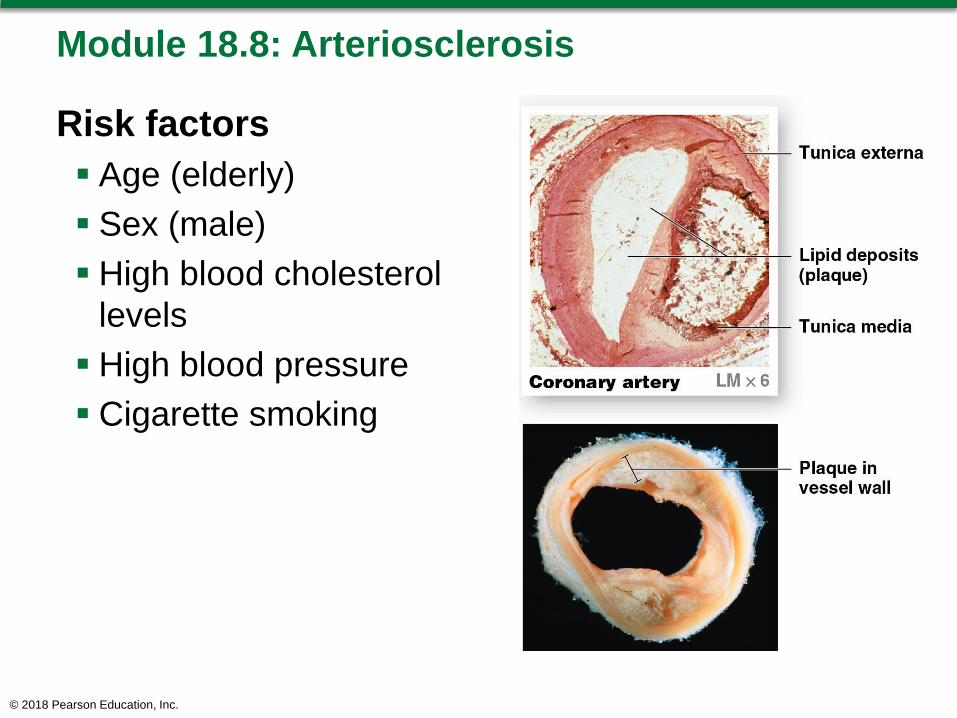
Module 18.8: Arteriosclerosis
Risk factors
Age (elderly)
Sex (male)
High blood cholesterol
levels
High blood pressure
Cigarette smoking
© 2018 Pearson Education, Inc.

Module 18.8: Arteriosclerosis
Atherosclerosis treatment
Replace damaged segment of the vessel
Compressing plaque with balloon angioplasty
• Catheter inserted past blockage; balloon inflated to
press plaque against wall and open vessel
• Most effective for small, soft plaques
• Very low surgical mortality rate (about 1%)
• Very high success
rate (>90%)
• Can be outpatient
© 2018 Pearson Education, Inc.

Module 18.8: Arteriosclerosis
Coronary artery disease (CAD)
= Areas of partial or complete blockage of coronary
circulation
• Reduces blood flow to area (coronary ischemia)
• Usually from atherosclerosis in a coronary artery or
associated blood clot (thrombus)
• Seen in digital subtraction angiography (DSA)
• May treat with wire-mesh tube (stent) to hold vessel
open
© 2018 Pearson Education, Inc.

Module 18.8: Arteriosclerosis
© 2018 Pearson Education, Inc.

Module 18.8: Arteriosclerosis
© 2018 Pearson Education, Inc.

Module 18.8: Review
A. Compare arteriosclerosis with atherosclerosis.
B. What is coronary ischemia?
C. Describe the purpose of a stent.
Learning Outcome: Define arteriosclerosis, and
explain its significance to health.
© 2018 Pearson Education, Inc.

Section 2: Cardiac Cycle
Learning Outcomes
18.9 Explain the complete round of cardiac
systole and diastole.
18.10 Explain the events of the cardiac cycle, and
relate the heart sounds to specific events.
18.11 Describe an action potential in cardiac
muscle, and explain the role of calcium ions.
18.12 Describe the components and functions of
the conducting system of the heart.
© 2018 Pearson Education, Inc.

Section 2: Cardiac Cycle
Learning Outcomes (continued)
18.13 Identify the electrical events shown on an
electrocardiogram.
18.14 Describe the factors affecting the heart rate.
18.15 Describe the variables that influence stroke
volume.
18.16 Explain how stroke volume and cardiac
output are coordinated.
© 2018 Pearson Education, Inc.
Module 18.9: The cardiac cycle is a complete round of systole and diastole
Cardiac cycle = period between start of one
heartbeat and the next; heart rate = number of
beats per minute
Two atria contract first to fill ventricles; two ventricles
then contract to pump blood into pulmonary
and systemic circuits
© 2018 Pearson Education, Inc.

Module 18.9: The cardiac cycle is a complete round of systole and diastole
Cardiac cycle (continued)
Two phases:
• Contraction (systole)—blood leaves the chamber
• Relaxation (diastole)—chamber refills
© 2018 Pearson Education, Inc.

Module 18.9: The cardiac cycle
Sequence of contractions
1. Atria contract together first (atrial systole)
• Push blood into the ventricles
• Ventricles are relaxed (diastole) and filling
2. Ventricles contract together next (ventricular systole)
• Push blood into the pulmonary and systemic circuits
• Atria are relaxed (diastole) and filling
Typical cardiac cycle lasts 800 msec
© 2018 Pearson Education, Inc.

Phases of cardiac cycle for a heart rate of 75 bpm
© 2018 Pearson Education, Inc.

Module 18.9: Review
A. Define cardiac cycle.
B. Give the alternate terms for heart contraction
and heart relaxation.
C. Compare the duration of atrial and ventricular
systole at a representative heart rate of 75 bpm.
Learning Outcome: Explain the complete round of
cardiac systole and diastole.
© 2018 Pearson Education, Inc.

Module 18.10: The cardiac cycle creates pressure gradients that maintain blood flow
Phases of cardiac cycle, diagrammed for heart
rate of 75 bpm
1. Cardiac cycle begins—all four chambers are
relaxed (diastole; ventricles are passively refilling)
© 2018 Pearson Education, Inc.

Module 18.10: The cardiac cycle creates pressure gradients that maintain blood flow
Phases of cardiac cycle (continued)
2. Atrial systole (100 msec)—atria contract; finish
filling ventricles
© 2018 Pearson Education, Inc.

Module 18.10: The cardiac cycle creates pressure gradients that maintain blood flow
Phases of cardiac cycle (continued)
3. Atrial diastole (270 msec)—continues until start
of next cardiac cycle (through ventricular systole)
© 2018 Pearson Education, Inc.

Module 18.10: The cardiac cycle creates pressure gradients that maintain blood flow
Phases of cardiac cycle (continued)
4. Ventricular systole—first phase. Contracting
ventricles push AV valves closed but not enough
pressure to open semilunar valves
(= isovolumetric
contraction—no
volume change)
© 2018 Pearson Education, Inc.
Module 18.10: The cardiac cycle creates pressure gradients that maintain blood flow
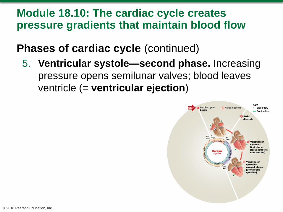
Phases of cardiac cycle (continued)
5. Ventricular systole—second phase. Increasing
pressure opens semilunar valves; blood leaves
ventricle (= ventricular ejection)
© 2018 Pearson Education, Inc.

Module 18.10: The cardiac cycle creates pressure gradients that maintain blood flow
Phases of cardiac cycle (continued)
6. Ventricular diastole—early. Ventricles relax
and their pressure drops; blood in aorta and
pulmonary trunk backflows, closes semilunar
valves
© 2018 Pearson Education, Inc.

Module 18.10: The cardiac cycle creates pressure gradients that maintain blood flow
Phases of cardiac cycle (continued)
7. Isovolumetric relaxation. All valves closed; no
volume change; blood passively filling atria
© 2018 Pearson Education, Inc.

Module 18.10: The cardiac cycle creates pressure gradients that maintain blood flow
Phases of cardiac cycle (continued)
8. Ventricular diastole—late. All chambers relaxed;
AV valves open; ventricles fill passively to ~70%
© 2018 Pearson Education, Inc.

SmartArt Video: The Cardiac Cycle
© 2018 Pearson Education, Inc.

Module 18.10: The cardiac cycle creates pressure gradients that maintain blood flow
Pressure changes in aorta during the cardiac
cycle
Increase in pressure with opening of aortic valve
Drop in pressure with closing of aortic valve
• Followed by a short pressure rise as aortic elastic
walls recoil
• Produces a valley in pressure tracing called the
dicrotic notch (dikrotos, double beating)
© 2018 Pearson Education, Inc.
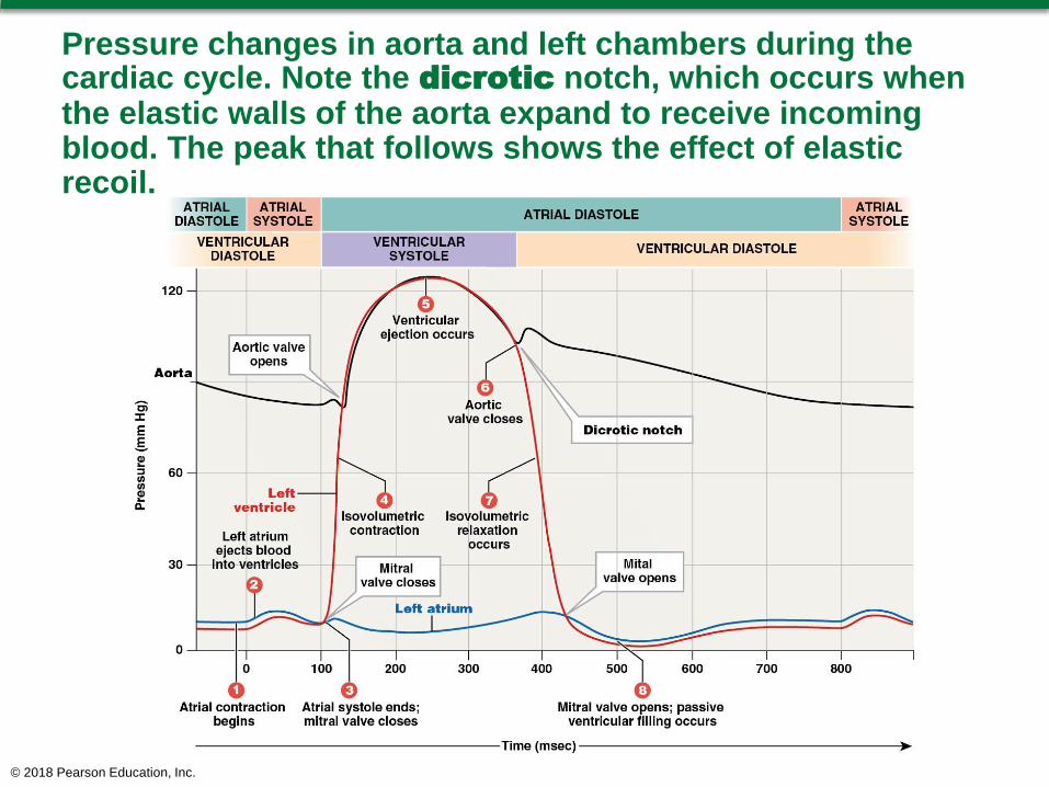
Pressure changes in aorta and left chambers during the cardiac cycle. Note the dicrotic notch, which occurs when the elastic walls of the aorta expand to receive incoming blood. The peak that follows shows the effect of elastic recoil.
© 2018 Pearson Education, Inc.

Module 18.10: The cardiac cycle creates pressure gradients that maintain blood flow
Heart sounds
S1 ( “lubb”)—when AV valves close; marks start of
ventricular contraction
S2 (“dupp”)—when semilunar valves close
S3 and S4—very faint; rarely heard in adults
• S3—blood flowing into ventricles
• S4—atrial contraction
© 2018 Pearson Education, Inc.

Module 18.10: Review
A. List the phases of the cardiac cycle.
B. What are the two phases of ventricular systole?
C. Is the heart always pumping blood when
pressure in the left ventricle is rising? Explain.
Learning Outcome: Explain the events of the
cardiac cycle, and relate the heart sounds to
specific events.
© 2018 Pearson Education, Inc.

Module 18.11: Cardiac muscle cell contractions last longer than skeletal muscle fiber contractions primarily because of differences in calcium ion membrane permeability
© 2018 Pearson Education, Inc.
Skeletal Muscle Cardiac Muscle
• Brief action potential; ends
as short twitch contraction
begins.
• Contraction ends when
sarcoplasmic reticulum
reclaims Ca2+
• Short refractory period ends
before peak tension
develops
• Twitches can summate;
tetanus can occur
• Long action potential
• Ca2+ enters cells over
prolonged period
• Long contraction
(~250 msec)
• Refractory period continues
into relaxation
• No tetanic contractions
occur (heart couldn’t pump
blood)

Skeletal Muscle
Skeletal muscle fiber contraction
© 2018 Pearson Education, Inc.

Skeletal Muscle
Cardiac muscle cell contraction
© 2018 Pearson Education, Inc.

Module 18.11: Cardiac muscle cell contraction
Three stages of a cardiac muscle action
potential
1. Rapid depolarization
2. Plateau
3. Repolarization
© 2018 Pearson Education, Inc.

Module 18.11: Cardiac muscle cell contraction
1. Rapid depolarization—similar to that in
skeletal muscle
At threshold, voltage-gated fast sodium channels
open
Massive, rapid Na+ influx
Channels
open quickly
and very
briefly
© 2018 Pearson Education, Inc.

Module 18.11: Cardiac muscle cell contraction
2. Plateau
Membrane potential stays near 0 mV due to 2
opposing factors:
• Fast sodium channels close as potential nears +30
mV
• Cell actively pumps Na+ out
Voltage-gated slow calcium channels open—
Ca2+ influx (open
slowly/stay open
~175 msec)
© 2018 Pearson Education, Inc.

Module 18.11: Cardiac muscle cell contraction.
3. Repolarization
Slow calcium channels close
Slow potassium channels open; K+ rushes out;
causes rapid repolarization and restores resting
potential
© 2018 Pearson Education, Inc.

Module 18.11: Review
A. Why does tetany not occur in cardiac muscle?
B. List the three stages of an action potential in a
cardiac muscle cell.
C. Describe slow calcium channels and the
significance of their activity.
Learning Outcome: Describe an action potential in
cardiac muscle, and explain the role of calcium
ions.
© 2018 Pearson Education, Inc.

Module 18.12: Electrical events of pacemaker cells and conducting cells establish the heart rate
Cardiac output (CO) = amount of blood pumped
from the left ventricle each minute
Determined by heart rate and stroke volume
Precisely adjusted to meet needs of tissues
© 2018 Pearson Education, Inc.

Module 18.12: Cardiac conducting system
To calculate cardiac output:
Cardiac Output = HR × SV
Heart rate (HR) = # contractions/minute (beats per
minute)
Stroke volume = volume of blood pumped out of
ventricle per contraction
By changing either or both HR and SV, cardiac output
is precisely controlled to meet changing needs of
tissues.© 2018 Pearson Education, Inc.

Module 18.12: Cardiac conducting system
Autorhythmicity = cardiac muscle’s ability to
contract at its own pace independent of neural or
hormonal stimulation
Conducting system = network of specialized
cardiac muscle cells (pacemaker and conducting
cells) that initiate/distribute a stimulus to contract
Components of conducting system:
1. Sinoatrial node (SA node)
2. Internodal pathways
3. Atrioventricular node (AV node)
4. AV bundle and bundle branches
5. Purkinje fibers
© 2018 Pearson Education, Inc.

Module 18.12: Cardiac conducting system
Conducting system (continued)
1. Sinoatrial (SA) node
• = pacemaker
• Each heartbeat begins
with action potential
generated here
• In posterior wall of
right atrium, near
superior vena cava
• Impulse is initiated here
and spreads through adjacent cells
• Average 60–100 bpm
© 2018 Pearson Education, Inc.

Module 18.12: Cardiac conducting system
Conducting system (continued)
2. Internodal pathways
• Formed by
conducting cells
• Distribute signal
through both atria
© 2018 Pearson Education, Inc.

Module 18.12: Cardiac conducting system
Conducting system (continued)
3. Atrioventricular
(AV) node
• At junction between
atria and ventricles
• Relays signals from
atria to ventricles
• Has pacemaker cells
that can take over
pacing if SA node fails
• AV pacing is slower—40 to 60 bpm
© 2018 Pearson Education, Inc.

Module 18.12: Cardiac conducting system
Conducting system (continued)
4. AV bundle
• Conducting cells
transmit signal from
AV node down through
interventricular septum
• Usually only
electrical connection
between atria/
ventricles
© 2018 Pearson Education, Inc.

Module 18.12: Cardiac conducting system
Conducting system (continued)
5. Bundle branches
• Right and left
branches
• Left bundle branch
larger
• Conducting cells
transmit signal to
apex of heart, then
spreading out in
ventricular walls
© 2018 Pearson Education, Inc.

Module 18.12: Cardiac conducting system
Conducting system (continued)
6. Purkinje fibers
• Radiate upward
through ventricular
walls
• Large-diameter
conducting cells
• Propagate action
potentials as fast as
myelinated neurons
• Stimulate ventricular
myocardium and trigger contraction
© 2018 Pearson Education, Inc.

Components of the cardiac conducting system
© 2018 Pearson Education, Inc.

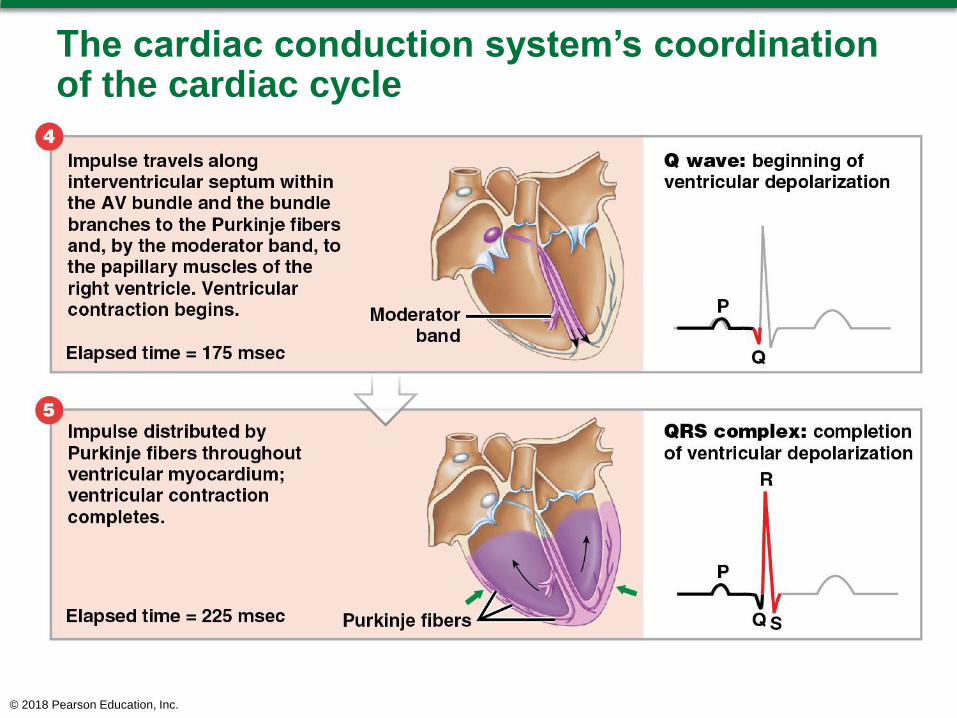
The cardiac conduction system’s coordination of the cardiac cycle
© 2018 Pearson Education, Inc.
The cardiac conduction system’s coordination of the cardiac cycle
© 2018 Pearson Education, Inc.

The cardiac conduction system’s coordination of the cardiac cycle
© 2018 Pearson Education, Inc.

SmartArt Video: The Conducting System of the Heart
© 2018 Pearson Education, Inc.

Module 18.12: Review
A. Define autorhythmicity.
B. If the cells of the SA node failed to function, how
would the heart rate be affected?
C. Why is it important for impulses from the atria to
be delayed at the AV node before they pass into
the ventricles?
Learning Outcome: Describe the components and
functions of the conducting system of the heart.
© 2018 Pearson Education, Inc.

Module 18.13: Normal and abnormal cardiac activity can be detected in an electrocardiogram
Electrocardiogram (ECG or EKG)
Recording of heart’s electrical activities from body
surface
To assess performance of
nodal, conducting, and
contractile components
If part of heart is damaged
by heart attack, may see
abnormal ECG pattern
Appearance varies with placement and number of
electrodes (leads)
© 2018 Pearson Education, Inc.
Module 18.13: ECG
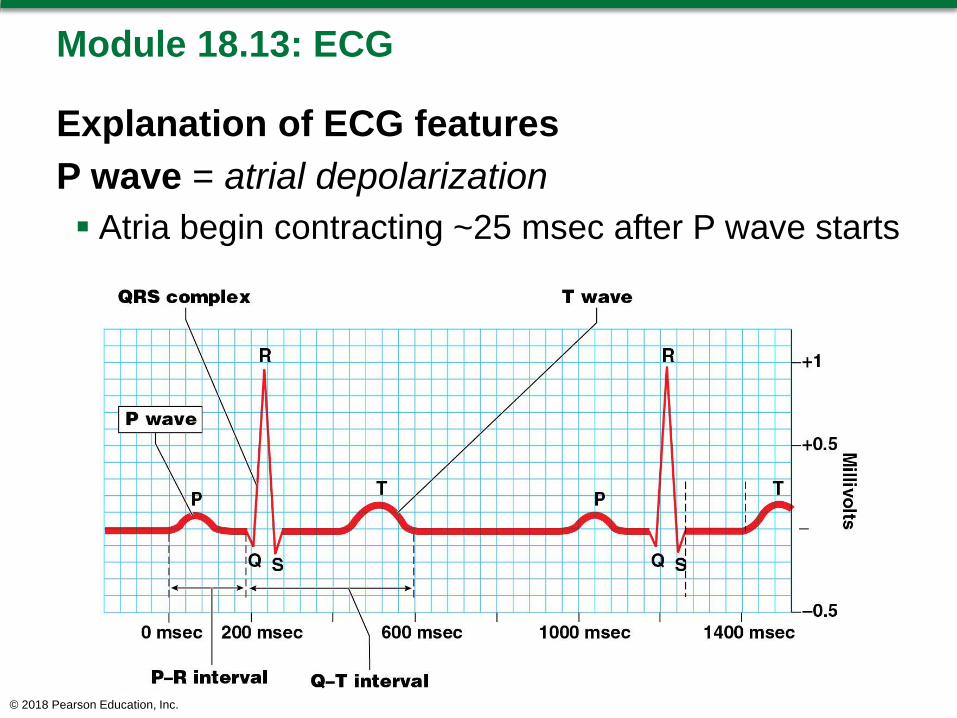
Explanation of ECG features
P wave = atrial depolarization
Atria begin contracting ~25 msec after P wave starts
© 2018 Pearson Education, Inc.

Module 18.13: ECG
Explanation of ECG features (continued)
QRS complex = ventricular depolarization
Larger wave due to larger ventricle muscle mass
Ventricles begin contracting shortly after R wave
peak
Atrial repolarization also occurs now but is masked
by QRS
© 2018 Pearson Education, Inc.

Module 18.13: ECG
Explanation of ECG features (continued)
T wave = ventricular repolarization
© 2018 Pearson Education, Inc.

Module 18.13: ECG
Explanation of ECG features (continued)
P–R interval
Period from start of atrial depolarization to start of
ventricular depolarization
>200 msec may mean damage to conducting
pathways or AV node
© 2018 Pearson Education, Inc.

Module 18.13: ECG
Explanation of ECG features (continued)
Q–T interval
Time for ventricles to undergo a single cycle
May be lengthened by electrolyte disturbances,
medications, conduction problems, coronary
ischemia, myocardial damage
© 2018 Pearson Education, Inc.

Module 18.13: ECG and arrhythmias
ECGs valuable for detecting/and diagnosing
arrhythmias
Cardiac arrhythmias = abnormal patterns of
cardiac electrical activity
About 5% of healthy people experience a few
abnormal heartbeats each day
Not a clinical problem unless pumping efficiency is
reduced
© 2018 Pearson Education, Inc.

Module 18.13: Arrhythmias
Cardiac arrhythmias (continued)
Premature atrial contractions (PACs)
• Often occur in healthy people
• Normal atrial rhythm momentarily interrupted by
“surprise” atrial contraction
• Increased incidences caused by stress, caffeine,
various drugs that increase permeability of the SA
pacemakers
• Normal ventricular contraction follows the atrial beat
© 2018 Pearson Education, Inc.
Module 18.13: Arrhythmias
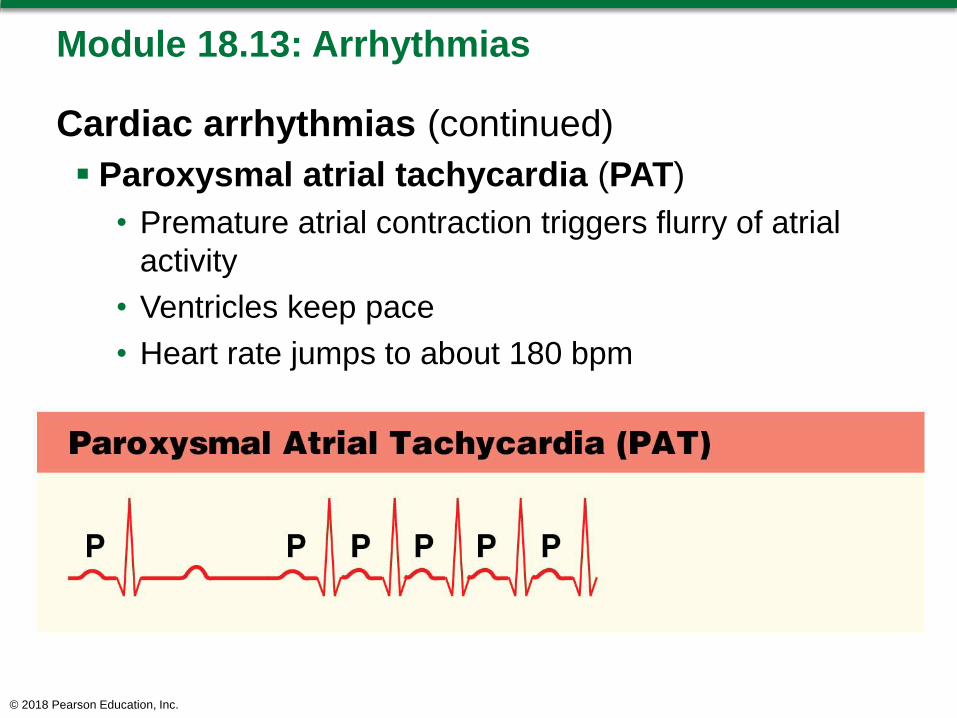
Cardiac arrhythmias (continued)
Paroxysmal atrial tachycardia (PAT)
• Premature atrial contraction triggers flurry of atrial
activity
• Ventricles keep pace
• Heart rate jumps to about 180 bpm
© 2018 Pearson Education, Inc.

Module 18.13: Arrhythmias
Cardiac arrhythmias (continued)
Atrial fibrillation
• Impulses move over atrial surface at up to 500 bpm
• Atria quiver—not organized contraction
• Ventricular rate cannot follow, may remain fairly
normal
• Atria nonfunctional, but ventricles still fill passively
• Person may not realize there is an arrhythmia
© 2018 Pearson Education, Inc.

Module 18.13: Arrhythmias
Cardiac arrhythmias (continued)
Premature ventricular contractions (PVCs)
• Purkinje cell or ventricular myocardial cell
depolarizes; triggers premature contraction
– Cell responsible called an ectopic pacemaker
(pacemaker other than the SA node)
• Single PVCs common, not dangerous
• Frequency increased by epinephrine, stimulatory
drugs, or ionic changes that depolarize cardiac
muscle cells
© 2018 Pearson Education, Inc.

Module 18.13: Arrhythmias
Cardiac arrhythmias (continued)
Ventricular tachycardia
• Also known as VT or V-tach
• Defined as four or more PVCs without intervening
normal beats
• Multiple PVCs and V-tach may indicate serious
cardiac problems
© 2018 Pearson Education, Inc.
Module 18.13: Arrhythmias
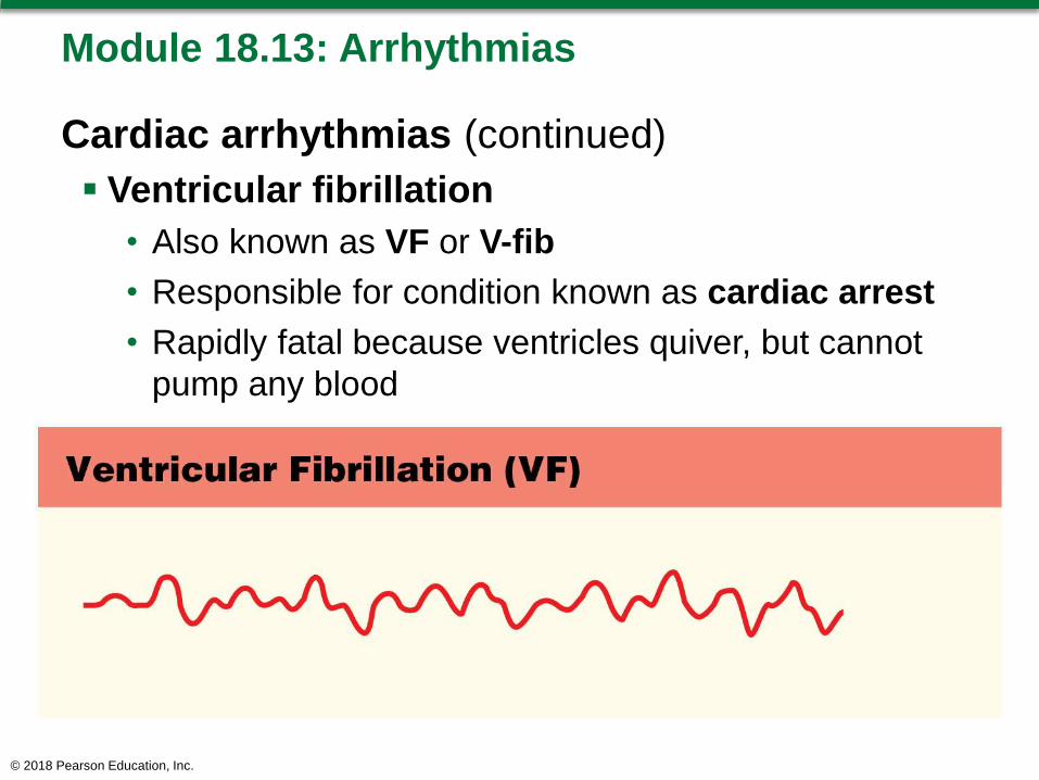
Cardiac arrhythmias (continued)
Ventricular fibrillation
• Also known as VF or V-fib
• Responsible for condition known as cardiac arrest
• Rapidly fatal because ventricles quiver, but cannot
pump any blood
© 2018 Pearson Education, Inc.

Module 18.13: Review
A. Define electrocardiogram.
B. List the important features of the ECG, and
indicate what each represents.
C. Why is ventricular fibrillation fatal?
Learning Outcome: Identify the electrical events
shown on an electrocardiogram.
© 2018 Pearson Education, Inc.

Module 18.14: The intrinsic heart rate can be altered by autonomic activity
Pacemaker potential
Pacemaker cells in SA/AV nodes cannot maintain a
stable resting membrane potential; membrane drifts
toward threshold
• Pacemaker potential = the gradual spontaneous
depolarization
© 2018 Pearson Education, Inc.

Module 18.14: The intrinsic heart rate can be altered by autonomic activity
Pacemaker potential (continued)
• Pacemaker potential in SA node cells occurs
80–100 times/min
– Establishes heart rate
– SA node brings AV nodal cells to threshold before they
reach it on their own, thus SA node paces the heart
© 2018 Pearson Education, Inc.

Module 18.14: Autonomic effects on heart rate
Parasympathetic influence
Parasympathetic stimulation decreases heart rate
ACh from parasympathetic neurons:
• Opens K+ channels in plasma membrane
• Hyperpolarizes membrane
• Slows rate of spontaneous depolarization
• Lengthens repolarization
© 2018 Pearson Education, Inc.

Module 18.14: Autonomic effects on heart rate
Sympathetic influence
Sympathetic stimulation increases heart rate
Binding of norepinephrine to beta-1 receptors opens
ion channels
• Increases rate of depolarization
• Decreases repolarization
© 2018 Pearson Education, Inc.

Module 18.14: Autonomic effects on heart rate
Resting heart rate
Varies with age, general health, physical
conditioning
Normal range is 60–100 bpm
Bradycardia
• Heart rate slower than normal (<60 bpm)
Tachycardia
• Heart rate faster than normal (>100 bpm)
© 2018 Pearson Education, Inc.

Module 18.14: Autonomic effects on heart rate
Cardiac centers of the medulla oblongata
Cardioinhibitory center
• Controls parasympathetic neurons; slows heart rate
• Parasympathetic supply to heart via vagus nerve (X);
synapse in cardiac plexus
• Postganglionic fibers to SA/AV nodes, atrial
musculature
Cardioacceleratory center
• Controls sympathetic neurons; increases heart rate
• Sympathetic innervation to heart via postganglionic
fibers in cardiac nerves; innervate nodes, conducting
system, atrial and ventricular myocardium
© 2018 Pearson Education, Inc.

The cardiac centers and innervation of the heart
© 2018 Pearson Education, Inc.

Module 18.14: Review
A. Compare bradycardia with tachycardia.
B. Describe the sites and actions of the
cardioinhibitory and cardioacceleratory centers.
C. Caffeine has effects on conducting cells and
contractile cells that are similar to those of NE.
What effect would drinking large amounts of
caffeinated beverages have on the heart rate?
Learning Outcome: Describe the factors affecting
the heart rate.
© 2018 Pearson Education, Inc.

Module 18.15: Stroke volume depends on the relationship between end-diastolic volume and end-systolic volume
Stroke volume analogy
Stroke volume can be compared to pumping water
with a manual pump
• Amount pumped varies with pump handle movement
Heart has two pumps (ventricles) that pump same
volume
• Can use single pump as model
© 2018 Pearson Education, Inc.

Module 18.15: Stroke volume analogy
As pump handle raises,
pressure in cylinder
decreases; water enters
through one-way valve.
Corresponds to passive
filling during ventricular
diastole
© 2018 Pearson Education, Inc.

Module 18.15: Stroke volume analogy
At the start of pumping
(cardiac) cycle:
Water in pump cylinder = blood in ventricle at end of ventricular diastole or the end-diastolic volume(EDV)
© 2018 Pearson Education, Inc.

Module 18.15: Stroke volume analogy
As the pump handle
comes down, water is
forced out.
Corresponds to
ventricular ejection
© 2018 Pearson Education, Inc.

Module 18.15: Stroke volume analogy
Handle fully depressed,
water remaining in
cylinder = blood in
ventricles at end of
systole, or end-systolic
volume (ESV)
Amount of water
pumped out = stroke
volume
Stroke volume =
EDV – ESV
© 2018 Pearson Education, Inc.

Module 18.15: Factors affecting stroke volume
Factors affecting stroke volume
End-diastolic volume (EDV)
• Venous return = amount of
venous blood returned to the right
atrium
– Varies directly with blood volume,
muscular activity, and rate of blood
flow
• Filling time = length of ventricular
diastole; the longer it is, the more
filling occurs (higher EDV)
© 2018 Pearson Education, Inc.

Module 18.15: Factors affecting stroke volume
Factors affecting stroke volume
(continued)
• Preload = amount of myocardial
stretch
– Greater EDV causes greater
preload; more stretching causes
stronger contractions and more
blood being ejected (Frank-Starling
law of the heart)
© 2018 Pearson Education, Inc.

Module 18.15: Factors affecting stroke volume
Factors affecting stroke volume
Influences on ESV
• Contractility = amount of force
produced during contraction at a
given preload
– Increased by sympathetic
stimulation, some hormones
(epinephrine, norepinephrine,
thyroid hormone, glucagon)
– Reduced by “beta blockers” and
calcium channel blockers
© 2018 Pearson Education, Inc.

Module 18.15: Factors affecting stroke volume
Factors affecting stroke volume
(continued)
• Afterload = ventricular tension
required to open semilunar valves
and empty
– As afterload increases, stroke
volume decreases
– Afterload increases whenever
blood flow is restricted, such as
with vasoconstriction
© 2018 Pearson Education, Inc.
Module 18.15: Review
A. Define end-diastolic volume (EDV) and end-
systolic volume (ESV).
B. What effect would an increase in venous return
have on the stroke volume?
C. What effect would an increase in sympathetic
stimulation of the heart have on the end-systolic
volume (ESV)?
Learning Outcome: Describe the variables that
influence stroke volume.
© 2018 Pearson Education, Inc.

Module 18.16: Cardiac output is regulated by adjustments in heart rate and stroke volume
Factors affecting cardiac
output
Cardiac output varies widely to
meet metabolic demands
Cardiac output can be changed
by affecting either heart rate or
stroke volume
Heart failure = condition in which
the heart cannot meet the
demands of peripheral tissues
© 2018 Pearson Education, Inc.

Module 18.16: Cardiac output adjustment
© 2018 Pearson Education, Inc.

Module 18.15: Review
A. Define heart failure.
B. Compute Joe’s stroke volume if his end-systolic
volume (ESV) is 40 mL and his end-diastolic
volume (EDV) is 125 mL.
Learning Outcome: Explain how stroke volume and
cardiac output are coordinated.
© 2018 Pearson Education, Inc.