Embed Size (px)
Citation preview
COPDand pulmonary function
in heart failure:
a matter of definition
Armine Minasian

COPD and pulmonary function in heart failure: a matter of definitionA.G. Minasian
PhD thesis, Radboud University Nijmegen Medical Centre, Nijmegen, The Netherlands
The research presented in this thesis was funded by an unrestricted educational grant of GlaxoSmithKline B.V.
The printing of this thesis was financially supported by the Radboud University Nijmegen Medical Centre, Department of Pulmonology of Rijnstate Hospital, het Rijnstate Vriendenfonds, Chiesi Pharmaceuticals B.V., Therabel Pharma Nederland B.V., Teva Nederland B.V., Boehringer Ingelheim B.V., and ABN AMRO. Also, financial support by the Dutch Heart Foundation for the publication of this thesis is gratefully acknowledged. Takeda Nederland B.V. kindly sponsored the course ‘Presenteren en Promoveren’.
ISBN
978-90-9029128-4
Cover page Glass art by Henry Summa, photographer David Wicks
Lay-out
Promotie In Zicht, Arnhem
Ipskamp Drukkers, Enschede
Copyright © A.G. Minasian, Arnhem, The Netherlands
All rights are reserved. No part of this book may be reproduced, distributed, stored in a retrieval system, or transmitted in any form or by any means, without prior written permission of the author.
Proefschrift
ter verkrijging van de graad van doctoraan de Radboud Universiteit Nijmegen
op gezag van de rector magnificus prof. dr. Th.L.M. Engelen,volgens besluit van het college van decanen
in het openbaar te verdedigen op donderdag 8 oktober 2015om 12:30 uur precies
door
Armine Gagikovna Minasiangeboren op 6 juli 1983
te Jerevan, Armenië
COPDand pulmonary function
in heart failure:
a matter of definition

Promotor Prof. Dr. P.N.R. Dekhuijzen
Copromotoren Dr. F.J.J. van den Elshout Dr. Y.F. Heijdra Dr. P.J.E. Vos
Manuscriptcommissie Prof. M.J. de Boer Prof. E.F.M. Wouters (MUMC) Prof. W.J.J. Assendelft
Paranimfen N.V. Melnikova M.H.M. Derikx

Voor Daniël

Table of contents
Chapter 1 Introduction and outline of this thesis 9
Chapter 2 Serial pulmonary function tests to diagnose COPD in chronic heart failure
Translational Respiratory Medicine. 2014;2:12.
55
Chapter 3 COPD in chronic heart failure: less common than previously thought?
Heart Lung. 2013;42:365-71.
73
Chapter 4 Pulmonary function impairment in patients with chronic heart failure: lower limit of normal versus conventional cutoff values
Heart Lung. 2014;43:311-6.
93
Chapter 5 Using the lower limit of normal instead of the conventional cutoff values to define predictors of pulmonary function impairment in subjects with chronic heart failure
Respiratory Care. 2015; accepted for publication.
111
Chapter 6 Bronchodilator responsiveness in patients with chronic heart failure
Heart Lung. 2013;42:208-14.
135
Chapter 7 Summary, conclusions, general discussion, and future perspectives
169
Chapter 8 Samenvatting, conclusies en toekomstperspectief 195
Chapter 9 List of publicationsList of abbreviationsAuthors’ affiliationsDankwoordCurriculum Vitae
207208210211217


Introduction and outline of this thesis
CHAPTER 1

11
Introduction and outline of this thesis
11.1 Chronic Obstructive Pulmonary Disease
Chronic Obstructive Pulmonary Disease (COPD), a common preventable and treatable disease, is one of the leading causes of morbidity and mortality in adults all over the world resulting in a substantial socioeconomic burden.1 According to predictions of the World Health Organization, COPD will become the third leading cause of death, exceeded only by ischemic heart disease and cerebrovascular disease, and the fifth leading cause of disability worldwide by the year 2020.2 Estimates of COPD prevalence in the general population vary substantially across studies due to geographical variations, differences in survey methods, study population, and collection of spirometric data.3 Moreover, different diagnostic criteria have been applied, yielding varying COPD prevalence rates.3 A systematic review and meta-analyses of studies carried out in 28 countries between 1990 and 2004 reported a pooled prevalence of “patient-reported and physician diagnosed COPD” of ~ 5%, reflecting the widespread underrecognition and underdiagnosis of COPD.3 The pooled prevalence from 26 “spirometric estimates” was ~ 9%, with the Global Initiative for Chronic Obstructive Lung Disease (GOLD) being the most common spirometric definition used.3 Prevalence rates were higher in elderly (≥ 40 years), smokers, males, and urban residents.3 The Burden of Obstructive Lung Disease (BOLD) study, including 9425 participants ≥ 40 years old from 12 sites worldwide, has documented higher COPD prevalence rates (11 – 26%) and more advanced staging of “spirometrically confirmed COPD” (GOLD criteria) than previously reported (GOLD stage ≥ II = overall 10%).4 Using the lower limit of normal (LLN) to define COPD, however, resulted in lower prevalence rates of COPD (~ 8-19%) than the GOLD criteria.5 Globally, the burden and mortality of COPD is projected to increase in coming decades because of continued exposure to COPD risk factors, ageing of the population, and a decrease of mortality due to other diseases like cardiovascular disease in industrialized countries and infectious disease in developing countries.6, 7
COPD is a heterogeneous disease process that varies greatly from person to person with respect to lung pathology, natural history of disease, and co-morbidity.6 It is characterized by persistent airflow limitation that is usually progressive and associated with an enhanced chronic inflammatory response in the airways and the lung to cumulative exposures over decades to noxious particles or gases.1 Tobacco smoking is the main risk factor for the development of COPD. However, other factors, such as genetic factors, exposure to indoor and outdoor air pollution, occupational hazards, infections, and chronic asthma are also important.6, 8 The inflammatory process results in hypersecretion of mucus, structural changes and narrowing of the small airways (chronic obstructive bronchiolitis), and enlargement of air spaces, destruction of lung parenchyma, loss of elasticity, and closure of small airways (emphysema).1, 9
12
Chapter 1
The characteristic symptoms of COPD are chronic and progressive dyspnea, cough, and sputum production that can be variable from day-to-day. Additional symptoms may include wheezing, chest tightness, fatigue, weight loss, anorexia, and symptoms of depression/anxiety and cor pulmonale.1 With increasing severity of disease, patients experience acute exacerbations of COPD, characterized by acute worsening of respiratory symptoms that is beyond normal day-to-day variations and requires change in medication.1 Exacerbations can be precipitated by several factors, including respiratory tract infections (viral or bacterial), exposure to pollutants, and interruption of maintenance therapy. However, the cause of about one-third of severe COPD exacerbation cannot be identified.1 Importantly, exacerbations can negatively affect a patient’s quality of life, accelerate the rate of lung function decline, and are associated with significant mortality and high socioeconomic costs. One of the treatment goals of COPD is therefore to reduce the frequency and severity of exacerbations.1
Spirometry is required for the diagnosis of COPD by measuring persistent airflow limitation. However, there is no consensus on how to define airflow limitation.10-20 This is further elaborated in subsection 1.5 of the introduction of this thesis. It is increasingly recognized that COPD is a complex multi-component disease that extends beyond the lungs. Many patients have systemic manifestations and co-morbidities that can further impair functional capacity and health-related quality of life and can increase the risk of hospitalization, mortality, and costs.21, 22 Indeed, patients with COPD more frequently die from other causes than COPD,23 with cardiovascular disease and lung cancer being the commonest causes of death,24-27 especially in those with mild to moderate COPD.28 Moreover, co-morbidities may interfere with COPD management.1 Co-morbidities and extra-pulmonary effects of COPD include cardiovascular disease, skeletal muscle dysfunction, cachexia, pulmonary hypertension, metabolic syndrome, lung cancer, osteoporosis, depression, anemia, and obstructive sleep apnea.21 Co-morbidities are more commonly seen in association with severe COPD, but they may also be associated with milder disease.22 Although the mechanism linking COPD to systemic manifestations and co-morbidities is not yet certain, it is though that systemic inflammation is crucial in the pathogenesis.21, 22, 29, 30 Other possible mechanisms include shared genetic predispositions, cigarette smoking, accelerated ageing, physical inactivity, and chronic hypoxia.21, 22
The cornerstone of COPD treatment is smoking cessation which has the greatest capacity to influence the natural history of COPD.31 Other non-pharmacological therapeutic options are rehabilitation, nutritional advice, oxygen therapy, ventilatory support, lung volume reduction therapies such as bullectomy and valve placement,

13
Introduction and outline of this thesis
1lung transplantation, and palliative care. Pharmacological treatment of COPD includes bronchodilators, corticosteroids, phosfodiesterase-4 inhibitors, vaccines, alpha-1 antitrypsin augmentation therapy, antibiotics, mucolytic and antioxidant agents, antitussives, and narcotics (morphine).1
1.2 Heart failure
Heart failure (HF) is defined according to the European Society of Cardiology (ESC) guidelines32 as a clinical syndrome in which patients have typical symptoms and signs resulting from an abnormality of cardiac structure or function. HF often results in diminished quality of life, declining functional capacity, episodes of decompensation leading to hospital admission, and premature death, usually due to pump failure or a ventricular arrhythmia.32, 33 Typical symptoms of HF include breathlessness, orthopnea, paroxysmal nocturnal dyspnea, reduced exercise tolerance, ankle swelling, and fatigue. Less typical symptoms are nocturnal cough, wheezing, weight gain or loss, bloated feeling, loss of appetite, confusion, depression, palpitations, and syncope.32 There is a poor relationship between symptoms and the severity of cardiac dysfunction. Symptoms do relate more closely to prognosis if persistent after therapy and can be used to classify the severity of HF (New York Heart Association (NYHA) class) and to monitor the effects of therapy.32 Signs of HF may include elevated jugular venous pressure, hepatojugular reflux, third heart sound, laterally displaced apex beat, cardiac murmur, peripheral edema, pulmonary crackles, reduced air entry and dullness to percussion at lung bases, tachycardia, irregular pulse, tachypnea, hepatomegaly, ascites, and cachexia.32 Many of the symptoms and signs of HF are not specific and, therefore, of limited diagnostic value. Demonstration of an underlying cardiac cause for these symptoms and signs by obtaining objective evidence of a structural or functional cardiac abnormality is therefore central to the diagnosis of HF.32 This is usually myocardial disease causing systolic ventricular dysfunction. However, abnormalities of ventricular diastolic function or of the valves, pericardium, endocardium, heart rhythm, and conduction can also cause HF and more than one abnormality can be present.32
Approximately 1–2% of the adult population in developed countries has HF, with the prevalence rising to ≥ 10% among persons 70 years of age or older, while persons younger than 50 years are hardly ever found to have HF.32, 33 The life time risk of developing HF is approximately 20% for all individuals older than 40 years.34 The number of patients with HF is predicted to increase due to the ageing of the population, improvements in the treatment of acute coronary syndromes, and a longer survival of patients with HF.33
14
Chapter 1
Two types of HF related to the measured ejection fraction (EF) have been recognized, i.e. HF with a reduced EF (HF-REF), or ‘systolic HF’, and HF with a preserved EF (HF-PEF), or ‘diastolic HF’.32 Approximately half of patients with HF have preserved EF.35, 36 Whether HF-PEF and HF-REF are two distinct entities or two ends of a common spectrum remains a matter of debate.36, 37 Amongst many causes of HF-REF, coronary artery disease is the cause of approximately two-thirds of cases of HF-REF.32 Patients with HF-PEF, on the contrary, are less likely to have coronary heart disease and more likely to have hypertension and atrial fibrillation (AF).35, 38, 39 Moreover, patients with HF-PEF are older, more often female and obese, and seem to have a better prognosis than those with HF-REF,33, 35, 39 although not all studies could confirm this latter finding.38, 40, 41 Also, the diagnosis of HF-PEF is more challenging than the diagnosis of HF-REF, because it is largely one of exclusion; potential non-cardiac causes of the patient’s symptoms, such as anemia or chronic lung disease, must first be discounted.32 Moreover, there is no universally agreed upon definition to diagnose HF-PEF.41 The left ventricular EF (LVEF) is normal or only mildly reduced in HF-PEF.32 A cutoff point of 50% LVEF was used in the consensus statement on the diagnosis of HF with normal LVEF by the HF and echocardiography associations of the ESC.42 Patients with a LVEF in the range 35–50% represent a ‘grey area’ and most probably have primarily mild systolic dysfunction.32 Usually, patients with HF-PEF do not have a dilated heart and many have an increase in LV wall thickness and increased left atrial size. Most have evidence of diastolic dysfunction, which is generally accepted as the likely cause of HF in these patients.32 Diastolic LV dysfunction, however, is not unique to patients with HF-PEF, but also occurs in patients with HF-REF, in whom it even correlates better to symptoms than LVEF.42
It is important to identify the underlying cardiac problem, as the precise pathology determines the specific treatment used.32 The goals of treatment are to relieve symptoms and signs, prevent hospital admission, and improve survival.32 Although several pharmacological treatment options exist for patients with chronic HF-REF, such as diuretics, angiotensin-converting enzyme inhibitors (ACE-I) or angiotensin receptor blockers (ARBs) in case of intolerance, beta-blockers, and mineralocorti-coid/aldosterone receptor antagonists, no effective treatment has been identified in patients with HF-PEF in order to reduce morbidity and mortality.32, 36, 43 Diuretics are used to relieve symptoms and signs in patients with HF-PEF. Also, adequate treatment of hypertension, myocardial ischemia, and AF is considered to be important for which calcium-channel blockers may be used, contrary to patients with HF-REF, in whom their negative inotropic action can be dangerous.32 Other treatment modalities for HF are described elsewhere in detail,32 including treatment of acute HF/cardiogenic shock, exercise training, implantable cardioverter-defibrillator, cardiac resynchronization therapy, treatment of arrhythmia or severe bradycardia/conduction
15
Introduction and outline of this thesis
1disturbances, coronary revascularization, surgery (valve surgery, ventricular assist devices, transplantation), and palliative care. Finally, management of co-morbidities such as COPD is important, as they may influence treatment and prognosis of HF.44, 45
1.3 COPD in HF and vice versa
COPD and HF are both common diseases with significant morbidity, mortality, and health care use.44, 45 Moreover, COPD and HF seem to coexist more frequently than expected from their separate population prevalences.46 Several factors might explain the high coexistence of these two diseases, including sharing of environmental (mainly smoking) or genetic risk factors, advanced age, systemic inflammation,45, 47-53 and a relationship between a reduction in pulmonary and heart function.54-58 Also, COPD patients are at an increased risk of co-morbidities, such as metabolic syndrome, which in turn are an important risk for cardiovascular disease.59 Furthermore, factors that increase stress on the cardiovascular system or precipitate arrhythmic events can also explain the association between COPD and cardiovascular disease, including hypoxemia, hyperinflation, hyperventilation, neurohumoral disturbances, increased work of breathing and oxygen consumption, pulmonary hypertension, and the use of pulmonary medication.48, 53, 60-65 Finally, other factors, such as oxidative stress, endothelial dysfunction, arterial stiffness, hypercoagulable state, and connective tissue degradation have also been suggested to play a role.48, 60, 62, 66
The combination of both diseases presents many diagnostic and therapeutic challenges as well as adverse prognostic implications.44-47, 67-75 Several difficulties in diagnosing COPD in patients with HF and vice versa have been put forward, all of which may lead to misdiagnosis or delay in the diagnosis. These include the overlap in symptoms, signs, and risk factors, misinterpretation of radiological evidence of HF due to chest hyperinflation and pulmonary vascular remodeling, poor acoustic windows of transthoracic echocardiography due to air trapping, lower diagnostic accuracy of B-type natriuretic peptide (BNP) in stable patients with COPD and chronic HF (CHF), even more challenging diagnosis of HF-PEF in patients with COPD, the underuse of spirometry, and difficulties with interpreting spirometry results, especially in patients with decompensated HF, who may have both restrictive and obstructive ventilatory defects.44, 72, 76, 77
Therapeutic challenges have been stressed as well. COPD may interfere with treatment and clinical course of HF and oppositely. Especially the opposite pharma-cological effects of beta-blockers for HF and beta-agonists for COPD have been a reason for concern leading to underuse of beta-blockers.78-81 Although cardioselective
16
Chapter 1
beta1-blockers are considered to be safe,82-85 even during hospitalization for COPD,86 and therefore should not be withheld from patients with COPD and coexisting cardiovascular diseases, the safety profile of non-selective beta-blockers is not as well-established.82, 85-97 In addition, the cardiovascular safety of beta2-agonists is disputable.82 Beta2-agonists have been reported to increase the risk of adverse cardiovascular events in patients with obstructive airway disease,98 with a significant increase in sinus tachycardia and a non-significant trend toward an increase in major cardiovascular events, including ventricular tachycardia, AF, syncope, congestive HF, myocardial infarction, cardiac arrest, and sudden death.99 On the other hand, a pooled analysis of cardiovascular safety data including 17 randomized clinical trials did not find an increased risk of cardiovascular adverse events as well as deaths comparing salmeterol treatment to placebo in patients with COPD.100 Similar findings were reported when patients were stratified for age of > 65 years or the presence of known cardiovascular disease.100 Similarly, cardiovascular mortality and cardio-vascular related adverse events were not greater in the salmeterol (+/- fluticasone) group compared to placebo in more than 6000 patients with COPD in the TORCH trial.25, 101 Observational studies have shown worse outcomes with bronchodilator use in patients with HF, including increased risk of HF hospitalization, increased mortality rates, in-hospital mechanical ventilation, intravenous vasodilator use, and major cardiovascular events associated with the use of beta-agonists,82, 102-104 although not all could confirm these findings.105 Possible mechanisms for adverse cardiovascular outcomes are arrythmogenesis, ischemia, hypoxemia, inflammation, cardiac remodeling, QTc prolongation, metabolic alterations (hypokalemia), and/or attenuation of beta- blocker benefits.82, 98, 103 In addition, hepatic metabolism of beta2-agonists may be diminished in patients with HF, leading to prolonged plasma half-life and accumulation with repeated doses.106
Inhaled anticholinergics have also been reported to be associated with adverse cardiovascular effects among patients with COPD, including an increased risk of cardio vascular death, myocardial infarction, or stroke,107-116 although recently reassuring cardiovascular safety data have been reported on the long-acting anticholinergic bronchodilator tiotropium HandiHaler110, 115, 117-123 as well as Respimat.124 However, HF patients are usually excluded from clinical trials and the impact of bronchodilators on outcomes has never been prospectively evaluated in patients with HF.103 Furthermore, methodological limitations of most studies require further investigation of reported adverse events.82, 103 Until then, bronchodilators, in particular beta-agonists, must be used with caution in patients with underlying cardiac condition such as HF, given the paucity of data in such patients.
17
Introduction and outline of this thesis
1Similarly, other medications prescribed for one disease may have detrimental effects on the other. High dosages of diuretics can cause metabolic alkalosis which may blunt the respiratory drive of COPD patients, causing hypoventilation with subsequent worsening of hypercapnia.75 Moreover, in HF patients with diuretic medication the potassium lowering effect of additive administration of beta2-agonists has to be considered, as it can provoke arrhythmias.51 Digitalis can cause pulmonary vasocon-striction, reduced venous return and cardiac output, and cardiac arrhythmias due to hypoxia and acidosis in patients with COPD.75 Also, it has the potential to increase airway obstruction.68 ACE-I are associated with side-effects such as cough, although according to the review of Packard et. al125 patients with asthma or COPD were not at an increased risk of developing cough and bronchoconstriction as a result of therapy with ACE-I. Theofylline can predispose to tachyarrhythmias even in the absence of elevated serum drug levels.126 Finally, oral corticosteroids cause sodium and water retention, potentially leading to worsening of HF, but this is not believed to be a problem with inhaled corticosteroids.32, 127 Moreover, systemic corticosteroids, unlike inhaled corticosteroids, have been associated with the development of atrial fibrillation and ventricular arrhythmias,126, 128 possibly due to local potassium efflux, mineralocorticosteroid effect leading to hypertension, development of late potentials, vasodilation, and possible anaphylaxis.129
On the other hand, cardiovascular drugs, including statins and ACE-I/ARBs, could have beneficial effects in patients with COPD;130-135 statins due to their anti-inflammatory, anti-oxidative, and immunomodulatory pleiotropic effects,131, 132, 136-140 as well as their potential to inhibit smoke-induced airway epithelial injury,141 and ACE-I/ARBs due to their anti-inflammatory and vasodilator effects as well as their effects on alveolar epithelial cell apoptosis and lung fibroblast growth.131, 140 However, a recent large multicenter randomized controlled trial (RCT) of 885 moderate to severe COPD patients at high risk for exacerbations and without an indication for statin use did not show beneficial effects of daily treatment with 40 mg simvastatin during 12-36 months versus placebo on exacerbation rate, time to the first exacerbation, or the severity of exacerbations.142 Simvastatin also had no effect on lung function, general or disease specific quality of life, serious adverse events, or the number of deaths.142 The underuse of statins in persons with cardiac risk factors who have been included in prior retrospective studies may account in part for the differences in findings. Beta-blockers could theoretically exert beneficial effects in patients with COPD by tempering the sympathetic nervous system or by reducing the ischemic burden.65, 131 Moreover, chronic use of beta-blockers may decrease airway hyperresponsiveness and inflammation as suggested by animal data and a small pilot study in humans.131 Recently, long-term treatment with beta-blockers has been shown to reduce the risk of exacerbations and improve survival in patients with COPD, even in the absence of
18
Chapter 1
overt cardiovascular disease, possibly as a result of dual cardiopulmonary protective properties.143, 144 Furthermore, thrombocytosis is associated with increased short- and long-term mortality after exacerbation of COPD and antiplatelet therapy may have a protective role by lowering 1-year mortality after COPD exacerbation.145, 146 Similarly, bronchodilation may have positive effects on cardiovascular function by alleviating dynamic hyperinflation,147 which reduces intrathoracic pressure, thus improving venous return and cardiac output, and lessens the effort of breathing by unloading respiratory muscles.148 Finally, corticosteroids may play a role in improving cardiovascular outcomes in COPD due to their anti-inflammatory properties,149-153 although a recent systematic review and meta-analysis of 23 RCTs did not show significant association between the use of inhaled corticosteroids with the reduction in risk of myocardial infarction, cardiovascular death, or mortality.154 In the observational studies, on the other hand, inhaled corticosteroids were associated with a significant reduction in cardiovascular death and mortality, most probably due to methodological issues.154
Patients with concurrent COPD and CHF may benefit from higher levels of positive end-expiratory pressure (PEEP) when managed with supported invasive mechanical ventilation. On the other hand, PEEP levels of 10 cm H2O or greater should be used cautiously in patients with COPD alone, since this can lead to increased dead space ventilation and work of breathing and as a consequence a decrease in minute alveolar ventilation and increased PaCO2 levels.155
The prognosis of coexistent COPD and HF is poorer than that in either disease alone. COPD is an independent predictor of death and HF hospitalization in patients with HF,44, 88, 156-168 although not all studies could confirm this.169-172 A recent study found COPD to be associated with increased risk of HF hospi talization and major adverse cardiovascular events, but not with 24 months survival in ambulatory HF patients.173 Similarly, after adjustment for prognostic risk factors, beta-blocker use, and randomized treatment (ivabradine/placebo), coexistent COPD was associated with all-cause hospitalization and hospitalization for worsening HF, but not with all-cause or cardiovascular mortality in ambulatory patients with stable systolic HF in another study.174 Higher GOLD stage is a predictor of worse prognosis.175 Long-term prognosis of patients with coexistent HF and COPD is similar in patients with HF-REF compared to those with HF-PEF,175 although mortality risk has been shown to be higher in patients with HF-PEF in another study.160 Furthermore, having COPD is associated with decreased functional capacity as measured by the 6-minute walking test176 and cardiopulmonary exercise testing.177 Similarly, concurrent HF is an independent predictor of mortality in patients with both stable and exacerbated COPD.178-182 Furthermore, congestive HF has been found to be associated with a higher risk for
19
Introduction and outline of this thesis
1COPD related emergency department visit,183 hospitalization,183 and worse self-rated health.184 Finally, healthcare utilization and costs for concomitant COPD and HF are greater than that in patients with either condition alone.80, 185, 186
Despite the close relationship between lung and heart diseases, pulmonologists and cardiologists often focus on their own field of specialization.70 Meanwhile, HF and COPD often remain an ignored combination73 and the degree of awareness is low among both cardiologists as well as pulmonologists.170 In view of diagnostic, therapeutic, and prognostic implications of the coexistence of COPD and HF, more attention should be paid to the concomitant presence of both diseases in clinical practice and research.73 Moreover, knowledge about the concomitant prevalence facilitates the decision on additional testing in daily practice.46 Although the occurrence of HF in patients with COPD has been assessed extensively in prior research, the occurrence of COPD in patients with HF has received much less attention.46
The prevalence of unrecognized HF has been reported to be 21% in both stable COPD patients from a general practice (54% systolic, 46% isolated diastolic, 0% right-sided HF),187 as well as patients with a history of COPD or asthma presenting to the emergency department with acute dyspnea.188 A history of ischemic heart disease, a laterally displaced apex beat, a body mass index > 30 kg/m2, and a raised heart rate (> 90 beats/min) were independent clinical indicators of the presence of concomitant HF in elderly patients with stable COPD. Raised N-terminal-pro-BNP (> 14.75 pmol/l) and abnormalities on electrocardiography further improved the diagnostic accuracy.189 The prevalence of LV systolic dysfunction (LVSD, i.e. LVEF < 40-50%) in stable COPD patients without known coronary artery disease (CAD) varied between 0% and 16% (4 out of 9 studies found a prevalence of 0%).73 When LVSD was found, it was generally mild and mainly in patients with a history of cor pulmonale.46 In patients without known CAD who were experiencing COPD exacerbation, prevalence varied between 0% and 32%.73 In groups without exclusion of patients with known CAD, prevalence rates of LVSD ranged from 0% to 46%.73, 170, 190 In another study, echocardiographic LV dysfunction was found in 51% of patients admitted for a severe acute exacerbation of COPD without an obvious cause, of which 64% was systolic, 23% diastolic, and 13% both. After discharge, an expert panel could confirm in 31% of the acute exacerbations a definite association with LV dysfunction.191 In a recent study,192 echocardiography was performed in 342 patients with COPD 3 months after discharge from their first hospital admission for COPD exacerbation. Significant cardiac alterations were present in 64% of the patients; 27% left- and 48% right-heart disorders. The most common were right ventricle enlargement (30%) and pulmonary hypertension (19%; 33% in severe disease and 7% in mild disease). The magnitude of pulmonary hypertension was mild in the majority of
20
Chapter 1
patients and only 3% of patients had systolic pulmonary artery pressures close to out-of-proportion pulmonary hypertension in COPD. Left ventricle enlargement was present in 6%, LV systolic dysfunction in 13% (9% LVEF 40-50%, 4% LVEF 30-40%), LV diastolic impairment in 12%, and left atrial dilatation in 29%. Echocardiographic abnormalities were unrelated to COPD severity and they were also observed in 63% of patients without known cardiac disease or cardiovascular risk factors other than smoking, although left heart abnormalities were more frequent in patients with previous cardiac disease compared to those without known cardiac disease (47% versus 27%).192 Finally, LV diastolic dysfunction has been found to be highly prevalent (48-88%) in patients with COPD in other studies.193-198 Interestingly, a recent study found 50% of advanced COPD patients with diastolic dysfunction but without known risk factors for diastolic dysfunction (hypertension, diabetes, ischemic heart disease, and hypothyroidism) to have reversible perfusion defect on stress SPECT myocardial perfusion imaging that were not apparent with stress electrocardiography.195 This observation suggests that myocardial ischemia is a possible cause of diastolic dysfunction in patients with advanced COPD without risk factors for LV relaxation abnormalities.195 Several other mechanisms of diastolic dysfunction in COPD have been put forward in the literature, including co-morbidities leading to impaired diastolic relaxation, interventricular dependence with impaired LV filling, hypoxia, prolonged use of beta2-agonists, pericardial restriction due to hyperinflation,195, 199 accelerated ageing,200 myocardial fibrosis,200 and LV hypertrophy which is present in 30% of COPD patients, even when normotensive and normoxemic.201
Pulmonary hypertension is a frequent complication of COPD, especially in those with advanced disease and hypoxemia.202 Estimates of the prevalence of pulmonary hypertension vary widely between 30% and 70% due to differences in definitions of pulmonary hypertension, study population, and methods used to determine pulmonary pressures.202 The true prevalence of pulmonary hypertension in patients with mild to moderate COPD is unknown because of the absence of large-scale epidemiologic studies.202 Severe pulmonary hypertension, defined as mean pulmonary artery pressure > 40 mmHg, is uncommon (< 5%) and is typically associated with less severe airflow limitation on the one hand, but more severe hypoxemia and diffusion impairment on the other hand.202 The pathophysiology of pulmonary hypertension is likely multifactorial: destruction of lung parenchyma with accompanying loss of vascular surface area, hypoxic vasoconstriction and inflammation leading to pulmonary vascular remodeling, and genetic susceptibility.202 Finally, other co-morbid conditions, such as left-sided heart disease, may contribute.202 Increase in the pulmonary vascular resistance and pulmonary artery pressure presents an increased afterload to the right ventricle which may eventually lead to ventricular remodeling with hypertrophy and later dilatation of the right
21
Introduction and outline of this thesis
1ventricle (cor pulmonale) with subsequent dysfunction.68, 202, 203 Also, pressure overload of the right ventricle and hypoxemia may lead to right ventricle ischemia, which may further aggravate ventricular dysfunction.202, 203 Finally, decreased right ventricular preload due to decreased venous return associated with hyperinflation may contribute to right ventricular dysfunction.54, 57, 202, 204 The same is true for the LV.57,
200, 204 On the other hand, the increase in the end-diastolic volume (preload) of the right ventricle due to dilatation maintains the cardiac output even as the right ventricle EF decreases.68 Most patients with COPD-associated pulmonary hypertension have preserved right ventricular contractility during stable conditions, while right ventricular systolic failure may be present in the acutely decompensated state.202 However, subclinical right ventricular dysfunction may be present in patients with mild airflow obstruction205 and without pulmonary hypertension,206 while right ventricular hypertrophy has been shown in patients with mild to moderate COPD196 and in those without pulmonary hypertension206 and without hypoxemia,207 suggesting that right ventricular morphological and functional changes could be early signs of pressure overload developing in the initial disease stages.208 Right ventricular dysfunction may ultimately lead to deterioration of LV function due to interventricular dependence.68, 202,
203 As the two ventricles are in series, the reduced right ventricular output reduces the LV preload.55, 68, 202 Moreover, a dilatation of the right ventricle shifts the interventricular septum into the left, changing LV geometry and thereby reducing LV diastolic compliance and end-diastolic volume.68, 202, 203, 209 On the other hand, the increased right ventricular end-systolic pressure also serves to augment the LV emptying.68, 210 Due to this complex interplay of opposite forces the LVEF is relatively preserved, even in advanced emphysema.68
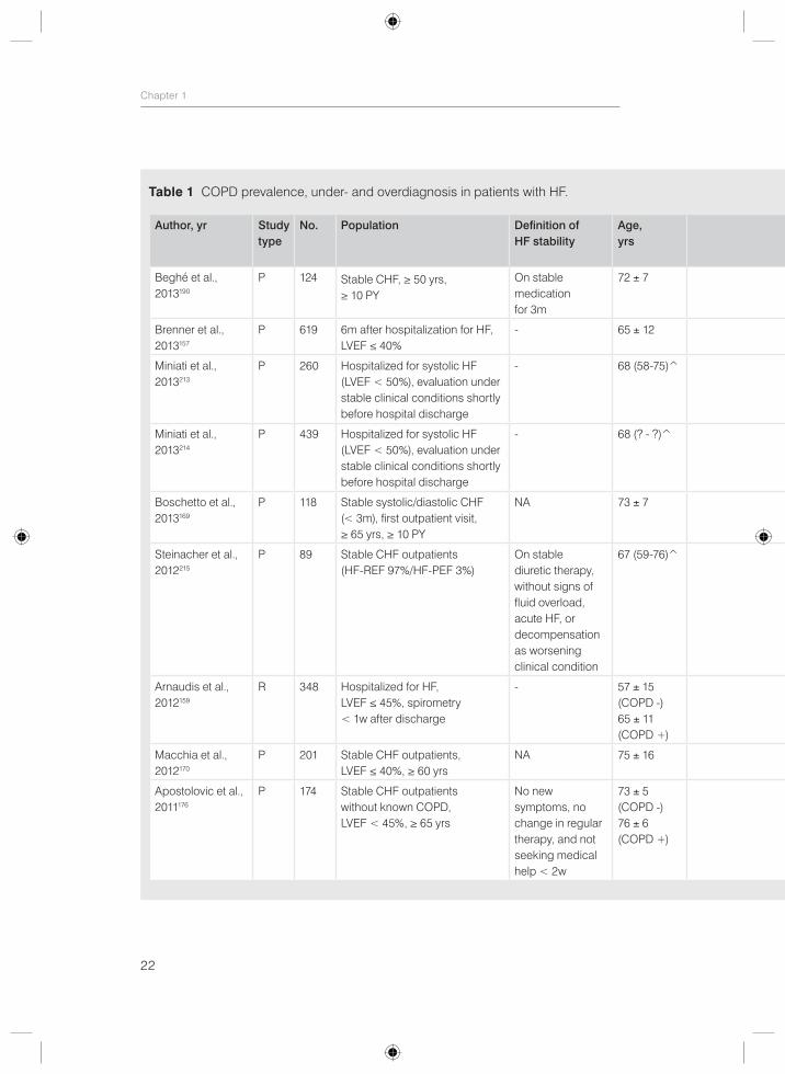
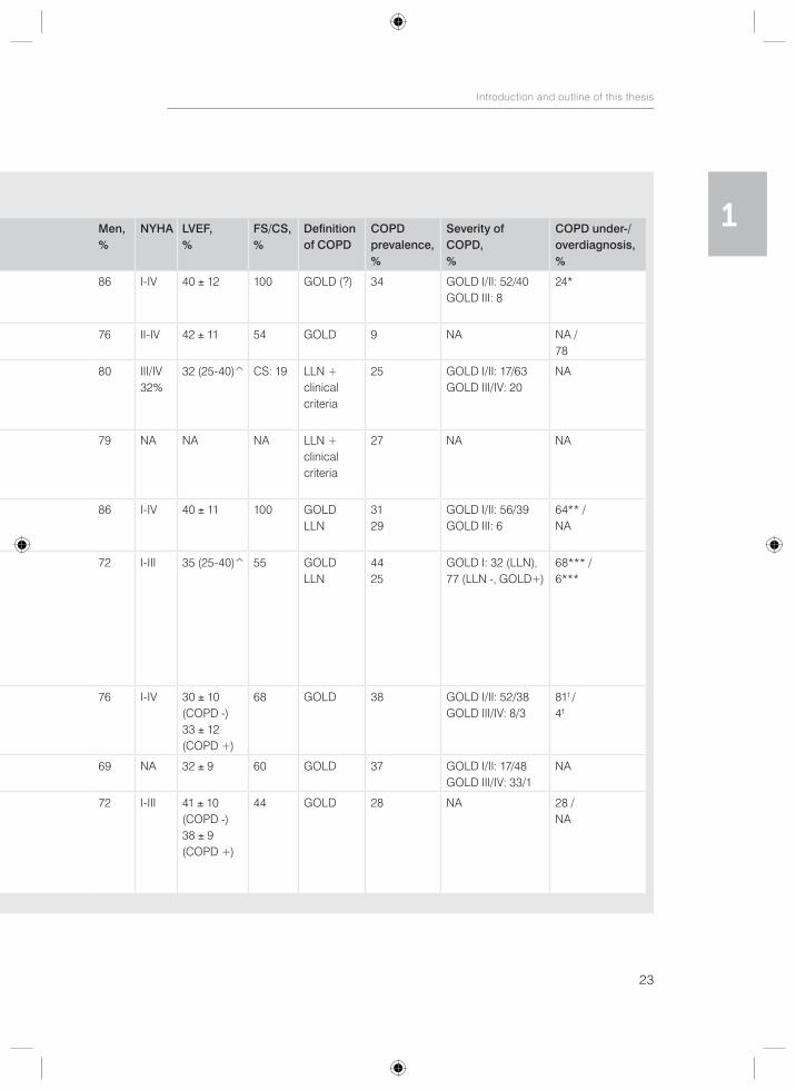
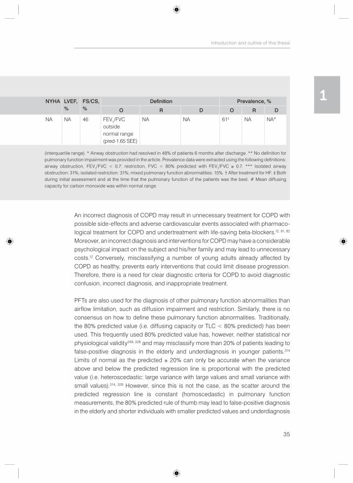
Estimates of COPD prevalence in patients with HF vary substantially between 9% and 52% in earlier reports that relied on clinical data, disease codes, or self-reported COPD, which is a very inaccurate method for establishing the diagnosis.44 Indeed, relying on self-report or physician diagnosis of COPD results in approximately one-third to one-half of patients being labeled as having COPD who actually do not fulfill the GOLD criteria for COPD.77, 187, 211, 212 Although spirometry is considered to be the gold standard for the diagnosis of COPD, data on the prevalence of COPD based on spirometry in patients with HF are scarce and spirometry is widely underused, even in a tertiary care setting.77 Fortunately, during the past six years more spirometric data on the prevalence of COPD in HF has become available. Nevertheless, prevalence rates of COPD still vary considerably between 9% and 44%, depending on study design, population, and diagnostic criteria used (Table 1). Also, a great number of patients is either under- or overdiagnosed with COPD, although exact figures vary across the studies (Table 1). Since airway obstruction is a dynamic phenomenon in HF, as it may be present in congestive HF and may disappear with
22
Chapter 1
Table 1 COPD prevalence, under- and overdiagnosis in patients with HF.
Author, yr Study type
No. Population Definition of HF stability
Age,yrs
Men,%
NYHA LVEF,%
FS/CS, %
Definition of COPD
COPD prevalence, %
Severity of COPD, %
COPD under-/ overdiagnosis, %
Beghé et al.,2013190
P 124 Stable CHF, ≥ 50 yrs, ≥ 10 PY
On stable medication for 3m
72 ± 7 86 I-IV 40 ± 12 100 GOLD (?) 34 GOLD I/II: 52/40GOLD III: 8
24*
Brenner et al.,2013157
P 619 6m after hospitalization for HF, LVEF ≤ 40%
- 65 ± 12 76 II-IV 42 ± 11 54 GOLD 9 NA NA /78
Miniati et al.,2013213
P 260 Hospitalized for systolic HF (LVEF < 50%), evaluation under stable clinical conditions shortly before hospital discharge
- 68 (58-75)̂ 80 III/IV 32%
32 (25-40)̂ CS: 19 LLN + clinical criteria
25 GOLD I/II: 17/63GOLD III/IV: 20
NA
Miniati et al.,2013214
P 439 Hospitalized for systolic HF (LVEF < 50%), evaluation under stable clinical conditions shortly before hospital discharge
- 68 (? - ?)̂ 79 NA NA NA LLN + clinical criteria
27 NA NA
Boschetto et al., 2013169
P 118 Stable systolic/diastolic CHF (< 3m), first outpatient visit, ≥ 65 yrs, ≥ 10 PY
NA 73 ± 7 86 I-IV 40 ± 11 100 GOLDLLN
3129
GOLD I/II: 56/39GOLD III: 6
64** /NA
Steinacher et al., 2012215
P 89 Stable CHF outpatients (HF-REF 97%/HF-PEF 3%)
On stable diuretic therapy, without signs of fluid overload, acute HF, or decompensation as worsening clinical condition
67 (59-76)̂ 72 I-III 35 (25-40)̂ 55 GOLDLLN
4425
GOLD I: 32 (LLN), 77 (LLN -, GOLD+)
68*** /6***
Arnaudis et al.,2012159
R 348 Hospitalized for HF, LVEF ≤ 45%, spirometry < 1w after discharge
- 57 ± 15(COPD -)65 ± 11(COPD +)
76 I-IV 30 ± 10(COPD -)33 ± 12(COPD +)
68 GOLD 38 GOLD I/II: 52/38GOLD III/IV: 8/3
81† /4†
Macchia et al.,2012170
P 201 Stable CHF outpatients, LVEF ≤ 40%, ≥ 60 yrs
NA 75 ± 16 69 NA 32 ± 9 60 GOLD 37 GOLD I/II: 17/48GOLD III/IV: 33/1
NA
Apostolovic et al., 2011176
P 174 Stable CHF outpatients without known COPD, LVEF < 45%, ≥ 65 yrs
No new symptoms, no change in regular therapy, and not seeking medical help < 2w
73 ± 5(COPD -)76 ± 6(COPD +)
72 I-III 41 ± 10(COPD -)38 ± 9(COPD +)
44 GOLD 28 NA 28 /NA
23
Introduction and outline of this thesis
1Table 1 COPD prevalence, under- and overdiagnosis in patients with HF.
Author, yr Study type
No. Population Definition of HF stability
Age,yrs
Men,%
NYHA LVEF,%
FS/CS, %
Definition of COPD
COPD prevalence, %
Severity of COPD, %
COPD under-/ overdiagnosis, %
Beghé et al.,2013190
P 124 Stable CHF, ≥ 50 yrs, ≥ 10 PY
On stable medication for 3m
72 ± 7 86 I-IV 40 ± 12 100 GOLD (?) 34 GOLD I/II: 52/40GOLD III: 8
24*
Brenner et al.,2013157
P 619 6m after hospitalization for HF, LVEF ≤ 40%
- 65 ± 12 76 II-IV 42 ± 11 54 GOLD 9 NA NA /78
Miniati et al.,2013213
P 260 Hospitalized for systolic HF (LVEF < 50%), evaluation under stable clinical conditions shortly before hospital discharge
- 68 (58-75)̂ 80 III/IV 32%
32 (25-40)̂ CS: 19 LLN + clinical criteria
25 GOLD I/II: 17/63GOLD III/IV: 20
NA
Miniati et al.,2013214
P 439 Hospitalized for systolic HF (LVEF < 50%), evaluation under stable clinical conditions shortly before hospital discharge
- 68 (? - ?)̂ 79 NA NA NA LLN + clinical criteria
27 NA NA
Boschetto et al., 2013169
P 118 Stable systolic/diastolic CHF (< 3m), first outpatient visit, ≥ 65 yrs, ≥ 10 PY
NA 73 ± 7 86 I-IV 40 ± 11 100 GOLDLLN
3129
GOLD I/II: 56/39GOLD III: 6
64** /NA
Steinacher et al., 2012215
P 89 Stable CHF outpatients (HF-REF 97%/HF-PEF 3%)
On stable diuretic therapy, without signs of fluid overload, acute HF, or decompensation as worsening clinical condition
67 (59-76)̂ 72 I-III 35 (25-40)̂ 55 GOLDLLN
4425
GOLD I: 32 (LLN), 77 (LLN -, GOLD+)
68*** /6***
Arnaudis et al.,2012159
R 348 Hospitalized for HF, LVEF ≤ 45%, spirometry < 1w after discharge
- 57 ± 15(COPD -)65 ± 11(COPD +)
76 I-IV 30 ± 10(COPD -)33 ± 12(COPD +)
68 GOLD 38 GOLD I/II: 52/38GOLD III/IV: 8/3
81† /4†
Macchia et al.,2012170
P 201 Stable CHF outpatients, LVEF ≤ 40%, ≥ 60 yrs
NA 75 ± 16 69 NA 32 ± 9 60 GOLD 37 GOLD I/II: 17/48GOLD III/IV: 33/1
NA
Apostolovic et al., 2011176
P 174 Stable CHF outpatients without known COPD, LVEF < 45%, ≥ 65 yrs
No new symptoms, no change in regular therapy, and not seeking medical help < 2w
73 ± 5(COPD -)76 ± 6(COPD +)
72 I-III 41 ± 10(COPD -)38 ± 9(COPD +)
44 GOLD 28 NA 28 /NA
24
Chapter 1
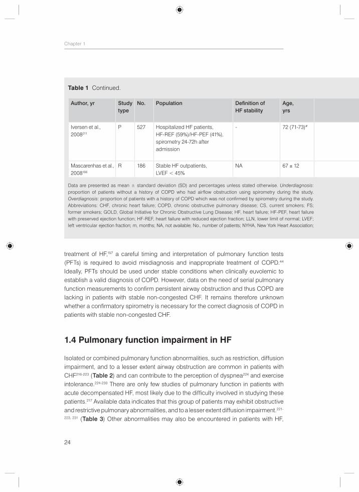
treatment of HF,157 a careful timing and interpretation of pulmonary function tests (PFTs) is required to avoid misdiagnosis and inappropriate treatment of COPD.44 Ideally, PFTs should be used under stable conditions when clinically euvolemic to establish a valid diagnosis of COPD. However, data on the need of serial pulmonary function measurements to confirm persistent airway obstruction and thus COPD are lacking in patients with stable non-congested CHF. It remains therefore unknown whether a confirmatory spirometry is necessary for the correct diagnosis of COPD in patients with stable non-congested CHF.
1.4 Pulmonary function impairment in HF
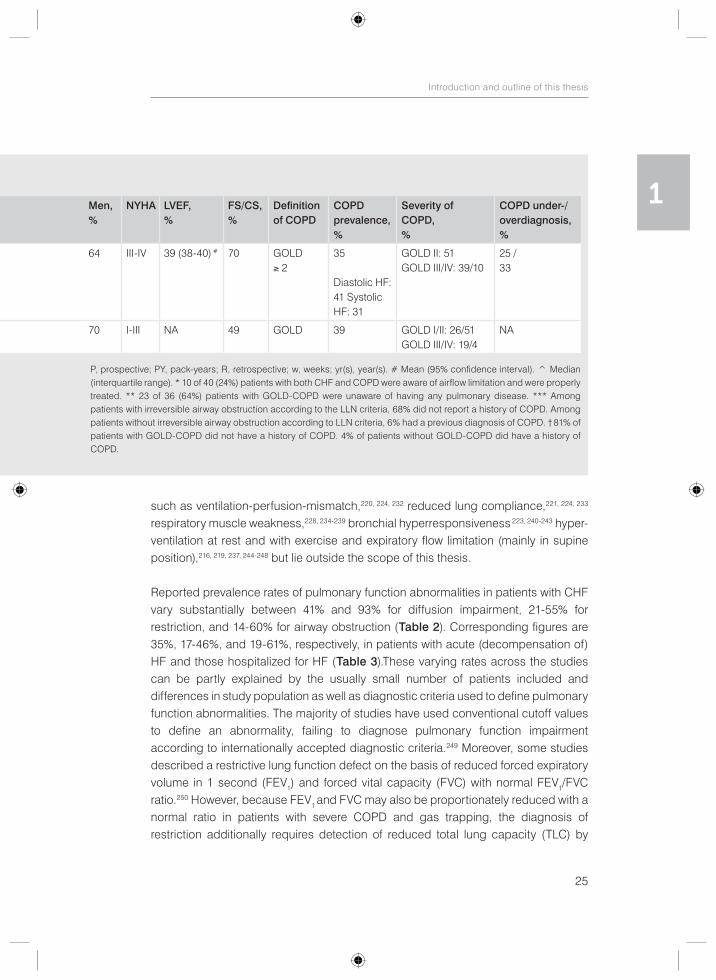
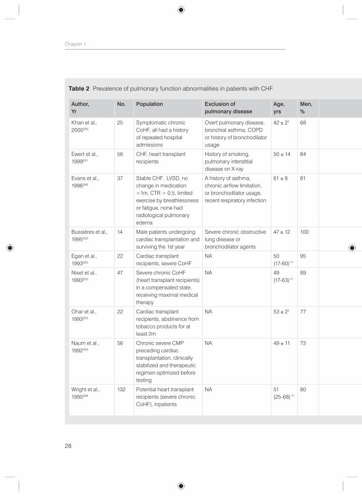
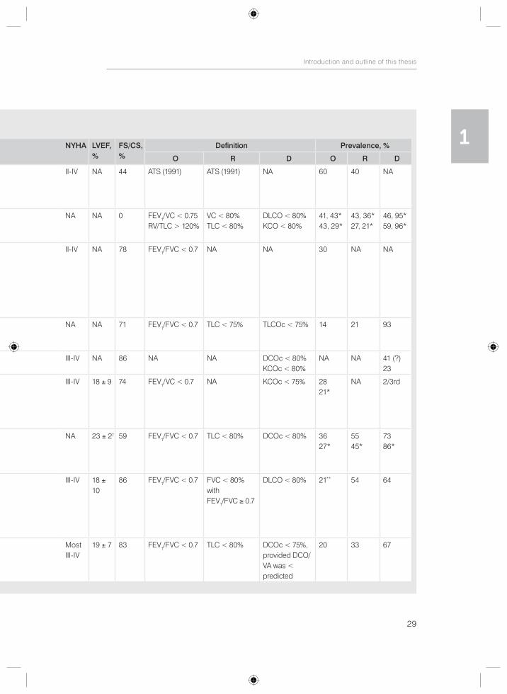
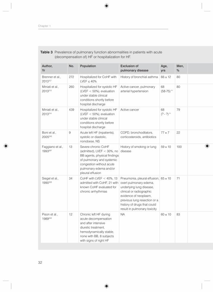
Isolated or combined pulmonary function abnormalities, such as restriction, diffusion impairment, and to a lesser extent airway obstruction are common in patients with CHF216-223 (Table 2) and can contribute to the perception of dyspnea224 and exercise intolerance.224-230 There are only few studies of pulmonary function in patients with acute decompensated HF, most likely due to the difficulty involved in studying these patients.217 Available data indicates that this group of patients may exhibit obstructive and restrictive pulmonary abnormalities, and to a lesser extent diffusion impairment.221-
223, 231 (Table 3) Other abnormalities may also be encountered in patients with HF,
Table 1 Continued.
Author, yr Study type
No. Population Definition of HF stability
Age,yrs
Men,%
NYHA LVEF,%
FS/CS, %
Definition of COPD
COPD prevalence, %
Severity of COPD, %
COPD under-/ overdiagnosis, %
Iversen et al.,2008211
P 527 Hospitalized HF patients, HF-REF (59%)/HF-PEF (41%), spirometry 24-72h after admission
- 72 (71-73)# 64 III-IV 39 (38-40) # 70 GOLD ≥ 2
35
Diastolic HF: 41 Systolic HF: 31
GOLD II: 51GOLD III/IV: 39/10
25 /33
Mascarenhas et al., 2008166
R 186 Stable HF outpatients, LVEF < 45%
NA 67 ± 12 70 I-III NA 49 GOLD 39 GOLD I/II: 26/51GOLD III/IV: 19/4
NA
Data are presented as mean ± standard deviation (SD) and percentages unless stated otherwise. Underdiagnosis: proportion of patients without a history of COPD who had airflow obstruction using spirometry during the study. Overdiagnosis: proportion of patients with a history of COPD which was not confirmed by spirometry during the study. Abbreviations: CHF, chronic heart failure; COPD, chronic obstructive pulmonary disease; CS, current smokers; FS; former smokers; GOLD, Global Initiative for Chronic Obstructive Lung Disease; HF, heart failure; HF-PEF, heart failure with preserved ejection function; HF-REF, heart failure with reduced ejection fraction; LLN, lower limit of normal; LVEF; left ventricular ejection fraction; m, months; NA, not available; No., number of patients; NYHA, New York Heart Association;
25
Introduction and outline of this thesis
1
such as ventilation-perfusion-mismatch,220, 224, 232 reduced lung compliance,221, 224, 233 respiratory muscle weakness,228, 234-239 bronchial hyperresponsiveness 223, 240-243 hyper-ventilation at rest and with exercise and expiratory flow limitation (mainly in supine position),216, 219, 237, 244-248 but lie outside the scope of this thesis.
Reported prevalence rates of pulmonary function abnormalities in patients with CHF vary substantially between 41% and 93% for diffusion impairment, 21-55% for restriction, and 14-60% for airway obstruction (Table 2). Corresponding figures are 35%, 17-46%, and 19-61%, respectively, in patients with acute (decompensation of) HF and those hospitalized for HF (Table 3).These varying rates across the studies can be partly explained by the usually small number of patients included and differences in study population as well as diagnostic criteria used to define pulmonary function abnormalities. The majority of studies have used conventional cutoff values to define an abnormality, failing to diagnose pulmonary function impairment according to internationally accepted diagnostic criteria.249 Moreover, some studies described a restrictive lung function defect on the basis of reduced forced expiratory volume in 1 second (FEV1) and forced vital capacity (FVC) with normal FEV1/FVC ratio.250 However, because FEV1 and FVC may also be proportionately reduced with a normal ratio in patients with severe COPD and gas trapping, the diagnosis of restriction additionally requires detection of reduced total lung capacity (TLC) by
Table 1 Continued.
Author, yr Study type
No. Population Definition of HF stability
Age,yrs
Men,%
NYHA LVEF,%
FS/CS, %
Definition of COPD
COPD prevalence, %
Severity of COPD, %
COPD under-/ overdiagnosis, %
Iversen et al.,2008211
P 527 Hospitalized HF patients, HF-REF (59%)/HF-PEF (41%), spirometry 24-72h after admission
- 72 (71-73)# 64 III-IV 39 (38-40) # 70 GOLD ≥ 2
35
Diastolic HF: 41 Systolic HF: 31
GOLD II: 51GOLD III/IV: 39/10
25 /33
Mascarenhas et al., 2008166
R 186 Stable HF outpatients, LVEF < 45%
NA 67 ± 12 70 I-III NA 49 GOLD 39 GOLD I/II: 26/51GOLD III/IV: 19/4
NA
P, prospective; PY, pack-years; R, retrospective; w, weeks; yr(s), year(s). # Mean (95% confidence interval). ^ Median (interquartile range). * 10 of 40 (24%) patients with both CHF and COPD were aware of airflow limitation and were properly treated. ** 23 of 36 (64%) patients with GOLD-COPD were unaware of having any pulmonary disease. *** Among patients with irreversible airway obstruction according to the LLN criteria, 68% did not report a history of COPD. Among patients without irreversible airway obstruction according to LLN criteria, 6% had a previous diagnosis of COPD. † 81% of patients with GOLD-COPD did not have a history of COPD. 4% of patients without GOLD-COPD did have a history of COPD.
26
Chapter 1
plethysmography. Finally, a large number of studies have included (potential) heart transplant recipients, who represent one extreme of the HF spectrum.250-256 Therefore, it is less known to what extent CHF patients who do not belong to the most severe category of HF have pulmonary function abnormalities and which of these abnormalities prevail.
Several factors have been implied to play a role in the etiology of pulmonary function impairment in patients with HF, including the effects of HF itself on pulmonary function in addition to (previously undiagnosed) underlying pulmonary disease and confounding influences such as smoking, coronary artery bypass grafting (CABG), and obesity.216-223, 232, 257
Diffusion impairment has been thought to be related to the thickening of alveolar- capillary membrane due to hydrostatic mechanical injury, interstitial edema, remodeling, and fibrosis.216-218, 221 Pressure or volume overload of the lung micro-circulation in HF causes structural adaptations of the alveolar-capillary membrane.217,
218 In acute HF, hydrostatic mechanical injury can cause breaks and discontinuities in the endothelial and epithelial layers of the blood-gas barrier, the so-called alveolar- capillary stress failure,258 and impair the cellular pathways involved in fluid filtration and reabsorption.217, 218 This process, which is generally reversible, leads to resistance to gas transfer.217, 218 In CHF, in which sustained neurohormonal activation has a significant pathophysiological role, a remodeling process may take place that is characterized by fixed extracellular matrix collagen proliferation. These changes may be protective against edema development on the one hand and may impair diffusing capacity on the other hand.217, 218, 232 Importantly, the damage caused to the alveolar- capillary membrane in CHF has been suggested to be permanent and may explain why heart transplantation does not affect (or may even worsen) pulmonary diffusing capacity despite an improvement in hemodynamic status and lung volumes.251-255,
259-266 Also, although ultrafiltration reduces lung fluid content, it does not improve diffusing capacity in CHF.267 In addition, although cardiac resynchronization therapy increases static and dynamic lung volumes, diffusing capacity for carbon monoxide remains unchanged.268 Furthermore, a relationship has been established between the time course of HF and extent of gas transfer alterations, suggesting that reversibility of diffusion impairment might depend on the disease time course.260 However, infusion of saline in patients with CHF is associated with a reduction in diffusing capacity,269-272 suggesting that abnormal pulmonary diffusion in CHF has a variable component that could be amenable to therapeutic intervention. Indeed, several pharmacological interventions were found to increase diffusing capacity, including spironolactone,273 ACE-I enalapril,269, 274-277 and type 5 phosphodiesterase inhibitor sildenafil,278 while hydralazine-isosorbide dinitrate,274, 277 ARB losartan,269, 276
27
Introduction and outline of this thesis
1and non-selective beta-blocker carvedilol279 did not affect diffusing capacity. Furthermore, aerobic exercise training may also favorably affect gas exchange.280 Little is known about the effects of other diuretics than spironolactone on diffusing capacity. Treatment for congestive HF did not result in improvement in diffusing capacity in two small studies.281, 282 On the other hand, diffusing capacity did increase after 1 year of treatment with diuretics and ACE-I/ARBs in 20 patients with newly diagnosed congestive HF.283 However, 87% of the patients were already taking diuretics prior to the study and therefore the improvement in diffusing capacity may be related to the addition of ACE-I rather than the effect of diuretics.
Other possible causes of diffusion impairment in HF include reduced lung and pulmonary capillary blood volumes, ventilation-perfusion mismatch, recurrent pulmonary emboli, smoking, and cardiopulmonary bypass.220-222, 257
Restriction has been linked to cardiomegaly, pleural effusion, respiratory muscle weakness, CABG, fibrosis from chronic congestion, and reduced lung compliance due to chronic vascular engorgement, interstitial/alveolar fluid accumulation, and chronic remodeling of the pulmonary vasculature due to elevated left atrial pressure.217,
221, 257, 284-286 This restrictive dysfunction generally improves after treatment of HF either by drug therapy,242, 281, 287 ultrafiltration,267, 288, 289 or following cardiac transplantation,252,
255, 261, 264, 290 most likely due to the reduction in lung water and cardiac size. However, other studies could not demonstrate significant improvement in lung volumes after pharmacological treatment for HF231, 283 or following cardiac transplantation.254, 291
Airway obstruction in HF has been attributed to alveolar fluid accumulation, bronchial mucosal swelling, peribronchial edema and fibrosis, squamous metaplasia of bronchial epithelial cells induces by transforming growth factor-ß from the failing heart, geometric decrease in airway size from reduction in lung volume, abnormalities of autonomic control, neurohumoral bronchoconstriction, bronchial hyperrespon-siveness, and smoking.220-223, 257, 292 Airway obstruction generally improves following treatment for HF,231, 287, 293 although not all studies could confirm this.242, 281 In addition, treatment directed at reversing airway obstruction with bronchodilators may have an additional role in the management of HF. There are, however, only few studies concerning the beneficial effects of bronchodilators in patients with HF. Improvements in pulmonary function,106, 293-298 dyspnea,296 and exercise performance298 have been reported. Some investigators have even observed an increase of mean FEV1 greater than 200 mL and 12% in patients with HF,293-295 especially in those with airway obstruction,295 LVSD,294 and during acute decompensation of HF.293 However, contrasting results have also been published. In the study of Witte et al.296 nebulized salbutamol (5 mg) and ipratropium bromide (0.5 mg) reduced peripheral airways
28
Chapter 1
Table 2 Prevalence of pulmonary function abnormalities in patients with CHF.
Author, Yr
No. Population Exclusion of pulmonary disease
Age,yrs
Men,%
NYHA LVEF,%
FS/CS, %
Definition Prevalence, %
O R D O R D
Khan et al.,2000295
25 Symptomatic chronic CoHF, all had a history of repeated hospital admissions
Overt pulmonary disease, bronchial asthma, COPD or history of bronchodilator usage
42 ± 2† 68 II-IV NA 44 ATS (1991) ATS (1991) NA 60 40 NA
Ewert et al., 1999251
56 CHF, heart transplant recipients
History of smoking, pulmonary interstitial disease on X-ray
50 ± 14 84 NA NA 0 FEV1/VC < 0.75RV/TLC > 120%
VC < 80%TLC < 80%
DLCO < 80%KCO < 80%
41, 43*43, 29*
43, 36*27, 21*
46, 95*59, 96*
Evans et al.,1996240
37 Stable CHF, LVSD, no change in medication <1m, CTR > 0.5, limited exercise by breathlessness or fatigue, none had radiological pulmonary edema
A history of asthma, chronic airflow limitation, or bronchodilator usage, recent respiratory infection
61 ± 8 81 II-IV NA 78 FEV1/FVC < 0.7 NA NA 30 NA NA
Bussières et al., 1995252
14 Male patients undergoing cardiac transplantation and surviving the 1st year
Severe chronic obstructive lung disease or bronchodilator agents
47 ± 12 100 NA NA 71 FEV1/FVC < 0.7 TLC < 75% TLCOc < 75% 14 21 93
Egan et al.,1993254
22 Cardiac transplant recipients, severe CoHF
NA 50(17-60)̂
95 III-IV NA 86 NA NA DCOc < 80% KCOc < 80%
NA NA 41 (?) 23
Niset et al.,1993255
47 Severe chronic CoHF (heart transplant recipients) in a compensated state, receiving maximal medical therapy
NA 49(17-63)̂
89 III-IV 18 ± 9 74 FEV1/VC < 0.7 NA KCOc < 75% 2821*
NA 2/3rd
Ohar et al.,1993253
22 Cardiac transplant recipients, abstinence from tobacco products for at least 2m
NA 53 ± 2† 77 NA 23 ± 2† 59 FEV1/FVC < 0.7 TLC < 80% DCOc < 80% 3627*
5545*
7386*
Naum et al., 1992250
56 Chronic severe CMP preceding cardiac transplantation, clinically stabilized and therapeutic regimen optimized before testing
NA 49 ± 11 73 III-IV 18 ± 10
86 FEV1/FVC < 0.7 FVC < 80% withFEV1/FVC ≥ 0.7
DLCO < 80% 21** 54 64
Wright et al., 1990256
132 Potential heart transplant recipients (severe chronic CoHF), inpatients
NA 51(25-68)̂
80 MostIII-IV
19 ± 7 83 FEV1/FVC < 0.7 TLC < 80% DCOc < 75%,provided DCO/VA was < predicted
20 33 67
29
Introduction and outline of this thesis
1Table 2 Prevalence of pulmonary function abnormalities in patients with CHF.
Author, Yr
No. Population Exclusion of pulmonary disease
Age,yrs
Men,%
NYHA LVEF,%
FS/CS, %
Definition Prevalence, %
O R D O R D
Khan et al.,2000295
25 Symptomatic chronic CoHF, all had a history of repeated hospital admissions
Overt pulmonary disease, bronchial asthma, COPD or history of bronchodilator usage
42 ± 2† 68 II-IV NA 44 ATS (1991) ATS (1991) NA 60 40 NA
Ewert et al., 1999251
56 CHF, heart transplant recipients
History of smoking, pulmonary interstitial disease on X-ray
50 ± 14 84 NA NA 0 FEV1/VC < 0.75RV/TLC > 120%
VC < 80%TLC < 80%
DLCO < 80%KCO < 80%
41, 43*43, 29*
43, 36*27, 21*
46, 95*59, 96*
Evans et al.,1996240
37 Stable CHF, LVSD, no change in medication <1m, CTR > 0.5, limited exercise by breathlessness or fatigue, none had radiological pulmonary edema
A history of asthma, chronic airflow limitation, or bronchodilator usage, recent respiratory infection
61 ± 8 81 II-IV NA 78 FEV1/FVC < 0.7 NA NA 30 NA NA
Bussières et al., 1995252
14 Male patients undergoing cardiac transplantation and surviving the 1st year
Severe chronic obstructive lung disease or bronchodilator agents
47 ± 12 100 NA NA 71 FEV1/FVC < 0.7 TLC < 75% TLCOc < 75% 14 21 93
Egan et al.,1993254
22 Cardiac transplant recipients, severe CoHF
NA 50(17-60)̂
95 III-IV NA 86 NA NA DCOc < 80% KCOc < 80%
NA NA 41 (?) 23
Niset et al.,1993255
47 Severe chronic CoHF (heart transplant recipients) in a compensated state, receiving maximal medical therapy
NA 49(17-63)̂
89 III-IV 18 ± 9 74 FEV1/VC < 0.7 NA KCOc < 75% 2821*
NA 2/3rd
Ohar et al.,1993253
22 Cardiac transplant recipients, abstinence from tobacco products for at least 2m
NA 53 ± 2† 77 NA 23 ± 2† 59 FEV1/FVC < 0.7 TLC < 80% DCOc < 80% 3627*
5545*
7386*
Naum et al., 1992250
56 Chronic severe CMP preceding cardiac transplantation, clinically stabilized and therapeutic regimen optimized before testing
NA 49 ± 11 73 III-IV 18 ± 10
86 FEV1/FVC < 0.7 FVC < 80% withFEV1/FVC ≥ 0.7
DLCO < 80% 21** 54 64
Wright et al., 1990256
132 Potential heart transplant recipients (severe chronic CoHF), inpatients
NA 51(25-68)̂
80 MostIII-IV
19 ± 7 83 FEV1/FVC < 0.7 TLC < 80% DCOc < 75%,provided DCO/VA was < predicted
20 33 67
30
Chapter 1
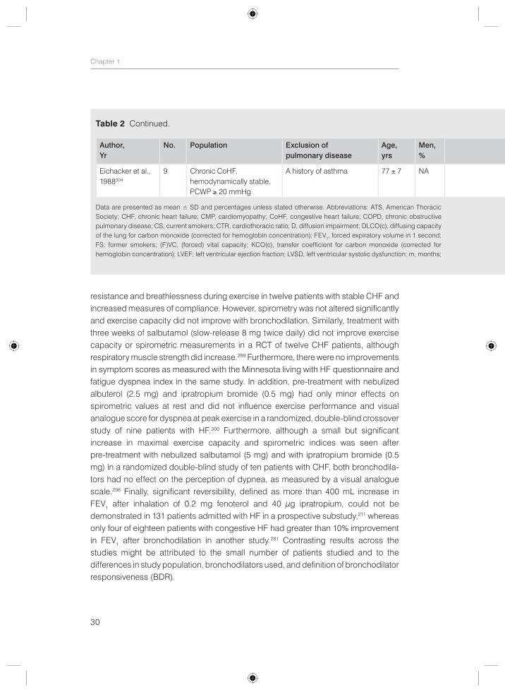
resistance and breathlessness during exercise in twelve patients with stable CHF and increased measures of compliance. However, spirometry was not altered significantly and exercise capacity did not improve with bronchodilation. Similarly, treatment with three weeks of salbutamol (slow-release 8 mg twice daily) did not improve exercise capacity or spirometric measurements in a RCT of twelve CHF patients, although respiratory muscle strength did increase.299 Furthermore, there were no improvements in symptom scores as measured with the Minnesota living with HF questionnaire and fatigue dyspnea index in the same study. In addition, pre-treatment with nebulized albuterol (2.5 mg) and ipratropium bromide (0.5 mg) had only minor effects on spirometric values at rest and did not influence exercise performance and visual analogue score for dyspnea at peak exercise in a randomized, double-blind crossover study of nine patients with HF.300 Furthermore, although a small but significant increase in maximal exercise capacity and spirometric indices was seen after pre-treatment with nebulized salbutamol (5 mg) and with ipratropium bromide (0.5 mg) in a randomized double-blind study of ten patients with CHF, both bronchodila-tors had no effect on the perception of dypnea, as measured by a visual analogue scale.298 Finally, significant reversibility, defined as more than 400 mL increase in FEV1 after inhalation of 0.2 mg fenoterol and 40 μg ipratropium, could not be demonstrated in 131 patients admitted with HF in a prospective substudy,211 whereas only four of eighteen patients with congestive HF had greater than 10% improvement in FEV1 after bronchodilation in another study.281 Contrasting results across the studies might be attributed to the small number of patients studied and to the differences in study population, bronchodilators used, and definition of bronchodilator responsiveness (BDR).
Table 2 Continued.
Author, Yr
No. Population Exclusion of pulmonary disease
Age,yrs
Men,%
NYHA LVEF,%
FS/CS, %
Definition Prevalence, %
O R D O R D
Eichacker et al., 1988304
9 Chronic CoHF, hemodynamically stable, PCWP ≥ 20 mmHg
A history of asthma 77 ± 7 NA IV 24 ± 6 56 FEV1/FVC < 0.75
NA NA 33 NA NA
Data are presented as mean ± SD and percentages unless stated otherwise. Abbreviations: ATS, American Thoracic Society; CHF, chronic heart failure; CMP, cardiomyopathy; CoHF, congestive heart failure; COPD, chronic obstructive pulmonary disease; CS, current smokers; CTR, cardiothoracic ratio; D, diffusion impairment; DLCO(c), diffusing capacity of the lung for carbon monoxide (corrected for hemoglobin concentration); FEV1, forced expiratory volume in 1 second; FS; former smokers; (F)VC, (forced) vital capacity; KCO(c), transfer coefficient for carbon monoxide (corrected for hemoglobin concentration); LVEF; left ventricular ejection fraction; LVSD, left ventricular systolic dysfunction; m, months;
31
Introduction and outline of this thesis
1
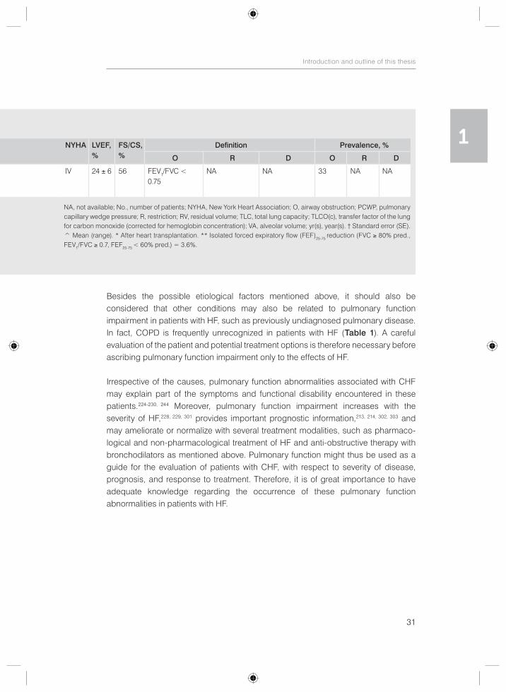
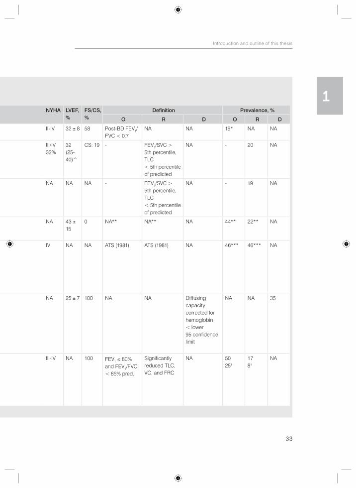
Besides the possible etiological factors mentioned above, it should also be considered that other conditions may also be related to pulmonary function impairment in patients with HF, such as previously undiagnosed pulmonary disease. In fact, COPD is frequently unrecognized in patients with HF (Table 1). A careful evaluation of the patient and potential treatment options is therefore necessary before ascribing pulmonary function impairment only to the effects of HF.
Irrespective of the causes, pulmonary function abnormalities associated with CHF may explain part of the symptoms and functional disability encountered in these patients.224-230, 244 Moreover, pulmonary function impairment increases with the severity of HF,228, 229, 301 provides important prognostic information,213, 214, 302, 303 and may ameliorate or normalize with several treatment modalities, such as pharmaco-logical and non-pharmacological treatment of HF and anti-obstructive therapy with bronchodilators as mentioned above. Pulmonary function might thus be used as a guide for the evaluation of patients with CHF, with respect to severity of disease, prognosis, and response to treatment. Therefore, it is of great importance to have adequate knowledge regarding the occurrence of these pulmonary function abnormalities in patients with HF.
Table 2 Continued.
Author, Yr
No. Population Exclusion of pulmonary disease
Age,yrs
Men,%
NYHA LVEF,%
FS/CS, %
Definition Prevalence, %
O R D O R D
Eichacker et al., 1988304
9 Chronic CoHF, hemodynamically stable, PCWP ≥ 20 mmHg
A history of asthma 77 ± 7 NA IV 24 ± 6 56 FEV1/FVC < 0.75
NA NA 33 NA NA
NA, not available; No., number of patients; NYHA, New York Heart Association; O, airway obstruction; PCWP, pulmonary capillary wedge pressure; R, restriction; RV, residual volume; TLC, total lung capacity; TLCO(c), transfer factor of the lung for carbon monoxide (corrected for hemoglobin concentration); VA, alveolar volume; yr(s), year(s). † Standard error (SE). ^ Mean (range). * After heart transplantation. ** Isolated forced expiratory flow (FEF)25-75 reduction (FVC ≥ 80% pred., FEV1/FVC ≥ 0.7, FEF25-75 < 60% pred.) = 3.6%.
32
Chapter 1
Table 3 Prevalence of pulmonary function abnormalities in patients with acute (decompensation of) HF or hospitalization for HF.
Author, Yr
No. Population Exclusion of pulmonary disease
Age,yrs
Men,%
NYHA LVEF,%
FS/CS, %
Definition Prevalence, %
O R D O R D
Brenner et al., 2013157
272 Hospitalized for CoHF with LVEF ≤ 40%
History of bronchial asthma 66 ± 12 80 II-IV 32 ± 8 58 Post-BD FEV1/FVC < 0.7
NA NA 19* NA NA
Miniati et al., 2013213
260 Hospitalized for systolic HF (LVEF < 50%), evaluation under stable clinical conditions shortly before hospital discharge
Active cancer, pulmonary arterial hypertension
68 (58-75)̂
80 III/IV 32%
32 (25-40)̂
CS: 19 - FEV1/SVC > 5th percentile, TLC < 5th percentile of predicted
NA - 20 NA
Miniati et al., 2013214
439 Hospitalized for systolic HF (LVEF < 50%), evaluation under stable clinical conditions shortly before hospital discharge
Active cancer 68 (? - ?)̂
79 NA NA NA - FEV1/SVC > 5th percentile, TLC < 5th percentile of predicted
NA - 19 NA
Boni et al.,2005245
9 Acute left HF (inpatients), systolic or diastolic, nonobese, NS
COPD, bronchodilators, corticosteroids, antibiotics
77 ± 7 22 NA 43 ± 15
0 NA** NA** NA 44** 22** NA
Faggiano et al., 1993287
13 Severe chronic CoHF (admitted), LVEF < 30%, no BB agents, physical findings of pulmonary and systemic congestion without acute pulmonary edema and/or pleural effusion
History of smoking or lung disease
59 ± 10 100 IV NA NA ATS (1981) ATS (1981) NA 46*** 46*** NA
Siegel et al., 1990305
34 CoHF with LVEF < 40%, 13 admitted with CoHF, 21 with known CoHF evaluated for chronic arrhythmias
Pneumonia, pleural effusion, overt pulmonary edema, underlying lung disease, clinical or radiographic evidence of neoplasm, previous lung resection or a history of drugs that could result in pulmonary toxicity
65 ± 10 71 NA 25 ± 7 100 NA NA Diffusing capacity corrected for hemoglobin < lower 95 confidence limit
NA NA 35
Pison et al.,1989242
12 Chronic left HF during acute decompensation and after intensive diuretic treatment, hemodynamically stable, none with BB, 8 subjects with signs of right HF
NA 60 ± 10 83 III-IV NA 100 FEV1 ≤ 80% and FEV1/FVC < 85% pred.
Significantly reduced TLC, VC, and FRC
NA 5025†
178†
NA
33
Introduction and outline of this thesis
1Table 3 Prevalence of pulmonary function abnormalities in patients with acute
(decompensation of) HF or hospitalization for HF.
Author, Yr
No. Population Exclusion of pulmonary disease
Age,yrs
Men,%
NYHA LVEF,%
FS/CS, %
Definition Prevalence, %
O R D O R D
Brenner et al., 2013157
272 Hospitalized for CoHF with LVEF ≤ 40%
History of bronchial asthma 66 ± 12 80 II-IV 32 ± 8 58 Post-BD FEV1/FVC < 0.7
NA NA 19* NA NA
Miniati et al., 2013213
260 Hospitalized for systolic HF (LVEF < 50%), evaluation under stable clinical conditions shortly before hospital discharge
Active cancer, pulmonary arterial hypertension
68 (58-75)̂
80 III/IV 32%
32 (25-40)̂
CS: 19 - FEV1/SVC > 5th percentile, TLC < 5th percentile of predicted
NA - 20 NA
Miniati et al., 2013214
439 Hospitalized for systolic HF (LVEF < 50%), evaluation under stable clinical conditions shortly before hospital discharge
Active cancer 68 (? - ?)̂
79 NA NA NA - FEV1/SVC > 5th percentile, TLC < 5th percentile of predicted
NA - 19 NA
Boni et al.,2005245
9 Acute left HF (inpatients), systolic or diastolic, nonobese, NS
COPD, bronchodilators, corticosteroids, antibiotics
77 ± 7 22 NA 43 ± 15
0 NA** NA** NA 44** 22** NA
Faggiano et al., 1993287
13 Severe chronic CoHF (admitted), LVEF < 30%, no BB agents, physical findings of pulmonary and systemic congestion without acute pulmonary edema and/or pleural effusion
History of smoking or lung disease
59 ± 10 100 IV NA NA ATS (1981) ATS (1981) NA 46*** 46*** NA
Siegel et al., 1990305
34 CoHF with LVEF < 40%, 13 admitted with CoHF, 21 with known CoHF evaluated for chronic arrhythmias
Pneumonia, pleural effusion, overt pulmonary edema, underlying lung disease, clinical or radiographic evidence of neoplasm, previous lung resection or a history of drugs that could result in pulmonary toxicity
65 ± 10 71 NA 25 ± 7 100 NA NA Diffusing capacity corrected for hemoglobin < lower 95 confidence limit
NA NA 35
Pison et al.,1989242
12 Chronic left HF during acute decompensation and after intensive diuretic treatment, hemodynamically stable, none with BB, 8 subjects with signs of right HF
NA 60 ± 10 83 III-IV NA 100 FEV1 ≤ 80% and FEV1/FVC < 85% pred.
Significantly reduced TLC, VC, and FRC
NA 5025†
178†
NA
34
Chapter 1
1.5 Different definitions of COPD and pulmonary function impairment: the lower limit of normal versus conventional cutoff values
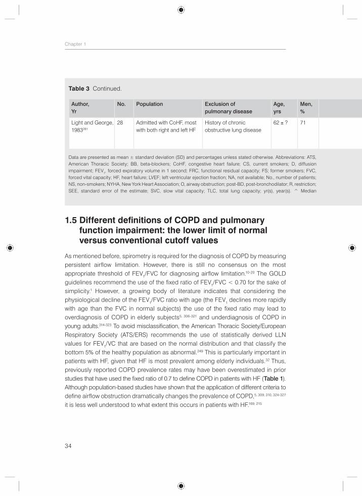
As mentioned before, spirometry is required for the diagnosis of COPD by measuring persistent airflow limitation. However, there is still no consensus on the most appropriate threshold of FEV1/FVC for diagnosing airflow limitation.10-20 The GOLD guidelines recommend the use of the fixed ratio of FEV1/FVC < 0.70 for the sake of simplicity.1 However, a growing body of literature indicates that considering the physiological decline of the FEV1/FVC ratio with age (the FEV1 declines more rapidly with age than the FVC in normal subjects) the use of the fixed ratio may lead to overdiagnosis of COPD in elderly subjects5, 306-321 and underdiagnosis of COPD in young adults.314-323 To avoid misclassification, the American Thoracic Society/European Respiratory Society (ATS/ERS) recommends the use of statistically derived LLN values for FEV1/VC that are based on the normal distribution and that classify the bottom 5% of the healthy population as abnormal.249 This is particularly important in patients with HF, given that HF is most prevalent among elderly individuals.32 Thus, previously reported COPD prevalence rates may have been overestimated in prior studies that have used the fixed ratio of 0.7 to define COPD in patients with HF (Table 1). Although population-based studies have shown that the application of different criteria to define airflow obstruction dramatically changes the prevalence of COPD,5, 309, 310, 324-327 it is less well understood to what extent this occurs in patients with HF.169, 215
Table 3 Continued.
Author, Yr
No. Population Exclusion of pulmonary disease
Age,yrs
Men,%
NYHA LVEF,%
FS/CS, %
Definition Prevalence, %
O R D O R D
Light and George,1983281
28 Admitted with CoHF, most with both right and left HF
History of chronic obstructive lung disease
62 ± ? 71 NA NA 46 FEV1/FVC outside normal range (pred-1.65 SEE)
NA NA 61‡ NA NA#
Data are presented as mean ± standard deviation (SD) and percentages unless stated otherwise. Abbreviations: ATS, American Thoracic Society; BB, beta-blockers; CoHF, congestive heart failure; CS, current smokers; D, diffusion impairment; FEV1, forced expiratory volume in 1 second; FRC, functional residual capacity; FS; former smokers; FVC, forced vital capacity; HF, heart failure; LVEF; left ventricular ejection fraction; NA, not available; No., number of patients; NS, non-smokers; NYHA, New York Heart Association; O, airway obstruction; post-BD, post-bronchodilator; R, restriction; SEE, standard error of the estimate; SVC, slow vital capacity; TLC, total lung capacity; yr(s), year(s). ^ Median
35
Introduction and outline of this thesis
1
An incorrect diagnosis of COPD may result in unnecessary treatment for COPD with possible side-effects and adverse cardiovascular events associated with pharmaco-logical treatment for COPD and undertreatment with life-saving beta-blockers.12, 81, 82 Moreover, an incorrect diagnosis and interventions for COPD may have a considerable psychological impact on the subject and his/her family and may lead to unnecessary costs.12 Conversely, misclassifying a number of young adults already affected by COPD as healthy, prevents early interventions that could limit disease progression. Therefore, there is a need for clear diagnostic criteria for COPD to avoid diagnostic confusion, incorrect diagnosis, and inappropriate treatment.
PFTs are also used for the diagnosis of other pulmonary function abnormalities than airflow limitation, such as diffusion impairment and restriction. Similarly, there is no consensus on how to define these pulmonary function abnormalities. Traditionally, the 80% predicted value (i.e. diffusing capacity or TLC < 80% predicted) has been used. This frequently used 80% predicted value has, however, neither statistical nor physiological validity249, 328 and may misclassify more than 20% of patients leading to false-positive diagnosis in the elderly and underdiagnosis in younger patients.314 Limits of normal as the predicted ± 20% can only be accurate when the variance above and below the predicted regression line is proportional with the predicted value (i.e. heteroscedastic: large variance with large values and small variance with small values).314, 328 However, since this is not the case, as the scatter around the predicted regression line is constant (homoscedastic) in pulmonary function measurements, the 80% predicted rule of thumb may lead to false-positive diagnosis in the elderly and shorter individuals with smaller predicted values and underdiagnosis
Table 3 Continued.
Author, Yr
No. Population Exclusion of pulmonary disease
Age,yrs
Men,%
NYHA LVEF,%
FS/CS, %
Definition Prevalence, %
O R D O R D
Light and George,1983281
28 Admitted with CoHF, most with both right and left HF
History of chronic obstructive lung disease
62 ± ? 71 NA NA 46 FEV1/FVC outside normal range (pred-1.65 SEE)
NA NA 61‡ NA NA#
(interquartile range). * Airway obstruction had resolved in 48% of patients 6 months after discharge. ** No definition for pulmonary function impairment was provided in the article. Prevalence data were extracted using the following definitions: airway obstruction, FEV1/FVC < 0.7; restriction, FVC < 80% predicted with FEV1/FVC ≥ 0.7. *** Isolated airway obstruction: 31%; isolated restriction: 31%; mixed pulmonary function abnormalities: 15%. † After treatment for HF. ‡ Both during initial assessment and at the time that the pulmonary function of the patients was the best. # Mean diffusing capacity for carbon monoxide was within normal range.
36
Chapter 1
in younger and taller patients with larger predicted values.314, 328 Misinterpretation of PFT results may lead to incorrect diagnosis of disease in elderly patients with HF and as a consequence unnecessary treatment. Moreover, results may be interpreted as having more severe or unstable HF due to the effects of HF on pulmonary function and as a result unnecessary intensified treatment for HF. Finally, misdiagnosis may interfere with interpretation of research aiming to understand the impact of HF and several clinical variables on pulmonary function.329, 330 To avoid misclassification, ATS/ERS guidelines249 again recommend the use of the fifth percentile LLN values, which are calculated by subtracting 1.64 times the residual standard deviation (RSD) from the predicted value. However, studies using the LLN values to assess the prevalence of pulmonary function abnormalities and their predictors in patients with HF are lacking.
1.6 Outline of this thesis
Given the gaps in current knowledge as described in the general introduction, the aim of this thesis is to provide more insight in the occurrence of COPD and pulmonary function abnormalities using different definitions in patients with CHF.
The main objectives of this thesis are:
1. To determine the prevalence of COPD in patients with CHF according to two definitions of airflow obstruction: the LLN versus the fixed ratio of FEV1/FVC < 0.70.
2. To assess the extent of underdiagnosis and overdiagnosis of COPD in patients with CHF.
3. To examine whether serial PFTs are necessary for the correct diagnosis of COPD in patients with stable non-congested CHF.
4. To determine the prevalence of pulmonary function abnormalities in patients with CHF according to recent ATS/ERS guidelines using the LLN compared to conventional cutoff values.
5. To assess predictors of pulmonary function impairment in patients with CHF according to the LLN in comparison to conventional cutoff values.
6. To evaluate the effect of inhaled bronchodilators on pulmonary function and dyspnea in patients with CHF.
37
Introduction and outline of this thesis
1After a general introduction in chapter 1, the 2nd chapter of this thesis describes the prevalence of COPD, including over- and underdiagnosis of COPD, in patients with CHF according to the widely used GOLD criteria. In addition, it provides an answer to the question whether serial PFTs are necessary for the correct diagnosis of COPD in patients with stable non-congested CHF. Chapter 3 compares two definitions of airflow limitation, namely the LLN versus the fixed ratio of FEV1/FVC < 0.70, regarding the prevalence of COPD in patients with CHF. Chapter 4 provides prevalence rates of pulmonary function abnormalities in patients with CHF according to the LLN versus conventional cutoff values. Chapter 5 describes several predictors of these pulmonary function abnormalities and the effect of using different diagnostic criteria, namely the LLN versus the conventional cutoff values. Chapter 6 evaluates the effect of inhaled bronchodilators on pulmonary function and dyspnea in patients with CHF. Finally, chapters 7 and 8 of this thesis provide a summary and discussion of all studies and the main findings. Subsequently, a perspective on the future is provided.
38
Chapter 1
References
1. Global Initiative for Chronic Obstructive Lung Disease. Global strategy for the diagnosis, management, and prevention of chronic obstructive pulmonary disease. NHBLI/WHO workshop report. Avaliable at: http://www.goldcopd.org; 2014.
2. Murray CJ, Lopez AD. Alternative projections of mortality and disability by cause 1990-2020: Global Burden of Disease Study. Lancet. 1997;349:1498-504.
3. Halbert RJ, Natoli JL, Gano A, Badamgarav E, Buist AS, Mannino DM. Global burden of COPD: systematic review and meta-analysis. Eur Respir J. 2006;28:523-32.
4. Buist AS, McBurnie MA, Vollmer WM, Gillespie S, Burney P, Mannino DM, et al. International variation in the prevalence of COPD (the BOLD Study): a population-based prevalence study. Lancet. 2007;370:741-50.
5. Vollmer WM, Gislason T, Burney P, Enright PL, Gulsvik A, Kocabas A, et al. Comparison of spirometry criteria for the diagnosis of COPD: results from the BOLD study. Eur Respir J. 2009;34:588-97.
6. Mannino DM, Buist AS. Global burden of COPD: risk factors, prevalence, and future trends. Lancet. 2007;370:765-73.
7. Feenstra TL, van Genugten ML, Hoogenveen RT, Wouters EF, Rutten-van Molken MP. The impact of aging and smoking on the future burden of chronic obstructive pulmonary disease: a model analysis in the Netherlands. Am J Respir Crit Care Med. 2001;164:590-6.
8. Eisner MD, Anthonisen N, Coultas D, Kuenzli N, Perez-Padilla R, Postma D, et al. An official American Thoracic Society public policy statement: Novel risk factors and the global burden of chronic obstructive pulmonary disease. Am J Respir Crit Care Med. 2010;182:693-718.
9. Barnes PJ. Chronic obstructive pulmonary disease. N Engl J Med. 2000;343:269-80.10. van Dijk WD, Gupta N, Tan WC, Bourbeau J. Clinical relevance of diagnosing COPD by fixed ratio or
lower limit of normal: a systematic review. Copd. 2014;11:113-20.11. Mohamed Hoesein FA, Zanen P, Lammers JW. Lower limit of normal or FEV(1)/FVC <0.70 in diagnosing
COPD: An evidence-based review. Respir Med. 2011;105:907-15.12. Quanjer PH, Enright PL, Miller MR, Stocks J, Ruppel G, Swanney MP, et al. Open letter: the need to
change the method for defining mild airway obstruction. Prim Care Respir J. 2010;19:288-91.13. Celli BR, Halbert RJ. Point: should we abandon FEV/FVC <0.70 to detect airway obstruction? No.
Chest. 2010;138:1037-40.14. Hansen JE. Lower limit of normal is better than 70% or 80%. CHEST. 2011;139:6-8.15. Enright P, Brusasco V. Counterpoint: should we abandon FEV/FVC < 0.70 to detect airway obstruction?
Yes. Chest. 2010;138:1040-2 [discussion 42-4].16. Pellegrino R, Brusasco V, Viegi G, Crapo RO, Burgos F, Casaburi R, et al. Definition of COPD: based
on evidence or opinion? Eur Respir J. 2008;31:681-2.17. Mannino DM. Should we be using statistics to define disease? Thorax. 2008;63:1031-2.18. Bhatt NY, Wood KL. What defines abnormal lung function in older adults with chronic obstructive
pulmonary disease? Drugs Aging. 2008;25:717-28.19. Townsend MC. Conflicting definitions of airways obstruction: Drawing the line between normal and
abnormal. Chest. 2007;131:335-6.20. Schermer TR, Quanjer PH. COPD screening in primary care: who is sick? Prim Care Respir J.
2007;16:49-53.21. Barnes PJ, Celli BR. Systemic manifestations and comorbidities of COPD. Eur Respir J. 2009;33:1165-85.22. Barnes PJ. Chronic obstructive pulmonary disease: effects beyond the lungs. PLoS Med. 2010;7:e1000220.23. Mannino DM, Doherty DE, Sonia Buist A. Global Initiative on Obstructive Lung Disease (GOLD)
classification of lung disease and mortality: findings from the Atherosclerosis Risk in Communities (ARIC) study. Respir Med. 2006;100:115-22.
24. Huiart L, Ernst P, Suissa S. Cardiovascular morbidity and mortality in COPD. Chest. 2005;128:2640-6.25. Calverley PM, Anderson JA, Celli B, Ferguson GT, Jenkins C, Jones PW, et al. Salmeterol and
fluticasone propionate and survival in chronic obstructive pulmonary disease. N Engl J Med. 2007;356:775-89.
39
Introduction and outline of this thesis
126. Terzano C, Conti V, Di Stefano F, Petroianni A, Ceccarelli D, Graziani E, et al. Comorbidity, hospitaliza-
tion, and mortality in COPD: results from a longitudinal study. Lung. 2010;188:321-9.27. Rodriguez-Roisin R, Soriano JB. Chronic Obstructive Pulmonary Disease with Lung Cancer and/or
Cardiovascular Disease. Proc Am Thorac Soc. 2008;5:842-7.28. Sin DD, Anthonisen NR, Soriano JB, Agusti AG. Mortality in COPD: Role of comorbidities. Eur Respir
J. 2006;28:1245-57.29. Agusti AG. Systemic effects of chronic obstructive pulmonary disease. Proc Am Thorac Soc.
2005;2:367-70; discussion 71-2.30. Gan WQ, Man SF, Senthilselvan A, Sin DD. Association between chronic obstructive pulmonary
disease and systemic inflammation: a systematic review and a meta-analysis. Thorax. 2004;59:574-80.31. Anthonisen NR, Connett JE, Kiley JP, Altose MD, Bailey WC, Buist AS, et al. Effects of smoking
intervention and the use of an inhaled anticholinergic bronchodilator on the rate of decline of FEV1. The Lung Health Study. Jama. 1994;272:1497-505.
32. McMurray JJ, Adamopoulos S, Anker SD, Auricchio A, Bohm M, Dickstein K, et al. ESC guidelines for the diagnosis and treatment of acute and chronic heart failure 2012: The Task Force for the Diagnosis and Treatment of Acute and Chronic Heart Failure 2012 of the European Society of Cardiology. Developed in collaboration with the Heart Failure Association (HFA) of the ESC. Eur J Heart Fail. 2012;14:803-69.
33. Mosterd A, Hoes AW. Clinical epidemiology of heart failure. Heart. 2007;93:1137-46.34. Lloyd-Jones DM, Larson MG, Leip EP, Beiser A, D’Agostino RB, Kannel WB, et al. Lifetime risk for
developing congestive heart failure: the Framingham Heart Study. Circulation. 2002;106:3068-72.35. Hogg K, Swedberg K, McMurray J. Heart failure with preserved left ventricular systolic function;
epidemiology, clinical characteristics, and prognosis. J Am Coll Cardiol. 2004;43:317-27.36. Borlaug BA, Paulus WJ. Heart failure with preserved ejection fraction: pathophysiology, diagnosis, and
treatment. Eur Heart J. 2011;32:670-9.37. Komajda M, Lam CS. Heart failure with preserved ejection fraction: a clinical dilemma. Eur Heart J.
2014;35:1022-32.38. Quiroz R, Doros G, Shaw P, Liang CS, Gauthier DF, Sam F. Comparison of characteristics and
outcomes of patients with heart failure preserved ejection fraction versus reduced left ventricular ejection fraction in an urban cohort. Am J Cardiol. 2014;113:691-6.
39. Lenzen MJ, Scholte op Reimer WJ, Boersma E, Vantrimpont PJ, Follath F, Swedberg K, et al. Differences between patients with a preserved and a depressed left ventricular function: a report from the EuroHeart Failure Survey. Eur Heart J. 2004;25:1214-20.
40. Almeida P, Rodrigues J, Lourenco P, Maciel MJ, Bettencourt P. Prognostic significance of applying the European Society of Cardiology consensus algorithm for heart failure with preserved systolic function diagnosis. Clin Cardiol. 2012;35:770-6.
41. Kaila K, Haykowsky MJ, Thompson RB, Paterson DI. Heart failure with preserved ejection fraction in the elderly: scope of the problem. Heart Fail Rev. 2012;17:555-62.
42. Paulus WJ, Tschope C, Sanderson JE, Rusconi C, Flachskampf FA, Rademakers FE, et al. How to diagnose diastolic heart failure: a consensus statement on the diagnosis of heart failure with normal left ventricular ejection fraction by the Heart Failure and Echocardiography Associations of the European Society of Cardiology. Eur Heart J. 2007;28:2539-50.
43. Paulus WJ, van Ballegoij JJ. Treatment of heart failure with normal ejection fraction: an inconvenient truth! J Am Coll Cardiol. 2010;55:526-37.
44. Hawkins NM, Petrie MC, Jhund PS, Chalmers GW, Dunn FG, McMurray JJ. Heart failure and chronic obstructive pulmonary disease: diagnostic pitfalls and epidemiology. Eur J Heart Fail. 2009;11:130-9.
45. Hawkins NM, Virani S, Ceconi C. Heart failure and chronic obstructive pulmonary disease: the challenges facing physicians and health services. Eur Heart J. 2013;34:2795-803.
46. Hannink JD, van Helvoort HA, Dekhuijzen PN, Heijdra YF. Heart failure and COPD: partners in crime? Respirology. 2010;15:895-901.
47. de Miguel Diez J, Chancafe Morgan J, Jimenez Garcia R. The association between COPD and heart failure risk: a review. Int J Chron Obstruct Pulmon Dis. 2013;8:305-12.
40
Chapter 1
48. Ghoorah K, De Soyza A, Kunadian V. Increased cardiovascular risk in patients with chronic obstructive pulmonary disease and the potential mechanisms linking the two conditions: a review. Cardiol Rev. 2013;21:196-202.
49. Mullerova H, Agusti A, Erqou S, Mapel DW. Cardiovascular comorbidity in COPD: systematic literature review. Chest. 2013;144:1163-78.
50. Van Eeden S, Leipsic J, Paul Man SF, Sin DD. The relationship between lung inflammation and cardiovascular disease. Am J Respir Crit Care Med. 2012;186:11-6.
51. Ukena C, Mahfoud F, Kindermann M, Kindermann I, Bals R, Voors AA, et al. The cardiopulmonary continuum systemic inflammation as ‘common soil’ of heart and lung disease. Int J Cardiol. 2010;145:172-6.
52. Fabbri LM, Rabe KF. From COPD to chronic systemic inflammatory syndrome? Lancet. 2007;370:797-9.53. Sin DD, Man SF. Chronic obstructive pulmonary disease: a novel risk factor for cardiovascular disease.
Can J Physiol Pharmacol. 2005;83:8-13.54. Grau M, Barr RG, Lima JA, Hoffman EA, Bluemke DA, Carr JJ, et al. Percent emphysema and right
ventricular structure and function: the Multi-Ethnic Study of Atherosclerosis-Lung and Multi-Ethnic Study of Atherosclerosis-Right Ventricle Studies. Chest. 2013;144:136-44.
55. Barr RG, Bluemke DA, Ahmed FS, Carr JJ, Enright PL, Hoffman EA, et al. Percent emphysema, airflow obstruction, and impaired left ventricular filling. N Engl J Med. 2010;362:217-27.
56. Vonk-Noordegraaf A. The shrinking heart in chronic obstructive pulmonary disease. N Engl J Med. 2010;362:267-8.
57. Watz H, Waschki B, Meyer T, Kretschmar G, Kirsten A, Claussen M, et al. Decreasing cardiac chamber sizes and associated heart dysfunction in COPD - role of hyperinflation. Chest. 2010;138:32-8.
58. Malerba M, Ragnoli B, Salameh M, Sennino G, Sorlini ML, Radaeli A, et al. Sub-clinical left ventricular diastolic dysfunction in early stage of chronic obstructive pulmonary disease. J Biol Regul Homeost Agents. 2011;25:443-51.
59. Watz H, Waschki B, Kirsten A, Muller KC, Kretschmar G, Meyer T, et al. The metabolic syndrome in patients with chronic bronchitis and COPD: frequency and associated consequences for systemic inflammation and physical inactivity. Chest. 2009;136:1039-46.
60. Macnee W, Maclay J, McAllister D. Cardiovascular injury and repair in chronic obstructive pulmonary disease. Proc Am Thorac Soc. 2008;5:824-33.
61. Finkelstein J, Cha E, Scharf SM. Chronic obstructive pulmonary disease as an independent risk factor for cardiovascular morbidity. Int J Chron Obstruct Pulmon Dis. 2009;4:337-49.
62. Malerba M, Romanelli G. Early cardiovascular involvement in chronic obstructive pulmonary disease. Monaldi Arch Chest Dis. 2009;71:59-65.
63. Theofilogiannakos EK, Anogeianaki A, Tsekoura P, Glouftsios P, Ilonidis G, Hatzitolios A, et al. Arrhyth-mogenesis in patients with stable chronic obstructive pulmonary disease. J Cardiovasc Med (Hagerstown). 2008;9:89-93.
64. Chhabra SK, De S. Cardiovascular autonomic neuropathy in chronic obstructive pulmonary disease. Respir Med. 2005;99:126-33.
65. Andreas S, Anker SD, Scanlon PD, Somers VK. Neurohumoral activation as a link to systemic manifestations of chronic lung disease. Chest. 2005;128:3618-24.
66. Mills NL, Miller JJ, Anand A, Robinson SD, Frazer GA, Anderson D, et al. Increased arterial stiffness in patients with chronic obstructive pulmonary disease: a mechanism for increased cardiovascular risk. Thorax. 2008;63:306-11.
67. Mascarenhas J, Azevedo A, Bettencourt P. Coexisting chronic obstructive pulmonary disease and heart failure: implications for treatment, course and mortality. Curr Opin Pulm Med. 2010;16:106-11.
68. Chhabra SK, Gupta M. Coexistent chronic obstructive pulmonary disease-heart failure: mechanisms, diagnostic and therapeutic dilemmas. Indian J Chest Dis Allied Sci. 2010;52:225-38.
69. Villar Alvarez F, Mendez Bailon M, de Miguel Diez J. [Chronic obstructive pulmonary disease and heart failure]. Arch Bronconeumol. 2009;45:387-93.
70. Lusuardi M, Garuti G, Massobrio M, Spagnolatti L, Bendinelli S. Heart and lungs in COPD. Close friends in real life--separate in daily medical practice? Monaldi Arch Chest Dis. 2008;69:11-7.
41
Introduction and outline of this thesis
171. Padeletti M, Jelic S, LeJemtel TH. Coexistent chronic obstructive pulmonary disease and heart failure
in the elderly. Int J Cardiol. 2008;125:209-15.72. Le Jemtel TH, Padeletti M, Jelic S. Diagnostic and therapeutic challenges in patients with coexistent
chronic obstructive pulmonary disease and chronic heart failure. J Am Coll Cardiol. 2007;49:171-80.73. Rutten FH, Cramer MJ, Lammers JW, Grobbee DE, Hoes AW. Heart failure and chronic obstructive
pulmonary disease: An ignored combination? Eur J Heart Fail. 2006;8:706-11.74. Dahlstrom U. Frequent non-cardiac comorbidities in patients with chronic heart failure. Eur J Heart
Fail. 2005;7:309-16.75. Aronow WS. Treatment of heart failure in older persons. Dilemmas with coexisting conditions: diabetes
mellitus, chronic obstructive pulmonary disease, and arthritis. Congest Heart Fail. 2003;9:142-7.76. Guder G, Rutten FH, Brenner S, Angermann CE, Berliner D, Ertl G, et al. The impact of heart failure on
the classification of COPD severity. J Card Fail. 2012;18:637-44.77. Damarla M, Celli BR, Mullerova HX, Pinto-Plata VM. Discrepancy in the use of confirmatory tests in
patients hospitalized with the diagnosis of chronic obstructive pulmonary disease or congestive heart failure. Respir Care. 2006;51:1120-4.
78. Puente-Maestu L, Calle M, Ortega-Gonzalez A, Fuster A, Gonzalez C, Marquez-Martin E, et al. Multi - centric study on the beta-blocker use and relation with exacerbations in COPD. Respir Med. 2014;108:737-44.
79. Ege MR, Guray U, Guray Y, Yilmaz MB, Yucel O, Zorlu A, et al. Acute heart failure with accompanying chronic obstructive pulmonary disease: should we focus on beta blockers? Herz. 2012;37:796-800.
80. Hawkins NM, Jhund PS, Simpson CR, Petrie MC, Macdonald MR, Dunn FG, et al. Primary care burden and treatment of patients with heart failure and chronic obstructive pulmonary disease in Scotland. Eur J Heart Fail. 2010;12:17-24.
81. Egred M, Shaw S, Mohammad B, Waitt P, Rodrigues E. Under-use of beta-blockers in patients with ischaemic heart disease and concomitant chronic obstructive pulmonary disease. Qjm. 2005;98:493-7.
82. Hawkins NM, Petrie MC, Macdonald MR, Jhund PS, Fabbri LM, Wikstrand J, et al. Heart failure and chronic obstructive pulmonary disease the quandary of Beta-blockers and Beta-agonists. J Am Coll Cardiol. 2011;57:2127-38.
83. Salpeter SR, Ormiston TM, Salpeter EE, Poole PJ, Cates CJ. Cardioselective beta-blockers for chronic obstructive pulmonary disease: a meta-analysis. Respir Med. 2003;97:1094-101.
84. Cazzola M, Matera MG. Beta-blockers are safe in patients with chronic obstructive pulmonary disease, but only with caution. Am J Respir Crit Care Med. 2008;178:661-2.
85. Andrus MR, Loyed JV. Use of beta-adrenoceptor antagonists in older patients with chronic obstructive pulmonary disease and cardiovascular co-morbidity: safety issues. Drugs Aging. 2008;25:131-44.
86. Stefan MS, Rothberg MB, Priya A, Pekow PS, Au DH, Lindenauer PK. Association between beta-blocker therapy and outcomes in patients hospitalised with acute exacerbations of chronic obstructive lung disease with underlying ischaemic heart disease, heart failure or hypertension. Thorax. 2012;67:977-84.
87. Mentz RJ, Wojdyla D, Fiuzat M, Chiswell K, Fonarow GC, O’Connor CM. Association of beta-blocker use and selectivity with outcomes in patients with heart failure and chronic obstructive pulmonary disease (from OPTIMIZE-HF). Am J Cardiol. 2013;111:582-7.
88. Mentz RJ, Schulte PJ, Fleg JL, Fiuzat M, Kraus WE, Pina IL, et al. Clinical characteristics, response to exercise training, and outcomes in patients with heart failure and chronic obstructive pulmonary disease: findings from Heart Failure and A Controlled Trial Investigating Outcomes of Exercise TraiNing (HF-ACTION). Am Heart J. 2013;165:193-9.
89. Lainscak M, Podbregar M, Kovacic D, Rozman J, von Haehling S. Differences between bisoprolol and carvedilol in patients with chronic heart failure and chronic obstructive pulmonary disease: a randomized trial. Respir Med. 2011;105 Suppl 1:S44-9.
90. Jabbour A, Macdonald PS, Keogh AM, Kotlyar E, Mellemkjaer S, Coleman CF, et al. Differences between beta-blockers in patients with chronic heart failure and chronic obstructive pulmonary disease: a randomized crossover trial. J Am Coll Cardiol. 2010;55:1780-7.
91. Chang CL, Mills GD, McLachlan JD, Karalus NC, Hancox RJ. Cardio-selective and non-selective beta-blockers in chronic obstructive pulmonary disease: effects on bronchodilator response and exercise. Intern Med J. 2010;40:193-200.
42
Chapter 1
92. van der Woude HJ, Zaagsma J, Postma DS, Winter TH, van Hulst M, Aalbers R. Detrimental effects of beta-blockers in COPD: a concern for nonselective beta-blockers. Chest. 2005;127:818-24.
93. Sirak TE, Jelic S, Le Jemtel TH. Therapeutic update: non-selective beta- and alpha-adrenergic blockade in patients with coexistent chronic obstructive pulmonary disease and chronic heart failure. J Am Coll Cardiol. 2004;44:497-502.
94. Kotlyar E, Keogh AM, Macdonald PS, Arnold RH, McCaffrey DJ, Glanville AR. Tolerability of carvedilol in patients with heart failure and concomitant chronic obstructive pulmonary disease or asthma. J Heart Lung Transplant. 2002;21:1290-5.
95. Krum H, Ninio D, MacDonald P. Baseline predictors of tolerability to carvedilol in patients with chronic heart failure. Heart. 2000;84:615-9.
96. George RB, Manocha K, Burford JG, Conrad SA, Kinasewitz GT. Effects of labetalol in hypertensive patients with chronic obstructive pulmonary disease. Chest. 1983;83:457-60.
97. McGavin CR, Williams IP. The effects of oral propranolol and metoprolol on lung function and exercise performance in chronic airways obstruction. Br J Dis Chest. 1978;72:327-32.
98. Cazzola M, Matera MG, Donner CF. Inhaled beta2-adrenoceptor agonists: cardiovascular safety in patients with obstructive lung disease. Drugs. 2005;65:1595-610.
99. Salpeter SR. Cardiovascular safety of beta(2)-adrenoceptor agonist use in patients with obstructive airway disease: a systematic review. Drugs Aging. 2004;21:405-14.
100. Ferguson GT, Funck-Brentano C, Fischer T, Darken P, Reisner C. Cardiovascular safety of salmeterol in COPD. Chest. 2003;123:1817-24.
101. Calverley PM, Anderson JA, Celli B, Ferguson GT, Jenkins C, Jones PW, et al. Cardiovascular events in patients with COPD: TORCH study results. Thorax. 2010;65:719-25.
102. Maak CA, Tabas JA, McClintock DE. Should acute treatment with inhaled beta agonists be withheld from patients with dyspnea who may have heart failure? J Emerg Med. 2011;40:135-45.
103. Mentz RJ, Fiuzat M, Kraft M, Lindenfeld J, O’Connor CM. Bronchodilators in heart failure patients with COPD: is it time for a clinical trial? J Card Fail. 2012;18:413-22.
104. Hawkins NM, Wang D, Petrie MC, Pfeffer MA, Swedberg K, Granger CB, et al. Baseline characteristics and outcomes of patients with heart failure receiving bronchodilators in the CHARM programme. Eur J Heart Fail. 2010;12:557-65.
105. Bermingham M, O’Callaghan E, Dawkins I, Miwa S, Samsudin S, McDonald K, et al. Are beta2-agonists responsible for increased mortality in heart failure? Eur J Heart Fail. 2011;13:885-91.
106. Ng TM, Munger MA, Lombardi WL, Doing TH, Ryujin DT, Young DC, et al. Chronically inhaled salmeterol improves pulmonary function in heart failure. J Cardiovasc Pharmacol. 2002;40:140-5.
107. Singh S, Loke YK, Enright P, Furberg CD. Pro-arrhythmic and pro-ischaemic effects of inhaled anticho-linergic medications. Thorax. 2013;68:114-6.
108. Singh S, Loke YK, Enright PL, Furberg CD. Mortality associated with tiotropium mist inhaler in patients with chronic obstructive pulmonary disease: systematic review and meta-analysis of randomised controlled trials. Bmj. 2011;342:d3215.
109. Singh S, Loke YK, Furberg CD. Inhaled anticholinergics and risk of major adverse cardiovascular events in patients with chronic obstructive pulmonary disease: a systematic review and meta-analysis. Jama. 2008;300:1439-50.
110. Dong YH, Lin HH, Shau WY, Wu YC, Chang CH, Lai MS. Comparative safety of inhaled medications in patients with chronic obstructive pulmonary disease: systematic review and mixed treatment comparison meta-analysis of randomised controlled trials. Thorax. 2013;68:48-56.
111. Gershon A, Croxford R, Calzavara A, To T, Stanbrook MB, Upshur R, et al. Cardiovascular safety of inhaled long-acting bronchodilators in individuals with chronic obstructive pulmonary disease. JAMA Intern Med. 2013;173:1175-85.
112. Verhamme KM, Afonso A, Romio S, Stricker BC, Brusselle GG, Sturkenboom MC. Use of tiotropium Respimat Soft Mist Inhaler versus HandiHaler and mortality in patients with COPD. Eur Respir J. 2013;42:606-15.
113. Beasley R. Tiotropium Respimat increases the risk of mortality: pro. Eur Respir J. 2013;42:584-9.
43
Introduction and outline of this thesis
1114. Wang MT, Tsai CL, Lo YW, Liou JT, Lee WJ, Lai IC. Risk of stroke associated with inhaled ipratropium
bromide in chronic obstructive pulmonary disease: A population-based nested case-control study. Int J Cardiol. 2012;158:279-84.
115. Karner C, Chong J, Poole P. Tiotropium versus placebo for chronic obstructive pulmonary disease. Cochrane Database Syst Rev. 2012;7:CD009285.
116. Ogale SS, Lee TA, Au DH, Boudreau DM, Sullivan SD. Cardiovascular events associated with ipratropium bromide in COPD. Chest. 2010;137:13-9.
117. Verhamme KM, Afonso AS, van Noord C, Haag MD, Koudstaal PJ, Brusselle GG, et al. Tiotropium Handihaler and the risk of cardio- or cerebrovascular events and mortality in patients with COPD. Pulm Pharmacol Ther. 2012;25:19-26.
118. Michele TM, Pinheiro S, Iyasu S. The safety of tiotropium--the FDA’s conclusions. N Engl J Med. 2010;363:1097-9.
119. Celli B, Decramer M, Leimer I, Vogel U, Kesten S, Tashkin DP. Cardiovascular safety of tiotropium in patients with COPD. Chest. 2010;137:20-30.
120. Salpeter SR. Do inhaled anticholinergics increase or decrease the risk of major cardiovascular events?: a synthesis of the available evidence. Drugs. 2009;69:2025-33.
121. Kesten S, Celli B, Decramer M, Leimer I, Tashkin D. Tiotropium HandiHaler in the treatment of COPD: a safety review. Int J Chron Obstruct Pulmon Dis. 2009;4:397-409.
122. Rodrigo GJ, Castro-Rodriguez JA, Nannini LJ, Plaza Moral V, Schiavi EA. Tiotropium and risk for fatal and nonfatal cardiovascular events in patients with chronic obstructive pulmonary disease: systematic review with meta-analysis. Respir Med. 2009;103:1421-9.
123. Oba Y, Zaza T, Thameem DM. Safety, tolerability and risk benefit analysis of tiotropium in COPD. Int J Chron Obstruct Pulmon Dis. 2008;3:575-84.
124. Wise RA, Anzueto A, Cotton D, Dahl R, Devins T, Disse B, et al. Tiotropium Respimat inhaler and the risk of death in COPD. N Engl J Med. 2013;369:1491-501.
125. Packard KA, Wurdeman RL, Arouni AJ. ACE inhibitor-induced bronchial reactivity in patients with respiratory dysfunction. Ann Pharmacother. 2002;36:1058-67.
126. Huerta C, Lanes SF, Garcia Rodriguez LA. Respiratory medications and the risk of cardiac arrhythmias. Epidemiology. 2005;16:360-6.
127. Souverein PC, Berard A, Van Staa TP, Cooper C, Egberts AC, Leufkens HG, et al. Use of oral glucocor-ticoids and risk of cardiovascular and cerebrovascular disease in a population based case-control study. Heart. 2004;90:859-65.
128. van der Hooft CS, Heeringa J, Brusselle GG, Hofman A, Witteman JC, Kingma JH, et al. Corticoste-roids and the risk of atrial fibrillation. Arch Intern Med. 2006;166:1016-20.
129. Falk JA, Kadiev S, Criner GJ, Scharf SM, Minai OA, Diaz P. Cardiac disease in chronic obstructive pulmonary disease. Proc Am Thorac Soc. 2008;5:543-8.
130. Lahousse L, Loth DW, Joos GF, Hofman A, Leufkens HG, Brusselle GG, et al. Statins, systemic inflammation and risk of death in COPD: the Rotterdam study. Pulm Pharmacol Ther. 2013;26:212-7.
131. Matera MG, Calzetta L, Rinaldi B, Cazzola M. Treatment of COPD: moving beyond the lungs. Curr Opin Pharmacol. 2012;12:315-22.
132. Janda S, Park K, FitzGerald JM, Etminan M, Swiston J. Statins in COPD: a systematic review. Chest. 2009;136:734-43.
133. Mortensen EM, Copeland LA, Pugh MJ, Restrepo MI, de Molina RM, Nakashima B, et al. Impact of statins and ACE inhibitors on mortality after COPD exacerbations. Respir Res. 2009;10:45.
134. Young RP, Hopkins R, Eaton TE. Potential benefits of statins on morbidity and mortality in chronic obstructive pulmonary disease: a review of the evidence. Postgrad Med J. 2009;85:414-21.
135. Mancini GB, Etminan M, Zhang B, Levesque LE, FitzGerald JM, Brophy JM. Reduction of morbidity and mortality by statins, angiotensin-converting enzyme inhibitors, and angiotensin receptor blockers in patients with chronic obstructive pulmonary disease. J Am Coll Cardiol. 2006;47:2554-60.
136. Quist-Paulsen P. Statins and inflammation: an update. Curr Opin Cardiol. 2010;25:399-405.137. Young RP, Hopkins R, Eaton TE. Pharmacological actions of statins: potential utility in COPD. Eur
Respir Rev. 2009;18:222-32.
44
Chapter 1
138. Ridker PM, Macfadyen J, Libby P, Glynn RJ. Relation of Baseline High-Sensitivity C-Reactive Protein Level to Cardiovascular Outcomes With Rosuvastatin in the Justification for Use of Statins in Prevention: An Intervention Trial Evaluating Rosuvastatin (JUPITER). Am J Cardiol. 2010;106:204-09.
139. Ridker PM, Danielson E, Fonseca FA, Genest J, Gotto AM, Jr., Kastelein JJ, et al. Rosuvastatin to prevent vascular events in men and women with elevated C-reactive protein. N Engl J Med. 2008;359: 2195-207.
140. Prasad K. C-reactive protein (CRP)-lowering agents. Cardiovasc Drug Rev. 2006;24:33-50.141. Davis BB, Zeki AA, Bratt JM, Wang L, Filosto S, Walby WF, et al. Simvastatin inhibits smoke-induced
airway epithelial injury: implications for COPD therapy. Eur Respir J. 2013;42:350-61.142. Criner GJ, Connett JE, Aaron SD, Albert RK, Bailey WC, Casaburi R, et al. Simvastatin for the prevention
of exacerbations in moderate-to-severe COPD. N Engl J Med. 2014;370:2201-10.143. Rutten FH, Zuithoff NP, Hak E, Grobbee DE, Hoes AW. Beta-blockers may reduce mortality and risk of
exacerbations in patients with chronic obstructive pulmonary disease. Arch Intern Med. 2010;170:880-7.144. Short PM, Lipworth SI, Elder DH, Schembri S, Lipworth BJ. Effect of beta blockers in treatment of
chronic obstructive pulmonary disease: a retrospective cohort study. Bmj. 2011;342:d2549.145. Harrison MT, Short P, Williamson PA, Singanayagam A, Chalmers JD, Schembri S. Thrombocytosis is
associated with increased short and long term mortality after exacerbation of chronic obstructive pulmonary disease: a role for antiplatelet therapy? Thorax. 2014;69:609-15.
146. Sin DD. The devastating power of platelets in COPD exacerbations: can aspirin save lives in COPD? Thorax. 2014;69:603-4.
147. Casaburi R, Porszasz J. Reduction of hyperinflation by pharmacologic and other interventions. Proc Am Thorac Soc. 2006;3:185-9.
148. Rennard SI. Clinical approach to patients with chronic obstructive pulmonary disease and cardio-vascular disease. Proc Am Thorac Soc. 2005;2:94-100.
149. Sin DD, Man SF. Steroids in COPD: still up in the air? Eur Respir J. 2010;35:949-51.150. Sin DD, Van Eeden SF, Man SF. Slaying the CVD dragon with steroids. Eur Respir J. 2010;36:466-8.151. Sin DD, Man SF. Pharmacotherapy for mortality reduction in chronic obstructive pulmonary disease.
Proc Am Thorac Soc. 2006;3:624-9.152. Sin DD, Man SF. Can inhaled steroids mend a broken heart in chronic obstructive pulmonary disease?
Eur Respir J. 2005;25:589-90.153. Sin DD, Lacy P, York E, Man SF. Effects of fluticasone on systemic markers of inflammation in chronic
obstructive pulmonary disease. Am J Respir Crit Care Med. 2004;170:760-5.154. Loke YK, Kwok CS, Singh S. Risk of myocardial infarction and cardiovascular death associated with
inhaled corticosteroids in COPD. Eur Respir J. 2010;35:1003-21.155. Bihari S, Bersten AD. Chronic heart failure modifies the response to positive end-expiratory pressure
in patients with chronic obstructive pulmonary disease. J Crit Care. 2012;27:639-46.156. Fisher K, Stefan M, Darling C, Lessard D, Goldberg RJ. Impact of COPD on the Mortality and Treatment
of Patients Hospitalized with Acute Decompensated Heart Failure (The Worcester Heart Failure Study). Chest. 2014;
157. Brenner S, Guder G, Berliner D, Deubner N, Frohlich K, Ertl G, et al. Airway obstruction in systolic heart failure - COPD or congestion? Int J Cardiol. 2013;168:1910-6.
158. Mentz RJ, Fiuzat M, Wojdyla DM, Chiswell K, Gheorghiade M, Fonarow GC, et al. Clinical characteris-tics and outcomes of hospitalized heart failure patients with systolic dysfunction and chronic obstructive pulmonary disease: findings from OPTIMIZE-HF. Eur J Heart Fail. 2012;14:395-403.
159. Arnaudis B, Lairez O, Escamilla R, Fouilloux A, Fournier P, Monteil B, et al. Impact of chronic obstructive pulmonary disease severity on symptoms and prognosis in patients with systolic heart failure. Clin Res Cardiol. 2012;101:717-26.
160. Ather S, Chan W, Bozkurt B, Aguilar D, Ramasubbu K, Zachariah AA, et al. Impact of noncardiac comorbidities on morbidity and mortality in a predominantly male population with heart failure and preserved versus reduced ejection fraction. J Am Coll Cardiol. 2012;59:998-1005.
161. Boldt LH, Schwenke C, Parwani AS, Huemer M, Wutzler A, Haverkamp W. Determinants of mortality in patients with heart failure and atrial fibrillation during long-term follow-up. Acta Cardiol. 2011;66:751-7.
45
Introduction and outline of this thesis
1162. De Blois J, Simard S, Atar D, Agewall S. COPD predicts mortality in HF: the Norwegian Heart Failure
Registry. J Card Fail. 2010;16:225-9.163. Martinez-Selles M, Martinez E, Cortes M, Prieto R, Gallego L, Fernandez-Aviles F. Determinants of
long-term survival in patients hospitalized for heart failure. J Cardiovasc Med (Hagerstown). 2010;11:164-9.164. Dunlay SM, Redfield MM, Weston SA, Therneau TM, Hall Long K, Shah ND, et al. Hospitalizations after
heart failure diagnosis a community perspective. J Am Coll Cardiol. 2009;54:1695-702.165. Lainscak M, Hodoscek LM, Dungen HD, Rauchhaus M, Doehner W, Anker SD, et al. The burden of
chronic obstructive pulmonary disease in patients hospitalized with heart failure. Wien Klin Wochenschr. 2009;121:309-13.
166. Mascarenhas J, Lourenco P, Lopes R, Azevedo A, Bettencourt P. Chronic obstructive pulmonary disease in heart failure. Prevalence, therapeutic and prognostic implications. Am Heart J. 2008;155:521-5.
167. Rusinaru D, Saaidi I, Godard S, Mahjoub H, Battle C, Tribouilloy C. Impact of chronic obstructive pulmonary disease on long-term outcome of patients hospitalized for heart failure. Am J Cardiol. 2008;101:353-8.
168. Staszewsky L, Wong M, Masson S, Barlera S, Carretta E, Maggioni AP, et al. Clinical, neurohormonal, and inflammatory markers and overall prognostic role of chronic obstructive pulmonary disease in patients with heart failure: data from the Val-HeFT heart failure trial. J Card Fail. 2007;13:797-804.
169. Boschetto P, Fucili A, Stendardo M, Malagu M, Parrinello G, Casimirri E, et al. Occurrence and impact of chronic obstructive pulmonary disease in elderly patients with stable heart failure. Respirology. 2013;18:125-30.
170. Macchia A, Rodriguez Moncalvo JJ, Kleinert M, Comignani PD, Gimeno G, Arakaki D, et al. Unrecognised ventricular dysfunction in COPD. Eur Respir J. 2012;39:51-8.
171. Mentz RJ, Schmidt PH, Kwasny MJ, Ambrosy AP, O’Connor CM, Konstam MA, et al. The impact of chronic obstructive pulmonary disease in patients hospitalized for worsening heart failure with reduced ejection fraction: an analysis of the EVEREST Trial. J Card Fail. 2012;18:515-23.
172. Lizak MK, Zakliczynski M, Jarosz A, Zembala M, Kalarus Z. Chronic obstructive pulmonary disease diagnosed based on the lower limit of normal does not worsen survival in chronic heart failure patients. Euro J Heart Fail Suppl. 2011;10(S1):S232.
173. O’Kelly N, Robertson W, Smith J, Dexter J, Carroll-Hawkins C, Ghosh S. Short-term outcomes in heart failure patients with chronic obstructive pulmonary disease in the community. World J Cardiol. 2012;4:66-71.
174. Tavazzi L, Swedberg K, Komajda M, Bohm M, Borer JS, Lainscak M, et al. Clinical profiles and outcomes in patients with chronic heart failure and chronic obstructive pulmonary disease: an efficacy and safety analysis of SHIFT study. Int J Cardiol. 2013;170:182-8.
175. Kwon BJ, Kim DB, Jang SW, Yoo KD, Moon KW, Shim BJ, et al. Prognosis of heart failure patients with reduced and preserved ejection fraction and coexistent chronic obstructive pulmonary disease. Eur J Heart Fail. 2010;12:1339-44.
176. Apostolovic S, Jankovic-Tomasevic R, Salinger-Martinovic S, Djordjevic-Radojkovic D, Stanojevic D, Pavlovic M, et al. Frequency and significance of unrecognized chronic obstructive pulmonary disease in elderly patients with stable heart failure. Aging Clin Exp Res. 2011;23:337-42.
177. Guazzi M, Myers J, Vicenzi M, Bensimhon D, Chase P, Pinkstaff S, et al. Cardiopulmonary exercise testing characteristics in heart failure patients with and without concomitant chronic obstructive pulmonary disease. Am Heart J. 2010;160:900-5.
178. Boudestein LC, Rutten FH, Cramer MJ, Lammers JW, Hoes AW. The impact of concurrent heart failure on prognosis in patients with chronic obstructive pulmonary disease. Eur J Heart Fail. 2009;11:1182-8.
179. Slenter RH, Sprooten RT, Kotz D, Wesseling G, Wouters EF, Rohde GG. Predictors of 1-year mortality at hospital admission for acute exacerbations of chronic obstructive pulmonary disease. Respiration. 2013;85:15-26.
180. Miller J, Edwards LD, Agusti A, Bakke P, Calverley PM, Celli B, et al. Comorbidity, systemic inflammation and outcomes in the ECLIPSE cohort. Respir Med. 2013;107:1376-84.
181. Singanayagam A, Schembri S, Chalmers JD. Predictors of mortality in hospitalized adults with acute exacerbation of chronic obstructive pulmonary disease. Ann Am Thorac Soc. 2013;10:81-9.
46
Chapter 1
182. Almagro P, Cabrera FJ, Diez J, Boixeda R, Alonso Ortiz MB, Murio C, et al. Comorbidities and short-term prognosis in patients hospitalized for acute exacerbation of COPD: the EPOC en Servicios de medicina interna (ESMI) study. Chest. 2012;142:1126-33.
183. Yeatts KB, Lippmann SJ, Waller AE, Hassmiller Lich K, Travers D, Weinberger M, et al. Popula-tion-based burden of COPD-related visits in the ED: return ED visits, hospital admissions, and comorbidity risks. Chest. 2013;144:784-93.
184. Putcha N, Puhan MA, Hansel NN, Drummond MB, Boyd CM. Impact of co-morbidities on self-rated health in self-reported COPD: an analysis of NHANES 2001-2008. Copd. 2013;10:324-32.
185. Simon-Tuval T, Scharf SM, Maimon N, Bernhard-Scharf BJ, Reuveni H, Tarasiuk A. Determinants of elevated healthcare utilization in patients with COPD. Respir Res. 2011;12:7.
186. Hutchinson A, Brand C, Irving L, Roberts C, Thompson P, Campbell D. Acute care costs of patients admitted for management of chronic obstructive pulmonary disease exacerbations: contribution of disease severity, infection and chronic heart failure. Intern Med J. 2010;40:364-71.
187. Rutten FH, Cramer MJ, Grobbee DE, Sachs AP, Kirkels JH, Lammers JW, et al. Unrecognized heart failure in elderly patients with stable chronic obstructive pulmonary disease. Eur Heart J. 2005;26:1887-94.
188. McCullough PA, Hollander JE, Nowak RM, Storrow AB, Duc P, Omland T, et al. Uncovering heart failure in patients with a history of pulmonary disease: rationale for the early use of B-type natriuretic peptide in the emergency department. Acad Emerg Med. 2003;10:198-204.
189. Rutten FH, Moons KG, Cramer MJ, Grobbee DE, Zuithoff NP, Lammers JW, et al. Recognising heart failure in elderly patients with stable chronic obstructive pulmonary disease in primary care: cross sectional diagnostic study. Bmj. 2005;331:1379.
190. Beghe B, Verduri A, Bottazzi B, Stendardo M, Fucili A, Balduzzi S, et al. Echocardiography, spirometry, and systemic acute-phase inflammatory proteins in smokers with COPD or CHF: an observational study. PLoS One. 2013;8:e80166.
191. Abroug F, Ouanes-Besbes L, Nciri N, Sellami N, Addad F, Hamda KB, et al. Association of left-heart dysfunction with severe exacerbation of chronic obstructive pulmonary disease: diagnostic performance of cardiac biomarkers. Am J Respir Crit Care Med. 2006;174:990-6.
192. Freixa X, Portillo K, Pare C, Garcia-Aymerich J, Gomez FP, Benet M, et al. Echocardiographic abnormalities in patients with COPD at their first hospital admission. Eur Respir J. 2013;41:784-91.
193. Caram LM, Ferrari R, Naves CR, Tanni SE, Coelho LS, Zanati SG, et al. Association between left ventricular diastolic dysfunction and severity of chronic obstructive pulmonary disease. Clinics (Sao Paulo). 2013;68:772-6.
194. Schoos MM, Dalsgaard M, Kjaergaard J, Moesby D, Jensen SG, Steffensen I, et al. Echocardiograph-ic predictors of exercise capacity and mortality in chronic obstructive pulmonary disease. BMC Cardiovasc Disord. 2013;13:84.
195. Bhattacharyya P, Acharjee D, Ray SN, Sharma RK, Tiwari P, Paul R, et al. Left ventricular diastolic dysfunction in COPD may manifest myocardial ischemia. Copd. 2012;9:305-9.
196. Cuttica MJ, Shah SJ, Rosenberg SR, Orr R, Beussink L, Dematte JE, et al. Right heart structural changes are independently associated with exercise capacity in non-severe COPD. PLoS One. 2011;6:e29069.
197. Gupta NK, Agrawal RK, Srivastav AB, Ved ML. Echocardiographic evaluation of heart in chronic obstructive pulmonary disease patient and its co-relation with the severity of disease. Lung India. 2011;28:105-9.
198. Boussuges A, Pinet C, Molenat F, Burnet H, Ambrosi P, Badier M, et al. Left atrial and ventricular filling in chronic obstructive pulmonary disease. An echocardiographic and Doppler study. Am J Respir Crit Care Med. 2000;162:670-5.
199. Funk GC, Lang I, Schenk P, Valipour A, Hartl S, Burghuber OC. Left ventricular diastolic dysfunction in patients with COPD in the presence and absence of elevated pulmonary arterial pressure. Chest. 2008;133:1354-9.
200. Smith BM, Prince MR, Hoffman EA, Bluemke DA, Liu CY, Rabinowitz D, et al. Impaired left ventricular filling in COPD and emphysema: is it the heart or the lungs? The Multi-Ethnic Study of Atherosclerosis COPD Study. Chest. 2013;144:1143-51.
47
Introduction and outline of this thesis
1201. Anderson WJ, Lipworth BJ, Rekhraj S, Struthers AD, George J. Left ventricular hypertrophy in COPD
without hypoxemia: the elephant in the room? Chest. 2013;143:91-7.202. Minai OA, Chaouat A, Adnot S. Pulmonary hypertension in COPD: epidemiology, significance, and
management: pulmonary vascular disease: the global perspective. Chest. 2010;137:39S-51S.203. Haddad F, Doyle R, Murphy DJ, Hunt SA. Right ventricular function in cardiovascular disease, part II:
pathophysiology, clinical importance, and management of right ventricular failure. Circulation. 2008;117:1717-31.
204. Jorgensen K, Muller MF, Nel J, Upton RN, Houltz E, Ricksten SE. Reduced intrathoracic blood volume and left and right ventricular dimensions in patients with severe emphysema: an MRI study. Chest. 2007;131:1050-7.
205. Sabit R, Bolton CE, Fraser AG, Edwards JM, Edwards PH, Ionescu AA, et al. Sub-clinical left and right ventricular dysfunction in patients with COPD. Respir Med. 2010;104:1171-8.
206. Hilde JM, Skjorten I, Grotta OJ, Hansteen V, Melsom MN, Hisdal J, et al. Right ventricular dysfunction and remodeling in chronic obstructive pulmonary disease without pulmonary hypertension. J Am Coll Cardiol. 2013;62:1103-11.
207. Vonk-Noordegraaf A, Marcus JT, Holverda S, Roseboom B, Postmus PE. Early changes of cardiac structure and function in COPD patients with mild hypoxemia. Chest. 2005;127:1898-903.
208. Rubin LJ. Cor pulmonale revisited. J Am Coll Cardiol. 2013;62:1112-3.209. Louie EK, Rich S, Levitsky S, Brundage BH. Doppler echocardiographic demonstration of the
differential effects of right ventricular pressure and volume overload on left ventricular geometry and filling. J Am Coll Cardiol. 1992;19:84-90.
210. Vonk Noordegraaf A, Marcus JT, Roseboom B, Postmus PE, Faes TJ, de Vries PM. The effect of right ventricular hypertrophy on left ventricular ejection fraction in pulmonary emphysema. Chest. 1997;112:640-5.
211. Iversen KK, Kjaergaard J, Akkan D, Kober L, Torp-Pedersen C, Hassager C, et al. Chronic obstructive pulmonary disease in patients admitted with heart failure. J Intern Med. 2008;264:361-9.
212. Joo MJ, Au DH, Fitzgibbon ML, McKell J, Lee TA. Determinants of spirometry use and accuracy of COPD diagnosis in primary care. J Gen Intern Med. 2011;26:1272-7.
213. Miniati M, Monti S, Bottai M, Pavlickova I, Passino C, Emdin M, et al. Prognostic value of alveolar volume in systolic heart failure: a prospective observational study. BMC Pulm Med. 2013;13:69.
214. Miniati M, Monti S, Bottai M, Passino C, Emdin M, Poletti R. Forced expiratory volume in one second: prognostic value in systolic heart failure. Int J Cardiol. 2013;168:1573-4.
215. Steinacher R, Parissis JT, Strohmer B, Eichinger J, Rottlaender D, Hoppe UC, et al. Comparison between ATS/ERS age- and gender-adjusted criteria and GOLD criteria for the detection of irreversible airway obstruction in chronic heart failure. Clin Res Cardiol. 2012;101:637-45.
216. Apostolo A, Giusti G, Gargiulo P, Bussotti M, Agostoni P. Lungs in heart failure. Pulm Med. 2012;2012:952741.
217. Kee K, Naughton MT. Heart failure and the lung. Circ J. 2010;74:2507-16.218. Guazzi M. Alveolar gas diffusion abnormalities in heart failure. J Card Fail. 2008;14:695-702.219. Agostoni P, Cattadori G, Bussotti M, Apostolo A. Cardiopulmonary interaction in heart failure. Pulm
Pharmacol Ther. 2007;20:130-4.220. Chua TP, Coats AJ. The lungs in chronic heart failure. Eur Heart J. 1995;16:882-7.221. Gehlbach BK, Geppert E. The pulmonary manifestations of left heart failure. Chest. 2004;125:669-82.222. Faggiano P. Abnormalities of pulmonary function in congestive heart failure. Int J Cardiol. 1994;44:1-8.223. Snashall PD, Chung KF. Airway obstruction and bronchial hyperresponsiveness in left ventricular
failure and mitral stenosis. Am Rev Respir Dis. 1991;144:945-56.224. Mancini DM. Pulmonary factors limiting exercise capacity in patients with heart failure. Prog Cardiovasc
Dis. 1995;37:347-70.225. Tzani P, Piepoli MF, Longo F, Aiello M, Serra W, Maurizio AR, et al. Resting lung function in the
assessment of the exercise capacity in patients with chronic heart failure. Am J Med Sci. 2010;339:210-5.226. Agostoni PG, Bussotti M, Palermo P, Guazzi M. Does lung diffusion impairment affect exercise
capacity in patients with heart failure? Heart. 2002;88:453-9.
48
Chapter 1
227. Faggiano P, D’Aloia A, Gualeni A, Giordano A. Relative contribution of resting haemodynamic profile and lung function to exercise tolerance in male patients with chronic heart failure. Heart. 2001;85:179-84.
228. Dimopoulou I, Daganou M, Tsintzas OK, Tzelepis GE. Effects of severity of long-standing congestive heart failure on pulmonary function. Respir Med. 1998;92:1321-5.
229. Puri S, Baker BL, Dutka DP, Oakley CM, Hughes JM, Cleland JG. Reduced alveolar-capillary membrane diffusing capacity in chronic heart failure. Its pathophysiological relevance and relationship to exercise performance. Circulation. 1995;91:2769-74.
230. Kraemer MD, Kubo SH, Rector TS, Brunsvold N, Bank AJ. Pulmonary and peripheral vascular factors are important determinants of peak exercise oxygen uptake in patients with heart failure. J Am Coll Cardiol. 1993;21:641-8.
231. Petermann W, Barth J, Entzian P. Heart failure and airway obstruction. Int J Cardiol. 1987;17:207-9.232. Smulyan H, Gilbert R, Eich RH. Pulmonary effects of heart failure. Surg Clin North Am. 1974;54:1077-87.233. Sharp JT, Griffith GT, Bunnell IL, Greene DG. Ventilatory mechanics in pulmonary edema in man. J Clin
Invest. 1958;37:111-7.234. Conraads VM, Bosmans JM, Vrints CJ. Chronic heart failure: an example of a systemic chronic
inflammatory disease resulting in cachexia. Int J Cardiol. 2002;85:33-49.235. Daganou M, Dimopoulou I, Alivizatos PA, Tzelepis GE. Pulmonary function and respiratory muscle
strength in chronic heart failure: comparison between ischaemic and idiopathic dilated cardiomyopathy. Heart. 1999;81:618-20.
236. McParland C, Resch EF, Krishnan B, Wang Y, Cujec B, Gallagher CG. Inspiratory muscle weakness in chronic heart failure: role of nutrition and electrolyte status and systemic myopathy. Am J Respir Crit Care Med. 1995;151:1101-7.
237. Ambrosino N, Opasich C, Crotti P, Cobelli F, Tavazzi L, Rampulla C. Breathing pattern, ventilatory drive and respiratory muscle strength in patients with chronic heart failure. Eur Respir J. 1994;7:17-22.
238. McParland C, Krishnan B, Wang Y, Gallagher CG. Inspiratory muscle weakness and dyspnea in chronic heart failure. Am Rev Respir Dis. 1992;146:467-72.
239. Hammond MD, Bauer KA, Sharp JT, Rocha RD. Respiratory muscle strength in congestive heart failure. Chest. 1990;98:1091-4.
240. Evans SA, Kinnear WJ, Watson L, Hawkins M, Cowley AJ, Johnston ID. Breathlessness and exercise capacity in heart failure: the role of bronchial obstruction and responsiveness. Int J Cardiol. 1996;57:233-40.
241. Brunnee T, Graf K, Kastens B, Fleck E, Kunkel G. Bronchial hyperreactivity in patients with moderate pulmonary circulation overload. Chest. 1993;103:1477-81.
242. Pison C, Malo JL, Rouleau JL, Chalaoui J, Ghezzo H, Malo J. Bronchial hyperresponsiveness to inhaled methacholine in subjects with chronic left heart failure at a time of exacerbation and after increasing diuretic therapy. Chest. 1989;96:230-5.
243. Cabanes LR, Weber SN, Matran R, Regnard J, Richard MO, Degeorges ME, et al. Bronchial hyperres-ponsiveness to methacholine in patients with impaired left ventricular function. N Engl J Med. 1989;320:1317-22.
244. Torchio R, Gulotta C, Greco-Lucchina P, Perboni A, Avonto L, Ghezzo H, et al. Orthopnea and tidal expiratory flow limitation in chronic heart failure. Chest. 2006;130:472-9.
245. Boni E, Bezzi M, Carminati L, Corda L, Grassi V, Tantucci C. Expiratory flow limitation is associated with orthopnea and reversed by vasodilators and diuretics in left heart failure. Chest. 2005;128:1050-7.
246. Duguet A, Tantucci C, Lozinguez O, Isnard R, Thomas D, Zelter M, et al. Expiratory flow limitation as a determinant of orthopnea in acute left heart failure. J Am Coll Cardiol. 2000;35:690-700.
247. Johnson BD, Beck KC, Olson LJ, O’Malley KA, Allison TG, Squires RW, et al. Ventilatory constraints during exercise in patients with chronic heart failure. Chest. 2000;117:321-32.
248. Clark AL, Volterrani M, Swan JW, Coats AJ. The increased ventilatory response to exercise in chronic heart failure: relation to pulmonary pathology. Heart. 1997;77:138-46.
249. Pellegrino R, Viegi G, Brusasco V, Crapo RO, Burgos F, Casaburi R, et al. Interpretative strategies for lung function tests. Eur Respir J. 2005;26:948-68.
49
Introduction and outline of this thesis
1250. Naum CC, Sciurba FC, Rogers RM. Pulmonary function abnormalities in chronic severe cardiomyopathy
preceding cardiac transplantation. Am Rev Respir Dis. 1992;145:1334-8.251. Ewert R, Wensel R, Bettmann M, Spiegelsberger S, Grauhan O, Hummel M, et al. Ventilatory and
diffusion abnormalities in long-term survivors after orthotopic heart transplantation. Chest. 1999;115: 1305-11.
252. Bussieres LM, Pflugfelder PW, Ahmad D, Taylor AW, Kostuk WJ. Evolution of resting lung function in the first year after cardiac transplantation. Eur Respir J. 1995;8:959-62.
253. Ohar J, Osterloh J, Ahmed N, Miller L. Diffusing capacity decreases after heart transplantation. Chest. 1993;103:857-61.
254. Egan JJ, Kalra S, Yonan N, Hasleton PS, Brooks N, Woodcock AA. Pulmonary diffusion abnormalities in heart transplant recipients. Relationship to cytomegalovirus infection. Chest. 1993;104:1085-9.
255. Niset G, Ninane V, Antoine M, Yernault JC. Respiratory dysfunction in congestive heart failure: correction after heart transplantation. Eur Respir J. 1993;6:1197-201.
256. Wright RS, Levine MS, Bellamy PE, Simmons MS, Batra P, Stevenson LW, et al. Ventilatory and diffusion abnormalities in potential heart transplant recipients. Chest. 1990;98:816-20.
257. Johnson BD, Beck KC, Olson LJ, O’Malley KA, Allison TG, Squires RW, et al. Pulmonary function in patients with reduced left ventricular function: influence of smoking and cardiac surgery. Chest. 2001; 120:1869-76.
258. West JB. Cellular responses to mechanical stress. Invited review: pulmonary-capillary stress failure. J Appl Physiol. 2000;89:2483-9.
259. Al-Rawas OA, Carter R, Stevenson RD, Naik SK, Wheatley DJ. The alveolar-capillary membrane diffusing capacity and the pulmonary capillary blood volume in heart transplant candidates. Heart. 2000;83:156-60.
260. Mettauer B, Lampert E, Charloux A, Zhao QM, Epailly E, Oswald M, et al. Lung membrane diffusing capacity, heart failure, and heart transplantation. Am J Cardiol. 1999;83:62-7.
261. Ulrik CS, Carlsen J, Arendrup H, Aldershvile J. Pulmonary function in chronic heart failure. Changes after heart transplantation. Scand Cardiovasc J. 1999;33:131-6.
262. al-Rawas OA, Carter R, Stevenson RD, Naik SK, Wheatley DJ. The time course of pulmonary transfer factor changes following heart transplantation. Eur J Cardiothorac Surg. 1997;12:471-8; discussion 78-9.
263. Egan JJ, Lowe L, Yonan N, Rahman AN, Campbell CA, Deiraniya AK, et al. Pulmonary diffusion impairment following heart transplantation: a prospective study. Eur Respir J. 1996;9:663-8.
264. Ravenscraft SA, Gross CR, Kubo SH, Olivari MT, Shumway SJ, Bolman RM, 3rd, et al. Pulmonary function after successful heart transplantation. One year follow-up. Chest. 1993;103:54-8.
265. Groen HJ, Bogaard JM, Balk AH, Kho SG, Hop WC, Hilvering C. Diffusion capacity in heart transplant recipients. Chest. 1992;102:456-60.
266. Casan P, Sanchis J, Cladellas M, Amengual MJ, Caralps JM. Diffusing lung capacity and cyclosporine in patients with heart transplants. J Heart Transplant. 1987;6:54-6.
267. Agostoni PG, Guazzi M, Bussotti M, Grazi M, Palermo P, Marenzi G. Lack of improvement of lung diffusing capacity following fluid withdrawal by ultrafiltration in chronic heart failure. J Am Coll Cardiol. 2000;36:1600-4.
268. Cundrle I, Jr., Johnson BD, Somers VK, Scott CG, Rea RF, Olson LJ. Effect of cardiac resynchroniza-tion therapy on pulmonary function in patients with heart failure. Am J Cardiol. 2013;112:838-42.
269. Guazzi M, Agostoni P, Guazzi MD. Modulation of alveolar-capillary sodium handling as a mechanism of protection of gas transfer by enalapril, and not by losartan, in chronic heart failure. J Am Coll Cardiol. 2001;37:398-406.
270. Puri S, Dutka DP, Baker BL, Hughes JM, Cleland JG. Acute saline infusion reduces alveolar-capillary membrane conductance and increases airflow obstruction in patients with left ventricular dysfunction. Circulation. 1999;99:1190-6.
271. Guazzi M, Agostoni P, Bussotti M, Guazzi MD. Impeded alveolar-capillary gas transfer with saline infusion in heart failure. Hypertension. 1999;34:1202-7.
272. Guazzi M, Agostoni P, Guazzi MD. Alveolar-capillary gas exchange and exercise performance in heart failure. Am J Cardiol. 2001;88:452-7.
50
Chapter 1
273. Agostoni P, Magini A, Andreini D, Contini M, Apostolo A, Bussotti M, et al. Spironolactone improves lung diffusion in chronic heart failure. Eur Heart J. 2005;26:159-64.
274. Guazzi M, Agostoni P. Angiotensin-converting enzyme inhibition restores the diffusing capacity for carbon monoxide in patients with chronic heart failure by improving the molecular diffusion across the alveolar capillary membrane. Clin Sci (Lond). 1999;96:17-22.
275. Guazzi M, Melzi G, Marenzi GC, Agostoni P. Angiotensin-converting enzyme inhibition facilitates alve-olar-capillary gas transfer and improves ventilation-perfusion coupling in patients with left ventricular dysfunction. Clin Pharmacol Ther. 1999;65:319-27.
276. Guazzi M, Melzi G, Agostoni P. Comparison of changes in respiratory function and exercise oxygen uptake with losartan versus enalapril in congestive heart failure secondary to ischemic or idiopathic dilated cardiomyopathy. Am J Cardiol. 1997;80:1572-6.
277. Guazzi M, Marenzi G, Alimento M, Contini M, Agostoni P. Improvement of alveolar-capillary membrane diffusing capacity with enalapril in chronic heart failure and counteracting effect of aspirin. Circulation. 1997;95:1930-6.
278. Guazzi M, Tumminello G, Di Marco F, Fiorentini C, Guazzi MD. The effects of phosphodiesterase-5 inhibition with sildenafil on pulmonary hemodynamics and diffusion capacity, exercise ventilatory efficiency, and oxygen uptake kinetics in chronic heart failure. J Am Coll Cardiol. 2004;44:2339-48.
279. Guazzi M, Agostoni P, Matturri M, Pontone G, Guazzi MD. Pulmonary function, cardiac function, and exercise capacity in a follow-up of patients with congestive heart failure treated with carvedilol. Am Heart J. 1999;138:460-7.
280. Guazzi M, Reina G, Tumminello G, Guazzi MD. Improvement of alveolar-capillary membrane diffusing capacity with exercise training in chronic heart failure. J Appl Physiol. 2004;97:1866-73.
281. Light RW, George RB. Serial pulmonary function in patients with acute heart failure. Arch Intern Med. 1983;143:429-33.
282. Bedell GN, Suzuki Y, Wilson WR. Pulmonary abnormalities in congestive heart failure. J. Lab & Clin. Med. 1961;November:798.
283. Petersen CL, Kjaer A. Impact of medical treatment on lung diffusion capacity in elderly patients with heart failure. Baseline characteristics and 1-year follow up after medical treatment. Int J Cardiol. 2005;98:453-7.
284. Olson TP, Beck KC, Johnson BD. Pulmonary function changes associated with cardiomegaly in chronic heart failure. J Card Fail. 2007;13:100-7.
285. Olson TP, Beck KC, Johnson JB, Johnson BD. Competition for intrathoracic space reduces lung capacity in patients with chronic heart failure: a radiographic study. Chest. 2006;130:164-71.
286. Agostoni P, Cattadori G, Guazzi M, Palermo P, Bussotti M, Marenzi G. Cardiomegaly as a possible cause of lung dysfunction in patients with heart failure. Am Heart J. 2000;140:e24.
287. Faggiano P, Lombardi C, Sorgato A, Ghizzoni G, Spedini C, Rusconi C. Pulmonary function tests in patients with congestive heart failure: effects of medical therapy. Cardiology. 1993;83:30-5.
288. Agostoni PG, Marenzi GC. Sustained benefit from ultrafiltration in moderate congestive heart failure. Cardiology. 2001;96:183-9.
289. Marenzi GC, Lauri G, Guazzi M, Perego GB, Agostoni PG. Ultrafiltration in moderate heart failure. Exercise oxygen uptake as a predictor of the clinical benefits. Chest. 1995;108:94-8.
290. Hosenpud JD, Stibolt TA, Atwal K, Shelley D. Abnormal pulmonary function specifically related to congestive heart failure: comparison of patients before and after cardiac transplantation. Am J Med. 1990;88:493-6.
291. Ewert R, Walde T, Bettmann M, Wensel R, Bauer U, Kleber FX, et al. Long-term persistence of lung function abnormalities after heart transplantation. Transplant Proc. 1998;30:1889-91.
292. Tanabe T, Kanoh S, Moskowitz WB, Rubin BK. Cardiac asthma: TGF-beta from the failing heart leads to squamous metaplasia in human airway cells and in the murine lung. Chest. 2012;142:1274-83.
293. Rolla G, Bucca C, Brussino L, Gallo W, Malara D. Bronchodilating effect of ipratropium bromide in heart failure. Eur Respir J. 1993;6:1492-5.
294. Kurzawa R, Baron T, Grodzicki T. Pulmonary function test abnormalities in the elderly with systolic heart failure. Kardiol Pol. 2007;65:875-80; discussion 81-2.
51
Introduction and outline of this thesis
1295. Khan KA, Jalal S, Jan VM, Lone GM, Jan R, Alai MS, et al. Pulmonary function profile in chronic
congestive heart failure and the effect of ipratropium bromide. Indian Heart J. 2000;52:297-300.296. Witte KK, Morice A, Cleland JG, Clark AL. The reversibility of increased airways resistance in chronic
heart failure measured by impulse oscillometry. J Card Fail. 2004;10:149-54.297. Kindman LA, Vagelos RH, Willson K, Prikazky L, Fowler M. Abnormalities of pulmonary function in
patients with congestive heart failure, and reversal with ipratropium bromide. Am J Cardiol. 1994;73:258-62.
298. Uren NG, Davies SW, Jordan SL, Lipkin DP. Inhaled bronchodilators increase maximum oxygen consumption in chronic left ventricular failure. Eur Heart J. 1993;14:744-50.
299. Harrington D, Chua TP, Coats AJ. The effect of salbutamol on skeletal muscle in chronic heart failure. Int J Cardiol. 2000;73:257-65.
300. Moore DP, Weston A, Hughes JM, Oakley CM, Cleland JG. Bronchial hyperresponsiveness in heart failure. N Engl J Med. 1993;328:1424-5.
301. Agostoni P, Bussotti M, Cattadori G, Margutti E, Contini M, Muratori M, et al. Gas diffusion and alveo-lar-capillary unit in chronic heart failure. Eur Heart J. 2006;27:2538-43.
302. Iversen KK, Kjaergaard J, Akkan D, Kober L, Torp-Pedersen C, Hassager C, et al. The prognostic importance of lung function in patients admitted with heart failure. Eur J Heart Fail. 2010;12:685-91.
303. Guazzi M, Pontone G, Brambilla R, Agostoni P, Reina G. Alveolar--capillary membrane gas conductance: a novel prognostic indicator in chronic heart failure. Eur Heart J. 2002;23:467-76.
304. Eichacker PQ, Seidelman MJ, Rothstein MS, Lejemtel T. Methacholine bronchial reactivity testing in patients with chronic congestive heart failure. Chest. 1988;93:336-8.
305. Siegel JL, Miller A, Brown LK, DeLuca A, Teirstein AS. Pulmonary diffusing capacity in left ventricular dysfunction. Chest. 1990;98:550-3.
306. Runarsdottir SB, Gudmundsson G, Aspelund T, Harris TB, Launer LJ, Gudnason V, et al. Prevalence of airflow obstruction in nonsmoking older individuals using different spirometric criteria: the AGES Reykjavik Study. Copd. 2013;10:493-9.
307. Maio S, Sherrill DL, MacNee W, Lange P, Costabel U, Dahlen SE, et al. The European Respiratory Society spirometry tent: a unique form of screening for airway obstruction. Eur Respir J. 2012;39:1458-67.
308. Garcia-Rio F, Soriano JB, Miravitlles M, Munoz L, Duran-Tauleria E, Sanchez G, et al. Overdiagnosing Subjects With COPD Using the 0.7 Fixed Ratio: Correlation With a Poor Health-Related Quality of Life. Chest. 2011;139:1072-80.
309. Swanney MP, Ruppel G, Enright PL, Pedersen OF, Crapo RO, Miller MR, et al. Using the lower limit of normal for the FEV1/FVC ratio reduces the misclassification of airway obstruction. Thorax. 2008;63:1046-51.
310. Shirtcliffe P, Weatherall M, Marsh S, Travers J, Hansell A, McNaughton A, et al. COPD prevalence in a random population survey: a matter of definition. Eur Respir J. 2007;30:232-9.
311. Schermer TR, Smeele IJ, Thoonen BP, Lucas AE, Grootens JG, van Boxem TJ, et al. Current clinical guideline definitions of airflow obstruction and COPD overdiagnosis in primary care. Eur Respir J. 2008;32:945-52.
312. Ko FW, Woo J, Tam W, Lai CK, Ngai J, Kwok T, et al. Prevalence and risk factors of airflow obstruction in an elderly Chinese population. Eur Respir J. 2008;32:1472-8.
313. Hardie JA, Buist AS, Vollmer WM, Ellingsen I, Bakke PS, Morkve O. Risk of over-diagnosis of COPD in asymptomatic elderly never-smokers. Eur Respir J. 2002;20:1117-22.
314. Miller MR, Quanjer PH, Swanney MP, Ruppel G, Enright PL. Interpreting lung function data using 80 percent of predicted and fixed thresholds misclassifies over 20% of patients. Chest. 2011;139:52-59.
315. Brazzale DJ, Upward AL, Pretto JJ. Effects of changing reference values and definition of the normal range on interpretation of spirometry. Respirology. 2010;15:1098-103.
316. Hwang YI, Kim CH, Kang HR, Shin T, Park SM, Jang SH, et al. Comparison of the prevalence of chronic obstructive pulmonary disease diagnosed by lower limit of normal and fixed ratio criteria. J Korean Med Sci. 2009;24:621-6.
317. Lau AC, Ip MS, Lai CK, Choo KL, Tang KS, Yam LY, et al. Variability of the prevalence of undiagnosed airflow obstruction in smokers using different diagnostic criteria. Chest. 2008;133:42-8.
52
Chapter 1
318. Hansen JE, Sun XG, Wasserman K. Spirometric criteria for airway obstruction: Use percentage of FEV1/FVC ratio below the fifth percentile, not < 70%. Chest. 2007;131:349-55.
319. Perez-Padilla R, Hallal PC, Vazquez-Garcia JC, Muino A, Maquez M, Lopez MV, et al. Impact of bronchodilator use on the prevalence of COPD in population-based samples. Copd. 2007;4:113-20.
320. Hnizdo E, Glindmeyer HW, Petsonk EL, Enright P, Buist AS. Case definitions for chronic obstructive pulmonary disease. Copd. 2006;3:95-100.
321. Roberts SD, Farber MO, Knox KS, Phillips GS, Bhatt NY, Mastronarde JG, et al. FEV1/FVC ratio of 70% misclassifies patients with obstruction at the extremes of age. Chest. 2006;130:200-6.
322. Cerveri I, Corsico AG, Accordini S, Niniano R, Ansaldo E, Anto JM, et al. Underestimation of airflow obstruction among young adults using FEV1/FVC <70% as a fixed cut-off: a longitudinal evaluation of clinical and functional outcomes. Thorax. 2008;63:1040-5.
323. Cerveri I, Corsico AG, Accordini S, Cervio G, Ansaldo E, Grosso A, et al. What defines airflow obstruction in asthma? Eur Respir J. 2009;34:568-73.
324. Glaser S, Schaper C, Obst A, Ittermann T, Volzke H, Felix SB, et al. Impact of different definitions of airflow limitation on the prevalence of chronic obstructive pulmonary disease in the general population. Respiration. 2010;80:292-300.
325. Lindberg A, Jonsson AC, Ronmark E, Lundgren R, Larsson LG, Lundback B. Prevalence of chronic obstructive pulmonary disease according to BTS, ERS, GOLD and ATS criteria in relation to doctor’s diagnosis, symptoms, age, gender, and smoking habits. Respiration. 2005;72:471-9.
326. Celli BR, Halbert RJ, Isonaka S, Schau B. Population impact of different definitions of airway obstruction. Eur Respir J. 2003;22:268-73.
327. Viegi G, Pedreschi M, Pistelli F, Di Pede F, Baldacci S, Carrozzi L, et al. Prevalence of airways obstruction in a general population: European Respiratory Society vs American Thoracic Society definition. Chest. 2000;117:339S-45S.
328. Sobol BJ, Sobol PG. Per cent of predicted as the limit of normal in pulmonary function testing: a statistically valid approach. Thorax. 1979;34:1-3.
329. Jordan RE, Miller MR, Lam KB, Cheng KK, Marsh J, Adab P. Sex, susceptibility to smoking and chronic obstructive pulmonary disease: the effect of different diagnostic criteria. Analysis of the Health Survey for England. Thorax. 2012;67:600-5.
330. de Marco R, Accordini S, Marcon A, Cerveri I, Anto JM, Gislason T, et al. Risk Factors for Chronic Obstructive Pulmonary Disease in a European Cohort of Young Adults. Am J Respir Crit Care Med. 2011;183:891-97.

53
Introduction and outline of this thesis
1

Armine G. Minasian Frank J.J. van den ElshoutP.N. Richard DekhuijzenPetra J.E. VosFrank F. WillemsPaul J.P.C. van den BerghYvonne F. Heijdra
Translational Respiratory Medicine. 2014;2:12.
Serial pulmonary function tests to diagnose COPD in chronic heart failure
CHAPTER 2
56
Chapter 2
Abstract
Background: It is unknown whether serial pulmonary function tests are necessary for the correct diagnosis of chronic obstructive pulmonary disease (COPD) in patients with stable non-congested chronic heart failure (CHF). The aim of this study was to determine the prevalence of COPD in outpatients with stable CHF without pulmonary congestion using initial as well as confirmatory spirometry three months after treatment for COPD.
Methods: Spirometry was performed in 187 outpatients with stable CHF without pulmonary congestion on chest radiograph who had a left ventricular ejection fraction < 40% (mean age 69 ± 10 years, 78% men). COPD was defined according to the Global Initiative for Chronic Obstructive Lung Disease guidelines. The diagnosis of COPD was confirmed three months after treatment with tiotropium in newly diagnosed COPD patients.
Results: Using a three month follow-up spirometry to confirm initial diagnosis of de novo COPD did not change COPD prevalence significantly: 32.6% initially versus 32.1% after three months of follow-up. Only 1 of 25 (4%) patients with newly diagnosed COPD was not reproducibly obstructed at follow-up. COPD was greatly under- (19%) and overdiagnosed (32%).
Conclusions: Spirometry should be used under stable and euvolemic conditions to decrease the burden of undiagnosed or overdiagnosed COPD in patients with CHF. Under these conditions, a confirmatory spirometry is unnecessary, as it does not change a newly established diagnosis of COPD in the vast majority of patients with CHF.
Keywords: chronic obstructive pulmonary disease, chronic heart failure, prevalence, serial pulmonary function tests, underdiagnosis, overdiagnosis.
57
Serial pulmonary function tests to diagnose COPD in chronic heart failure
2
Background
Heart failure (HF) is a common clinical condition with high mortality and morbidity rates.1 Chronic obstructive pulmonary disease (COPD) frequently coexists with HF, leading to poor prognosis as well as diagnostic and therapeutic challenges.2-17 Estimates of COPD prevalence in patients with HF vary substantially between 9% and 52% in earlier reports that relied on clinical data, disease codes, or self-reported COPD for establishing the diagnosis.2 Spirometry is considered to be the gold standard for the diagnosis of COPD,18 but is unfortunately still underutilised.19 Studies that used spirometry have also reported varying prevalence rates of COPD (9 - 44%) depending on study design, population, and diagnostic criteria.14-17, 20-23 Airway obstruction is a dynamic phenomenon in HF, as it may be present in congestive HF and may disappear with treatment of HF.14 Therefore, a careful timing and interpretation of pulmonary function tests (PFTs) is required to avoid misdiagnosis and inappropriate treatment.2 Ideally, serial PFTs should be used under stable conditions when clinically euvolemic to establish a valid diagnosis of COPD by confirming persistent airway obstruction. However, data on the need of serial pulmonary function measurements are scarce and even lacking in patients with stable chronic HF (CHF). It is therefore unknown whether a confirmatory spirometry is necessary for the correct diagnosis of COPD in patients with stable non-congested CHF.
The present study determined the prevalence of COPD in outpatients with stable CHF without pulmonary congestion using initial as well as confirmatory spirometry three months after treatment with tiotropium in patients with newly diagnosed COPD.
Methods
Study design and participantsAll patients attending two outpatient cardiology departments of a large general hospital in The Netherlands were screened for inclusion in this prospective study between October 2009 and December 2010. In addition, existing patient lists were used to ensure that the majority of HF population had been examined for eligibility. Inclusion criteria were CHF1 with left ventricular ejection fraction (LVEF) < 40%, New York Heart Association (NYHA) class I-IV, and age of ≥ 18 years. CHF was defined according to European Society of Cardiology guidelines.1 Echocardiography was performed in patients without a recent (≤ 6 months) echocardiography to confirm persisting left ventricular systolic dysfunction (LVSD). Patients were classified as having stable HF in the absence of hospitalization due to progression of HF within 3 months, change in diuretics within 1 month, 3% or more weight gain within 3 days,
58
Chapter 2
and more than 50% increase of N-terminal pro-B natriuretic peptide (NT-pro-BNP) within 1 month when the baseline NT-pro-BNP was 100 pmol/L or higher or more than 100 pmol/L increase of NT-pro-BNP within 1 month when baseline NT-pro-BNP was below 100 pmol/L.24 Pulmonary congestion was evaluated on standard posterior- anterior and lateral chest radiographs for the presence or absence of alveolar edema, pleural effusion, Kerley-B lines, and/or the redistribution of pulmonary blood flow by independent radiologists who qualitatively assessed the chest radiographs with an overall clinical impression. We excluded patients who were not able to cooperate or undergo spirometry or who had asthma according to their medical chart. Other exclusion criteria were malignancy with a poor prognosis (survival < 6 months) and participation in another cardiology study. Patients who had been hospitalized in the pulmonary department in the past six weeks were included six weeks after discharge to ensure that their pulmonary function was stable at the time of spirometry testing.
Measurements and data collection At baseline, a first blood sample was taken for the measurement of NT-pro-BNP according to the standard methods used at the hospital laboratory. One month later, the participants visited the hospital for an interview with the investigator and several examinations, including height and weight measurement, spirometry, and a chest radiograph. In addition, a second blood sample (NT-pro-BNP) was taken to determine the stability of HF. The Minnesota Living with Heart Failure Questionnaire,25 modified Medical Research Council dyspnea scale,26 and 10-point Borg score27 were used to evaluate quality of life, effect of breathlessness on daily activities, and dyspnea at rest, respectively. Additional data were collected from medical records and personal interviews. Arterial blood gas analysis was carried out in patients with severe airway obstruction to determine whether they had chronic respiratory failure.18
Patients with newly diagnosed COPD were followed up three months after standard treatment for COPD with once-daily 18 μg tiotropium. A third blood sample (NT-pro-BNP) was taken and spirometry was repeated to confirm persistent airway obstruction characteristic of COPD and exclude asthma as much as possible. Thus only patients with persistent airway obstruction on three months of follow-up were classified as having COPD.
Spirometry testingSpirometry (MasterLab Pro; Jaeger; Würzburg, Germany) was performed by trained and certified operators using standard techniques and according to European Respiratory Society standards for acceptability and reproducibility.28 The reference values of the European Community for Coal and Steel were used.28 Subjects with airway obstruction underwent post-bronchodilator spirometry 30 minutes after
59
Serial pulmonary function tests to diagnose COPD in chronic heart failure
2
inhalation of four doses of 100 μg aerosolised salbutamol and four doses of 20 μg aerosolised ipratropium via Volumatic spacer. Participants were instructed not to take bronchodilators 6-24 hours before the tests, depending on the type of bronchodilator used. At follow-up, salbutamol and ipratropium were used, as previously described, when patients discontinued the use of tiotropium > 24 hours prior to spirometry. Care was taken to match the timing of the second spirometry testing to the first to reduce variations that may occur over a 24-hour period.
DefinitionsCOPD was defined according to the Global Initiative for Chronic Obstructive Lung Disease (GOLD) guidelines as post-bronchodilator ratio of forced expiratory volume in one second to forced vital capacity (FEV1/FVC) < 0.7.18 COPD severity staging was determined on the basis of FEV1 percent predicted according to GOLD criteria: FEV1 ≥ 80% predicted (stage I, mild), 50% ≤ FEV1 < 80% predicted (stage II, moderate), 30% ≤ FEV1 < 50% predicted (stage III, severe), and FEV1 < 30% predicted or FEV1 < 50% predicted plus chronic respiratory failure (stage IV, very severe).18
Smoking status was defined as never (< 100 cigarettes in a lifetime), former (≥ 3 months ago), or current smoker (< 3 months). Smoking pack-years (PY) were based only on the tobacco cigarette history and one PY was defined as smoking 20 cigarettes a day for 1 year.
Dyspnea was defined as resting dyspnea or dyspnea at any level of exertion, chronic cough as cough ≥ 3 months prior to the study, chronic sputum production as regular production of sputum for ≥ 3 months in 2 consecutive years, and aspecific bronchial hyperreactivity (ABHR) as respiratory symptoms in response to perfumes, scent of baking or paint, fog, cold air, or temperature changes.
Ethical considerationsThe study was approved by the regional Research Ethics Committee Arnhem-Nijmegen in The Netherlands (2009/101, NL27798.091.09, ClinicalTrials.gov Identifier NCT01429376) and complies with the Declaration of Helsinki. All participants gave written informed consent.
Statistical analysisDescriptive data are presented as mean ± standard deviation (SD) or as number (%). Baseline characteristics of patients with and without COPD were compared using the independent t-test or Mann-Whitney U test for continuous variables and the chi-square or Fisher’s exact test for categorical variables, as appropriate. Correlations between COPD and LVEF and between COPD and NT-pro-BNP were examined using the
60
Chapter 2
Pearson’s and Spearman’s correlation coefficient tests, respectively. Association between COPD and NYHA class was examined using the chi-square test. Statistical analyses were performed using the Statistical Package for Social Science (SPSS, version 15.0). All statistical tests were two-sided and a p-value < 0.05 was considered significant.
Results
Patient characteristicsAfter screening of the entire HF population, a cohort of 337 patients with CHF was initially enrolled in this study. Thirty-eight patients withdrew informed consent and 65 patients were excluded due to several other reasons as specified in Figure 1. The remaining 234 patients were finally included of whom 187 had stable CHF without signs of pulmonary congestion. The characteristics of these patients are shown in Tables 1 and 2. Mean age and LVEF were 69 ± 10 years and 29 ± 7%, respectively, and 78% were men. The majority of patients (72%) had NYHA class II, while only 16% and 12% had NYHA class I and III/IV, respectively. Almost 60% had an ischemic etiology of HF. Other causes of HF were idiopathic (24%), hypertension (6%), valve disease (6%), tachycardiomyopathy (3%), and other (2%). Most patients were former or current smokers (83%) and reported symptoms of dyspnea (82%). Other respiratory symptoms were less common (cough 36%, sputum 23%, ABHR 29%). The patients received optimized, individually tailored drug treatment as maintenance therapy for their CHF.
COPD prevalence Initially, 61 (32.6%) CHF patients were diagnosed with COPD based on spirometry of whom 34 had a history of obstructive lung disease (OLD). Subsequently, 27 patients with newly diagnosed COPD were followed up three months after standard treatment for COPD. Two patients were lost to follow-up; one deceased and the other withdrew informed consent. One of the remaining 25 patients no longer had airway obstruction at follow-up, which was classified as mild upon initial assessment. Thus, COPD prevalence was 32.1% [25.4-38.8%] after three months of follow-up.
COPD prevalence tended to be higher in men than women (p = 0.051). It was also higher in former (36%) and current smokers (43%) than in non-smokers (9%), with no significant differences between current and former smokers (Figure 2A). None of the 9 patients aged between 31 and 50 years were diagnosed with COPD. COPD prevalence according to other age categories is shown in Figure 2B. Most patients had mild (46.7%) or moderate (40.0%) COPD, while only 8.3% and 5.0% had severe or very severe COPD, respectively.
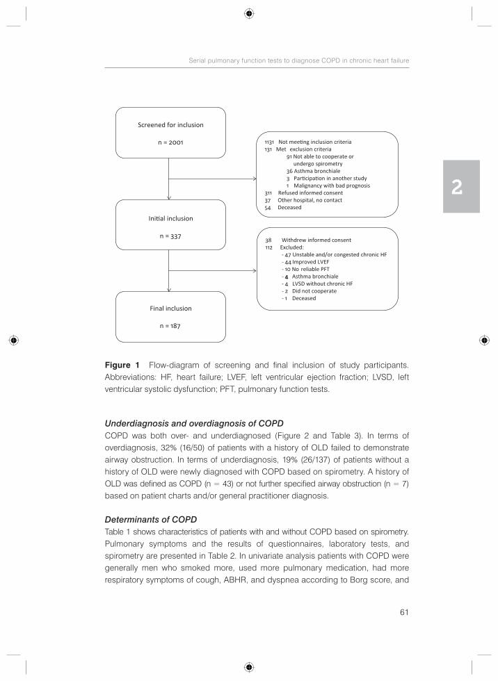
61
Serial pulmonary function tests to diagnose COPD in chronic heart failure
2
Underdiagnosis and overdiagnosis of COPDCOPD was both over- and underdiagnosed (Figure 2 and Table 3). In terms of overdiagnosis, 32% (16/50) of patients with a history of OLD failed to demonstrate airway obstruction. In terms of underdiagnosis, 19% (26/137) of patients without a history of OLD were newly diagnosed with COPD based on spirometry. A history of OLD was defined as COPD (n = 43) or not further specified airway obstruction (n = 7) based on patient charts and/or general practitioner diagnosis.
Determinants of COPDTable 1 shows characteristics of patients with and without COPD based on spirometry. Pulmonary symptoms and the results of questionnaires, laboratory tests, and spirometry are presented in Table 2. In univariate analysis patients with COPD were generally men who smoked more, used more pulmonary medication, had more respiratory symptoms of cough, ABHR, and dyspnea according to Borg score, and
Figure 1 Flow-diagram of screening and final inclusion of study participants. Abbreviations: HF, heart failure; LVEF, left ventricular ejection fraction; LVSD, left ventricular systolic dysfunction; PFT, pulmonary function tests.
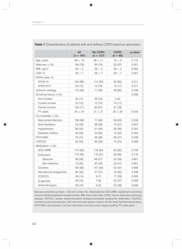
62
Chapter 2
Table 1 Characteristics of patients with and without COPD based on spirometry.
All (n = 187)
No COPD (n = 127)
COPD (n = 60)
p-value
Age, years 69 ± 10 68 ± 11 70 ± 9 0.173
Male sex, n (%) 146 (78) 94 (74) 52 (87) 0.051
BMI, kg/m2 28 ± 5 29 ± 5 28 ± 5 0.064
LVEF, % 29 ± 7 29 ± 7 29 ± 7 0.401
NYHA class, %
NYHA I-II 164 (88) 114 (90) 50 (83) 0.211
NYHA III-IV 23 (12) 13 (10) 10 (17) 0.211
Ischemic etiology 110 (59) 71 (56) 39 (65) 0.238
Smoking history, n (%) 0.008
Non-smoker 32 (17) 29 (23) 3 (5)
Current smoker 23 (12) 13 (10) 10 (17)
Former smoker 132 (71) 85 (67) 47 (78)
PY, years 24 ± 24 21 ± 21 30 ± 28 0.016
Co-morbidity, n (%)
Myocardial infarction 109 (58) 71 (56) 38 (63) 0.336
Atrial fibrillation 54 (29) 38 (30) 16 (27) 0.647
Hypertension 80 (43) 51 (40) 29 (48) 0.291
Diabetes mellitus 46 (25) 33 (26) 13 (22) 0.522
PCI/CABG 76 (41) 48 (38) 28 (47) 0.249
CRT/ICD 64 (34) 49 (39) 15 (25) 0.068
Medication, n (%)
ACE-I/ARB 174 (93) 119 (94) 55 (92) 0.759
β-blockers 172 (92) 116 (91) 56 (93) 0.778
Selective 99 (58) 66 (57) 33 (59) 0.801
Non-selective 73 (42) 50 (43) 23 (41) 0.801
Diuretics 159 (85) 107 (84) 52 (87) 0.666
Aldosterone-antagonists 65 (35) 47 (37) 18 (30) 0.348
ICS/OCS 26 (14) 9 (7) 17 (28) 0.000
β-agonists 29 (16) 7 (6) 22 (37) 0.000
Anticholinergics 29 (16) 6 (5) 23 (38) 0.000
Data are presented as mean ± SD and number (%). Abbreviations: ACE-I/ARB, angiotensin-converting enzyme inhibitor/angiotensin receptor blocker; BMI, body mass index; COPD, chronic obstructive pulmonary disease; CRT/ICD, cardiac resynchronization therapy/implantable cardioverter defibrillator; ICS/OCS, inhalation/oral corticosteroids; LVEF, left ventricular ejection fraction; NYHA, New York Heart Association; PCI/CABG, percutaneous coronary intervention/coronary artery bypass grafting; PY, pack-years.
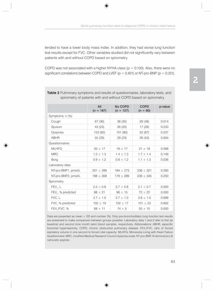
63
Serial pulmonary function tests to diagnose COPD in chronic heart failure
2
tended to have a lower body mass index. In addition, they had worse lung function test results except for FVC. Other variables studied did not significantly vary between patients with and without COPD based on spirometry.
COPD was not associated with a higher NYHA class (p = 0.130). Also, there were no significant correlations between COPD and LVEF (p = 0.401) or NT-pro-BNP (p = 0.251).
Table 2 Pulmonary symptoms and results of questionnaires, laboratory tests, and spirometry of patients with and without COPD based on spirometry.
All (n = 187)
No COPD (n = 127)
COPD (n = 60)
p-value
Symptoms, n (%)
Cough 67 (36) 38 (30) 29 (48) 0.014
Sputum 43 (23) 26 (20) 17 (28) 0.233
Dyspnea 153 (82) 101 (80) 52 (87) 0.237
ABHR 55 (29) 29 (23) 26 (43) 0.004
Questionnaires
MLHFQ 20 ± 17 19 ± 17 21 ± 18 0.568
MRC 1.5 ± 1.3 1.4 ± 1.3 1.7 ± 1.4 0.149
Borg 0.9 ± 1.2 0.8 ± 1.2 1.1 ± 1.3 0.036
Laboratory data
NT-pro-BNP1, pmol/L 201 ± 289 184 ± 273 236 ± 321 0.290
NT-pro-BNP2, pmol/L 198 ± 308 179 ± 289 239 ± 345 0.250
Spirometry
FEV1, L 2.5 ± 0.8 2.7 ± 0.8 2.1 ± 0.7 0.000
FEV1, % predicted 88 ± 21 96 ± 15 72 ± 22 0.000
FVC, L 3.7 ± 1.0 3.7 ± 1.0 3.8 ± 1.0 0.699
FVC, % predicted 102 ± 19 102 ± 17 101 ± 23 0.602
FEV1/FVC, % 68 ± 11 74 ± 5 55 ± 10 0.000
Data are presented as mean ± SD and number (%). Only pre-bronchodilator lung function test results are presented to make comparison between groups possible. Laboratory data 1 and 2 refer to first (at baseline) and second (one month later) blood samples, respectively. Abbreviations: ABHR, aspecific bronchial hyperreactivity; COPD, chronic obstructive pulmonary disease; FEV1/FVC, ratio of forced expiratory volume in one second to forced vital capacity; MLHFQ, Minnesota Living with Heart Failure Questionnaire; MRC, modified Medical Research Council dyspnea scale; NT-pro-BNP, N-terminal pro-B natriuretic peptide.
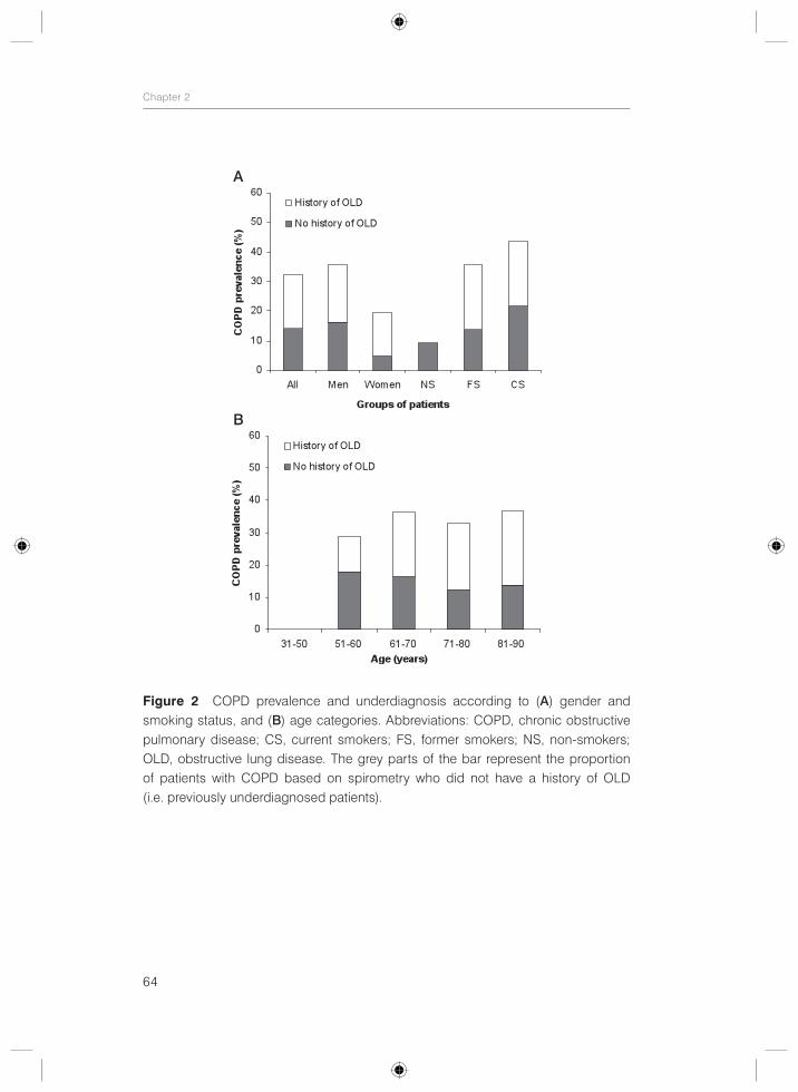
64
Chapter 2
Figure 2 COPD prevalence and underdiagnosis according to (A) gender and smoking status, and (B) age categories. Abbreviations: COPD, chronic obstructive pulmonary disease; CS, current smokers; FS, former smokers; NS, non-smokers; OLD, obstructive lung disease. The grey parts of the bar represent the proportion of patients with COPD based on spirometry who did not have a history of OLD (i.e. previously underdiagnosed patients).
A
B
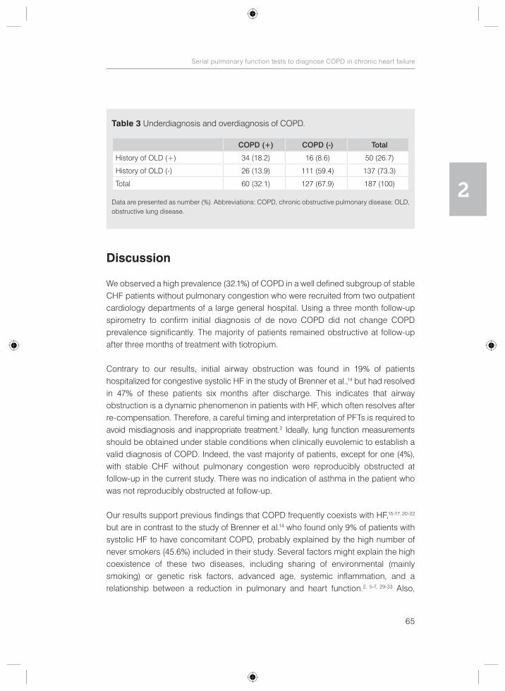
65
Serial pulmonary function tests to diagnose COPD in chronic heart failure
2
Discussion
We observed a high prevalence (32.1%) of COPD in a well defined subgroup of stable CHF patients without pulmonary congestion who were recruited from two outpatient cardiology departments of a large general hospital. Using a three month follow-up spirometry to confirm initial diagnosis of de novo COPD did not change COPD prevalence significantly. The majority of patients remained obstructive at follow-up after three months of treatment with tiotropium.
Contrary to our results, initial airway obstruction was found in 19% of patients hospitalized for congestive systolic HF in the study of Brenner et al.,14 but had resolved in 47% of these patients six months after discharge. This indicates that airway obstruction is a dynamic phenomenon in patients with HF, which often resolves after re-compensation. Therefore, a careful timing and interpretation of PFTs is required to avoid misdiagnosis and inappropriate treatment.2 Ideally, lung function measurements should be obtained under stable conditions when clinically euvolemic to establish a valid diagnosis of COPD. Indeed, the vast majority of patients, except for one (4%), with stable CHF without pulmonary congestion were reproducibly obstructed at follow-up in the current study. There was no indication of asthma in the patient who was not reproducibly obstructed at follow-up.
Our results support previous findings that COPD frequently coexists with HF,15-17, 20-22 but are in contrast to the study of Brenner et al.14 who found only 9% of patients with systolic HF to have concomitant COPD, probably explained by the high number of never smokers (45.6%) included in their study. Several factors might explain the high coexistence of these two diseases, including sharing of environmental (mainly smoking) or genetic risk factors, advanced age, systemic inflammation, and a relationship between a reduction in pulmonary and heart function.2, 5-7, 29-33 Also,
Table 3 Underdiagnosis and overdiagnosis of COPD.
COPD (+) COPD (-) Total
History of OLD (+) 34 (18.2) 16 (8.6) 50 (26.7)
History of OLD (-) 26 (13.9) 111 (59.4) 137 (73.3)
Total 60 (32.1) 127 (67.9) 187 (100)
Data are presented as number (%). Abbreviations: COPD, chronic obstructive pulmonary disease; OLD, obstructive lung disease.
66
Chapter 2
COPD patients are at an increased risk of co-morbidities such as type 2 diabetes, which in turn are an important risk for cardiovascular disease.30 Furthermore, factors that increase stress on the cardiovascular system or precipitate arrhythmic events can also explain the association between COPD and cardiovascular disease, including hypoxemia, hyperinflation, hyperventilation, neurohumoral disturbances, increased work of breathing and oxygen consumption, pulmonary hypertension, and the use of pulmonary medication.29, 30, 32, 34 Finally, other factors, such as oxidative stress, endothelial dysfunction, arterial stiffness, and connective tissue degradation have also been suggested to play a role.32, 34
Since spirometry is still underutilized even in a tertiary-care facility,19 it seems reasonable to consider routine spirometry testing in patients with CHF to diagnose or rule out COPD, a co-morbidity with important therapeutic and prognostic implications2,
10-16, 22 which is still greatly under- and overdiagnosed as found in this study (19% and 32%, respectively). Indeed, diagnostic difficulties have been stressed before, including the overlap in signs, symptoms, and risk factors, the underuse of spirometry despite the fact that objective evidence of airway obstruction is mandatory for diagnosing COPD, and difficulties with interpreting spirometry results, especially in patients with decompensated HF.2, 4-7, 14 This raises concerns regarding possible inappropriate treatment of COPD in an already vulnerable group of patients and as a result possible adverse impact on health and outcome.
Unfortunately, the current study was not large enough to find predictors of newly diagnosed COPD to make specific recommendations regarding which subgroup of patients should be tested. Also, it should be noted that a large proportion (69,2%) of newly diagnosed COPD patients had only mild airway obstruction that may represent a physiological decline of lung function with age instead of a disease.35, 36 It is unknown whether an additional diagnosis and treatment of COPD in these patients would improve health outcomes and change their prognosis. This warrants further research to establish the effectiveness of screening of patients with CHF for COPD in terms of symptomatic relief and improvement of the outcome as well as cost- effectiveness of such a policy. Until then, spirometry could be used in CHF patients with pulmonary symptoms despite an adequate treatment for their HF, especially in the presence of risk factors for COPD, such as a smoking history of ≥ 10 PY and occupational exposures. Importantly, spirometry should be used when clinically euvolemic to avoid both misdiagnosis and inappropriate treatment of COPD.2
The current study has some limitations that deserve further discussion. It is important to realize that the results may not be applicable to all patients with CHF, since we did not include patients with preserved systolic function. The diagnosis of HF with
67
Serial pulmonary function tests to diagnose COPD in chronic heart failure
2
preserved systolic function is challenging and particularly difficult to establish in patients with COPD.5 Thus, to avoid possible overestimation of COPD prevalence in our population, we only included patients with LVSD. Also, patients with more severe HF could have been under-represented in this study because of inability to participate. COPD prevalence may therefore have been somewhat underestimated. Likewise, there may be a recruitment bias in the cohort, given the refusal of 311 patients to provide informed consent. Finally, another limitation is the relatively small number of patients, particularly with COPD. However, included patient numbers are comparable to other recently published studies.16, 20-22
Despite these limitations, our findings have potential clinical implications. Our results indicate that confirmatory spirometry does not change a newly established diagnosis of COPD in the vast majority of patients with CHF, provided that PFTs are obtained during stable and non-congested conditions. The results also highlight the need for extensive use of spirometry to decrease the burden of undiagnosed or overdiagnosed COPD in patients with CHF. Evidently, the frequent underdiagnosis and overdiagnosis of COPD is not only a concern in the general population, but also in patients who are regularly monitored in outpatient cardiology clinics. Physicians should bear in mind that both diseases often coexist with important diagnostic and therapeutic difficulties and prognostic implications. Thus, both conditions must be simultaneously assessed and collaboration between cardiologists and pulmonologists is essential.
Conclusions
In conclusion, COPD is a frequent co-morbidity in patients with stable CHF without pulmonary congestion, but is often unrecognized or overdiagnosed. To avoid this and thus ensure adequate treatment of COPD in CHF, PFTs should be routinely obtained in a stable and non-congested condition. Under these conditions a confirmatory spirometry is unnecessary, as it does not change a newly established diagnosis of COPD in the vast majority of patients with CHF.
AcknowledgmentsThe authors would like to thank the participants in this study for their cooperation. We also would like to thank the nurse practitioners P.A. Ninaber, W.A.M. Janssen, W.H. van Zimmeren-Feijen, and heart failure nurse J.G. Froon-Elferink for their contribution to inclusion of participants and data collection. The authors owe much gratitude to the pulmonary function and clinical chemistry and haematology laboratories as well as the echocardiography and radiology departments for their assistance in data collection. Also, we would like to acknowledge biostatistician

68
Chapter 2
A.R.T. Donders for his contribution to statistical analysis. Finally, we would like to thank GlaxoSmithKline for supporting this study by an unrestricted grant. Glaxo-SmithKline had no involvement in study design, data collection, data analysis, interpretation of data, writing of the manuscript, or in the decision to submit the manuscript for publication.
69
Serial pulmonary function tests to diagnose COPD in chronic heart failure
2
References
1. Dickstein K, Cohen-Solal A, Filippatos G, McMurray JJ, Ponikowski P, Poole-Wilson PA, et al. ESC Guidelines for the diagnosis and treatment of acute and chronic heart failure 2008: the Task Force for the Diagnosis and Treatment of Acute and Chronic Heart Failure 2008 of the European Society of Cardiology. Developed in collaboration with the Heart Failure Association of the ESC (HFA) and endorsed by the European Society of Intensive Care Medicine (ESICM). Eur Heart J. 2008;29:2388-442.
2. Hawkins NM, Petrie MC, Jhund PS, Chalmers GW, Dunn FG, McMurray JJ. Heart failure and chronic obstructive pulmonary disease: diagnostic pitfalls and epidemiology. Eur J Heart Fail. 2009;11:130-9.
3. Hannink JD, van Helvoort HA, Dekhuijzen PN, Heijdra YF. Heart failure and COPD: partners in crime? Respirology. 2010;15:895-901.
4. Rutten FH, Cramer MJ, Lammers JW, Grobbee DE, Hoes AW. Heart failure and chronic obstructive pulmonary disease: An ignored combination? Eur J Heart Fail. 2006;8:706-11.
5. Le Jemtel TH, Padeletti M, Jelic S. Diagnostic and therapeutic challenges in patients with coexistent chronic obstructive pulmonary disease and chronic heart failure. J Am Coll Cardiol. 2007;49:171-80.
6. Lusuardi M, Garuti G, Massobrio M, Spagnolatti L, Bendinelli S. Heart and lungs in COPD. Close friends in real life--separate in daily medical practice? Monaldi Arch Chest Dis. 2008;69:11-7.
7. Padeletti M, Jelic S, LeJemtel TH. Coexistent chronic obstructive pulmonary disease and heart failure in the elderly. Int J Cardiol. 2008;125:209-15.
8. Dahlstrom U. Frequent non-cardiac comorbidities in patients with chronic heart failure. Eur J Heart Fail. 2005;7:309-16.
9. Hawkins NM, Jhund PS, Simpson CR, Petrie MC, Macdonald MR, Dunn FG, et al. Primary care burden and treatment of patients with heart failure and chronic obstructive pulmonary disease in Scotland. Eur J Heart Fail. 2010;12:17-24.
10. Rusinaru D, Saaidi I, Godard S, Mahjoub H, Battle C, Tribouilloy C. Impact of chronic obstructive pulmonary disease on long-term outcome of patients hospitalized for heart failure. Am J Cardiol. 2008;101:353-8.
11. Macchia A, Monte S, Romero M, D’Ettorre A, Tognoni G. The prognostic influence of chronic obstructive pulmonary disease in patients hospitalised for chronic heart failure. Eur J Heart Fail. 2007;9:942-8.
12. Staszewsky L, Wong M, Masson S, Barlera S, Carretta E, Maggioni AP, et al. Clinical, neurohormonal, and inflammatory markers and overall prognostic role of chronic obstructive pulmonary disease in patients with heart failure: data from the Val-HeFT heart failure trial. J Card Fail. 2007;13:797-804.
13. De Blois J, Simard S, Atar D, Agewall S. COPD predicts mortality in HF: the Norwegian Heart Failure Registry. J Card Fail. 2010;16:225-9.
14. Brenner S, Guder G, Berliner D, Deubner N, Frohlich K, Ertl G, et al. Airway obstruction in systolic heart failure - COPD or congestion? Int J Cardiol. 2013;168:1910-6.
15. Arnaudis B, Lairez O, Escamilla R, Fouilloux A, Fournier P, Monteil B, et al. Impact of chronic obstructive pulmonary disease severity on symptoms and prognosis in patients with systolic heart failure. Clin Res Cardiol. 2012;101:717-26.
16. Mascarenhas J, Lourenco P, Lopes R, Azevedo A, Bettencourt P. Chronic obstructive pulmonary disease in heart failure. Prevalence, therapeutic and prognostic implications. Am Heart J. 2008;155:521-5.
17. Iversen KK, Kjaergaard J, Akkan D, Kober L, Torp-Pedersen C, Hassager C, et al. Chronic obstructive pulmonary disease in patients admitted with heart failure. J Intern Med. 2008;264:361-9.
18. Rabe KF, Hurd S, Anzueto A, Barnes PJ, Buist SA, Calverley P, et al. Global strategy for the diagnosis, management, and prevention of chronic obstructive pulmonary disease: GOLD executive summary. Am J Respir Crit Care Med. 2007;176:532-55.
19. Damarla M, Celli BR, Mullerova HX, Pinto-Plata VM. Discrepancy in the use of confirmatory tests in patients hospitalized with the diagnosis of chronic obstructive pulmonary disease or congestive heart failure. Respir Care. 2006;51:1120-4.
20. Steinacher R, Parissis JT, Strohmer B, Eichinger J, Rottlaender D, Hoppe UC, et al. Comparison between ATS/ERS age- and gender-adjusted criteria and GOLD criteria for the detection of irreversible airway obstruction in chronic heart failure. Clin Res Cardiol. 2012;101:637-45.
70
Chapter 2
21. Boschetto P, Fucili A, Stendardo M, Malagu M, Parrinello G, Casimirri E, et al. Occurrence and impact of chronic obstructive pulmonary disease in elderly patients with stable heart failure. Respirology. 2013;18:125-30.
22. Macchia A, Rodriguez Moncalvo JJ, Kleinert M, Comignani PD, Gimeno G, Arakaki D, et al. Unrecognised ventricular dysfunction in COPD. Eur Respir J. 2012;39:51-8.
23. Minasian AG, van den Elshout FJ, Dekhuijzen PN, Vos PJ, Willems FF, van den Bergh PJ, et al. COPD in chronic heart failure: less common than previously thought? Heart Lung. 2013;42:365-71.
24. O’Hanlon R, O’Shea P, Ledwidge M, O’Loughlin C, Lange S, Conlon C, et al. The biologic variability of B-type natriuretic peptide and N-terminal pro-B-type natriuretic peptide in stable heart failure patients. J Card Fail. 2007;13:50-5.
25. Rector TS, Kubo SH, Cohn JN. Patients’ self-assessment of their congestive heart failure. Part 2: Content, reliability and validity of a new measure, the Minnesota Living with Heart Failure questionnaire. Heart Failure. 1987;Oct/Nov:198-209.
26. Bestall JC, Paul EA, Garrod R, Garnham R, Jones PW, Wedzicha JA. Usefulness of the Medical Research Council (MRC) dyspnoea scale as a measure of disability in patients with chronic obstructive pulmonary disease. Thorax. 1999;54:581-6.
27. Borg GA. Psychophysical bases of perceived exertion. Med Sci Sports Exerc. 1982;14:377-81.28. Quanjer PH, Tammeling GJ, Cotes JE, Pedersen OF, Peslin R, Yernault JC. Lung volumes and forced
ventilatory flows. Report Working Party Standardization of Lung Function Tests, European Community for Steel and Coal. Official Statement of the European Respiratory Society. Eur Respir J 1993;16:5-40.
29. Sin DD, Man SF. Chronic obstructive pulmonary disease: a novel risk factor for cardiovascular disease. Can J Physiol Pharmacol. 2005;83:8-13.
30. Finkelstein J, Cha E, Scharf SM. Chronic obstructive pulmonary disease as an independent risk factor for cardiovascular morbidity. Int J Chron Obstruct Pulmon Dis. 2009;4:337-49.
31. Barr RG, Bluemke DA, Ahmed FS, Carr JJ, Enright PL, Hoffman EA, et al. Percent emphysema, airflow obstruction, and impaired left ventricular filling. N Engl J Med. 2010;362:217-27.
32. Malerba M, Romanelli G. Early cardiovascular involvement in chronic obstructive pulmonary disease. Monaldi Arch Chest Dis. 2009;71:59-65.
33. Malerba M, Ragnoli B, Salameh M, Sennino G, Sorlini ML, Radaeli A, et al. Sub-clinical left ventricular diastolic dysfunction in early stage of chronic obstructive pulmonary disease. J Biol Regul Homeost Agents. 2011;25:443-51.
34. Macnee W, Maclay J, McAllister D. Cardiovascular injury and repair in chronic obstructive pulmonary disease. Proc Am Thorac Soc. 2008;5:824-33.
35. Hardie JA, Buist AS, Vollmer WM, Ellingsen I, Bakke PS, Morkve O. Risk of over-diagnosis of COPD in asymptomatic elderly never-smokers. Eur Respir J. 2002;20:1117-22.
36. Enright P, Brusasco V. Counterpoint: should we abandon FEV/FVC < 0.70 to detect airway obstruction? Yes. Chest. 2010;138:1040-2 [discussion 42-4].

71
Serial pulmonary function tests to diagnose COPD in chronic heart failure
2

Armine G. Minasian Frank J.J. van den ElshoutP.N. Richard DekhuijzenPetra J.E. VosFrank F. WillemsPaul J.P.C. van den BerghYvonne F. Heijdra
Heart Lung. 2013;42:365-71.
COPD in chronic heart failure: less common than previously thought?
CHAPTER 3
74
Chapter 3
Abstract
Background: Using the fixed ratio of forced expiratory volume in 1 s to forced vital capacity (FEV1/FVC) < 0.70 instead of the lower limit of normal (LLN) to define chronic obstructive pulmonary disease (COPD) may lead to overdiagnosis of COPD in elderly patients with heart failure (HF) and consequently unnecessary treatment with possible adverse health effects.
Objective: The aim of this study was to determine COPD prevalence in patients with chronic HF according to two definitions of airflow obstruction.
Methods: Spirometry was performed in 187 outpatients with stable chronic HF without pulmonary congestion who had a left ventricular ejection fraction < 40% (mean age 69 ± 10 years, 78% men). COPD diagnosis was confirmed three months after standard treatment with tiotropium in newly diagnosed COPD patients.
Results: COPD prevalence varied substantially between 19.8% (LLN-COPD) and 32.1% (GOLD-COPD). Twenty-three of 60 patients (38.3%) with GOLD-COPD were potentially misclassified as having COPD (FEV1/FVC < 0.7 but > LLN). In contrast to patients with LLN-COPD, potentially misclassified patients did not differ significantly from those without COPD regarding respiratory symptoms and risk factors for COPD.
Conclusions: One fifth, rather than one third, of the patients with chronic HF had concomitant COPD using the LLN instead of the fixed ratio. LLN may identify clinically more important COPD than the fixed ratio of 0.7.
Keywords: Chronic heart failure; chronic obstructive pulmonary disease; prevalence; fixed ratio; lower limit of normal.
75
COPD in chronic heart failure: less common than previously thought?
3
Introduction
Chronic obstructive pulmonary disease (COPD) frequently coexists with heart failure (HF), leading to poor prognosis as well as diagnostic and therapeutic challenges.1 However, estimates of COPD prevalence in patients with HF with reduced or preserved left ventricular ejection fraction (LVEF) vary substantially between 9% and 52%, depending on study design, population (age, gender, smoking habits, inpatients versus outpatients, acute versus chronic HF, primary, secondary, or tertiary care), and diagnostic criteria.1 Although spirometry is considered to be the gold standard for the diagnosis of COPD,2 data on the prevalence of COPD based on spirometry in patients with HF are scarce.3-7 Moreover, even when spirometry is used, in general there is still no consensus on how to define COPD.8-11 The Global Initiative for Chronic Obstructive Lung Disease (GOLD) guidelines recommend the use of the fixed ratio of forced expiratory volume in 1 s to forced vital capacity (FEV1/FVC) < 0.70 for the sake of simplicity.2 However, a growing body of literature indicates that considering the physiological decline of the FEV1/FVC ratio with age the use of the fixed ratio may lead to overdiagnosis of COPD in elderly subjects12-19 and underdiagnosis of COPD in young adults.15-17, 20 Therefore, to avoid misclassification, the American Thoracic Society/European Respiratory Society (ATS/ERS) recommends the use of statistically derived lower limit of normal (LLN) values for FEV1/VC that are based on the normal distribution and that classify the bottom 5% of the healthy population as abnormal.21 This is particularly important in patients with HF, given that HF is most prevalent among elderly individuals.22 Thus, COPD prevalence of 30-44% may have been over - estimated in prior studies that used the fixed ratio of 0.7 to define COPD in patients with HF.3-7 Subsequently, an incorrect diagnosis of COPD may result in unnecessary treatment for COPD and undertreatment with beta-blockers, with possible adverse health effects.8, 23, 24
Although population-based studies have shown that the application of different criteria to define airflow obstruction dramatically changes the prevalence of COPD,18, 25-30 it is less well understood to what extent this occurs in patients with chronic HF.6, 7 Therefore, the primary aim of this study was to determine COPD prevalence according to two definitions of airflow obstruction (FEV1/FVC < 0.70 versus FEV1/FVC < LLN) in outpatients with stable chronic HF with left ventricular systolic dysfunction (LVSD). The secondary aim of this study was to determine whether patients potentially misclassified as having COPD (FEV1/FVC < 0.7 but > LLN) had clinical features of COPD or those of the healthy population.
76
Chapter 3
Methods
Study design and participantsAll patients visiting two outpatient cardiology departments of a large general hospital in The Netherlands were screened for inclusion in this prospective observational study between October 2009 and December 2010. In addition, existing patient lists were used to ensure that the majority of the HF population had been examined for eligibility. Inclusion criteria were stable chronic HF with LVSD, i.e., LVEF < 40%, without pulmonary congestion, New York Heart Association (NYHA) class I-IV, and age ≥ 18 years. Chronic HF was defined according to the European Society of Cardiology guidelines.22 Echocardiography was performed in patients without a recent (≤ 6 months) echocardiography to confirm persisting LVSD. Patients were classified as having stable HF in the absence of hospitalization due to progression of HF within 3 months, change in diuretics within 1 month, 3% or more weight gain within 3 days, and more than 50% increase of N-terminal pro-B natriuretic peptide (NT-pro-BNP) within 1 month when the baseline NT-pro-BNP was 100 pmol/L or higher or more than 100 pmol/L increase of NT-pro-BNP within 1 month when baseline NT-pro-BNP was below 100 pmol/L.31 Pulmonary congestion was evaluated on standard posterior-an-terior and lateral chest radiographs for the presence or absence of alveolar edema, pleural effusion, Kerley-B lines, and/or the redistribution of pulmonary blood flow by independent radiologists who qualitatively assessed the chest radiographs with an overall clinical impression. Patients who were not able to cooperate or undergo spirometry or who had asthma according to their medical chart were excluded. Other exclusion criteria were malignancy with a poor prognosis (survival < 6 months) and participation in another study. Patients who had been hospitalized in the pulmonary department in the past six weeks were included six weeks after discharge to ensure that their pulmonary function was stable at the time of spirometry testing.
In conformity with the ethical guidelines of the 1975 Declaration of Helsinki, this study was conducted with the approval of the regional Research Ethics Committee Arnhem- Nijmegen in The Netherlands (2009/101, NL27798.091.09, ClinicalTrials.gov Identifier NCT01429376). All patients gave written informed consent.
Measurements and data collection At baseline, a first blood sample was taken for the measurement of NT-pro-BNP according to standard methods used in the hospital laboratory. One month later, the participants visited the hospital for an interview with the investigator and several examinations, including height and weight measurement, spirometry, and a chest radiograph. In addition, a second blood sample (NT-pro-BNP) was taken to determine the stability of HF. A 10-point Borg score32 was used to evaluate dyspnea at rest.
77
COPD in chronic heart failure: less common than previously thought?
3
Additional data were collected from medical records and personal interviews. Arterial blood gas analysis was performed on patients with severe airflow obstruction to determine whether they had chronic respiratory failure.2
Patients with newly diagnosed COPD according to either definition were followed up three months after standard treatment for COPD with once-daily 18 μg tiotropium. A third blood sample (NT-pro-BNP) was taken, and spirometry was repeated to confirm persistent airflow obstruction characteristic of COPD in an attempt to exclude asthma as much as possible. Thus only patients with persistent airflow obstruction on three months of follow-up were classified as having COPD.
Spirometry testingSpirometry (MasterLab Pro; Jaeger; Würzburg, Germany) was performed by trained and certified operators using standard techniques and according to ERS standards for acceptability and reproducibility.33 The reference values of the European Community for Coal and Steel were used.33 Subjects with airflow obstruction according to either definition underwent post-bronchodilator spirometry 30 minutes after inhalation of four doses of 100 μg aerosolized salbutamol and four doses of 20 μg aerosolized ipratropium via Volumatic spacer. Participants were instructed not to take broncho-dilators 6-24 hours before the tests, depending on the type of bronchodilator used. At follow-up, salbutamol and ipratropium were used, as previously described, when patients discontinued the use of tiotropium > 24 hours prior to spirometry. Care was taken to match the timing of the second spirometry testing to the first to reduce variations that may occur over a 24-hour period.
DefinitionsCOPD was defined according to two criteria: post-bronchodilator FEV1/FVC < 0.7 (GOLD-COPD)2 and post-bronchodilator FEV1/FVC < LLN (LLN-COPD).21 LLN was regarded as the lower fifth percentile of the frequency distribution of a healthy reference population and was calculated by subtracting 1.64 times the residual standard deviation from the predicted value. The investigator who identified the GOLD or LLN criteria was not blinded to the other rating.
Smoking status was defined as never (< 100 cigarettes in a lifetime), former (≥ 3 months ago), or current smoker (< 3 months). Smoking pack-years (PY) were based only on the tobacco cigarette history, and one PY was defined as smoking 20 cigarettes a day for 1 year.
Dyspnea was defined as resting dyspnea or dyspnea at any level of exertion, chronic cough as cough ≥ 3 months prior to the study, chronic sputum production as the
78
Chapter 3
regular production of sputum for ≥ 3 months in 2 consecutive years, and aspecific bronchial hyperreactivity (ABHR) as respiratory symptoms in response to perfumes, the scent of baking or paint, fog, cold air, or temperature changes.
Variables and measuresGOLD-COPD severity staging was determined on the basis of FEV1 percent predicted according to GOLD criteria: FEV1 ≥ 80% predicted (stage I, mild), 50% ≤ FEV1 < 80% predicted (stage II, moderate), 30% ≤ FEV1 < 50% predicted (stage III, severe), and FEV1 < 30% predicted or FEV1 < 50% predicted plus chronic respiratory failure (stage IV, very severe).2 LLN-COPD severity staging was determined on the basis of FEV1 percent predicted according to ATS/ERS criteria: FEV1 ≥ 70% predicted (mild), FEV1
60-69% predicted (moderate), FEV1 50-59% predicted (moderately severe), FEV1
35-49% predicted (severe), and FEV1 < 35% predicted (very severe).
Statistical analysisDescriptive data are presented as the mean ± SD or as a number (%). Baseline characteristics of patients with and without GOLD-COPD were compared using an independent t-test or a Mann-Whitney U test for continuous variables and a chi-square or Fisher’s exact test for categorical variables, as appropriate. Differences in continuous variables between three groups of patients (patients with LLN-COPD, patients with potentially misclassified COPD (FEV1/FVC < 0.7 but > LLN), and patients without COPD) were examined with independent analysis of variance. Post hoc analyses were performed using Fisher’s LSD test, and a Games-Howell test was used when the assumption of homogeneity of variance was not met. Log transformation was applied to achieve normal distribution when the assumption of normal distribution was not met. Differences in categorical variables between the aforementioned three groups of patients were analyzed with a chi-square test, and the Bonferroni correction was used to control for type I errors. Statistical analyses were performed using the Statistical Package for Social Science version 15.0. All statistical tests were two-sided, and a p-value < 0.05 was considered significant.
Results
Patient characteristicsA cohort of 337 patients with chronic HF was initially included in this study. Thirty-eight patients withdrew informed consent, and 65 patients were excluded for several reasons, as specified in Fig. 1. The remaining 234 patients were included in the study, of whom 187 had stable chronic HF without signs of congestion on chest radiograph. Table 1 shows the characteristics of these patients. The mean age was 69 ± 10 years,
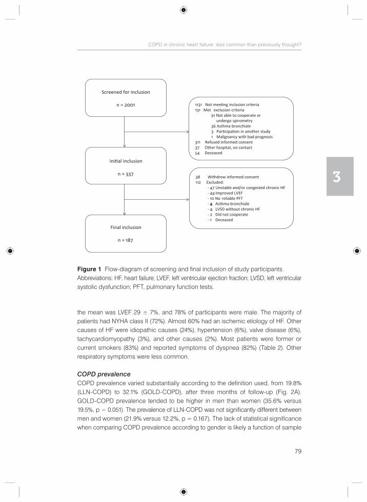
79
COPD in chronic heart failure: less common than previously thought?
3
the mean was LVEF 29 ± 7%, and 78% of participants were male. The majority of patients had NYHA class II (72%). Almost 60% had an ischemic etiology of HF. Other causes of HF were idiopathic causes (24%), hypertension (6%), valve disease (6%), tachycardiomyopathy (3%), and other causes (2%). Most patients were former or current smokers (83%) and reported symptoms of dyspnea (82%) (Table 2). Other respiratory symptoms were less common.
COPD prevalenceCOPD prevalence varied substantially according to the definition used, from 19.8% (LLN-COPD) to 32.1% (GOLD-COPD), after three months of follow-up (Fig. 2A). GOLD-COPD prevalence tended to be higher in men than women (35.6% versus 19.5%, p = 0.051). The prevalence of LLN-COPD was not significantly different between men and women (21.9% versus 12.2%, p = 0.167). The lack of statistical significance when comparing COPD prevalence according to gender is likely a function of sample
Figure 1 Flow-diagram of screening and final inclusion of study participants.Abbreviations: HF, heart failure; LVEF, left ventricular ejection fraction; LVSD, left ventricular systolic dysfunction; PFT, pulmonary function tests.
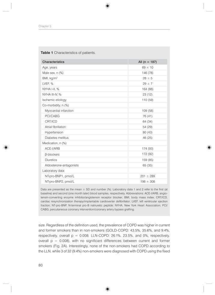
80
Chapter 3
size. Regardless of the definition used, the prevalence of COPD was higher in current and former smokers than in non-smokers (GOLD-COPD: 43.5%, 35.6%, and 9.4%, respectively, overall p = 0.008; LLN-COPD: 26.1%, 23.5%, and 0%, respectively, overall p = 0.008), with no significant differences between current and former smokers (Fig. 2A). Interestingly, none of the non-smokers had COPD according to the LLN, while 3 of 32 (9.4%) non-smokers were diagnosed with COPD using the fixed
Table 1 Characteristics of patients.
Characteristics All (n = 187)
Age, years 69 ± 10
Male sex, n (%) 146 (78)
BMI, kg/m2 28 ± 5
LVEF, % 29 ± 7
NYHA I-II, % 164 (88)
NYHA III-IV, % 23 (12)
Ischemic etiology 110 (59)
Co-morbidity, n (%)
Myocardial infarction 109 (58)
PCI/CABG 76 (41)
CRT/ICD 64 (34)
Atrial fibrillation 54 (29)
Hypertension 80 (43)
Diabetes mellitus 46 (25)
Medication, n (%)
ACE-I/ARB 174 (93)
β-blockers 172 (92)
Diuretics 159 (85)
Aldosterone-antagonists 65 (35)
Laboratory data
NT-pro-BNP1, pmol/L 201 ± 289
NT-pro-BNP2, pmol/L 198 ± 308
Data are presented as the mean ± SD and number (%). Laboratory data 1 and 2 refer to the first (at baseline) and second (one month later) blood samples, respectively. Abbreviations: ACE-I/ARB, angio-tensin-converting enzyme inhibitor/angiotensin receptor blocker; BMI, body mass index; CRT/ICD, cardiac resynchronization therapy/implantable cardioverter defibrillator; LVEF, left ventricular ejection fraction; NT-pro-BNP, N-terminal pro-B natriuretic peptide; NYHA, New York Heart Association; PCI/CABG, percutaneous coronary intervention/coronary artery bypass grafting.
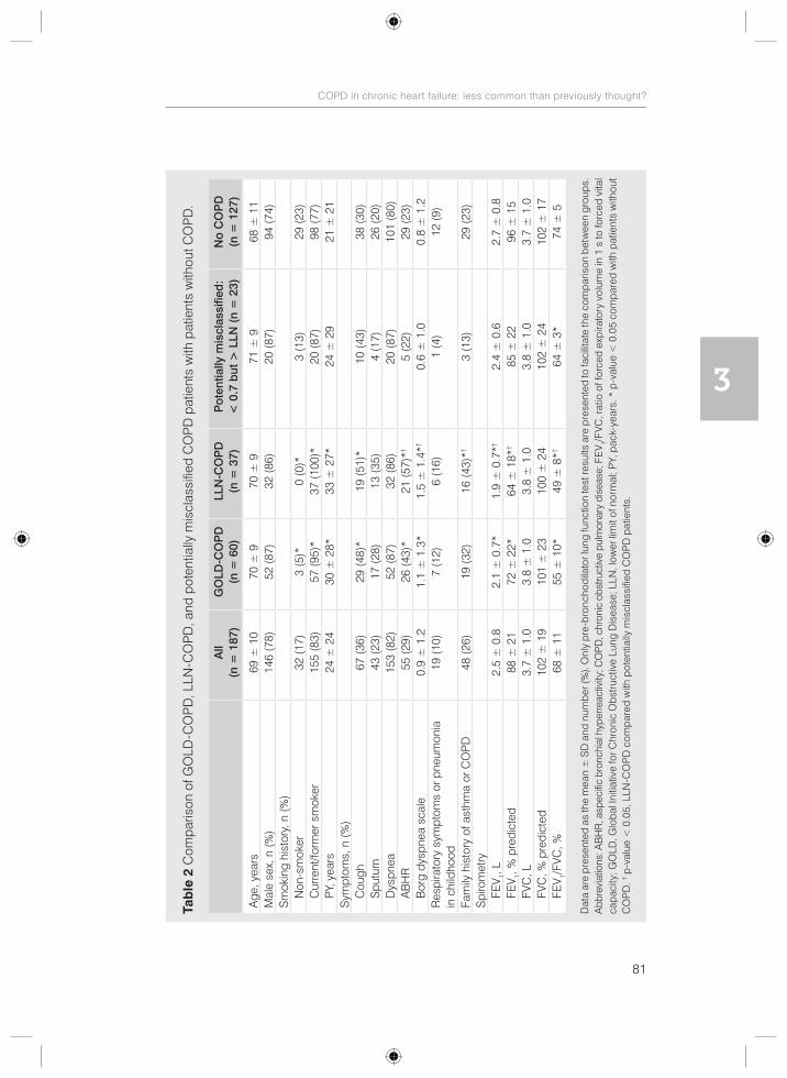
81
COPD in chronic heart failure: less common than previously thought?
3
Tab
le 2
Com
paris
on o
f GO
LD-C
OP
D, L
LN-C
OP
D, a
nd p
oten
tially
mis
clas
sifie
d C
OP
D p
atie
nts
with
pat
ient
s w
ithou
t CO
PD
.
A
ll (n
= 1
87)
GO
LD-C
OP
D(n
= 6
0)LL
N-C
OP
D
(n =
37)
Pot
entia
lly m
iscl
assi
fied
:<
0.7
but
> L
LN (
n =
23)
No
CO
PD
(n
= 1
27)
Age
, yea
rs69
± 1
070
± 9
70 ±
971
± 9
68 ±
11
Mal
e se
x, n
(%)
146
(78)
52 (8
7)32
(86)
20 (8
7)94
(74)
Sm
okin
g hi
stor
y, n
(%)
N
on-s
mok
er32
(17)
3 (5
)*0
(0)*
3 (1
3)29
(23)
C
urre
nt/fo
rmer
sm
oker
155
(83)
57 (9
5)*
37 (1
00)*
20 (8
7)98
(77)
PY
, yea
rs24
± 2
430
± 2
8*33
± 2
7*24
± 2
921
± 2
1S
ympt
oms,
n (%
)
Cou
gh67
(36)
29 (4
8)*
19 (5
1)*
10 (4
3)38
(30)
S
putu
m43
(23)
17 (2
8)13
(35)
4 (1
7)26
(20)
D
yspn
ea15
3 (8
2)52
(87)
32 (8
6)20
(87)
101
(80)
A
BH
R55
(29)
26 (4
3)*
21 (5
7)*†
5 (2
2)29
(23)
B
org
dysp
nea
scal
e0.
9 ±
1.2
1.1
± 1
.3*
1.5
± 1
.4*†
0.6
± 1
.00.
8 ±
1.2
Res
pira
tory
sym
ptom
s or
pne
umon
ia
in c
hild
hood
19 (1
0)7
(12)
6 (1
6)1
(4)
12 (9
)
Fam
ily h
isto
ry o
f ast
hma
or C
OP
D48
(26)
19 (3
2)16
(43)
*†3
(13)
29 (2
3)S
piro
met
ry
FE
V 1, L
2.5
± 0
.82.
1 ±
0.7
*1.
9 ±
0.7
*†2.
4 ±
0.6
2.7
± 0
.8
FEV 1,
% p
redi
cted
88 ±
21
72 ±
22*
64 ±
18*
†85
± 2
296
± 1
5
FVC
, L
3.7
± 1
.03.
8 ±
1.0
3.8
± 1
.03.
8 ±
1.0
3.7
± 1
.0
FVC
, % p
redi
cted
102
± 1
910
1 ±
23
100
± 2
410
2 ±
24
102
± 1
7
FEV 1/
FVC
, %68
± 1
155
± 1
0*49
± 8
*†64
± 3
*74
± 5
Dat
a ar
e p
rese
nted
as
the
mea
n ±
SD
and
num
ber (
%).
Onl
y p
re-b
ronc
hodi
lato
r lun
g fu
nctio
n te
st re
sults
are
pre
sent
ed to
faci
litat
e th
e co
mpa
rison
bet
wee
n gr
oups
. A
bbre
viat
ions
: AB
HR
, asp
ecifi
c br
onch
ial h
yper
reac
tivity
; CO
PD, c
hron
ic o
bstru
ctiv
e pu
lmon
ary
dise
ase;
FE
V 1/F
VC, r
atio
of f
orce
d ex
pira
tory
vol
ume
in 1
s to
forc
ed v
ital
capa
city
; GO
LD, G
loba
l Ini
tiativ
e fo
r Chr
onic
Obs
truc
tive
Lung
Dis
ease
; LLN
, low
er li
mit
of n
orm
al; P
Y, p
ack-
year
s. *
p-v
alue
< 0
.05
com
pare
d w
ith p
atie
nts
with
out
CO
PD
. † p-v
alue
< 0
.05,
LLN
-CO
PD
com
pare
d w
ith p
oten
tially
mis
clas
sifie
d C
OP
D p
atie
nts.
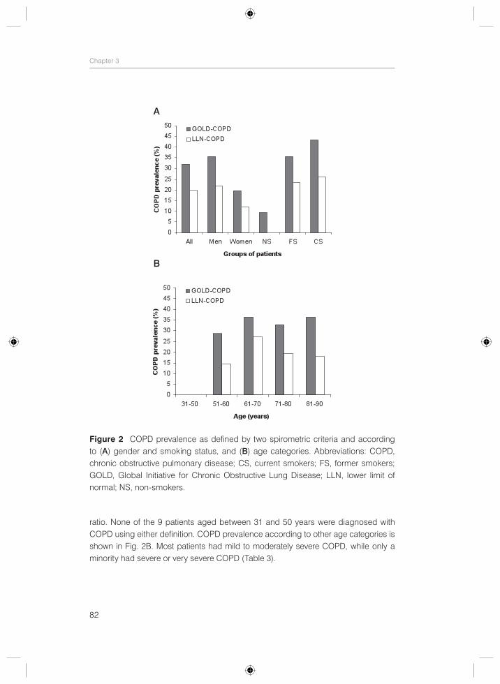
82
Chapter 3
ratio. None of the 9 patients aged between 31 and 50 years were diagnosed with COPD using either definition. COPD prevalence according to other age categories is shown in Fig. 2B. Most patients had mild to moderately severe COPD, while only a minority had severe or very severe COPD (Table 3).
Figure 2 COPD prevalence as defined by two spirometric criteria and according to (A) gender and smoking status, and (B) age categories. Abbreviations: COPD, chronic obstructive pulmonary disease; CS, current smokers; FS, former smokers; GOLD, Global Initiative for Chronic Obstructive Lung Disease; LLN, lower limit of normal; NS, non-smokers.
A
B
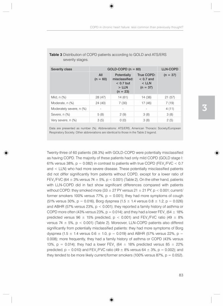
83
COPD in chronic heart failure: less common than previously thought?
3
Twenty-three of 60 patients (38.3%) with GOLD-COPD were potentially misclassified as having COPD. The majority of these patients had only mild COPD (GOLD stage I: 61% versus 38%, p = 0.082) in contrast to patients with true COPD (FEV1/FVC < 0.7 and < LLN) who had more severe disease. These potentially misclassified patients did not differ significantly from patients without COPD, except for a lower ratio of FEV1/FVC (64 ± 3% versus 74 ± 5%, p < 0.001) (Table 2). On the other hand, patients with LLN-COPD did in fact show significant differences compared with patients without COPD: they smoked more (33 ± 27 PY versus 21 ± 21 PY, p < 0.001; current/former smokers 100% versus 77%, p = 0.001); they had more symptoms of cough (51% versus 30%, p = 0.016), Borg dyspnea (1.5 ± 1.4 versus 0.8 ± 1.2, p = 0.026) and ABHR (57% versus 23%, p < 0.001); they reported a family history of asthma or COPD more often (43% versus 23%, p = 0.014); and they had a lower FEV1 (64 ± 18% predicted versus 96 ± 15% predicted, p < 0.001) and FEV1/FVC ratio (49 ± 8% versus 74 ± 5%, p < 0.001) (Table 2). Moreover, LLN-COPD patients also differed significantly from potentially misclassified patients: they had more symptoms of Borg dyspnea (1.5 ± 1.4 versus 0.6 ± 1.0, p = 0.019) and ABHR (57% versus 22%, p = 0.008); more frequently, they had a family history of asthma or COPD (43% versus 13%, p = 0.014); they had a lower FEV1 (64 ± 18% predicted versus 85 ± 22% predicted, p = 0.010) and FEV1/FVC ratio (49 ± 8% versus 64 ± 3%, p = 0.002); and they tended to be more likely current/former smokers (100% versus 87%, p = 0.052).
Table 3 Distribution of COPD patients according to GOLD and ATS/ERS severity stages.
Severity class GOLD-COPD (n = 60) LLN-COPD
All(n = 60)
Potentially misclassified:
< 0.7 but > LLN
(n = 23)
True COPD: < 0.7 and
< LLN (n = 37)
(n = 37)
Mild, n (%) 28 (47) 14 (61) 14 (38) 21 (57)
Moderate, n (%) 24 (40) 7 (30) 17 (46) 7 (19)
Moderately severe, n (%) - - - 4 (11)
Severe, n (%) 5 (8) 2 (9) 3 (8) 3 (8)
Very severe, n (%) 3 (5) 0 (0) 3 (8) 2 (5)
Data are presented as number (%). Abbreviations: ATS/ERS, American Thoracic Society/European Respiratory Society. Other abbreviations are identical to those in the Table 2 legend.
84
Chapter 3
Patients with GOLD-COPD differed significantly from patients without COPD on corresponding variables as those with LLN-COPD, except for one variable; in contrast to patients with LLN-COPD who had a family history of asthma or COPD significantly more often than those without COPD, this was not true for patients with GOLD-COPD (Table 2).
Thirty-four of 60 patients (56.7%) with GOLD-COPD had previously been diagnosed with obstructive lung disease, compared with 25 of 37 patients (67.6%) with LLN-COPD. On the other hand, 16 of 50 (32.0%) patients with a history of obstructive lung disease did not have GOLD-COPD according to their spirometry. The corresponding figure was 25/50 (50.0%) for LLN-COPD.
Discussion
This is one of the few studies to determine the prevalence of COPD in patients with chronic HF using two definitions of COPD, namely, the fixed ratio of 0.7 and the LLN.6, 7 Our results support previous findings that COPD frequently coexists with chronic HF.3-7 However, the exact definition of airflow obstruction alters COPD prevalence substantially; one fifth, rather than one third, of the patients with chronic HF had concomitant COPD using the LLN instead of the fixed ratio. LLN may identify clinically more important COPD than the fixed ratio of 0.7 as patients who were potentially misclassified as having COPD, in contrast to patients with LLN-COPD, did not differ significantly from those without COPD in terms of respiratory symptoms and risk factors for COPD.
As expected, using the fixed ratio of 0.7 resulted in a considerably higher prevalence of COPD compared with using the LLN (32.1% versus 19.8%, respectively). This finding is in line with the results of Steinacher et al.7 who reported COPD prevalence rates of 43.8% according to the GOLD criteria (fixed ratio) and 24.7% according to ATS/ERS criteria (LLN) in 89 outpatients with stable chronic HF. However, this finding was not as well supported in another study of 118 elderly (≥ 65 years) patients with stable chronic HF, most likely because of the selection of patients with ≥ 10 PY (COPD prevalence 30.5% versus 28.8%).6
Given the physiological decline of the FEV1/FVC ratio with age, using the fixed ratio instead of the LLN may result in a diagnosis of COPD in elderly individuals who may not actually have COPD12-19 which is of particular concern in patients with HF, given that most patients affected by HF are elderly22 (80.2% of the patients in the current study were older than 60 years). Almost 40% of the patients who were diagnosed with GOLD-COPD based on spirometry were potentially misclassified as having COPD because their FEV1/FVC ratio was above the LLN.
85
COPD in chronic heart failure: less common than previously thought?
3
In clinical practice, decisions are not made on the basis of a single test. Clinical findings, including history and exposure to risk factors, can facilitate the diagnosis of COPD,2 and the physician ultimately determines the medical significance of an abnormal spirometric value based on these clinical findings. However, these decisions are more complicated in patients with HF due to overlap in signs and symptoms as well as risk factors,1 which explains, at least in part, the considerable over- and underdiagnosis of COPD observed in this study. The majority (74%) of patients with potentially misclassified COPD had respiratory symptoms (dyspnea, cough, and/or sputum production) combined with a smoking history (current or former smoker), which was also true for patients without COPD, of whom 64% had respiratory symptoms combined with a smoking history. Subsequently, an incorrect diagnosis of COPD and unnecessary treatment as a consequence may be associated with side-effects of pharmacological interventions,34 especially in elderly patients with HF who usually have several co-morbidities and are prone to polypharmacy. In addition, concerns have been raised regarding the cardiovascular safety profile of bronchodilators. Beta-agonists have been reported to increase the risk for adverse cardiovascular events in patients with obstructive airway disease, with a significant increase in sinus tachycardia and a non-significant trend toward an increase in major cardiovascular events, including ventricular tachycardia, atrial fibrillation, syncope, congestive heart failure, myocardial infarction, cardiac arrest, and sudden death.35 Moreover, observational studies have shown worse outcomes with bronchodilator use in patients with HF, including increased risk of HF hospitalization, increased mortality rates, in-hospital mechanical ventilation, intravenous vasodilator use, and major cardiovascular events associated with the use of beta-agonists, although further investigation is warranted.23, 36 Furthermore, inhaled anticholinergics have been reported to be associated with an increased risk of cardiovascular death, myocardial infarction, or stroke among patients with COPD,37 although recently reassuring cardiovascular safety data have been reported on the long-acting an-ticholinergic bronchodilator tiotropium (HandiHaler).38, 39 Nevertheless, bronchodila-tors, in particular beta-agonists, must be used with caution in patients with underlying cardiac condition such as HF.
Aside from the possible side-effects and adverse cardiovascular events associated with pharmacological treatment for COPD, incorrect diagnosis and interventions for COPD may have a considerable psychological impact on the subject and his/her family and may cause an unnecessary financial burden on society.8 Furthermore, incorrect diagnosis of COPD may lead to undertreatment with life-saving beta-blockers.23, 24 Therefore, there is a need for clear diagnostic criteria for COPD to avoid diagnostic confusion, incorrect diagnosis, and inappropriate treatment.

86
Chapter 3
Unfortunately, there is no gold standard for the diagnosis of COPD. The hallmark of the disease is the presence of airflow limitation that is not fully reversible and is usually progressive in nature.2 However, there is no consensus on the spirometric criteria for the diagnosis of COPD.8-11 Considering the physiological decline of the FEV1/FVC ratio with age, the application of statistically derived LLN values for FEV1/FVC that are based on the normal distribution and that classify the bottom 5% of the healthy population as abnormal should be preferred to avoid overdiagnosis of COPD in elderly subjects12-19 and underdiagnosis in young adults.15-17, 20 However, little is known about the clinical impact of these different criteria and contrasting results have been reported.12, 20, 40-47 More longitudinal studies are needed to determine which criterion is better and clinically more relevant.48
In the current study, patients who were potentially misclassified as having COPD did not differ significantly from those without COPD in terms of respiratory symptoms and risk factors for COPD. On the contrary, patients with LLN-COPD did in fact show significant differences when they were compared with patients without COPD in terms of respiratory symptoms (cough, Borg dyspnea, and ABHR) and risk factors for COPD (smoking history and a family history of asthma or COPD). Moreover, patients with LLN-COPD more frequently had respiratory symptoms (Borg dyspnea and ABHR) and a family history of asthma or COPD compared with patients with potentially misclassified COPD, and they more frequently tended to be current or former smokers. Although this study is not longitudinal in nature, these findings imply that using the LLN may identify clinically more important COPD than the fixed ratio of 0.7. In support of this implication, 67.6% of the patients with LLN-COPD were previously diagnosed with obstructive lung disease, compared to only 39.1% of patients with potentially misclassified COPD.
A limitation of the LLN criterion is its dependence on the prediction equations and on the reference population from which the prediction equations have been drawn. In the USA, ethnically appropriate National Health and Nutrition Examination Survey (NHANES) III reference equations are recommended for those aged 8-80 years.49 In Europe, the combined reference equations published in the 1993 ERS statement are often used for people aged 18-70 years, with a height range of 155-195 cm in males, and 145-180 cm in females.33 Recent ATS/ERS guidelines do not recommend any specific set of equations to be used in Europe, but they do suggest the need for a new Europe-wide study to derive updated reference equations.21 Recently, multi-ethnic spirometric prediction equations for the 3-95 years age range that include appropriate age-dependent lower limits of normal and that can be applied globally have become available.50
87
COPD in chronic heart failure: less common than previously thought?
3
A limitation to our study is the lack of follow-up to determine how the different spirometric definitions of COPD relate to outcomes such as pulmonary function decline, hospitalization, and mortality. Furthermore, it is important to realize that the prevalence of COPD found in this study may not be applicable to all patients with chronic HF because we did not include patients with preserved systolic function. Additionally, patients with more severe HF could have been under-represented in this study because of their inability to participate.
Clinical implications and implications for future researchDespite these limitations, our findings have potential clinical implications and implications for future research. The results stress the need for a clear definition of COPD, especially in patients with HF in whom the diagnosis of COPD is already complicated due to overlap in signs, symptoms, and risk factors and who are prone to the adverse effects of pharmacological treatment for COPD. Using the LLN results in considerably lower COPD prevalence rates when compared with using the fixed ratio of 0.7: one fifth, rather than one third, of the patients with chronic HF had concomitant COPD. Moreover, LLN may identify clinically more important COPD than the fixed ratio. More longitudinal studies are needed to determine which criterion is better and clinically more relevant in terms of morbidity (symptoms, exercise tolerance, health-related quality of life, exacerbations, hospitalization, pulmonary function decline, use of health recourses, systemic effects such as co-morbidities and systemic biomarkers) and mortality. Finally, future research should focus on the clinical benefit of treating COPD according to either definition in patients with HF and the cardiovascular safety profile of bronchodilators, especially in patients with underlying cardiac condition such as HF.
AcknowledgmentsThe authors would like to thank the participants in this study for their cooperation. We also would like to thank the nurse practitioners P.A. Ninaber, W.A.M. Janssen, W.H. van Zimmeren-Feijen, and heart failure nurse J.G. Froon-Elferink for their contribution to inclusion of participants and data collection. The authors owe much gratitude to the pulmonary function and clinical chemistry and hematology laboratories as well as the echocardiography and radiology departments for their assistance in data collection. Also, we would like to acknowledge biostatistician A.R.T. Donders for his contribution to statistical analysis. Finally, we thank GlaxoSmithKline for supporting this study by an unrestricted grant.
88
Chapter 3
References
1. Hawkins NM, Petrie MC, Jhund PS, Chalmers GW, Dunn FG, McMurray JJ. Heart failure and chronic obstructive pulmonary disease: diagnostic pitfalls and epidemiology. Eur J Heart Fail. 2009;11:130-9.
2. Global Initiative for Chronic Obstructive Lung Disease. Global strategy for the diagnosis, management, and prevention of chronic obstructive pulmonary disease. NHBLI/WHO workshop report. Available at: http://www.goldcopd.org; 2011.
3. Iversen KK, Kjaergaard J, Akkan D, Kober L, Torp-Pedersen C, Hassager C, et al. Chronic obstructive pulmonary disease in patients admitted with heart failure. J Intern Med. 2008;264:361-9.
4. Mascarenhas J, Lourenco P, Lopes R, Azevedo A, Bettencourt P. Chronic obstructive pulmonary disease in heart failure. Prevalence, therapeutic and prognostic implications. Am Heart J. 2008;155:521-5.
5. Macchia A, Rodriguez Moncalvo JJ, Kleinert M, Comignani PD, Gimeno G, Arakaki D, et al. Unrecognised ventricular dysfunction in COPD. Eur Respir J. 2012;39:51-8.
6. Boschetto P, Fucili A, Stendardo M, Malagu M, Parrinello G, Casimirri E, et al. Occurrence and impact of chronic obstructive pulmonary disease in elderly patients with stable heart failure. Respirology. 2013;18:125-30.
7. Steinacher R, Parissis JT, Strohmer B, Eichinger J, Rottlaender D, Hoppe UC, et al. Comparison between ATS/ERS age- and gender-adjusted criteria and GOLD criteria for the detection of irreversible airway obstruction in chronic heart failure. Clin Res Cardiol. 2012;101:637-45.
8. Quanjer PH, Enright PL, Miller MR, Stocks J, Ruppel G, Swanney MP, et al. Open letter: the need to change the method for defining mild airway obstruction. Prim Care Respir J. 2010;19:288-91.
9. Celli BR, Halbert RJ. Point: should we abandon FEV/FVC <0.70 to detect airway obstruction? No. Chest. 2010;138:1037-40.
10. Enright P, Brusasco V. Counterpoint: should we abandon FEV/FVC < 0.70 to detect airway obstruction? Yes. Chest. 2010;138:1040-2 [discussion 42-4].
11. Townsend MC. Conflicting definitions of airways obstruction: Drawing the line between normal and abnormal. Chest. 2007;131:335-6.
12. Garcia-Rio F, Soriano JB, Miravitlles M, Munoz L, Duran-Tauleria E, Sanchez G, et al. Overdiagnosing Subjects With COPD Using the 0.7 Fixed Ratio: Correlation With a Poor Health-Related Quality of Life. Chest. 2011;139:1072-80.
13. Hansen JE, Sun XG, Wasserman K. Spirometric criteria for airway obstruction: Use percentage of FEV1/FVC ratio below the fifth percentile, not < 70%. Chest. 2007;131:349-55.
14. Hardie JA, Buist AS, Vollmer WM, Ellingsen I, Bakke PS, Morkve O. Risk of over-diagnosis of COPD in asymptomatic elderly never-smokers. Eur Respir J. 2002;20:1117-22.
15. Hnizdo E, Glindmeyer HW, Petsonk EL, Enright P, Buist AS. Case definitions for chronic obstructive pulmonary disease. Copd. 2006;3:95-100.
16. Miller MR, Quanjer PH, Swanney MP, Ruppel G, Enright PL. Interpreting lung function data using 80 percent of predicted and fixed thresholds misclassifies over 20% of patients. Chest. 2011;139:52-59.
17. Roberts SD, Farber MO, Knox KS, Phillips GS, Bhatt NY, Mastronarde JG, et al. FEV1/FVC ratio of 70% misclassifies patients with obstruction at the extremes of age. Chest. 2006;130:200-6.
18. Swanney MP, Ruppel G, Enright PL, Pedersen OF, Crapo RO, Miller MR, et al. Using the lower limit of normal for the FEV1/FVC ratio reduces the misclassification of airway obstruction. Thorax. 2008;63:1046-51.
19. Schermer TR, Smeele IJ, Thoonen BP, Lucas AE, Grootens JG, van Boxem TJ, et al. Current clinical guideline definitions of airflow obstruction and COPD overdiagnosis in primary care. Eur Respir J. 2008;32:945-52.
20. Cerveri I, Corsico AG, Accordini S, Niniano R, Ansaldo E, Anto JM, et al. Underestimation of airflow obstruction among young adults using FEV1/FVC <70% as a fixed cut-off: a longitudinal evaluation of clinical and functional outcomes. Thorax. 2008;63:1040-5.
21. Pellegrino R, Viegi G, Brusasco V, Crapo RO, Burgos F, Casaburi R, et al. Interpretative strategies for lung function tests. Eur Respir J. 2005;26:948-68.
22. Dickstein K, Cohen-Solal A, Filippatos G, McMurray JJ, Ponikowski P, Poole-Wilson PA, et al. ESC Guidelines for the diagnosis and treatment of acute and chronic heart failure 2008: the Task Force for the Diagnosis and Treatment of Acute and Chronic Heart Failure 2008 of the European Society of
89
COPD in chronic heart failure: less common than previously thought?
3
Cardiology. Developed in collaboration with the Heart Failure Association of the ESC (HFA) and endorsed by the European Society of Intensive Care Medicine (ESICM). Eur Heart J. 2008;29:2388-442.
23. Hawkins NM, Petrie MC, Macdonald MR, Jhund PS, Fabbri LM, Wikstrand J, et al. Heart failure and chronic obstructive pulmonary disease the quandary of Beta-blockers and Beta-agonists. J Am Coll Cardiol. 2011;57:2127-38.
24. Egred M, Shaw S, Mohammad B, Waitt P, Rodrigues E. Under-use of beta-blockers in patients with ischaemic heart disease and concomitant chronic obstructive pulmonary disease. Qjm. 2005;98:493-7.
25. Celli BR, Halbert RJ, Isonaka S, Schau B. Population impact of different definitions of airway obstruction. Eur Respir J. 2003;22:268-73.
26. Glaser S, Schaper C, Obst A, Ittermann T, Volzke H, Felix SB, et al. Impact of different definitions of airflow limitation on the prevalence of chronic obstructive pulmonary disease in the general population. Respiration. 2010;80:292-300.
27. Lindberg A, Jonsson AC, Ronmark E, Lundgren R, Larsson LG, Lundback B. Prevalence of chronic obstructive pulmonary disease according to BTS, ERS, GOLD and ATS criteria in relation to doctor’s diagnosis, symptoms, age, gender, and smoking habits. Respiration. 2005;72:471-9.
28. Shirtcliffe P, Weatherall M, Marsh S, Travers J, Hansell A, McNaughton A, et al. COPD prevalence in a random population survey: a matter of definition. Eur Respir J. 2007;30:232-9.
29. Vollmer WM, Gislason T, Burney P, Enright PL, Gulsvik A, Kocabas A, et al. Comparison of spirometry criteria for the diagnosis of COPD: results from the BOLD study. Eur Respir J. 2009;34:588-97.
30. Viegi G, Pedreschi M, Pistelli F, Di Pede F, Baldacci S, Carrozzi L, et al. Prevalence of airways obstruction in a general population: European Respiratory Society vs American Thoracic Society definition. Chest. 2000;117:339S-45S.
31. O’Hanlon R, O’Shea P, Ledwidge M, O’Loughlin C, Lange S, Conlon C, et al. The biologic variability of B-type natriuretic peptide and N-terminal pro-B-type natriuretic peptide in stable heart failure patients. J Card Fail. 2007;13:50-5.
32. Borg GA. Psychophysical bases of perceived exertion. Med Sci Sports Exerc. 1982;14:377-81.33. Quanjer PH, Tammeling GJ, Cotes JE, Pedersen OF, Peslin R, Yernault JC. Lung volumes and forced
ventilatory flows. Report Working Party Standardization of Lung Function Tests, European Community for Steel and Coal. Official Statement of the European Respiratory Society. Eur Respir J 1993;16:5-40.
34. http://www.fk.cvz.nl/.35. Salpeter SR, Ormiston TM, Salpeter EE. Cardiovascular effects of beta-agonists in patients with
asthma and COPD: a meta-analysis. Chest. 2004;125:2309-21.36. Maak CA, Tabas JA, McClintock DE. Should acute treatment with inhaled beta agonists be withheld
from patients with dyspnea who may have heart failure? J Emerg Med. 2011;40:135-45.37. Singh S, Loke YK, Furberg CD. Inhaled anticholinergics and risk of major adverse cardiovascular
events in patients with chronic obstructive pulmonary disease: a systematic review and meta-analysis. Jama. 2008;300:1439-50.
38. Salpeter SR. Do inhaled anticholinergics increase or decrease the risk of major cardiovascular events?: a synthesis of the available evidence. Drugs. 2009;69:2025-33.
39. Michele TM, Pinheiro S, Iyasu S. The safety of tiotropium--the FDA’s conclusions. N Engl J Med. 2010; 363:1097-9.
40. Mannino DM, Diaz-Guzman E. Interpreting lung function data using 80% predicted and fixed thresholds identifies patients at increased risk of mortality. Chest. 2012;141:73-80.
41. Mannino DM, Sonia Buist A, Vollmer WM. Chronic obstructive pulmonary disease in the older adult: what defines abnormal lung function? Thorax. 2007;62:237-41.
42. Izquierdo Alonso JL, De Lucas Ramos P, Rodriguez Glez-Moro JM. The use of the lower limit of normal as a criterion for COPD excludes patients with increased morbidity and high consumption of health-care resources. Arch Bronconeumol. 2012;48:223-8.
43. Akkermans RP, Berrevoets MA, Smeele IJ, Lucas AE, Thoonen BP, Grootens-Stekelenburg JG, et al. Lung function decline in relation to diagnostic criteria for airflow obstruction in respiratory symptomatic subjects. BMC Pulm Med. 2012;12:12.

90
Chapter 3
44. Guder G, Brenner S, Angermann CE, Ertl G, Held M, Sachs AP, et al. “GOLD or lower limit of normal definition? A comparison with expert-based diagnosis of chronic obstructive pulmonary disease in a prospective cohort-study”. Respir Res. 2012;13:13.
45. Brusasco V. Spirometric definition of COPD: exercise in futility or factual debate? Thorax. 2012;67:569-70.46. Lamprecht B, Schirnhofer L, Kaiser B, Buist SA, Mannino DM, Studnicka M. Subjects with Discordant
Airways Obstruction: Lost between Spirometric Definitions of COPD. Pulm Med. 2011;2011:780215.47. Mohamed Hoesein FA, Zanen P, Sachs AP, Verheij TJ, Lammers JW, Broekhuizen BD. Spirometric
thresholds for diagnosing COPD: 0.70 or LLN, pre- or post-dilator values? Copd. 2012;9:338-43.48. Mohamed Hoesein FA, Zanen P, Lammers JW. Lower limit of normal or FEV(1)/FVC <0.70 in diagnosing
COPD: An evidence-based review. Respir Med. 2011;105:907-15.49. Hankinson JL, Odencrantz JR, Fedan KB. Spirometric reference values from a sample of the general
U.S. population. Am J Respir Crit Care Med. 1999;159:179-87.50. Quanjer PH, Stanojevic S, Cole TJ, Baur X, Hall GL, Culver BH, et al. Multi-ethnic reference values for
spirometry for the 3-95-yr age range: the global lung function 2012 equations. Eur Respir J. 2012;40: 1324-43.

91
COPD in chronic heart failure: less common than previously thought?
3

Armine G. Minasian Frank J.J. van den ElshoutP.N. Richard DekhuijzenPetra J.E. VosFrank F. WillemsPaul J.P.C. van den BerghYvonne F. Heijdra
Heart Lung. 2014;43:311-6.
Pulmonary function impairment in patients with chronic heart failure:
lower limit of normal versus conventional cutoff values
CHAPTER 4
94
Chapter 4
Abstract
Objective: To determine the prevalence of pulmonary function abnormalities in patients with chronic heart failure (HF) according to recent American Thoracic Society/European Respiratory Society (ATS/ERS) guidelines using the lower limit of normal (LLN) compared to conventional cutoff values.
Background: Recent ATS/ERS guidelines recommend the use of the LLN instead of the conventional cutoff values to define pulmonary function impairment to avoid misclassification of patients. However, studies addressing the prevalence of pulmonary function abnormalities according to both definitions in patients with chronic HF are lacking.
Results: Diffusion impairment and airway obstruction were found in 44-58% and 26-37% of the patients, respectively, depending on the definition used (LLN versus conventional cutoff values, p < 0.05). However, restriction was infrequent, irrespective of the definition used (7% versus 5%, respectively, p > 0.05). The LLN identified fewer patients with abnormal lung function, whereas the conventional cutoff values classified more patients with diffusion impairment, airway obstruction, or a mixed category. Twenty-seven percent of patients were misclassified by the conventional cutoff values.
Conclusion: Pulmonary function abnormalities, especially diffusion impairment and airway obstruction, were highly prevalent in patients with chronic HF. Conventional cutoff values classified more patients with diffusion impairment, airway obstruction, or a mixed category compared to the LLN.
Keywords: Chronic heart failure, conventional cutoff values, lower limit of normal, prevalence, pulmonary function impairment.
95
Pulmonary function impairment in chronic heart failure
4
Introduction
Isolated or combined pulmonary function abnormalities, such as restriction, diffusion impairment, and to a lesser extent airway obstruction are common in patients with chronic heart failure (HF)1-7 and can contribute to the perception of dyspnea8 and exercise intolerance.8-12 Reported prevalence rates vary between 41% and 93% for diffusion impairment,13-19 between 21% and 55% for restriction,13-17, 20 and between 14% and 60% for airway obstruction.13-18, 20, 21 These varying rates across the studies can be partly explained by the usually small number of patients included and differences in study population as well as diagnostic criteria used to define pulmonary function abnormalities. All studies except for one20 have used conventional cutoff values to define an abnormality. However, the 80% predicted value (i.e. diffusing capacity or total lung capacity < 80% predicted) and the fixed ratio of 0.7 (i.e. forced expiratory volume in 1s to forced vital capacity (FEV1/FVC) < 0.7) that are traditionally used may misclassify more than 20% of patients leading to false-positive diagnosis in the elderly and underdiagnosis in younger patients.22 This is explained by the physiological decrease of the FEV1/FVC ratio with age because the FEV1 declines more rapidly with age than the FVC in normal subjects.23 Furthermore, the frequently used 80% predicted value has neither statistical nor physiological validity.24 To avoid misclassifi-cation, recent American Thoracic Society/European Respiratory Society (ATS/ERS) guidelines25 recommend the use of statistically derived lower limit of normal (LLN) values that are based on the normal distribution and that classify the bottom 5% of the healthy population as abnormal. However, studies using the LLN values to assess the prevalence of pulmonary function abnormalities in patients with HF are lacking. Therefore, the purpose of this study was to determine the prevalence of pulmonary function abnormalities according to recent ATS/ERS guidelines using the LLN in comparison to conventional cutoff values in outpatients with chronic HF.
Methods
Study design and participantsThis study was part of a larger prospective cross-sectional study evaluating the prevalence of chronic obstructive pulmonary disease (COPD) in patients with chronic HF (ClinicalTrials.gov Identifier NCT01429376). All patients visiting two outpatient cardiology departments of a large hospital in The Netherlands were screened for inclusion between October 2009 and December 2010. Inclusion criteria were chronic HF26 with left ventricular systolic dysfunction (LVSD), i.e. left ventricular ejection fraction (LVEF) < 40%, and New York Heart Association class (NYHA) I-IV. Chronic HF was defined according to the European Society of Cardiology guidelines as a clinical
96
Chapter 4
syndrome with symptoms and signs typical of HF and objective evidence of a structural or functional abnormality of the heart at rest.26 Echocardiography was performed in patients without a recent (≤ 6 months) echocardiography to confirm persisting LVSD. Patients who were not able to cooperate or undergo reliable spirometry according to ERS standards for acceptability and reproducibility27 or who had a history of asthma were excluded. Other exclusion criteria were malignancy with a poor prognosis (survival < 6 months) and participation in another study. For the current study we also excluded patients with known pulmonary, pleural (with exception of pleural effusion due to HF), neuromuscular, collagen vascular, or other diseases that could affect pulmonary function. Patients with a body mass index (BMI) above 35 were excluded from the restriction prevalence analysis.
Patients were classified as having stable HF in the absence of hospitalization due to progression of HF within 3 months, change in diuretics within 1 month, 3% or more weight gain within 3 days, and more than 50% increase of N-terminal pro-B natriuretic peptide (NT-pro-BNP) within 1 month when the baseline NT-pro-BNP was 100 pmol/L or higher or more than 100 pmol/L increase of NT-pro-BNP within 1 month when baseline NT-pro-BNP was below 100 pmol/L.28
Measurements and data collection At baseline, a first blood sample was taken for the measurement of NT-pro-BNP. One month later, the participants visited the hospital for an interview with the investigator and several examinations, including height and weight measurement, pulmonary function tests (PFTs), a chest radiograph, and a second blood sample (hemoglobin, NT-pro-BNP). Additional data were collected from medical records and personal interviews. Smoking status was defined as never (< 100 cigarettes in a lifetime), former (≥ 3 months ago), or current smoker (< 3 months). Smoking pack-years (PY) were based only on the tobacco cigarette history and one PY was defined as smoking twenty cigarettes a day for one year.
Pulmonary function testsAll participants underwent pre-bronchodilator spirometry (MasterLab Pro, Jaeger, Würzburg, Germany) and measurement of the transfer factor of the lungs for carbon monoxide (TLCO). TLCO was measured with standard single-breath technique and was corrected for the subject’s hemoglobin concentration (TLCOc). Body plethys-mography was only performed in patients with airway obstruction and/or signs of restriction on spirometry including FVC and/or largest VC < LLN and/or < 80% predicted, since patients with normal spirometry results were not expected to have abnormal findings of body plethysmography. PFTs were performed by trained and certified operators using standard techniques and according to the ERS standards
97
Pulmonary function impairment in chronic heart failure
4
for acceptability and reproducibility.27 The European Community for Coal and Steel (ECCS) reference equations were used to calculate predicted values.27
Diffusion impairment was defined as TLCOc < LLN (ATS/ERS)25 and < 80% predicted (conventional cutoff value). Restriction was defined as total lung capacity (TLC) < LLN (ATS/ERS)25 and < 80% predicted (conventional cutoff value). Airway obstruction was defined as FEV1/VC < LLN (ATS/ERS)25 and FEV1/FVC < 0.7 (conventional cutoff value).29 LLN was regarded as the lower fifth percentile of the frequency distribution of a healthy reference population and it was calculated by subtracting 1.64 times the residual standard deviation from the predicted value based on the ECCS reference equations. Severity of diffusion impairment and airway obstruction using the LLN criteria was categorized according to ATS/ERS guidelines25 and severity of airway obstruction using the fixed ratio of 0.7 was categorized according to the Global Initiative for Chronic Obstructive Lung Disease (GOLD) guidelines.29
Chest radiographsStandard posterior-anterior and lateral chest radiographs were performed and evaluated for the presence or absence of cardiomegaly (cardiothoracic ratio > 0.5), congestion (alveolar edema, pleural effusion, Kerley-B lines, and/or redistribution of pulmonary blood flow) and conditions that belonged to the exclusion criteria. Independent radiologists qualitatively assessed the chest radiographs with an overall clinical impression.
Ethical considerationsThe study was approved by the regional Research Ethics Committee Arnhem-Nijmegen in The Netherlands (ClinicalTrials.gov Identifier NCT01429376) and complies with the Declaration of Helsinki. All patients gave written informed consent.
Statistical analysisDescriptive data are presented as the mean ± SD or as a number (%). Differences in prevalence of pulmonary function abnormalities according to different definitions were analyzed with McNemar’s test that compares paired proportions. Differences between groups were analyzed using an independent t-test for continuous variables and a chi-square or Fisher’s exact test for categorical variables, as appropriate. Statistical analyses were performed using the Statistical Package for Social Science version 21.0. All statistical tests were two-sided, and a p-value < 0.05 was considered significant.
98
Chapter 4
Results
Patient characteristicsAfter screening of the entire HF population, a cohort of 164 chronic HF patients was selected for the current study of whom 78% were men (Table 1). The mean age was 68 ± 10 years and the mean LVEF was 28 ± 7%. Seventeen percent were in NYHA class I, 71% in NYHA class II, and 12% in NYHA class III. The majority had stable chronic HF (86%) without signs of congestion on chest radiograph (89%). Other patient characteristics and results of PFTs are presented in Table 1 and Table 2, respectively.
Reliable diffusion measurement could not be obtained in eleven patients due to inability to meet ERS standards for acceptability and reproducibility. These patients were included only in the airway obstruction and restriction prevalence analysis. Similarly, reliable body plethysmography results could not be obtained in three patients. Two of these patients were consequently excluded from the restriction prevalence analysis, because suspected restriction on spirometry could not be confirmed by reliable body plethysmography results. Nine patients had a BMI above 35 and were subsequently excluded from the restriction prevalence analysis.
Pulmonary function impairmentThe most noted pulmonary function abnormality was diffusion impairment, which was more prevalent using the conventional cutoff value of 80% instead of the LLN (57.5% versus 44.4%, respectively, p < 0.05). The second most prevalent abnormality was airway obstruction, which was more frequent using the fixed ratio of 0.7 instead of the LLN (37.2% versus 25.6%, respectively, p < 0.05). In contrast to the high occurrence of diffusion impairment and airway obstruction, restriction was less common, irrespective of the definition used (5.2% versus 7.2%, respectively, p > 0.05). This was also true when the two patients with suspected restriction on spirometry but without a reliable body plethysmography result to confirm this were regarded as having restriction (6.5% versus 8.4%, respectively, p > 0.05). The frequency of pulmonary function abnormalities according to either definition was not significantly different between current/former smokers and non-smokers. However, patients who had smoked ≥ 10 PY had diffusion impairment and airway obstruction more often compared to those with < 10 PY using the LLN (50.5% versus 34.5% and 31.0% versus 17.2%, respectively, p-value ≤ 0.05). This relationship was lost when using conventional cutoff values (62.1% versus 50.0%, p = 0.142, and 40.0% versus 32.8%, p = 0.353, respectively).
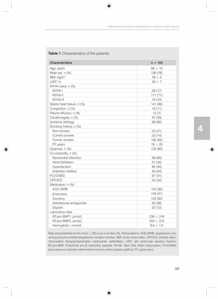
99
Pulmonary function impairment in chronic heart failure
4
Table 1 Characteristics of the patients.
Characteristics n = 164
Age, years 68 ± 10Male sex, n (%) 128 (78)BMI, kg/m2 28 ± 5LVEF, % 28 ± 7NYHA class, n (%) NYHA I 28 (17) NYHA II 117 (71) NYHA III 19 (12)Stable heart failure, n (%) 141 (86)Congestion, n (%) 18 (11)Pleural effusion, n (%) 12 (7)Cardiomegaly, n (%) 97 (59)Ischemic etiology 98 (60)Smoking history, n (%) Non-smoker 35 (21) Current smoker 23 (14) Former smoker 106 (65) PY, years 19 ± 20Dyspnea, n (%) 132 (80)Co-morbidity, n (%) Myocardial infarction 99 (60) Atrial fibrillation 57 (35) Hypertension 66 (40) Diabetes mellitus 40 (24)PCI/CABG 67 (41)CRT/ICD 52 (32)Medication, n (%) ACE-I/ARB 153 (93)
β-blockers 149 (91)
Diuretics 135 (82) Aldosterone-antagonists 63 (38) Digoxin 20 (12)Laboratory data NT-pro-BNP1, pmol/L 236 ± 316 NT-pro-BNP2, pmol/L 250 ± 375 Hemoglobin, mmol/L 8.6 ± 1.0
Data are presented as the mean ± SD or as a number (%). Abbreviations: ACE-I/ARB, angiotensin-con-verting enzyme inhibitor/angiotensin receptor blocker; BMI, body mass index; CRT/ICD, cardiac resyn-chronization therapy/implantable cardioverter defibrillator; LVEF, left ventricular ejection fraction; NT-pro-BNP, N-terminal pro-B natriuretic peptide; NYHA, New York Heart Association; PCI/CABG, percutaneous coronary intervention/coronary artery bypass grafting; PY, pack-years.
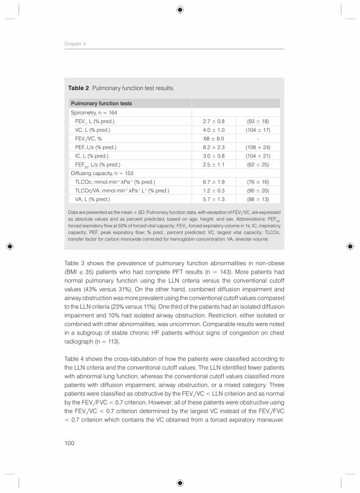
100
Chapter 4
Table 3 shows the prevalence of pulmonary function abnormalities in non-obese (BMI ≤ 35) patients who had complete PFT results (n = 143). More patients had normal pulmonary function using the LLN criteria versus the conventional cutoff values (43% versus 31%). On the other hand, combined diffusion impairment and airway obstruction was more prevalent using the conventional cutoff values compared to the LLN criteria (23% versus 11%). One third of the patients had an isolated diffusion impairment and 10% had isolated airway obstruction. Restriction, either isolated or combined with other abnormalities, was uncommon. Comparable results were noted in a subgroup of stable chronic HF patients without signs of congestion on chest radiograph (n = 113).
Table 4 shows the cross-tabulation of how the patients were classified according to the LLN criteria and the conventional cutoff values. The LLN identified fewer patients with abnormal lung function, whereas the conventional cutoff values classified more patients with diffusion impairment, airway obstruction, or a mixed category. Three patients were classified as obstructive by the FEV1/VC < LLN criterion and as normal by the FEV1/FVC < 0.7 criterion. However, all of these patients were obstructive using the FEV1/VC < 0.7 criterion determined by the largest VC instead of the FEV1/FVC < 0.7 criterion which contains the VC obtained from a forced expiratory maneuver.
Table 2 Pulmonary function test results.
Pulmonary function tests
Spirometry, n = 164
FEV1, L (% pred.) 2.7 ± 0.8 (93 ± 18)
VC, L (% pred.) 4.0 ± 1.0 (104 ± 17)
FEV1/VC, % 68 ± 8.0 -
PEF, L/s (% pred.) 8.2 ± 2.3 (108 ± 24)
IC, L (% pred.) 3.0 ± 0.8 (104 ± 21)
FEF50, L/s (% pred.) 2.5 ± 1.1 (62 ± 25)
Diffusing capacity, n = 153
TLCOc, mmol.min-1.kPa-1 (% pred.) 6.7 ± 1.9 (76 ± 16)
TLCOc/VA, mmol.min-1.kPa-1.L-1 (% pred.) 1.2 ± 0.3 (90 ± 20)
VA, L (% pred.) 5.7 ± 1.3 (88 ± 13)
Data are presented as the mean ± SD. Pulmonary function data, with exception of FEV1/VC, are expressed as absolute values and as percent predicted, based on age, height, and sex. Abbreviations: FEF50, forced expiratory flow at 50% of forced vital capacity; FEV1, forced expiratory volume in 1s; IC, inspiratory capacity; PEF, peak expiratory flow; % pred., percent predicted; VC, largest vital capacity; TLCOc, transfer factor for carbon monoxide corrected for hemoglobin concentration; VA, alveolar volume.
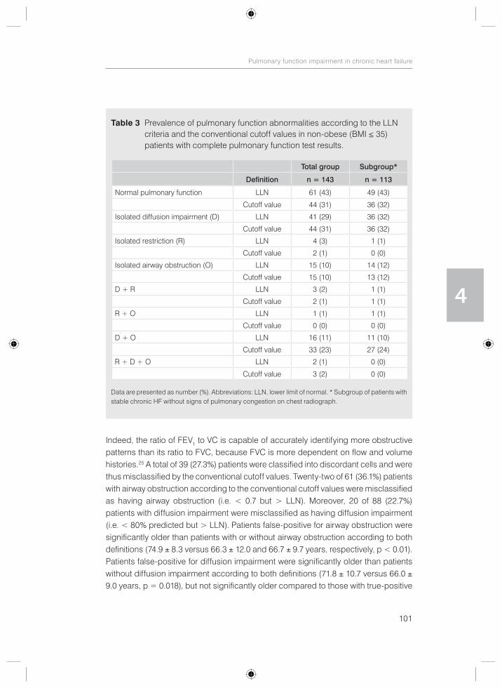
101
Pulmonary function impairment in chronic heart failure
4
Indeed, the ratio of FEV1 to VC is capable of accurately identifying more obstructive patterns than its ratio to FVC, because FVC is more dependent on flow and volume histories.25 A total of 39 (27.3%) patients were classified into discordant cells and were thus misclassified by the conventional cutoff values. Twenty-two of 61 (36.1%) patients with airway obstruction according to the conventional cutoff values were misclassified as having airway obstruction (i.e. < 0.7 but > LLN). Moreover, 20 of 88 (22.7%) patients with diffusion impairment were misclassified as having diffusion impairment (i.e. < 80% predicted but > LLN). Patients false-positive for airway obstruction were significantly older than patients with or without airway obstruction according to both definitions (74.9 ± 8.3 versus 66.3 ± 12.0 and 66.7 ± 9.7 years, respectively, p < 0.01). Patients false-positive for diffusion impairment were significantly older than patients without diffusion impairment according to both definitions (71.8 ± 10.7 versus 66.0 ± 9.0 years, p = 0.018), but not significantly older compared to those with true-positive
Table 3 Prevalence of pulmonary function abnormalities according to the LLN criteria and the conventional cutoff values in non-obese (BMI ≤ 35) patients with complete pulmonary function test results.
Total group Subgroup*
Definition n = 143 n = 113
Normal pulmonary function LLN 61 (43) 49 (43)
Cutoff value 44 (31) 36 (32)
Isolated diffusion impairment (D) LLN 41 (29) 36 (32)
Cutoff value 44 (31) 36 (32)
Isolated restriction (R) LLN 4 (3) 1 (1)
Cutoff value 2 (1) 0 (0)
Isolated airway obstruction (O) LLN 15 (10) 14 (12)
Cutoff value 15 (10) 13 (12)
D + R LLN 3 (2) 1 (1)
Cutoff value 2 (1) 1 (1)
R + O LLN 1 (1) 1 (1)
Cutoff value 0 (0) 0 (0)
D + O LLN 16 (11) 11 (10)
Cutoff value 33 (23) 27 (24)
R + D + O LLN 2 (1) 0 (0)
Cutoff value 3 (2) 0 (0)
Data are presented as number (%). Abbreviations: LLN, lower limit of normal. * Subgroup of patients with stable chronic HF without signs of pulmonary congestion on chest radiograph.
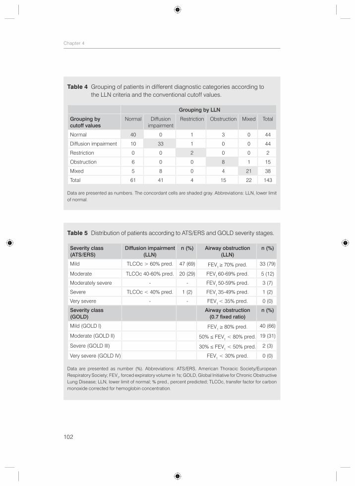
102
Chapter 4
Table 5 Distribution of patients according to ATS/ERS and GOLD severity stages.
Severity class(ATS/ERS)
Diffusion impairment(LLN)
n (%) Airway obstruction(LLN)
n (%)
Mild TLCOc > 60% pred. 47 (69) FEV1 ≥ 70% pred. 33 (79)
Moderate TLCOc 40-60% pred. 20 (29) FEV1 60-69% pred. 5 (12)
Moderately severe - - FEV1 50-59% pred. 3 (7)
Severe TLCOc < 40% pred. 1 (2) FEV1 35-49% pred. 1 (2)
Very severe - - FEV1 < 35% pred. 0 (0)
Severity class(GOLD)
Airway obstruction(0.7 fixed ratio)
n (%)
Mild (GOLD I) FEV1 ≥ 80% pred. 40 (66)
Moderate (GOLD II) 50% ≤ FEV1 < 80% pred. 19 (31)
Severe (GOLD III) 30% ≤ FEV1 < 50% pred. 2 (3)
Very severe (GOLD IV) FEV1 < 30% pred. 0 (0)
Data are presented as number (%). Abbreviations: ATS/ERS, American Thoracic Society/European Respiratory Society; FEV1, forced expiratory volume in 1s; GOLD, Global Initiative for Chronic Obstructive Lung Disease; LLN, lower limit of normal; % pred., percent predicted; TLCOc, transfer factor for carbon monoxide corrected for hemoglobin concentration.
Table 4 Grouping of patients in different diagnostic categories according to the LLN criteria and the conventional cutoff values.
Grouping by LLN
Grouping by cutoff values
Normal Diffusion impairment
Restriction Obstruction Mixed Total
Normal 40 0 1 3 0 44
Diffusion impairment 10 33 1 0 0 44
Restriction 0 0 2 0 0 2
Obstruction 6 0 0 8 1 15
Mixed 5 8 0 4 21 38
Total 61 41 4 15 22 143
Data are presented as numbers. The concordant cells are shaded gray. Abbreviations: LLN, lower limit of normal.
103
Pulmonary function impairment in chronic heart failure
4
diffusion impairment (71.8 ± 10.7 versus 67.5 ± 11.4 years, p = 0.135). There were no significant differences between these groups regarding the gender.
The majority of patients had mild to moderate severity of diffusion impairment and airway obstruction (Table 5). Only 2 patients with restriction had a TLC between 55% and 64%, whereas the TLC of the remaining patients was between 76% and 83%.
Discussion
The current study defined pulmonary function impairment in patients with chronic HF according to different diagnostic criteria, namely the LLN versus the conventional cutoff values. Our results demonstrate that the LLN identifies fewer HF patients with abnormal lung function, whereas the conventional cutoff values classify more patients as having diffusion impairment, airway obstruction, or a mixed pulmonary impairment. Using the conventional cutoff values instead of the LLN led to misclassification of 27% of the patients. Importantly, the cutoff values failed to identify correctly 34% of patients with normal lung function placing them falsely within a pulmonary dysfunction category. This is explained by the physiological decrease of the FEV1/FVC ratio with age. The FEV1 declines more rapidly with age than the FVC in normal subjects.23 As a result, the fixed ratio of 0.7, that is traditionally used because of its simplicity, may lead to overdiagnosis in the elderly and underdiagnosis in younger patients. Furthermore, the frequently used 80% predicted value has neither statistical nor physiological validity.24 Limits of normal as the predicted ± 20% can only be accurate when the variance above and below the predicted regression line is proportional with the predicted value (i.e. heteroscedastic: large variance with large values and small variance with small values). However, since this is not the case as the scatter around the predicted regression line is constant (homoscedastic) in pulmonary function measurements, the 80% predicted rule of thumb may lead to false-positive diagnosis in the elderly and shorter individuals with smaller predicted values and underdiagnosis in younger and taller patients with larger predicted values.22, 24
Misinterpretation of pulmonary function test results may lead to incorrect diagnosis of disease in elderly patients with HF, such as chronic obstructive pulmonary disease, and as a consequence unnecessary treatment with possible side effects, detrimental psychological impact, and unnecessary financial burden on society. Moreover, results may be interpreted as having more severe or unstable HF due to the effects of HF on pulmonary function and as a result unnecessary intensified treatment for HF. Finally, misdiagnosis may interfere with interpretation of research aimed at under -standing the impact of heart failure and several clinical variables on pulmonary
104
Chapter 4
function. For example, patients who had smoked ≥ 10 PY in our study had diffusion impairment and airway obstruction significantly more often compared to those with < 10 PY using the LLN. This relationship was, however, lost when using conventional cutoff values and implies that inclusion of patients who are incorrectly labelled as having pulmonary dysfunction distorted the effect of smoking on pulmonary function by adding noise to our data.
To avoid misclassification, recent ATS/ERS guidelines25 recommend the use of statistically derived LLN values that are based on the normal distribution and that classify the bottom 5% of the healthy population as abnormal, thus limiting the chance of false-positive diagnosis to a maximum of 5%. However, studies using the LLN values to assess the prevalence of pulmonary function abnormalities in patients with HF are lacking and pulmonary function abnormalities may have been over -estimated in previous studies that have used conventional cutoff values. Indeed, in this population of mainly old patients with chronic HF significantly higher prevalence rates of diffusion impairment and airway obstruction were noted with the 80% predicted criterion and the fixed ratio of 0.7, respectively, compared to the LLN. Thus, a substantially number of mainly older patients were misclassified as having airway obstruction and diffusion impairment using the conventional cutoff values instead of the LLN.
Even though we used the LLN to better account for age, the occurrence of diffusion impairment and airway obstruction was still high (44% and 26%, respectively). This was also true for patients in a stable clinical condition who did not have signs of pulmonary congestion. Prevalence rates of diffusion impairment and airway obstruction were 41-93%13-19 and 14-60%,13-18, 20, 21 respectively, in prior studies that have often included heart transplant recipients. Our results extend these studies by demonstrating that these abnormalities are also highly prevalent in patients with less severe heart failure who were mainly in NYHA class II. However, in contrast to previous reports that have demonstrated high prevalence rates of restriction (21-55%),13-17, 20 this was quite infrequent in the current study, independent of the definition used. Contrasting results are not explained by the fact that body plethysmography was performed only in patients with an abnormal spirometry result and not in the entire study population, as the chance of missing a restrictive defect is less than 3% when the VC is within the normal range using spirometry. Spirometry is therefore very useful at excluding a restrictive defect.30 Contrasting results can, however, be attributed to differences in patient characteristics as less severe and mainly stable chronic HF patients without signs of congestion on chest radiograph were enrolled in the current study. In addition, we excluded other diseases that could affect pulmonary function, including obesity. Finally, in contrast to some studies,16 we have confirmed suspected
105
Pulmonary function impairment in chronic heart failure
4
restriction on spirometry with body plethysmography, as a reduced VC with normal FEV1/VC ratio does not prove a restrictive ventilatory defect.25
Several factors have been implied to play a role in the etiology of pulmonary function impairment in patients with HF, including the effects of HF itself on pulmonary function in addition to (previously undiagnosed) underlying pulmonary disease and confounding influences such as smoking, coronary artery bypass grafting (CABG), and obesity.4-7, 31 Irrespective of the causes, pulmonary function abnormalities associated with chronic HF may explain part of the symptoms and functional disability encountered in these patients.8-12 Moreover, pulmonary function impairment increases with the severity of HF,9, 11 provides important prognostic information,32, 33 and may ameliorate or normalize with several treatment modalities, such as pharma-cological and non-pharmacological treatment of HF 2, 14, 18, 34-36 and anti-obstructive therapy with bronchodilators.20, 37-42 Pulmonary function might thus be used as a guide for the evaluation of patients with chronic HF, with respect to severity of disease, prognosis, and response to treatment. More studies involving therapeutic approaches to improve pulmonary function in chronic HF are warranted to determine whether treatment directed at correcting pulmonary function impairment may lead to symptomatic relief, increased exercise capacity, and improvement of the outcome. Furthermore, since little is known about the clinical impact of different criteria of pulmonary dysfunction, longitudinal studies are needed to determine which criterion is clinically more relevant in terms of morbidity (symptoms, exercise tolerance, health-related quality of life, hospitalization, use of health resources) and mortality.
Some limitations of this study deserve further discussion. It is important to realize that these results may not be applicable to all patients with chronic HF, since we did not include patients with preserved systolic function, who seem to have less impaired pulmonary function.43 The diagnosis of HF with preserved systolic function is challenging and only few patients met our pre-defined criteria of HF with preserved systolic function. Consequently, only patients with LVSD were enrolled in the current study. Furthermore, patients with more severe HF could have been underrepresented in this study because of inability to participate and thus pulmonary function abnormalities might have been underestimated. Finally, since we did not follow the patients prospectively, it remains unknown whether pulmonary function impairment had prognostic implications in our study population and whether this is influenced by different definitions of pulmonary dysfunction. However, pulmonary function did provide significant prognostic information in patients with HF in other studies.32, 33
In conclusion, pulmonary function impairment, especially diffusion impairment and airway obstruction, was highly prevalent in patients with chronic HF with LVSD, even
106
Chapter 4
in a stable and non-congested condition and even though we used the LLN to better account for age. The LLN identifies fewer HF patients with abnormal lung function, whereas the conventional cutoff values classify more patients as having diffusion impairment, airway obstruction, or a mixed pulmonary impairment, leading to mis-classification of 27% of the patients. In contrast to previous reports, restriction was found to be infrequent in this population of less severe chronic HF, irrespective of the definition used.
AcknowledgmentsThe authors would like to thank the participants in this study for their cooperation. We also would like to thank the nurse practitioners P.A. Ninaber, W.A.M. Janssen, W.H. van Zimmeren-Feijen, and heart failure nurse J.G. Froon-Elferink for their contribution to inclusion of participants and data collection. The authors owe much gratitude to the pulmonary function and clinical chemistry and haematology laboratories as well as the echocardiography and radiology departments for their assistance in data collection. Also, we would like to acknowledge Prof. Dr. P.H. Quanjer for his contribution to the manuscript and biostatistician A.R.T. Donders for his contribution to statistical analysis. Finally, we would like to thank GlaxoSmithKline for supporting this study by an unrestricted grant.
107
Pulmonary function impairment in chronic heart failure
4
References
1. Kee K, Naughton MT. Heart failure and the lung. Circ J. 2010;74:2507-162. Guazzi M. Alveolar gas diffusion abnormalities in heart failure. J Card Fail. 2008;14:695-702.3. Agostoni P, Cattadori G, Bussotti M, Apostolo A. Cardiopulmonary interaction in heart failure. Pulm
Pharmacol Ther. 2007;20:130-4.4. Gehlbach BK, Geppert E. The pulmonary manifestations of left heart failure. Chest. 2004;125:669-82.5. Chua TP, Coats AJ. The lungs in chronic heart failure. Eur Heart J. 1995;16:882-7.6. Faggiano P. Abnormalities of pulmonary function in congestive heart failure. Int J Cardiol. 1994;44:1-8.7. Snashall PD, Chung KF. Airway obstruction and bronchial hyperresponsiveness in left ventricular
failure and mitral stenosis. Am Rev Respir Dis. 1991;144:945-56.8. Mancini DM. Pulmonary factors limiting exercise capacity in patients with heart failure. Prog Cardiovasc
Dis. 1995;37:347-70.9. Puri S, Baker BL, Dutka DP, Oakley CM, Hughes JM, Cleland JG. Reduced alveolar-capillary
membrane diffusing capacity in chronic heart failure. Its pathophysiological relevance and relationship to exercise performance. Circulation. 1995;91:2769-74.
10. Kraemer MD, Kubo SH, Rector TS, Brunsvold N, Bank AJ. Pulmonary and peripheral vascular factors are important determinants of peak exercise oxygen uptake in patients with heart failure. J Am Coll Cardiol. 1993;21:641-8.
11. Dimopoulou I, Daganou M, Tsintzas OK, Tzelepis GE. Effects of severity of long-standing congestive heart failure on pulmonary function. Respir Med. 1998;92:1321-5.
12. Tzani P, Piepoli MF, Longo F, Aiello M, Serra W, Maurizio AR, et al. Resting lung function in the assessment of the exercise capacity in patients with chronic heart failure. Am J Med Sci. 2010;339:210-5.
13. Wright RS, Levine MS, Bellamy PE, Simmons MS, Batra P, Stevenson LW, et al. Ventilatory and diffusion abnormalities in potential heart transplant recipients. Chest. 1990;98:816-20.
14. Bussieres LM, Pflugfelder PW, Ahmad D, Taylor AW, Kostuk WJ. Evolution of resting lung function in the first year after cardiac transplantation. Eur Respir J. 1995;8:959-62.
15. Ewert R, Wensel R, Bettmann M, Spiegelsberger S, Grauhan O, Hummel M, et al. Ventilatory and diffusion abnormalities in long-term survivors after orthotopic heart transplantation. Chest. 1999;115:1305-11.
16. Naum CC, Sciurba FC, Rogers RM. Pulmonary function abnormalities in chronic severe cardiomyopathy preceding cardiac transplantation. Am Rev Respir Dis. 1992;145:1334-8.
17. Ohar J, Osterloh J, Ahmed N, Miller L. Diffusing capacity decreases after heart transplantation. Chest. 1993;103:857-61.
18. Niset G, Ninane V, Antoine M, Yernault JC. Respiratory dysfunction in congestive heart failure: correction after heart transplantation. Eur Respir J. 1993;6:1197-201.
19. Egan JJ, Kalra S, Yonan N, Hasleton PS, Brooks N, Woodcock AA. Pulmonary diffusion abnormalities in heart transplant recipients. Relationship to cytomegalovirus infection. Chest. 1993;104:1085-9.
20. Khan KA, Jalal S, Jan VM, Lone GM, Jan R, Alai MS, et al. Pulmonary function profile in chronic congestive heart failure and the effect of ipratropium bromide. Indian Heart J. 2000;52:297-300.
21. Evans SA, Kinnear WJ, Watson L, Hawkins M, Cowley AJ, Johnston ID. Breathlessness and exercise capacity in heart failure: the role of bronchial obstruction and responsiveness. Int J Cardiol. 1996;57:233-40.
22. Miller MR, Quanjer PH, Swanney MP, Ruppel G, Enright PL. Interpreting lung function data using 80 percent of predicted and fixed thresholds misclassifies over 20% of patients. Chest. 2011;139:52-59.
23. Townsend MC. Conflicting definitions of airways obstruction: Drawing the line between normal and abnormal. Chest. 2007;131:335-6.
24. Sobol BJ, Sobol PG. Per cent of predicted as the limit of normal in pulmonary function testing: a statistically valid approach. Thorax. 1979;34:1-3.
25. Pellegrino R, Viegi G, Brusasco V, Crapo RO, Burgos F, Casaburi R, et al. Interpretative strategies for lung function tests. Eur Respir J. 2005;26:948-68.
26. Dickstein K, Cohen-Solal A, Filippatos G, McMurray JJ, Ponikowski P, Poole-Wilson PA, et al. ESC Guidelines for the diagnosis and treatment of acute and chronic heart failure 2008: the Task Force for the Diagnosis and Treatment of Acute and Chronic Heart Failure 2008 of the European Society of
108
Chapter 4
Cardiology. Developed in collaboration with the Heart Failure Association of the ESC (HFA) and endorsed by the European Society of Intensive Care Medicine (ESICM). Eur Heart J. 2008;29:2388-442.
27. Quanjer PH, Tammeling GJ, Cotes JE, Pedersen OF, Peslin R, Yernault JC. Lung volumes and forced ventilatory flows. Report Working Party Standardization of Lung Function Tests, European Community for Steel and Coal. Official Statement of the European Respiratory Society. Eur Respir J 1993;16:5-40.
28. O’Hanlon R, O’Shea P, Ledwidge M, O’Loughlin C, Lange S, Conlon C, et al. The biologic variability of B-type natriuretic peptide and N-terminal pro-B-type natriuretic peptide in stable heart failure patients. J Card Fail. 2007;13:50-5.
29. Rabe KF, Hurd S, Anzueto A, Barnes PJ, Buist SA, Calverley P, et al. Global strategy for the diagnosis, management, and prevention of chronic obstructive pulmonary disease: GOLD executive summary. Am J Respir Crit Care Med. 2007;176:532-55.
30. Aaron SD, Dales RE, Cardinal P. How accurate is spirometry at predicting restrictive pulmonary impairment? Chest. 1999;115:869-73.
31. Johnson BD, Beck KC, Olson LJ, O’Malley KA, Allison TG, Squires RW, et al. Pulmonary function in patients with reduced left ventricular function: influence of smoking and cardiac surgery. Chest. 2001; 120:1869-76.
32. Guazzi M, Pontone G, Brambilla R, Agostoni P, Reina G. Alveolar--capillary membrane gas conductance: a novel prognostic indicator in chronic heart failure. Eur Heart J. 2002;23:467-76.
33. Iversen KK, Kjaergaard J, Akkan D, Kober L, Torp-Pedersen C, Hassager C, et al. The prognostic importance of lung function in patients admitted with heart failure. Eur J Heart Fail. 2010;12:685-91.
34. Light RW, George RB. Serial pulmonary function in patients with acute heart failure. Arch Intern Med. 1983;143:429-33.
35. Faggiano P, Lombardi C, Sorgato A, Ghizzoni G, Spedini C, Rusconi C. Pulmonary function tests in patients with congestive heart failure: effects of medical therapy. Cardiology. 1993;83:30-5.
36. Petersen CL, Kjaer A. Impact of medical treatment on lung diffusion capacity in elderly patients with heart failure. Baseline characteristics and 1-year follow up after medical treatment. Int J Cardiol. 2005;98:453-7.
37. Minasian AG, van den Elshout FJ, Dekhuijzen PN, Vos PJ, Willems FF, van den Bergh PJ, et al. Bronchodilator responsiveness in patients with chronic heart failure. Heart Lung. 2013;42:208-214.
38. Kindman LA, Vagelos RH, Willson K, Prikazky L, Fowler M. Abnormalities of pulmonary function in patients with congestive heart failure, and reversal with ipratropium bromide. Am J Cardiol. 1994;73:258-62.
39. Uren NG, Davies SW, Jordan SL, Lipkin DP. Inhaled bronchodilators increase maximum oxygen consumption in chronic left ventricular failure. Eur Heart J. 1993;14:744-50.
40. Witte KK, Morice A, Cleland JG, Clark AL. The reversibility of increased airways resistance in chronic heart failure measured by impulse oscillometry. J Card Fail. 2004;10:149-54.
41. Rolla G, Bucca C, Brussino L, Gallo W, Malara D. Bronchodilating effect of ipratropium bromide in heart failure. Eur Respir J. 1993;6:1492-5.
42. Ng TM, Munger MA, Lombardi WL, Doing TH, Ryujin DT, Young DC, et al. Chronically inhaled salmeterol improves pulmonary function in heart failure. J Cardiovasc Pharmacol. 2002;40:140-5.
43. Kurzawa R, Baron T, Grodzicki T. Pulmonary function test abnormalities in the elderly with systolic heart failure. Kardiol Pol. 2007;65:875-80. discussion 81-2.

109
Pulmonary function impairment in chronic heart failure
4

Armine G. Minasian Frank J.J. van den ElshoutP.N. Richard DekhuijzenPetra J.E. VosFrank F. WillemsPaul J.P.C. van den BerghYvonne F. Heijdra
Respiratory Care. 2015; accepted for publication.
Using the lower limit of normal instead of the conventional cutoff values
to define predictors of pulmonary function impairment in subjects with
chronic heart failure
CHAPTER 5
112
Chapter 5
Abstract
Introduction: Using the newer lower limit of normal (LLN) criterion instead of the conventional cutoff values to define pulmonary function abnormalities may result in different predictors of pulmonary function impairment in patients with heart failure (HF). Therefore, we assessed predictors of pulmonary function impairment in subjects with chronic HF according to the LLN in comparison to conventional cutoff values.
Methods: In this prospective cross-sectional study, 164 chronic HF subjects (age 68 ± 10 years, 78% men, 88% New York Heart Association class I-II) with left ventricular ejection fraction < 40% underwent pulmonary function tests. Predictors of pulmonary function impairment were assessed using the LLN and conventional cutoff values (i.e. 80% predicted value and the fixed ratio of forced expiratory volume in 1s to forced vital capacity < 0.7).
Results: The LLN criterion identified an extra independent predictor of diffusion impairment compared to the 80% predicted value; in addition to body mass index, pack-years (PY), and alveolar volume, female gender also turned out to be an independent predictor. A smoking history of ≥ 10 PY was a significant predictor of diffusion impairment and airway obstruction using the LLN criterion, but not using the conventional cutoff values. However, lowering the cutoff points of conventional criteria to match the more stringent LLN and thus avoid overdiagnosis of diffusion impairment and airway obstruction in the elderly, produced similar results as the LLN.
Conclusion: The LLN identifies more predictors of diffusion impairment and airway obstruction compared to conventional cutoff values in subjects with chronic HF with left ventricular systolic dysfunction. However, lowering the conventional cutoff points yielded similar results as the LLN.
Keywords: Chronic heart failure, conventional cutoff values, lower limit of normal, predictors, pulmonary function impairment.
Trial registration: ClinicalTrials.gov Identifier NCT01429376.
113
Predictors of pulmonary function impairment in chronic heart failure
5
Introduction
Isolated or combined pulmonary function abnormalities, such as restriction, diffusion impairment, and to a lesser extent airway obstruction are common in patients with chronic heart failure (HF)1-7 and can contribute to the perception of dyspnea8 and exercise intolerance.8-12 Several factors have been implied to play a role in the etiology of pulmonary function impairment in patients with HF, including the effects of HF itself on pulmonary function in addition to (previously undiagnosed) underlying pulmonary disease and confounding influences such as smoking, coronary artery bypass grafting (CABG), and obesity.4-7, 13 However, results are not consistent across the studies. For example, although smoking and a history of CABG was associated with more impaired pulmonary function in the study of Johnson et. al.13 with also weak associations between left ventricular function and both lung volumes as well as diffusing capacity, none of the described pulmonary function abnormalities were found to be related to either smoking status, use of cardiac drugs, chest radiographic changes, hemodynamic findings, or clinical features, including the duration of HF in the study of Wright et. al.14 Misdiagnosis of pulmonary function abnormalities may have interfered with the interpretation of prior research aiming to investigate the impact of HF and several clinical variables on pulmonary function in this group of patients. Traditionally, the 80% predicted value and the fixed ratio of forced expiratory volume in 1s to forced vital capacity (FEV1/FVC) < 0.7 have been used to define pulmonary function abnormalities. However, these conventional cutoff values have neither statistical nor physiological validity15-17 and may misclassify more than 20% of patients leading to false-positive diagnosis in the elderly and underdiagnosis in younger patients.18 To avoid misclassification, recent American Thoracic Society/European Respiratory Society (ATS/ERS) guidelines16 recommend the use of statistically derived lower limit of normal (LLN) values that are based on the normal distribution and that classify the bottom 5% of the healthy population as abnormal. However, studies using the LLN to assess predictors of pulmonary function impairment in patients with chronic HF are lacking. Therefore, the purpose of this study was to assess predictors of pulmonary function impairment in subjects with chronic HF according to the LLN in comparison to conventional cutoff values (percent predicted and the fixed ratio of FEV1/FVC).
Methods
Study design and participantsThis study was part of a larger prospective cross-sectional study evaluating the prevalence of chronic obstructive pulmonary disease in subjects with chronic HF. All
114
Chapter 5
patients visiting two outpatient cardiology departments of a large hospital in The Netherlands were screened for inclusion between October 2009 and December 2010. Inclusion criteria were chronic HF19 with left ventricular systolic dysfunction (LVSD), i.e. left ventricular ejection fraction (LVEF) < 40%, and New York Heart Association class (NYHA) I-IV. Chronic HF was defined according to the European Society of Cardiology guidelines.19 Echocardiography was performed in subjects without a recent (≤ 6 months) echocardiography to confirm persisting LVSD. Subjects who were not able to cooperate or undergo spirometry or who had a history of asthma were excluded. Other exclusion criteria were malignancy with a poor prognosis (survival < 6 months) and participation in another study. For the current study we also excluded subjects with known pulmonary (including chronic obstructive pulmonary disease (COPD)), pleural (with exception of pleural effusion due to HF), neuromuscular, collagen vascular, or other diseases that could affect pulmonary function. Subjects with a body mass index (BMI) above 35 were excluded from the restriction prevalence analysis.
Subjects were classified as having stable HF in the absence of hospitalization due to progression of HF within 3 months, change in diuretics within 1 month, 3% or more weight gain within 3 days, and more than 50% increase of N-terminal pro-B natriuretic peptide (NT-pro-BNP) within 1 month when the baseline NT-pro-BNP was 100 pmol/L or higher or more than 100 pmol/L increase of NT-pro-BNP within 1 month when baseline NT-pro-BNP was below 100 pmol/L.20
Measurements and data collection At baseline, a first blood sample was taken for the measurement of NT-pro-BNP. One month later, the participants visited the hospital for an interview with the investigator and several examinations, including height and weight measurement, pulmonary function tests (PFTs), a chest radiograph, and a second blood sample (hemoglobin, NT-pro-BNP). Additional data were collected from medical records and personal interviews. Smoking status was defined as never (< 100 cigarettes in a lifetime), former (≥ 3 months ago), or current smoker (< 3 months). Smoking pack-years (PY) were based only on the tobacco cigarette history, and one PY was defined as smoking twenty cigarettes a day for one year. Dyspnea was defined as resting dyspnea or dyspnea at any level of exertion.
Pulmonary function testsAll participants underwent pre-bronchodilator spirometry (MasterLab Pro, Jaeger, Würzburg, Germany) and measurement of the transfer factor of the lungs for carbon monoxide (TLCO). TLCO was measured with standard single-breath technique and was corrected for the subject’s hemoglobin concentration (TLCOc). During the
115
Predictors of pulmonary function impairment in chronic heart failure
5
measurement of TLCO the alveolar volume (VA) was also obtained and the TLCOc was corrected for the VA (TLCOc/VA, i.e. transfer coefficient for carbon monoxide). Body plethysmography was performed in subjects with airway obstruction according to either definition to assess the presence of hyperinflation. In addition, it was performed in subjects with signs of restriction on spirometry, i.e. (F)VC < LLN and/or < 80% predicted with normal FEV1/(F)VC ratio, to confirm suspected restriction by measuring the total lung capacity (TLC). In other cases body plethysmography was omitted since abnormal findings of body plethysmography were not expected with normal spirometry results. PFTs were performed by trained and certified operators using standard techniques and according to the ERS standards for acceptability and reproducibility.21 The European Community for Coal and Steel reference equations were used to calculate predicted values.21
Diffusion impairment was defined as TLCOc < LLN (ATS/ERS)16 and < 80% predicted (conventional cutoff value). Restriction was defined as TLC < LLN (ATS/ERS)16 and < 80% predicted (conventional cutoff value). Airway obstruction was defined as FEV1/VC < LLN (ATS/ERS)16 and FEV1/FVC < 0.7 (conventional cutoff value).22 VC was regarded as the largest vital capacity (either slow, forced, inspiratory or expiratory) and FVC as the forced vital capacity obtained during a forced expiratory maneuver. LLN was regarded as the lower fifth percentile of the frequency distribution of a healthy reference population and it was calculated by subtracting 1.64 times the residual standard deviation (RSD) from the predicted value (Table 1). Hyperinflation was defined as the absolute ratio of residual volume (RV) to TLC > 40%.23
Chest radiographsStandard posterior-anterior and lateral chest radiographs were performed and evaluated for the presence or absence of cardiomegaly (cardiothoracic ratio > 0.5), congestion (alveolar edema, pleural effusion, Kerley-B lines, and/or redistribution of pulmonary blood flow) and conditions that belonged to the exclusion criteria. Independent radiologists qualitatively assessed the chest radiographs with an overall clinical impression.
Statistical analysisDescriptive data are presented as the mean ± SD or as a number (%). Differences in prevalence of pulmonary function abnormalities according to different definitions were analysed with McNemar’s test that compares paired proportions. Differences between groups were analyzed using an independent t-test for continuous variables and a chi-square or Fisher’s exact test for categorical variables, as appropriate. Differences in pulmonary function between groups of subjects according to NYHA class were analyzed with an independent analysis of variance. Post hoc analyses
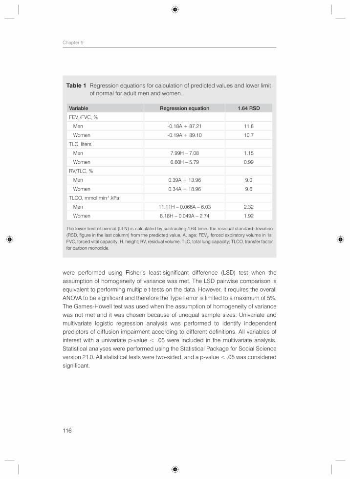
116
Chapter 5
were performed using Fisher’s least-significant difference (LSD) test when the assumption of homogeneity of variance was met. The LSD pairwise comparison is equivalent to performing multiple t-tests on the data. However, it requires the overall ANOVA to be significant and therefore the Type I error is limited to a maximum of 5%. The Games-Howell test was used when the assumption of homogeneity of variance was not met and it was chosen because of unequal sample sizes. Univariate and multivariate logistic regression analysis was performed to identify independent predictors of diffusion impairment according to different definitions. All variables of interest with a univariate p-value < .05 were included in the multivariate analysis. Statistical analyses were performed using the Statistical Package for Social Science version 21.0. All statistical tests were two-sided, and a p-value < .05 was considered significant.
Table 1 Regression equations for calculation of predicted values and lower limit of normal for adult men and women.
Variable Regression equation 1.64 RSD
FEV1/FVC, %
Men -0.18A + 87.21 11.8
Women -0.19A + 89.10 10.7
TLC, liters
Men 7.99H – 7.08 1.15
Women 6.60H – 5.79 0.99
RV/TLC, %
Men 0.39A + 13.96 9.0
Women 0.34A + 18.96 9.6
TLCO, mmol.min-1.kPa-1
Men 11.11H – 0.066A – 6.03 2.32
Women 8.18H – 0.049A – 2.74 1.92
The lower limit of normal (LLN) is calculated by subtracting 1.64 times the residual standard deviation (RSD, figure in the last column) from the predicted value. A, age; FEV1, forced expiratory volume in 1s; FVC, forced vital capacity; H, height; RV, residual volume; TLC, total lung capacity; TLCO, transfer factor for carbon monoxide.
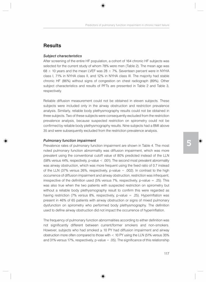
117
Predictors of pulmonary function impairment in chronic heart failure
5
Results
Subject characteristicsAfter screening of the entire HF population, a cohort of 164 chronic HF subjects was selected for the current study of whom 78% were men (Table 2). The mean age was 68 ± 10 years and the mean LVEF was 28 ± 7%. Seventeen percent were in NYHA class I, 71% in NYHA class II, and 12% in NYHA class III. The majority had stable chronic HF (86%) without signs of congestion on chest radiograph (89%). Other subject characteristics and results of PFTs are presented in Table 2 and Table 3, respectively.
Reliable diffusion measurement could not be obtained in eleven subjects. These subjects were included only in the airway obstruction and restriction prevalence analysis. Similarly, reliable body plethysmography results could not be obtained in three subjects. Two of these subjects were consequently excluded from the restriction prevalence analysis, because suspected restriction on spirometry could not be confirmed by reliable body plethysmography results. Nine subjects had a BMI above 35 and were subsequently excluded from the restriction prevalence analysis.
Pulmonary function impairmentPrevalence rates of pulmonary function impairment are shown in Table 4. The most noted pulmonary function abnormality was diffusion impairment, which was more prevalent using the conventional cutoff value of 80% predicted instead of the LLN (58% versus 44%, respectively, p-value < .001). The second most prevalent abnormality was airway obstruction, which was more frequent using the fixed ratio of 0.7 instead of the LLN (37% versus 26%, respectively, p-value = .002). In contrast to the high occurrence of diffusion impairment and airway obstruction, restriction was infrequent, irrespective of the definition used (5% versus 7%, respectively, p-value = .25). This was also true when the two patients with suspected restriction on spirometry but without a reliable body plethysmography result to confirm this were regarded as having restriction (7% versus 8%, respectively, p-value = .25). Hyperinflation was present in 46% of 65 patients with airway obstruction or signs of mixed pulmonary dysfunction on spirometry who performed body plethysmography. The definition used to define airway obstruction did not impact the occurrence of hyperinflation.
The frequency of pulmonary function abnormalities according to either definition was not significantly different between current/former smokers and non-smokers. However, subjects who had smoked ≥ 10 PY had diffusion impairment and airway obstruction more often compared to those with < 10 PY using the LLN (51% versus 35% and 31% versus 17%, respectively, p-value = .05). The significance of this relationship
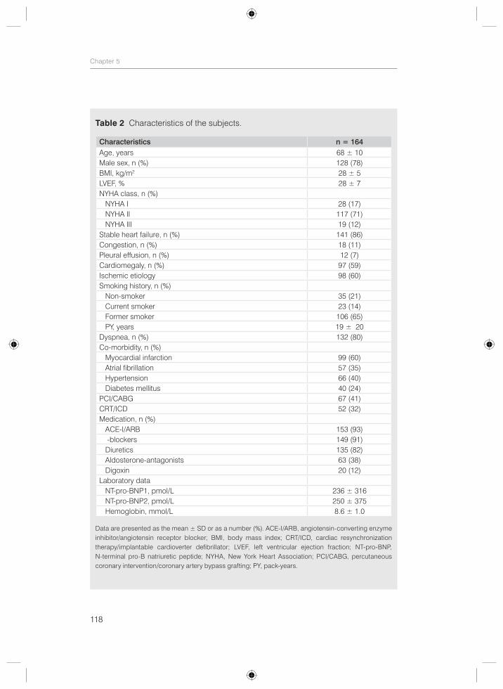
118
Chapter 5
Table 2 Characteristics of the subjects.
Characteristics n = 164Age, years 68 ± 10Male sex, n (%) 128 (78)BMI, kg/m2 28 ± 5LVEF, % 28 ± 7NYHA class, n (%) NYHA I 28 (17) NYHA II 117 (71) NYHA III 19 (12)Stable heart failure, n (%) 141 (86)Congestion, n (%) 18 (11)Pleural effusion, n (%) 12 (7)Cardiomegaly, n (%) 97 (59)Ischemic etiology 98 (60)Smoking history, n (%) Non-smoker 35 (21) Current smoker 23 (14) Former smoker 106 (65) PY, years 19 ± 20Dyspnea, n (%) 132 (80)Co-morbidity, n (%) Myocardial infarction 99 (60) Atrial fibrillation 57 (35) Hypertension 66 (40) Diabetes mellitus 40 (24)PCI/CABG 67 (41)CRT/ICD 52 (32)Medication, n (%) ACE-I/ARB 153 (93) β-blockers 149 (91) Diuretics 135 (82) Aldosterone-antagonists 63 (38) Digoxin 20 (12)Laboratory data NT-pro-BNP1, pmol/L 236 ± 316 NT-pro-BNP2, pmol/L 250 ± 375 Hemoglobin, mmol/L 8.6 ± 1.0
Data are presented as the mean ± SD or as a number (%). ACE-I/ARB, angiotensin-converting enzyme inhibitor/angiotensin receptor blocker; BMI, body mass index; CRT/ICD, cardiac resynchronization therapy/implantable cardioverter defibrillator; LVEF, left ventricular ejection fraction; NT-pro-BNP, N-terminal pro-B natriuretic peptide; NYHA, New York Heart Association; PCI/CABG, percutaneous coronary intervention/coronary artery bypass grafting; PY, pack-years.
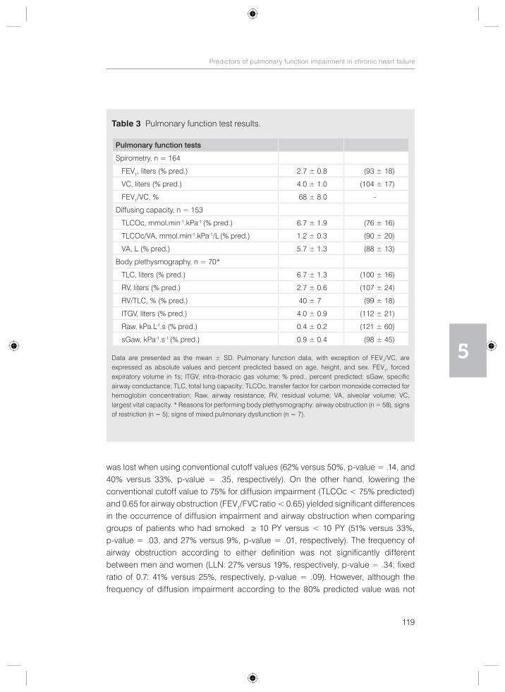
119
Predictors of pulmonary function impairment in chronic heart failure
5
was lost when using conventional cutoff values (62% versus 50%, p-value = .14, and 40% versus 33%, p-value = .35, respectively). On the other hand, lowering the conventional cutoff value to 75% for diffusion impairment (TLCOc < 75% predicted) and 0.65 for airway obstruction (FEV1/FVC ratio < 0.65) yielded significant differences in the occurrence of diffusion impairment and airway obstruction when comparing groups of patients who had smoked ≥ 10 PY versus < 10 PY (51% versus 33%, p-value = .03, and 27% versus 9%, p-value = .01, respectively). The frequency of airway obstruction according to either definition was not significantly different between men and women (LLN: 27% versus 19%, respectively, p-value = .34; fixed ratio of 0.7: 41% versus 25%, respectively, p-value = .09). However, although the frequency of diffusion impairment according to the 80% predicted value was not
Table 3 Pulmonary function test results.
Pulmonary function tests
Spirometry, n = 164
FEV1, liters (% pred.) 2.7 ± 0.8 (93 ± 18)
VC, liters (% pred.) 4.0 ± 1.0 (104 ± 17)
FEV1/VC, % 68 ± 8.0 -
Diffusing capacity, n = 153
TLCOc, mmol.min-1.kPa-1 (% pred.) 6.7 ± 1.9 (76 ± 16)
TLCOc/VA, mmol.min-1.kPa-1/L (% pred.) 1.2 ± 0.3 (90 ± 20)
VA, L (% pred.) 5.7 ± 1.3 (88 ± 13)
Body plethysmography, n = 70*
TLC, liters (% pred.) 6.7 ± 1.3 (100 ± 16)
RV, liters (% pred.) 2.7 ± 0.6 (107 ± 24)
RV/TLC, % (% pred.) 40 ± 7 (99 ± 18)
ITGV, liters (% pred.) 4.0 ± 0.9 (112 ± 21)
Raw, kPa.L-1.s (% pred.) 0.4 ± 0.2 (121 ± 60)
sGaw, kPa-1.s-1 (% pred.) 0.9 ± 0.4 (98 ± 45)
Data are presented as the mean ± SD. Pulmonary function data, with exception of FEV1/VC, are expressed as absolute values and percent predicted based on age, height, and sex. FEV1, forced expiratory volume in 1s; ITGV, intra-thoracic gas volume; % pred., percent predicted; sGaw, specific airway conductance; TLC, total lung capacity; TLCOc, transfer factor for carbon monoxide corrected for hemoglobin concentration; Raw, airway resistance; RV, residual volume; VA, alveolar volume; VC, largest vital capacity. * Reasons for performing body plethysmography: airway obstruction (n = 58), signs of restriction (n = 5); signs of mixed pulmonary dysfunction (n = 7).
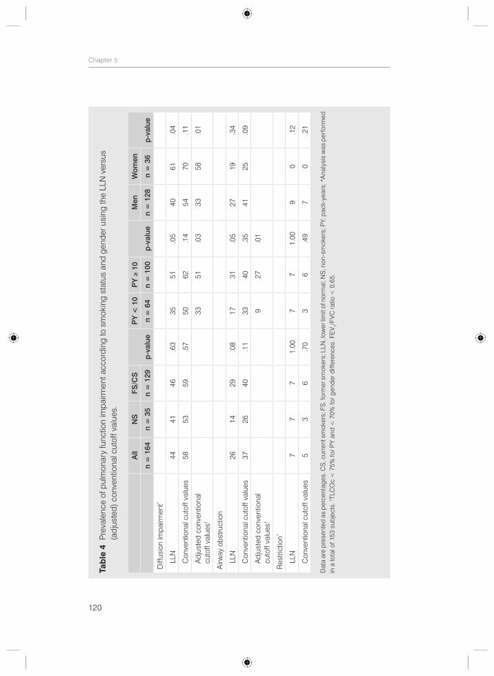
120
Chapter 5
Tab
le 4
Pre
vale
nce
of p
ulm
onar
y fu
nctio
n im
pairm
ent a
ccor
ding
to s
mok
ing
stat
us a
nd g
ende
r usi
ng th
e LL
N v
ersu
s
(adj
uste
d) c
onve
ntio
nal c
utof
f val
ues.
All
NS
FS/C
S
p-v
alue
PY
< 1
0P
Y ≥
10
p-v
alue
Men
Wom
en
p-v
alue
n =
164
n =
35
n =
129
n =
64
n =
100
n =
128
n =
36
Diff
usio
n im
pairm
ent*
LL
N44
4146
.63
3551
.05
4061
.04
C
onve
ntio
nal c
utof
f val
ues
5853
59.5
750
62.1
454
70.1
1
A
djus
ted
conv
entio
nal
cuto
ff va
lues
†
3351
.03
3358
.01
Airw
ay o
bstru
ctio
n
LL
N26
1429
.08
1731
.05
2719
.34
C
onve
ntio
nal c
utof
f val
ues
3726
40.1
133
40.3
541
25.0
9
A
djus
ted
conv
entio
nal
cuto
ff va
lues
†
927
.01
Res
trict
ion*
LL
N7
77
1.00
77
1.00
90
.12
C
onve
ntio
nal c
utof
f val
ues
53
6.7
03
6.4
97
0.2
1
Dat
a ar
e p
rese
nted
as
per
cent
ages
. CS
, cur
rent
sm
oker
s; F
S, f
orm
er s
mok
ers;
LLN
, low
er li
mit
of n
orm
al; N
S, n
on-s
mok
ers;
PY,
pac
k-ye
ars;
*A
naly
sis
was
per
form
ed
in a
tota
l of 1
53 s
ubje
cts.
† TLC
Oc
< 7
5% fo
r PY
and
< 7
0% fo
r gen
der d
iffer
ence
s. F
EV 1/
FVC
ratio
< 0
.65.
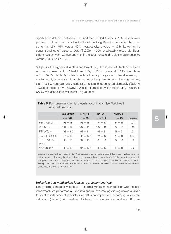
121
Predictors of pulmonary function impairment in chronic heart failure
5
significantly different between men and women (54% versus 70%, respectively, p-value = .11), women had diffusion impairment significantly more often than men using the LLN (61% versus 40%, respectively, p-value = .04). Lowering the conventional cutoff value to 70% (TLCOc < 70% predicted) yielded significant differences between women and men in the occurrence of diffusion impairment (58% versus 33%, p-value = .01).
Subjects with a higher NYHA class had lower FEV1, TLCOc, and VA (Table 5). Subjects who had smoked ≥ 10 PY had lower FEV1, FEV1/VC ratio and TLCOc than those with < 10 PY (Table 6). Subjects with pulmonary congestion, pleural effusion, or cardiomegaly on chest radiograph had lower lung volumes and diffusing capacity than those without pulmonary congestion, pleural effusion, or cardiomegaly (Table 7). TLCOc corrected for VA, however, was comparable between the groups. A history of CABG was associated with lower lung volumes.
Univariate and multivariate logistic regression analysisSince the most frequently observed abnormality in pulmonary function was diffusion impairment, we performed a univariate and multivariate logistic regression analysis to identify independent predictors of diffusion impairment according to different definitions (Table 8). All variables of interest with a univariate p-value < .05 were
Table 5 Pulmonary function test results according to New York Heart Association class.
Total group NYHA I NYHA II NYHA III
n = 164 n = 28 n = 117 n = 19 p-value
FEV1, % pred. 93 ± 18 98 ± 18† 94 ± 17 84 ± 19 .03
VC, % pred. 104 ± 17 107 ± 16 104 ± 16 97 ± 21 .12
FEV1/VC, % 68 ± 8.0 68 ± 8 68 ± 8 68 ± 9 .91
TLCOc, % pred.‡ 76 ± 16 85 ± 10*† 74 ± 16 73 ± 15 < .001
TLCOc/VA, % pred.‡
90 ± 20 94 ± 15 88 ± 20 92 ± 23 .33
VA, % pred.‡ 88 ± 13 94 ± 10*† 88 ± 13 83 ± 15 .03
Data are presented as mean ± SD. Abbreviations as in Table 2 and 3 legends. P-values refer to differences in pulmonary function between groups of subjects according to NYHA class (independent analysis of variance). * p-value < .05, NYHA I versus NYHA II; †p-value < .05, NYHA I versus NYHA III. No significant differences in pulmonary function were found between NYHA class II and III. ‡ Analysis was performed in a total of 153 subjects.
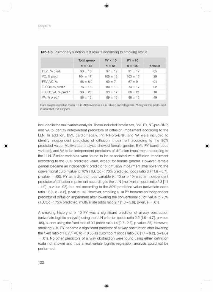
122
Chapter 5
included in the multivariate analysis. These included female sex, BMI, PY, NT-pro-BNP, and VA to identify independent predictors of diffusion impairment according to the LLN. In addition, BMI, cardiomegaly, PY, NT-pro-BNP, and VA were included to identify independent predictors of diffusion impairment according to the 80% predicted value. Multivariate analysis showed female gender, BMI, PY (continuous variable), and VA to be independent predictors of diffusion impairment according to the LLN. Similar variables were found to be associated with diffusion impairment according to the 80% predicted value, except for female gender. However, female gender became an independent predictor of diffusion impairment after lowering the conventional cutoff value to 70% (TLCOc < 70% predicted, odds ratio 3.7 [1.6 – 8.7], p-value = .00). PY as a dichotomous variable (< 10 or ≥ 10) was an independent predictor of diffusion impairment according to the LLN (multivariate odds ratio 2.3 [1.1 - 4.9], p-value .03), but not according to the 80% predicted value (univariate odds ratio 1.6 [0.8 - 3.2], p-value .14). However, smoking ≥ 10 PY became an independent predictor of diffusion impairment after lowering the conventional cutoff value to 75% (TLCOc < 75% predicted, multivariate odds ratio 2.7 [1.3 – 5.9], p-value = .01).
A smoking history of ≥ 10 PY was a significant predictor of airway obstruction (univariate logistic analysis) using the LLN criterion (odds ratio 2.2 [1.0 - 4.7], p-value .05), but not using the fixed ratio of 0.7 (odds ratio 1.4 [0.7 - 2.6], p-value .35). However, smoking ≥ 10 PY became a significant predictor of airway obstruction after lowering the fixed ratio of FEV1/FVC to < 0.65 as cutoff point (odds ratio 3.6 [1.4 – 9.2], p-value = .01). No other predictors of airway obstruction were found using either definition (data not shown) and thus a multivariate logistic regression analysis could not be performed.
Table 6 Pulmonary function test results according to smoking status.
Total group PY < 10 PY ≥ 10
n = 164 n = 64 n = 100 p-value
FEV1, % pred. 93 ± 18 97 ± 19 91 ± 17 .05
VC, % pred. 104 ± 17 105 ± 19 103 ± 15 .39
FEV1/VC, % 68 ± 8.0 69 ± 7 67 ± 9 .04
TLCOc, % pred.* 76 ± 16 80 ± 13 74 ± 17 .02
TLCOc/VA, % pred.* 90 ± 20 93 ± 17 88 ± 21 .10
VA, % pred.* 88 ± 13 89 ± 13 88 ± 13 .49
Data are presented as mean ± SD. Abbreviations as in Table 2 and 3 legends. *Analysis was performed in a total of 153 subjects.
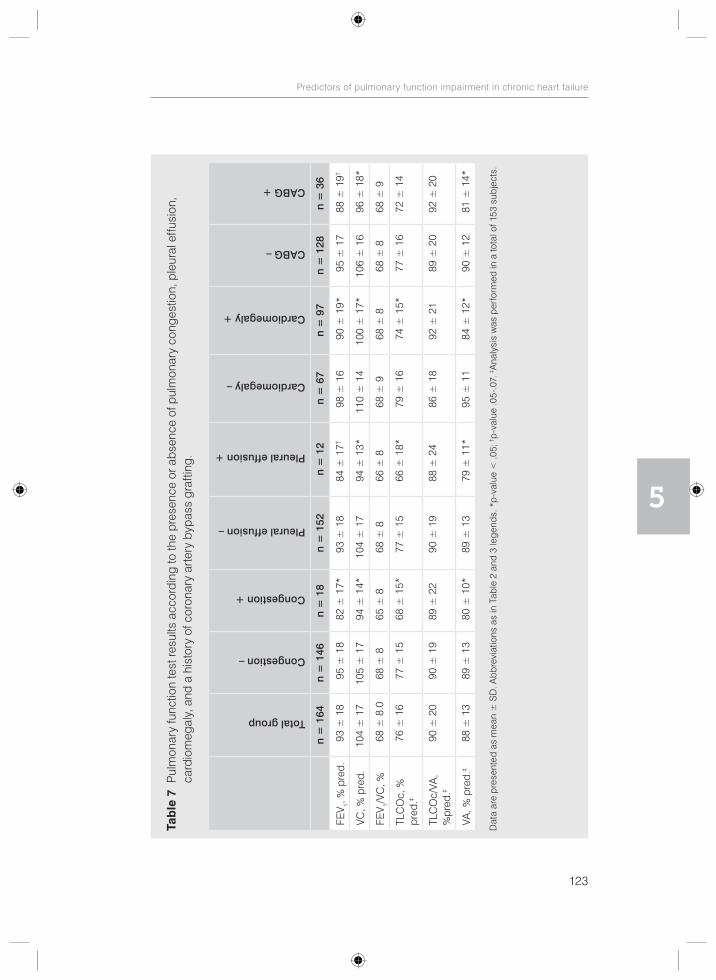
123
Predictors of pulmonary function impairment in chronic heart failure
5
Tab
le 7
Pul
mon
ary
func
tion
test
resu
lts a
ccor
ding
to th
e pr
esen
ce o
r abs
ence
of p
ulm
onar
y co
nges
tion,
ple
ural
effu
sion
, ca
rdio
meg
aly,
and
a h
isto
ry o
f cor
onar
y ar
tery
byp
ass
graf
ting.
Total group
Congestion –
Congestion +
Pleural effusion –
Pleural effusion +
Cardiomegaly –
Cardiomegaly +
CABG –
CABG +
n =
164
n =
146
n =
18
n =
152
n =
12
n =
67
n =
97
n =
128
n =
36
FEV 1,
% p
red.
93 ±
18
95 ±
18
82 ±
17*
93 ±
18
84 ±
17†
98 ±
16
90 ±
19*
95 ±
17
88 ±
19†
VC, %
pre
d.10
4 ±
17
105
± 1
794
± 1
4*10
4 ±
17
94 ±
13*
110
± 1
410
0 ±
17*
106
± 1
696
± 1
8*
FEV 1/
VC, %
68 ±
8.0
68 ±
865
± 8
68 ±
866
± 8
68 ±
968
± 8
68 ±
868
± 9
TLC
Oc,
%
pred
.‡
76 ±
16
77 ±
15
68 ±
15*
77 ±
15
66 ±
18*
79 ±
16
74 ±
15*
77 ±
16
72 ±
14
TLC
Oc/
VA,
%pr
ed.‡
90 ±
20
90 ±
19
89 ±
22
90 ±
19
88 ±
24
86 ±
18
92 ±
21
89 ±
20
92 ±
20
VA, %
pre
d.‡
88 ±
13
89 ±
13
80 ±
10*
89 ±
13
79 ±
11*
95 ±
11
84 ±
12*
90 ±
12
81 ±
14*
Dat
a ar
e p
rese
nted
as
mea
n ±
SD
. Abb
revi
atio
ns a
s in
Tab
le 2
and
3 le
gend
s. *
p-v
alue
< .0
5; † p
-val
ue .0
5-.0
7. ‡ A
naly
sis
was
per
form
ed in
a to
tal o
f 153
sub
ject
s.

124
Chapter 5
Tab
le 8
Det
erm
inan
ts o
f diff
usio
n im
pairm
ent a
ccor
ding
to th
e LL
N c
riter
ia a
nd c
onve
ntio
nal c
utof
f val
ues.
Diff
usio
n im
pai
rmen
t
TLC
Oc
< L
LNTL
CO
c <
LLN
TLC
Oc
< 8
0% p
red
.TL
CO
c <
80%
pre
d.
Uni
varia
teM
ultiv
aria
te†
Uni
varia
teM
ultiv
aria
te‡
Age
, yea
rs1.
001
(0.9
71 -
1.03
2)1.
023
(0.9
92 -
1.05
5)
Fem
ale
sex
2.30
8 (1
.049
– 5
.075
)*2.
970
(1.2
57 –
7.0
19)*
1.94
6 (0
.853
– 4
.440
)
BM
I, kg
/m2
0.91
6 (0
.847
- 0.
990)
*0.
898
(0.8
21 –
0.9
82)*
0.90
5 (0
.838
– 0
.977
)*0.
872
(0.7
96 –
0.9
56)*
LVE
F, %
0.96
2 (0
.920
- 1.
006)
0.96
4 (0
.921
– 1
.008
)
NYH
A c
lass
I vs
III
0.29
8 (0
.077
– 1
.145
)0.
424
(0.1
24 –
1.4
51)
NYH
A c
lass
II v
s III
1.27
3 (0
.467
– 3
.470
)1.
380
(0.5
04 –
3.7
81)
Con
gest
ion
2.00
8 (0
.678
- 5.
954)
3.26
3 (0
.881
- 12
.080
)
Car
diom
egal
y1.
823
(0.9
44 -
3.52
1)2.
374
(1.2
27 –
4.5
92)*
PY, y
ears
1.02
1 (1
.003
- 1.
038)
*1.
023
(1.0
04 –
1.0
43)*
1.00
2 (1
.003
– 1
.041
)*1.
025
(1.0
03 –
1.0
48)*
CA
BG
1.22
1 (0
.554
- 2.
690)
1.72
4 (0
.749
- 3.
966)
NT-
pro-
BN
P, p
mol
/L1.
001
(1.0
00 -
1.00
2)*
1.00
2 (1
.000
– 1
.003
)*
VA, %
pre
d.0.
968
(0.9
43 –
0.9
94)*
0.96
5 (0
.936
– 0
.995
)*0.
962
(0.9
36 –
0.9
89)*
0.96
1 (0
.928
– 0
.994
)*
AC
E-I
0.94
4 (0
.467
- 1.
908)
1.04
1 (0
.513
– 2
.111
)
Ald
oste
rone
-ant
agon
ists
1.80
0 (0
.930
- 3.
484)
1.52
0 (0
.778
– 2
.972
)
Dat
a ar
e p
rese
nted
as
odds
rat
io’s
and
con
fiden
ce in
terv
als.
LLN
, lo
wer
lim
it of
nor
mal
. O
ther
abb
revi
atio
ns a
s in
Tab
le 2
and
3 le
gend
s. *
p <
.05
. † N
agel
kerk
e R
2 = .2
2. ‡ N
agel
kerk
e R
2 = .2
3.
125
Predictors of pulmonary function impairment in chronic heart failure
5
Discussion
The current study showed that the definition used for pulmonary function impairment impacts the role of gender and smoking on pulmonary function in subjects with chronic HF with LVSD. The LLN criterion identified an extra independent predictor of diffusion impairment compared to the 80% predicted value; in addition to BMI, PY, and VA, female gender also turned out to be an independent predictor. A smoking history of ≥ 10 PY was a significant predictor of diffusion impairment and airway obstruction using the LLN criterion, but not using the conventional cutoff values. However, making the conventional cutoff values more stringent by lowering the cutoff point yielded similar results as the LLN. Lower lung volumes were found in subjects with pulmonary congestion, cardiomegaly, and a history of CABG.
In the current study, the conventional cutoff values classified more patients as having diffusion impairment and airway obstruction compared to the LLN. This is explained by the physiological decrease of the FEV1/FVC ratio with age. The FEV1 declines more rapidly with age than the FVC in normal subjects.24 As a result, the fixed ratio of 0.7, that is traditionally used because of its simplicity, may lead to overdiagnosis in the elderly and underdiagnosis in younger patients.24 Furthermore, the frequently used 80% predicted value has neither statistical nor physiological validity.15-17 Limits of normal as the predicted ± 20% can only be accurate when the variance above and below the predicted regression line is proportional with the predicted value (i.e. het-eroscedastic: large variance with large values and small variance with small values). However, since this is not the case as the scatter around the predicted regression line is constant (homoscedastic) in pulmonary function measurements, the 80% predicted rule of thumb may lead to false-positive diagnosis in the elderly and shorter individuals with smaller predicted values and underdiagnosis in younger and taller patients with larger predicted values.15-17
Misdiagnosis of pulmonary function abnormalities by the conventional cutoff values may have interfered with the interpretation of prior research aiming to investigate the impact of HF and several clinical variables on pulmonary function. This may explain part of the inconsistencies across the studies. In fact, a smoking history of ≥ 10 PY was a significant predictor of diffusion impairment and airway obstruction using the LLN criterion, but not using the conventional cutoff values. This implies that inclusion of subjects who are incorrectly labeled as having pulmonary dysfunction distorted the effect of smoking on pulmonary function. Indeed, lowering the conventional cutoff values to match the more stringent LLN and thus avoid overdiagnosis of diffusion impairment and airway obstruction in the elderly, produced similar results as the LLN. Similarly, female gender was an independent predictor of diffusion impairment
126
Chapter 5
according to the LLN, but not according to the 80% predicted value. However, decreasing the cutoff point to define diffusion impairment showed comparable findings to the LLN. On the other hand, by increasing the LLN to the 10th percentile, the association between female gender and diffusion impairment was lost (data not shown). This is explained by the fact that the lower ranges of diffusing capacity represented relatively more women than men. In summary, the LLN criterion identified more predictors of diffusion impairment and airway obstruction compared to conventional cutoff values in subjects with chronic HF with LVSD. However, when conventional cutoff points were lowered to match the more stringent LLN criterion, the same effects were seen.
The effect of different definitions has also been put forward in the study of de Marco et al.,25 who have shown that the role of age, sex, former smoking, and low BMI on the development of COPD differs according to the definition used to define COPD. They suggested the need for a definition of COPD that is not exclusively based on spirometry.
Little is known about the clinical impact of different criteria of pulmonary dysfunction. Mannino and Diaz-Guzman26 followed up the mortality data of a large number of subjects from the NHANES III classified as normal, obstructed, or restricted using conventional cutoff values and the LLN. They found that subjects classified as normal using the LLN but obstructed or restricted using conventional cutoff values had a higher risk of mortality than normal subjects in up to 18 years of follow-up. This finding suggests that conventional criteria may identify at-risk patients who would have been missed using the LLN. This study was limited by the lack of post-bronchodilator pulmonary function test results, other outcome parameters than mortality, and the lack of comparison between subjects with mild airway obstruction according to conventional cutoff values (FEV1/FVC < 0.7 and FEV1 ≥ 80% predicted) and normal subjects according to the LLN (FEV1/FVC, FEV1, and FVC ≥ LLN). More longitudinal studies are warranted to determine which criterion is clinically more relevant in terms of morbidity (symptoms, exercise tolerance, health-related quality of life, hospitalization, use of health recourses) and mortality. Since we did not follow our subjects prospectively, it remains unknown whether pulmonary function impairment had prognostic implications in our study population and whether this is influenced by different definitions of pulmonary dysfunction.
Several factors have been implied to play a role in the etiology of pulmonary function impairment in patients with HF, including the effects of HF itself on pulmonary function in addition to (previously undiagnosed) underlying pulmonary disease and confounding influences such as smoking, CABG, and obesity.4-7, 13
127
Predictors of pulmonary function impairment in chronic heart failure
5
Diffusion impairment has been thought to be related to the thickening of alveolar- capillary membrane due to hydrostatic mechanical injury, interstitial edema, remodeling, and fibrosis.1, 2, 4-6, 27 Because heart transplantation does not affect or may even worsen pulmonary diffusing capacity despite an improvement in hemodynamic status and lung volumes,28 it has been suggested that reduced diffusing capacity in CHF may be related to permanent damage to the alveolar-capillary membrane.2 Other possible causes of diffusion impairment in HF include reduced lung and pulmonary capillary blood volumes, ventilation-perfusion mismatch, recurrent pulmonary emboli, smoking, and cardiopulmonary bypass.4-6, 13 The results of our study showed a higher NYHA class, smoking of ≥ 10 PY, pulmonary congestion, pleural effusion, and cardiomegaly to be associated with more impaired diffusing capacity, the latter three probably due to their negative effects on lung volume. Indeed, diffusing capacity corrected for VA was not significantly different between the groups. Also, although VA turned out to be an independent predictor of diffusion impairment, pulmonary congestion and cardiomegaly were not. In contrast to previous reports,29-31 the use of angiotensin-converting enzyme inhibitors and aldosterone-antagonists was not associated with increased diffusing capacity. Also, diffusing capacity was not significantly different between groups of subjects with or without a history of CABG. Independent predictors of diffusion impairment were BMI, PY (continuous variable), and VA, whereas the role of gender and having smoked ≥ 10 PY depended on the definition used to define diffusion impairment. Although the underlying mechanisms are not clear, women seemed to be more sensitive to the detrimental effects of HF on diffusing capacity. Gender differences in pulmonary function have been recognized before, but not specifically in the HF population. Adult women have been reported to have lower resting lung diffusing capacity corrected for hemoglobin, smaller lung volumes, and lower maximal expiratory flow rates, even when corrected for age and standing height relative to men.32 It has been suggested that these gender differences in part can be explained by pulmonary structural differences (fewer total number of alveoli and smaller airway diameter relative to lung size) and hormonal influences in women.32 More research is needed regarding the influence of gender on pulmonary function in general and specifically in the HF population. The protective association between a higher BMI and less likelihood of having diffusion impairment has to our knowledge not been described before in subjects with chronic HF. However, some studies in healthy obese non-smokers have suggested that diffusing capacity may be increased in extremely obese subjects, probably as a result of the increase in blood volume.33
Restriction has been linked to cardiomegaly, pleural effusion, respiratory muscle weakness, CABG, fibrosis from chronic congestion, and reduced lung compliance due to chronic vascular engorgement, interstitial/alveolar fluid accumulation, and chronic remodeling of the pulmonary vasculature due to elevated left atrial pressure.1, 4-6, 13, 34, 35
128
Chapter 5
In line with expectations, we found lung volumes to be lower in subjects with pulmonary congestion, pleural effusion, cardiomegaly, and a history of CABG.
Airway obstruction has been attributed to alveolar fluid accumulation, bronchial mucosal swelling, peribronchial edema and fibrosis, squamous metaplasia of bronchial epithelial cells induces by transforming growth factor-ß from the failing heart, geometric decrease in airway size from reduction in lung volume, abnormalities of autonomic control, neurohumoral bronchoconstriction, bronchial hyperrespon-siveness, and smoking,4-7, 13, 36 although results are not consistent.14 Our study showed more impaired FEV1/VC ratio in patients who had smoked ≥ 10 PY. Also, having smoked ≥ 10 PY was a significant predictor of airway obstruction, but this depended on the definition used to define airway obstruction. Although we have excluded patients with known COPD or other obstructive lung disease, we cannot rule out the possibility that some of the patients with airway obstruction had previously undiagnosed COPD, as most patients were current or former smokers. In fact, 16% (LLN) to 24% (conventional cutoff values) of the patients had post-bronchodilator airway obstruction after inhaling 400 mcg salbutamol and 80 mcg ipratropium. These patients had more symptoms of cough and sputum than those without post- bronchodilator airway obstruction (data not shown). Also, patients who were current or former smokers tended to have more often post-bronchodilator airway obstruction than those who had never smoked (LLN: 19% versus 6%, p = .06; conventional cutoff values: 27% versus 11%, p = .05). However, hyperinflation, which has been found to be a valid indicator of true COPD in patients with congested HF,37 was not significantly different between groups of patients with persistent airway obstruction after broncho-dilation and those with reversible airway obstruction (data not shown). Although airway obstruction in HF has also been attributed to pulmonary congestion, FEV1/VC ratio was not significantly different between groups of subjects with and without pulmonary congestion in our study. This, however, does not exclude the contribution of pulmonary congestion to small airways obstruction.
Importantly, airway obstruction may lead to hyperinflation of the lungs due to expiratory flow limitation and air trapping, which was found in almost half of our patients with airway obstruction as defined by an increased RV/TLC ratio. This may contribute to symptoms of dyspnea, poor exercise tolerance, increased work of breathing and oxygen consumption, respiratory muscle dysfunction, and adverse impact on cardiac function by decreasing the preload.38 Thus, irrespective of the causes, pulmonary function abnormalities associated with chronic HF may explain part of the symptoms and functional disability encountered in these patients.8-12 Moreover, pulmonary function impairment increases with the severity of HF,9, 11 provides important prognostic information,39-42 and may ameliorate or normalize with
129
Predictors of pulmonary function impairment in chronic heart failure
5
several treatment modalities, such as pharmacological and non-pharmacological treatment of HF2, 28, 43-46 and anti-obstructive therapy with bronchodilators.47-53 Pulmonary function might thus be used as a guide for the evaluation of patients with chronic HF, with respect to severity of disease, prognosis, and response to treatment.
Some limitations of this study deserve further discussion. It is important to realize that these results may not be applicable to all patients with chronic HF, since we did not include subjects with preserved systolic function, who seem to have less impaired pulmonary function.54 Furthermore, patients with more severe HF could have been underrepresented in this study because of inability to participate and thus pulmonary function abnormalities might have been underestimated. Finally, considering the relatively small number of subjects included in the current study, in particular women and non-smokers, further research is needed to confirm our results.
In conclusion, the LLN identifies gender as an extra independent predictor of diffusion impairment and a smoking history of ≥ 10 PY as an additional predictor of both diffusion impairment and airway obstruction compared to conventional cutoff values in subjects with chronic HF with LVSD. However, when conventional cutoff points were lowered to match the more stringent LLN criterion, the same effects were seen. Our results stress the need for clear definitions of pulmonary function abnormalities. More longitudinal studies are warranted to determine which criterion is clinically more relevant. Specifically, future research should focus on better characterizing the potentially misclassified group of patients who are above the LLN but below the conventional cutoff values. Do these patients have worse outcome with higher morbidity and mortality rates that is amenable to treatment or do they present clinical features similar to those with CHF but without pulmonary dysfunction? Finally, more research is needed regarding the influence of gender on pulmonary function in the HF population and the possible underlying pathophysiologic mechanisms.
AcknowledgmentsThe authors would like to thank the participants in this study for their cooperation. We also would like to thank the nurse practitioners P.A. Ninaber, W.A.M. Janssen, W.H. van Zimmeren-Feijen, and heart failure nurse J.G. Froon-Elferink for their contribution to inclusion of participants and data collection. The authors owe much gratitude to the pulmonary function and clinical chemistry and haematology laboratories as well as the echocardiography and radiology departments for their assistance in data collection. Also, we would like to acknowledge Prof. Dr. P.H. Quanjer for his contribution to the manuscript and biostatistician A.R.T. Donders for his contribution to statistical analysis. Finally, we would like to thank GlaxoSmithKline for supporting this study by an unrestricted grant.

130
Chapter 5
References
1. Kee K, Naughton MT. Heart failure and the lung. Circ J. 2010;74:2507-16.2. Guazzi M. Alveolar gas diffusion abnormalities in heart failure. J Card Fail. 2008;14:695-702.3. Agostoni P, Cattadori G, Bussotti M, Apostolo A. Cardiopulmonary interaction in heart failure. Pulm
Pharmacol Ther. 2007;20:130-4.4. Gehlbach BK, Geppert E. The pulmonary manifestations of left heart failure. Chest. 2004;125:669-82.5. Chua TP, Coats AJ. The lungs in chronic heart failure. Eur Heart J. 1995;16:882-7.6. Faggiano P. Abnormalities of pulmonary function in congestive heart failure. Int J Cardiol. 1994;44:1-8.7. Snashall PD, Chung KF. Airway obstruction and bronchial hyperresponsiveness in left ventricular failure
and mitral stenosis. Am Rev Respir Dis. 1991;144:945-56.8. Mancini DM. Pulmonary factors limiting exercise capacity in patients with heart failure. Prog Cardiovasc
Dis. 1995;37:347-70.9. Puri S, Baker BL, Dutka DP, Oakley CM, Hughes JM, Cleland JG. Reduced alveolar-capillary membrane
diffusing capacity in chronic heart failure. Its pathophysiological relevance and relationship to exercise performance. Circulation. 1995;91:2769-74.
10. Kraemer MD, Kubo SH, Rector TS, Brunsvold N, Bank AJ. Pulmonary and peripheral vascular factors are important determinants of peak exercise oxygen uptake in patients with heart failure. J Am Coll Cardiol. 1993;21:641-8.
11. Dimopoulou I, Daganou M, Tsintzas OK, Tzelepis GE. Effects of severity of long-standing congestive heart failure on pulmonary function. Respir Med. 1998;92:1321-5.
12. Tzani P, Piepoli MF, Longo F, Aiello M, Serra W, Maurizio AR, et al. Resting lung function in the assessment of the exercise capacity in patients with chronic heart failure. Am J Med Sci. 2010;339:210-5.
13. Johnson BD, Beck KC, Olson LJ, O’Malley KA, Allison TG, Squires RW, et al. Pulmonary function in patients with reduced left ventricular function: influence of smoking and cardiac surgery. Chest. 2001;120:1869-76.
14. Wright RS, Levine MS, Bellamy PE, Simmons MS, Batra P, Stevenson LW, et al. Ventilatory and diffusion abnormalities in potential heart transplant recipients. Chest. 1990;98:816-20.
15. Sobol BJ, Sobol PG. Per cent of predicted as the limit of normal in pulmonary function testing: a statistically valid approach. Thorax. 1979;34:1-3.
16. Pellegrino R, Viegi G, Brusasco V, Crapo RO, Burgos F, Casaburi R, et al. Interpretative strategies for lung function tests. Eur Respir J. 2005;26:948-68.
17. Minasian AG, van den Elshout FJ, Dekhuijzen PN, Vos PJ, Willems FF, van den Bergh PJ, et al. Pulmonary function impairment in patients with chronic heart failure: Lower limit of normal versus conventional cutoff values. Heart Lung. 2014;43:311-16.
18. Miller MR, Quanjer PH, Swanney MP, Ruppel G, Enright PL. Interpreting lung function data using 80 percent of predicted and fixed thresholds misclassifies over 20% of patients. Chest. 2011;139:52-59.
19. Dickstein K, Cohen-Solal A, Filippatos G, McMurray JJ, Ponikowski P, Poole-Wilson PA, et al. ESC Guidelines for the diagnosis and treatment of acute and chronic heart failure 2008: the Task Force for the Diagnosis and Treatment of Acute and Chronic Heart Failure 2008 of the European Society of Cardiology. Developed in collaboration with the Heart Failure Association of the ESC (HFA) and endorsed by the European Society of Intensive Care Medicine (ESICM). Eur Heart J. 2008;29:2388-442.
20. O’Hanlon R, O’Shea P, Ledwidge M, O’Loughlin C, Lange S, Conlon C, et al. The biologic variability of B-type natriuretic peptide and N-terminal pro-B-type natriuretic peptide in stable heart failure patients. J Card Fail. 2007;13:50-5.
21. Quanjer PH, Tammeling GJ, Cotes JE, Pedersen OF, Peslin R, Yernault JC. Lung volumes and forced ventilatory flows. Report Working Party Standardization of Lung Function Tests, European Community for Steel and Coal. Official Statement of the European Respiratory Society. Eur Respir J 1993;16:5-40.
22. Rabe KF, Hurd S, Anzueto A, Barnes PJ, Buist SA, Calverley P, et al. Global strategy for the diagnosis, management, and prevention of chronic obstructive pulmonary disease: GOLD executive summary. Am J Respir Crit Care Med. 2007;176:532-55.
131
Predictors of pulmonary function impairment in chronic heart failure
5
23. Bourjeily-Habr G, Rochester CL, Palermo F, Snyder P, Mohsenin V. Randomised controlled trial of transcutaneous electrical muscle stimulation of the lower extremities in patients with chronic obstructive pulmonary disease. Thorax. 2002;57:1045-9.
24. Townsend MC. Conflicting definitions of airways obstruction: Drawing the line between normal and abnormal. Chest. 2007;131:335-6.
25. de Marco R, Accordini S, Marcon A, Cerveri I, Anto JM, Gislason T, et al. Risk factors for chronic obstructive pulmonary disease in a European cohort of young adults. Am J Respir Crit Care Med. 2011;183:891-7.
26. Mannino DM, Diaz-Guzman E. Interpreting lung function data using 80% predicted and fixed thresholds identifies patients at increased risk of mortality. Chest. 2012;141:73-80.
27. Guazzi M. Alveolar-capillary membrane dysfunction in chronic heart failure: pathophysiology and therapeutic implications. Clin Sci (Lond). 2000;98:633-41.
28. Bussieres LM, Pflugfelder PW, Ahmad D, Taylor AW, Kostuk WJ. Evolution of resting lung function in the first year after cardiac transplantation. Eur Respir J. 1995;8:959-62.
29. Guazzi M, Melzi G, Agostoni P. Comparison of changes in respiratory function and exercise oxygen uptake with losartan versus enalapril in congestive heart failure secondary to ischemic or idiopathic dilated cardiomyopathy. Am J Cardiol. 1997;80:1572-6.
30. Guazzi M, Marenzi G, Alimento M, Contini M, Agostoni P. Improvement of alveolar-capillary membrane diffusing capacity with enalapril in chronic heart failure and counteracting effect of aspirin. Circulation. 1997;95:1930-6.
31. Guazzi M, Agostoni P, Guazzi MD. Modulation of alveolar-capillary sodium handling as a mechanism of protection of gas transfer by enalapril, and not by losartan, in chronic heart failure. J Am Coll Cardiol. 2001;37:398-406.
32. Harms CA, Rosenkranz S. Sex differences in pulmonary function during exercise. Med Sci Sports Exerc. 2008;40:664-8.
33. Salome CM, King GG, Berend N. Physiology of obesity and effects on lung function. J Appl Physiol (1985). 2010;108:206-11.
34. Olson TP, Beck KC, Johnson BD. Pulmonary function changes associated with cardiomegaly in chronic heart failure. J Card Fail. 2007;13:100-7.
35. Agostoni P, Cattadori G, Guazzi M, Palermo P, Bussotti M, Marenzi G. Cardiomegaly as a possible cause of lung dysfunction in patients with heart failure. Am Heart J. 2000;140:e24.
36. Tanabe T, Kanoh S, Moskowitz WB, Rubin BK. Cardiac asthma: TGF-beta from the failing heart leads to squamous metaplasia in human airway cells and in the murine lung. Chest. 2012;142:1274-83.
37. Brenner S, Guder G, Berliner D, Deubner N, Frohlich K, Ertl G, et al. Airway obstruction in systolic heart failure - COPD or congestion? Int J Cardiol. 2013;168:1910-6.
38. Rossi A, Aisanov Z, Avdeev S, Di Maria G, Donner CF, Izquierdo JL, et al. Mechanisms, assessment and therapeutic implications of lung hyperinflation in COPD. Respir Med. 2015;109:785-802.
39. Guazzi M, Pontone G, Brambilla R, Agostoni P, Reina G. Alveolar--capillary membrane gas conductance: a novel prognostic indicator in chronic heart failure. Eur Heart J. 2002;23:467-76.
40. Iversen KK, Kjaergaard J, Akkan D, Kober L, Torp-Pedersen C, Hassager C, et al. The prognostic importance of lung function in patients admitted with heart failure. Eur J Heart Fail. 2010;12:685-91.
41. Hawkins NM, Petrie MC, Jhund PS, Chalmers GW, Dunn FG, McMurray JJ. Heart failure and chronic obstructive pulmonary disease: diagnostic pitfalls and epidemiology. Eur J Heart Fail. 2009;11:130-9.
42. O’Kelly N, Robertson W, Smith J, Dexter J, Carroll-Hawkins C, Ghosh S. Short-term outcomes in heart failure patients with chronic obstructive pulmonary disease in the community. World J Cardiol. 2012;4:66-71.
43. Light RW, George RB. Serial pulmonary function in patients with acute heart failure. Arch Intern Med. 1983;143:429-33.
44. Faggiano P, Lombardi C, Sorgato A, Ghizzoni G, Spedini C, Rusconi C. Pulmonary function tests in patients with congestive heart failure: effects of medical therapy. Cardiology. 1993;83:30-5.
45. Niset G, Ninane V, Antoine M, Yernault JC. Respiratory dysfunction in congestive heart failure: correction after heart transplantation. Eur Respir J. 1993;6:1197-201.

132
Chapter 5
46. Petersen CL, Kjaer A. Impact of medical treatment on lung diffusion capacity in elderly patients with heart failure. Baseline characteristics and 1-year follow up after medical treatment. Int J Cardiol. 2005;98:453-7.
47. Minasian AG, van den Elshout FJ, Dekhuijzen PN, Vos PJ, Willems FF, van den Bergh PJ, et al. Bronchodilator responsiveness in patients with chronic heart failure. Heart Lung. 2012;42:208-14.
48. Kindman LA, Vagelos RH, Willson K, Prikazky L, Fowler M. Abnormalities of pulmonary function in patients with congestive heart failure, and reversal with ipratropium bromide. Am J Cardiol. 1994;73:258-62.
49. Uren NG, Davies SW, Jordan SL, Lipkin DP. Inhaled bronchodilators increase maximum oxygen consumption in chronic left ventricular failure. Eur Heart J. 1993;14:744-50.
50. Khan KA, Jalal S, Jan VM, Lone GM, Jan R, Alai MS, et al. Pulmonary function profile in chronic congestive heart failure and the effect of ipratropium bromide. Indian Heart J. 2000;52:297-300.
51. Witte KK, Morice A, Cleland JG, Clark AL. The reversibility of increased airways resistance in chronic heart failure measured by impulse oscillometry. J Card Fail. 2004;10:149-54.
52. Rolla G, Bucca C, Brussino L, Gallo W, Malara D. Bronchodilating effect of ipratropium bromide in heart failure. Eur Respir J. 1993;6:1492-5.
53. Ng TM, Munger MA, Lombardi WL, Doing TH, Ryujin DT, Young DC, et al. Chronically inhaled salmeterol improves pulmonary function in heart failure. J Cardiovasc Pharmacol. 2002;40:140-5.
54. Kurzawa R, Baron T, Grodzicki T. Pulmonary function test abnormalities in the elderly with systolic heart failure. Kardiol Pol. 2007;65:875-80; discussion 81-2.

133
Predictors of pulmonary function impairment in chronic heart failure
5

Armine G. Minasian Frank J.J. van den ElshoutP.N. Richard DekhuijzenPetra J.E. VosFrank F. WillemsPaul J.P.C. van den BerghYvonne F. Heijdra
Heart Lung. 2013;42:208-14.
Bronchodilator responsiveness in patients with chronic heart failure
CHAPTER 6
136
Chapter 6
Abstract
Objective: The aim of this study was to evaluate the effect of inhaled bronchodilators on pulmonary function and dyspnea in patients with chronic heart failure (HF).
Background: Conflicting data exist on whether bronchodilators may improve pulmonary function and dyspnea in patients with chronic HF.
Methods: In this retrospective observational study we analyzed data of 116 chronic HF outpatients with systolic dysfunction who underwent spirometry and Borg dyspnea measurements before and after inhalation of 400 μg salbutamol and 80 μg ipratropium. Patients with chronic obstructive pulmonary disease (COPD) or asthma were excluded.
Results: Bronchodilators fully reversed airway obstruction (AO) in 25 of 64 (39.1%) patients with pre-bronchodilator AO. All spirometric measurements, except for forced vital and inspiratory capacities, improved significantly post-bronchodilation. Absolute and percent improvements in forced expiratory volume in 1 s (FEV1) were more pronounced in patients with persistent AO post-bronchodilation compared to those without AO (0.19 ± 0.18L and 8.4 ± 7.3% versus 0.11 ± 0.12L and 4.3 ± 4.0%, p < 0.05). Significant bronchodilator responsiveness of FEV1 (> 200 mL and > 12%) was noted in 12.1% and was more frequent in patients with persistent AO and fully reversible AO than in those without AO (23.1% and 16.0% versus 1.9%, p < 0.05). We measured a small, albeit significant improvement in dyspnea (0.7 ± 1.2 versus 0.9 ± 1.3, p = 0.002).
Conclusions: Inhaled bronchodilators may have an additional role in the management of patients with chronic HF because of their potential to improve pulmonary function, especially in those with AO. The clinical usefulness and possible adverse events of bronchodilators need to be further established.
Keywords: Chronic heart failure; bronchodilator responsiveness; salbutamol; ipratropium; airway obstruction.
137
Bronchodilator responsiveness in patients with chronic heart failure
6
Introduction
Pulmonary function abnormalities, such as diffusion impairment, restriction, and airway obstruction (AO), are common in patients with heart failure (HF)1, 2 and may contribute to the perception of dyspnea and exercise intolerance.3 Treatment directed at reversing AO with bronchodilators may have an additional role in the management of these patients. There are, however, only few studies concerning the beneficial effects of bronchodilators in patients with HF. Improvements in pulmonary function,4-10 dyspnea,10 and exercise performance7 have been reported. Some investigators have even observed an increase of mean forced expiratory volume in 1 s (FEV1) greater than 200 mL and 12% in patients with HF,6, 8, 9 especially in those with AO,8 left ventricular systolic dysfunction (LVSD),9 and during acute decompensation of HF.6 However, not all could confirm these findings.7, 10-14 Contrasting results across the studies might be attributed to the small number of patients studied and to the differences in study population, bronchodilators used, and definition of bronchodilator responsiveness (BDR).
The purpose of this study was to evaluate the effect of maximal bronchodilation with combined inhaled salbutamol and ipratropium bromide on pulmonary function and dyspnea in patients with chronic HF. In addition, we determined the proportion of patients with significant BDR (i.e. responder), defined as post-bronchodilator (BD) increase in FEV1 greater than 200 mL and 12% from the baseline value.15
Methods
Study design and participantsIn this retrospective observational study we analyzed data of chronic HF16 outpatients with left ventricular ejection fraction (LVEF) below 40%, New York Heart Association (NYHA) class I to IV, and age ≥ 18 years. These patients were recruited prospectively in another study from two outpatient cardiology departments of a large general hospital in The Netherlands between October 2009 and December 2010 for the purpose of evaluating the prevalence of chronic obstructive pulmonary disease (COPD) and pulmonary function impairment in chronic HF (ClinicalTrials.gov Identifier NCT01429376). Only patients with both pre- and post-BD spirometry results were included in the current study. Patients who were not able to cooperate or undergo spirometry or had a history of asthma17 were excluded. Other exclusion criteria were malignancy with a bad prognosis (survival < 6 months) and participation in another study. In addition, patients with a history of COPD15 or not further specified obstructive lung disease (OLD) were excluded from the current study. Echocardiography was performed in patients without a recent (≤ 6 months) echocardiography to confirm persisting LVSD.
138
Chapter 6
Patients were classified as having stable HF in the absence of hospitalization due to HF ≤ 3 months, change in diuretics ≤ 1 month, ≥ 3% weight gain ≤ 3 days, and > 50% increase of N-terminal pro-B natriuretic peptide (NT-pro-BNP) ≤ 1 month (baseline NT-pro-BNP ≥ 100 pmol/L) or > 100 pmol/L increase of NT-pro-BNP ≤ 1 month (baseline NT-pro-BNP < 100 pmol/L).18
Measurements and data collection At baseline, a first blood sample was taken for the measurement of NT-pro-BNP. One month later, the participants visited the hospital for an interview with the investigator and several examinations, including height and weight measurement, spirometry and a 10-point Borg dyspnea score19 before and after bronchodilators, a chest radiograph, and a second blood sample (NT-pro-BNP). Additional data were collected from medical records and personal interviews. Smoking status was defined as never (< 100 cigarettes in a lifetime), former (≥ 3 months ago), or current smoker (< 3 months). Smoking pack-years (PY) were based only on the tobacco cigarette history and one PY was defined as smoking 20 cigarettes a day for 1 year. Pulmonary function testsParticipants performed spirometry (MasterLab Pro, Jaeger, Würzburg, Germany) before and 30 minutes after inhalation of four doses of 100 μg aerosolized salbutamol and four doses of 20 μg aerosolized ipratropium bromide via Volumatic spacer. Beta-blockers were not discontinued prior to testing. Spirometry was performed by trained and certified operators using standard techniques and according to European Respiratory Society standards for acceptability and reproducibility.20 Reference values of the European Community for Coal and Steel were used.20 AO was defined as the ratio of FEV1 to forced vital capacity (FEV1/FVC) < 0.7.15 Chest radiographsStandard posteroanterior and lateral chest radiographs were performed and evaluated on the presence or absence of congestion: alveolar edema, pleural effusion, Kerley-B lines, and/or redistribution of pulmonary blood flow.
Ethical considerationsThe study was approved by the regional Research Ethics Committee Arnhem-Nijmegen in The Netherlands. All patients gave written informed consent.
Statistical analysisDescriptive data are presented as mean ± SD or as number (%). Differences between pre- and post-BD spirometry results were analyzed with the dependent t-test and the Wilcoxon signed-rank test was used to analyze the effect of bronchodilators on
139
Bronchodilator responsiveness in patients with chronic heart failure
6
dyspnea. Relationships between improvements in pulmonary function and LVEF, baseline pulmonary function, and changes in Borg score were examined using the Pearson’s or Spearman’s correlation coefficient tests, as appropriate. Differences in BDR between three groups of patients were examined with independent analysis of variance; group 1: patients with persistent AO after bronchodilation, group 2: patients with fully reversible AO, group 3: patients without AO. Post hoc analyses were performed using Fisher’s LSD procedure while Games-Howell procedure was used when the assumption of homogeneity of variance was not met. Comparisons between two groups of patients were analyzed using an independent t-test or Mann-Whitney U test for continuous variables and a chi-square or Fisher’s exact test for categorical variables, as appropriate (not all data shown). Statistical analyses were performed using the Statistical Package for Social Science version 15.0. All statistical tests were two-sided and a p-value < 0.05 was considered significant.
Results
Patient characteristicsOf the 234 patients recruited in the prospective study, 154 had both pre- and post-BD spirometry results of whom 38 had a history of COPD or not further specified OLD. The remaining 116 patients (50%) were selected for the current study (Figure 1). Table 1 shows the characteristics of these patients. Mean age was 70 ± 10 years and 83% were men. LVEF was 28 ± 7% and 86% had stable chronic HF. Only 14% had signs of congestion on chest radiograph. Over 60% had an ischemic etiology of HF. Other causes of HF were idiopathic (18%), hypertension (6%), valve disease (9%), tachycardio-myopathy (2%), and other (3%). The majority had NYHA class II (72%) and most patients were current or former smokers (81%). Symptoms of dyspnea at rest and/or on exertion were present in 80%.
Bronchodilator responsiveness AO was present in 64 of 116 (55.2%) patients before administration of inhaled bronchodilators. Of these patients, 39 (60.9%) had persistent AO after bronchodilation. Thus, bronchodilators fully reversed AO in 25 of 64 (39.1%) patients.
Significant improvements in pulmonary function were noted in all spirometric indices except for FVC and inspiratory capacity (IC) after bronchodilation (Table 2). Mean absolute and percent baseline changes in FEV1 were 0.15 ± 0.14L and 6.1 ± 5.9%, respectively. Corresponding changes for FVC and IC were 0.04 ± 0.23L (1.4 ± 6.5%) and 0.06 ± 0.37L (2.5 ± 13.0%), respectively. Figure 2 shows mean absolute and percent baseline changes in FEV1, FVC, and IC according to three groups of patients:
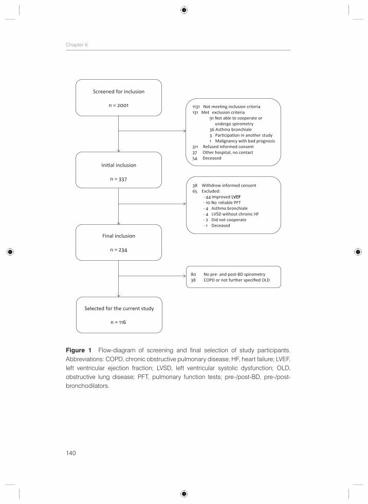
140
Chapter 6
Figure 1 Flow-diagram of screening and final selection of study participants. Abbreviations: COPD, chronic obstructive pulmonary disease; HF, heart failure; LVEF, left ventricular ejection fraction; LVSD, left ventricular systolic dysfunction; OLD, obstructive lung disease; PFT, pulmonary function tests; pre-/post-BD, pre-/post- bronchodilators.
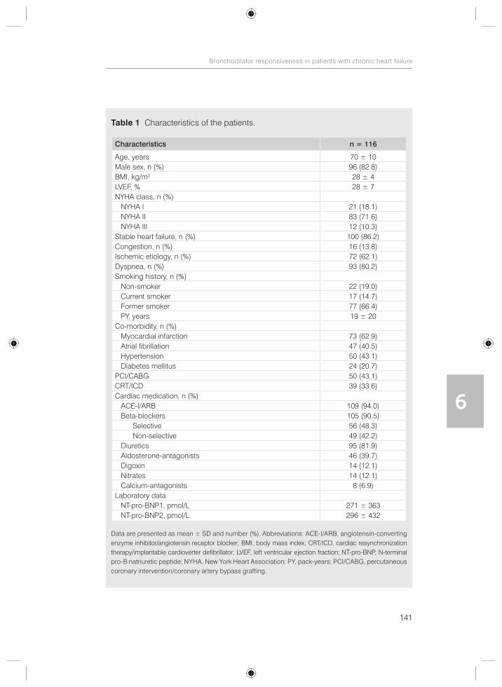
141
Bronchodilator responsiveness in patients with chronic heart failure
6
Table 1 Characteristics of the patients.
Characteristics n = 116
Age, years 70 ± 10Male sex, n (%) 96 (82.8)BMI, kg/m2 28 ± 4LVEF, % 28 ± 7NYHA class, n (%) NYHA I 21 (18.1) NYHA II 83 (71.6) NYHA III 12 (10.3)Stable heart failure, n (%) 100 (86.2)Congestion, n (%) 16 (13.8)Ischemic etiology, n (%) 72 (62.1)Dyspnea, n (%) 93 (80.2)Smoking history, n (%) Non-smoker 22 (19.0) Current smoker 17 (14.7) Former smoker 77 (66.4) PY, years 19 ± 20Co-morbidity, n (%) Myocardial infarction 73 (62.9) Atrial fibrillation 47 (40.5) Hypertension 50 (43.1) Diabetes mellitus 24 (20.7)PCI/CABG 50 (43.1)CRT/ICD 39 (33.6)Cardiac medication, n (%) ACE-I/ARB 109 (94.0) Beta-blockers 105 (90.5) Selective 56 (48.3) Non-selective 49 (42.2) Diuretics 95 (81.9) Aldosterone-antagonists 46 (39.7) Digoxin 14 (12.1) Nitrates 14 (12.1) Calcium-antagonists 8 (6.9)Laboratory data NT-pro-BNP1, pmol/L 271 ± 363 NT-pro-BNP2, pmol/L 296 ± 432
Data are presented as mean ± SD and number (%). Abbreviations: ACE-I/ARB, angiotensin-converting enzyme inhibitor/angiotensin receptor blocker; BMI, body mass index; CRT/ICD, cardiac resynchronization therapy/implantable cardioverter defibrillator; LVEF, left ventricular ejection fraction; NT-pro-BNP, N-terminal pro-B natriuretic peptide; NYHA, New York Heart Association; PY, pack-years; PCI/CABG, percutaneous coronary intervention/coronary artery bypass grafting.
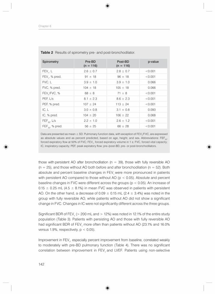
142
Chapter 6
those with persistent AO after bronchodilation (n = 39), those with fully reversible AO (n = 25), and those without AO both before and after bronchodilation (n = 52). Both absolute and percent baseline changes in FEV1 were more pronounced in patients with persistent AO compared to those without AO (p < 0.05). Absolute and percent baseline changes in FVC were different across the groups (p < 0.05). An increase of 0.15 ± 0.25 mL (4.5 ± 8.1%) in mean FVC was observed in patients with persistent AO. On the other hand, a decrease of 0.09 ± 0.15 mL (2.4 ± 3.4%) was noted in the group with fully reversible AO, while patients without AO did not show a significant change in FVC. Changes in IC were not significantly different across the three groups.
Significant BDR of FEV1 (> 200 mL and > 12%) was noted in 12.1% of the entire study population (Table 3). Patients with persisting AO and those with fully reversible AO had significant BDR of FEV1 more often than patients without AO (23.1% and 16.0% versus 1.9%, respectively, p < 0.05).
Improvement in FEV1, especially percent improvement from baseline, correlated weakly to moderately with pre-BD pulmonary function (Table 4). There was no significant correlation between improvement in FEV1 and LVEF. Patients using non-selective
Table 2 Results of spirometry pre- and post-bronchodilator.
Spirometry Pre-BD(n = 116)
Post-BD(n = 116)
p-value
FEV1, L 2.6 ± 0.7 2.8 ± 0.7 <0.001
FEV1, % pred. 91 ± 18 96 ± 18 <0.001
FVC, L 3.9 ± 1.0 3.9 ± 1.0 0.066
FVC, % pred. 104 ± 18 105 ± 18 0.066
FEV1/FVC, % 68 ± 8 71 ± 8 <0.001
PEF, L/s 8.1 ± 2.3 8.6 ± 2.3 <0.001
PEF, % pred. 107 ± 24 113 ± 24 <0.001
IC, L 3.0 ± 0.8 3.1 ± 0.8 0.093
IC, % pred. 104 ± 20 106 ± 22 0.068
FEF50, L/s 2.2 ± 1.0 2.6 ± 1.2 <0.001
FEF50, % pred. 56 ± 25 66 ± 28 <0.001
Data are presented as mean ± SD. Pulmonary function data, with exception of FEV1/FVC, are expressed as absolute values and as percent predicted, based on age, height, and sex. Abbreviations: FEF50, forced expiratory flow at 50% of FVC; FEV1, forced expiratory volume in 1 s; FVC, forced vital capacity; IC, inspiratory capacity; PEF, peak expiratory flow; pre-/post-BD, pre- or post-bronchodilators.
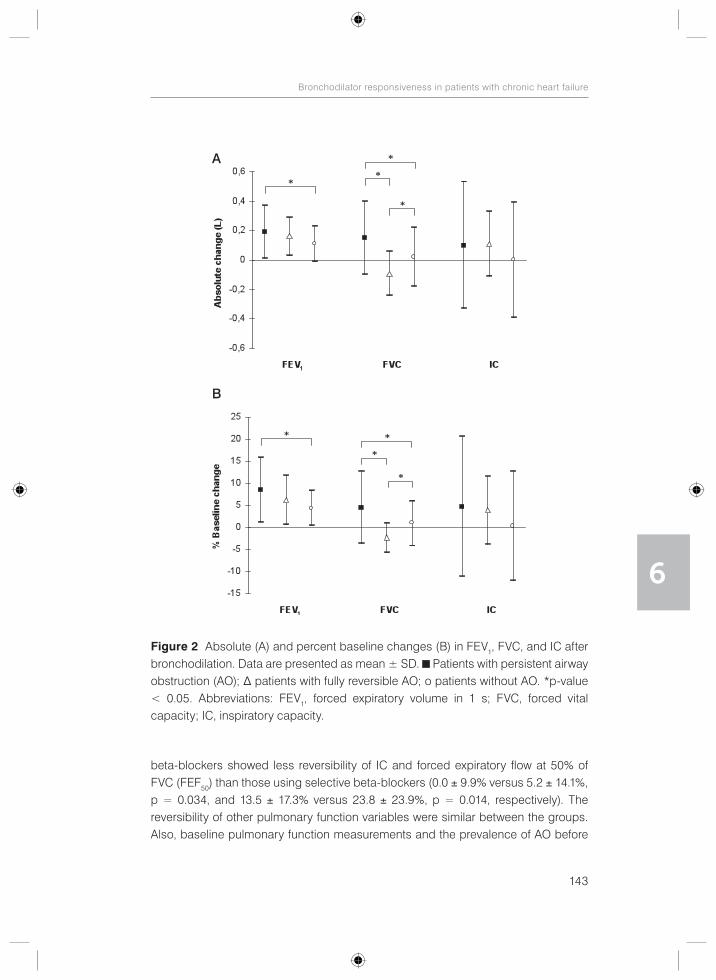
143
Bronchodilator responsiveness in patients with chronic heart failure
6
beta-blockers showed less reversibility of IC and forced expiratory flow at 50% of FVC (FEF50) than those using selective beta-blockers (0.0 ± 9.9% versus 5.2 ± 14.1%, p = 0.034, and 13.5 ± 17.3% versus 23.8 ± 23.9%, p = 0.014, respectively). The reversibility of other pulmonary function variables were similar between the groups. Also, baseline pulmonary function measurements and the prevalence of AO before
Figure 2 Absolute (A) and percent baseline changes (B) in FEV1, FVC, and IC after bronchodilation. Data are presented as mean ± SD. Patients with persistent airway obstruction (AO); Δ patients with fully reversible AO; o patients without AO. *p-value < 0.05. Abbreviations: FEV1, forced expiratory volume in 1 s; FVC, forced vital capacity; IC, inspiratory capacity.
A
B
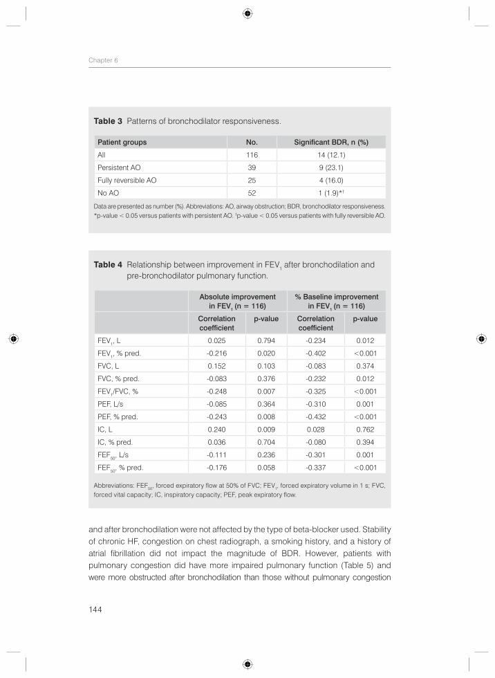
144
Chapter 6
and after bronchodilation were not affected by the type of beta-blocker used. Stability of chronic HF, congestion on chest radiograph, a smoking history, and a history of atrial fibrillation did not impact the magnitude of BDR. However, patients with pulmonary congestion did have more impaired pulmonary function (Table 5) and were more obstructed after bronchodilation than those without pulmonary congestion
Table 3 Patterns of bronchodilator responsiveness.
Patient groups No. Significant BDR, n (%)
All 116 14 (12.1)
Persistent AO 39 9 (23.1)
Fully reversible AO 25 4 (16.0)
No AO 52 1 (1.9)*†
Data are presented as number (%). Abbreviations: AO, airway obstruction; BDR, bronchodilator responsiveness. *p-value < 0.05 versus patients with persistent AO. †p-value < 0.05 versus patients with fully reversible AO.
Table 4 Relationship between improvement in FEV1 after bronchodilation and pre-bronchodilator pulmonary function.
Absolute improvement in FEV1 (n = 116)
% Baseline improvement in FEV1 (n = 116)
Correlation coefficient
p-value Correlation coefficient
p-value
FEV1, L 0.025 0.794 -0.234 0.012
FEV1, % pred. -0.216 0.020 -0.402 <0.001
FVC, L 0.152 0.103 -0.083 0.374
FVC, % pred. -0.083 0.376 -0.232 0.012
FEV1/FVC, % -0.248 0.007 -0.325 <0.001
PEF, L/s -0.085 0.364 -0.310 0.001
PEF, % pred. -0.243 0.008 -0.432 <0.001
IC, L 0.240 0.009 0.028 0.762
IC, % pred. 0.036 0.704 -0.080 0.394
FEF50, L/s -0.111 0.236 -0.301 0.001
FEF50, % pred. -0.176 0.058 -0.337 <0.001
Abbreviations: FEF50, forced expiratory flow at 50% of FVC; FEV1, forced expiratory volume in 1 s; FVC, forced vital capacity; IC, inspiratory capacity; PEF, peak expiratory flow.
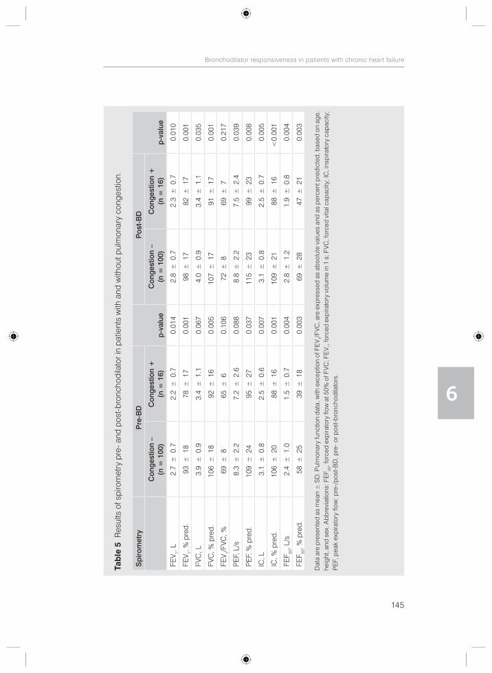
145
Bronchodilator responsiveness in patients with chronic heart failure
6
Tab
le 5
Res
ults
of s
piro
met
ry p
re- a
nd p
ost-
bron
chod
ilato
r in
patie
nts
with
and
with
out p
ulm
onar
y co
nges
tion.
Sp
irom
etry
P
re-B
D
p-v
alue
P
ost-
BD
p-v
alue
Con
ges
tion
–
(n =
100
)C
ong
estio
n +
(n
= 1
6)C
ong
estio
n –
(n
= 1
00)
Con
ges
tion
+
(n =
16)
FEV 1,
L 2.
7±
0.7
2.2
±0.
70.
014
2.8
±0.
72.
3±
0.7
0.01
0
FEV 1,
% p
red.
93±
1878
±17
0.00
198
±17
82±
170.
001
FVC
, L
3.9
±0.
93.
4±
1.1
0.06
74.
0±
0.9
3.4
±1.
10.
035
FVC
, % p
red.
106
±18
92±
160.
005
107
±17
91±
170.
001
FEV 1/
FVC
, %69
±8
65±
60.
106
72±
869
±7
0.21
7
PE
F, L
/s
8.3
±2.
27.
2±
2.6
0.08
88.
8±
2.2
7.5
±2.
40.
039
PE
F, %
pre
d.10
9±
2495
±27
0.03
711
5±
2399
±23
0.00
8
IC, L
3.
1±
0.8
2.5
±0.
60.
007
3.1
±0.
82.
5±
0.7
0.00
5
IC, %
pre
d.10
6±
2088
±16
0.00
110
9±
2188
±16
<0.
001
FEF 50
, L/s
2.4
±1.
01.
5±
0.7
0.00
42.
8±
1.2
1.9
±0.
80.
004
FEF 50
, % p
red.
58±
2539
±18
0.00
369
±28
47±
210.
003
Dat
a ar
e p
rese
nted
as
mea
n ±
SD
. Pul
mon
ary
func
tion
data
, with
exc
eptio
n of
FE
V 1/F
VC, a
re e
xpre
ssed
as
abso
lute
val
ues
and
as p
erce
nt p
redi
cted
, bas
ed o
n ag
e,
heig
ht, a
nd s
ex. A
bbre
viat
ions
: FE
F 50, f
orce
d ex
pira
tory
flow
at 5
0% o
f FVC
; FE
V 1, fo
rced
exp
irato
ry v
olum
e in
1 s
; FVC
, for
ced
vita
l cap
acity
; IC
, ins
pira
tory
cap
acity
; P
EF,
pea
k ex
pira
tory
flow
; pre
-/p
ost-
BD
, pre
- or p
ost-
bron
chod
ilato
rs.
146
Chapter 6
(56.3% versus 30%, p = 0.039). Current and former smokers had significantly lower pre- and post-BD FEV1/FVC ratio than non-smokers (67 ± 8% versus 72 ± 7%, p = 0.022, and 70 ± 8% versus 75 ± 7%, p = 0.011, respectively), but the prevalence of pre- and post-BD AO was not significantly different between the groups (pre-BD 57.4% versus 45.5%, p = 0.309, and post-BD 37.2% versus 18.2%, p = 0.089, respectively). Also, the proportion of patients with completely reversible airway obstruction after bronchodilation was not significantly different between current/former smokers and non-smokers (35% versus 60%, p = 0.170).
Clinically small, albeit statistically significant improvements in the Borg dyspnea score were observed after bronchodilation in the entire group (0.7 ± 1.2 versus 0.9 ± 1.3, p = 0.002). Improvement in Borg score was not significantly different between responders or non-responders and it was not correlated to the improvement of FEV1. Also, the type of beta-blocker used, congestion on chest radiograph, a smoking history, and a history of atrial fibrillation did not impact the magnitude of Borg score improvement.
Discussion
The results of this study confirm that inhaled bronchodilators have the potential to improve pulmonary function in patients with chronic HF, especially in those with AO. Importantly, full reversal of AO was seen in approximately 40% of patients who had pre-BD AO. However, improvement in dyspnea at rest after bronchodilation was small and did not correlate to improvement in pulmonary function.
Our findings agree with the notion that airway narrowing associated with HF has a reversible component, as almost 40% of patients without a history of COPD or asthma who had pre-BD AO fully recovered after bronchodilation. The bronchodilatory effect is most probably explained by a reduction in the increased bronchial tone which may contribute to AO in patients with HF. Also, acutely delivered beta-agonists may act by improving cardiovascular hemodynamics and resorption of pulmonary edema.21 However, considering the speed with which bronchodilation took place and the fact that most patients were not congested on chest radiograph, this was likely not the main mechanism of action.
Similar rates of fully reversible AO after bronchodilator testing were reported in a population-based survey with reductions in AO prevalence between 32% and 39%, depending on the definition used to describe airflow obstruction.22 Consistent with the findings of that study we observed an increase in mean FEV1 and a decrease in
147
Bronchodilator responsiveness in patients with chronic heart failure
6
mean FVC in the fully reversible group of patients while the group with persistent AO did in fact show an increase in both parameters. This may explain why the group with persistent AO maintains a low ratio of FEV1/FVC after bronchodilation while this ratio normalizes in the group with reversible AO. We cannot exclude the possibility that some of the patients with complete reversal of AO in our study may have had previously undiagnosed asthma, as new-onset asthma may occur at any age and is often underrecognised or misdiagnosed in the elderly.23 However, there was no indication of asthma based on symptoms and physical examination in these patients except for one patient who was diagnosed with asthma after having participated in our study. Excluding this patient from the analysis still yielded similar rates of fully reversible AO (38.1%).
The incomplete reversal of AO in other patients may be due to irreversible anatomical alterations of the bronchial wall. In fact, AO in HF has been attributed to increased thickness of the airway wall due to bronchial mucosal swelling, peribronchial edema, and fibrosis. In addition, alveolar fluid accumulation may have contributed.2 Although most patients were not congested on chest radiograph and pulmonary congestion did not influence the magnitude of BDR, occult congestion with small airways compression may still have been present. In support, although pulmonary function improved with treatment for cardiac decompensation in the study of Light and George,13 a great number of patients without a history of chronic obstructive lung disease still had evidence of airway obstruction. In the same study, airway obstruction did not improve after the administration of nebulized bronchodilators in the majority of the patients. Another plausible explanation for the persistent AO could be previously undiagnosed pulmonary disease, such as COPD, which is underrecognized in patients with HF.14 In support of this explanation, our patients with persistent AO without a previous diagnosis of COPD had comparable BDR to 38 chronic HF patients with known COPD of similar age, gender, and LVEF (data not shown). Thus, patients with post-BD AO, irrespective of a previous diagnosis of COPD, might similarly benefit from bronchodilators. On the other hand, although smoking, which is a major risk factor for COPD, was associated with lower FEV1/FVC ratio, the prevalence of post-BD AO was not significantly different between current/former smokers and non-smokers. This may be, however, the result of a small number of non-smokers included in our study. Other differential diagnosis of fixed AO should also be considered, such as bronchiectases and late-onset asthma.23 Finally, considering the physiological decline of the FEV1/FVC ratio with age, the low ratio of FEV1/FVC below 0.7 may still be normal for the age of the patients when using the lower limit of normal to define AO.24
148
Chapter 6
An interesting finding of our study was that the use of non-selective beta-blockers instead of selective beta-blockers resulted in less reversibility of IC and FEF50. This finding is in line with prior evidence that non-selective beta-blockers may antagonize beta-agonists.25 In contrast, baseline pulmonary function, the prevalence of AO, and improvement of dyspnea were not affected by the type of beta-blocker used.
Prior research concerning the beneficial effects of bronchodilators in patients with HF is scarce. Improvements in pulmonary function,4-10 dyspnea,10 and exercise performance7 have been reported. Some investigators have even observed an increase of mean FEV1 greater than 200 mL and 12% in patients with HF,6, 8, 9 especially in those with AO,8 LVSD,9 and during acute decompensation of HF.6 However, contrasting results have also been published. In the study of Witte et al.10 nebulized salbutamol (5 mg) and ipratropium bromide (0.5 mg) reduced peripheral airways resistance and breathlessness during exercise in twelve patients with stable chronic HF and increased measures of compliance. However, spirometry was not altered significantly and exercise capacity did not improve with bronchodilation. Similarly, treatment with three weeks of salbutamol (slow-release 8 mg twice daily) did not improve exercise capacity or spirometric measurements in a randomized placebo-controlled trial of twelve chronic HF patients, although respiratory muscle strength did increase.11 Furthermore, there were no improvements in symptom scores as measured with the Minnesota living with HF questionnaire and fatigue dyspnea index in the same study. In addition, pre-treatment with nebulized albuterol (2.5 mg) and ipratropium bromide (0.5 mg) had only minor effects on spirometric values at rest and did not influence exercise performance and visual analogue score for dyspnea at peak exercise in a randomized, double-blind crossover study of nine patients with HF.12 Furthermore, although a small but significant increase in maximal exercise capacity and spirometric indices was seen after pre-treatment with nebulized salbutamol (5 mg) and with ipratropium bromide (0.5 mg) in a randomized double-blind study of ten patients with chronic HF, both bronchodilators had no effect on the perception of dypnoea, as measured by a visual analogue scale.7 Finally, significant reversibility, defined as more than 400 mL increase in FEV1 after inhalation of 0.2 mg fenoterol and 40 μg ipratropium, could not be demonstrated in 131 patients admitted with HF in a prospective substudy,14 whereas only four of eighteen patients with congestive HF had greater than 10% improvement in FEV1 after bronchodilation in another study.13 Contrasting results across the studies might be attributed to the small number of patients studied and to the differences in study population, bronchodilators used, and definition of BDR.
149
Bronchodilator responsiveness in patients with chronic heart failure
6
There is no consensus about the drug, dose, or mode of administering a bronchodilator to test BDR.24 Since BDR may differ with the bronchodilator used, we sought to obtain maximal bronchodilation by combining two bronchodilators with different mechanisms of action. In addition, we used the most recent criteria to define significant BDR.15 Whether the presence or absence of significant BDR in patients with chronic HF predicts prognostic outcome and clinical response to bronchodilators has not been investigated before and needs further research. The small improvement in dyspnea after bronchodilation was not significantly different between responders or non- responders in our patients with chronic HF. Similarly, BDR status did not predict clinical benefit of bronchodilators in patients with COPD in the UPLIFT trial.26 Clinical implicationsBased on our results, inhaled bronchodilators may have an additional role in the management of patients with chronic HF because of their potential to improve pulmonary function, especially in those with AO. This may contribute to decrease in perceived dyspnea and exercise limitation as demonstrated by others.7, 10 Although post-BD improvements in resting dyspnea were clinically small and not related to improvements in pulmonary function in this study, greater symptomatic relief might be possible in more symptomatic patients and with longer duration of treatment. Further randomized-control trials are needed to examine the clinical benefits as well as potential adverse events of bronchodilators in the treatment of chronic HF.27 Concerns have been raised regarding the cardiovascular safety profile of these drugs.25, 27-30 Recently, reassuring cardiovascular safety data have been reported on long-acting anticholinergic bronchodilator tiotropium (HandiHaler) in patients with COPD.31, 32 However, HF patients are usually excluded from clinical trials and the impact of bronchodilators on outcomes has never been prospectively evaluated in patients with HF.27 Observational studies showed worse outcomes with bronchodilator use in patients with HF, including increased risk of HF hospitalization, increased mortality rates, in-hospital mechanical ventilation, intravenous vasodilator use, and major cardiovascular events. Possible mechanisms for adverse cardiovascular outcomes are arrythmogenesis, ischemia, hypoxemia, inflammation, cardiac remodeling, metabolic alterations (hypokalemia), and/or attenuation of beta-blocker benefits.25, 27 In addition, physicians must be wary of diminished hepatic metabolism of beta-agonists in patients with HF, leading to prolonged plasma half-life and accumulation with repeated doses.5
Methodological limitations of most studies require further investigation of reported adverse events.25, 27 Until then, bronchodilators, in particular beta-agonists, must be used with caution in patients with cardiac disease.
150
Chapter 6
Study limitationsThis retrospective observational study has several limitations that need to be addressed. First, it is important to realize that results may not be applicable to all patients with chronic HF, since we did not include patients with preserved systolic function, who seem to have less impaired pulmonary function.9 Therefore, the bronchodilator effect may have been greater in our patients with decreased systolic function. Also, patients with more severe and unstable HF could have been under represented in this study because of inability to participate. There is evidence to indicate greater bronchodilator effect during acute exacerbation of heart failure,6 although both stability of HF and congestion on chest radiograph did not impact the magnitude of BDR in the current study. Second, since we did not measure the effects of salbutamol and ipratropium bromide separately, comparison between both agents was not possible. It is not unlikely that some patients may respond more to salbutamol than ipratropium bromide and vice versa. However, similar improvements in FEV1 after salbutamol or ipratropium were noticed by Uren et al., although peak expiratory flow rates increased more with salbutamol.7 Third, we did not perform spirometry following placebo inhalations. However, no significant changes with placebo were observed in other studies,6, 7 suggesting genuine effect of bronchodilators. Fourth, since we did not include a control group, we could not establish whether improvements in pulmonary function observed in patients with chronic HF are a reflection of the response observed in normal subjects. However, one in five patients with pre-BD AO had a significant BDR of FEV1 which is considered to exceed spontaneous variability, response to placebo inhalations, and the response observed in healthy individuals. Fifth, we did not measure the effect of bronchodilators on airway resistance. Patients who do not improve on spirometry may still benefit from bronchodilators due to a decrease in airway resistance.10 Also, we did not measure the effect of bronchodilators on heart rate and blood pressure. However, no cardiac adverse effects of broncho dilators such as palpitations and angina were reported during the study. Finally, it is important to realize that our findings are based on a single measurement of BDR and results may differ with serial measurements.26
Conclusion
In conclusion, inhaled bronchodilators may have an additional role in the management of patients with chronic HF because of their potential to improve pulmonary function, especially in those with AO. Despite excluding patients with a history of COPD or asthma, more than half of the chronic HF population studied had pre-BD AO that was fully reversible in approximately 40% of these patients. However, the clinical usefulness and potential adverse events of bronchodilators in the treatment of chronic HF need to be further established in randomized placebo-controlled trials.

151
Bronchodilator responsiveness in patients with chronic heart failure
6
AcknowledgementsThe authors would like to thank the participants in this study for their cooperation. We also would like to thank the nurse practitioners P.A. Ninaber, W.A.M. Janssen, W.H. van Zimmeren-Feijen, and heart failure nurse J.G. Froon-Elferink for their contribution to inclusion of participants and data collection. The authors owe much gratitude to the pulmonary function and clinical chemistry and hematology laboratories as well as the echocardiography and radiology departments for their assistance in data collection. Finally, we would like to acknowledge biostatistician A.R.T. Donders for his contribution to statistical analysis.

152
Chapter 6
References
1. Guazzi M. Alveolar gas diffusion abnormalities in heart failure. J Card Fail. 2008;14:695-702.2. Gehlbach BK, Geppert E. The pulmonary manifestations of left heart failure. Chest. 2004;125:669-82.3. Mancini DM. Pulmonary factors limiting exercise capacity in patients with heart failure. Prog Cardiovasc
Dis. 1995;37:347-70.4. Kindman LA, Vagelos RH, Willson K, Prikazky L, Fowler M. Abnormalities of pulmonary function in
patients with congestive heart failure, and reversal with ipratropium bromide. Am J Cardiol. 1994;73:258-62.5. Ng TM, Munger MA, Lombardi WL, Doing TH, Ryujin DT, Young DC, et al. Chronically inhaled salmeterol
improves pulmonary function in heart failure. J Cardiovasc Pharmacol. 2002;40:140-5.6. Rolla G, Bucca C, Brussino L, Gallo W, Malara D. Bronchodilating effect of ipratropium bromide in
heart failure. Eur Respir J. 1993;6:1492-5.7. Uren NG, Davies SW, Jordan SL, Lipkin DP. Inhaled bronchodilators increase maximum oxygen
consumption in chronic left ventricular failure. Eur Heart J. 1993;14:744-50.8. Khan KA, Jalal S, Jan VM, Lone GM, Jan R, Alai MS, et al. Pulmonary function profile in chronic
congestive heart failure and the effect of ipratropium bromide. Indian Heart J. 2000;52:297-300.9. Kurzawa R, Baron T, Grodzicki T. Pulmonary function test abnormalities in the elderly with systolic
heart failure. Kardiol Pol. 2007;65:875-80. discussion 81-2.10. Witte KK, Morice A, Cleland JG, Clark AL. The reversibility of increased airways resistance in chronic
heart failure measured by impulse oscillometry. J Card Fail. 2004;10:149-54.11. Harrington D, Chua TP, Coats AJ. The effect of salbutamol on skeletal muscle in chronic heart failure.
Int J Cardiol. 2000;73:257-65.12. Moore DP, Weston A, Hughes JM, Oakley CM, Cleland JG. Bronchial hyperresponsiveness in heart
failure. N Engl J Med. 1993;328:1424-5.13. Light RW, George RB. Serial pulmonary function in patients with acute heart failure. Arch Intern Med.
1983;143:429-33.14. Iversen KK, Kjaergaard J, Akkan D, Kober L, Torp-Pedersen C, Hassager C, et al. Chronic obstructive
pulmonary disease in patients admitted with heart failure. J Intern Med. 2008;264:361-9.15. Global Initiative for Chronic Obstructive Lung Disease. Global strategy for the diagnosis, management,
and prevention of chronic obstructive pulmonary disease. NHBLI/WHO workshop report. Available at: http://www.goldcopd.org; 2010.
16. Dickstein K, Cohen-Solal A, Filippatos G, McMurray JJ, Ponikowski P, Poole-Wilson PA, et al. ESC Guidelines for the diagnosis and treatment of acute and chronic heart failure 2008: the Task Force for the Diagnosis and Treatment of Acute and Chronic Heart Failure 2008 of the European Society of Cardiology. Developed in collaboration with the Heart Failure Association of the ESC (HFA) and endorsed by the European Society of Intensive Care Medicine (ESICM). Eur Heart J. 2008;29:2388-442.
17. Global Initiative for asthma. Global strategy for asthma management and prevention. NHBLI/WHO workshop report. Available at: http://www.ginasthma.org; 2010.
18. O’Hanlon R, O’Shea P, Ledwidge M, O’Loughlin C, Lange S, Conlon C, et al. The biologic variability of B-type natriuretic peptide and N-terminal pro-B-type natriuretic peptide in stable heart failure patients. J Card Fail. 2007;13:50-5.
19. Borg GA. Psychophysical bases of perceived exertion. Med Sci Sports Exerc. 1982;14:377-81.20. Quanjer PH, Tammeling GJ, Cotes JE, Pedersen OF, Peslin R, Yernault JC. Lung volumes and forced
ventilatory flows. Report Working Party Standardization of Lung Function Tests, European Community for Steel and Coal. Official Statement of the European Respiratory Society. Eur Respir J 1993;16:5-40.
21. Maak CA, Tabas JA, McClintock DE. Should acute treatment with inhaled beta agonists be withheld from patients with dyspnea who may have heart failure? J Emerg Med. 2011;40:135-45.
22. Perez-Padilla R, Hallal PC, Vazquez-Garcia JC, Muino A, Maquez M, Lopez MV, et al. Impact of bronchodilator use on the prevalence of COPD in population-based samples. Copd. 2007;4:113-20.
23. Hanania NA, King MJ, Braman SS, Saltoun C, Wise RA, Enright P, et al. Asthma in the elderly: Current understanding and future research needs--a report of a National Institute on Aging (NIA) workshop. J Allergy Clin Immunol. 2011;128:S4-24.
153
Bronchodilator responsiveness in patients with chronic heart failure
6
24. Pellegrino R, Viegi G, Brusasco V, Crapo RO, Burgos F, Casaburi R, et al. Interpretative strategies for lung function tests. Eur Respir J. 2005;26:948-68.
25. Hawkins NM, Petrie MC, Macdonald MR, Jhund PS, Fabbri LM, Wikstrand J, et al. Heart failure and chronic obstructive pulmonary disease the quandary of Beta-blockers and Beta-agonists. J Am Coll Cardiol. 2011;57:2127-38.
26. Hanania NA, Sharafkhaneh A, Celli B, Decramer M, Lystig T, Kesten S, et al. Acute bronchodilator responsiveness and health outcomes in COPD patients in the UPLIFT trial. Respir Res. 2011;12:6.
27. Mentz RJ, Fiuzat M, Kraft M, Lindenfeld J, O’Connor CM. Bronchodilators in heart failure patients with COPD: is it time for a clinical trial? J Card Fail. 2012;18:413-22.
28. Salpeter SR, Ormiston TM, Salpeter EE. Cardiovascular effects of beta-agonists in patients with asthma and COPD: a meta-analysis. Chest. 2004;125:2309-21.
29. Singh S, Loke YK, Furberg CD. Inhaled anticholinergics and risk of major adverse cardiovascular events in patients with chronic obstructive pulmonary disease: a systematic review and meta-analysis. Jama. 2008;300:1439-50.
30. Singh S, Loke YK, Enright PL, Furberg CD. Mortality associated with tiotropium mist inhaler in patients with chronic obstructive pulmonary disease: systematic review and meta-analysis of randomised controlled trials. Bmj. 2011;342:d3215.
31. Salpeter SR. Do inhaled anticholinergics increase or decrease the risk of major cardiovascular events?: a synthesis of the available evidence. Drugs. 2009;69:2025-33.
32. Michele TM, Pinheiro S, Iyasu S. The safety of tiotropium--the FDA’s conclusions. N Engl J Med. 2010;363:1097-9.

Armine G. Minasian Frank J.J. van den ElshoutP.N. Richard DekhuijzenPetra J.E. VosFrank F. WillemsPaul J.P.C. van den BerghYvonne F. Heijdra
Heart Lung. 2013;42:388-9.
Comment on the letter to the editor entitled ‘Bronchodilator responsiveness
in patients with chronic heart failure’

157
Bronchodilator responsiveness in patients with chronic heart failure
6
Reply to letter to the editor
We thank Dr. Alkhuja and colleagues for their comments on our recent article ‘Bronchodilator responsiveness in patients with chronic heart failure’.1 They stressed the presence of airway obstruction (AO) and the role of bronchial hyperreactivity / hyperresponsiveness in patients with pulmonary congestive changes secondary to heart failure (HF) in the absence of chronic obstructive pulmonary disease (COPD). They suggested a lower threshold to the administration of inhaled bronchodilators in patients with heart failure without COPD, because of their potential to completely reverse the bronchial obstruction. Finally, they mentioned that the high prevalence of current or former smokers (81%) in our study may explain the presence of AO and the bronchodilator response found in our group of patients.
Several factors have been implied to play a role in the etiology of AO in patients with HF, including the effects of HF itself on pulmonary function as well as confounding influences such as smoking which may lead to COPD. In addition, a careful history taking, physical examination, and additional investigation should be carried out to exclude other co-morbidities that may be the cause of AO, such as bronchial asthma and bronchiectasis.
AO due to HF itself has been attributed to several factors such as alveolar fluid accumulation, bronchial mucosal swelling, peribronchial edema and fibrosis, squamous metaplasia of bronchial epithelial cells induces by transforming growth factor-ß from the failing heart, geometric decrease in airway size from reduction in lung volume, abnormalities of autonomic control, neural (vagal reflex) bronchoconstriction secondary to pulmonary edema, and bronchial hyperresponsiveness which is thought to be mediated at least in part by dilatation of the bronchial vessels.2-5 Brenner et al.6 found transient AO to be an important clinical feature of systolic HF, as AO resolved in 24 of 51 (47%) patients under more stable condition six months after discharge from the hospital. This finding is supported by other studies showing that AO due to HF may be partially or completely reversible by means of cardiac therapy alone.7, 8 Thus, the first step in treating AO in patients with HF should include optimizing treatment directed at the failing heart to achieve a more compensated state. Patients with chronic HF with persisting AO under maximal treatment for their HF may benefit from inhaled bronchodilators as additional treatment because of their potential to improve pulmonary function,1 dyspnea,1 and exercise performance.9 As an example, in our subgroup of patients with stable chronic HF without pulmonary congestion (n = 90) bronchodilators fully reversed AO in 21 of 48 patients (43.8%) who had AO before administration of bronchodilators. Mean absolute and percent baseline changes in FEV1 were 0.16 ± 0.15L and 6.4 ± 5.9%, respectively. However, contrasting results
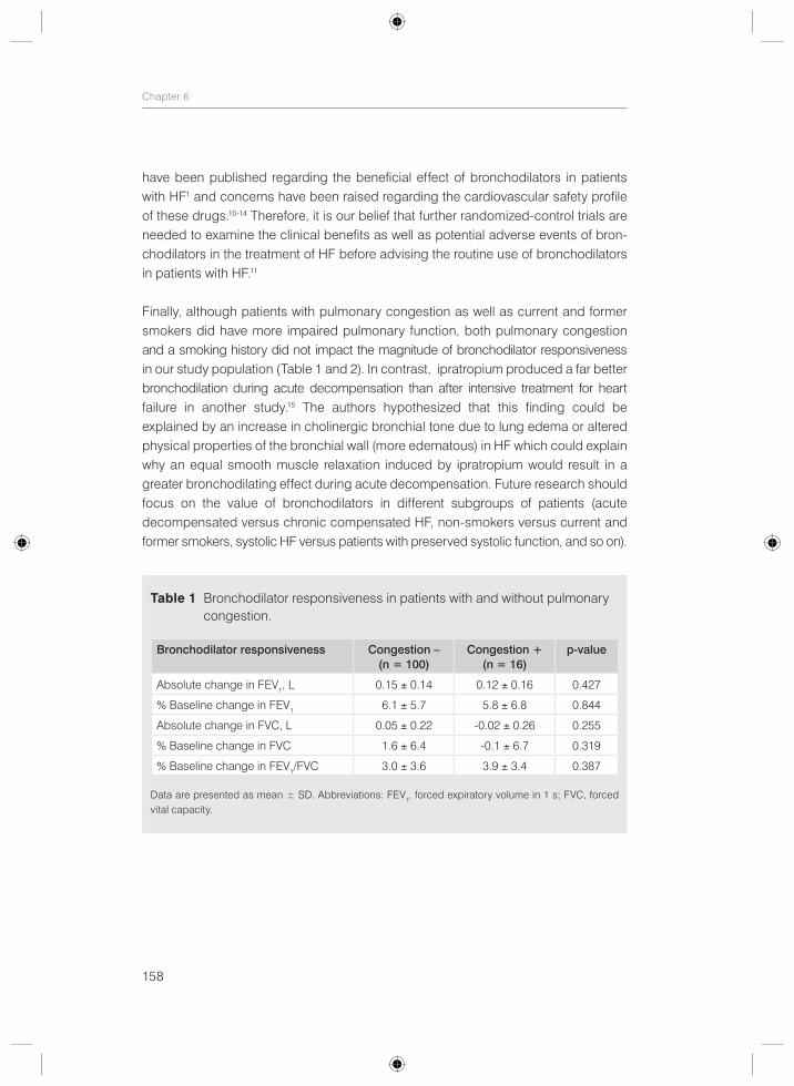
158
Chapter 6
have been published regarding the beneficial effect of bronchodilators in patients with HF1 and concerns have been raised regarding the cardiovascular safety profile of these drugs.10-14 Therefore, it is our belief that further randomized-control trials are needed to examine the clinical benefits as well as potential adverse events of bron-chodilators in the treatment of HF before advising the routine use of bronchodilators in patients with HF.11
Finally, although patients with pulmonary congestion as well as current and former smokers did have more impaired pulmonary function, both pulmonary congestion and a smoking history did not impact the magnitude of bronchodilator responsiveness in our study population (Table 1 and 2). In contrast, ipratropium produced a far better bronchodilation during acute decompensation than after intensive treatment for heart failure in another study.15 The authors hypothesized that this finding could be explained by an increase in cholinergic bronchial tone due to lung edema or altered physical properties of the bronchial wall (more edematous) in HF which could explain why an equal smooth muscle relaxation induced by ipratropium would result in a greater bronchodilating effect during acute decompensation. Future research should focus on the value of bronchodilators in different subgroups of patients (acute decompensated versus chronic compensated HF, non-smokers versus current and former smokers, systolic HF versus patients with preserved systolic function, and so on).
Table 1 Bronchodilator responsiveness in patients with and without pulmonary congestion.
Bronchodilator responsiveness Congestion – (n = 100)
Congestion + (n = 16)
p-value
Absolute change in FEV1, L 0.15 ± 0.14 0.12 ± 0.16 0.427
% Baseline change in FEV1 6.1 ± 5.7 5.8 ± 6.8 0.844
Absolute change in FVC, L 0.05 ± 0.22 -0.02 ± 0.26 0.255
% Baseline change in FVC 1.6 ± 6.4 -0.1 ± 6.7 0.319
% Baseline change in FEV1/FVC 3.0 ± 3.6 3.9 ± 3.4 0.387
Data are presented as mean ± SD. Abbreviations: FEV1, forced expiratory volume in 1 s; FVC, forced vital capacity.
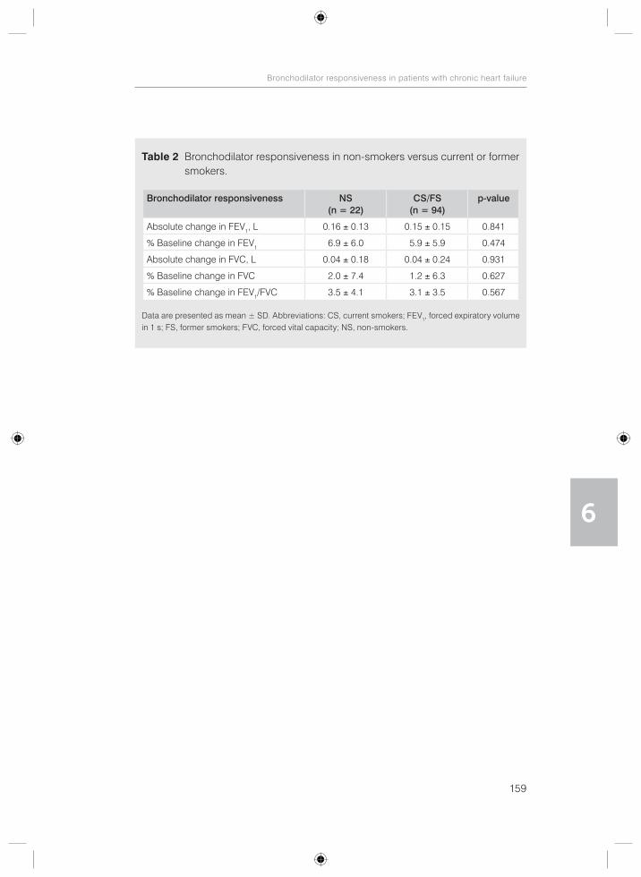
159
Bronchodilator responsiveness in patients with chronic heart failure
6
Table 2 Bronchodilator responsiveness in non-smokers versus current or former smokers.
Bronchodilator responsiveness NS (n = 22)
CS/FS (n = 94)
p-value
Absolute change in FEV1, L 0.16 ± 0.13 0.15 ± 0.15 0.841
% Baseline change in FEV1 6.9 ± 6.0 5.9 ± 5.9 0.474
Absolute change in FVC, L 0.04 ± 0.18 0.04 ± 0.24 0.931
% Baseline change in FVC 2.0 ± 7.4 1.2 ± 6.3 0.627
% Baseline change in FEV1/FVC 3.5 ± 4.1 3.1 ± 3.5 0.567
Data are presented as mean ± SD. Abbreviations: CS, current smokers; FEV1, forced expiratory volume in 1 s; FS, former smokers; FVC, forced vital capacity; NS, non-smokers.

160
Chapter 6
References
1. Minasian AG, van den Elshout FJ, Dekhuijzen PN, Vos PJ, Willems FF, van den Bergh PJ, et al. Bronchodilator responsiveness in patients with chronic heart failure. Heart Lung. 2013; 42:208-14.
2. Snashall PD, Chung KF. Airway obstruction and bronchial hyperresponsiveness in left ventricular failure and mitral stenosis. Am Rev Respir Dis. 1991;144:945-56.
3. Tanabe T, Kanoh S, Moskowitz WB, Rubin BK. Cardiac asthma: TGF-beta from the failing heart leads to squamous metaplasia in human airway cells and in the murine lung. Chest. 2012;142:1274-83.
4. Brunnee T, Graf K, Kastens B, Fleck E, Kunkel G. Bronchial hyperreactivity in patients with moderate pulmonary circulation overload. Chest. 1993;103:1477-81.
5. Cabanes LR, Weber SN, Matran R, Regnard J, Richard MO, Degeorges ME, et al. Bronchial hyperres-ponsiveness to methacholine in patients with impaired left ventricular function. N Engl J Med. 1989;320:1317-22.
6. Brenner S, Guder G, Berliner D, Deubner N, Frohlich K, Ertl G, et al. Airway obstruction in systolic heart failure - COPD or congestion? Int J Cardiol. 2013;168:1910-6.
7. Petermann W, Barth J, Entzian P. Heart failure and airway obstruction. Int J Cardiol. 1987;17:207-9.8. Faggiano P, Lombardi C, Sorgato A, Ghizzoni G, Spedini C, Rusconi C. Pulmonary function tests in
patients with congestive heart failure: effects of medical therapy. Cardiology. 1993;83:30-5.9. Uren NG, Davies SW, Jordan SL, Lipkin DP. Inhaled bronchodilators increase maximum oxygen
consumption in chronic left ventricular failure. Eur Heart J. 1993;14:744-50.10. Hawkins NM, Petrie MC, Macdonald MR, Jhund PS, Fabbri LM, Wikstrand J, et al. Heart failure and
chronic obstructive pulmonary disease the quandary of Beta-blockers and Beta-agonists. J Am Coll Cardiol. 2011;57:2127-38.
11. Mentz RJ, Fiuzat M, Kraft M, Lindenfeld J, O’Connor CM. Bronchodilators in heart failure patients with COPD: is it time for a clinical trial? J Card Fail. 2012;18:413-22.
12. Salpeter SR. Cardiovascular safety of beta(2)-adrenoceptor agonist use in patients with obstructive airway disease: a systematic review. Drugs Aging. 2004;21:405-14.
13. Singh S, Loke YK, Enright PL, Furberg CD. Mortality associated with tiotropium mist inhaler in patients with chronic obstructive pulmonary disease: systematic review and meta-analysis of randomised controlled trials. Bmj. 2011;342:d3215.
14. Singh S, Loke YK, Furberg CD. Inhaled anticholinergics and risk of major adverse cardiovascular events in patients with chronic obstructive pulmonary disease: a systematic review and meta-analysis. Jama. 2008;300:1439-50.
15. Rolla G, Bucca C, Brussino L, Gallo W, Malara D. Bronchodilating effect of ipratropium bromide in heart failure. Eur Respir J. 1993;6:1492-5.

161
Bronchodilator responsiveness in patients with chronic heart failure
6

Armine G. Minasian Frank J.J. van den ElshoutP.N. Richard DekhuijzenPetra J.E. VosFrank F. WillemsPaul J.P.C. van den BerghYvonne F. Heijdra
Heart Lung. 2013;42:229-30.
Comment on the letter to the editor entitled ‘Heart failure: not only reduced
left ventricular ejection fraction but also reserved ejection fraction!

165
Bronchodilator responsiveness in patients with chronic heart failure
6
Reply to letter to the editor
We thank Dr. Aydogan and colleagues for their comments on our recent article ‘Bronchodilator responsiveness in patients with chronic heart failure’.1 Main results of our study were the improvement in almost all spirometric measurements of pulmonary function with inhaled bronchodilators in patients with chronic heart failure (HF) with left ventricular systolic dysfunction (LVSD), especially in those with airway obstruction. Importantly, full reversal of airway obstruction was seen in approximately 40% of patients who had pre-bronchodilator airway obstruction. However, improvement in Borg dyspnea scores at rest after bronchodilation was small (0.7 ± 1.2 versus 0.9 ± 1.3, p = 0.002) and did not correlate to improvement in pulmonary function. Dr. Aydogan and colleagues suggested that undiagnosed or untreated pulmonary hypertension may be the reason for this result. They also noticed that the results of our study may be different in HF patients with diastolic HF.
Indeed, up to 60% of patients with severe LVSD and up to 70% of patients with isolated LV diastolic dysfunction may present with pulmonary hypertension which may be one of the reasons why patients with chronic HF complain of breathlessness.2 However, other potential factors, alone or in combination, may also explain dyspnea experienced by these patients, including cardiac dysfunction, pulmonary function impairment (airway obstruction, restriction, diffusion impairment, decreased lung compliance, ventilation/perfusion mismatch), respiratory muscle dysfunction, expansion of the chest wall leading to activation of chest wall position sensors, direct stimulation of nerve endings by vascular distention and interstitial edema, and co-morbidities such as anemia and previously undiagnosed pulmonary disease.3, 4-5 This multi- factorial etiology of dyspnea in patients with chronic HF may explain the only small improvement in dyspnea despite significant improvement in pulmonary function in our study. However, we believe that the main reason for this rather small improvement in dyspnea and the lack of correlation with improvement in pulmonary function was probably the fact that the majority of our patients had stable chronic HF without signs of pulmonary congestions and therefore did not report symptoms of dyspnea at rest (67.2% had Borg scores between 0 and 0.5, mean Borg score 0.9 ± 1.3). Greater symptomatic relief might be possible in more symptomatic patients and with longer duration of treatment. In addition, selection of other outcome variables such as other dyspnea scales or exercise capacity may lead to different results. There is unfortunately no consensus on which tool should be used to measure breathlessness severity, despite breathlessness being an important limiting problem for patients with chronic HF.6 Also, since we did not measure the effect of bronchodilators on airway resistance, it remains unknown whether improvement in airway resistance correlates to improvement in dyspnea.
166
Chapter 6
Unfortunately, we do not have data on echocardiographic estimates of pulmonary artery pressure (PAP) in all of the patients studied. Not all patients underwent echo-cardiography during this study, as it was performed only in patients without a recent (≤ 6 months) echocardiography to confirm persisting LVSD. Consequently, estimates of PAP obtained a few months before the study may not be representative of PAP during the study, as PAP depends on the actual volemic state of HF. In addition, echocardiography was not performed before and after bronchodilation in the current study and it therefore remains unknown whether bronchodilators may improve dyspnea by decreasing PAP. Evidence shows that acutely delivered beta-2 agonists in patients with chronic HF increase cardiac output/index and decrease systemic vascular resistance and left-sided cardiac filling pressures due to a combination of afterload reduction through vasodilatation and enhanced contractile state.7 Moreover, beta-2 agonists are known to decrease pulmonary edema in animal studies of HF and acute lung injury by increasing transepithelial sodium and chloride transport.7 Thus these mechanisms, in addition to the bronchodilator effect of beta-2 agonists, provide a theoretical benefit of beta-2 agonists to patients with dyspnea due to HF. This hypothesis deserves further investigation.
Finally, as mentioned in the limitation section of our article, the results of our study indeed may not be applicable to all patients with chronic HF, since we did not include patients with preserved systolic function, who seem to have less impaired pulmonary function.8 Future studies should also focus on HF patients with preserved systolic function.

167
Bronchodilator responsiveness in patients with chronic heart failure
6
References
1. Minasian AG, van den Elshout FJ, Dekhuijzen PN, Vos PJ, Willems FF, van den Bergh PJ, et al. Bronchodilator responsiveness in patients with chronic heart failure. Heart Lung. 2013;42:208-214.
2. Galie N, Hoeper MM, Humbert M, Torbicki A, Vachiery JL, Barbera JA, et al. Guidelines for the diagnosis and treatment of pulmonary hypertension: the Task Force for the Diagnosis and Treatment of Pulmonary Hypertension of the European Society of Cardiology (ESC) and the European Respiratory Society (ERS), endorsed by the International Society of Heart and Lung Transplantation (ISHLT). Eur Heart J. 2009;30:2493-537.
3. Mancini DM. Pulmonary factors limiting exercise capacity in patients with heart failure. Prog Cardiovasc Dis. 1995;37:347-70.
4. Witte KK, Clark AL. Why does chronic heart failure cause breathlessness and fatigue? Prog Cardiovasc Dis. 2007;49:366-84.
5. Gehlbach BK, Geppert E. The pulmonary manifestations of left heart failure. Chest. 2004;125:669-82.6. Johnson MJ, Oxberry SG, Cleland JG, Clark AL. Measurement of breathlessness in clinical trials in
patients with chronic heart failure: the need for a standardized approach: a systematic review. Eur J Heart Fail. 2010;12:137-47.
7. Maak CA, Tabas JA, McClintock DE. Should acute treatment with inhaled beta agonists be withheld from patients with dyspnea who may have heart failure? J Emerg Med. 2011;40:135-45.
8. Kurzawa R, Baron T, Grodzicki T. Pulmonary function test abnormalities in the elderly with systolic heart failure. Kardiol Pol. 2007;65:875-80. Discussion 881-2.


Summary
Conclusions
General discussion
Future perspectives
CHAPTER 7

171
Summary, conclusions, general discussion, and future perspectives
7
Summary
The aim of this thesis is to provide more insight in the occurrence of COPD and pulmonary function abnormalities using different definitions in patients with CHF. In addition, we aimed to determine under- and overdiagnosis of COPD, the necessity of performing serial PFTs to correctly diagnose COPD, and predictors of pulmonary function abnormalities using different definitions in patients with CHF. Finally, we aimed to evaluate the effect of inhaled bronchodilators on pulmonary function and dyspnea in patients with CHF.
In chapter 2 we determined the prevalence as well as over- and underdiagnosis of COPD according to the widely used GOLD criteria in 187 outpatients with stable non-congested CHF with LVSD. In addition, we investigated whether serial PFTs are necessary for the correct diagnosis of COPD in this group of patients by using initial as well as confirmatory spirometry three months after treatment with tiotropium in patients with newly diagnosed COPD. We found COPD to be a frequent co-morbidity in patients with CHF, occurring in approximately one-third of the patients. Importantly, it was often unrecognized (19%) or overdiagnosed (32%). Furthermore, we concluded that under stable and euvolemic conditions a confirmatory spirometry is unnecessary for the correct diagnosis of COPD, as it did not change a newly established diagnosis of COPD in the vast majority of patients with CHF.
In chapter 3 we determined the prevalence of COPD in 187 outpatients with stable non-congested CHF with LVSD according to two definitions of airflow obstruction: the LLN (ATS/ERS guidelines) versus the fixed ratio of FEV1/FVC < 0.70 (GOLD guidelines). We found that the exact definition of airflow obstruction alters COPD prevalence substantially; one fifth, rather than one third, of the patients with CHF had concomitant COPD using the LLN instead of the fixed ratio. Importantly, the LLN seemed to identify clinically more important COPD than the fixed ratio of 0.7; 38% of patients with GOLD-COPD who were potentially misclassified as having COPD (FEV1/FVC < 0.7 but > LLN) did not differ significantly from those without COPD in terms of respiratory symptoms and risk factors for COPD, whereas patients with LLN-COPD did.
In chapter 4 we investigated the occurrence of pulmonary function abnormalities in 164 outpatients with CHF with LVSD according to the LLN versus conventional cutoff values (i.e. FEV1/VC < LLN versus FEV1/FVC < 0.7 for airway obstruction, TLCOc < LLN versus < 80% predicted for diffusion impairment, and TLC < LLN versus < 80% predicted for restriction). We excluded patients with known pulmonary, pleural, neuromuscular, collagen vascular, or other diseases that could affect pulmonary function. Patients
172
Chapter 7
with a BMI above 35 were excluded from the restriction prevalence analysis. Pulmonary function abnormalities, especially diffusion impairment and airway obstruction, were highly prevalent in patients with CHF, even in a stable and non-congested condition and even though we used the LLN to better account for age. However, prevalence rates varied significantly according to the definition used between 44% and 58% for diffusion impairment and between 26% and 37% for airway obstruction using the LLN versus conventional cutoff values, respectively. In contrast to previous reports, restriction was found to be infrequent in this population of less severe and mainly stable CHF patients without pulmonary congestion, irrespective of the definition used (7% versus 5%, respectively). The conventional cutoff values classified more patients as having diffusion impairment, airway obstruction, or a mixed category compared to the LLN and failed to identify correctly 34% of patients with normal lung function placing them falsely within a pulmonary dysfunction category. Using the conventional cutoff values instead of the LLN led to misclassification of 27% of the patients.
In chapter 5 we assessed predictors of pulmonary function impairment in 164 outpatients with CHF with LVSD according to the LLN in comparison to conventional cutoff values. The same exclusion criteria as described in chapter 4 were applied. We found that the LLN criterion identified an extra independent predictor of diffusion impairment compared to the 80% predicted value; in addition to BMI, PY, and VA, female gender also turned out to be an independent predictor of diffusion impairment using the LLN. A smoking history of ≥ 10 PY was a significant predictor of diffusion impairment and airway obstruction using the LLN criterion, but not using the conventional cutoff values. However, lowering the cutoff points of conventional criteria to match the more stringent LLN and thus avoid overdiagnosis of diffusion impairment and airway obstruction in the elderly, produced similar results as the LLN. Lower lung volumes were found in patients with pulmonary congestion, cardiomegaly, and a history of CABG.
Finally, in chapter 6 we evaluated retrospectively the effect of inhaled bronchodilators (combined 400 μg salbutamol and 80 μg ipratropium bromide) on pulmonary function and dyspnea in 116 outpatients with CHF and LVSD. Patients with a history of COPD or asthma were excluded. The results of our study confirmed that inhaled broncho-dilators have the potential to improve pulmonary function in patients with CHF, especially in those with airway obstruction. Importantly, full reversal of airway obstruction was seen in approximately 40% of patients without a history of COPD or asthma who had pre-bronchodilator airway obstruction. Significant improvements in pulmonary function were noted in all spirometric indices except for FVC and inspiratory capacity (IC) in the entire study population. Significant BDR of FEV1 (> 200 mL and > 12% from the baseline value) was noted in 12%. Patients with both

173
Summary, conclusions, general discussion, and future perspectives
7
persisting and fully reversible airway obstruction had significant BDR of FEV1 more often than patients without airway obstruction (23%, 16%, and 2%, respectively). Improvement in dyspnea at rest after bronchodilation was, however, small and did not correlate to improvement in pulmonary function in this selected study population.
174
Chapter 7
Conclusions
§COPD is a frequent co-morbidity in patients with stable non-congested CHF with LVSD, but is often unrecognized or overdiagnosed.
§Spirometry should be used under stable and euvolemic conditions to decrease the burden of undiagnosed or overdiagnosed COPD in patients with CHF. Under these conditions, a confirmatory spirometry is unnecessary, as it does not change a newly established diagnosis of COPD in the vast majority of patients with CHF.
§The exact definition of airflow obstruction alters COPD prevalence substantially; one fifth, rather than one third, of the patients with CHF have concomitant COPD using the LLN instead of the fixed ratio.
§LLN may identify clinically more important COPD than the fixed ratio of 0.7 as patients who are potentially misclassified as having COPD, in contrast to patients with LLN-COPD, do not differ significantly from those without COPD in terms of respiratory symptoms and risk factors for COPD.
§Pulmonary function abnormalities, especially diffusion impairment and airway obstruction, are highly prevalent in patients with CHF.
§Conventional cutoff values classify more patients as having diffusion impairment, airway obstruction, or a mixed category compared to the LLN, leading to mis-classification of 27% of the patients.
§The LLN identifies more predictors of diffusion impairment and airway obstruction compared to the conventional cutoff values. However, lowering the conventional cutoff points to match the more stringent LLN, produces similar results as the LLN.
§Inhaled bronchodilators may have an additional role in the management of patients with CHF because of their potential to improve pulmonary function, especially in those with airway obstruction. Improvement in dyspnea is, however, small and does not correlate to improvement in pulmonary function in this selected study population.
175
Summary, conclusions, general discussion, and future perspectives
7
General discussion
1. COPD in CHF COPD and HF are both common diseases with significant morbidity, mortality, and health care use.1, 2 The combination of both diseases presents many diagnostic and therapeutic challenges as well as adverse prognostic implications.1-13 Although the occurrence of HF in patients with COPD has been assessed extensively in prior research, the occurrence of COPD in patients with HF has received much less attention.4 Moreover, although spirometry is considered to be the gold standard for the diagnosis of COPD, data on the prevalence of COPD based on spirometry in patients with HF are scarce and spirometry remains widely underused.14 Furthermore, since airway obstruction is a dynamic phenomenon in HF, as it may be present in congestive HF and may disappear with treatment of HF,15 a careful timing and interpretation of PFTs is required to avoid misdiagnosis and inappropriate treatment of COPD.1 Ideally, PFTs should be used under stable conditions when clinically euvolemic to establish a valid diagnosis of COPD. However, data on the need of serial pulmonary function measurements to confirm persistent airway obstruction and thus COPD are lacking in patients with stable non-congested CHF. We therefore aimed to determine the prevalence of COPD using spirometry in outpatients with stable CHF without pulmonary congestion. In addition, we investigated whether serial PFTs are necessary for the correct diagnosis of COPD in this group of patients by using initial as well as confirmatory spirometry three months after treatment with tiotropium in patients with newly diagnosed COPD. We observed a high prevalence of COPD (32.1%) according to the GOLD criteria in a well defined subgroup of 187 stable non-congested CHF outpatients with LVSD (i.e. LVEF < 40%) (chapter 2). Using a follow-up spirometry after three months of treatment with tiotropium to confirm initial diagnosis of de novo COPD did not change COPD prevalence significantly: 32.6% initially versus 32.1% after three months of follow-up. Importantly, COPD was often unrecognized (19%) or overdiagnosed (32%). Our results support previous findings that COPD frequently coexists with HF (chapter 1.3, Table 1), but are in contrast to the study of Brenner et al.15 who found only 9% of patients with systolic HF to have concomitant COPD, probably explained by the high number of never smokers (46%) included in their study. Several factors might explain the high coexistence of these two diseases, as described in the general introduction. Since spirometry is still underutilized, even in a tertiary-care facility,14 it seems reasonable to consider routine spirometry testing in patients with CHF to diagnose or rule out COPD, a co-morbidity with important therapeutic and prognostic implications,1 which is still greatly under- and overdiagnosed as found in our study. Indeed, diagnostic difficulties have been stressed before, including the overlap in signs, symptoms, and risk factors, underuse of spirometry, and lower diagnostic accuracy of several tests,
176
Chapter 7
including difficulties with interpreting spirometry results.1, 10, 14, 16 This raises concerns regarding possible inappropriate treatment of COPD in an already vulnerable group of patients and as a result possible adverse impact on health and outcome.
Unfortunately, the current study was not large enough to find predictors of newly diagnosed COPD to make specific recommendations regarding which subgroup of patients should be tested. Also, it should be noted that a large proportion (69%) of newly diagnosed COPD patients according to GOLD criteria had only mild airway obstruction that may represent a physiological decline of lung function with age instead of a disease.17, 18 It is unknown whether an additional diagnosis and treatment of COPD in these patients would improve health outcomes and change their prognosis. This warrants further research to establish the effectiveness of screening of patients with CHF for COPD in terms of symptomatic relief and improvement of the outcome as well as cost-effectiveness of such a policy. Until then, spirometry could be used in CHF patients with pulmonary symptoms despite an adequate treatment for their HF, especially in the presence of risk factors for COPD, such as a smoking history of ≥ 10 PY and occupational exposures. Importantly, spirometry should be used when clinically stable and euvolemic to avoid both misdiagnosis and inappropriate treatment of COPD.1, 15 Under these conditions, a confirmatory spirometry seems to be unnecessary, as it did not change a newly established diagnosis of COPD in the vast majority of patients with CHF in our study (chapter 2). Finally, awareness of the co-occurrence of both diseases is essential among pulmonologists and cardiologists and collaboration between the two specialists is crucial to ensure optimal management of these patients. A better recognition and treatment of the two diseases may lead to less morbidity and mortality.
2. COPD in CHF: the LLN versus the fixed ratio of FEV1/FVCSpirometry is required for the diagnosis of COPD by measuring persistent airflow limitation.19 However, there is still no consensus on the most appropriate threshold of FEV1/FVC for diagnosing airflow limitation.18, 20-29 The GOLD guidelines recommend the use of the fixed ratio of FEV1/FVC < 0.70 for the sake of simplicity.19 However, a growing body of literature indicates that considering the physiological decline of the FEV1/FVC ratio with age the use of the fixed ratio may lead to overdiagnosis of COPD in elderly subjects17, 30-45 and underdiagnosis of COPD in young adults.38-47 To avoid misclassification, the ATS/ERS recommends the use of statistically derived LLN values for FEV1/VC that are based on the normal distribution and that classify the bottom 5% of the healthy population as abnormal.48 This is particularly important in patients with HF, given that HF is most prevalent among elderly individuals.49 Thus, the earlier mentioned (chapter 2) and previously reported COPD prevalence rates (chapter 1.3, Table 1) based on the fixed ratio of 0.7 may have overestimated the true
177
Summary, conclusions, general discussion, and future perspectives
7
prevalence of COPD in patients with HF. Although population-based studies have shown that the application of different criteria to define airflow obstruction dramatically changes the prevalence of COPD,33-35, 50-53 it is less well understood to what extent this occurs in patients with HF.54, 55 Therefore, the aim of the study presented in chapter 3 was to determine COPD prevalence in patients with CHF according to two definitions of airflow obstruction: the LLN versus the fixed ratio of FEV1/FVC < 0.7. We performed additional analysis in the same study population as described in chapter 2. The LLN was calculated by subtracting 1.64 times the RSD from the predicted value based on the reference values of the European Community for Coal and Steel.56 We found that the exact definition of airflow obstruction altered COPD prevalence substantially; one fifth, rather than one third, of the patients with CHF had concomitant COPD using the LLN instead of the fixed ratio. This finding is in line with the results of Steinacher et al.54 who reported COPD prevalence rates of 44% according to the GOLD criteria (fixed ratio) and 25% according to the ATS/ERS criteria (LLN) in 89 outpatients with stable CHF. However, this finding was not as well supported in another study of 118 elderly (≥ 65 years) patients with stable CHF, most likely because of the selection of patients with ≥ 10 PY (COPD prevalence 31% versus 29%).55
Thirty-eight percent of patients with GOLD-COPD in our study (chapter 3) were potentially misclassified as having COPD (FEV1/FVC < 0.7 but > LLN). In contrast to patients with LLN-COPD, potentially misclassified patients did not differ significantly from those without COPD regarding respiratory symptoms and risk factors for COPD. LLN may therefore identify clinically more important COPD than the fixed ratio of 0.7.
An incorrect diagnosis of COPD may result in unnecessary treatment for COPD with possible side-effects and adverse cardiovascular events associated with pharmaco-logical treatment for COPD and undertreatment with life-saving beta-blockers.22, 57, 58 Moreover, an incorrect diagnosis and interventions for COPD may have a considerable psychological impact on the subject and his/her family and may lead to unnecessary costs.22 Conversely, misclassifying a number of young adults already affected by COPD as healthy, prevents early interventions that could limit disease progression. Therefore, there is a need for clear diagnostic criteria for COPD to avoid diagnostic confusion, incorrect diagnosis, and inappropriate treatment.
Unfortunately, there is no gold standard for the diagnosis of COPD. The hallmark of the disease is the presence of airflow limitation that is not fully reversible and is usually progressive in nature.19 However, there is no consensus on the spirometric criteria for the diagnosis of COPD.18, 20-29 In theory, the most appropriate threshold for FEV1/FVC in diagnosing COPD is the one with the best combination of sensitivity and specificity.21 However, since there is no real gold standard for COPD calculating these numbers is
178
Chapter 7
not possible and a true comparison of the fixed value and the LLN is not possible.21 Considering the physiological decline of the FEV1/FVC ratio with age, the application of statistically derived LLN values for FEV1/FVC should be preferred to avoid overdiagnosis of COPD in elderly subjects17, 30-45 and underdiagnosis in young adults.38-47 However, when choosing appropriate cutoff values to define disease one should not only consider the statistical range of normality, but also clinically meaningful disease outcomes. An increasing number of studies have focused on the clinical impact of these different criteria. However, contrasting results have been reported and a recent review of 11 studies could not reveal a clear preference for one criterion over the other.20 The results of some studies are in favor of using the fixed ratio of FEV1/FVC < 0.7. Mannino et. al reported higher adjusted mortality risk59, 60 and increased risk of COPD-related hospitalizations59 in patients with FEV1/FVC ratio < 0.7 but > LLN (‘in-between group’) in comparison to subjects with normal lung function, suggesting that the fixed ratio of 0.7 identifies at-risk patients who would have been classified as normal according to the LLN. In addition, this in-between group has been reported to have a worse health-related quality of life than the non-COPD group in the study of Garcίa-Rio et.al.32 However, no differences between the two groups were found in the same study for respiratory exacerbations, 6-minute walk distance, daily physical activity, systemic biomarkers, and cardiovascular disease, whereas patients with mild COPD did have more respiratory exacerbations, greater risk for cardiovascular disease, and greater serum concentrations of nitrites/nitrates than the control group.32 Another cross-sectional study found this in-between group to have similar consumption of health-care resources (number of emergency room visits, hospitalizations due to COPD exacerbation, pharmacological treatment) and associated cardiovascular disorders, with exception of ischemic heart disease, compared to patients with LLN-COPD.61 Furthermore, adopting an expert panel as the reference standard, the fixed ratio of FEV1/FVC < 0.7 was more accurate to detect COPD in a symptomatic primary care population than the LLN.62 Finally, subjects with airflow obstruction by fixed ratio only had a greater degree of emphysema, air wall thickening, and gas trapping using quantitative CT as gold standard than smoking controls without airflow obstruction.63, 64 In addition, they had higher TLC, lower diffusing capacity, and more respiratory symptoms.64 However, all of these variables were even more impaired in patients with airflow obstruction according to both the fixed ratio and the LLN.63, 64 On follow-up, the fixed ratio only group had more exacerbations, higher scores on St. George’s Respiratory Questionnaire, and more frequent initiation of oxygen therapy than smoking controls.63
The results of other studies are more in favor of using the LLN. Patients with FEV1/FVC ratio < 0.7 but > LLN did not show accelerated post-bronchodilator FEV1 decline or more frequent exacerbations, whereas subjects with airway obstruction according to
179
Summary, conclusions, general discussion, and future perspectives
7
the LLN did.65, 66 Also, these patients did not seem to have clinically important airway obstruction in terms of the presence of respiratory symptoms, although they did report a diagnosis of heart disease more often than subject with normal lung function, suggesting that they might be at risk and should be followed carefully.67 Additionally, the risk of death, COPD hospitalization, and the odds of having respiratory symptoms were only elevated amongst participants with FEV1/FVC ratio < LLN.68, 69 Furthermore, COPD according to the LLN criterion showed a stronger association with cardiac and all-cause hospitalization than COPD according to the fixed ratio.70 Finally, misidentified subjects with lung function below the normal range (FEV1/FVC ratio < LLN but > 0.7) had a significantly higher risk of developing COPD and a higher use of health resources because of breathing problems than subjects without airflow obstruction.46, 71
The results of Wollmer et. al did not favor either the LLN or the fixed ratio, but found the group of patients with FEV1/FVC ratio < 0.7 but > LLN to form an intermediate group with respect to lung function impairment and mortality rates,72 implicating that careful evaluation of patient history and extended PFTs may be warranted in this group of patients. More longitudinal studies are needed to determine which criterion is better and clinically more relevant.20, 21
In clinical practice, decisions are not made on the basis of a single test and there are diagnostic difficulties that go beyond the discussion regarding which definition should be used. It is not possible to categorically separate sick from healthy individuals based on spirometry, since there is some overlap in FEV1/FVC ratio between those with and without respiratory disease, independent of the definition used. Therefore, there is always the risk of false positive and false negative results, especially in the grey area between normal and abnormal pulmonary function. Moreover, both definitions cannot exclude airflow obstruction with certainty because, in a minority of cases, FEV1 and FVC may be decreased proportionally as a result of an isolated increase in RV,48 which may lead to a false diagnosis of restriction instead of obstruction. Also, conditions that increase lung stiffness, such as interstitial edema in HF, may mask airflow obstruction.73Additional body plethysmography may therefore be necessary for the correct diagnosis. Furthermore, reduced FEV1/FVC ratio with normal FEV1 may represent a physiological variant possibly due to dysanaptic lung growth, by which lung volume is disproportionately large compared with airway size.73 Finally, structural lung changes (emphysema) may already be present before or even in the absence of airflow limitation.73 In light of these diagnostic difficulties, a more comprehensive approach is required, taking into account clinical features, pulmonary function tests, and radiological findings. Clinical findings, including history and exposure to risk factors, can facilitate the diagnosis of COPD,19 and the physician ultimately determines the medical significance of an abnormal spirometric value
180
Chapter 7
based on these clinical findings. In support, an expert diagnosis of COPD has been found to better predict COPD exacerbations, hospitalizations, and mortality than GOLD or LLN criteria.74 However, these decisions are more complicated in patients with HF due to overlap in signs and symptoms as well as risk factors.1 Extended PFTs, including diffusion capacity and body plethysmography, may be warranted to facilitate correct diagnosis in this group of patients. Indeed, from all PFT variables (besides FEV1/FVC), FEV1, RV/TLC, and diffusing capacity performed best in predicting an expert diagnosis of COPD.74 Moreover, incorporating FEV1 and RV/TLC into the GOLD-COPD and LLN-based definition improved the diagnostic accuracy significantly and brought both definitions closer to expert panel diagnosis of COPD and to daily clinical practice.74 Finally, radiological findings (computed tomography) may advance further decision making.
3 Pulmonary function impairment in CHF Isolated or combined pulmonary function abnormalities, such as restriction, diffusion impairment, and to a lesser extent airway obstruction are common in patients with CHF75-82 and can contribute to the perception of dyspnea83 and exercise intolerance.83-
89 Reported prevalence rates of pulmonary function abnormalities in patients with CHF vary substantially between 41% and 93% for diffusion impairment, 21-55% for restriction, and 14-60% for airway obstruction (chapter 1.4, Table 2). These varying rates across the studies can be partly explained by the usually small number of patients included and differences in study population as well as diagnostic criteria used to define pulmonary function abnormalities. The majority of studies have used conventional cutoff values to define an abnormality, failing to diagnose pulmonary function impairment according to internationally accepted diagnostic criteria.48 These conventional cutoff values have neither statistical nor physiological validity48, 90 and may misclassify more than 20% of patients leading to false-positive diagnosis in the elderly and underdiagnosis in younger patients.38 Therefore, pulmonary function abnormalities may have been overestimated in previous studies of mainly old CHF patients. Moreover, some studies described a restrictive lung function defect on the basis of reduced FEV1 and FVC with normal FEV1/FVC ratio.91 However, because FEV1 and FVC may also be proportionately reduced with a normal ratio in patients with severe COPD and gas trapping, the diagnosis of restriction additionally requires detection of reduced TLC by plethysmography. Finally, a large number of studies have included (potential) heart transplant recipients, who represent one extreme of the HF spectrum.91-97 Therefore, it is less known to what extent CHF patients who do not belong to the most severe category of HF have pulmonary function abnormalities and which of these abnormalities prevail. Given the gaps in current knowledge we investigated the occurrence of pulmonary function abnormalities in 164 outpatients with CHF with LVSD according to the LLN versus conventional cutoff values (i.e.
181
Summary, conclusions, general discussion, and future perspectives
7
FEV1/VC < LLN versus FEV1/FVC < 0.7 for airway obstruction, TLCOc < LLN versus < 80% predicted for diffusion impairment, and TLC < LLN versus < 80% predicted for restriction) (chapter 4). We excluded patients with known pulmonary, pleural (with exception of pleural effusion due to HF), neuromuscular, collagen vascular, or other diseases that could affect pulmonary function. Patients with a BMI above 35 were excluded from the restriction prevalence analysis. Pulmonary function abnormalities, especially diffusion impairment and airway obstruction, were found to be highly prevalent in patients with CHF, even in a stable and non-congested condition and even though we used the LLN to better account for age. However, prevalence rates varied significantly according to the definition used between 44% and 58% for diffusion impairment and between 26% and 37% for airway obstruction using the LLN versus conventional cutoff values, respectively. In contrast to previous reports, restriction was found to be infrequent in this population of less severe and mainly stable CHF patients without pulmonary congestion, irrespective of the definition used (7% versus 5%, respectively). The conventional cutoff values classified more patients as having diffusion impairment, airway obstruction, or a mixed category compared to the LLN and failed to identify correctly 34% of patients with normal lung function placing them falsely within a pulmonary dysfunction category. Using the conventional cutoff values instead of the LLN led to misclassification of 27% of the patients.
Misinterpretation of pulmonary function test results may lead to incorrect diagnosis of disease in elderly patients with HF, such as COPD, and as a consequence unnecessary treatment with possible side effects, detrimental psychological impact, and needless costs. Moreover, results may be interpreted as having more severe or unstable HF due to the effects of HF on pulmonary function and as a result unnecessary intensified treatment for HF. Finally, misdiagnosis may have interfered with the interpretation of prior research aiming to investigate the impact of HF and several clinical variables on pulmonary function. Therefore, the purpose of the study presented in chapter 5 was to assess predictors of pulmonary function impairment in 164 outpatients with CHF with LVSD according to the LLN in comparison to conventional cutoff values (percent predicted and the fixed ratio of FEV1/FVC). The same exclusion criteria as described in chapter 4 were applied. We found that the LLN criterion identified an extra independent predictor of diffusion impairment compared to the 80% predicted value; in addition to BMI, PY, and VA, female gender also turned out to be an independent predictor of diffusion impairment using the LLN. Furthermore, a smoking history of ≥ 10 PY was a significant predictor of diffusion impairment and airway obstruction using the LLN criterion, but not using the conventional cutoff values, implying that inclusion of patients who are incorrectly labeled as having pulmonary dysfunction distorted the effect of smoking on pulmonary function. Lowering the cutoff points of conventional criteria to match the
182
Chapter 7
more stringent LLN and thus avoid overdiagnosis of diffusion impairment and airway obstruction in the elderly, produced similar results as the LLN. Finally, lower lung volumes were found in patients with pulmonary congestion, cardiomegaly, and a history of CABG.
Several factors have been implied to play a role in the etiology of pulmonary function impairment in patients with HF, including the effects of HF itself on pulmonary function in addition to confounding influences such as smoking, CABG, and obesity.75-82, 98, 99 Physicians should also consider the possibility that pulmonary function impairment in patients with HF represents previously undiagnosed pulmonary disease such as COPD. In fact, COPD is frequently unrecognized in patients with HF (chapter 1.3, Table 1). A careful evaluation of the patient and potential treatment options is therefore necessary before ascribing pulmonary function impairment only to the effects of HF.
Although the underlying mechanisms are not clear, women seemed to be more sensitive to the detrimental effects of HF on diffusing capacity in our study. Gender differences in pulmonary function have been recognized before, but not specifically in the HF population. Adult women have been reported to have lower resting lung diffusing capacity corrected for hemoglobin, smaller lung volumes, and lower maximal expiratory flow rates, even when corrected for age and standing height relative to men.100 It has been suggested that these gender differences in part can be explained by pulmonary structural differences (fewer total number of alveoli and smaller airway diameter relative to lung size) and hormonal influences in women.100 More research is needed regarding the influence of gender on pulmonary function in general and specifically in the HF population.
The protective association between a higher BMI and less likelihood of having diffusion impairment has not been described before in patients with CHF. However, some studies in healthy obese nonsmokers have suggested that diffusing capacity may be increased in extremely obese subjects, probably as a result of the increase in blood volume.101
Irrespective of the causes, pulmonary function abnormalities associated with CHF may explain part of the symptoms and functional disability encountered in these patients.83-89, 102 Moreover, pulmonary function impairment increases with the severity of HF,87, 88, 103 provides important prognostic information,104-107 and may ameliorate or normalize with several treatment modalities, such as pharmacological and non-phar-macological treatment of HF and anti-obstructive therapy with bronchodilators as described in the general introduction. Pulmonary function might thus be used as a guide for the evaluation of patients with CHF, with respect to severity of disease,
183
Summary, conclusions, general discussion, and future perspectives
7
prognosis, and response to treatment. More studies involving therapeutic approaches to improve pulmonary function in CHF are warranted to determine whether treatment directed at correcting pulmonary function impairment may lead to symptomatic relief, increased exercise capacity, and improvement of the outcome.
4. The role of inhaled bronchodilators in CHFAs mentioned before, airway obstruction may be present in patients with CHF. Treatment directed at reversing airway obstruction with bronchodilators may thus have an additional role in the management of HF. There are, however, only few studies concerning the beneficial effects of bronchodilators in patients with HF. Improvements in pulmonary function,108-114 dyspnea,111 and exercise performance114 have been reported. Some investigators have even observed an increase of mean FEV1 greater than 200 mL and 12% in patients with HF,108-110 especially in those with airway obstruction,109 LVSD,108 and during acute decompensation of HF.110 However, not all could confirm these findings.111, 114-118 Contrasting results across the studies might be attributed to the small number of patients studied and to the differences in study population, bronchodilators used, and definition of BDR. The purpose of the retrospective observational study presented in chapter 6 was to evaluate the effect of maximal bronchodilation with combined inhaled salbutamol (400 μg) and ipratropium bromide (80 μg) on pulmonary function and dyspnea in 116 patients with CHF without a history of COPD or asthma. In addition, we determined the proportion of patients with significant BDR (i.e. responder), defined as post-bronchodilator increase in FEV1 greater than 200 mL and 12% from the baseline value. Since there is no consensus about the drug, dose, or mode of administering a bronchodilator to test BDR,48 and BDR may differ with the bronchodilator used, we sought to obtain maximal bronchodilation by combining two bronchodilators with different mechanisms of action. The results of our study confirmed that inhaled bronchodilators have the potential to improve pulmonary function in patients with CHF, especially in those with airway obstruction. Importantly, full reversal of airway obstruction was seen in approximately 40% of patients without a history of COPD or asthma who had pre-bronchodilator airway obstruction. Significant improvements in pulmonary function were noted in all spirometric indices except for FVC and IC in the entire study population. Significant BDR of FEV1 was noted in 12%. Patients with persisting airway obstruction and those with fully reversible airway obstruction had significant BDR of FEV1 more often than patients without airway obstruction (23%, 16%, and 2%, respectively). Improvement in dyspnea at rest after bronchodilation was, however, small and did not correlate to improvement in pulmonary function.
The bronchodilatory effect is most probably explained by a reduction in the increased bronchial tone which may contribute to airway obstruction in patients with HF. Also,
184
Chapter 7
acutely delivered beta-agonists may act by improving cardiovascular hemodynamics and resorption of pulmonary edema.119 However, considering the speed with which bronchodilation took place and the fact that most patients were not congested on chest radiograph, this was likely not the main mechanism of action. Also, we observed an increase in mean FEV1 and a decrease in mean FVC in the fully reversible group of patients, while the group with persistent airway obstruction did in fact show an increase in both parameters. This may explain why the group with persistent airway obstruction maintained a low ratio of FEV1/FVC after bronchodilation while this ratio normalized in the group with reversible airway obstruction. Finally, we cannot exclude the possibility that some of the patients with complete reversal of airway obstruction in our study may have had previously undiagnosed asthma, as new-onset asthma may occur at any age and is often underrecognised or misdiagnosed in the elderly.120 However, there was no indication of asthma based on symptoms and physical examination in these patients except for one patient who was diagnosed with asthma after having participated in our study. Excluding this patient from the analysis still yielded similar rates of fully reversible airway obstruction (38%).
The incomplete reversal of airway obstruction in other patients may be due to irreversible anatomical alterations of the bronchial wall or subclinical pulmonary congestion. In addition, previously undiagnosed pulmonary disease, such as COPD, bronchiectases, and late-onset asthma, should also be considered in the differential diagnosis of fixed airway obstruction.
Another interesting finding of our study was that the use of non-selective beta-blockers instead of selective beta-blockers resulted in less reversibility of IC and FEF50. This finding is in line with prior evidence that non-selective beta-blockers may antagonize beta-agonists.57 In contrast, baseline pulmonary function, the prevalence of airway obstruction, and improvement of dyspnea were not affected by the type of beta- blocker used.
Although post-bronchodilator improvements in resting dyspnea were clinically small and not related to improvements in pulmonary function in this selected study population, greater symptomatic relief might be possible in more symptomatic patients and with longer duration of treatment. Further RCTs are needed to examine the clinical benefits as well as potential adverse events of bronchodilators in the treatment of CHF.121 Concerns have been raised regarding the cardiovascular safety profile of these drugs.57, 121-124 Recently, reassuring cardiovascular safety data have been reported on long-acting anticholinergic bronchodilator tiotropium HandiHaler125-133 as well as Respimat.134 However, HF patients are usually excluded from clinical trials and the impact of bronchodilators on outcomes has never been prospectively evaluated in

185
Summary, conclusions, general discussion, and future perspectives
7
patients with HF.121 Furthermore, methodological limitations of most studies require further investigation of reported adverse events.57, 121 Until then, bronchodilators, in particular beta-agonists, must be used with caution in patients with underlying cardiac condition such as HF, given the paucity of data in such patients.
186
Chapter 7
Future perspectives
Although this thesis has provided answers to many questions, there are still many challenges for future research.
Despite the close relationship between lung and heart diseases, pulmonologists and cardiologists often focus on their own field of specialization.8 Meanwhile, HF and COPD often remain an ignored combination11 and the degree of awareness is low among both cardiologists as well as pulmonologists.135 In view of diagnostic, therapeutic, and prognostic implications of the coexistence of COPD and HF, more attention should be paid to the concomitant presence of both diseases in clinical practice and research.11 A more combined and integrated approach in the diagnosis and treatment of concurrent COPD and HF is required.
Although we found COPD to be frequently unrecognized in patients with CHF (chapter 2 and 3), it is unknown whether an additional diagnosis and treatment of COPD in these patients will improve health outcomes and change their prognosis. This warrants further research to establish the effectiveness of screening of patients with CHF for COPD in terms of symptomatic relief and improvement of the outcome as well as cost-effectiveness of such a policy. Additionally, future research should focus on the cardiovascular safety profile of bronchodilators, especially in patients with underlying cardiac condition such as HF.
Furthermore, our findings stress the need for a clear definition of COPD, especially in patients with HF in whom the diagnosis of COPD is already complicated and who are prone to the adverse effects of pharmacological treatment for COPD. Considering the ongoing debate regarding the most appropriate threshold for FEV1/FVC in diagnosing COPD, more longitudinal studies are needed to determine which criterion is better and clinically more relevant in terms of morbidity (symptoms, exercise tolerance, health-related quality of life, exacerbations, hospitalization, pulmonary function decline, use of health recourses, systemic effects such as co-morbidities and systemic biomarkers) and mortality. The question is whether patients potentially misclassified as having COPD (FEV1/FVC < 0.7 but > LLN) actually show clinical features that justify a COPD diagnosis or whether they behave more like a healthy population. Until a consensus is reached, we prefer the application of statistically derived LLN values to avoid overdiagnosis of COPD in the elderly and underdiagnosis in younger patients, and advocate a more comprehensive approach, taking into account clinical features, the results of extended PFTs, and radiological findings.
187
Summary, conclusions, general discussion, and future perspectives
7
Similarly, little is known about the clinical impact of different criteria of pulmonary dysfunction and this therefore requires more longitudinal research. Although we found pulmonary function abnormalities to be highly prevalent in patients with CHF, it remains unknown, due to lack of follow-up, whether pulmonary function impairment had prognostic implications in our study population and whether this has been influenced by different definitions of pulmonary dysfunction. Also, more studies involving therapeutic approaches to improve pulmonary function in CHF are warranted to determine whether treatment directed at correcting pulmonary function impairment may lead to symptomatic relief, increased exercise capacity, and improvement of the outcome. The clinical usefulness and possible adverse events of bronchodilators need to be further established. The results of our retrospective observational study regarding the effects of inhaled bronchodilators on pulmonary function and dyspnea in patients with CHF (chapter 6) should be confirmed in large RCTs in both stable and decompensated conditions of HF and short-term as well as long-term treatment and follow-up. Moreover, the effects of salbutamol and ipratropium should also be studied separately. In addition, the effects of bronchodi-lators on other outcomes should be investigated, including quality of life, exercise performance, airway resistance, hospitalization, and survival. Furthermore, whether the presence or absence of significant BDR in patients with CHF predicts prognostic outcome and clinical response to bronchodilators has not been investigated before and needs further research. The small improvement in dyspnea after bronchodilation was not significantly different between responders or non-responders in our patients with CHF.
Although the underlying mechanisms are not clear, women seemed to be more sensitive to the detrimental effects of HF on diffusing capacity. This needs to be confirmed and further explored in future studies.
Finally, since we only included patients with HF-REF, our results cannot be extended to patients with HF-PEF and more research is warranted in this group of patients.
188
Chapter 7
References
1. Hawkins NM, Petrie MC, Jhund PS, Chalmers GW, Dunn FG, McMurray JJ. Heart failure and chronic obstructive pulmonary disease: diagnostic pitfalls and epidemiology. Eur J Heart Fail. 2009;11:130-9.
2. Hawkins NM, Virani S, Ceconi C. Heart failure and chronic obstructive pulmonary disease: the challenges facing physicians and health services. Eur Heart J. 2013;34:2795-803.
3. de Miguel Diez J, Chancafe Morgan J, Jimenez Garcia R. The association between COPD and heart failure risk: a review. Int J Chron Obstruct Pulmon Dis. 2013;8:305-12.
4. Hannink JD, van Helvoort HA, Dekhuijzen PN, Heijdra YF. Heart failure and COPD: partners in crime? Respirology. 2010;15:895-901.
5. Mascarenhas J, Azevedo A, Bettencourt P. Coexisting chronic obstructive pulmonary disease and heart failure: implications for treatment, course and mortality. Curr Opin Pulm Med. 2010;16:106-11.
6. Chhabra SK, Gupta M. Coexistent chronic obstructive pulmonary disease-heart failure: mechanisms, diagnostic and therapeutic dilemmas. Indian J Chest Dis Allied Sci. 2010;52:225-38.
7. Villar Alvarez F, Mendez Bailon M, de Miguel Diez J. [Chronic obstructive pulmonary disease and heart failure]. Arch Bronconeumol. 2009;45:387-93.
8. Lusuardi M, Garuti G, Massobrio M, Spagnolatti L, Bendinelli S. Heart and lungs in COPD. Close friends in real life--separate in daily medical practice? Monaldi Arch Chest Dis. 2008;69:11-7.
9. Padeletti M, Jelic S, LeJemtel TH. Coexistent chronic obstructive pulmonary disease and heart failure in the elderly. Int J Cardiol. 2008;125:209-15.
10. Le Jemtel TH, Padeletti M, Jelic S. Diagnostic and therapeutic challenges in patients with coexistent chronic obstructive pulmonary disease and chronic heart failure. J Am Coll Cardiol. 2007;49:171-80.
11. Rutten FH, Cramer MJ, Lammers JW, Grobbee DE, Hoes AW. Heart failure and chronic obstructive pulmonary disease: An ignored combination? Eur J Heart Fail. 2006;8:706-11.
12. Dahlstrom U. Frequent non-cardiac comorbidities in patients with chronic heart failure. Eur J Heart Fail. 2005;7:309-16.
13. Aronow WS. Treatment of heart failure in older persons. Dilemmas with coexisting conditions: diabetes mellitus, chronic obstructive pulmonary disease, and arthritis. Congest Heart Fail. 2003;9:142-7.
14. Damarla M, Celli BR, Mullerova HX, Pinto-Plata VM. Discrepancy in the use of confirmatory tests in patients hospitalized with the diagnosis of chronic obstructive pulmonary disease or congestive heart failure. Respir Care. 2006;51:1120-4.
15. Brenner S, Guder G, Berliner D, Deubner N, Frohlich K, Ertl G, et al. Airway obstruction in systolic heart failure - COPD or congestion? Int J Cardiol. 2013;168:1910-6.
16. Guder G, Rutten FH, Brenner S, Angermann CE, Berliner D, Ertl G, et al. The impact of heart failure on the classification of COPD severity. J Card Fail. 2012;18:637-44.
17. Hardie JA, Buist AS, Vollmer WM, Ellingsen I, Bakke PS, Morkve O. Risk of over-diagnosis of COPD in asymptomatic elderly never-smokers. Eur Respir J. 2002;20:1117-22.
18. Enright P, Brusasco V. Counterpoint: should we abandon FEV/FVC < 0.70 to detect airway obstruction? Yes. Chest. 2010;138:1040-2 [discussion 42-4].
19. Global Initiative for Chronic Obstructive Lung Disease. Global strategy for the diagnosis, management, and prevention of chronic obstructive pulmonary disease. NHBLI/WHO workshop report. Avaliable at: http://www.goldcopd.org; 2014.
20. van Dijk WD, Gupta N, Tan WC, Bourbeau J. Clinical relevance of diagnosing COPD by fixed ratio or lower limit of normal: a systematic review. Copd. 2014;11:113-20.
21. Mohamed Hoesein FA, Zanen P, Lammers JW. Lower limit of normal or FEV(1)/FVC <0.70 in diagnosing COPD: An evidence-based review. Respir Med. 2011;105:907-15.
22. Quanjer PH, Enright PL, Miller MR, Stocks J, Ruppel G, Swanney MP, et al. Open letter: the need to change the method for defining mild airway obstruction. Prim Care Respir J. 2010;19:288-91.
23. Celli BR, Halbert RJ. Point: should we abandon FEV/FVC <0.70 to detect airway obstruction? No. Chest. 2010;138:1037-40.
24. Hansen JE. Lower limit of normal is better than 70% or 80%. CHEST. 2011;139:6-8.
189
Summary, conclusions, general discussion, and future perspectives
7
25. Pellegrino R, Brusasco V, Viegi G, Crapo RO, Burgos F, Casaburi R, et al. Definition of COPD: based on evidence or opinion? Eur Respir J. 2008;31:681-2.
26. Mannino DM. Should we be using statistics to define disease? Thorax. 2008;63:1031-2.27. Bhatt NY, Wood KL. What defines abnormal lung function in older adults with chronic obstructive
pulmonary disease? Drugs Aging. 2008;25:717-28.28. Townsend MC. Conflicting definitions of airways obstruction: Drawing the line between normal and
abnormal. Chest. 2007;131:335-6.29. Schermer TR, Quanjer PH. COPD screening in primary care: who is sick? Prim Care Respir J.
2007;16:49-53.30. Runarsdottir SB, Gudmundsson G, Aspelund T, Harris TB, Launer LJ, Gudnason V, et al. Prevalence of
airflow obstruction in nonsmoking older individuals using different spirometric criteria: the AGES Reykjavik Study. Copd. 2013;10:493-9.
31. Maio S, Sherrill DL, MacNee W, Lange P, Costabel U, Dahlen SE, et al. The European Respiratory Society spirometry tent: a unique form of screening for airway obstruction. Eur Respir J. 2012;39:1458-67.
32. Garcia-Rio F, Soriano JB, Miravitlles M, Munoz L, Duran-Tauleria E, Sanchez G, et al. Overdiagnosing Subjects With COPD Using the 0.7 Fixed Ratio: Correlation With a Poor Health-Related Quality of Life. Chest. 2011;139:1072-80.
33. Vollmer WM, Gislason T, Burney P, Enright PL, Gulsvik A, Kocabas A, et al. Comparison of spirometry criteria for the diagnosis of COPD: results from the BOLD study. Eur Respir J. 2009;34:588-97.
34. Swanney MP, Ruppel G, Enright PL, Pedersen OF, Crapo RO, Miller MR, et al. Using the lower limit of normal for the FEV1/FVC ratio reduces the misclassification of airway obstruction. Thorax. 2008;63:1046-51.
35. Shirtcliffe P, Weatherall M, Marsh S, Travers J, Hansell A, McNaughton A, et al. COPD prevalence in a random population survey: a matter of definition. Eur Respir J. 2007;30:232-9.
36. Schermer TR, Smeele IJ, Thoonen BP, Lucas AE, Grootens JG, van Boxem TJ, et al. Current clinical guideline definitions of airflow obstruction and COPD overdiagnosis in primary care. Eur Respir J. 2008;32:945-52.
37. Ko FW, Woo J, Tam W, Lai CK, Ngai J, Kwok T, et al. Prevalence and risk factors of airflow obstruction in an elderly Chinese population. Eur Respir J. 2008;32:1472-8.
38. Miller MR, Quanjer PH, Swanney MP, Ruppel G, Enright PL. Interpreting lung function data using 80 percent of predicted and fixed thresholds misclassifies over 20% of patients. Chest. 2011;139:52-59.
39. Brazzale DJ, Upward AL, Pretto JJ. Effects of changing reference values and definition of the normal range on interpretation of spirometry. Respirology. 2010;15:1098-103.
40. Hwang YI, Kim CH, Kang HR, Shin T, Park SM, Jang SH, et al. Comparison of the prevalence of chronic obstructive pulmonary disease diagnosed by lower limit of normal and fixed ratio criteria. J Korean Med Sci. 2009;24:621-6.
41. Lau AC, Ip MS, Lai CK, Choo KL, Tang KS, Yam LY, et al. Variability of the prevalence of undiagnosed airflow obstruction in smokers using different diagnostic criteria. Chest. 2008;133:42-8.
42. Hansen JE, Sun XG, Wasserman K. Spirometric criteria for airway obstruction: Use percentage of FEV1/FVC ratio below the fifth percentile, not < 70%. Chest. 2007;131:349-55.
43. Perez-Padilla R, Hallal PC, Vazquez-Garcia JC, Muino A, Maquez M, Lopez MV, et al. Impact of bronchodilator use on the prevalence of COPD in population-based samples. Copd. 2007;4:113-20.
44. Hnizdo E, Glindmeyer HW, Petsonk EL, Enright P, Buist AS. Case definitions for chronic obstructive pulmonary disease. Copd. 2006;3:95-100.
45. Roberts SD, Farber MO, Knox KS, Phillips GS, Bhatt NY, Mastronarde JG, et al. FEV1/FVC ratio of 70% misclassifies patients with obstruction at the extremes of age. Chest. 2006;130:200-6.
46. Cerveri I, Corsico AG, Accordini S, Niniano R, Ansaldo E, Anto JM, et al. Underestimation of airflow obstruction among young adults using FEV1/FVC <70% as a fixed cut-off: a longitudinal evaluation of clinical and functional outcomes. Thorax. 2008;63:1040-5.
47. Cerveri I, Corsico AG, Accordini S, Cervio G, Ansaldo E, Grosso A, et al. What defines airflow obstruction in asthma? Eur Respir J. 2009;34:568-73.
48. Pellegrino R, Viegi G, Brusasco V, Crapo RO, Burgos F, Casaburi R, et al. Interpretative strategies for lung function tests. Eur Respir J. 2005;26:948-68.
190
Chapter 7
49. McMurray JJ, Adamopoulos S, Anker SD, Auricchio A, Bohm M, Dickstein K, et al. ESC guidelines for the diagnosis and treatment of acute and chronic heart failure 2012: The Task Force for the Diagnosis and Treatment of Acute and Chronic Heart Failure 2012 of the European Society of Cardiology. Developed in collaboration with the Heart Failure Association (HFA) of the ESC. Eur J Heart Fail. 2012;14:803-69.
50. Glaser S, Schaper C, Obst A, Ittermann T, Volzke H, Felix SB, et al. Impact of different definitions of airflow limitation on the prevalence of chronic obstructive pulmonary disease in the general population. Respiration. 2010;80:292-300.
51. Lindberg A, Jonsson AC, Ronmark E, Lundgren R, Larsson LG, Lundback B. Prevalence of chronic obstructive pulmonary disease according to BTS, ERS, GOLD and ATS criteria in relation to doctor’s diagnosis, symptoms, age, gender, and smoking habits. Respiration. 2005;72:471-9.
52. Celli BR, Halbert RJ, Isonaka S, Schau B. Population impact of different definitions of airway obstruction. Eur Respir J. 2003;22:268-73.
53. Viegi G, Pedreschi M, Pistelli F, Di Pede F, Baldacci S, Carrozzi L, et al. Prevalence of airways obstruction in a general population: European Respiratory Society vs American Thoracic Society definition. Chest. 2000;117:339S-45S.
54. Steinacher R, Parissis JT, Strohmer B, Eichinger J, Rottlaender D, Hoppe UC, et al. Comparison between ATS/ERS age- and gender-adjusted criteria and GOLD criteria for the detection of irreversible airway obstruction in chronic heart failure. Clin Res Cardiol. 2012;101:637-45.
55. Boschetto P, Fucili A, Stendardo M, Malagu M, Parrinello G, Casimirri E, et al. Occurrence and impact of chronic obstructive pulmonary disease in elderly patients with stable heart failure. Respirology. 2013;18:125-30.
56. Quanjer PH, Tammeling GJ, Cotes JE, Pedersen OF, Peslin R, Yernault JC. Lung volumes and forced ventilatory flows. Report Working Party Standardization of Lung Function Tests, European Community for Steel and Coal. Official Statement of the European Respiratory Society. Eur Respir J 1993;16:5-40.
57. Hawkins NM, Petrie MC, Macdonald MR, Jhund PS, Fabbri LM, Wikstrand J, et al. Heart failure and chronic obstructive pulmonary disease the quandary of Beta-blockers and Beta-agonists. J Am Coll Cardiol. 2011;57:2127-38.
58. Egred M, Shaw S, Mohammad B, Waitt P, Rodrigues E. Under-use of beta-blockers in patients with ischaemic heart disease and concomitant chronic obstructive pulmonary disease. Qjm. 2005;98:493-7.
59. Mannino DM, Sonia Buist A, Vollmer WM. Chronic obstructive pulmonary disease in the older adult: what defines abnormal lung function? Thorax. 2007;62:237-41.
60. Mannino DM, Diaz-Guzman E. Interpreting lung function data using 80% predicted and fixed thresholds identifies patients at increased risk of mortality. Chest. 2012;141:73-80.
61. Izquierdo Alonso JL, De Lucas Ramos P, Rodriguez Glez-Moro JM. The use of the lower limit of normal as a criterion for COPD excludes patients with increased morbidity and high consumption of health-care resources. Arch Bronconeumol. 2012;48:223-8.
62. Mohamed Hoesein FA, Zanen P, Sachs AP, Verheij TJ, Lammers JW, Broekhuizen BD. Spirometric thresholds for diagnosing COPD: 0.70 or LLN, pre- or post-dilator values? Copd. 2012;9:338-43.
63. Bhatt SP, Sieren JC, Dransfield MT, Washko GR, Newell JD, Jr., Stinson DS, et al. Comparison of spirometric thresholds in diagnosing smoking-related airflow obstruction. Thorax. 2013;69:409-14.
64. Mohamed Hoesein FA, de Jong PA, Lammers JW, Mali WP, Schmidt M, de Koning HJ, et al. Computed tomography structural lung changes in discordant airflow limitation. PLoS One. 2013;8:e65177.
65. Akkermans RP, Berrevoets MA, Smeele IJ, Lucas AE, Thoonen BP, Grootens-Stekelenburg JG, et al. Lung function decline in relation to diagnostic criteria for airflow obstruction in respiratory symptomatic subjects. BMC Pulm Med. 2012;12:12.
66. Akkermans RP, Biermans M, Robberts B, Ter Riet G, Jacobs A, van Weel C, et al. COPD prognosis in relation to diagnostic criteria for airflow obstruction in smokers. Eur Respir J. 2014;43:54-63.
67. Lamprecht B, Schirnhofer L, Kaiser B, Buist SA, Mannino DM, Studnicka M. Subjects with Discordant Airways Obstruction: Lost between Spirometric Definitions of COPD. Pulm Med. 2011;2011:780215.
68. Vaz Fragoso CA, Concato J, McAvay G, Van Ness PH, Rochester CL, Yaggi HK, et al. The Ratio of FEV1 to FVC as a Basis for Establishing Chronic Obstructive Pulmonary Disease. Am J Respir Crit Care Med. 2010;181:446-51.
191
Summary, conclusions, general discussion, and future perspectives
7
69. Vaz Fragoso CA, Concato J, McAvay G, Van Ness PH, Gill TM. Respiratory impairment and COPD hos-pitalisation in older persons: a competing risk analysis. Eur Respir J. 2012;40:37-44.
70. Lizak MK, Zakliczynski M, Jarosz A, Zembala M, Kalarus Z. Chronic obstructive pulmonary disease diagnosed based on the lower limit of normal does not worsen survival in chronic heart failure patients. Euro J Heart Fail Suppl. 2011;10(S1):S232.
71. Albers M, Schermer T, Heijdra Y, Molema J, Akkermans R, van Weel C. Predictive value of lung function below the normal range and respiratory symptoms for progression of chronic obstructive pulmonary disease. Thorax. 2008;63:201-7.
72. Wollmer P, Engstrom G. Fixed ratio or lower limit of normal as cut-off value for FEV1/VC: an outcome study. Respir Med. 2013;107:1460-2.
73. Brusasco V, Barisione G, Crimi E. Pulmonary physiology: Future directions for lung function testing in COPD. Respirology. 2014;doi: 10.1111/resp.12388.
74. Guder G, Brenner S, Angermann CE, Ertl G, Held M, Sachs AP, et al. “GOLD or lower limit of normal definition? A comparison with expert-based diagnosis of chronic obstructive pulmonary disease in a prospective cohort-study”. Respir Res. 2012;13:13.
75. Apostolo A, Giusti G, Gargiulo P, Bussotti M, Agostoni P. Lungs in heart failure. Pulm Med. 2012; 2012:952741.
76. Kee K, Naughton MT. Heart failure and the lung. Circ J. 2010;74:2507-16.77. Guazzi M. Alveolar gas diffusion abnormalities in heart failure. J Card Fail. 2008;14:695-702.78. Agostoni P, Cattadori G, Bussotti M, Apostolo A. Cardiopulmonary interaction in heart failure. Pulm
Pharmacol Ther. 2007;20:130-4.79. Chua TP, Coats AJ. The lungs in chronic heart failure. Eur Heart J. 1995;16:882-7.80. Gehlbach BK, Geppert E. The pulmonary manifestations of left heart failure. Chest. 2004;125:669-82.81. Faggiano P. Abnormalities of pulmonary function in congestive heart failure. Int J Cardiol. 1994;44:1-8.82. Snashall PD, Chung KF. Airway obstruction and bronchial hyperresponsiveness in left ventricular failure
and mitral stenosis. Am Rev Respir Dis. 1991;144:945-56.83. Mancini DM. Pulmonary factors limiting exercise capacity in patients with heart failure. Prog Cardiovasc
Dis. 1995;37:347-70.84. Tzani P, Piepoli MF, Longo F, Aiello M, Serra W, Maurizio AR, et al. Resting lung function in the assessment
of the exercise capacity in patients with chronic heart failure. Am J Med Sci. 2010;339:210-5.85. Agostoni PG, Bussotti M, Palermo P, Guazzi M. Does lung diffusion impairment affect exercise capacity
in patients with heart failure? Heart. 2002;88:453-9.86. Faggiano P, D’Aloia A, Gualeni A, Giordano A. Relative contribution of resting haemodynamic profile and
lung function to exercise tolerance in male patients with chronic heart failure. Heart. 2001;85:179-84.87. Dimopoulou I, Daganou M, Tsintzas OK, Tzelepis GE. Effects of severity of long-standing congestive
heart failure on pulmonary function. Respir Med. 1998;92:1321-5.88. Puri S, Baker BL, Dutka DP, Oakley CM, Hughes JM, Cleland JG. Reduced alveolar-capillary membrane
diffusing capacity in chronic heart failure. Its pathophysiological relevance and relationship to exercise performance. Circulation. 1995;91:2769-74.
89. Kraemer MD, Kubo SH, Rector TS, Brunsvold N, Bank AJ. Pulmonary and peripheral vascular factors are important determinants of peak exercise oxygen uptake in patients with heart failure. J Am Coll Cardiol. 1993;21:641-8.
90. Sobol BJ, Sobol PG. Per cent of predicted as the limit of normal in pulmonary function testing: a statistically valid approach. Thorax. 1979;34:1-3.
91. Naum CC, Sciurba FC, Rogers RM. Pulmonary function abnormalities in chronic severe cardiomyopathy preceding cardiac transplantation. Am Rev Respir Dis. 1992;145:1334-8.
92. Ewert R, Wensel R, Bettmann M, Spiegelsberger S, Grauhan O, Hummel M, et al. Ventilatory and diffusion abnormalities in long-term survivors after orthotopic heart transplantation. Chest. 1999;115:1305-11.
93. Bussieres LM, Pflugfelder PW, Ahmad D, Taylor AW, Kostuk WJ. Evolution of resting lung function in the first year after cardiac transplantation. Eur Respir J. 1995;8:959-62.
94. Ohar J, Osterloh J, Ahmed N, Miller L. Diffusing capacity decreases after heart transplantation. Chest. 1993;103:857-61.
192
Chapter 7
95. Egan JJ, Kalra S, Yonan N, Hasleton PS, Brooks N, Woodcock AA. Pulmonary diffusion abnormalities in heart transplant recipients. Relationship to cytomegalovirus infection. Chest. 1993;104:1085-9.
96. Niset G, Ninane V, Antoine M, Yernault JC. Respiratory dysfunction in congestive heart failure: correction after heart transplantation. Eur Respir J. 1993;6:1197-201.
97. Wright RS, Levine MS, Bellamy PE, Simmons MS, Batra P, Stevenson LW, et al. Ventilatory and diffusion abnormalities in potential heart transplant recipients. Chest. 1990;98:816-20.
98. Smulyan H, Gilbert R, Eich RH. Pulmonary effects of heart failure. Surg Clin North Am. 1974;54:1077-87.99. Johnson BD, Beck KC, Olson LJ, O’Malley KA, Allison TG, Squires RW, et al. Pulmonary function in
patients with reduced left ventricular function: influence of smoking and cardiac surgery. Chest. 2001;120:1869-76.
100. Harms CA, Rosenkranz S. Sex differences in pulmonary function during exercise. Med Sci Sports Exerc. 2008;40:664-8.
101. Salome CM, King GG, Berend N. Physiology of obesity and effects on lung function. J Appl Physiol (1985). 2010;108:206-11.
102. Torchio R, Gulotta C, Greco-Lucchina P, Perboni A, Avonto L, Ghezzo H, et al. Orthopnea and tidal expiratory flow limitation in chronic heart failure. Chest. 2006;130:472-9.
103. Agostoni P, Bussotti M, Cattadori G, Margutti E, Contini M, Muratori M, et al. Gas diffusion and alveo-lar-capillary unit in chronic heart failure. Eur Heart J. 2006;27:2538-43.
104. Miniati M, Monti S, Bottai M, Pavlickova I, Passino C, Emdin M, et al. Prognostic value of alveolar volume in systolic heart failure: a prospective observational study. BMC Pulm Med. 2013;13:69.
105. Miniati M, Monti S, Bottai M, Passino C, Emdin M, Poletti R. Forced expiratory volume in one second: prognostic value in systolic heart failure. Int J Cardiol. 2013;168:1573-4.
106. Iversen KK, Kjaergaard J, Akkan D, Kober L, Torp-Pedersen C, Hassager C, et al. The prognostic importance of lung function in patients admitted with heart failure. Eur J Heart Fail. 2010;12:685-91.
107. Guazzi M, Pontone G, Brambilla R, Agostoni P, Reina G. Alveolar--capillary membrane gas conductance: a novel prognostic indicator in chronic heart failure. Eur Heart J. 2002;23:467-76.
108. Kurzawa R, Baron T, Grodzicki T. Pulmonary function test abnormalities in the elderly with systolic heart failure. Kardiol Pol. 2007;65:875-80; discussion 81-2.
109. Khan KA, Jalal S, Jan VM, Lone GM, Jan R, Alai MS, et al. Pulmonary function profile in chronic congestive heart failure and the effect of ipratropium bromide. Indian Heart J. 2000;52:297-300.
110. Rolla G, Bucca C, Brussino L, Gallo W, Malara D. Bronchodilating effect of ipratropium bromide in heart failure. Eur Respir J. 1993;6:1492-5.
111. Witte KK, Morice A, Cleland JG, Clark AL. The reversibility of increased airways resistance in chronic heart failure measured by impulse oscillometry. J Card Fail. 2004;10:149-54.
112. Ng TM, Munger MA, Lombardi WL, Doing TH, Ryujin DT, Young DC, et al. Chronically inhaled salmeterol improves pulmonary function in heart failure. J Cardiovasc Pharmacol. 2002;40:140-5.
113. Kindman LA, Vagelos RH, Willson K, Prikazky L, Fowler M. Abnormalities of pulmonary function in patients with congestive heart failure, and reversal with ipratropium bromide. Am J Cardiol. 1994;73:258-62.
114. Uren NG, Davies SW, Jordan SL, Lipkin DP. Inhaled bronchodilators increase maximum oxygen consumption in chronic left ventricular failure. Eur Heart J. 1993;14:744-50.
115. Harrington D, Chua TP, Coats AJ. The effect of salbutamol on skeletal muscle in chronic heart failure. Int J Cardiol. 2000;73:257-65.
116. Moore DP, Weston A, Hughes JM, Oakley CM, Cleland JG. Bronchial hyperresponsiveness in heart failure. N Engl J Med. 1993;328:1424-5.
117. Light RW, George RB. Serial pulmonary function in patients with acute heart failure. Arch Intern Med. 1983;143:429-33.
118. Iversen KK, Kjaergaard J, Akkan D, Kober L, Torp-Pedersen C, Hassager C, et al. Chronic obstructive pulmonary disease in patients admitted with heart failure. J Intern Med. 2008;264:361-9.
119. Maak CA, Tabas JA, McClintock DE. Should acute treatment with inhaled beta agonists be withheld from patients with dyspnea who may have heart failure? J Emerg Med. 2011;40:135-45.
193
Summary, conclusions, general discussion, and future perspectives
7
120. Hanania NA, King MJ, Braman SS, Saltoun C, Wise RA, Enright P, et al. Asthma in the elderly: Current understanding and future research needs--a report of a National Institute on Aging (NIA) workshop. J Allergy Clin Immunol. 2011;128:S4-24.
121. Mentz RJ, Fiuzat M, Kraft M, Lindenfeld J, O’Connor CM. Bronchodilators in heart failure patients with COPD: is it time for a clinical trial? J Card Fail. 2012;18:413-22.
122. Salpeter SR, Ormiston TM, Salpeter EE. Cardiovascular effects of beta-agonists in patients with asthma and COPD: a meta-analysis. Chest. 2004;125:2309-21.
123. Singh S, Loke YK, Enright PL, Furberg CD. Mortality associated with tiotropium mist inhaler in patients with chronic obstructive pulmonary disease: systematic review and meta-analysis of randomised controlled trials. Bmj. 2011;342:d3215.
124. Singh S, Loke YK, Furberg CD. Inhaled anticholinergics and risk of major adverse cardiovascular events in patients with chronic obstructive pulmonary disease: a systematic review and meta-analysis. Jama. 2008;300:1439-50.
125. Dong YH, Lin HH, Shau WY, Wu YC, Chang CH, Lai MS. Comparative safety of inhaled medications in patients with chronic obstructive pulmonary disease: systematic review and mixed treatment comparison meta-analysis of randomised controlled trials. Thorax. 2013;68:48-56.
126. Karner C, Chong J, Poole P. Tiotropium versus placebo for chronic obstructive pulmonary disease. Cochrane Database Syst Rev. 2012;7:CD009285.
127. Verhamme KM, Afonso AS, van Noord C, Haag MD, Koudstaal PJ, Brusselle GG, et al. Tiotropium Handihaler and the risk of cardio- or cerebrovascular events and mortality in patients with COPD. Pulm Pharmacol Ther. 2012;25:19-26.
128. Michele TM, Pinheiro S, Iyasu S. The safety of tiotropium--the FDA’s conclusions. N Engl J Med. 2010;363:1097-9.
129. Celli B, Decramer M, Leimer I, Vogel U, Kesten S, Tashkin DP. Cardiovascular safety of tiotropium in patients with COPD. Chest. 2010;137:20-30.
130. Salpeter SR. Do inhaled anticholinergics increase or decrease the risk of major cardiovascular events?: a synthesis of the available evidence. Drugs. 2009;69:2025-33.
131. Kesten S, Celli B, Decramer M, Leimer I, Tashkin D. Tiotropium HandiHaler in the treatment of COPD: a safety review. Int J Chron Obstruct Pulmon Dis. 2009;4:397-409.
132. Rodrigo GJ, Castro-Rodriguez JA, Nannini LJ, Plaza Moral V, Schiavi EA. Tiotropium and risk for fatal and nonfatal cardiovascular events in patients with chronic obstructive pulmonary disease: systematic review with meta-analysis. Respir Med. 2009;103:1421-9.
133. Oba Y, Zaza T, Thameem DM. Safety, tolerability and risk benefit analysis of tiotropium in COPD. Int J Chron Obstruct Pulmon Dis. 2008;3:575-84.
134. Wise RA, Anzueto A, Cotton D, Dahl R, Devins T, Disse B, et al. Tiotropium Respimat inhaler and the risk of death in COPD. N Engl J Med. 2013;369:1491-501.
135. Macchia A, Rodriguez Moncalvo JJ, Kleinert M, Comignani PD, Gimeno G, Arakaki D, et al. Unrecognised ventricular dysfunction in COPD. Eur Respir J. 2012;39:51-8.


Samenvatting
Conclusies
Toekomstperspectief
CHAPTER 8

197
Samenvatting, conclusies en toekomstperspectief
8
Samenvatting
Het doel van dit proefschrift is het verschaffen van meer inzicht in het vóórkomen van chronisch obstructieve longziekte (COPD) en longfunctiestoornissen, gebruikmakende van verschillende definities, bij patiënten met chronisch hartfalen (CHF). Daarnaast was ons streven om onder- en overdiagnostiek van COPD in kaart te brengen en de noodzaak te bepalen van het herhalen van longfunctietesten om een correcte diagnose van COPD te stellen. Tevens wilden wij mogelijke voorspellers van long-functiestoornissen bij patiënten met CHF identificeren. Wederom hebben wij hiervoor gebruikgemaakt van verschillende definities van longfunctiestoornissen. Tenslotte wilden wij het effect van luchtwegverwijdende medicijnen bepalen op de longfunctie en kortademigheid bij patiënten met CHF.
In hoofdstuk 2 bepaalden wij het vóórkomen van COPD alsook de over- en onder-diagnostiek hiervan volgens de veel gebruikte GOLD definitie bij 187 poliklinische patiënten met stabiel CHF. Alle patiënten hadden een verminderde pompfunctie van de linker hartkamer zonder aanwijzingen voor longvaatovervulling. Daarnaast onderzochten wij of het nodig is om longfunctietesten te herhalen om daarmee een correcte diagnose van COPD te kunnen stellen. Hiertoe maakten wij gebruik van een initiële alsook een vervolgspirometrie drie maanden na behandeling met tiotropium, een luchtwegverwijdend medicijn, bij patiënten met een nieuw gestelde COPD diagnose. COPD kwam vaak voor bij patiënten met CHF; het werd gevonden bij ongeveer eenderde van de patiënten. Bovendien bleek COPD vaak onder- (19%) of juist overgediagnosticeerd (32%) te zijn. Wij concludeerden dat onder stabiele en niet-overvulde omstandigheden het niet noodzakelijk is om spirometrie te herhalen voor het stellen van een correcte diagnose van COPD, aangezien een nieuw gestelde COPD diagnose gehandhaafd bleef bij de meerderheid van de patiënten met CHF.
In hoofdstuk 3 bepaalden wij het vóórkomen van COPD in dezelfde studiepopulatie als beschreven in hoofdstuk 2, waarbij we twee definities van luchtwegvernauwing vergeleken: de ‘lower limit of normal’ (LLN) (ATS/ERS richtlijn) en de vaste afkapwaarde van het geforceerde expiratoire volume in één seconde ten opzichte van de geforceerde vitale capaciteit (FEV1/FVC) < 0.7 (GOLD richtlijn). Wij vonden dat COPD substantieel minder vaak voorkwam volgens de LLN dan volgens de vaste afkapwaarde; slechts één op de vijf, in plaats van één op de drie, patiënten met CHF bleek tevens COPD te hebben. Bovendien bleken patiënten met COPD volgens de LLN definitie klinisch meer relevant COPD te hebben dan patiënten met COPD volgens de conventionele afkapwaarde. Achtendertig procent van de patiënten met GOLD-COPD die mogelijk onterecht bestempeld waren als COPD patiënt (FEV1/FVC < 0.7 maar > LLN) verschilden namelijk niet significant van degenen zonder COPD
198
Chapter 8
in termen van longklachten en risicofactoren voor COPD, dit in tegenstelling tot patiënten met LLN-COPD.
In hoofdstuk 4 onderzochten wij het vóórkomen van longfunctiestoornissen bij 164 poliklinische CHF patiënten met een verminderde pompfunctie van de linker hartkamer. Hiertoe hebben wij gebruikgemaakt van de LLN versus de conventionele afkapwaarden, oftewel FEV1/VC < LLN versus FEV1/FVC < 0.7 voor het definiëren van luchtwegvernauwing, diffusiecapaciteit < LLN versus < 80% van voorspeld voor het definiëren van verminderde zuurstofopname en totale longcapaciteit < LLN versus < 80% van voorspeld voor het definiëren van een kleine longinhoud. Wij sloten patiënten uit met bekende aandoeningen aan long, longvlies, zenuw- en spierstelsel, bindweefsel en vaten, of andere aandoeningen die de longfunctie konden beïnvloeden. Patiënten met een ‘body mass index’ (BMI) boven de 35 werden uitgesloten van de analyse naar het vóórkomen van een kleine longinhoud. Long-functiestoornissen, met name verminderde zuurstofopname en luchtwegvernauwing, kwamen vaak voor bij patiënten met CHF. Dit was zelfs het geval gedurende een stabiele en niet-overvulde klinische toestand en ondanks het gebruik van de LLN om rekening te houden met de gevorderde leeftijd van de patiënten. Echter, zowel een verminderde zuurstof- opname alsook luchtwegvernauwing kwamen significant minder vaak voor volgens de LLN dan wanneer we gebruikmaakten van de conventionele afkapwaarden: respectievelijk 44% versus 58% en 26% versus 37%. In tegenstelling tot eerdere studies werd een kleine longinhoud weinig waargenomen, onafhankelijk van de gebruikte definitie, in deze populatie van minder ernstig aangedane en hoofdzakelijk stabiele CHF patiënten zonder longvaatovervulling (respectievelijk 7% en 5%). De conventionele afkapwaarden classificeerden meer patiënten tot het hebben van een verminderde zuurstofopname, luchtwegvernauwing, of gecombineerde longfunctiestoornis in vergelijking met de LLN. Daarbij waren ze nalatig in het correct identificeren van 34% van de patiënten met een normale longfunctie, waardoor deze onterecht in de groep met longfunctiestoornissen geclassificeerd werden. Het gebruik van de conventionele afkapwaarden in plaats van de LLN leidde tot misclassificatie van 27% van de patiënten.
In hoofdstuk 5 onderzochten wij voorspellers van longfunctiestoornissen volgens de LLN in vergelijking met de conventionele afkapwaarden bij 164 poliklinische CHF patiënten met een verminderde pompfunctie van de linker hartkamer. Dezelfde uit-sluitingscriteria als beschreven in hoofdstuk 4 werden gebruikt. De LLN identificeerde een extra onafhankelijke voorspeller van verminderde zuurstofopname in vergelijking met de 80% voorspelde waarde; naast de BMI, het aantal gerookte pakjaren en het longvolume, bleek het vrouwelijk geslacht ook een onafhankelijke voorspeller te zijn van verminderde zuurstofopname op basis van de LLN. Het roken van ≥ 10 pakjaren
199
Samenvatting, conclusies en toekomstperspectief
8
was een significante voorspeller van zowel verminderde zuurstofopname als lucht-wegvernauwing gebruikmakende van de LLN, maar niet volgens de conventionele afkapwaarden. Echter, door het verlagen van de conventionele afkapwaarden om de meer strenge LLN te evenaren en hiermee overdiagnostiek van verminderde zuurstof- opname en luchtwegvernauwing bij ouderen te voorkomen, werden vergelijkbare resultaten verkregen als met de LLN. Lagere longvolumina werden gevonden bij patiënten met longvaatovervulling, hartvergroting, en een voorgeschiedenis van bypassoperatie aan de kransslagaders.
Tenslotte evalueerden wij in hoofdstuk 6 retrospectief het effect van luchtwegverwij-dende medicijnen (gecombineerd 400 μg salbutamol en 80 μg ipratropium bromide) op de longfunctie en kortademigheid bij 116 poliklinische CHF patiënten met verminderde pompfunctie van de linker hartkamer. Patiënten met een voorgeschie-denis van COPD of astma werden uitgesloten. De resultaten van onze studie bevestigden dat luchtwegverwijdende medicijnen het vermogen hebben om de longfunctie bij patiënten met CHF te verbeteren, voornamelijk bij degenen met een luchtwegvernauwing. Bij ongeveer 40% van de patiënten met luchtwegvernauwing zonder bekend COPD of astma trad een volledig herstel op van de longfunctie na toediening van luchtwegverwijdende medicijnen. Alle longfunctionele waarden verbeterden significant in de gehele studiepopulatie, met uitzondering van de FVC en de inspiratoire capaciteit. Bij 12% werd een aanzienlijke verbetering van de FEV1 waargenomen (> 200 mL en > 12% van de uitgangswaarde) na toediening van luchtwegverwijdende medicijnen. Patiënten met luchtwegvernauwing, zowel degenen met als zonder volledig herstel na toediening van luchtwegverwijdende medicijnen, vertoonden vaker een aanzienlijke verbetering in FEV1 dan patiënten zonder luchtwegvernauwing (respectievelijk 23%, 16% en 2%). Verbetering van kortademigheid in rust na luchtwegverwijding was daarentegen klein en bleek niet gecorreleerd te zijn met de verbeteringen in longfunctie in deze geselecteerde stu-diepopulatie.
200
Chapter 8
Conclusies
§COPD is een veelvoorkomende aandoening bij patiënten met stabiel CHF die een verminderde pompfunctie van de linker hartkamer hebben zonder aanwijzingen voor longvaatovervulling. COPD wordt echter vaak onder- of juist overgediagnosticeerd.
§Spirometrie dient gebruikt te worden tijdens een stabiele en gerecompenseerde klinische toestand om de hoge mate van onderdiagnostiek danwel overdiagnostiek van COPD te verminderen bij patiënten met CHF. Onder deze omstandigheden is het niet noodzakelijk om spirometrie te herhalen ter bevestiging van de diagnose COPD, aangezien dit een nieuw gestelde COPD diagnose niet verandert bij de overgrote meerderheid van de patiënten met CHF.
§De gebruikte definitie van luchtwegvernauwing verandert de mate van voorkomen van COPD substantieel; eenvijfde, in plaats van eenderde, van de patiënten met CHF heeft tevens COPD gebruikmakende van de LLN in plaats van de vaste afkapwaarde.
§De LLN definitie lijkt patiënten te identificeren met klinisch meer relevant COPD dan de vaste afkapwaarde van 0.7. Dit vloeit voort uit de bevinding dat patiënten die potentieel onterecht bestempeld zijn als COPD patiënt, in tegenstelling tot degenen met LLN-COPD, niet significant verschillen van degenen zonder COPD in termen van longklachten en risicofactoren voor COPD.
§Longfunctiestoornissen, met name verminderde zuurstofopname en luchtweg-vernauwing, zijn veelvoorkomend bij patiënten met CHF.
§De conventionele afkapwaarden classificeren meer patiënten tot het hebben van een verminderde zuurstofopname, luchtwegvernauwing danwel gecombineerde longfunctiestoornis in vergelijking met de LLN, hetgeen leidt tot misclassificatie van 27% van de patiënten.
§De LLN identificeert meer voorspellers van verminderde zuurstofopname en lucht-wegvernauwing dan de conventionele afkapwaarden. Echter, lagere conventionele afkapwaarden resulteren in vergelijkbare uitkomsten als de strengere LLN.
§Luchtwegverwijdende medicijnen zijn mogelijk van toegevoegde waarde bij de behandeling van patiënten met CHF in verband met hun vermogen om de longfunctie te verbeteren, voornamelijk bij degenen met luchtwegvernauwing. Verbetering van kortademigheid daarentegen is klein en niet gecorreleerd met de verbeteringen in longfunctie in deze geselecterde studiepopulatie.
201
Samenvatting, conclusies en toekomstperspectief
8
Toekomstperspectief
Hoewel dit proefschrift vele vragen heeft beantwoord, zijn er nog meer dan genoeg uitdagingen voor toekomstige studies.
Ondanks de nauwe relatie tussen long- en hartaandoeningen richten longartsen en hartspecialisten zich vaak teveel op hun eigen vakgebied.1 Ondertussen blijven hartfalen en COPD dikwijls een onderschatte combinatie2 en de mate van besef hiervan is laag onder zowel hartspecialisten als longartsen.3 Aangezien het gezamenlijk voorkomen van COPD en hartfalen gevolgen kan hebben voor diagnostiek, behandeling en prognose van deze patiëntengroep, is zowel meer aandacht in de kliniek vereist als meer onderzoek naar de gelijktijdige aanwezigheid van beide ziektes.2 Een meer gecombineerde en geïntegreerde aanpak van diagnostiek en behandeling van COPD en hartfalen is noodzakelijk.
Hoewel COPD dikwijls onderschat blijkt te zijn bij patiënten met CHF (hoofdstuk 2 en 3), is het onbekend of een additionele diagnose en behandeling van COPD in deze patiëntenpopulatie hun gezondheidsuitkomsten zal verbeteren en de prognose zal veranderen. Dit behoeft verder onderzoek om de effectiviteit van het screenen van patiënten met CHF op COPD te bepalen in termen van symptoomverlichting en verbetering van de gezondheidsuitkomsten alsook de kosteneffectiviteit van een dergelijke aanpak. Daarnaast dient toekomstig onderzoek zich te richten op de cardiovasculaire veiligheid van luchtwegverwijdende medicijnen, voornamelijk bij patiënten met een onderliggende hartziekte als hartfalen.
Voorts benadrukken onze bevindingen de noodzaak van een duidelijke definitie van COPD, in het bijzonder bij patiënten met hartfalen bij wie de diagnose van COPD reeds bemoeilijkt is en die gevoelig zijn voor de bijwerkingen van medicamenteuze behandeling van COPD. Gezien de lopende discussie omtrent de meest geschikte afkapwaarde voor FEV1/FVC om de diagnose COPD te stellen, zijn meer lange-termijn studies nodig om te bepalen welke definitie beter en klinisch meer relevant is in termen van mortaliteit en ziektelast (symptomen, inspanningstolerantie, gezond-heidsgerelateerde kwaliteit van leven, opvlamming van ziekte, ziekenhuisopnames, achteruitgang van de longfunctie, gebruik van gezondheidszorg, systemische effecten zoals het tegelijk voorkomen van meerdere ziektes en systemische biomarkers). De hamvraag is of patiënten die mogelijk onterecht bestempeld zijn als COPD patiënt (FEV1/FVC < 0.7 maar > LLN) daadwerkelijk klinische kenmerken vertonen die een diagnose van COPD rechtvaardigen of dat ze toch meer gelijkenis vertonen met de gezonde populatie. Totdat consensus is bereikt, geven wij de voorkeur aan het gebruik van statistisch afgeleide LLN om overdiagnostiek van
202
Chapter 8
COPD te vermijden bij de ouderen en onderdiagnostiek bij de jongere patiënten. Tevens pleiten wij voor een meer uitgebreide aanpak, rekening houdende met klinische kenmerken, resultaten van uitgebreidere longfunctietesten en radiologische bevindingen.
Evenzo is er slechts weinig bekend over de klinische relevantie van verschillende definities van longfunctiestoornissen en is dan ook meer longitudinaal onderzoek vereist. Ondanks onze bevinding dat longfunctiestoornissen veelvoorkomend zijn bij patiënten met CHF, blijft het zonder follow-up onbekend of de aanwezigheid van longfunctiestoornissen prognostische gevolgen had in onze studiepopulatie en of dit beïnvloed werd door het gebruik van verschillende definities voor longfunctiestoor-nissen. Tevens zijn meer studies nodig gericht op de vraag of behandeling van long-functiestoornissen bij hartfalen leidt tot symptoomverlichting, toegenomen inspan-ningstolerantie en verbetering van de uitkomsten. De klinische toepasbaarheid en mogelijke bijwerkingen van luchtwegverwijdende medicijnen dienen verder te worden onderzocht. De resultaten van onze retrospectieve observationele studie betreffende de effecten van luchtwegverwijdende medicijnen op de longfunctie en kortademigheid bij patiënten met CHF (hoofdstuk 6) dienen te worden bevestigd in grote gerando-miseerde en gecontroleerde studies met zowel stabiele als gedecompenseerde hartfalen patiënten alsook kortdurende en langdurige behandeling en follow-up. Bovendien dienen de effecten van salbutamol en ipratropium ook apart te worden onderzocht. Daarnaast dienen de effecten van luchtwegverwijdende medicijnen op andere uitkomstparameters te worden onderzocht, zoals kwaliteit van leven, inspan-ningstolerantie, luchtwegweerstand, ziekenhuis opnames en overleving. Tevens is het onbekend of de aan- of afwezigheid van significante verbetering van de longfunctie bij patiënten met CHF voorspellend is voor hun prognose en klinische respons op luchtwegverwijdende medicatie. Dit behoeft dan ook verder onderzoek. De kleine verbetering in kortademigheid na luchtwegverwijdende medicijnen was niet significant verschillend bij onze CHF patiënten met of zonder significante verbetering van de longfunctie.
Ofschoon de onderliggende mechanismen vooralsnog onbekend zijn, bleken vrouwen meer vatbaar te zijn voor de nadelige gevolgen van hartfalen op zuurstofopname. Dit dient te worden bevestigd en verder toegelicht in toekomstige studies.
Tenslotte, aangezien we enkel hartfalen patiënten met een verminderde pompfunctie hebben onderzocht, zijn onze resultaten niet toepasbaar op hartfalen patiënten met een behouden pompfunctie. Meer onderzoek is nodig in deze patiëntengroep.

203
Samenvatting, conclusies en toekomstperspectief
8
Referenties
1. Lusuardi M, Garuti G, Massobrio M, Spagnolatti L, Bendinelli S. Heart and lungs in COPD. Close friends in real life--separate in daily medical practice? Monaldi Arch Chest Dis. 2008;69:11-7.
2. Rutten FH, Cramer MJ, Lammers JW, Grobbee DE, Hoes AW. Heart failure and chronic obstructive pulmonary disease: An ignored combination? Eur J Heart Fail. 2006;8:706-11.
3. Macchia A, Rodriguez Moncalvo JJ, Kleinert M, Comignani PD, Gimeno G, Arakaki D, et al. Unrecognised ventricular dysfunction in COPD. Eur Respir J. 2012;39:51-8.


List of publications
List of abbreviations
Authors’ affiliations
Dankwoord
Curriculum Vitae
CHAPTER 9

207
List of publications
9
List of publications
Minasian AG, van den Elshout FJ, Dekhuijzen PN, Vos PJ, Willems FF, van den Bergh PJ, Heijdra YF. Serial pulmonary function tests to diagnose COPD in chronic heart failure. Translational Respiratory Medicine. 2014;2:12.
Minasian AG, van den Elshout FJ, Dekhuijzen PN, Vos PJ, Willems FF, van den Bergh PJ, Heijdra YF. COPD in chronic heart failure: less common than previously thought? Heart Lung. 2013;42:365-71.
Minasian AG, van den Elshout FJ, Dekhuijzen PN, Vos PJ, Willems FF, van den Bergh PJ, Heijdra YF. Pulmonary function impairment in patients with chronic heart failure: lower limit of normal versus conventional cutoff values. Heart Lung. 2014;43:311-6.
Minasian AG, van den Elshout FJ, Dekhuijzen PN, Vos PJ, Willems FF, van den Bergh PJ, Heijdra YF. Using the lower limit of normal instead of the conventional cutoff values to define predictors of pulmonary function impairment in subjects with chronic heart failure. Respiratory Care. 2015; accepted for publication.
Minasian AG, van den Elshout FJ, Dekhuijzen PN, Vos PJ, Willems FF, van den Bergh PJ, Heijdra YF. Bronchodilator responsiveness in patients with chronic heart failure. Heart Lung. 2013;42:208-14.
Minasian AG, van den Elshout FJ, Dekhuijzen PN, Vos PJ, Willems FF, van den Bergh PJ, Heijdra YF. Comment on the letter to the editor entitled ‘Bronchodilator responsiveness in patients with chronic heart failure’. Heart Lung. 2013;42:388-9.
Minasian AG, van den Elshout FJ, Dekhuijzen PN, Vos PJ, Willems FF, van den Bergh PJ, Heijdra YF. Comment on the letter to the editor entitled ‘Heart failure: not only reduced left ventricular ejection fraction but also reserved ejection fraction! Heart Lung. 2013;42:229-30.
208
Chapter 9
List of abbreviations
A AgeABHR Aspecific bronchial hyperreactivityACE-I Angiotensin-converting enzyme inhibitorAF Atrial fibrillationAO Airway obstruction ARB Angiotensin receptor blockerATS/ERS American Thoracic Society/European Respiratory Society BB Beta-blockersBDR Bronchodilator responsiveness BMI Body mass indexBNP B-type natriuretic peptideBOLD Burden of Obstructive Lung DiseaseCABG Coronary artery bypass graftingCAD Coronary artery diseaseCHF Chronic heart failureCMP CardiomyopathyCoHF Congestive heart failureCOPD Chronic obstructive pulmonary diseaseCRT Cardiac resynchronization therapyCS Current smokersCTR Cardiothoracic ratioD Diffusion impairmentDLCO(c) Diffusion capacity of the lung for carbon monoxide (corrected for hemoglobin concentration)ECCS European Community for Coal and Steel ESC European Society of CardiologyFEF50 Forced expiratory flow at 50% of forced vital capacityFEF25-75 Forced expiratory flow at 25-75% of forced vital capacityFEV1 Forced expiratory volume in 1 secondFEV1/FVC Ratio of forced expiratory volume in 1 second to forced vital capacity FRC Functional residual capacity (= ITGV) FS Former smokersFVC Forced vital capacityGOLD Global Initiative for Chronic Obstructive Lung DiseaseH HeightHF Heart failure HF-PEF Heart failure with preserved ejection fractionHF-REF Heart failure with reduced ejection fractionIC Inspiratory capacityICD Implantable cardioverter defibrillator ICS/OCS Inhalation/oral corticosteroidsITGV Intra-thoracic gas volume (= FRC)KCO(c) Transfer coefficient for carbon monoxide (corrected for hemoglobin concentration)
= TLCO(c)/VALLN Lower limit of normal LVEF Left ventricular ejection fraction LVSD Left ventricular systolic dysfunction M MonthsMLHFQ Minnesota Living with Heart Failure Questionnaire

209
List of abbreviations
9
MRC Modified Medical Research Council dyspnea scaleNA Not availableNHANES National Health and Nutrition Examination Survey No. Number of patientsNS Non-smokersNT-pro-BNP N-terminal pro-B natriuretic peptide NYHA New York Heart AssociationO Airway obstructionOLD Obstructive lung disease P ProspectivePAP Pulmonary artery pressure PCI Percutaneous coronary interventionPCWP Pulmonary capillary wedge pressurePEF Peak expiratory flowPEEP Positive end-expiratory pressurePFTs Pulmonary function testsPre-/post-BD Pre/post-bronchodilator% Pred. Percent predictedPY Pack-years R Restriction; retrospective (Table 1)Raw Airway resistanceRCT Randomized controlled trialRSD Residual standard deviationRV Residual volumeSD Standard deviation SE(E) Standard error (of the estimate)sGaw Specific airway conductanceSPSS Statistical Package for Social ScienceSVC Slow vital capacityTLC Total lung capacity TLCO(c) Transfer factor of the lung for carbon monoxide (corrected for hemoglobin concentration)TLCOc/VA Transfer coefficient for carbon monoxide (corrected for hemoglobin concentration) = KCOc VA Alveolar volumeVC Largest vital capacityW WeeksYr(s) Year(s)

210
Chapter 9
Authors’ affiliations
Armine G. Minasian1 Frank J.J. van den Elshout1 P.N. Richard Dekhuijzen2
Petra J.E. Vos1 Frank F. Willems3 Paul J.P.C. van den Bergh3 Yvonne F. Heijdra2
1 Department of Pulmonary Diseases, Rijnstate Hospital, Arnhem, The Netherlands 2 Department of Pulmonary Diseases, Radboud University Nijmegen Medical Centre, Nijmegen, The Netherlands 3 Department of Cardiology, Rijnstate Hospital, Arnhem, The Netherlands

211
Dankwoord
9
Dankwoord
Ik wil een ieder die bijgedragen heeft aan de opzet en uitvoering van mijn promotie- onderzoek, danwel de totstandkoming van dit proefschrift ontzettend bedanken. Een aantal mensen in het bijzonder.
Allereerst gaat mijn dank en waardering uit naar alle deelnemers die bereid waren om deel te nemen aan mijn onderzoek. Zonder hun inzet, enthousiasme en doorzettings-vermogen om nog één goede longfunctie curve te blazen zou dit proefschrift er niet zijn geweest.
Professor Dekhuijzen, beste Richard, met jouw enorme wetenschappelijke kennis, enthousiasme en behoud van overzicht wist je me altijd op de juiste koers te houden en hield je van een afstand alles nauwlettend in de gaten. Aan één woord/zin had ik genoeg om te weten dat ik op de juiste weg zat.
Beste Yvonne, voor jouw passie voor het verrichten van wetenschappelijk onderzoek heb ik grote bewondering. Jouw inspirerende inbreng was dan ook onmisbaar voor mijn proefschrift en ik had je niet als mijn co-promotor willen missen. Binnen no-time kreeg ik waardevol commentaar met kritische noten terug op mijn artikelen en kwam je steeds weer met goede voorstellen voor nieuwe publicaties. Ondanks de tegen - slagen die ik gekend heb, m.n. in de publicatiefase van mijn artikelen, heb je me altijd opnieuw weten te motiveren om door te gaan. Je inzet voor promovendi buiten werktijd om is mij niet ontgaan en na een werkbezoek bij jou thuis kon ik telkens verder met mijn onderzoek en wist ik weer precies wat me te doen stond.
Beste Frank, het verbreden van onze horizon door het doen van onderzoek naast de opleiding heb je altijd gepromoot. Je hebt dan ook alles in het werk gesteld voor de mogelijkheid om te kunnen promoveren bij de longgeneeskunde in het Rijnstate Ziekenhuis. Allerlei opstartproblemen door tegenwerking van de lokale toetsings-commissie werden dan ook doeltreffend overwonnen. Tevens wist je naast je drukke baan als longarts en opleider wanneer nodig tijd vrij te maken voor mij en interesse te tonen; niet alleen op werkgebied, maar ook op privévlak. Jouw oog voor de mens achter de promovendus/AIOS siert jou als opleider.
Beste Petra, jouw praktische manier van aanpak, het reduceren van moeilijke problemen tot iets makkelijks en alle tips en trucs betreffende het opstarten van een onderzoek hebben mij enorm geholpen. Je wist me te doen focussen op wat belangrijk was voor mijn onderzoek en te behoeden voor het afdwalen van het hoofddoel. Ook heb je me gewezen op goed time-management, waar ik de rest van mijn onderzoek

212
Chapter 9
profijt van heb gehad. Jouw verhalen over de combinatie van werk en moederschap heb ik voor altijd in mijn hart gesloten en ik hoop een even betrokken en toegewijde moeder te worden als jij!
Beste Frank, hartelijk dank voor jouw bijdrage aan dit proefschift vanuit cardiologisch oogpunt. Samen hebben wij een degelijke definitie voor stabiel chronisch hartfalen kunnen opstellen, iets wat nog steeds niet makkelijk is naar ik begreep. Dank voor jouw steun gedurende mijn onderzoek, jouw vertrouwen in een goede afloop en uiteraard voor de financiële bijdrage vanuit de maatschap cardiologie.
Beste Paul, het openzetten van de deuren voor mijn onderzoek van de destijds nog aparte cardiologie maatschap in Zevenaar heeft mij enorm geholpen in het vergroten van mijn populatie. Het werken in Zevenaar heb ik als zeer prettig ervaren en iedereen was altijd bereid om mij daar goed verder te helpen. Hartelijk dank ook voor het meebeoordelen van de moeilijk te interpreteren echo’s.
Beste professor Quanjer, hartelijk dank voor de tijd en moeite die u genomen heeft om één van mijn artikelen te reviewen. Ik heb genoten en veel geleerd van onze e-mailwisselingen betreffende de LLN t.o.v. de conventionele afkapwaarden. Ik heb veel gehad aan uw advies om het vooral kort en simpel te houden, of zoals u het zelf zo mooi formuleerde met een Duits spreekwoord; “In der Beschränkung zeigt sich der Meister”.
Leden van de manuscriptcommissie, prof. de Boer, prof. Wouters en prof. Assendelft, hartelijk dank voor de nauwkeurige beoordeling van dit proefschrift en de goedkeuring ervan.
Mijn dank gaat tevens uit naar GlaxoSmithKline voor het verstrekken van een onvoor-waardelijke opleidingssubsidie die de onderzoeken van dit proefschrift mogelijk hebben gemaakt.
Patricia, Wim, Will en Joke, hartelijk dank voor jullie bijdrage aan de inclusie van de proefpersonen en de dataverzameling. Ik vond de samenwerking met jullie erg plezierig en stel jullie belangstelling en inzet voor wetenschappelijk onderzoek naast de drukke poli’s zeer op prijs.
Longfunctie-laboranten, hartelijk dank voor al jullie hulp bij het afnemen van de long-functietesten en het mij aanleren om zelf betrouwbare longfunctietesten af te nemen. Mark, Martijn (helaas niet meer in het Rijnstate Ziekenhuis werkzaam), Hans en Maurice, hartelijk dank voor het aanmaken van een persoonlijke longfunctie uitdraai
213
Dankwoord
9
en het feit dat ik altijd bij jullie terecht kon voor de wat moeilijkere vragen over longfunctie.
Alle echocardiografie laboranten, in het bijzonder Bob, Paul, Wim, Erik, en Maarten, hartelijk dank voor het verrichten van de echocardiografieën en alle uitleg en hulp die ik van jullie heb gehad.
Annet, dank voor al je inspanningen en ‘geregel’ tijdens mijn onderzoek en nu ook tijdens mijn opleiding tot longarts. Je bent onmisbaar voor de afdeling longgenees-kunde! Zonder jouw handige adresstickers en meer van dat soort handigheidjes had de ‘administratieve rompslomp’ van mijn onderzoek mij veel kostbare tijd gekost. Je oprechte belangstelling in mij als persoon en jouw luisterend oor betreffende mijn zwangerschap heb ik zeer gewaardeerd.
Maatschap longartsen te Arnhem, hartelijk dank voor de mogelijkheid om dit onderzoek te verrichten op jullie afdeling. Dank ook voor alle adviezen en het meedenken tijdens mijn praatjes. Jullie interesse in mijn onderzoek en de totstandkoming van dit proefschrift heb ik zeer gewaardeerd en dit motiveerde mij om door te zetten. Ik hoop in de toekomst nog veel van jullie te mogen leren. Toby, mijn mentor, dank voor al je adviezen en persoonlijke verhalen over jouw eigen promotietraject. Daar heb ik veel aan gehad.
Mijn collega-AIOS en ANIOS, waarvan ik ondertussen meerdere heb zien komen en gaan, dank voor al jullie hulp, belangstelling, en op momenten oprechte bezorgdheid en medeleven (‘ben je nu alweer hier voor je onderzoek?’). Ook tijdens mijn onderzoek maakte ik deel uit van jullie gezellige groep en voelde ik me daardoor niet alleen, ondanks het feit dat ik de enige promovendus was. Gerben, dank voor al je hulp, adviezen en motiverende verhalen. Vaak heb je me een hart onder de riem moeten steken, dat heb ik enorm gewaardeerd. Safir, ik geef de wijsheid door aan jou: volhouden, uiteindelijk komt alles goed!
Natasha en Monique, ik ben er trots op en heel blij dat jullie vandaag mijn paranifmen zijn. Natasha, onze vriendschap gaat veel verder terug dan vandaag en door al die jaren ben je er altijd voor mij geweest, zo ook vandaag. Mijn vriendschap met jou heeft mijn verhuizing naar en verblijf in Arnhem een stuk aangenamer gemaakt. Ook al zien wij elkaar helaas minder dan voorheen door de combinatie van werk en gezin, hebben wij net zoveel plezier tijdens onze ‘meiden uitjes’. Ik hoop dat we elkaar nooit uit het oog zullen verliezen.Monique, voor mij ben je een voorbeeld van een menselijke, hardwerkende dokter en onderzoekster met een eindeloos doorzettingsvermogen. Hoe jij een zeven daagse

214
Chapter 9
werkweek in Boston hebt volgehouden gedurende een half jaar is mij nog steeds een raadsel. Jouw drive om je promotietraject tot een goed einde te brengen, en de op - offeringen die je daarvoor over had, hebben mij gestimuleerd om ook mijn onderzoek succesvol af te ronden.
Lieve vrienden en vriendinnen, bedankt voor jullie belangstelling, luisterend oor en bemoedigende woorden, maar bovenal voor de gezellige tijd die we samen hebben doorgebracht en nog zullen doorbrengen. Onze gezellige uitjes en gesprekken waren voor mij altijd een welkome afwisseling en maakten dat ik het e.e.a. beter kon relativeren. Hoe belangrijk en relevant ook; er is meer in het leven dan werk en onderzoek.
Lieve Paul en Ellie, mijn schoonouders, bedankt voor jullie steun en belangstellling. Jullie staan altijd voor ons klaar met goede raad en hulp waar nodig. Dank voor jullie inzet in de opvoeding van Daniël. Ik had mij geen betere schoonouders kunnen wensen!
Lieve pap en mam, zonder jullie dappere zoektocht naar een betere toekomst elders, was ik nu überhaupt niet in Nederland geweest en stond ik vandaag niet te promoveren. Al jullie wijze adviezen heb ik voor altijd in mijn hart gesloten en ik ben door jullie de persoon geworden die ik nu ben. Van jullie heb ik geleerd om door te zetten, het beste uit mezelf te halen, te streven naar meer, maar ook tevreden te zijn met wat ik heb. Jullie wijze raad zal ik op mijn beurt doorgeven aan mijn eigen kinderen. Dank voor jullie steun, onvoorwaardelijke liefde en geloof in mij. Ik houd van jullie.
Lieve Paul, mijn broer, het is bewonderenswaardig hoe jij, door jouw eeuwige nieuws-gierigheid, altijd streeft naar het vergaren van meer kennis. Zoals ze zeggen, kennis is macht. Ook jij hebt je moeten bewijzen en boven jezelf moeten uitstijgen. Jouw masteropleiding en werkjaren in Londen, verwijderd van alles en iedereen, hebben je veel gebracht en je verder gevormd. Daar pluk je nu de vruchten van. Ik ben jaloers op je gemak waarmee jij aan nieuwe hoofdstukken in je leven begint en ik ben er trots op jouw zus te mogen zijn.
Lieve Erik, mijn lieve man, mijn grote liefde, de vader van mijn kind, mijn maatje, mijn steun en toeverlaat, mijn klaagmuur, mijn held, mijn Excel expert….kortom mijn alles. Dank voor al je bemoedigende woorden om door te gaan, je relativeringsvermogen als ik het weer eens niet zag zitten, je eindeloze geduld, en je geloof en vertrouwen in mij en een goede afloop van dit traject. Het ‘hobbyen’ naast mijn werk is eindelijk afgelopen en ik hoop vanaf nu meer tijd met jou en Daniël te kunnen doorbrengen. Ik kan het niet vaak genoeg zeggen: es qez sirumem (oftewel ik houd van jou)!

215
Dankwoord
9
Lieve Daniël, mijn allerliefste en langverwachte schat, jouw geboorte was mijn deadline en ‘schop-onder-de-kont’. Ik wil namelijk zoveel mogelijk tijd met jou doorbrengen. Ik ben heel gelukkig en trots om jouw mamma te mogen zijn. Sommige dromen komen dan toch echt uit!

217
Curriculum Vitae
9
Curriculum Vitae
The author of this thesis was born on 6 July of 1983 in Yerevan, Armenia. At the age of 11 she emigrated with her family to The Netherlands, where she graduated from high school (Gymnasium Trevianum, Sittard) in 2002 cum laude. From 2002 until 2008 she studied Medicine at the University of Maastricht with a cum laude result. During her Medicine study she worked as ‘student-assistant’ at the department of Anatomy and Embryology and she got familiar with the health care system at Malta (department of pulmonology, St. Luke’s Hospital). Her first research experience at the department of Pulmonology of Maastricht University Medical Centre under supervision of Prof. Dr. G. Wesseling resulted in the writing of an abstract called ‘Psychological Functioning and Quality of Life of Cystic Fibrosis Adults and Adolescents’ that was presented at the 22nd Annual North American CF Conference in Orlando, FL., in 2008. After a short clinical experience of 3 months as an internal resident at the department of Pulmonology of Deventer Hospital, she started her PhD research in October of 2008 at the department of Pulmonology of Rijnstate Hospital in Arnhem, which resulted in this thesis. Hereafter she started her specialist training to become a pulmonologist, which consists of two years Internal Medicine (2012-2013, Rijnstate Hospital, Dr. Mattijssen) and four years of Pulmonology (2014-2017, Rijnstate Hospital, Dr. Van den Elshout). She is married to Erik and they have 1 son (Daniël).