Embed Size (px)
Citation preview
RESEARCH Open Access
The healing effect of platelet-rich plasmaon xenograft in peri-implant bone defectsin rabbitsWang Peng1, Il-kyu Kim1,3*, Hyun-young Cho1, Ji-Hoon Seo1, Dong-Hwan Lee1, Jun-Min Jang1
and Seung-Hoon Park2
Abstract
Background: The association of biomaterial combined with repair factor-like platelet-rich plasma (PRP) has prospectivevalues. Bovine-derived xenograft has been identified as an osteoconductive and biocompatible grafting material thatprovides osseointegration ability. PRP has become a valuable adjunctive agent to promote healing in a lot of dentaland oral surgery procedures. However, there are controversies with respect to the regenerative capacity of PRP and thereal benefits of its use in bone grafts. The purpose of this study was to assess the influence of PRP combined withxenograft for the repair of peri-implant bone defects.
Methods: Twelve rabbits were used in this study, and the experimental surgery with implant installation wasperformed simultaneously. Autologous PRP was prepared before the surgical procedure. An intrabony defect(7.0 mm in diameter and 3.0 mm deep) was created in the tibia of each rabbit; then, 24 titanium dental implants(3.0 mm in diameter and 8.5 mm long) were inserted into these osteotomy sites. Thus, a standardized gap (4.0 mm)was established between the surrounding bony walls and the implant surface. The gaps were treated with eitherxenograft alone (control group) or xenograft combined with PRP (experimental group). After healing for 1, 2, 3, 4, 5,and 6 weeks, the rabbits were sacrificed with an overdose of KCl solution. Two rabbits were killed at each time, and thesamples including dental implants and surrounding bone were collected and processed for histological analysis.
Results: More newly formed bone and a better bone healing process were observed in control group. Thehistomorphometric analysis revealed that the mean percentage of bone-to-implant contact in the control group wassignificantly higher than that of the experimental group (25.23 vs. 8.16 %; P < 0.05, independent-simple t test, analysis ofvariance [ANOVA]).
Conclusions: The results indicate that in the addition of PRP to bovine-derived xenograft in the repair of bone defectsaround the implant, PRP may delay peri-implant bone healing.
Keywords: Bone healing, Dental implant, Platelet-rich plasma, Xenograft, Rabbit
BackgroundThe use of a dental implant has become a commontreatment and an important part of modern dentistry.Immediate implantation into fresh extraction socketshas been recommended as a means to minimize boneloss and shorten the prosthetic treatment time [1].
However, the residual bone defects, between the implantneck and the residual bone walls, may cause cell migra-tion from the connective and epithelial tissue into thedefect area, possibly preventing osseointegration [2], andjeopardize the success of immediate implant procedures[3]. For such defects, bone augmentation procedures incombination with the implant placement are necessary.There is a continuous search for bone substitutes to
minimize or avoid the need for autogenous bone grafts.Several materials, synthetically derived or processedfrom skeletal structures of other species (xenografts),
* Correspondence: [email protected] of Oral and Maxillofacial Surgery, College of Medicine, InhaUniversity, Incheon, South Korea3Department of OMFS, Dentistry, College of Medicine, Inha University,#7-206, 3rd St. Shinheung-dong, Choong-gu, Incheon 400-711, South KoreaFull list of author information is available at the end of the article
© 2016 Peng et al. Open Access This article is distributed under the terms of the Creative Commons Attribution 4.0International License (http://creativecommons.org/licenses/by/4.0/), which permits unrestricted use, distribution, andreproduction in any medium, provided you give appropriate credit to the original author(s) and the source, provide a link tothe Creative Commons license, and indicate if changes were made.
Peng et al. Maxillofacial Plastic and Reconstructive Surgery (2016) 38:16 DOI 10.1186/s40902-016-0061-5
have been used alternative to the autogenous bone har-vest. Bovine-derived xenograft has been widely used as abone graft material due to abundant sources and access-ible processing, which can provide an osteoconductivescaffold and has a mineral content comparable to that ofhuman bone allowing it to integrate with the host bone.It is by far the best documented bone substitute materialused in combination with guided bone regeneration [4].For several years, platelet-rich plasma (PRP) has been
thought to promote bone healing, in particular, bonegrafting material mixed with PRP has been reported toenhance bone formation. Marx et al. [5] found that themixture of PRP and autogenous bone grafts can increasethe rate of osteogenesis and qualitatively enhance boneformation. Moreover, Trisi et al. [6] reported that PRP,adding to a mixture of autogenous bone and Biogran,could improve the new bone formation, with a reductionin the time needed for graft healing and a greateramount of bone formed after only 5 to 6 months. Re-cently, PRP has become a valuable adjunct in many den-tal and oral surgery procedures, such as ablative surgicalprocedures, periodontal plastic surgery, and treatment ofinfrabony periodontal defects, as well as procedures re-lating to the placement of osseointegrated implants [7].PRP can be defined as a volume of autogenous plasma
that has a platelet concentration above the baseline, andit is produced by centrifugation of the patient’s ownblood. Platelets release multiple wound-healing growthfactors and cytokines, including platelet-derived growthfactor (PDGF), transforming growth factor β1 and β2(TGF-β1 and β2), vascular endothelial growth factor(VEGF), platelet-derived endothelial growth factor, basicfibroblast growth factor, and platelet-activating factor-4[8]. So, PRP is the suspension of growth factors that hasbeen demonstrated to induce healing and regenerationin soft as well as hard tissues [9].However, there are contradicting reports about its
biologic effect on the enhancement of bone healing.Hatakeyama et al. [10] analyzed the bone healing in thecalvarial defects of rabbits by using an autogenous graftwith or without PRP, and they found that the associ-ation of PRP with autogenous bone did not improvethe bone healing process. Furthermore, Jensen et al.[11] investigated the effect of PRP on bone regener-ation in an allograft using dog models. They demon-strated that the addition of PRP into an allograft has noeffect on new bone formation in the graft.The inconsistency of these results prompted this
study on the effect of PRP on bone healing in axenograft. Therefore, this experiment was designedto assess the influence of PRP used as an adjunctcombined with bovine-derived xenograft for therepair of bone defects adjacent to titanium dentalimplants in rabbits.
MethodsExperimental materialsA total of 24 titanium dental implants (Osstem ImplantCo., Busan, Korea), 8.5 mm in length and 3 mm in diam-eter, were used in this study. Collagen membranes (Bioland,Chungnam, Korea), as barrier membrane, were used tocover the entire surgical site. Twelve healthy female NewZealand rabbits, 5–6 months old, weighing between 3.2 and3.7 kg, were selected as the animal models. Animal selec-tion and management, surgical protocol, and preparationfollowed routines were approved by the Institutional Ani-mal Care and Use Committee at the College of Medicine,Inha University, Incheon, Korea.
Preparation of PRPBased on the method described by Okuda et al. [12], thePRP was prepared by the transfusion laboratory (InhaUniversity Hospital, Incheon, Korea) using a hematologysystem (Advia 2120i, Siemens, Erlangen, Germany).Briefly, 4 mL of autologous blood withdrawn from eachrabbit, using a 23-gauge needle attached to a 5-mL ster-ilized syringe, then added to a tube (BD Vacutainer®, BDCo., NJ, USA) containing sodium citrate and mixed. Theblood was initially centrifuged at 2400 rpm for 10 minto separate the PRP and platelet-poor plasma (PPP) por-tions from the red blood cell fraction. The PRP and PPPportions were again centrifuged at 3600 rpm for 15 minto separate the PRP from the PPP. The approximate vol-ume of PRP obtained was 0.5 mL, and then plateletcounts were performed. In our experimental animals,the numbers of platelets in the whole blood and PRPwere 236 × 103/μL and 625 × 103/μL, respectively. So,the concentration of platelets in PRP is at least 2.6 timeshigher than the baseline value of platelets by using thishematology system. Before application, the PRP was acti-vated with 10 % calcium chloride solution and 1 KU ofbovine thrombin (Sigma-Aldrich, St. Louis, MO, USA).After activation, PRP turned into a gel-like substanceand mixed with xenograft (Bio-Oss®, Osstem ImplantCo., Busan, Korea) in a ratio of 0.5 mL PRP with 0.5 mgof xenograft.
Surgical procedureSurgery was conducted on all rabbits under sterile con-ditions. General anesthesia was induced by intramuscu-lar injection of 5 mg/kg xylazine HCl and 40 mg/kgketamine HCl, and was maintained by injection of thesame mixture with half doses. Then, the incision sitewas shaved and sterilized. An injection of 2 % lidocaineHCl 1.8 mL containing 1:100,000 epinephrine was usedas a local anesthesia at the incision site to reduce sub-cutaneous hemorrhage. The bone surface of the tibiawas exposed by an approximately 3 cm in length inci-sion made on the internal side along the tibial long axis.
Peng et al. Maxillofacial Plastic and Reconstructive Surgery (2016) 38:16 Page 2 of 9
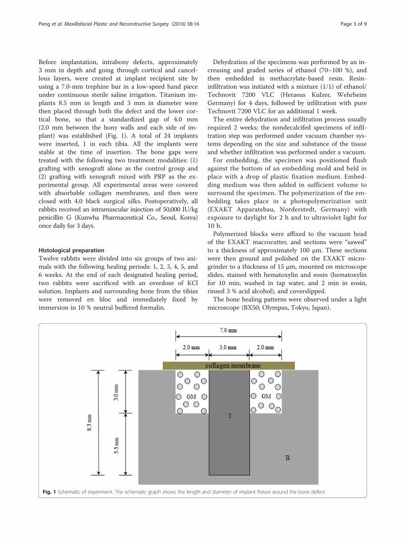
Before implantation, intrabony defects, approximately3 mm in depth and going through cortical and cancel-lous layers, were created at implant recipient site byusing a 7.0-mm trephine bur in a low-speed hand pieceunder continuous sterile saline irrigation. Titanium im-plants 8.5 mm in length and 3 mm in diameter werethen placed through both the defect and the lower cor-tical bone, so that a standardized gap of 4.0 mm(2.0 mm between the bony walls and each side of im-plant) was established (Fig. 1). A total of 24 implantswere inserted, 1 in each tibia. All the implants werestable at the time of insertion. The bone gaps weretreated with the following two treatment modalities: (1)grafting with xenograft alone as the control group and(2) grafting with xenograft mixed with PRP as the ex-perimental group. All experimental areas were coveredwith absorbable collagen membranes, and then wereclosed with 4.0 black surgical silks. Postoperatively, allrabbits received an intramuscular injection of 50,000 IU/kgpenicillin G (Kunwha Pharmaceutical Co., Seoul, Korea)once daily for 3 days.
Histological preparationTwelve rabbits were divided into six groups of two ani-mals with the following healing periods: 1, 2, 3, 4, 5, and6 weeks. At the end of each designated healing period,two rabbits were sacrificed with an overdose of KClsolution. Implants and surrounding bone from the tibiaswere removed en bloc and immediately fixed byimmersion in 10 % neutral buffered formalin.
Dehydration of the specimens was performed by an in-creasing and graded series of ethanol (70–100 %), andthen embedded in methacrylate-based resin. Resin-infiltration was initiated with a mixture (1/1) of ethanol/Technovit 7200 VLC (Heraeus Kulzer, WehrheimGermany) for 4 days, followed by infiltration with pureTechnovit 7200 VLC for an additional 1 week.The entire dehydration and infiltration process usually
required 2 weeks; the nondecalcifed specimens of infil-tration step was performed under vacuum chamber sys-tems depending on the size and substance of the tissueand whether infiltration was performed under a vacuum.For embedding, the specimen was positioned flush
against the bottom of an embedding mold and held inplace with a drop of plastic fixation medium. Embed-ding medium was then added in sufficient volume tosurround the specimen. The polymerization of the em-bedding takes place in a photopolymerization unit(EXAKT Apparatebau, Norderstedt, Germany) withexposure to daylight for 2 h and to ultraviolet light for10 h.Polymerized blocks were affixed to the vacuum head
of the EXAKT macrocutter, and sections were “sawed”to a thickness of approximately 100 μm. These sectionswere then ground and polished on the EXAKT micro-grinder to a thickness of 15 μm, mounted on microscopeslides, stained with hematoxylin and eosin (hematoxylinfor 10 min, washed in tap water, and 2 min in eosin,rinsed 3 % acid alcohol), and coverslipped.The bone healing patterns were observed under a light
microscope (BX50; Olympus, Tokyo, Japan).
Fig. 1 Schematic of experiment. The schematic graph shows the length and diameter of implant fixture around the bone defect
Peng et al. Maxillofacial Plastic and Reconstructive Surgery (2016) 38:16 Page 3 of 9
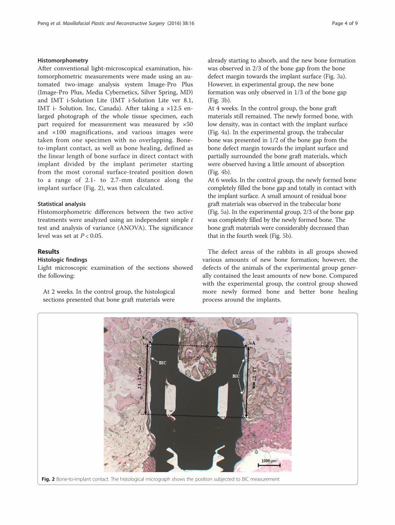
HistomorphometryAfter conventional light-microscopical examination, his-tomorphometric measurements were made using an au-tomated two-image analysis system Image-Pro Plus(Image-Pro Plus, Media Cybernetics, Silver Spring, MD)and IMT i-Solution Lite (IMT i-Solution Lite ver 8.1,IMT i- Solution. Inc, Canada). After taking a ×12.5 en-larged photograph of the whole tissue specimen, eachpart required for measurement was measured by ×50and ×100 magnifications, and various images weretaken from one specimen with no overlapping. Bone-to-implant contact, as well as bone healing, defined asthe linear length of bone surface in direct contact withimplant divided by the implant perimeter startingfrom the most coronal surface-treated position downto a range of 2.1- to 2.7-mm distance along theimplant surface (Fig. 2), was then calculated.
Statistical analysisHistomorphometric differences between the two activetreatments were analyzed using an independent simple ttest and analysis of variance (ANOVA). The significancelevel was set at P < 0.05.
ResultsHistologic findingsLight microscopic examination of the sections showedthe following:
At 2 weeks. In the control group, the histologicalsections presented that bone graft materials were
already starting to absorb, and the new bone formationwas observed in 2/3 of the bone gap from the bonedefect margin towards the implant surface (Fig. 3a).However, in experimental group, the new boneformation was only observed in 1/3 of the bone gap(Fig. 3b).At 4 weeks. In the control group, the bone graftmaterials still remained. The newly formed bone, withlow density, was in contact with the implant surface(Fig. 4a). In the experimental group, the trabecularbone was presented in 1/2 of the bone gap from thebone defect margin towards the implant surface andpartially surrounded the bone graft materials, whichwere observed having a little amount of absorption(Fig. 4b).At 6 weeks. In the control group, the newly formed bonecompletely filled the bone gap and totally in contact withthe implant surface. A small amount of residual bonegraft materials was observed in the trabecular bone(Fig. 5a). In the experimental group, 2/3 of the bone gapwas completely filled by the newly formed bone. Thebone graft materials were considerably decreased thanthat in the fourth week (Fig. 5b).
The defect areas of the rabbits in all groups showedvarious amounts of new bone formation; however, thedefects of the animals of the experimental group gener-ally contained the least amounts of new bone. Comparedwith the experimental group, the control group showedmore newly formed bone and better bone healingprocess around the implants.
Fig. 2 Bone-to-implant contact. The histological micrograph shows the position subjected to BIC measurement
Peng et al. Maxillofacial Plastic and Reconstructive Surgery (2016) 38:16 Page 4 of 9
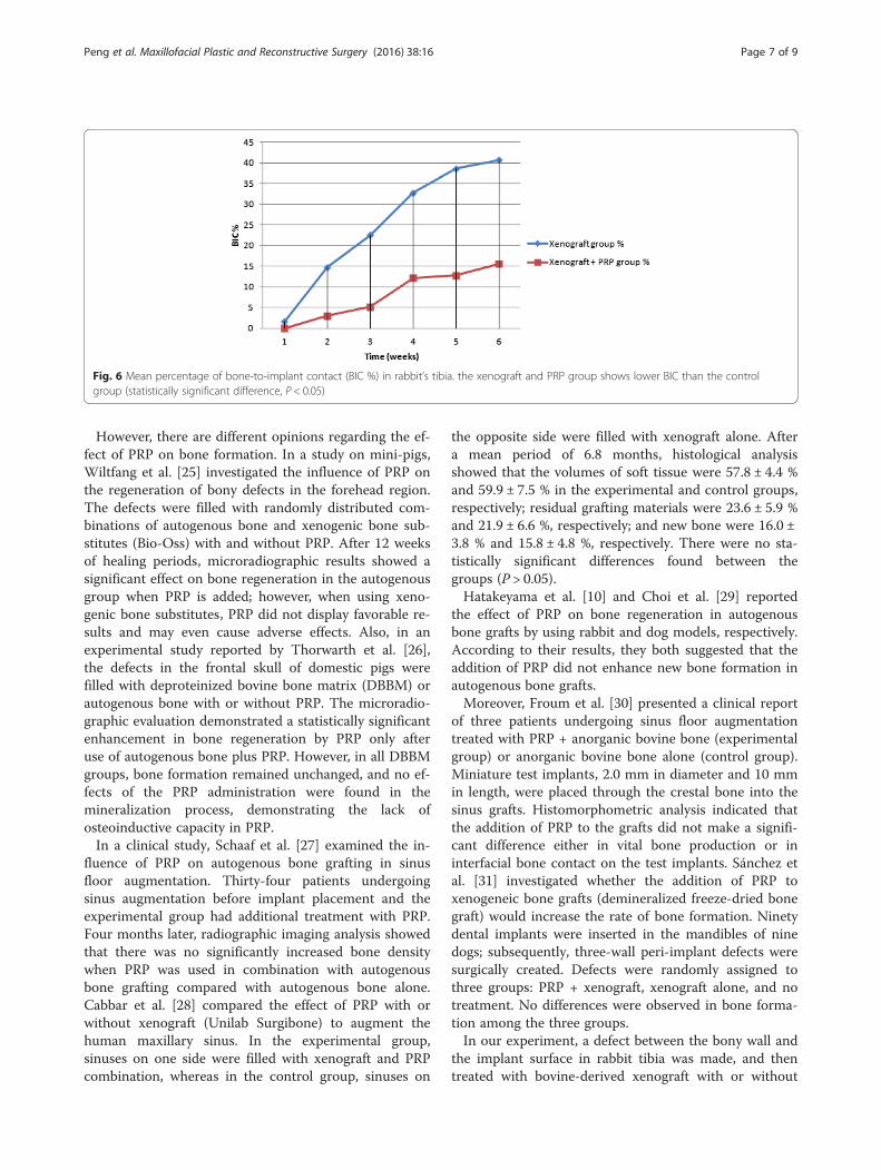
Histomorphometric analysisThe mean percentages of direct bone-to-implant con-tact in the two groups are shown in Table 1 and Fig. 6.The quantitative morphometric analysis showed signifi-cantly more bone-to-implant contact in the controlgroup. The bone-to-implant contact was significantlyhigher (P < 0.05) in the control group (25.23 ± 15.15 %)than in the experimental group (8.16 ± 6.26 %).
DiscussionAlthough autogenous grafts are commonly used in oraland maxillofacial surgery for treatment of bone loss andfunctional rehabilitation, the need for additional inter-vention increases the duration of surgery and the risk ofinfection, pain, and discomfort at the donor site. Duringthe last decade, several bone grafting materials produced
from bovine bone, with physicochemical characteristicssimilar to those of human bone, have been developed foruse in oral and orthopedic surgeries as an alternative toautogenous grafts [13]. In a study on rabbit, Jensen et al.[14] found that Bio-Oss became completely incorporatedin newly formed bone. In comparison with other threebone substitute materials (ceramic hydroxyapatite, corallinehydroxylapatite, and coral calcium carbonate), Bio-Ossshowed a higher degree of integration in the surroundingbone. Berglundh and Lindhe [15] also concluded thatBio-Oss became integrated with the host bone and sub-sequently replaced by newly formed bone. Bio-Oss con-tains pores with different sizes, intracrystalline spaces(3–26 nm), micro pores (vascular marrow canals), andmacro pores (300–1500 μm), which result in a highoverall porosity of 70–75 % [16]. As a consequence of
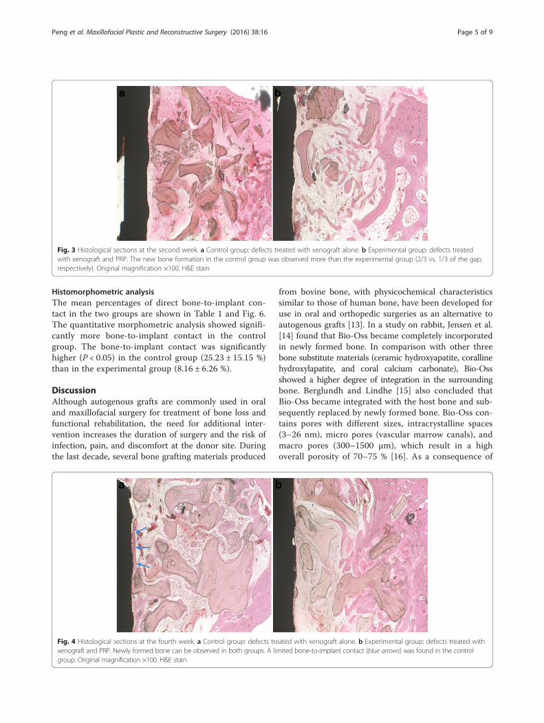
Fig. 3 Histological sections at the second week. a Control group: defects treated with xenograft alone. b Experimental group: defects treatedwith xenograft and PRP. The new bone formation in the control group was observed more than the experimental group (2/3 vs. 1/3 of the gap,respectively). Original magnification ×100. H&E stain
Fig. 4 Histological sections at the fourth week. a Control group: defects treated with xenograft alone. b Experimental group: defects treated withxenograft and PRP. Newly formed bone can be observed in both groups. A limited bone-to-implant contact (blue arrows) was found in the controlgroup. Original magnification ×100. H&E stain
Peng et al. Maxillofacial Plastic and Reconstructive Surgery (2016) 38:16 Page 5 of 9
this highly porous structure, Bio-Oss can be easily in-vaded by blood vessels resulting in subsequent migra-tion of osteoblasts. Therefore, Bio-Oss is considered tobe a biocompatible grafting material with remarkableosteoconductive ability, which does not cause signifi-cant inflammatory reaction [17].Some authors such as Marx et al., Magesh et al. [18],
and Aimetti et al. [19] evaluated the effect of PRP onbone regeneration in human. In their studies, the bonedefects were treated with an autogenous bone graftalone or in combination with PRP, and they all demon-strated that the use of PRP along with an autogenousbone graft were advantageous since it enhanced thequantity of newly formed bone. Yilmaz et al. [20] investi-gated the effectiveness of PRP and a bovine-derivedxenograft (BDX) combination on early wound healing indeep intrabony defects. A total of 85 intrabony defectswere selected in 20 advanced chronic periodontitispatients. Defects were surgically treated with PRP/BDX.One year after surgery, the results showed that PRP incombination with BDX leads to a significantly favorable
clinical improvement in deep intrabony periodontaldefects.Nagata et al. [21] analyzed the effect of PRP on healing
of autogenous bone (AB) grafts placed in surgically cre-ated critical-size defects in rabbit calvaria. The results in-dicated that AB/PRP significantly improved boneformation, and a beneficial effect of PRP was limited to aninitial healing period of 4 weeks. In the study of Kurikchyet al. [22], they assessed the effect of PRP on the bonehealing process either alone or mixed with xenogenic graft(Gen-Ox-lyophilized bovine bone organic matrix) in thefemur bone defects of rabbit models. The results showedthat in the use of PRP in combination with the xenogenicbone graft, new bone formation and neovascularizationwere enhanced significantly when compared with xeno-genic graft alone.Furthermore, Torres et al. [23] evaluated the clinical
efficacy of PRP in a sinus augmentation procedure withimplant placement. Eighty-seven patients underwent144 sinus floor augmentation procedures using anor-ganic bovine bone (ABB) alone or ABB + PRP. A totalof 286 implants were placed in the augmented bone.After a follow-up period of 24 months, the histologicalanalysis in the five edentulous patients revealed thatbone augmentation was significantly higher in sitestreated with ABB + PRP (P < 0.05). Cho et al. [24] inves-tigated the effect of PRP on dental implant osseointe-gration. Sixteen dental implants 4 mm in diameter and8 mm in length were placed into each tibia of fourdogs. The experimental groups were treated with0.5 mL PRP; the control groups were instilled with0.5 mL of saline. Four weeks after implantation, theexperimental group showed significantly faster bone re-generation and increased bone activity compared to thecontrol group (P < 0.05).
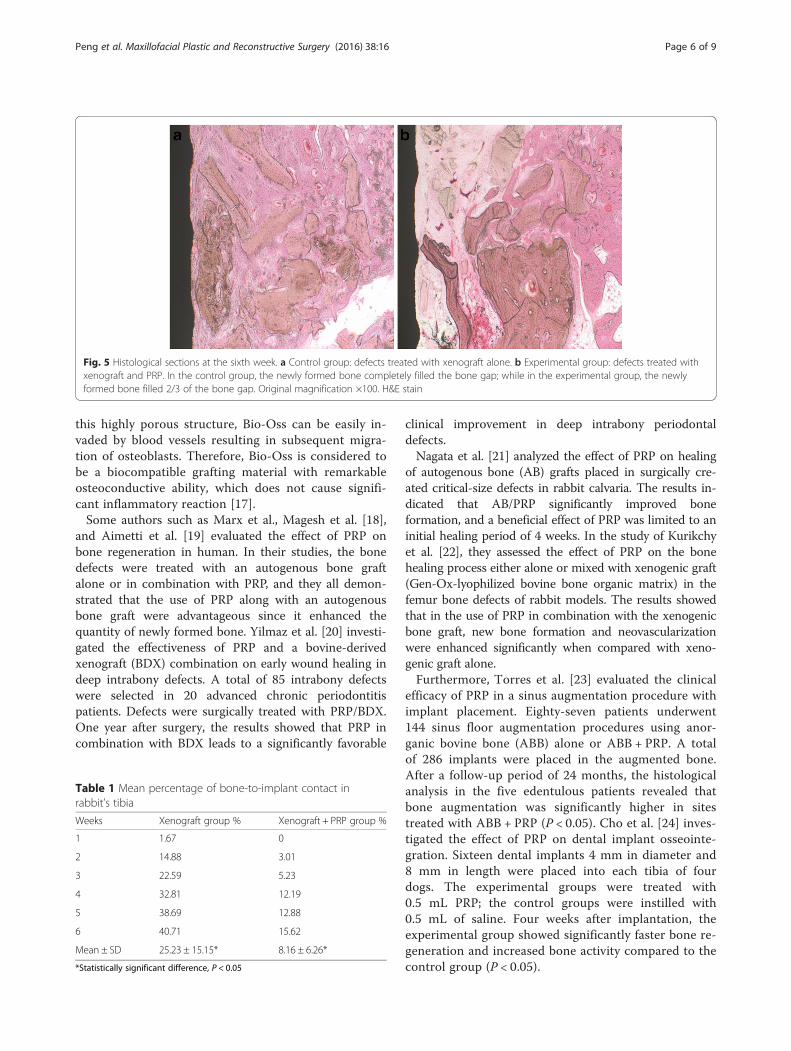
Fig. 5 Histological sections at the sixth week. a Control group: defects treated with xenograft alone. b Experimental group: defects treated withxenograft and PRP. In the control group, the newly formed bone completely filled the bone gap; while in the experimental group, the newlyformed bone filled 2/3 of the bone gap. Original magnification ×100. H&E stain
Table 1 Mean percentage of bone-to-implant contact inrabbit’s tibia
Weeks Xenograft group % Xenograft + PRP group %
1 1.67 0
2 14.88 3.01
3 22.59 5.23
4 32.81 12.19
5 38.69 12.88
6 40.71 15.62
Mean ± SD 25.23 ± 15.15* 8.16 ± 6.26*
*Statistically significant difference, P < 0.05
Peng et al. Maxillofacial Plastic and Reconstructive Surgery (2016) 38:16 Page 6 of 9
However, there are different opinions regarding the ef-fect of PRP on bone formation. In a study on mini-pigs,Wiltfang et al. [25] investigated the influence of PRP onthe regeneration of bony defects in the forehead region.The defects were filled with randomly distributed com-binations of autogenous bone and xenogenic bone sub-stitutes (Bio-Oss) with and without PRP. After 12 weeksof healing periods, microradiographic results showed asignificant effect on bone regeneration in the autogenousgroup when PRP is added; however, when using xeno-genic bone substitutes, PRP did not display favorable re-sults and may even cause adverse effects. Also, in anexperimental study reported by Thorwarth et al. [26],the defects in the frontal skull of domestic pigs werefilled with deproteinized bovine bone matrix (DBBM) orautogenous bone with or without PRP. The microradio-graphic evaluation demonstrated a statistically significantenhancement in bone regeneration by PRP only afteruse of autogenous bone plus PRP. However, in all DBBMgroups, bone formation remained unchanged, and no ef-fects of the PRP administration were found in themineralization process, demonstrating the lack ofosteoinductive capacity in PRP.In a clinical study, Schaaf et al. [27] examined the in-
fluence of PRP on autogenous bone grafting in sinusfloor augmentation. Thirty-four patients undergoingsinus augmentation before implant placement and theexperimental group had additional treatment with PRP.Four months later, radiographic imaging analysis showedthat there was no significantly increased bone densitywhen PRP was used in combination with autogenousbone grafting compared with autogenous bone alone.Cabbar et al. [28] compared the effect of PRP with orwithout xenograft (Unilab Surgibone) to augment thehuman maxillary sinus. In the experimental group,sinuses on one side were filled with xenograft and PRPcombination, whereas in the control group, sinuses on
the opposite side were filled with xenograft alone. Aftera mean period of 6.8 months, histological analysisshowed that the volumes of soft tissue were 57.8 ± 4.4 %and 59.9 ± 7.5 % in the experimental and control groups,respectively; residual grafting materials were 23.6 ± 5.9 %and 21.9 ± 6.6 %, respectively; and new bone were 16.0 ±3.8 % and 15.8 ± 4.8 %, respectively. There were no sta-tistically significant differences found between thegroups (P > 0.05).Hatakeyama et al. [10] and Choi et al. [29] reported
the effect of PRP on bone regeneration in autogenousbone grafts by using rabbit and dog models, respectively.According to their results, they both suggested that theaddition of PRP did not enhance new bone formation inautogenous bone grafts.Moreover, Froum et al. [30] presented a clinical report
of three patients undergoing sinus floor augmentationtreated with PRP + anorganic bovine bone (experimentalgroup) or anorganic bovine bone alone (control group).Miniature test implants, 2.0 mm in diameter and 10 mmin length, were placed through the crestal bone into thesinus grafts. Histomorphometric analysis indicated thatthe addition of PRP to the grafts did not make a signifi-cant difference either in vital bone production or ininterfacial bone contact on the test implants. Sánchez etal. [31] investigated whether the addition of PRP toxenogeneic bone grafts (demineralized freeze-dried bonegraft) would increase the rate of bone formation. Ninetydental implants were inserted in the mandibles of ninedogs; subsequently, three-wall peri-implant defects weresurgically created. Defects were randomly assigned tothree groups: PRP + xenograft, xenograft alone, and notreatment. No differences were observed in bone forma-tion among the three groups.In our experiment, a defect between the bony wall and
the implant surface in rabbit tibia was made, and thentreated with bovine-derived xenograft with or without
Fig. 6 Mean percentage of bone-to-implant contact (BIC %) in rabbit’s tibia. the xenograft and PRP group shows lower BIC than the controlgroup (statistically significant difference, P < 0.05)
Peng et al. Maxillofacial Plastic and Reconstructive Surgery (2016) 38:16 Page 7 of 9
PRP. As the result, a better bone healing process andmore amount of new bone formation were observed inthe control group. The percentage of bone-to-implantcontact in the control group was 25.23 ± 15.15 %,whereas in the experimental group, the percentage wasonly 8.16 ± 6.26 %. According to the results of histo-logical and histomorphometric examinations, our studyis in agreement with the findings from previous studiesin which there was no effect of PRP on new bone forma-tion in the PRP-treated bone graft.It is not very clear why the PRP-treated grafts exhib-
ited decreased bone formation when compared with thenon-PRP-treated grafts. Between and within the species,the baseline values of the platelet numbers have a greatvariation [32]. This variation of the platelet concentra-tion may have an important effect in the conflicting re-sults reported in various animal experimental studiesusing PRP. Regarding several studies carried out inhumans indicating an advantageous effect of PRP, it maybe possible that human PRP is more potent than animal-derived PRP. It should be born in mind that these hu-man studies are designed for comprehensible reasons,but without randomized prospective, have no controlsites and comprise a heterogeneous group of patients. Arecently performed animal experiment by Plachokova etal. [33] supports this suggestion. In their experiment,they compared the bone regenerative effect of PRPs ofdifferent species (rat, goat, and human). PRPs in com-bination with human bone or HA/TCP (hydroxyapatite-tricalcium phosphate) were used in nude rat models withcritical-size cranial defects. As a result, no effect of ratPRP and goat PRP was seen, while human PRP mixedwith human bone significantly enhanced new bone for-mation, but only after 2 weeks postoperatively. The au-thors noted that, in comparison with the preparation ofhuman PRP, the method of animal PRP preparationshould be changed for different animal species. The au-thors also noted the importance of defining the differentcritical effective amounts of platelet and growth factorlevels in PRP according to different animal species. Forthis reason, the critical effective amount of platelets ineach type of animal should be defined by experimentalstudies.Another factor may be related to the concentration of
PRP within the grafts. When a small amount of bonegraft is mixed with a large volume of PRP, the activationof bone cells in the adjacent tissue or graft and enhance-ment of new bone formation would be expected. Wei-brich et al. [34] analyzed the effect of the platelet countin PRP on bone regeneration. In their study, three typesof platelet concentrations in PRP were used in the rabbitmodels, including low-platelet concentrations (164,000–373,000 platelets/μL), intermediate platelet concentra-tions (503,000–1,729,000 platelets/μL), and high platelet
concentrations (1,845,000–3,200,000 platelets/μL). Com-paring the bone regeneration after 4 weeks, the onlyslightly significant difference was seen with intermediateplatelet concentrations (P = 0.004), but in analyzing thebone-to-implant contact, no differences were found forthe three platelet concentration groups. In our study, thebaseline value of platelets in the whole blood was 236 ×103/μL, whereas the platelet concentration of PRP was625 × 103/μL. Thus, a 265 % increase in platelet countwas observed. However, the results failed to show an in-crease in bone-to-implant contact when this concentra-tion of PRP was used.PRP is commonly used in different clinical situations
in an attempt to improve soft and hard tissue healing. Inspite of this, the results from our study showed that theaddition of PRP to bovine-derived xenograft in the de-fects around the implants in the present animal modeldid not result in increased new bone formation or bone-to-implant contacts. More basic researches into animalspecies, optimal concentration of PRP, grafting materials,and presence of implants are necessary to capitalize onthe ability of platelet growth factors to enhance boneformation in a graft.
ConclusionsThis study reported the healing effect of PRP on bovine-derived xenograft in peri-implant defects. Intrabony defectswere created in the tibia of rabbits, and dental implantswere installed in defects filled with either xenograft aloneor mixture of PRP and xenograft. The mean percentage ofbone-to-implant contact in the defects treated with thexenograft alone was 25.23 ± 15.15 %, whereas in the defectstreated with xenograft combined with PRP, the percentagewas only 8.16 ± 6.26 %. On the basis of these findings, itcan be concluded that the addition of PRP into bovine-derived xenograft around titanium dental implants maydelay peri-implant bone healing. So far, the scientific evi-dence regarding the efficacy and efficiency of PRP is stillcontroversial.
Statement of ethics approvalAnimal protocol was approved by INHA University, In-stitutional Animal Care, and the approval number isINHA 150716-372.
Competing interestsThe authors declare that they have no competing interests.
Authors’ contributionsWP and IKK made contributions to the conception of the report and carriedout the experiment. JHS, DHL, and JMJ carried out the experiment. HYC andSHP participated in the collection of data and drafting of the manuscript. Allauthors read and approved the final manuscript.
Authors’ informationAll of the authors have no affiliations with or involvement in anyorganization or entity with any financial interest or non-financial interest in
Peng et al. Maxillofacial Plastic and Reconstructive Surgery (2016) 38:16 Page 8 of 9
this manuscript. This manuscript represents original works and is not beingconsidered for publication elsewhere.
Author details1Department of Oral and Maxillofacial Surgery, College of Medicine, InhaUniversity, Incheon, South Korea. 2Department of Oral and MaxillofacialSurgery, International St. Mary’s Hospital, Catholic Kwandong UniversityCollege of Medicine, Incheon, South Korea. 3Department of OMFS, Dentistry,College of Medicine, Inha University, #7-206, 3rd St. Shinheung-dong,Choong-gu, Incheon 400-711, South Korea.
Received: 20 January 2016 Accepted: 8 March 2016
References1. Lang N, Bragger U, Hammerle C (1994) Immediate transmucosal implants
using the principle of guided tissue regeneration. I. Rationale, clinicalprocedures and 30-month results. Clin Oral Implants Res 5:154–163
2. Landsberg CJ (1997) Socket seal surgery combined with immediate implantplacement: a novel approach for single tooth replacement. Int JPeriodontics Restorative Dent 17:141–149
3. Becker BE, Becker W, Ricci A (1998) A prospective clinical trial of endosseousscrew-shaped implants placed at the time of tooth extraction withoutaugmentation. J Periodontol 69:920–926
4. Retzepi M, Donos N (2010) Guided bone regeneration: biological principlesand theurapeutic applications. Clin Oral Implants Res 21:567–576
5. Marx RE, Carlson ER, Eichstaedt RM (1998) Platelet-rich plasma: growthfactor enhancement for bone grafts. Oral Surg Oral Med Oral Pathol OralRadiol Endod 85:638–646
6. Trisi P, Rebaudi A, Calvari F (2006) Sinus graft with biogran, autogenousbone, and PRP: a report of three cases with histology and micro-CT. Int JPeriodontics Restorative Dent 26:113–125
7. Albanese A, Licata ME, Polizzi B (2013) Platelet-rich plasma (PRP) in dentaland oral surgery: from the wound healing to bone regeneration. ImmunAgeing 10:23
8. Ganio C, Tenewitz FE, Wilson RC (1993) The treatment of chronicnonhealing wounds using autologous platelet-derived growth factors.J Foot Ankle Surg 32:263–268
9. Tözum TF, Demirlap B (2003) Platelet rich plasma. A promising innovation indentistry. J Can Dent Assoc 69:664
10. Hatakeyama M, Beletti ME, Zanetta-Barbosa D (2008) Radiographic andhistomorphometric analysis of bone healing using autogenous graftassociated with platelet-rich plasma obtained by 2 different methods. OralSurg Oral Med Oral Pathol Oral Radiol Endod 105:13–18
11. Jensen TB, Rahbek O, Overgaard S (2004) Platelet rich plasma and freshfrozen bone allograft as enhancement of implant fixation. An experimentalstudy in dogs. J Orthop Res 22:653–658
12. Okuda K, Kawase T, Momose M (2003) Platelet-rich plasma contains highlevels of platelet-derived growth factor and transforming growth factor-βand modulates the proliferation of periodontally related cells in vitro.J Periodontol 74:849–857
13. Vajda EG, Kneissel M, Muggenburg B (1999) Increased intracortical boneremodeling during lactation in Beagle dogs. Biol Reprod 61:1439–1444
14. Jensen SS, Aaboe M, Pinholt EM (1996) Tissue reaction and materialcharacteristics of four bone substitutes. Int J Oral Maxillofac Implants11:55–66
15. Berglundh T, Lindhe J (1997) Healing around implants placed in bonedefects treated with Bio-Oss. An experimental study in the dog. Clin OralImplants Res 8:117–124
16. Hammerle CH, Olah AJ, Schmid J (1997) The biological effect of naturalbone mineral on bone neoformation on the rabbit skull. Clin Oral ImplantsRes 8:198–207
17. Clergeau LP, Danan M, ClergeauGuerithault S (1996) Healing response toanorganic bone implantation in periodontal intrabony defects in dogs. PartI. Bone regeneration. A microradiographic study. J Periodontol 67:140–149
18. Magesh DP, Kumaravelu C, Maheshwari G (2013) Efficacy of PRP in thereconstruction of mandibular segmental defects using iliac bone grafts.J Maxillofac Oral Surg 12:160–167
19. Aimetti M, Romano F, Dellavia C (2008) Sinus grafting using autogenousbone and platelet-rich plasma: histologic outcomes in humans. Int JPeriodontics Restorative Dent 28:585–591
20. Yilmaz S, Cakar G, Kuru B (2009) Platelet-rich plasma in combination withbovine derived xenograft in the treatment of deep intrabony periodontaldefects: a report of 20 consecutively treated patients. Platelets 20:432–440
21. Nagata MJ, Melo LG, Messora MR (2009) Effect of platelet-rich plasma onbone healing of autogenous bone grafts in critical-size defects. J ClinPeriodontol 36:775–783
22. Kurikchy MQ, Al-Rawi NH, Ayoub RS (2013) Histological evaluation of bonehealing using organic bovine bone in combination with platelet-richplasma (an experimental study on rabbits). Clin Oral Investig 17:897–904
23. Torres J, Tamimi F, Martinez PP (2009) Effect of platelet-rich plasma on sinuslifting: a randomized-controlled clinical trial. J Clin Periodontol 36:677–687
24. Cho K, Kim JM, Kim MH (2013) Scintigraphic evaluation of osseointegrativeresponse around calcium phosphate-coated titanium implants in tibia bone:effect of platelet-rich plasma on bone healing in dogs. Eur Surg Res51:138–145
25. Wiltfang J, Kloss FR, Kessler P (2004) Effects of platelet rich plasma on bonehealing in combination with autogenous bone and bone substitutes incritical-size defects. An animal experiment. Clin Oral Implants Res15:187–193
26. Thorwarth M, Wehrhan F, Schultze-Mosgau S (2006) PRP modulatesexpression of bone matrix proteins in vivo without long-term effects onbone formation. Bone 38:30–40
27. Schaaf H, Streckbein P, Lendeckel S (2008) Sinus lift augmentation usingautogenous bone grafts and platelet-rich plasma: radiographic results. OralSurg Oral Med Oral Pathol Oral Radiol Endod 106:673–678
28. Cabbar F, Güler N, Kürkcü M (2011) The effect of bovine bone graft with orwithout platelet-rich plasma on maxillary sinus floor augmentation. J OralMaxillofac Surg 69:2537–2547
29. Choi BH, Im CJ, Huh JY (2004) Effect of platelet-rich plasma on boneregeneration in autogenous bone graft. Int J Oral Maxillofac Surg 33:56–59
30. Froum SJ, Wallace SS, Tarnow DP (2002) Effect of platelet-rich plasma onbone growth and osseointegration in human maxillary sinus grafts: threebilateral case reports. Int J Periodontics Restorative Dent 22:45–53
31. Sánchez AR, Sheridan PJ, Eckert SE (2005) Influence of platelet-rich plasmaadded to xenogeneic bone grafts in periimplant defects: a vitalfluorescence study in dogs. Clin Implant Dent Relat Res 7:61–69
32. Merck Veterinary Manual 2006: Table 6 Hematologic references ranges.33. Plachokova AS, van den Dolder J, van den Beucken JJ (2009) Bone
regenerative properties of rat, goat and human platelet-rich plasma. Int JOral Maxillofac Surg 38:861–869
34. Weibrich G, Hansen T, Kleis W (2004) Effect of platelet concentration inplatelet-rich plasma on peri-implant bone regeneration. Bone 34:665–671
Submit your manuscript to a journal and benefi t from:
7 Convenient online submission
7 Rigorous peer review
7 Immediate publication on acceptance
7 Open access: articles freely available online
7 High visibility within the fi eld
7 Retaining the copyright to your article
Submit your next manuscript at 7 springeropen.com
Peng et al. Maxillofacial Plastic and Reconstructive Surgery (2016) 38:16 Page 9 of 9


![The effect of autologous leukocyte platelet rich fibrin on ... · these two platelet concentrates. [15,16] Although they are both clinically effective in accelerating the healing](https://img.pdfslide.us/doc/110x75/5f74cde1d357e407be22081f/the-effect-of-autologous-leukocyte-platelet-rich-fibrin-on-these-two-platelet.jpg)