Embed Size (px)
Citation preview
www.sciencedirect.com
c o r t e x 4 9 ( 2 0 1 3 ) 4 1 1e4 2 2
Available online at
Journal homepage: www.elsevier.com/locate/cortex
Special issue: Research report
The functional anatomy of suggested limb paralysis
Quinton Deeley a,*, David A. Oakley b, Brian Toone c, Vaughan Bell d, Eamonn Walsh a,Andre F. Marquand d, Vincent Giampietro d, Michael J. Brammer d, Steven C.R. Williams d,Mitul A. Mehta d and Peter W. Halligan e
aCultural and Social Neuroscience Research Group, Forensic and Neurodevelopmental Sciences, Kings College London, Institute of Psychiatry,
London, UKbDivision of Psychology and Language Sciences, University College London, UKcKings College London, Institute of Psychiatry, London, UKdCultural and Social Neuroscience Research Group, Centre for Neuroimaging Sciences, Kings College London, Institute of Psychiatry,
London, UKe School of Psychology, Cardiff University, UK
a r t i c l e i n f o
Article history:
Received 20 January 2012
Reviewed 17 May 2012
Revised 21 July 2012
Accepted 14 September 2012
Action editor H. Branch Coslett
Published online 21 December 2012
Keywords:
Dissociation
Conversion disorder
Hypnosis
fMRI
Brain
Cultural neuroscience
* Corresponding author. Cultural and SocialLondon, Institute of Psychiatry, De Crespign
E-mail address: [email protected] (Q0010-9452/$ e see front matter ª 2012 Elsevhttp://dx.doi.org/10.1016/j.cortex.2012.09.016
a b s t r a c t
Suggestions of limb paralysis in highly hypnotically suggestible subjects have been
employed to successfully model conversion disorders, revealing similar patterns of brain
activation associated with attempted movement of the affected limb. However, previous
studies differ with regard to the executive regions involved during involuntary inhibition of
the affected limb. This difference may have arisen as previous studies did not control for
differences in hypnosis depth between conditions and/or include subjective measures to
explore the experience of suggested paralysis. In the current study we employed functional
magnetic resonance imaging (fMRI) to examine the functional anatomy of left and right
upper limb movements in eight healthy subjects selected for high hypnotic suggestibility
during (i) hypnosis (NORMAL) and (ii) attempted movement following additional left upper
limb paralysis suggestions (PARALYSIS). Contrast of left upper limb motor function during
NORMAL relative to PARALYSIS conditions revealed greater activation of contralateral
M1/S1 and ipsilateral cerebellum, consistent with the engagement of these regions in the
completion of movements. By contrast, two significant observations were noted in PA-
RALYSIS relative to NORMAL conditions. In conjunction with reports of attempts to move
the paralysed limb, greater supplementary motor area (SMA) activation was observed,
a finding consistent with the role of SMA in motor intention and planning. The anterior
cingulate cortex (ACC, BA 24) was also significantly more active in PARALYSIS relative to
NORMAL conditions e suggesting that ACC (BA 24) may be implicated in involuntary, as
well as voluntary inhibition of prepotent motor responses.
ª 2012 Elsevier Ltd. All rights reserved.
Neuroscience Research Group, Forensic and Neurodevelopmental Sciences, Kings Collegey Park, Denmark Hill, London SE5 8AF, UK.. Deeley).ier Ltd. All rights reserved.
c o r t e x 4 9 ( 2 0 1 3 ) 4 1 1e4 2 2412
1. Introduction movement in the suggested paralysis condition, in keeping
Patients with “functional”, “psychogenic” or dissociative
conversion disorders present with symptoms that resemble
neurological illnesses e such as paralyses and loss of sensa-
tion for which no organic or neurological cause has been
established (Akagi andHouse, 2001; Fink et al., 2006; Stone and
Zeman, 2001). These disorders are common, have a poor
prognosis and contribute considerable morbidity and costs to
health services (Stone et al., 2003). Between 30% and 40% of
patients attending neurology outpatient clinics have symp-
toms that cannot be totally or partially explained by known
organic illness (Carson et al., 2000). Similar rates have been
reported for both in- and outpatient clinics in other European
countries (Halligan et al., 2000).
While the specific neurocognitive mechanisms underlying
dissociative conversion disorders remain largely unknown,
similarities between conversion symptoms and phenomena
produced by suggestions in hypnosis have been noted from
the 19th century onwards (Bell et al., 2011; Bliss, 1984; Charcot,
1888; Janet, 1907; Kihlstrom, 1994; Oakley, 1999b). In particular
both hypnotic phenomena and conversion disorder symp-
toms are experienced as being ‘real’ or ‘involuntary’, and yet
in the latter, medical and neurological examinations fail to
demonstrate specific neurological abnormalities that could
account for the reported symptoms. The similarities in
reported experiences between patients and highly hypnotis-
able subjects responding to suggestions of paralysis raise the
possibility that such symptoms are generated by the same
kinds of cognitive and neural process (Bell et al., 2011; Oakley,
1999b, 2001).
The first evidence for shared neural mechanisms under-
lying both dissociative conversion and suggested limb paral-
ysis was provided by two single-subject Positron Emission
Tomography (PET) studies. The first study of a 45-year-old
right-handed woman with a dissociative paralysis of her left
leg revealed that attempts to move her paralysed limb com-
pared to rest were associatedwith activation of right premotor
cortex and left cerebellum, which was interpreted as evidence
of preparation to move the limb (Marshall et al., 1997). In
addition, non-activation of contralateral sensorimotor cortex
during attempted movement was taken as being consistent
with the failure to produce actual movement in the paralysed
limb. Further, activation of right anterior cingulate cortex
(ACC) and right orbitofrontal cortex (OFC) was interpreted as
the basis for an unconscious inhibition of the planned (“wil-
led”) execution of the movement (Marshall et al., 1997).
A follow-up PET study used hypnotic suggestion as an
experimental tool (Oakley and Halligan, 2009) to model left leg
conversion paralysis in a healthy 25-year-old male. The pat-
terns of brain activations during attempted left leg move-
ments following the paralysis suggestion for this hypnotised
participant were similar to those in the dissociative conver-
sion patient. In both instances, the experienced paralysis
appeared to be produced by comparable brain areas involved
in the inhibition of voluntary movement (Halligan et al., 2000).
A further PET group study comparing the functional anat-
omy of suggested and feigned limb paralysis similarly found
activation of right OFC in association with attempted limb
with the prior single-case studies of dissociative and sug-
gested limb paralysis (Ward et al., 2003). In contrast, the
feigned paralysis condition was uniquely associated with ac-
tivity in the left ventrolateral prefrontal cortex and right
posterior cortical structures, supporting a view that the par-
alyses reported in the two earlier studies were unlikely to be
the product of faking or malingering (Ward et al., 2003). Hence
there is converging evidence of similar patterns of brain ac-
tivity involving inhibition of primary motor areas by OFC and/
or ACC underlying dissociative and suggested limb paralysis,
that are distinct from the neural correlates of feigned limb
paralysis (Spence et al., 2000).
An alternative explanation for this ‘executive control’ ac-
count of dissociative and suggested limb paralysis was put
forward by Cojan et al. in two studies (Cojan et al., 2009a,
2009b). The first used a goeno go task of response inhibition to
examine brain activation with fMRI during movement prep-
aration, execution, and inhibition in a patientwith left handed
dissociative conversion paralysis in comparison with a group
of healthy controls (Cojan et al., 2009a). The second used the
same experimental task in a group selected for high hypnotic
suggestibility with separate suggested and feigned paralysis
conditions (Cojan et al., 2009b). As with their conversion pa-
tient, the study with highly hypnotically suggestible partici-
pants found normal motor cortex activation during
a movement preparation phase, which was again taken as
evidence that movement inhibition was not working through
suppression of motor intention. They also reported that right
ACC, bilateral OFC and extrastriate visual area activity were
increased as a main effect of hypnosis irrespective of motor
task condition, which they interpreted as suggesting that ac-
tivity in these areas may be indicative of state-related hyp-
nosis changes rather than a specific inhibitorymechanism. As
with their study on hysterical paralysis, they found a greater
degree of functional connectivity between the motor cortex
and precuneus, and similarly argued that motor inhibition
may be mediated through imagery based and self-reflective pro-
cessing rather than direct manipulation of topedown execu-
tive control, though the precise mechanism by which this
might be achieved was not specified. Nevertheless, it is
notable that the attempt to move the left limb to GO stimuli in
the suggested paralysis condition e a contrast which re-
sembles those of the prior PET studies e was associated with
increased activation in left inferior (IFG) and superior frontal
gyri (SFG) compared to execution of movement in response to
the GO stimulus in the normal (non-hypnotised) state (Cojan
et al., 2009b). These regions have previously been reported to
be involved in motor inhibition (Simmonds et al., 2008). A
recent study of left-hand hypnotically induced paralysis using
resting state fMRI (Pyka et al., 2011) also argued for paralysis
mediated by modified self representation, associated with
posterior cingulate cortex and precuneus modulation. How-
ever, clear functional coupling was also found between these
areas and the right dorsolateral prefrontal cortex, right
angular gyrus and the left somatosensory cortex, suggesting
motor inhibition may still be an essential part of the neuro-
cognitive mechanism for suggested paralysis. Collectively,
such findings could be taken as further evidence for the
c o r t e x 4 9 ( 2 0 1 3 ) 4 1 1e4 2 2 413
engagement of prefrontal ‘executive’ regions in inhibiting
implementation of motor intentions in a manner that is
experienced by the subjects as involuntary.
In summary, a small number of prior PET and fMRI studies
employing hypnotic suggestion to model dissociative con-
version paralyses provide evidence of engagement of a range
of prefrontal ‘executive’ inhibitory regions when attempting
to move the paralysed limb. However, while earlier PET
studies implicated OFC and ACC in involuntary inhibition of
movement (Halligan et al., 2000; Marshall et al., 1997; Ward
et al., 2003), a subsequent fMRI study attributed these activa-
tions to the main effects of hypnosis (Cojan et al., 2009b). The
latter study also found increased activation of IFG and SFG,
which are known to be associated with inhibitory function
(Simmonds et al., 2008), in the comparable contrast of
attempted left limb movement following hypnotically sug-
gested paralysis versus completed movement in the no-
hypnosis condition. Notwithstanding differences in inter-
pretation, these studies had several limitations. First, they did
not control for differences in depth of hypnosis between and
within experimental conditions, which could potentially
confound interpretation of activations that would otherwise
be attributed to the effects of suggested limb paralysis. Sec-
ondly, one of these prior studies contrasted a hypnosis plus
limb paralysis condition with movement in the normal alert
state, potentially confounding the effects of hypnotic induc-
tion with specific effects of suggested paralysis (Cojan et al.,
2009b). Also, the previous PET studies were constrained by
limited spatial resolution. This may critically restrict inves-
tigation of the modulation of motor control systems by sug-
gestive processes, given the anatomical proximity of regions
such as lateral premotor cortices, supplementary and pre-
supplementary motor areas (SMAs), ACC, and sensorimotor
cortices (Mayka et al., 2006; Picard and Strick, 2001). Finally,
prior imaging studies [with the exception of Cojan et al.
(2009a), (2009b)] did not examine the behavioural and func-
tional anatomical effects of suggested limb paralysis on the
contralateral (non-paralysed) limb. Establishing whether
these effects are limited to a target domain (limb) is important
if hypnotic suggestions are to be considered an effective tool
for modelling dissociative processes.
Consequently in this study, we employed functional mag-
netic resonance imaging (fMRI) to investigate the functional
anatomy of (upper) limb movement in eight healthy subjects
selected for high hypnotic suggestibility. We measured brain
activity during limb movement following induction of hyp-
nosis, with and without suggestions of left upper limb paral-
ysis (in keeping with the choice of the left limb in prior
studies). As an additional control condition, we alsomeasured
brain activity during movement of the right limb following
induction of hypnosis, both before and after suggestions of
paralysis in the contralateral (left) upper limb. The main hy-
potheses were that
(i) attempts to move the left upper limb in the paralysed
compared to the non-paralysed condition would be
associated with increased activity in motor control re-
gions involved in preparation and intention to move,
particularly the medial premotor region of SMA (Mayka
et al., 2006; Picard and Strick, 2001); and
(ii) attempts to move the limb in the paralysis condition
compared to the unparalysed condition would be asso-
ciated with (a) behavioural evidence of reduced move-
ment compared to the unparalysed condition; (b)
decreased activity in contralateral primary sensorimotor
cortices and ipsilateral cerebellum; and (c) self-report
evidence of greater perceived difficulty of movement
and loss of control (involuntariness) for the paralysed
limb associated with (d) increased activity in executive
inhibitory systems of OFC and ACC (Athwal et al., 2001).
In other words, we assumed that the latter would con-
stitute the neuroanatomical locations mediating the subjec-
tively involuntary inhibition of the intended movements.
Finally, we tested the supplementary hypothesis that the
functional anatomy of movement of the right limb would be
unaffected by the administration of paralysis suggestions in
the (left) contralateral limb.
2. Materials and methods
2.1. Subjects
Eight right-handed healthy volunteers (university un-
dergraduates or graduates in psychology and medicine at
University College London) were studied. There were three
male and five female participants in the group with a mean
age of 22.63 years [range 19e36, standard deviation (SD)
�5.55]. Participants were selected from a larger sample tested
on the Harvard Group Scale of Hypnotic Susceptibility (Form
A) (HGSS:A) (Shor and Orne, 1962) as being of medium to high
hypnotic suggestibility (9þ out of 12). They had experienced at
least two subsequent standardised hypnotic induction pro-
cedures before the present study, including an individually
administered test of responsiveness to ideomotor suggestion,
which they all passed. Their mean HGSS:A score was 10.5
(range 9e12, SD �1.2). All participants were screened to
exclude co-morbid psychiatric illness (e.g., schizophrenia,
major depression) and neurological and extracerebral disor-
ders that might affect brain function (e.g., epilepsy or hyper-
tension). One participant had a past history of a single episode
of mild depression, and was completing a course of antide-
pressants (citalopram 20 mg od), but was not depressed at the
time of recruitment or scanning. Ethical approval was
obtained from the Ethical Committee of the South London and
Maudsley Trust and Institute of Psychiatry. After complete
description of the study to the subjects, written informed
consent was obtained.
2.2. Hypnosis procedure
All hypnosis induction procedures as well as administration
and reversal of motor suggestions were carried out while the
participants were lying inside a GE Signa 1.5 T (General Elec-
tric, Milwaukee, WI) whole-body magnet, up to their waist. A
quadrature birdcage head coil was used for radiofrequency
(RF) transmission and reception. All instructions from the
experimenter were conveyed via earphones from a micro-
phone located in the scanning control room. The hypnosis
c o r t e x 4 9 ( 2 0 1 3 ) 4 1 1e4 2 2414
induction procedure, motor instructions and induction and
reversal of left upper limb paralysis have previously been
described in detail (Oakley et al., 2007). Briefly, the hypnosis
induction procedure was based on Gruzelier’s 3-Stage model
(Gruzelier, 1998), and involved: (1) visual fixation on a pro-
jected central cross-hair and listening to the experimenter’s
voice; (2) suggestions of ocular fatigue at continued fixation,
eye closure and deep physical (muscle) relaxation along with
counting 1e20; and (3) instructions for relaxed and passive
multimodal imagery (“Special Place” or “Safe Place”) (Heap
and Aravind, 2002). Subjects were also asked to rate the sub-
jective depth of their hypnotic experience immediately before
and after each of the motor tests on a scale of 1e10 (Tart,
1970), where 0 was defined as ‘not hypnotised at all’ and 10
was ‘as deeply hypnotised as you have ever been’. Numbers
above 10 could be used if the subject felt they were more
deeply hypnotised than they had been on any previous occa-
sion. As full hypnotic depth is achieved by the end of Stage 2 of
this particular induction procedure (Oakley et al., 2007), Stage
3 of the induction procedure (Special Place) was reversed
before the motor tests in the hypnosis condition. Termination
of hypnosis consisted of a reversal of induction Stages 1 and 2.
2.3. Motor testing and induction of motor symptoms
Hand and wrist movements of each subject were tested using
two, identical standard computer analogue joysticks adapted
to remove most metal. The filtered signal from each joystick
was fed to the computers via a custom-built interface box.
Subjects were instructed to move the left and right joysticks
once forward, once backward, and then to a neutral central
position upon hearing respective verbal instructions (‘right’,
‘left’ depending upon experimental epoch) presented via
a digital recording. They also received epochs of ‘rest’ in-
structions that signified that no response was required. It was
additionally suggested that irrespective of relaxation or pa-
ralysis suggestions the participant’s fingers/hand would
retain their grip on the relevant joystick throughout.
Presentation ofmotor epochs and concurrent acquisition of
fMRI data followed a blocked periodic design, involving
repeated alternation between the baseline (‘rest’ instruction)
and activation epochs (‘right’ or ‘left’ instructions respectively),
to optimise efficiency of estimation of time series regression
models used to measure cognitive activation. The order of
‘rest’, ‘right’, and ‘left’ epochswas pseudo-randomised in order
to reduce habituation effects. Each of themotor-testing blocks,
during which MR activations were acquired, comprised a total
of 15 30-sec epochs (five “rest”, five “right”, and five “left”), each
epoch consisting of 10 consecutive repetitions of the relevant
command repeated at 3-sec intervals. The total stimulus time
for each motor block was 7 min, 30 sec resulting in acquisition
of 150 images. The specific wording preceding each motor-
testing block and the script for administering left upper limb
paralysis are described in detail elsewhere (Oakley et al., 2007).
We acquired data formotor function following induction of
hypnosis with no suggestions of left upper limb paralysis
(MOTOR NORMAL) and motor function following additional
suggestions of left upper limb paralysis (PARALYSIS). These
contrasts allowed us to test our prior hypotheses regarding
the effects of suggested limb paralysis on brain function
whilst controlling for depth of hypnosis, and also to test the
effects of the paralysis suggestion on the functional anatomy
of contralateral limb movement.
2.4. Image acquisition parameters
Gradient-echo echo planar MR imaging (EPI) data were ac-
quired at 1.5 Tesla (T) at the Maudsley Hospital, UK. A GE LX
NVi scanner equipped with ultra-fast SR150 field gradients
was employed, allowing a maximum gradient amplitude of
40 mT/m (General Electric, Milwaukee WI, USA). fMRI exami-
nations were conducted using the following scanner param-
eters: pulse sequence ¼ echo planar, gradient echo, repetition
time ¼ 3000 msec, echo time ¼ 40 msec, RF flip angle ¼ 90�,slice orientation ¼ near-axial, number of slices ¼ 38, slice
thickness ¼ 3 mm, gap between slices ¼ .3 mm, acquisition
matrix resolution ¼ 64 � 64, acquisition mode ¼ interleaved,
K-space sampling ¼ FULL, ramp sampling ON, frequency
direction ¼ righteleft, number of dummy acquisitions ¼ 0,
total number of images per slice ¼ 150. An additional four
volumes were acquired before the experiment started in order
for the magnetic field to reach a steady state, but these were
removed before the start of the analysis process.
2.5. Neuroimaging data analysis
2.5.1. Summary of methodData were analyzed with software developed at the Institute of
Psychiatry, London, using a non-parametric approach (i.e., one
that not does assume that fMRI data are normally distributed)
(Thirion et al., 2007). For each experimental condition, whole-
brain maps of the changes in blood-oxygen-level-dependent
(BOLD) signal generated when contrasting MOVE blocks rela-
tive to REST blocks were separately generated for each subject
(1st level analysis). Each individual brain activation map was
then spatially normalized, first by rigid body transformation of
the fMRI data into a high-resolution inversion recovery image of
the same subject, and then by affine transformation onto
a Talairach template (Talairach and Tournoux, 1988) to allow
comparisons between experimental conditions at the group
level (2nd level analysis). Group level maps of changes in BOLD
signal over the whole brain to MOVE relative to REST blocks for
eachexperimentalcondition(MOTORNORMAL,PARALYSIS)and
hand (RIGHT and LEFT) were calculated. These contrasts were
undertaken to confirm that the motor paradigm significantly
engaged activation in brain regions known to be involved in
motor control and implementation. Repeated measures Ana-
lyses of Variants of BOLD responses to completed or attempted
movement across the NORMAL and PARALYSIS conditions for
the left and right limbs were calculated respectively. These
contrasts were undertaken to test our main hypotheses about
the effects of left upper limb paralysis suggestions on activation
of regions involved in motor control and implementation for
both the left and right limbs. A more detailed description of our
brain image analysis method is included below.
2.5.2. Individual brain activation mapsData were initially processed to minimise motion related ar-
tifacts (Bullmore et al., 1999a). A 3D volume consisting of the
average intensity at each voxel over the whole experiment
c o r t e x 4 9 ( 2 0 1 3 ) 4 1 1e4 2 2 415
was calculated and used as a template. The 3D image volume
at each time point was then realigned to this template.
Following realignment, data were then smoothed using
a Gaussian filter (full width half maximum (FWHM) 7.2mm) to
improve the signal to noise characteristics of the images.
Experimental responses were analyzed by convolving each
contrast of interest (activations associated with responses to
‘right’ instructions vs ‘rest’, ‘left’ instructions vs ‘rest’, and
‘right’ vs ‘left’) with two gamma variate functions (peak re-
sponses at 4 and 8 sec). These two functions were chosen to
encompass the known range of times to peak response fol-
lowing stimulus onset for BOLD effects. The best fit between
the weighted sum of these convolutions and the time series at
each voxel was computed using a constrained BOLD effect
model of Friman et al. (2003). This constrained model com-
putation was used to minimise the possibility of mathemati-
cally plausible but physiologically implausible results.
Following computation of the model fit, a goodness-of-fit
statistic was computed. This consisted of the ratio of the
sum of squares of deviations from the mean image intensity
(over the whole time series) due to the model to the sum of
squares of deviations due to the residuals (SSQ ratio). The data
were permuted by the wavelet-based method described in
Bullmore et al. (2001). This allows the data-driven calculation
of the null distribution of SSQ ratios under the assumption of
no experimentally determined response. Using the distribu-
tion computed by refitting the model to the permuted time
series at each voxel 20 times, and subsequent combination of
the parameters derived from the model fit over all voxels, it is
possible to calculate the critical value of SSQ ratio needed to
threshold the maps at any desired type I error rate. The
detection of activated voxels is extended from voxel to cluster
level using the method described in detail by Bullmore et al.
(1999b).
2.5.3. Group brain activation mapsGroup brain activation maps were calculated for the eight
subjects to generate basic contrasts of relative activation of
brain regions during limbmovement compared to rest for both
limbs,underbothMOTORNORMALandPARALYSIS conditions.
These contrasts were undertaken to confirm that the motor
paradigm significantly engaged activation in brain regions
known to be involved in motor control and implementation.
The observed and permuted SSQ ratio maps for each in-
dividual were transformed into a standard space (Talairach
and Tournoux, 1988), using a two stage warping procedure
(Brammer et al., 1997). This involves first computing the
average image intensity map for each individual over the
course of the experiment. The transformations required to
map this image to the structural scan for each individual and
then from “structural space” to the Talairach template are
then computed by maximizing the correlation between the
images at each stage. The SSQ ratio maps are then trans-
formed into Talairach space using these transformations.
Group activationmaps are then computed by determining the
median SSQ ratio at each voxel (over all individuals) in the
observed and permuted data maps (medians are used to
minimise outlier effects). The distribution of median SSQ ra-
tios over all intracerebral voxels from the permuted data is
then used to derive the null distribution of SSQ ratios and can
be thresholded to produce group activation maps at any
desired voxel or cluster-level type I error rate. Cluster level
maps are thresholded at <1 expected type I error cluster per
brain. The computation of a standardised measure of effect
SSQ ratio at the individual level, followed by analysis of the
median SSQ ratio maps over all individuals treats intra- and
inter-subject variations in effect separately, constituting
amixed effect approach to analysis which is deemed desirable
in fMRI. The probability associated with each 3D cluster is
given in the results tables. The tables report the most acti-
vated voxel in each 3D cluster (with the relevant Talairach
coordinates indicated by an asterisk), in addition to further
most activated voxels from ‘slices’ of the cluster (no asterisk).
These slices in turn are derived from decomposition of the
main cluster into 3.33 mm contiguous slices in the z (vertical)
dimension, noting that such derived cluster ‘slices’ do not
have separate probability values from the parent cluster. Each
cluster and its component slices may also encompass brain
regions other than those containing the most activated voxel.
For example, all clusters we report where the most activated
voxel lies in S1 also encompass M1, and vice versa (see
Results).
2.5.4. Analysis of variance (ANOVA)Repeated measures ANOVA of neural responses to completed
or attempted movement across the NORMAL and PARALYSIS
conditions for the left and right limbs were calculated
respectively. These contrasts were undertaken to test our
main hypotheses about the effects of left upper limb paralysis
suggestions on activation of regions involved in motor control
and implementation for both the left and right limbs.
Comparisons of responses between groups or exper-
imental conditions were performed by fitting the data at each
intracerebral voxel at which all subjects have non-zero data
using a linear model of the type.
Y ¼ aþ bXþ e
where Y is the vector of SSQ ratio for each individual, X is the
contrast matrix for the particular inter condition contrast
required, a is the mean effect across all individuals in the
various conditions/groups, b is the computed condition dif-
ference and e is a vector of residual errors. The model is fitted
by minimising the sum of absolute deviations rather than the
sums of squares to reduce outlier effects. The null distribution
of bwas computed robustly by a permutation test inwhich the
SSQs (standardised fMRI activation data) were randomly
reassigned between conditions (assuming the null hypothesis
of no effect of experimental condition or group membership)
and refitting the above model. Between condition difference
maps are computed at voxel or cluster level by appropriate
thresholding of the null distribution of b. Such a permutation
approach has been shown to lead to a significant increase in
reliability of results by reducing the impact of problems of
non-normality in fMRI data at inter-subject level (Thirion
et al., 2007).
Hence in the present experiment, voxel- and cluster-wise
differences in BOLD signal (indexed by SSQ) to ‘right’ and
‘left’ versus ‘rest’ (baseline) across the two experimental
stages conditions (NORMAL and PARALYSIS) were separately
examined for right and left upper limbs using repeated
c o r t e x 4 9 ( 2 0 1 3 ) 4 1 1e4 2 2416
measures ANOVA. Cluster level maps were thresholded at <1
expected type I error cluster over the whole brain. Probabil-
ities associated to each 3D cluster are given in the results ta-
bles, but no such probability values can be given for the
derived clusters as these are simply the results of slicing up
the 3D clusters and are not generated by a statistical process.
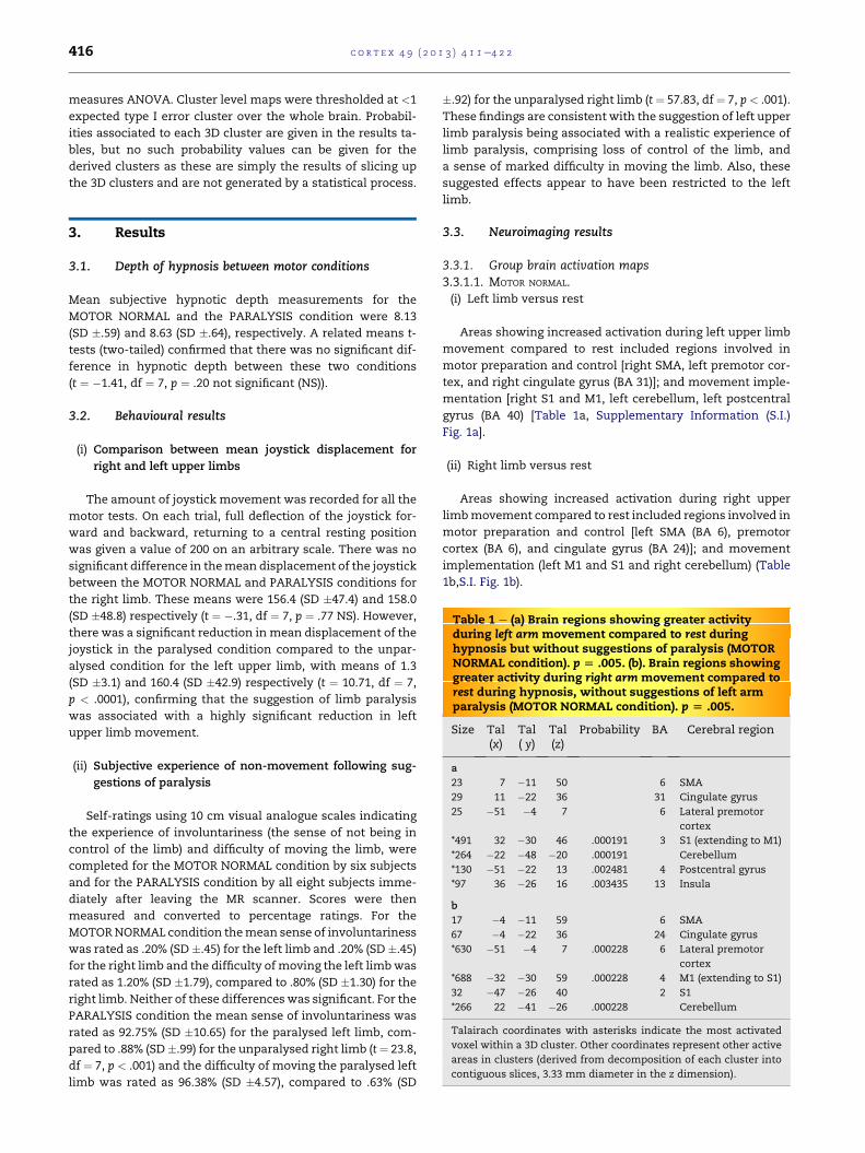
Table 1 e (a) Brain regions showing greater activityduring left arm movement compared to rest duringhypnosis but without suggestions of paralysis (MOTORNORMAL condition). p [ .005. (b). Brain regions showinggreater activity during right arm movement compared torest during hypnosis, without suggestions of left armparalysis (MOTOR NORMAL condition). p [ .005.
Size Tal(x)
Tal( y)
Tal(z)
Probability BA Cerebral region
a
23 7 �11 50 6 SMA
29 11 �22 36 31 Cingulate gyrus
25 �51 �4 7 6 Lateral premotor
cortex
*491 32 �30 46 .000191 3 S1 (extending to M1)
*264 �22 �48 �20 .000191 Cerebellum
*130 �51 �22 13 .002481 4 Postcentral gyrus
*97 36 �26 16 .003435 13 Insula
b
17 �4 �11 59 6 SMA
67 �4 �22 36 24 Cingulate gyrus
*630 �51 �4 7 .000228 6 Lateral premotor
cortex
*688 �32 �30 59 .000228 4 M1 (extending to S1)
32 �47 �26 40 2 S1
*266 22 �41 �26 .000228 Cerebellum
Talairach coordinates with asterisks indicate the most activated
voxel within a 3D cluster. Other coordinates represent other active
areas in clusters (derived from decomposition of each cluster into
contiguous slices, 3.33 mm diameter in the z dimension).
3. Results
3.1. Depth of hypnosis between motor conditions
Mean subjective hypnotic depth measurements for the
MOTOR NORMAL and the PARALYSIS condition were 8.13
(SD �.59) and 8.63 (SD �.64), respectively. A related means t-
tests (two-tailed) confirmed that there was no significant dif-
ference in hypnotic depth between these two conditions
(t ¼ �1.41, df ¼ 7, p ¼ .20 not significant (NS)).
3.2. Behavioural results
(i) Comparison between mean joystick displacement for
right and left upper limbs
The amount of joystick movement was recorded for all the
motor tests. On each trial, full deflection of the joystick for-
ward and backward, returning to a central resting position
was given a value of 200 on an arbitrary scale. There was no
significant difference in themean displacement of the joystick
between the MOTOR NORMAL and PARALYSIS conditions for
the right limb. These means were 156.4 (SD �47.4) and 158.0
(SD �48.8) respectively (t ¼ �.31, df ¼ 7, p ¼ .77 NS). However,
there was a significant reduction in mean displacement of the
joystick in the paralysed condition compared to the unpar-
alysed condition for the left upper limb, with means of 1.3
(SD �3.1) and 160.4 (SD �42.9) respectively (t ¼ 10.71, df ¼ 7,
p < .0001), confirming that the suggestion of limb paralysis
was associated with a highly significant reduction in left
upper limb movement.
(ii) Subjective experience of non-movement following sug-
gestions of paralysis
Self-ratings using 10 cm visual analogue scales indicating
the experience of involuntariness (the sense of not being in
control of the limb) and difficulty of moving the limb, were
completed for the MOTOR NORMAL condition by six subjects
and for the PARALYSIS condition by all eight subjects imme-
diately after leaving the MR scanner. Scores were then
measured and converted to percentage ratings. For the
MOTORNORMAL condition themean sense of involuntariness
was rated as .20% (SD �.45) for the left limb and .20% (SD �.45)
for the right limb and the difficulty of moving the left limbwas
rated as 1.20% (SD �1.79), compared to .80% (SD �1.30) for the
right limb. Neither of these differences was significant. For the
PARALYSIS condition the mean sense of involuntariness was
rated as 92.75% (SD �10.65) for the paralysed left limb, com-
pared to .88% (SD�.99) for the unparalysed right limb (t¼ 23.8,
df ¼ 7, p < .001) and the difficulty of moving the paralysed left
limb was rated as 96.38% (SD �4.57), compared to .63% (SD
�.92) for the unparalysed right limb (t ¼ 57.83, df¼ 7, p < .001).
These findings are consistent with the suggestion of left upper
limb paralysis being associated with a realistic experience of
limb paralysis, comprising loss of control of the limb, and
a sense of marked difficulty in moving the limb. Also, these
suggested effects appear to have been restricted to the left
limb.
3.3. Neuroimaging results
3.3.1. Group brain activation maps3.3.1.1. MOTOR NORMAL.(i) Left limb versus rest
Areas showing increased activation during left upper limb
movement compared to rest included regions involved in
motor preparation and control [right SMA, left premotor cor-
tex, and right cingulate gyrus (BA 31)]; and movement imple-
mentation [right S1 and M1, left cerebellum, left postcentral
gyrus (BA 40) [Table 1a, Supplementary Information (S.I.)
Fig. 1a].
(ii) Right limb versus rest
Areas showing increased activation during right upper
limbmovement compared to rest included regions involved in
motor preparation and control [left SMA (BA 6), premotor
cortex (BA 6), and cingulate gyrus (BA 24)]; and movement
implementation (left M1 and S1 and right cerebellum) (Table
1b,S.I. Fig. 1b).
c o r t e x 4 9 ( 2 0 1 3 ) 4 1 1e4 2 2 417
3.3.1.2. PARALYSIS.(i) Left limb versus rest
During attempted left upper limb movement compared to
rest following a suggestion of left limb paralysis, brain areas
showing increased activation included regions involved in
motor preparation and control [bilateral SMA (BA 6), left pre-
central gyrus (BA 44), and right anterior cingulate gyrus (BA
24)]; and movement implementation (right S1 and M1; and
bilateral cerebellum) (Table 2a, S.I. Fig. 2a). In addition, a single
cluster in left S1 (BA 2) showed a task induced deactivation
(i.e., greater activity at rest compared to during attempted
movement of the left upper limb) (Table 2b).
(ii) Right limb versus rest
Following the suggestion of left limb paralysis brain areas
showing increased activation during right upper limb move-
ment compared to rest included regions involved in motor
preparation and control [left SMA (BA 6)] and cingulate gyrus
(BA 31); and regions involved in movement implementation
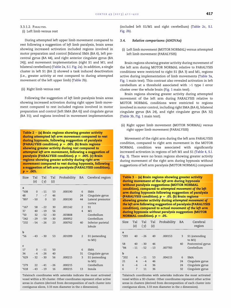
Table 2 e (a) Brain regions showing greater activityduring attempted left arm movement compared to restduring hypnosis, following suggestion of paralysis(PARALYSIS condition). p [ .005. (b) Brain regionsshowing greater activity during rest compared toattempted left arm movement, following a suggestion ofparalysis (PARALYSIS condition). p [ .005. (c) Brainregions showing greater activity during right armmovement compared to rest during hypnosis, followinga suggestion of left arm paralysis (PARALYSIS condition).p [ .005.
Size Tal(x)
Tal( y)
Tal(z)
Probability BA Cerebral region
a
*508 0 �11 53 .000190 6 SMA
48 7 �7 46 24 Cingulate gyrus
*897 �50 0 10 .000190 44 Lateral premotor
cortex
*167 58 �22 30 .001142 2 S1
37 40 �19 56 4 M1
*50 32 �52 �30 .003808 Cerebellum
*242 �29 �59 �30 .000952 Cerebellum
*232 �54 �26 23 .000762 40 Inferior parietal
lobule
b
*56 �43 �30 53 .001099 2 S1 (extending
to M1)
c
17 0 �11 50 6 SMA
34 �4 �26 40 31 Cingulate gyrus
*629 �32 �30 56 .000215 3 S1 (extending
to M1)
*279 22 �41 �26 .000215 Cerebellum
*618 �43 �19 16 .000215 13 Insula
Talairach coordinates with asterisks indicate the most activated
voxel within a 3D cluster. Other coordinates represent other active
areas in clusters (derived from decomposition of each cluster into
contiguous slices, 3.33 mm diameter in the z dimension).
(included left S1/M1 and right cerebellum) (Table 2c, S.I.
Fig. 2b).
3.4. Relative comparisons (ANOVAs)
(i) Left limbmovement (MOTOR NORMAL) versus attempted
left limb movement (PARALYSIS)
Brain regions showing greater activity duringmovement of
the left arm during MOTOR NORMAL relative to PARALYSIS
conditions were restricted to right S1 (BA 3) and M1, regions
active during implementation of limb movements (Table 3a,
Fig. 1 main text). This contrast also revealed activation in left
cerebellum at a threshold associated with >1 type I error
cluster over the whole brain (Fig. 1 main text).
Brain regions showing greater activity during attempted
movement of the left arm during PARALYSIS relative to
MOTOR NORMAL conditions were restricted to regions
involved inmotor control, including right SMA (BA 6), bilateral
cingulate gyrus (BA 24), and right cingulate gyrus (BA 32)
(Table 3b, Fig. 1 main text).
(ii) Right upper limb movement (MOTOR NORMAL) versus
right upper limb movement (PARALYSIS)
Movement of the right arm during the left arm PARALYSIS
condition, compared to right arm movement in the MOTOR
NORMAL condition was associated with significantly
increased activation in regions of left M1 and S1 (Table 4, S.I.
Fig. 3). There were no brain regions showing greater activity
during movement of the right arm during hypnosis without
suggestions of left arm paralysis (MOTOR NORMAL) compared
Table 3 e (a) Brain regions showing greater activityduring movement of the left arm during hypnosiswithout paralysis suggestions (MOTOR NORMALcondition), compared to attempted movement of the leftarm during hypnosis following suggestion of paralysis(PARALYSIS condition). p [ .05. (b) Brain regionsshowing greater activity during attempted movement ofthe left arm following suggestion of paralysis (PARALYSIScondition), compared to actual movement of the left armduring hypnosis without paralysis suggestion (MOTORNORMAL condition). p [ .05.
Size Tal(x)
Tal( y)
Tal(z)
Probability BA Cerebralregion
a
*293 40 �26 49 .000153 3 S1 (extending
to M1)
58 40 �30 46 40 Postcentral gyrus
*84 �11 �52 �13 .007783 Cerebellum
b
*202 4 �11 53 .004115 6 SMA
21 4 �4 46 24 Cingulate gyrus
6 �4 �4 36 24 Cingulate gyrus
6 7 11 36 32 Cingulate gyrus
Talairach coordinates with asterisks indicate the most activated
voxel within a 3D cluster. Other coordinates represent other active
areas in clusters (derived from decomposition of each cluster into
contiguous slices, 3.33 mm diameter in the z dimension).
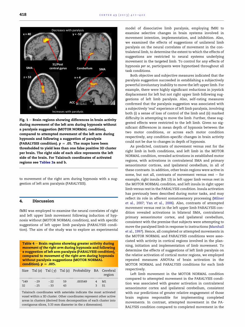
Fig. 1 e Brain regions showing differences in brain activity
during movement of the left arm during hypnosis without
a paralysis suggestion (MOTOR NORMAL condition),
compared to attempted movement of the left arm during
hypnosis and following a suggestion of paralysis
(PARALYSIS condition). p [ .05. The maps have been
thresholded to yield less than one false positive 3D cluster
per brain. The right side of each slice represents the left
side of the brain. For Talairach coordinates of activated
regions see Tables 3a and b.
c o r t e x 4 9 ( 2 0 1 3 ) 4 1 1e4 2 2418
to movement of the right arm during hypnosis with a sug-
gestion of left arm paralysis (PARALYSIS).
4. Discussion
fMRI was employed to examine the neural correlates of right
and left upper limb movement following induction of hyp-
nosis without (MOTOR NORMAL condition), and with specific
suggestions of left upper limb paralysis (PARALYSIS condi-
tion). The aim of the study was to explore an experimental
Table 4 e Brain regions showing greater activity duringmovement of the right arm during hypnosis and followinga suggestion of left arm paralysis (PARALYSIS condition),compared to movement of the right arm during hypnosiswithout paralysis suggestions (MOTOR NORMALcondition). p [ .005.
Size Tal (x) Tal ( y) Tal (z) Probability BA Cerebralregion
*149 �29 �22 59 .003549 4 M1
32 �25 �33 43 3 S1
Talairach coordinates with asterisks indicate the most activated
voxel within a 3D cluster. Other coordinates represent other active
areas in clusters (derived from decomposition of each cluster into
contiguous slices, 3.33 mm diameter in the z dimension).
model of dissociative limb paralysis, employing fMRI to
examine selective changes in brain systems involved in
movement intention, implementation, and inhibition. Also,
we examined the effects of suggestions of unilateral limb
paralysis on the neural correlates of movement in the con-
tralateral limb, to determine the extent to which the effects of
suggestions are restricted to neural systems underlying
movement in the targeted limb. To control for any effects of
hypnosis per se, participants were hypnotised throughout all
task conditions.
Both objective and subjective measures indicated that the
paralysis suggestion succeeded in establishing a subjectively
powerful involuntary inability tomove the left upper limb. For
example, there were highly significant reductions in joystick
displacement for left but not right upper limb following sug-
gestions of left limb paralysis. Also, self-rating measures
confirmed that the paralysis suggestion was associated with
a subjectively ‘real’ experience of left limb paralysis, involving
both (i) a sense of loss of control of the limb and (ii) marked
difficulty in attempting to move the limb. Further, these sug-
gested effects were restricted to the left limb. Given no sig-
nificant differences in mean depth of hypnosis between the
two motor conditions, or across each motor condition
respectively, any condition-specific changes in brain activity
could not be due to changes in depth of hypnosis.
As predicted, contrasts of movement versus rest for the
right limb in both conditions, and left limb in the MOTOR
NORMAL condition, revealed activations in established motor
regions, with activations in contralateral SMA and primary
sensorimotor cortices, and ipsilateral cerebellum, in all of
these contrasts. In addition, other brain regions were active in
some, but not all, contrasts of movement versus rest e for
example, right insula (BA 13) in left upper limb versus rest in
the MOTOR NORMAL condition, and left insula in right upper
limb versus rest in the PARALYSIS condition. Insula activation
has previously been described during motor tasks, and may
reflect its role in afferent somatosensory processing (Milner
et al., 2007; Yan et al., 2006). Also, contrasts of attempted
movement versus rest in the left upper limb PARALYSIS con-
dition revealed activations in bilateral SMA, contralateral
primary sensorimotor cortex, and ipsilateral cerebellum,
consistent with the premise that subjects were attempting to
move the paralysed limb in response to instructions (Marshall
et al., 1997). Hence, all completed or attempted movements in
the MOTOR NORMAL and PARALYSIS conditions were asso-
ciated with activity in cortical regions involved in the plan-
ning, initiation and implementation of limb movement. To
determine the effects of suggestions of left limb paralysis on
the relative activation of cortical motor regions, we employed
repeated measures ANOVAs of brain activation in the
MOTOR NORMAL and PARALYSIS conditions for each limb
respectively.
Left limb movement in the MOTOR NORMAL condition
compared to attempted movement in the PARALYSIS condi-
tion was associated with greater activation in contralateral
sensorimotor cortex and ipsilateral cerebellum, consistent
with our predictions of greater relative engagement of those
brain regions responsible for implementing completed
movements. In contrast, attempted movement in the PA-
RALYSIS condition compared to completed movement in the
c o r t e x 4 9 ( 2 0 1 3 ) 4 1 1e4 2 2 419
MOTOR NORMAL condition was associated with greater acti-
vation in medial premotor cortex (BA 6), with peak activation
in right SMA. SMA is active during motor planning and the
intention to move, and is considered as demonstrating sub-
jects’ attempts to comply with the instructions to move
(Grafton et al., 1992; Penfield and Welch, 1951; Picard and
Strick, 2001).
In summary, the present study provided compelling evi-
dence that following paralysis suggestions, where subjects
demonstrated significant reductions in left upper limb
movement, and subjectively reported loss of control over and
marked difficulty in moving the left upper limb, there was
evidence of significantly increased activation in brain regions
involved in motor intention and planning. These findings
raise the intriguing question of what neural systems could
provide for the experience of involuntary, selective inhibition
of the execution of an intended limb movement.
The present study showed increased activation in bilateral
regions of the ACC (especially BA 24) during the left upper limb
PARALYSIS compared to NORMAL conditions. ACC appears to
be a key motor control region, with strong connectivity to
premotor, SMA, and primary motor cortices (Botvinick et al.,
2004). ACC has been shown to be active under conditions of
conflict detection, response selection and inhibition in a vari-
ety of tasks, and is strongly activated when overriding pre-
potent motor responses (Botvinick et al., 2004). In the present
experiment, attempted left upper limb movement in the PA-
RALYSIS relative to NORMAL conditions was associated with
activation of an anterior rostral subregion of ACC (BA 32, x¼ 7,
y ¼ 11, z ¼ 36), engaged during conflict monitoring and
response selection; and a posterior rostral subregion of ACC
(BA 24, x¼ 4, y¼�4, z¼ 46), engaged during response selection
and inhibition (Picard and Strick, 2001).
One conclusion from the present findings is that the ACC
reversibly mediates selective inhibition (countermanding or
overriding) of intended movements in response to targeted
suggestions, and that this inhibition is experienced as invol-
untary. In other words it is possible that the ACC contributes
to inhibition of prepotent motor responses in both cases of
voluntary and involuntary inhibition, but does not itself
mediate the key subjective sense of involuntariness. Future
studies employing larger samples could correlate self-ratings
of perceived loss of control with ACC activation during the
attempt to move a limb following suggestions of limb paral-
ysis to test the extent to which ACC activity is associated with
perceived involuntariness.
The current findings extend prior PET findings of increased
SMA (Ward et al., 2003) and anterior cingulate activation
(Halligan et al., 2000) in associationwith attemptedmovement
in suggested limb paralysis. However, in contrast to previous
studies involving suggested lower limb paralysis (Halligan
et al., 2000; Ward et al., 2003), the present study did not find
evidence of increased OFC activity during attempted move-
ment of the paralysed limb. Lack of OFC activation could
however reflect the common problem of signal dropout in
fMRI of basal brain regions (Schmidt et al., 2005), which we
endeavoured to minimise by collecting the thinnest possible
slices. Alternatively, given differences in experimental con-
ditions and contrasts, and inconsistencies in the location of
OFC activation in prior PET studies of attempted movement
following suggested limb paralysis (Halligan et al., 2000; Ward
et al., 2003), the present findings may indicate that the ACC
constitutes a key cortical region involved in implementing the
selective inhibition of movements following targeted motor
suggestions (Botvinick et al., 2004). The current findings are
also not inconsistent with imaging literature on conversion
disorder, where increased cingulate cortex activity is reliably
found to be associated with functional movement disorder
(van Beilen et al., 2010; Vogt et al., 2000). In this view of the
regulatory role of ACC in suggested limb paralysis, SMA acti-
vation is associated with preparation to implement the pre-
potent motor response following instructions to move the
limb, which is then overridden by higher level control sys-
tems, with consequent reduced activation of structures
involved in motor implementation [sensorimotor cortex (M1
and S1) and cerebellum].
Alternatively, some studies indicate that SMA/pre-SMA
may also be involved in motor inhibition (such as STOP
tasks) (Isoda and Hikosaka, 2007; Sharp et al., 2010). Hence it is
possible that both SMA and ACC may be contributing to the
perceived loss of control of and difficulty in moving the left
limb during the PARALYSIS condition. Nevertheless, a net
contribution of SMA activity to preparation and intention to
move rather than inhibition alone in the present study would
help explain subjects’ reports of attempts to move (despite
perceived loss of limb control and difficulty inmoving). Future
studies using suggestions, fMRI and phenomenological mea-
sures which separate ‘intention’ and ‘movement’ phases that
vary both (i) the degree of intention to move (Blakemore et al.,
2003; Haggard et al., 2004) and (ii) the extent of difficulty (de-
gree of paralysis) could potentially help clarify the contribu-
tions of SMA to movement intention and inhibition
respectively during suggested limb paralysis.
Our findings of increased activation in SMA and ACC for
the left upper limb PARALYSIS versus NORMAL contrast were
accompanied by a trend to significant increases in bilateral
precentral gyrus (BA 44) extending to IFG (BA 44) on the right
side (i.e., the clusters were not present using the chosen level
of <1 expected type 1 error cluster). We note that significant
increases in left IFG activity were reported in the most com-
parable contrast of a recent study (Cojan et al., 2009b), which
also reported increased activation in left SFG. Both IFG and
SFG (and indeed precentral gyri (BA 44)) have previously been
reported to be involved in motor planning, control, and inhi-
bition (Hanakawa et al., 2008; Levy and Wagner, 2011;
Rushworth et al., 2004). With respect to the present study, our
findings of statistically significant increases in ACC and SMA
activity in the PARALYSIS compared to LEFTMOTOR condition
suggest that differential activity in these brain regions crit-
ically contributes to the phenomenon of suggested paralysis
relative to other executive and motor control regions. Never-
theless, the trend to significance of right IFG (and indeed
bilateral precentral gyrus) activity we report e if due to a type
II error e represents additional components of the motor
control network that may also contribute to inhibition of
movement in keeping with the general proposal that sugges-
tions of paralysis operate via their effects on ‘executive’ re-
gions (Bell et al., 2011; Oakley, 1999a). Differences in relative
activation of motor control regions within and between
studiesmay be attributable to differences in the task demands
c o r t e x 4 9 ( 2 0 1 3 ) 4 1 1e4 2 2420
and response sets required bearing in mind that the neural
substrates of motor inhibition are known to vary with the
experimental task employed (Simmonds et al., 2008). For
example, employing a goeno go task with suggestions of
unilateral limb paralysis in an event related design (Cojan
et al., 2009b) is likely to require the maintenance of a more
complex instructional and response set in working memory
than the block design involving ‘MOVE’ or ‘REST’ epochs of the
present study. Hence, the differences in regional brain acti-
vation between the present study and that of the study
employing a goeno go task (Cojan et al., 2009b) raise the
possibility that as with voluntary movement, the neural cor-
relates of suggested involuntary inhibition may vary
depending on the context or task demands in which a prepo-
tent motor response has to be inhibited (Simmonds et al.,
2008). If so, this would raise the question of what brain sys-
tems represent a suggested inability to move in a way that
modulates different inhibitory systems in accordance with
various experimental or contextual demands. This question
can be addressed in future studies by measuring the neural
correlates of both voluntary and involuntary inhibition in
highly hypnotisable subjects across a variety of tasks requir-
ing motor and non-motor inhibition.
Behavioural results did not show any significant reductions
in movement of the right limb following suggestions of left
limb paralysis (PARALYSIS) compared to MOTOR NORMAL.
Similarly, volunteers did not report any significant differences
in the subjective difficulty, or feelings of control, whenmoving
the right limb following suggestions of left limb paralysis.
Nevertheless, contrary to the prior hypothesis fMRI revealed
differences in brain activation associated with movement
between the conditions, with greater activation of left M1 and
S1 in the PARALYSIS compared to NORMAL condition for the
right arm. Influences of non-primary motor areas on M1 ac-
tivity have been demonstrated by magnetic stimulation
studies (Reis et al., 2008), including facilitation of M1 activa-
tion by repetitive transcranial magnetic stimulation (TMS) of
ipsilateral SMA (Matsunaga et al., 2005). In the PARALYSIS
block, significantly increased bilateral SMA activity was evi-
dent in the PARALYSIS versus NORMAL contrast for the left
but not right limb. It is not known if increased task-related
SMA activity during the left limb epochs of the PARALYSIS
block could have had a net facilitatory effect onM1 activity for
right limb movement during the PARALYSIS relative to NOR-
MAL blocks. While this specific hypothesis requires further
investigation, the broader point remains that suggestions of
unilateral limb paralysis may also affect neural systems
associated with movement in the contralateral limb. Hence, it
cannot be assumed that the effects of suggestions are fully
restricted to the targeted cognitive or sensorimotor domain,
highlighting the importance of introducing appropriate con-
trol conditions when investigating the functional anatomy of
suggested phenomena. Similarly, the functional anatomy of
movement in ostensibly unaffected limbs in dissociative dis-
orders may also be altered by disturbance of function in the
contralateral limb.
In conclusion, the present study combining objective and
subjective measures of movement in conjunction with mea-
surements of brain activity revealed the neural correlates of
involuntary inhibition of what would otherwise be perceived
as voluntary movements following the administration of
suggestions of limb paralysis. Greater SMA activation was
observed in PARALYSIS relative to NORMAL conditions in
conjunction with reports of attempts to move the paralysed
limb e a finding consistent with the role of SMA in motor
intention and planning. ACC (BA 24) was also significantly
more active in PARALYSIS relative to NORMAL conditions e
suggesting that ACC (BA 24) may be implicated in involuntary,
as well as voluntary inhibition of prepotent motor responses.
The findings also revealed how targeted suggestions can be
associated with alterations in brain activity in contralateral
sensorimotor domains, underlining the importance of con-
trolled experimental design when investigating the neural
effects of suggested phenomena. The generalisability of these
findings and respective contributions of SMA, ACC and other
executive regions such as IFG and SFG to involuntary inhibi-
tion should be assessed in future studies by measuring the
neural correlates of both voluntary and involuntary inhibition
in highly hypnotisable subjects across a variety of tasks
requiring motor and non-motor inhibition.
Acknowledgements
We gratefully acknowledge the financial support of the Psy-
chiatry Research Trust and Panacea Society and the assis-
tance of our volunteers.
Supplementary data
Supplementary data related to this article can be found at
http://dx.doi.org/10.1016/j.cortex.2012.09.016.
r e f e r e n c e s
Akagi H and House A. The epidemiology of conversion hysteria.In Halligan PW, Bass C, and Marshall JC (Eds), ContemporaryApproaches to the Study of Hysteria: Clinical and TheoreticalPerspectives. Oxford: Oxford University Press, 2001: 73e101.
Athwal BS, Halligan PW, Fink GR, Marshall JC, andFrackowiak RSJ. Imaging hysterical paralysis. In Halligan PW,Bass C, and Marshall JC (Eds), Contemporary Approach to theStudy of Hysteria: Clinical and Theoretical Perspectives. Oxford,UK: Oxford University Press, 2001: 216e234.
Bell V, Oakley DA, Halligan PW, and Deeley Q. Dissociation inhysteria and hypnosis: Evidence from cognitive neuroscience.Journal of Neurology, Neurosurgery and Psychiatry, 82(3): 332e339,2011.
Blakemore SJ, Oakley DA, and Frith CD. Delusions of alien controlin the normal brain. Neuropsychologia, 41(8): 1058e1067, 2003.
Bliss EL. Hysteria and hypnosis. Journal of Nervous and MentalDisease, 172: 203e206, 1984.
Botvinick MM, Cohen JD, and Carter CS. Conflict monitoring andanterior cingulate cortex: An update. Trends in CognitiveSciences, 8(12): 539e546, 2004.
Brammer MJ, Bullmore ET, Simmons A, Williams SCR, Grasby PM,Howard RJ, et al. Generic brain activation mapping infunctional magnetic resonance imaging: A nonparametricapproach. Magnetic Resonance Imaging, 15: 763e770, 1997.
c o r t e x 4 9 ( 2 0 1 3 ) 4 1 1e4 2 2 421
Bullmore E, Brammer M, Rabe-Hesketh S, Curtis V, Morris R,Williams S, et al. Methods for the diagnosis and treatment ofstimulus correlated motion in generic brain activation studiesusing fMRI. Human Brain Mapping, 7: 38e48, 1999a.
Bullmore E, Long C, Suckling J, Fadili J, Calvert G, Zelaya F, et al.Colored noise and computational inference inneurophysiological (fMRI) time series analysis: Resamplingmethods in time and wavelet domains. Human Brain Mapping,12(2): 61e78, 2001.
Bullmore E, Suckling J, Overmeyer S, Rabe-Hesketh S, Taylor E,and Brammer MJ. Global, voxel, and cluster tests, by theoryand permutation, for a difference between two groups ofstructural MR images of the brain. IEEE Transactions on MedicalImaging, 18(1): 32e42, 1999b.
Carson AJ, Ringbauer B, MacKenzie L, Warlow C, and Sharpe M.Neurological disease, emotional disorder, and disability: Theyare related: A study of 300 consecutive new referrals toa neurology outpatient department. Journal of Neurology,Neurosurgery and Psychiatry, 68(2): 202e206, 2000.
Charcot JM. Clinical Lectures on Certain Diseases of the NervousSystem. Detroit: Davis, 1888 (original work published in 1877).
Cojan Y, Waber L, Carruzzo A, and Vuilleumier P. Motor inhibitionin hysterical conversion paralysis. NeuroImage, 47(3):1026e1037, 2009a.
Cojan Y, Waber L, Schwartz S, Rossier L, Forster A, andVuilleumier P. The brain under self-control: Modulation ofinhibitory and monitoring cortical networks during hypnoticparalysis. Neuron, 62(6): 862e875, 2009b.
Fink GR, Halligan PW, and Marshal JC. Neuroimaging of hysteria.In Hallett M, Fahn S, Jankovic J, et al. (Eds), PsychogenicMovement Disorders: Neurology and Psychiatry. London:Lippincott Williams & Wilkins/Wolters Kluwer Health, 2006.
Friman O, Borga M, Lundberg P, and Knutsson H. Adaptiveanalysis of fMRI data. NeuroImage, 19(3): 837e845, 2003.
Grafton ST, Mazziotta JC, Woods RP, and Phelps ME. Humanfunctional anatomy of visually guided finger movements.Brain, 115(Pt 2): 565e587, 1992.
Gruzelier JH. A working model of the neurophysiology ofhypnosis: A review of the evidence. Contemporary Hypnosis, 15:3e21, 1998.
Haggard P, Cartledge P, Dafydd M, and Oakley DA. Anomalouscontrol: When ‘free-will’ is not conscious. Consciousness &Cognition, 13(3): 646e654, 2004.
Halligan PW, Athwal BS, Oakley DA, and Frackowiak RS. Imaginghypnotic paralysis: Implications for conversion hysteria.Lancet, 355(9208): 986e987, 2000.
Hanakawa T, Dimyan MA, and Hallett M. Motor planning,imagery, and execution in the distributed motor network: Atime-course study with functional MRI. Cerebral Cortex, 18(12):2775e2788, 2008.
Heap M and Aravind KK. Hartland’s Medical and Dental Hypnosis.4th ed. Edinburgh: Churchill Livingstone, 2002.
Isoda M and Hikosaka O. Switching from automatic to controlledaction by monkey medial frontal cortex. Nature Neuroscience,10(2): 240e248, 2007.
Janet P. TheMajor Symptoms of Hysteria. NewYork:Macmillan, 1907.Kihlstrom JS. One hundred years of hysteria. In Lynn SJ and
Rhue JW (Eds), Dissociation: Clinical and Theoretical Perspectives.New York: Guildford, 1994: 365e394.
Levy BJ and Wagner AD. Cognitive control and right ventrolateralprefrontal cortex: Reflexive reorienting, motor inhibition, andaction updating. Annals of the New York Academy of Sciences,1224(1): 40e62, 2011.
Marshall JC, Halligan PW, Fink GR, Wade DT, and Frackowiak RS.The functional anatomy of a hysterical paralysis. Cognition,64(1): B1eB8, 1997.
Matsunaga K, Maruyama A, Fujiwara T, Nakanishi R, Tsuji S, andRothwell JC. Increased corticospinal excitability after 5 Hz
rTMS over the human supplementary motor area. The Journalof Physiology, 562(1): 295e306, 2005.
Mayka MA, Corcos DM, Leurgans SE, and Vaillancourt DE. Three-dimensional locations and boundaries of motor and premotorcortices as defined by functional brain imaging: A meta-analysis. NeuroImage, 31(4): 1453e1474, 2006.
Milner TE, Franklin DW, Imamizu H, and Kawato M. Centralcontrol of grasp: Manipulation of objects with complex andsimple dynamics. NeuroImage, 36(2): 388e395, 2007.
Oakley D. Hypnosis and consciousness: A structural model.Contemporary Hypnosis, 16: 215e223, 1999a.
Oakley DA. Hypnosis and conversion hysteria: A unifying model.Cognitive Neuropsychiatry, 4(3): 243e265, 1999b.
Oakley DA. Hypnosis and suggestion in the treatment of hysteria.In Halligan PW, Bass C, and Marshal JC (Eds), ContemporaryApproaches to the Study of Hysteria. Oxford: Oxford UniversityPress, 2001: 312e329.
Oakley DA, Deeley Q, and Halligan PW. Hypnotic depth andresponse to suggestion under standardized conditions andduring FMRI scanning. International Journal of Clinical &Experimental Hypnosis, 55(1): 32e58, 2007.
Oakley DA and Halligan PW. Hypnotic suggestion and cognitiveneuroscience. Trends in Cognitive Sciences, 13(6): 264e270, 2009.
Penfield W and Welch K. The supplementary motor area of thecerebral cortex; A clinical and experimental study. AmericanMedical Association Archives of Neurology & Psychiatry, 66(3):289e317, 1951.
Picard N and Strick PL. Imaging the premotor areas. CurrentOpinion in Neurobiology, 11: 663e672, 2001.
Pyka M, Burgmer M, Lenzen T, Pioch R, Dannlowski U,Pfleiderer B, et al. Brain correlates of hypnotic paralysis e Aresting-state fMRI study. NeuroImage, 56(4): 2173e2182, 2011.
Reis J, Swayne OB, Vandermeeren Y, Camus M, Dimyan MA,Harris-Love M, et al. Contribution of transcranial magneticstimulation to the understanding of cortical mechanismsinvolved in motor control. The Journal of Physiology, 586(2):325e351, 2008.
Rushworth MFS, Walton ME, Kennerley SW, and Bannerman DM.Action sets and decisions in the medial frontal cortex. Trendsin Cognitive Sciences, 8(9): 410e417, 2004.
Schmidt CF, Boesiger P, and Ishai A. Comparison of fMRIactivation as measured with gradient- and spin-echo EPIduring visual perception. NeuroImage, 26(3): 852e859, 2005.
Sharp DJ, Bonnelle V, De Boissezon X, Beckmann CF, James SG,Patel MC, et al. Distinct frontal systems for responseinhibition, attentional capture, and error processing.Proceedings of the National Academy of Sciences of the United Statesof America, 107(13): 6106e6111, 2010.
Shor RE and Orne EC. Harvard Group Scale of Hypnotic Susceptibility,Form A. Palo Alto: Consulting Psychologists Press, 1962.
Simmonds DJ, Pekar JJ, and Mostofsky SH. Meta-analysis of go/no-go tasks demonstrating that fMRI activation associated withresponse inhibition is task-dependent. Neuropsychologia, 46(1):224e232, 2008.
Spence SA, Crimlisk HL, Cope H, Ron MA, and Grasby PM. Discreteneurophysiological correlates in prefrontal cortex duringhysterical and feigned disorder of movement. The Lancet,355(9211): 1243e1244, 2000.
Stone J, Sharpe M, Rothwell PM, and Warlow CP. The 12 yearprognosis of unilateral functional weakness and sensorydisturbance. Journal of Neurology, Neurosurgery and Psychiatry,74(5): 591e596, 2003.
Stone J and Zeman A. Hysterical conversion: The view fromclinical neurology. In Halligan PW, Bass C, and Marshall JC(Eds), Contemporary Approaches to the Study of Hysteria. Oxford:Oxford University Press, 2001.
Talairach J and Tournoux P. Co-planar Stereotaxic Atlas of theHuman Brain. Stuttgart: Thieme, 1988.

c o r t e x 4 9 ( 2 0 1 3 ) 4 1 1e4 2 2422
Tart CT. Self-report scales of hypnotic depth. International Journalof Clinical and Experimental Hypnosis, 18: 105e125, 1970.
Thirion B, Pinel P, Meriaux S, Roche A, Dehaene S, andPoline JB. Analysis of a large fMRI cohort: Statistical andmethodological issues for group analyses. NeuroImage, 35(1):105e120, 2007.
van Beilen M, Vogt BA, and Leenders KL. Increased activationin cingulate cortex in conversion disorder: What does itmean? Journal of the Neurological Sciences, 289(1e2): 155e158,2010.
Vogt BA, Absher JR, and Bush G. Human retrosplenial cortex:Where is it and is it involved in emotion? Trends inNeurosciences, 23(5): 195e196, 2000.
Ward NS, Oakley DA, Frackowiak RS, and Halligan PW.Differential brain activations during intentionally simulatedand subjectively experienced paralysis. CognitiveNeuropsychiatry, 8(4): 295e312, 2003.
Yan L, Wu D, Wang X, Zhou Z, Liu Y, Yao S, et al. Intratask andintertask asymmetry analysis of motor function. NeuroReport,17(11): 1143e1147, 2006.