Embed Size (px)
Citation preview
REVIEW ARTICLEpublished: 03 April 2012
doi: 10.3389/fneur.2012.00042
The frog vestibular system as a model for lesion-inducedplasticity: basic neural principles and implications forposture controlFrançois M. Lambert 1 and Hans Straka2*
1 Department of Physiology, University of Oslo, Oslo, Norway2 Biocenter-Martinsried, Department Biology II, Ludwig-Maximilians-University Munich, Planegg, Germany
Edited by:
Kenna Peusner, George WashingtonUniversity, USA
Reviewed by:
Kenna Peusner, George WashingtonUniversity, USAIan S. Curthoys, University of Sydney,Australia
*Correspondence:
Hans Straka, Biocenter-Martinsried,Department Biology II,Ludwig-Maximilians-UniversityMunich, Grosshaderner Strasse 2,82152 Planegg, Germany.e-mail: [email protected]
Studies of behavioral consequences after unilateral labyrinthectomy have a long traditionin the quest of determining rules and limitations of the central nervous system (CNS) toexert plastic changes that assist the recuperation from the loss of sensory inputs. Frogswere among the first animal models to illustrate general principles of regenerative capacityand reorganizational neural flexibility after a vestibular lesion. The continuous successfuluse of the latter animals is in part based on the easy access and identifiability of nervebranches to inner ear organs for surgical intervention, the possibility to employ whole brainpreparations for in vitro studies and the limited degree of freedom of postural reflexes forquantification of behavioral impairments and subsequent improvements. Major discoveriesthat increased the knowledge of post-lesional reactive mechanisms in the CNS include alter-ations in vestibular commissural signal processing and activation of cooperative changes inexcitatory and inhibitory inputs to disfacilitated neurons. Moreover, the observed increaseof synaptic efficacy in propriospinal circuits illustrates the importance of limb propriocep-tive inputs for postural recovery. Accumulated evidence suggests that the lesion-inducedneural plasticity is not a goal-directed process that aims toward a meaningful restorationof vestibular reflexes but rather attempts a survival of those neurons that have lost theirexcitatory inputs. Accordingly, the reaction mechanism causes an improvement of somecomponents but also a deterioration of other aspects as seen by spatio-temporally inap-propriate vestibulo-motor responses, similar to the consequences of plasticity processesin various sensory systems and species. The generality of the findings indicate that frogscontinue to form a highly amenable vertebrate model system for exploring molecular andphysiological events during cellular and network reorganization after a loss of vestibularfunction.
Keywords: utricle, vestibular, spinal cord, skeletal deformation, proprioception, semicircular canals, posture,
scoliosis
INTRODUCTIONBilateral vestibular afferent signals from semicircular canal andotolith organs are essential for the stabilization of gaze, controlof posture and locomotion, as well as for cognitive aspects ofbalance, self-orientation, spatial navigation, and vegetative home-ostasis (Olabi et al., 2009; Dutia, 2010; Smith et al., 2010). Damageto the inner ear or to the vestibular nerve, through accident,disease, or intended experimental manipulations results in a com-plex syndrome of static (in the absence of body motion) anddynamic (during body motion) ocular motor, postural and cog-nitive deficits. Initial static deficits such as the postural asymme-try and spontaneous nystagmus after unilateral labyrinthectomy(UL) disappear over time. This behavioral recovery is generallyassumed to represent the consequence of a process called “vestibu-lar compensation” that differs in its extent and time course fordifferent symptoms as well as for different vertebrate species assummarized by a large number of classical papers (e.g., Smithand Curthoys, 1989; Dieringer, 1995; Vibert et al., 1997, 1999;
Curthoys and Halmagyi, 1999; Curthoys, 2000; Darlington et al.,2002) as well as by more recent reviews (e.g., Cullen et al., 2009;Olabi et al., 2009; Shao et al., 2009; Dutia, 2010; Smith et al.,2010). Since the vestibular epithelium does not regenerate after aperipheral lesion and removal of the endorgans in postembryonicanimals, the subsequent progressive disappearance of the initialbehavioral symptoms is attributed to plasticity processes in thecentral nervous system (CNS) including various areas such asthe vestibular nuclei, cerebellum, spinal cord, and cortical struc-tures (Dutia, 2010). In fact, results from an increasing numberof studies suggest that different forms of neural plasticity occursimultaneously as well as consecutively at multiple sites in thebrain and spinal cord, compatible with the notion of “vestibu-lar compensation” as a multiple, distributed process (Llinás andWalton, 1979).
The unilateral loss of vestibular sensory inputs causes animbalanced activity along central pathways and in circuits thatare involved in vestibular signal processing. As a consequence,
www.frontiersin.org April 2012 | Volume 3 | Article 42 | 1

Lambert and Straka Vestibular plasticity in frog
this imbalance triggers objective behavioral symptoms, such asspontaneous nystagmus, postural asymmetry, and gaze instabil-ity but also subjective illusions such as vertigo (Dutia, 2010). Theasymmetric posture is one of the most prominent static deficitsand its time course of recovery is often correlated with the pro-gression and extent of CNS plasticity (Dieringer, 1995; Curthoys,2000). In contrast to dynamic deficits such as direction-specificimpaired vestibulo-ocular reflexes (VOR), which show only poorrecovery, static deficits, including asymmetric limb and body posi-tions, recover almost entirely and only reappear in chronic animalsunder certain conditions (Dieringer, 1995, 2003).
The removal of particular vestibular endorgans causes posturaldeficits that correlate with the loss of one or more semicircularcanal or otolith organs, respectively (Dieringer, 1995). Dependingon the absence of specific combinations of endorgans, differentstatic and dynamic deficits predominate. The various pathologiesare caused by the combinatorial accumulation of direct effects fol-lowing the loss of particular labyrinthine organs and secondaryeffects that are triggered by the postural asymmetry. The resultingdeficits after a complete UL are similar in all tetrapods studied sofar (Dieringer, 1995). However, some species-specific variations inthe extent of the deficiencies exist and are likely correlated withparticular skeletal configurations that mainly concern the lengthand curvature of the neck and the articulation of the head (Vidalet al., 1998).
Since the experiments by Precht et al. (1966) in cat, manystudies attempted to explain and link changes at cellular andnetwork levels with improvements of the behavioral symptomsafter a peripheral vestibular lesion. One of the key ideas has beenthe notion of “vestibular compensation” as a process that con-sequently leads to a recovery of the initial behavioral deficits.However, this view of a goal-directed process is contradicted bythe fact that a number of symptoms, in particular deficits in thedynamic components of vestibular reflexes, show no recovery, evenmany years after the lesion (Hamann et al., 1998). Moreover, theobserved improvement of some of the initial deficits is no proofthat mechanisms are at work that aim toward a restoration ofnormal function by a driven readjustment of cellular and net-work properties through specific sensory or intrinsic substitutionprocesses (Dieringer, 2003).
Most plastic neural changes were for obvious reasons observedin the bilateral central vestibular nuclei and include altered intrin-sic membrane and discharge properties of second-order vestibularneurons (2˚VN; Beraneck et al., 2003, 2004; Shao et al., 2009), dif-ferential modifications of GABAB receptor-mediated vestibularcommissural inhibition (Bergquist et al., 2008), increased efficacyof NMDA receptor-mediated components in ipsilesional 2˚VN(Knöpfel, 1987; see Smith and Darlington, 1997) and neurogenesisin the ipsilesional vestibular nucleus (Tighilet et al., 2007). Thesemostly molecular and cellular changes are supplemented by a moreglobal process of what is called sensory substitution by other bodymotion-related signals such as visual or proprioceptive signals (seeBrandt et al., 1997; Curthoys, 2000; Darlington and Smith, 2000) aswell as by contributions that depend on intact cerebellar circuitries(Kitahara et al., 1997; Cullen et al., 2009; Dutia, 2010).
The number of reported post-lesional alterations at molecular,cellular, network, and behavioral levels is paralleled by an equally
large spectrum of employed vertebrate models that ranges fromfish to mammals (Dieringer, 1995). Instead of adding another gen-eral review on plasticity processes in the vertebrate CNS after UL tothe multitude of existing comprehensive publications, this reviewwill mainly focus on general vertebrate principles of vestibularlesion-induced behavioral and neural plasticity discovered in frogand aims to extend the classical review by Dieringer (1995) byemphasizing more recent findings and adding new interpretationsof existing data.
Frogs were among the first species in which the consequencesof a unilateral labyrinthine lesion (Goltz, 1870; Ewald, 1892; Lau-denbach, 1899) or pharmacological elimination of labyrinthinefunction (Thauer and Peters, 1935) have been studied and a num-ber of basic concepts of post-lesional plasticity in the vestibularsystem of vertebrates have been described in this animal model(e.g., Dieringer and Precht, 1977; Straka and Dieringer, 1995; Gotoet al., 2000). The discovery of these general reaction principles wasfacilitated by a number of advantages of frogs, such as the easyaccess to bilateral vestibular endorgans for experimental manip-ulations (Figures 1A,B; Goto et al., 2002), the well-determinedspatially specific convergence of afferent semicircular canal andotolith inputs in individual 2˚VN (Straka et al., 2002) or the pos-sibility to use an isolated whole brain preparation for probingcellular and network physiology in vitro (Cochran et al., 1987). Therelatively small volume of ∼6 mm × 2 mm × 1.5 mm of the entirebrainstem along with the possibility to work at a temperature of14–16˚C in these poikilothermic animals allows a utilization ofthe latter preparation for electrophysiological recordings withoutnoticeable deterioration of up to 7 days (see Straka and Dieringer,2004). Even though, a distinction of the vestibular nucleus intothe different classical subnuclei on the basis of cytoarchitecturalfeatures is difficult in frog, it is possible to correlate stereotac-tically defined positions of recorded central vestibular neuronswith the intrinsic hindbrain segmental organization (Straka et al.,2003) and thus with the rhombomeric framework in these ani-mals (Straka et al., 2001). With respect to the determination ofplasticity events, this allows a sampling of larger numbers of neu-rons throughout the entire vestibular nuclei with reference tofixed external landmarks in the attempt to obtain a representa-tive overview of potential changes after UL that can be assignedto particular functional subgroups. The combination of all theseadvantages forms an excellent basis for the use of frogs as a modelto study questions related to post-lesional vestibular plasticity. Inthe following, particular aspects of behavioral and neural changesafter UL will be highlighted by illustrating several key resultsthat allowed deducing basic patterns and conceptual principlesthat also govern the behavioral recovery from a vestibular loss ordysfunction in other vertebrate species, including humans.
ANATOMICAL ARRANGEMENT OF PERIPHERALLABYRINTHINE ENDORGANS AND CONSEQUENCES OFSURGICAL MANIPULATIONSAmphibian labyrinthine endorgans consist of three semicircularcanals, three macula organs (utricle, lagena, and saccule), and twopapillary organs (basilar papilla and amphibian papilla) on eachside (for review see Straka and Dieringer, 2004). At variance withmammals, these sensory organs are not located in a bony labyrinth
Frontiers in Neurology | Neuro-otology April 2012 | Volume 3 | Article 42 | 2
Lambert and Straka Vestibular plasticity in frog
FIGURE 1 | Morphological organization and afferent innervation of
labyrinthine endorgans in amphibians. (A) Top view of the otic capsulewith semicircular canal and otolith organs of a stage 53 Xenopus laevistadpole. (B) Color-coded schematic drawing of the VIIIth nerve, ganglion ofScarpa (blue), separation into an anterior (ant ramus, red) and posterior(post ramus, green) branch, and innervations of individual labyrinthineendorgans in a ranid frog. HC, PC, AC, horizontal, posterior, and anteriorvertical canal; HCn, HCc, PCn PCc, ACn, ACc, nerve branches (n)innervating the cupula (c) of the semicircular canal organs, respectively; BP,basal papilla; AP, amphibian papilla; SA, saccule; UT, utricle; HB, hindbrain;ant/post ramus, anterior/posterior VIIIth nerve branch; modified andadapted from Goto et al. (2002).
but in a relatively spacious cartilaginous bulla with all organsplainly visible under the microscope (Figure 1A). This anatom-ical particularity is an enormous advantage for all experimentalmanipulations and facilitates the transection of specific nervebranches and/or the removal of individual endorgans under directvisual control. As in all other vertebrates, labyrinthine organs areinnervated by afferent fibers of the VIIIth nerve, which, at the levelof Scarpa’s ganglion, form an anterior (ramus anterior) and a pos-terior branch (ramus posterior) before entering the otic capsule(Figure 1B; de Burlet, 1929). The anterior branch innervates thehorizontal and the anterior vertical semicircular canal, the utricleand the saccule, while the posterior branch innervates the poste-rior vertical semicircular canal, the lagena, as well as the basilarand amphibian papillary organ (Figure 1B), with the latter two
organs being sensitive to air-borne sound (Lewis and Li, 1975).With the exception of the saccular innervation by fibers in theanterior branch as well as by additional variable contributionsof afferent fibers in the posterior branch in some species (e.g.,Xenopus laevis), this organizational scheme essentially applies toall amphibians (de Burlet, 1929). In frog, the largest numbers ofafferent fibers innervate the utricle (∼4000), whereas fewer fiberssupply the lagena (∼2000), the semicircular canals or the saccule(each up to 1500), respectively (Dunn,1978; Honrubia et al., 1989).
The three semicircular canals on one side in frogs are orientedapproximately perpendicular with respect to each other and formfunctional pairs with their respective coplanar partner canal onthe other side (Blanks and Precht, 1976). Morphometric detailsof frog semicircular canals are similar to those of other verte-brates, although some differences in duct diameter, curvature, anddimensions of the ampullar cristae exist (Ramprashad et al., 1986).The utricle is largely oriented in the plane of the horizontal semi-circular canals (Rohregger and Dieringer, 2002) and sensitive tolinear acceleration and changes of the head position with respectto the earth gravitational vector (Blanks and Precht, 1976). Asa complement, the two other amphibian otolith organs are ori-ented approximately vertically. While a clear graviceptive functionis demonstrated for the lagena (Caston et al., 1977), a sensitivityof the saccule for changes in head position has been reported insome studies (Gallé and Clemens, 1973; Lannou and Cazin, 1976)but not in others (Ashcroft and Hallpike, 1934).
At variance with this inconclusive role of the saccule in detect-ing static and/or dynamic body position in space, there is clearevidence that this latter otolith organ is highly sensitive to substratevibration (Koyama et al., 1982) and for a central connection withmidbrain auditory centers (see Straka and Dieringer, 2004). Sucha sensitivity to vibrational stimuli, however, is not restricted to thesaccule but has been reported to varying degrees for lagena (Cor-topassi and Lewis, 1996; Harada et al., 2001) and utricular afferentfibers (Jørgensen and Christensen-Dalsgaard, 1991) and is likelydue to the presence of particular hair cell types along the striola(Baird and Lewis, 1986). Thus, all three otolith organs might havea dual function in frog, even though to differential extents. Whilethe utricle and the lagena mainly serve as gravitoinertia-sensitiveorgans, the saccule is a highly sensitive detector for substrate vibra-tion. This differential organization with respect to their sensorysensitivity and their major functional role is important, wheninterpreting potential contributions of the lagena and the sacculeto deficits in vestibulo-motor responses after an ablation.
Unilateral ablation and removal of all vestibular endorganscauses a number of static and dynamic behavioral deficits in frogas in all other vertebrates. The most obvious symptom is a charac-teristic asymmetric posture of head, body, and limbs (Figure 2A),as first described by Goltz (1870) and Ewald (1892). The principalbehavioral deficit is a tonic head roll tilt toward the ipsilesional sidewith a magnitude of up to ∼45˚ that is caused by an asymmetricactivation of bilateral neck muscles (Dieringer, 1995). This tonichead roll along the longitudinal body axis is accompanied by asym-metric limb positions that consist of flexed and extended limbs onthe ipsi- and contralesional side, respectively (colored limbs inFigure 2A). However, the asymmetric limb position appears tobe a secondary effect and a consequence of the lesion-induced
www.frontiersin.org April 2012 | Volume 3 | Article 42 | 3

Lambert and Straka Vestibular plasticity in frog
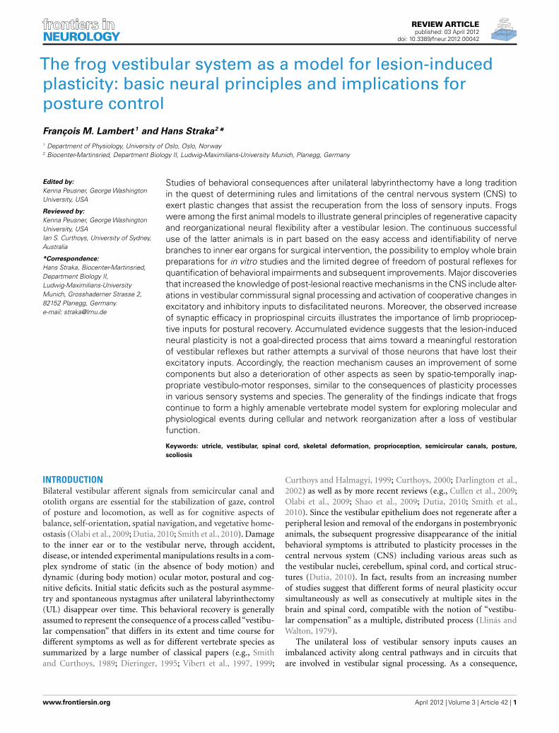
FIGURE 2 | Different postural consequences after selective lesion of the
VIIIth nerve in adult grass frog. (A–D) Induction of an ipsiversive head roll tiltand ipsilesional flexed (blue) and contralesional extended (violet) fore- andhindlimbs after unilateral section of the entire VIIIth nerve (A) or the utricular
(UT) nerve branch on the right side (C); absence of postural deficits after abilateral section of the VIIIth nerve (B) or of one horizontal semicircular canalnerve (D). Hlimb, hindlimb; Flimb, forelimb. Modified and adapted fromPrecht and Dieringer (1985).
torque of the neck, rather than an immediate consequence ofthe removal of one labyrinth (de Kleijn, 1914). This notion issupported by results of a more recent study where a weight wasmounted eccentrically on the head of a frog (Straka and Dieringer,1995). In the presence of the weight, a normal head posture wasmaintained against the neck torque and the forelimbs assumed asimilar, although somewhat less asymmetric posture as seen afterUL. The asymmetric UL-induced posture is complemented by atonic asymmetric deviation of the eyes toward the side of thelesion, even though its degree is rather marginal compatible withthe relatively small ocular motor range of adult frogs (Dieringerand Precht, 1982).
An attempt to determine the contribution of the differentvestibular endorgans to static and dynamic postural reflexes infrog was made by several extensive, almost heroic, series of lesionexperiments (McNally and Tait, 1925, 1933; Tait and McNally,1934; MacNaughton and McNally, 1946). These studies describe ingreat detail the differential functional contributions of individualsemicircular canal and otolith organs to the maintenance of a nor-mal posture in frogs, by interpreting the behavioral consequencesof postural deficits after uni- or bilateral removal of different com-binations of endorgans. The observed postural asymmetry afterthe loss of one labyrinth (Figure 2A) is essentially caused by a uni-lateral removal of the utricle (Figure 2C), since bilateral section of
the VIIIth nerve (Figure 2B) or lesion of one horizontal semicir-cular canal (Figure 2D) does not cause the obvious tonic posturaldeficits (Tait and McNally, 1934). A similar postural asymmetry,although temporally more transient, could be evoked by a unilat-eral lesion of the anterior and posterior vertical semicircular canalsthat caused a slight head roll tilt (10–15˚) toward the ipsilesionalside (McNally and Tait, 1933). However, as the former authorsemphasized, the animals could correct the residual head deviationat any time to resume a symmetric posture. This suggests that theabsence of the two vertical semicircular canals on one side essen-tially causes a dynamic deficit that corresponds in its direction tothe loss of the utricle on the same side but becomes apparent onlyduring passively induced or self-motion (Dieringer, 1995).
While tonic behavioral deficits were also absent after a uni-lateral horizontal canal lesion (Figure 2D), the dynamics of gaze-stabilizing compensatory eye/head movements during vertical axisrotation in these animals was severely impaired (Gribenski, 1963;Precht and Dieringer, 1985). The absence of horizontal semi-circular canal signals during rotation in the ipsilesional direc-tion in light are substituted by saccadic head movements thatappear to depend on head velocity-related proprioceptive reaffer-ent inputs (Dieringer,1988). These head saccades are accompaniedby smaller ocular quick phases and are still present after a bilat-eral section of the horizontal canal nerves or after removal of
Frontiers in Neurology | Neuro-otology April 2012 | Volume 3 | Article 42 | 4

Lambert and Straka Vestibular plasticity in frog
both labyrinths, suggesting a non-vestibular origin. Finally, uni-lateral ablation of the lagena provokes a slight upward tilt of thehead on the ipsilesional side, while a bilateral lesion has littleeffect on the posture, although it severely impairs the rightingreflex from a supine position (MacNaughton and McNally, 1946).In contrast, uni- or bilateral lesion of the saccular nerve neitherdisturbs posture nor deteriorates locomotor performance (Lau-denbach, 1899; McNally and Tait, 1925). This is compatible withthe well-established substrate vibration sensitivity (see above) ofthe saccule and indicates that this organ in frog has no detectablerole in vestibular function.
In summary, the principal static postural deficit after UL in frogis a head roll tilt toward the ipsilesional side. The absence of anadditional tonic head deviation in the horizontal plane as observedin, e.g., guinea pigs is related to differences in the skeletal geome-try of the cervical region (de Waele et al., 1989). In fact, the shortneck in frogs likely prevents a major deviation in the horizontalplane after ablation of one labyrinth at variance with the situationin most other vertebrates (Graf et al., 1995). With the exceptionof the latter species-specific particularity, primary and secondaryconsequences of a labyrinthine lesion in frogs on head/body/limbposition complies with the presumed general principles of vestibu-lar influences on skeletal geometry and postural behavior (Vidalet al., 1998).
CONSEQUENCES OF PRE- AND POSTGANGLIONIC LESIONSON VESTIBULAR NERVE AFFERENTSThe surgical manipulations that were used for studying neuraland behavioral consequences of a unilateral loss of vestibular sen-sation usually consist of a destruction and removal of the labyrinth(labyrinthectomy) and/or of cutting the vestibular nerve, withor without removal of Scarpa’s ganglion (vestibular neurectomy;see Dieringer, 1995, 2003). In studies that report on differentaspects following the loss of one labyrinth in frog, the vestibularnerve was sectioned either proximal or distal to Scarpa’s gan-glion (Figures 3A,B) and semicircular canal and otolith organswere removed (Kunkel and Dieringer, 1994). However, indepen-dent of conservation (preganglionic lesion; Figure 3A) or removalof the ganglion (postganglionic lesion; Figure 3B), the magni-tude of the induced behavioral asymmetry of body posture andlimb position was similar (Kunkel and Dieringer, 1994). This isnot too surprising since both experimental conditions eliminatethe vestibular nerve afferent resting discharge, which is the majorexcitatory drive of central vestibular neurons, thereby causing animbalanced activity on both sides (Dieringer, 1995). However, theanatomical consequences of the two types of interventions in frogdiffer considerably.
After a postganglionic lesion, axons and central terminals ofvestibular nerve afferents start degenerating almost immediatelyafter the lesion as visualized by a specific silver impregnationmethod (Figure 3D) and have entirely disappeared a few weekslater (Kunkel and Dieringer, 1994). In contrast to this degen-eration, vestibular afferents survive after a preganglionic lesion(Kunkel and Dieringer, 1994) as seen by the absence of degen-erating fibers (Figure 3C). This is similar to the situation in cat,where a large part of vestibular nerve afferents survives at leastfor 1 year after a UL that spared the vestibular ganglion (Gacek
and Schoonmaker, 1997) but disappears when the ganglion isdestroyed (Schuhknecht, 1982). Thus, the absence or presenceof afferent degeneration after a pre- versus postganglionic lesionappears to be a general reaction principle. The surviving affer-ent fibers after a preganglionic lesion in frog even maintain thesynaptic connectivity with their target neurons in the vestibularnuclei since electrical stimulation of the stump of the anatomi-cally persisting ipsilesional VIIIth nerve 2 month after the lesionevokes excitatory responses in 2˚VN (Figures 3E,F) with similarmagnitudes compared to controls (Kunkel and Dieringer, 1994;Goto et al., 2001).
TIME COURSE OF POSTURAL RECOVERYStatic, in contrast to dynamic symptoms after UL recover almostcompletely over time, although with different species-specific timecourses (see Dieringer, 1995; Straka et al., 2005). In grass frogs(Rana temporaria), the asymmetric posture after UL consists of ahead roll tilt to the operated side of up to 45˚ and, as a secondaryconsequence, bilateral asymmetrically flexed and extended limbpositions (Figure 2A). The angle of the head deviation normalizeswith an asymptotic time course (Figures 4A,B) and is more or lesscomplete after about 2 month (Kolb, 1955; Bienhold and Flohr,1980; Kunkel and Dieringer, 1994). Despite the considerable mor-phological consequences for the survival of afferent fibers afterpre- versus postganglionic lesions (Kunkel and Dieringer, 1994),there appears to be neither a major difference in the extent ofpostural deficits nor in the time course of the recovery whetherScarpa’s ganglion is spared or not. However, under both condi-tions, a normal posture is only attained during static positions ofthe animal, while dynamic positional changes, arousing stimulior locomotor activity is often accompanied by a decompensa-tion and reappearance of the acute postural syndrome (Dieringer,1995).
Additional removal of the remaining intact labyrinth at dif-ferent time intervals after the first lesion causes the classicalBechterew-symptom that is a mirror image of the behavioraldeficit induced by the first lesion (von Bechterew, 1883). Ingrass frogs, the minimal time interval for the induction of theBechterew-symptom is 5 days (Dieringer and Precht, 1981) andis followed by a postural normalization that progresses moreslowly and is less complete compared to the reaction after the firstlesion (Kolb, 1955). The mirror image-like head deviation afterthe delayed, second lesion has been interpreted as evidence for aCNS plasticity, since simultaneous removal of both labyrinths failsto provoke a postural syndrome (Dieringer and Precht, 1981).
Even though most experiments in amphibians have been con-ducted on grass frogs, a comparison with results from water frogs(R. esculenta) is of particular importance for understanding themechanisms that are at play during the postural recovery process.Interestingly, water frogs exhibit a postural normalization afterUL that is incomplete with a residual head deviation of 5–10˚(Figure 4A; Kolb, 1955). This difference between water and grassfrogs might be related to particularities in the life style of thetwo anuran species. The more aquatic water frogs mostly float inwater, which limits the use of body-weight-supporting limb pro-prioceptive signals as potential source for a sensory substitutionof absent vestibular signals (see below). In contrast, the terrestrial
www.frontiersin.org April 2012 | Volume 3 | Article 42 | 5

Lambert and Straka Vestibular plasticity in frog
FIGURE 3 | Different morphological consequences of VIIIth nerve
sections in adult grass frog that spare or include the ganglion of Scarpa.
(A,B) Schematic drawing of a transverse hindbrain section, depicting theVIIIth nerve entry, termination of labyrinthine afferent fibers in the vestibularnuclei (VN), and site of pre- (A, pre-GS) and postganglionic nerve sections (B,post-GS). (C,D) Bright field microphotographs of transverse sections throughthe VN (see inset in C) 2 days after a pre-GS (C) and post-GS (D); degenerating
vestibular nerve afferent fibers were visualized by a silver impregnationmethod. (E,F) Pre- (N0) and postsynaptic components (N1) of ipsilateral VIIIthnerve-evoked afferent field potentials (E) recorded in the VN on the intact andoperated side of chronic UL frogs with a pre-GS; mean amplitudes of the N1
afferent field potential component, recorded on the intact (control) andoperated side at different survival periods after a pre-GS (F); modified andadapted from Straka et al. (1993) and Kunkel and Dieringer (1994).
life style of grass frogs or toads and the resulting availability oflimb proprioceptive sensory signals favor a more or less completebehavioral normalization (Straka and Dieringer, 1995). Accord-ingly, the latter sensory signals might form the relevant referenceframe that allows a reestablishment of a normal, symmetric bodyposition in space. This interpretation is corroborated by results ofexperiments in grass frogs that showed an implication of asym-metrically enhanced neck reflexes after UL (Kolb, 1955) as aftereccentrically mounting a weight on the head (Straka and Dieringer,1995) for the postural recalibration.
Pharmacological alterations of the time course of the posturalrecovery after UL by systemic drug injections was used to elu-cidate a potential involvement of specific neurotransmitters andneuromodulators in the underlying central nervous plasticity (seeDieringer, 1995). Sedating and arousing drugs have been shownto delay and facilitate the normalization process, respectively
(Bienhold et al., 1981). A similar effect was observed with cholin-ergic drugs, which causes a postural decompensation (choli-nomimetics) or reduction (cholinolytics) of the postural asymme-try (Bienhold and Flohr, 1980; Bienhold et al., 1981), compatiblewith the established role of cholinergic synapses in vestibular sig-nal processing in frog (Rossi et al., 1980; Bernard et al., 1985;Kasik et al., 1986). A potential role of glutamate receptors forthe postural recovery was studied in a series of experiments thattested the influence of systemically applied NMDA receptor block-ers on the time course and magnitude of the lesion-inducedhead deviation (Flohr and Lüneburg, 1993). According to theresults of the latter study, NMDA receptors are involved in therecovery, but not in the maintenance of the normal head posi-tion once it has been resumed. While these pharmaco-behavioralresults have a relative importance for confining the post-lesionalperiod of presumed neural plasticity, the systemic approach of
Frontiers in Neurology | Neuro-otology April 2012 | Volume 3 | Article 42 | 6

Lambert and Straka Vestibular plasticity in frog
FIGURE 4 |Time course of recovery from the postural asymmetry after
a unilateral vestibular lesion in adult frog. (A) Angle of the head roll tiltas a function of postoperative time in grass frog (Rana temporaria) after ULor selective section of the anterior VIIIth nerve branch (RA) and in waterfrog (Rana esculenta) after UL; data on UL in Rana esculenta adopted fromKolb (1955) and in Rana temporaria from Flohr et al. (1981). (B) Comparisonof the residual head tilt angles in four specimen of Rana temporaria inwhich the same RA nerve branch was re-sectioned 100 days after the firstlesion; modified and adopted from Goto et al. (2002).
drug application in combination with the commonality and wide-spread occurrence of the tested transmitter systems in the CNSprevents further specification of the site of action. Despite theseknown limitations in the interpretation, central vestibular nucleiwere commonly assumed as the predominant site of the neuralplasticity processes.
PERIPHERAL AND CENTRAL VESTIBULAR PLASTICITY AFTERUNILATERAL VESTIBULAR LOSSA partial or complete unilateral loss of vestibular sensory inputstriggers a number of morpho-physiological changes that usuallyhave been associated with different aspects of the behavioral recov-ery (Dutia, 2010). The reliable improvement of acute posturaldeficits over time is generally interpreted as the consequence ofneural changes that cause spatio-temporal accurate compensa-tion or substitution of the impaired vestibular sensation (Flohret al., 1985). However, a number of experiments in frogs haveshown that even though some of the observed neural changes arein favor of a recalibration of central vestibular activity, other cel-lular and network modifications cause deterioration of the spatialresponse vectors of individual neuronal populations and therebypotentially alter the behavioral output and induce inappropriatemotor reactions (Goto et al., 2000, 2001; Rohregger and Dieringer,2003). Furthermore, the time course of plastic changes in centralvestibular signal processing differs from that of the postural recov-ery (Straka et al., 1993), thereby challenging the vestibulo-centricview of the “compensation” process.
In the following, different plasticity processes at the cellularand circuit level in the brainstem and spinal cord in frog aftera unilateral vestibular loss will be described. Potential relationswith the behavioral recovery of lesion-induced deficits and possi-ble consequences for the precision of vestibulo-motor responseswill be discussed. Major post-lesional changes in general includemodifications of intrinsic membrane properties, specifically aug-mented synaptic efficacy of available intact inputs, generation ofnew or extension of existing morphological connections withinthe vestibulo-motor circuitry, alterations in convergence patternsof intact vestibular signals and sensory substitution (e.g., Strakaet al., 2005; Dutia, 2010). A number of these conceptually dif-ferent plasticity processes were initially discovered in frogs (seeDieringer, 1995, 2003) and later confirmed in different vertebratespecies, whereas others appear to be part of a more amphibian-specific plasticity regime that includes regeneration of vestibularnerve afferents under certain experimental conditions. In contrast,changes in intrinsic membrane properties of vestibular neuronsafter a loss of one labyrinth as shown in guinea pig (Beranecket al., 2003, 2004) have so far not been reported in frog. However,this is not due to a general absence of such a plasticity phenomenonin frog, but rather due to the lack of respective studies that com-pare known intrinsic neuronal properties of frog tonic and phasic2˚VN (Straka et al., 2004; Beraneck et al., 2007) before and afterthe lesion. Assuming the presence of basic reaction mechanismsthat only differ in magnitude between different species, a generalhypothesis is derived from the results obtained in guinea pigs afterUL (Beraneck et al., 2003, 2004). Accordingly, on the ipsilesionalside in frog, the overall tonic response properties might increase,mainly by a process, whereby phasic 2˚VN express a more sustaineddischarge during current step-evoked depolarization. In addition,oppositely oriented changes are predicted for 2˚VN on the con-tralesional side. A potential mechanism that allows phasic neuronsto become more tonic is a reduction in the density of dendrotoxin-sensitive potassium channels (Beraneck et al., 2007), similar to thesituation of chicken tangential vestibular nucleus neurons dur-ing late embryogenesis (Gamkrelidze et al., 1998, 2000). Thus,
www.frontiersin.org April 2012 | Volume 3 | Article 42 | 7
Lambert and Straka Vestibular plasticity in frog
future studies in the frog model will reveal a potential homeosta-tic plasticity of electrophysiological properties that include mirrorimage-like alterations of ion channel conductances and their func-tional consequences in the bilateral vestibular nuclei after UL asdescribed for the synaptic charge transfer in the chicken tangentialvestibular nucleus (Shao et al., 2012).
REGENERATION OF VESTIBULAR NERVE AFFERENTS AFTER VIIITHNERVE SECTIONA particular reaction after transection of the VIIIth nerve withoutremoval of labyrinthine endorgans is a regeneration of afferentfibers and a reinnervation of the remaining epithelium in thesensory periphery. This plasticity phenomenon is common inamphibians (Sperry, 1945; Hernandez et al., 1998; Goto et al.,2002) and birds (Haque et al., 2008) but apparently absent inmammals. In frog, regeneration of vestibular afferents and rein-nervation occurs both within the sensory epithelium after a nervesection distal to Scarpa’s ganglion (Figure 4B; Hernandez et al.,1998) as well as centrally in the vestibular nuclei after a nervesection between the ganglion and the brainstem (Newman andHonrubia, 1992). Independent of the site of nerve section, periph-eral and central regenerating afferent fibers form appropriate pro-jections and reinnervate adequate vestibular endorgans and centralneurons in the vestibular nuclei, respectively (Newman et al., 1986,1987; Hernandez et al., 1998). The remarkable spatio-temporalprecision of this process is best illustrated by the reappearance ofhead motion-driven activity in afferent fibers and vestibulo-motorresponses with gains that are comparable to those of controls(Hernandez et al., 1998; Goto et al., 2002). With the exceptionof particular organizational differences in afferent fiber diametersand reinnervation patterns of hair cells in different areas of thesensory epithelium by thin and thick fibers, this plasticity mech-anism is specific and results in a complete functional recoveryof vestibular function if the sensory periphery remains preservedafter the lesion. In fact, the persisting intact receptor epitheliumexerts a presynaptic influence on the physiological characteristicsof regenerating and reconnecting axons, thereby ensuring a certainspatio-temporal precision (Hernandez et al., 1998). This mecha-nism, however, appears to be specific to non-mammalian speciesand thus has only limited implications for explaining “vestibularcompensation” in mammals.
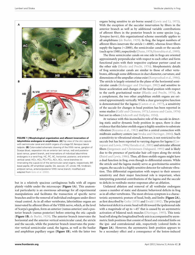
CHANGES IN VESTIBULAR COMMISSURAL RESPONSESBrainstem commissural connections play an important role forthe sensitivity of bilateral processing of head motion-related sen-sory signals (Shimazu, 1972; Graham and Dutia, 2001). Increasedamplitudes of commissural responses in ipsilesional 2˚VN ofchronic frogs were among the first neural alterations observedafter UL (Dieringer and Precht, 1977). Commissural field poten-tials, activated by electrical pulse stimulation of the contralateralVIIIth nerve in frog, typically consist of a disynaptic negativefield potential (Figure 5A) that mirrors intracellularly recordedcommissural EPSPs in 2˚VN (Figure 5C; Ozawa et al., 1974). Com-pared to controls, commissural (Figures 5A,B) but not afferentfield potentials (Figures 3E,F) are significantly enhanced (∼100%)2 month after UL, whereas regional distributions and depth pro-files remain unaltered. This finding was interpreted as a general
increase in synaptic efficacy of commissural excitatory inputs fromthe contralateral, intact side to ipsilesional 2˚VN (Dieringer andPrecht, 1977, 1979a,b; Kunkel and Dieringer, 1994), independentof degeneration (postganglionic lesion) or survival (preganglioniclesion) of the VIIIth nerve (Kunkel and Dieringer, 1994). Furtherspecification of this plastic change in synaptic responsiveness byintracellular recordings of ipsilesional 2˚VN in chronic UL frogsrevealed increased amplitudes and faster rise times of commis-sural EPSPs (compare black and gray traces in Figure 5C), which,in contrast to controls, are able to trigger partial and full actionpotentials (Dieringer and Precht, 1977, 1979a). Functionally, thischange is compatible with a reestablishment of the lesion-inducedloss of resting activity in central vestibular neurons, potentiallyfacilitating the recovery from behavioral deficits.
The survival of ipsilesional nerve afferents after a preganglionicUL allowed in chronic animals an electrical stimulation of thesefibers with similar current thresholds as in controls (Kunkel andDieringer, 1994). The absence of a general amplitude enhance-ment of VIIIth nerve afferent responses on the ipsilesional sideprecludes a global change in excitatory synaptic efficacy as the ori-gin of the observed plasticity and suggests the implementation ofmore specific alterations in the synaptic transmission of commis-sural responses in 2˚VN after lesion (Kunkel and Dieringer, 1994).Since excitatory commissural responses in frog 2˚VN are transmit-ted by glutamate that activates both AMPA and NMDA receptors(Cochran et al., 1987; Knöpfel and Dieringer, 1988), both receptorsubtypes could theoretically contribute to the observed increase inEPSP amplitude. However, NMDA components of commissuralEPSPs in chronic frogs are not increased compared to controls (seedashed black and gray traces in Figure 5C; Knöpfel and Dieringer,1988) and circumstantial evidence (Cochran et al., 1987) arguesagainst an increased number or sensitivity of AMPA receptors asthe cause for the facilitated commissural signals (for details seeDieringer, 1995). Moreover, the role of glutamate in transmit-ting both afferent and commissural signals onto 2˚VN along withthe restricted augmentation of the latter responses in ipsilesional2˚VN, was used as an argument to preclude a general supersen-sitivity of glutamate receptors as the origin of the plasticity inexcitatory commissural responses.
Accordingly, pre- and/or postsynaptic alterations of the synap-tic transmission process as possible cause for the observedincreased commissural excitation in ipsilesional 2˚VN of chronicfrogs were considered unlikely. This led to the suggestion of reac-tive synaptogenesis of excitatory commissural connections as analternative mechanism to explain, at least in part, the observedchanges. In particular, a morphological expansion of commis-sural synapses onto more proximal dendrites, in addition to thosepresent on distal dendrites as in controls, was hypothesized asa cause for the altered amplitude and rise time of the commis-sural excitation (Dieringer and Precht, 1979a). Even though, directmorphological evidence for this interpretation is missing (seeDieringer, 1995), more recent studies (Goto et al., 2000, 2001)using a specific vestibular nerve lesion paradigm appears to concurwith this idea (see below). Nonetheless, the still unsatisfying inter-pretation of the augmented commissural excitation in chronicfrogs after UL without a clearly confirmed underlying mechanismprevails until today.
Frontiers in Neurology | Neuro-otology April 2012 | Volume 3 | Article 42 | 8

Lambert and Straka Vestibular plasticity in frog
FIGURE 5 | Cellular origins of lesion-induced plastic changes in
vestibular commissural pathways in frog. (A,B) Pre- (cN0) and postsynapticcomponents (cN1) of vestibular commissural field potentials (A) recorded inthe vestibular nuclei on the intact and operated side of chronic UL frogsfollowing electrical stimulation of the contralateral VIIIth nerve (scheme);changes in vestibular commissural field potential amplitudes after a pre-(preG) and postganglionic (postG) UL (B); modified and adapted from Strakaet al. (1993) and Kunkel and Dieringer (1994). (C) Vestibular commissuralEPSPs in 2˚VN of controls (left) and on the operated side of chronic UL frogs(right) activated by electrical stimulation of the intact VIIIth nerve (scheme);bath application of D-APV (25 μM) in vitro unmasks similar contributions ofNMDA receptor-mediated components in responses of 2˚VN in controls and
on the operated side of chronic UL frogs; modified and adapted from Knöpfeland Dieringer (1988). (D) Commissural vestibular responses recorded in vitroin an identified 2˚ anterior vertical semicircular canal (AC) neuron followingelectrical stimulation of the entire contralateral VIIIth nerve (black trace) orindividual branches to the contralateral posterior canal (PC, green trace), AC(blue trace), and horizontal canal (HC, red trace). (E) Sensitivity of contralateralsemicircular canal (cAC, cPC) nerve-evoked vestibular commissural fieldpotentials (upper traces) and commissural EPSPs in 2˚VN (lower traces) toin vitro bath-applied antagonists that block the GABAergic (5 μM bicuculline)and glycinergic (2 μM strychnine) transmission; note the increase in amplitudeof extra- and intracellular recorded responses. (D,E) Modified and adaptedfrom Malinvaud et al. (2010).
www.frontiersin.org April 2012 | Volume 3 | Article 42 | 9

Lambert and Straka Vestibular plasticity in frog
Based on a recent functional reevaluation of frog vestibularcommissural organization (Holler and Straka, 2001; Malinvaudet al., 2010), there is an alternative interpretation of the data byDieringer and Precht (1977). The explanation for the increasedexcitatory vestibular commissural responses in chronic frogs afterUL in the initial report as in all subsequent studies (see Dieringer,1995) was based on the until then unquestioned finding thatfrogs, in contrast to cats, have an excitatory vestibular commis-sure. This was concluded from the presence of negative vestibularcommissural field potentials and EPSPs in frog 2˚VN (Ozawaet al., 1974) after stimulation of the entire contralateral VIIIthnerve (Figures 5A,C). More recent studies,however, that employeda specific electrical stimulation of individual labyrinthine nervebranches, revealed in individual semicircular canal-related 2˚VN aplane-specific commissural IPSP (green trace in Figure 5D) fromthe contralateral coplanar canal in frog (Holler and Straka, 2001;Malinvaud et al., 2010) as reported earlier in cat (Kasahara andUchino, 1971). This canal-specific IPSP, however, is masked infrog by commissural EPSPs from most non-coplanar semicircularcanal (blue and red traces in Figure 5D) and otolith organs, therebycausing a net slow rising excitation in 2˚VN when stimulating theentire VIIIth nerve (black trace in Figure 5D), consolidating thelatter results with the initial report of Ozawa et al. (1974).
Based on the recent, more elaborate functional organization ofcommissural connections in frog, the observed increase in ampli-tude and dynamics of the commissural excitation of 2˚VN inchronic UL frogs after VIIIth nerve stimulation (e.g., Dieringerand Precht, 1977, 1979a,b; Knöpfel and Dieringer, 1988; Kunkeland Dieringer, 1994) might be caused by a reduction of semicir-cular canal-specific commissural IPSP rather than by an increasein commissural EPSP amplitudes. In fact, evidence for such aninterpretation comes from in vitro experiments in brainstempreparations of intact frogs where the commissural inhibition waspharmacologically blocked by GABAergic and glycinergic antago-nists (Figure 5E), causing an augmentation of the net commissuralexcitation (lower traces in Figure 5E; Malinvaud et al., 2010) asseen in the altered responses of 2˚VN in chronic UL frogs. More-over, a lesion-induced reduction of the commissural inhibitionin ipsilesional semicircular canal-related 2˚VN is also present inchronic frogs after section of the anterior branch of the VIIIthnerve (Goto et al., 2001). Thus, the latter studies and the newlines of arguments offer an alternative explanation for the plas-tic changes that were previously interpreted by an increase investibular commissural excitation. Accordingly, the increase ofthe commissural excitation is not caused by an augmentation ofexcitatory response components but rather by a decrease in thecommissural inhibition to ipsilesional 2˚VN, in line with observa-tions on cellular mechanisms that are involved in rebalancing thecommissural system in mammalian species (Graham and Dutia,2001; see Olabi et al., 2009; Dutia 2010).
The plastic changes in the commissural system in chronic ULfrogs (Dieringer and Precht, 1977, 1979a) were initially interpretedas a mechanism that is directly involved in the postural recoveryby assisting the reestablishment of a bilateral symmetric sponta-neous activity in central vestibular neurons. In fact, the significantincrease of the commissural excitation 2 month after the lesionwas assumed to be the cause for the normalized posture in these
animals, partly because this increase saturated at the same time(Kunkel and Dieringer, 1994). However, experiments that deter-mined onset and time course of the post-lesional commissuralchanges indicated that a first significant increase in commissuralfield potential amplitudes occurs only ∼30 days after UL and pro-gresses slowly thereafter (Figure 5B). This, however, is too late andthus at variance with the considerably faster time course of thepostural recovery (Figure 4A) that proceeds asymptotically witha 50% recovery of the initial postural asymmetry after ∼15 days(Kunkel and Dieringer, 1994).
The delayed onset of plastic changes in commissural signal-ing and its presumed influence on the resting activity of centralvestibular neurons is in agreement with the time course of alter-ations in the metabolic activity in the vestibular nuclei after UL(Flohr et al., 1981, 1989). Similar to the time course of the increasein commissural responses, the recovery of the markedly reduceddeoxyglucose uptake in central vestibular neurons immediatelyafter the lesion is delayed by ∼30 days. Thus, the obvious discrep-ancy in the timing of changes in commissural signaling, meta-bolic activity in the vestibular nuclei, and effective body posture(Figures 4A and 5B; Straka et al., 1993) suggests that plasticityprocesses other than those in the vestibular commissural systemor the vestibular nuclei might be at the origin of the postural nor-malization. This assumption also concurs with the view on neuralplasticity after UL as a distributed process that also includes areasoutside the vestibular nuclei (Llinás and Walton, 1979).
SYNAPTIC REORGANIZATIONReactive synaptogenesis at multiple hierarchical levels of the CNSis a common morpho-physiological mechanism that has beenshown to cause reorganization of sensory signal processing afterperipheral or central lesions in vertebrates (Kaas, 2000). Withrespect to an experimentally induced peripheral loss of vestibu-lar inputs, only few studies have so far explored the interactionbetween synaptic inputs from sectioned, silenced and remain-ing, intact labyrinthine nerve afferents or central inputs to 2˚VN(Dieringer, 2003). The resulting competition between intact andsectioned fibers for synaptic access to 2˚VN after a partial vestibu-lar nerve section is at variance with the situation after a completeUL where all fibers are silenced. While the induced reactionmechanisms for the overall population of ipsilesional afferentfibers consequently differ between the two experimental manipu-lations, other simultaneously and/or sequentially occurring plas-ticity processes might only vary in magnitude. Thus, a partialvestibular nerve section is by definition not just a smaller versionof a complete UL but induces an interesting and unique conditionwith respect to potential afferent synaptic plasticity that might berelevant for the postural recovery after the lesion.
While a complete UL generates a relatively defined experimen-tal condition by silencing all vestibular nerve afferent fibers onone side, a partial removal of vestibular inputs and its provokedfunctional plasticity is relevant for understanding those eventsthat are triggered by various types of incomplete impairment ofvestibular sensation in human patients. Accordingly, the partialvestibular lesion model in frog offers an excellent possibility togain insight into the synaptic plasticity after a specific, restricteddisfacilitation of identified central neurons. A selective section of
Frontiers in Neurology | Neuro-otology April 2012 | Volume 3 | Article 42 | 10

Lambert and Straka Vestibular plasticity in frog
the entire anterior branch of the VIIIth nerve or of single branchesto individual endorgans creates a conflict situation between theremaining intact and the disfacilitated vestibular nerve afferentfibers with respect to the access to 2˚VN (Goto et al., 2000, 2001,2003). The section of the anterior VIIIth nerve branch eliminatessensory inputs from the utricle, the saccule, the anterior verticaland the horizontal semicircular canal, while the posterior VIIIthnerve branch that mediates sensory inputs from the posterior canaland the lagena (the vertical otolith organ in frog) remains intact.The preganglionic nature of this nerve section ensures survivalof the central projection of the sectioned anterior VIIIth nervebranch and thus offers the possibility for electrical activation ofthe silenced nerve fibers 2 months later, at a time when the posturalasymmetry, as relevant marker for immediate static post-lesionaldeficits, has been normalized. Moreover, the possibility to activatemonosynaptic EPSPs in ipsilesional 2˚VN from the severed nerve
stump facilitates the identification of those neurons that had beendeprived of sensory inputs.
Chronic frogs that had received a section of the anterior VIIIthnerve branch expressed considerable alterations in the synap-tic organization of afferent inputs from fibers in the remainingintact posterior VIIIth nerve branch on the operated side aswell as of commissural inputs from the contralateral labyrinth(Figures 6A–C; Goto et al., 2000, 2001). On the ipsilesional side,field potentials evoked by stimulation of the intact posterior verti-cal semicircular canal nerve were enlarged throughout the vestibu-lar nuclei (Figures 6A,B), and the total number of 2˚VN with amonosynaptic input from this semicircular canal was significantlyincreased compared to controls (Figure 6D; Goto et al., 2000),suggesting an expansion of the latter afferent inputs onto those2˚VN that had been deprived of afferent vestibular inputs fromthe sectioned anterior VIIIth nerve branch. However, this synaptic
FIGURE 6 | Synaptic reorganization of intact vestibular inputs after
labyrinthine nerve lesions. (A,B) Posterior semicircular canal (PC)nerve-evoked afferent pre- (N0) and postsynaptic (N1) field potentialcomponents (A) recorded on the intact and operated side of chronic frogswith a section of the anterior branch (RA) of the VIIIth nerve; the enhancedfield potentials on the operated side extend throughout the entire nucleusas indicated by the amplitude depth profile (B). (C) Contralateral horizontalsemicircular canal (HC) nerve-evoked postsynaptic commissural fieldpotentials (cN1) recorded in a control frog and on the operated side of achronic RA frog; the sites of lesion, electrical stimulation and recording areillustrated in the scheme. (D) Convergence pattern of monosynapticafferent responses in two 2˚VN after separate stimulation of the PC nerve(black traces) and the RA of the VIIIth nerve (green traces); after RA nervesection, the percentages of 2˚RA neurons and of 2˚RA + PC but not of
2˚PC neurons on the operated side of chronic RA frogs arecomplementarily altered, with respect to the pattern on the intact side(*P ≤ 0.01) or controls (#P ≤ 0.0001); data adopted from Goto et al. (2000).(E,F) Plasticity of commissural semicircular canal inputs in 2˚VN of chronicRA frogs; typical afferent and commissural responses of a 2˚PC neuron(E); the pattern consists of an afferent (i) PC nerve-evoked monosynapticEPSP and commissural (c) anterior vertical canal (AC) nerve-evoked IPSP,cHC, and cPC nerve-evoked EPSPs; ipsi- and contralateral stimulation andrecording sites are illustrated (scheme); the relative proportions ofIPSPs/2˚VN and EPSPs/2˚VN (F) in chronic RA frogs (gray bars) aresignificantly altered (*P ≤ 0.01, **P ≤ 0.001, ***P ≤ 0.0001) in acomplementary fashion on the operated side in 2˚RA and 2˚RA + PC butnot in 2˚PC neurons compared to controls (white bars); data adopted fromGoto et al. (2001).
www.frontiersin.org April 2012 | Volume 3 | Article 42 | 11
Lambert and Straka Vestibular plasticity in frog
reorganization is not restricted to the remaining intact afferentvestibular nerve inputs from the same side, but also extendsto commissural inputs from the contralateral semicircular canalnerves (Figures 6C,E,F; Goto et al., 2001).
After separate stimulation of the three contralateral, intactsemicircular canal nerve branches, inhibitory commissural inputswere reduced in number and amplitude, whereas the relativeoccurrence and magnitude of excitatory commissural responseswere augmented in those 2˚VN that were disfacilitated by theperipheral nerve lesion (Figures 6E,F). Thus, the inactivatedmonosynaptic inputs from the sectioned afferent fibers in theanterior VIIIth nerve branch are functionally substituted by anexpansion of inputs from afferents of the remaining intact homo-lateral posterior vertical canal and by cooperative changes in semi-circular canal-related commissural inputs from the contralateralside. These modifications are specific, since afferent and com-missural responses of posterior vertical canal 2˚VN on the sameside, which were not disfacilitated by the lesion, remain unaltered(Figures 6D,F; Goto et al., 2000, 2001).
The new excitatory response components from vestibular nerveafferents to the disfacilitated 2˚VN might either represent new con-nections, formed by axonal sprouting from the remaining intactposterior VIIIth nerve branch (afferents from the posterior verticalcanal and/or the lagena), or reactivated silent synapses that alreadyexisted before the lesion. In addition, new excitatory inputs to thedisfacilitated 2˚VN are not limited to intact afferent or commis-sural vestibular inputs but include excitatory synaptic inputs fromascending propriospinal afferent projections as evidenced by theincreased axonal arborization of this pathway in the ipsilesionalvestibular nuclei in chronic UL frogs (Dieringer et al., 1984).
Based on these results, a set of general rules can be extracted byincluding data obtained after a selective section of either utricularafferent fibers or after a combined section of the horizontal andanterior vertical canal nerve branch (Goto et al., 2002). Accord-ingly, the synaptic reorganization is activity-dependent, since theexpansion of excitatory signals is restricted to intact inputs fromvestibular nerve branches, commissural fibers or ascending spinalprojections. The retention of labyrinthine afferent synapses onto2˚VN after the preganglionic nerve section precludes a competi-tive interaction between active (intact) and inactive (axotomized)axon terminals. Rather, the presence of spontaneous or modulatedactivity of intact neurons is the likely trigger for an expansion oftheir synaptic inputs onto disfacilitated 2˚VN, compatible with thecomplete reversibility of the latter plasticity after regeneration ofthe sectioned peripheral axons and reinnervation of the previoussensory epithelium (Figure 4B; Goto et al., 2002). The synapticreorganization is gradual, and its extent appears to depend on theamount of sensory fibers that were silenced by the lesion (Gotoet al., 2002). Thus, a utricular (∼4000 fibers) but not semicir-cular canal nerve (∼1500 fibers) section provokes a significantexpansion of intact excitatory inputs onto those 2˚VN that weredisfacilitated by the lesion.
As after UL, the onset of synaptic changes in the vestibularnuclei, triggered by the partial VIIIth nerve lesion is delayed com-pared to the onset of the postural recovery from those lesions(Goto et al., 2002). This precludes changes in the synaptic arrange-ment of central vestibular neurons as a major contributor to the
reestablishment of a normal posture after a partial or complete lossof one labyrinth. The apparent substitution of the reduced synap-tic excitation of 2˚VN by intact excitatory inputs is analogous tothe situation of denervated muscle fibers that accept a heterony-mous innervation to avoid degeneration (Kaas, 2000). Therefore,cooperative changes that include expansion of excitatory and con-traction of inhibitory inputs to disfacilitated neurons appear torepresent a general, ubiquitous mechanism. The synaptic reorga-nization of vestibular circuitry in frog at a period when the initialasymmetric posture is normalized again, does not exclude thatother mechanisms, such as changes of intrinsic cellular properties,might contribute to the postural recovery at earlier post-lesionalperiods.
Reactive synaptogenesis, even though it appears to be advanta-geous for the survival of deafferented neurons, has an importantfunctional consequence. The newly acquired excitatory synapticinputs to 2˚VN originate from vestibular endorgans that con-vey inappropriate signals onto disfacilitated neurons and therebyimpair the spatio-temporal specificity of the sensory-motor trans-formation. Thus, the cooperative changes in synaptic inputs mightbe beneficial for the survival of deprived 2˚VN and likely assist inthe reduction of the asymmetry between the resting discharge ofbilateral 2˚VN, however, the rescue of the latter neurons is at theexpense of the precision in the tuning of vestibular reflexes. Sucha deterioration of spatio-temporal parameters of the signal pro-cessing in “compensated” animals after a peripheral nerve lesionalso complies with the notion that neural plasticity is not primar-ily oriented toward a functionally appropriate reestablishment ofimpaired vestibulo-motor responses.
INADEQUATE VESTIBULAR REFLEXES AFTER SYNAPTICREORGANIZATIONThe apparent absence of a targeted neural plasticity process thatcompensates functional deficits was evidenced by the results ofa series of experiments that used specific vestibular nerve lesionsin frog (Rohregger and Dieringer, 2003). The pronounced synap-tic reorganization of vestibular nerve afferent, commissural andascending spinal inputs at the level of 2˚VN after UL as after partialVIIIth nerve lesions suggests that the spatial and dynamic precisionof the disfacilitated 2˚VN is considerably altered by this process. Inparticular, the acquisition of new excitatory inputs from vestibularendorgans other than the original organ during the post-lesionalplasticity (Figures 7A,B) is not necessarily functionally mean-ingful and adaptive and in fact appears to be even detrimental(Goto et al., 2000, 2001; Rohregger and Dieringer, 2003). Thenewly acquired inputs after a selective section of the anterior VII-Ith nerve branch differ from the original afferent inputs in theirresponse vectors, and thus convey spatio-temporally inappropri-ate signals to the disfacilitated neurons (Figure 7B). Assumingthat the projections to efferent recipients of 2˚VN with new inputsremain unchanged, functionally inadequate signals are relayed tothe default motor targets and thus cause erroneous behavioralresponses. This is in fact the case as evidenced by the appearanceof inappropriate posterior vertical semicircular canal responsesin contralesional frog abducens motoneurons after a lesion ofthe anterior VIIIth nerve branch (Goto et al., 2000). Thus, as aresult, the spatial response vector of contra- but not ipsilesional
Frontiers in Neurology | Neuro-otology April 2012 | Volume 3 | Article 42 | 12

Lambert and Straka Vestibular plasticity in frog
FIGURE 7 | Modification of response vectors in abducens motoneurons
after labyrinthine nerve lesions. (A,B) Schematic diagram of signalingpathways before (A) and after section (B) of the anterior branch (RA) of theVIIIth nerve; afferent vestibular nerve signals during linear acceleration incontralateral (left) abducens motoneurons (AB) of controls (A) originatefrom a sector of hair cells (gray) on the right utricle (UT) and are mediatedby neurons in the vestibular nucleus (VN) that excite contralateral AB. Inchronic RA frogs (B) with a nerve section on the right side, the vestibularresponses in the left AB recovered but now originate during linearacceleration from hair cells located laterally with respect to striola on theleft, ipsilateral UT; after convergence with spatially inappropriate signalsfrom the lagena (LA, yellow) in the ipsilesional VN, these signals accesscontralateral AB through the default excitatory crossed projection; schememodified and adapted from Rohregger and Dieringer (2003).
abducens motoneurons to linear and angular head acceleration ismodified, which leads to a deterioration of the spatial tuning ofvestibulo-motor responses (Rohregger and Dieringer, 2003).
The change in vector orientation of these responses, however,is highly variable between different specimen, most likely due toindividual degrees of reorganization of signals from the remain-ing intact posterior vertical canal and the lagena (Figure 7B).Again, the deterioration in spatial precision of the VOR is at vari-ance with the idea of targeted plasticity processes that aim towarda functional compensation of lesion-induced behavioral deficits.More likely, cellular response cascades are triggered in the disfacil-itated neurons that ensure a general survival of these neurons by
preventing the induction of apoptotic cellular consequences. Thechanges in both synaptic circuitry and cellular properties of 2˚VNmight thus be the expression of a fundamental neural reaction pat-tern that is common between sensory modalities and vertebratespecies.
FUNCTIONAL ROLE OF PROPRIOSPINAL PLASTICITY FOR THEPOSTURAL RECOVERY AFTER ULThe obvious discrepancy between onset and timing of the neuralchanges in the vestibular nuclei (Figure 5B) and the recovery fromthe postural asymmetry after UL (Figure 4A) precludes a causalrelationship between the former and the latter plasticity process(Straka et al., 1993). In addition, the vestibular nuclei are by nomeans the only place at which the neural organization becomesaltered in response to a peripheral labyrinthine lesion. As a conse-quence, “vestibular compensation” is likely not accomplished by asingle, local process nor does it entirely rely on vestibular signalsfrom remaining intact vestibular endorgans. Rather, the functionalnormalization after the loss of vestibular sensory inputs repre-sents the integrated result of a multitude of distributed neuralmodifications that occur at different time intervals after the lesionas suggested earlier (Llinás and Walton, 1979; Dieringer, 1995).The observation of increased densities of synaptic terminationsof ascending propriospinal fibers within the ipsilesional vestibularnuclei after UL (Dieringer et al., 1984) suggests that propriocep-tive signals play an important role in the recovery process fromthe lesion-induced deficits. In terrestrial tetrapods, these sensoryinputs are excellently suited for providing an appropriate referenceframe that supplies information on the position of the “body inspace” after a unilateral loss of labyrinthine function. However, thedynamics and spatial precision for detecting and encoding relativepositions of head-body-limbs with respect to the ground is rel-atively low and therefore limited to substituting static vestibularreflexes such as tonic head/body deviations. Thus, altered synap-tic efficacy of the bilateral propriospinal circuitry might be theprincipal cause for the observed postural recovery.
Isolated brainstem-spinal cord preparations of frogs thathad recovered from the postural asymmetry after ULexhibit an increased efficacy of monosynaptic uncrossed pro-priospinal responses within the ipsilesional brachial cord region(Figures 8A,B; Straka and Dieringer, 1995). Importantly, theamplitude of the uncrossed dorsal root-evoked ventral root poten-tials (DR-VRP) on the operated side began to significantly increase7–15 days post-lesion, progressively augmented in parallel withthe recovery of a normal static posture and reached a steady stateafter 30–40 days (Figures 8C,D). This increase in propriospinalresponse gain persisted after isolation of the spinal cord from thebrainstem, and was therefore independent from changes at thecentral vestibular level (Straka and Dieringer, 1995).
Neural changes similar to those after UL were also seen 15 daysafter a selective unilateral section of the utricular nerve, but notafter a respective section of branches to the horizontal semicir-cular canal or sacculus (Figure 8E). This suggests that the neuralchanges were initiated either specifically by bilaterally asymmetricutricular afferent inputs or by asymmetric proprioceptive inputsas a consequence of the utricular lesion-induced postural deficits(Tait and McNally, 1934). The two possibilities were explored in
www.frontiersin.org April 2012 | Volume 3 | Article 42 | 13

Lambert and Straka Vestibular plasticity in frog
FIGURE 8 | Relation between propriospinal plasticity and postural
recovery after labyrinthine nerve lesion. (A) Schematic drawing of bilateralvestibulo- (iVIIIth, cVIIIth nerve, dashed blue lines) and propriospinal (iDR,cDR, solid green lines) projections to motoneurons (Mn) in the brachial spinalcord. (B) Ventral root (VR) potentials (VRP) recorded on the operated side afterbilateral electrical stimulation of the iDR, cDR are significantly increased inchronic UL frogs compared to those of controls. (C) Post-lesional time courseof changes in mean amplitude of uncrossed iDR-VRP on the operated andintact side (*P ≤ 0.05, **P ≤ 0.001, ***P ≤ 0.0001). (D) Comparison of thetime course of plastic changes after UL in the amplitude of commissuralvestibular field potentials (CFP, blue), uncrossed dorsal root-evoked ventralroot potentials (VRP, red) on the operated side, respectively, and of the
normalization of the head roll tilt (black). (E) Mean amplitudes of iDR-VRP onthe left and right side in controls (CO) and in frogs 15 days after differentselective labyrinthine nerve lesions or with a weight mounted eccentrically onthe head (WE). UT, SA, HC, unilateral utricular, saccular, horizontal canal nervesection on the right side, respectively; biUT, biHC, bilateral utricular, horizontalcanal nerve section; significance of difference between the left and right sideis indicated by orange asterisks and with respect to controls by asterisks inparentheses (*P ≤ 0.05; **P ≤ 0.001). (F) Schematic drawing of a spinal cordcross-section summarizing relative changes in amplitudes with respect tocontrols (*P ≤ 0.05) in propriospinal (iDR/cDR-VRP; left) and vestibulo-spinal(iVIIIth/cVIIIth nerve-VRP; right) pathways after UL; data adopted from Strakaet al. (1993) and Straka and Dieringer (1995).
frogs with a bilateral utricular nerve section, which fails to provokea postural asymmetry but disrupts the utriculo-vestibulo-spinalsignal transmission on both sides. In these animals, the DR-VRPrecorded on either side of the isolated brachial spinal cord is sig-nificantly enhanced (biUT in Figure 8E), demonstrating that theUL-induced spinal plasticity is directly related to the inactivationof utricular inputs and not a consequence of the lesion-inducedpostural deficits or asymmetric reafferent proprioceptive inputs(Straka and Dieringer, 1995).
The early changes in propriospinal efficacy after UL closelymatch the known time course of the postural recovery (red andblack curve in Figure 8D; Straka et al., 1993), which proceedsexponentially in frogs and reaches about 50% 2–3 weeks after thelesion. This is at variance with the considerably delayed onset of
the changes in the efficacy of synaptic inputs in the ipsilesionalvestibular nuclei (blue curve in Figure 8D). Thus, the normaliza-tion of the lesion-induced postural asymmetry appears to be anemergent property of adaptive processes that essentially includechanges in propriospinal circuits (Figure 8F) following utricu-lar afferent inactivation. The general validity of this finding hasbeen confirmed by the presence of asymmetric neck reflexes inhuman patients following a unilateral vestibular loss (Brandt et al.,1997). Accordingly, a major contributor to the recovery of pos-tural deficits after the loss of utricular function is the use of thereference frame that is implemented in the CNS of vertebratesby body-weight-supporting limb proprioceptive inputs. Thus as aprediction, tetrapods, which lack limb proprioceptive sensory sig-nals, such as larval or adult frogs with an aquatic life style should
Frontiers in Neurology | Neuro-otology April 2012 | Volume 3 | Article 42 | 14
Lambert and Straka Vestibular plasticity in frog
be unable to recover from the asymmetric posture after the loss ofone labyrinth.
POST-LESIONAL PLASTICITY IN THE ABSENCE OF LIMBPROPRIOCEPTIONThe extension of the classical adult frog model for decipheringbasic neural reaction patterns after an experimentally induced uni-lateral loss of vestibular function to premetamorphic larval stagesoffers the unique opportunity to further elucidate the potentialrole of limb proprioceptive signals in the plasticity that underlies“vestibular compensation.” The delayed ontogenetic developmentof limbs in many amphibian larvae in combination with thebuoyancy of the body in water restricts the potential use body-weight-supporting proprioceptive signals. This situation allowsstudying post-lesional neural and behavioral consequences in avertebrate model with a reduced functionality of the sensorysystem that appears to be significantly involved in the recoveryfrom the lesion-induced postural deficits. The dominant role oflimb proprioception and the increased synaptic efficacy of pro-priospinal circuits as the cause for the postural normalization afterUL (Straka and Dieringer, 1995) suggests that a lesion-inducedasymmetric vestibular tone might not be rebalanced in aquaticcompared to terrestrial tetrapods. Accordingly, Xenopus frogs withtheir permanent aquatic life style, at variance with adult terrestrialamphibians such as grass frogs, should be unable to reestablish anormal posture after a unilateral peripheral vestibular nerve lesionthat eliminates utricular signals.
Compatible with the latter prediction, larval and adult Xeno-pus, in which one labyrinth was surgically removed, develop apermanent postural asymmetry and asymmetric limb positionsthat remain uncompensated even several months after the lesion(Figure 9A; Lambert et al., 2009). Immediately after the surgery,Xenopus tadpoles exhibit a longitudinal torsion and a curvatureof the body/tail toward the side of the lesion (Figure 9A) that iscaused by a relatively weaker contraction of the axial body mus-culature on the intact side. The bending of the body/tail and theaccompanying circling movements are caused by the removal ofone utricle and persist throughout further development (Lambertet al., 2009). Moreover, after UL Xenopus larvae develop a hunch-back body shape and markedly asymmetric limb positions, bothof which are manifested during metamorphosis into adult frogs(Figure 9A). These young adult frogs exhibit a scoliotic posturalsyndrome with strong deformations of the spine and all vertebrae(Figure 9B). If the UL is performed early after metamorphosisin young adult frogs, the typical postural syndrome along with asevere perturbation of the locomotor pattern is observed as well,however, skeletal deformations are absent, although the behavioralasymmetries persist.
Quantification of the scoliotic syndrome elicited by the UL inXenopus tadpoles indicated a complex pattern of postural defor-mations of the skeletal geometry (Lambert et al., 2009). Eventhough the extent of the spine curvature varies in the frontalplane between different animals, it is consistently directed towardthe ipsilesional side, accompanied by a transverse rotation alongthe longitudinal body axis and a substantial deformation of thevertebrae (Figure 9B), closely resembling the overall structuraldeformations of human idiopathic scoliosis.
The development of skeletal asymmetries exclusively after aunilateral lesion of the utricle in Xenopus supports the notion thatthis vestibular endorgan plays a dominant role in the descendingcontrol of the tone of the axial musculature, the establishmentof symmetric skeletal components and a symmetric body axis(Figure 9C) as shown in guinea pig (de Waele et al., 1989). Interrestrial tetrapods, sensory substitution of eliminated vestibularinputs and in particular an increased efficacy of proprioceptivesignals is the dominant mechanism for postural recovery in frog(Dieringer, 1995) and likely also in humans (Brandt et al., 1997). Asa consequence, any asymmetric activity in descending vestibulo-and vestibulo-reticulo-spinal pathways after the lesion diminishesover a species-specific period. In contrast, in aquatic tetrapodssuch as larval and adult Xenopus, or to a certain extent waterfrogs (Figure 4A), an asymmetric tonic neuronal discharge ismanifested, since a compensation of postural deficits is absentas indicated by the continuous rolling and circling movementsof these animals during locomotion. This is most likely due tothe water-floating aquatic life style and the virtual absence ofgravitation-related limb proprioception in both larval and adultXenopus. Thus, the lack of the latter sensory signals that couldsubstitute for the impaired vestibular signals after UL preventsa recalibration of bilateral central activity with signals related tothe ground-based reference frame thereby causing a permanentpostural asymmetry.
The establishment of scoliotic skeletal deformations in theabsence of “vestibular compensation” in larval Xenopus alsoemphasizes the importance of utricular signals beyond the classicalrole in gaze- and posture-stabilizing reflexes (Straka and Dieringer,2004). Bilateral adequately adjusted descending utriculo-spinalsignals might be responsible for the formation of a symmetricbody blueprint during ontogeny. Any uncompensated, permanentasymmetry in vestibulo-spinal and vestibulo-reticulo-spinal cir-cuits would cause a continuous imbalance of the driving forceto spinal motoneurons and as a result a permanent tonic con-traction of particular populations of axial and limb extensor andflexor muscles (Figure 9D). Such a bilateral asymmetry in muscletension and pulling at individual joints and bones would progres-sively lead to deformations of the skeletal geometry, given that inXenopus larvae, most skeletal elements are still cartilaginous andnot, or only mildly ossified (Miura et al., 2008). The progressingossification will then “fix” the skeletal elements in the deformedstate during further development.
The direct causal relation between a unilateral vestibular lossand the development of scoliotic skeletal deformations in verte-brates under certain circumstances that are linked to the particulareco-physiology and anatomy of Xenopus is an important findingand helps to further elucidate the origin and mechanism of theclinical syndrome of adolescent idiopathic scoliosis in humans.This is due to the fact that the specific conditions under whichthe skeletal deformations were triggered in Xenopus are simi-lar to those that occur during human gestation, when the firststructural changes of this disease are implemented. Developinghuman embryos and newborn babies during the first year makerather limited use of limb proprioception that could serve asa substituting reference frame to recalibrate a naturally occur-ring vestibular imbalance (Assaiante et al., 2005). In addition,
www.frontiersin.org April 2012 | Volume 3 | Article 42 | 15

Lambert and Straka Vestibular plasticity in frog
FIGURE 9 | Postural deficits and scoliotic deformations in adult Xenopus
laevis after UL at larval stages. (A) Photographs of a stage 56 larval and astage 65 young adult Xenopus frog after removal of the left labyrinth (redasterisks), respectively, illustrating the acute tail bending in the tadpole andthe chronic postural asymmetry after metamorphosis in the adult. (B)
3D-reconstruction of a μ-CT scan (left row) depicting the skeleton of a youngadult Xenopus frog exposed to a UL (red asterisk) at larval stage 55;cross-sections in the middle row show the spatial rotation of vertebra 2 (V2),5 (V5), and 8 (V8) in the coronal (“α”) and medio-lateral (“β”) plane;
cross-sections in the right row show the structural deformation of V2, V5, andV8 as a function of the symmetry between left (red) and right (blue)hemi-vertebrae. (C,D) Presumed causality between a UL, development of apermanent postural asymmetry and skeletal deformation; thebalanced/imbalanced bilateral activity in descending brainstem-spinalpathways, spinal motoneurons and axial musculature determines the skeletalsymmetry in controls (C) and the deformation after permanent asymmetriccontractions following UL on the left side (D). Modified and adapted fromLambert et al. (2009).
skeletal elements during this period would also be relatively softand prone to deformations by a constant asymmetric pull of theinserting muscles. Thus, different developmental stages of frogs,embryos – larvae – adults, offer convenient models for study-ing various aspects of lesion-induced plasticity after a loss oflabyrinthine function. Further studies in amphibian models willnot only reveal specific details of general reaction patterns of cellu-lar and network properties in vertebrates, but will also illustrate theparticular importance of bilateral adequate vestibular signaling for
an appropriate assembly of the body blueprint during ontogeny(Straka, 2010).
ACKNOWLEDGMENTSThis project was supported by funds from the German FederalMinistry of Education and Research under the Grant code 01EO 0901 and by a joint travel grant from the German Acad-emic Exchange Service and the Norwegian Ministry of Science(Project-ID 50902500).
REFERENCESAshcroft, D. W., and Hallpike, C. S.
(1934). On the function of the sac-cule. J. Laryngol. 49, 450–458.
Assaiante, C., Mallau, S., Viel, S.,Jover, M., and Schmitz, C. (2005).Development of postural control inhealthy children: a functional
approach. Neural Plast. 12,109–118.
Baird, R. A., and Lewis, E. R. (1986).Correspondences between afferent
innervation patterns and responsedynamics in the bullfrog utri-cle and lagena. Brain Res. 369,48–64.
Frontiers in Neurology | Neuro-otology April 2012 | Volume 3 | Article 42 | 16

Lambert and Straka Vestibular plasticity in frog
Beraneck, M., Hachemaoui, M., Idoux,E., Ris, L., Uno, A., Godaux, E.,Vidal, P. P., Moore, L. E., and Vib-ert, N. (2003). Long-term plastic-ity of ipsilesional medial vestibu-lar nucleus neurons after unilaterallabyrinthectomy. J. Neurophysiol. 90,184–203.
Beraneck, M., Idoux, E., Uno, A., Vidal,P. P., Moore, L. E., and Vibert, N.(2004). Unilateral labyrinthectomymodifies the membrane propertiesof contralesional vestibular neurons.J. Neurophysiol. 92, 1668–1684.
Beraneck, M., Pfanzelt, S., Vassias, I.,Rohregger, M., Vibert, N., Vidal,P. P., Moore, L. E., and Straka,H. (2007). Differential intrinsicresponse dynamics determinesynaptic signal processing in frogvestibular neurons. J. Neurosci. 27,4283–4296.
Bergquist, F., Ludwig, M., and Dutia, M.B. (2008). Role of the commissuralinhibitory system in vestibular com-pensation in the rat. J. Physiol. 586,4441–4452.
Bernard, C., Cochran, S. L., and Precht,W. (1985). Presynaptic actions ofcholinergic agents upon the hair cell-afferent fiber synapse in the vestibu-lar labyrinth of the frog. Brain Res.338, 225–236.
Bienhold, H., Abeln, W., and Flohr, H.(1981). “Drug effects on vestibularcompensation,” in Lesion-InducedNeuronal Plasticity in SensorimotorSystems, eds H. Flohr and W. Precht(Berlin/Heidelberg: Springer),265–273.
Bienhold, H., and Flohr, H. (1980). Roleof cholinergic synapses in vestibu-lar compensation. Brain Res. 195,476–478.
Blanks, R. H., and Precht, W. (1976).Functional characterization ofprimary vestibular afferents inthe frog. Exp. Brain Res. 25,369–390.
Brandt, T., Strupp, M., Arbusow, V., andDieringer, N. (1997). Plasticity of thevestibular system: central compen-sation and sensory substitution forvestibular deficits. Adv. Neurol. 73,297–309.
Caston, J., Precht, W., and Blanks, R. H.I. (1977). Response characteristicsof frog’s lagena afferents to naturalstimulation. J. Comp. Physiol. A 118,273–289.
Cochran, S. L., Kasik, P., and Precht, W.(1987). Pharmacological aspects ofexcitatory synaptic transmission tosecond-order vestibular neurons inthe frog. Synapse 1, 102–123.
Cortopassi, K. A., and Lewis, E.R. (1996). High-frequency tun-ing properties of bullfrog lagenar
vestibular afferent fibers. J. Vestib.Res. 6, 105–119.
Cullen, K. E., Minor, L. B., Bera-neck, M., and Sadeghi, S. G.(2009). Neural substrates underly-ing vestibular compensation: con-tribution of peripheral versus cen-tral processing. J. Vestib. Res. 19,171–182.
Curthoys, I. S. (2000). Vestibular com-pensation and substitution. Curr.Opin. Neurol. 13, 27–30.
Curthoys, I. S., and Halmagyi, G.M. (1999). Vestibular compensation.Adv. Otorhinolaryngol. 55, 82–110.
Darlington, C. L., Dutia, M. B., andSmith, P. F. (2002). The contribu-tion of the intrinsic excitability ofvestibular nucleus neurons to recov-ery from vestibular damage. Eur. J.Neurosci. 15, 1719–1727.
Darlington, C. L., and Smith, P. F.(2000). Molecular mechanisms ofrecovery from vestibular damage inmammals: recent advances. Prog.Neurobiol. 62, 313–325.
de Burlet, H. M. (1929). Zur ver-gleichenden Anatomie derLabyrinthinnervation. J. Comp.Neurol. 47, 155–169.
de Kleijn, A. (1914). Zur Analyseder Folgezustände einseitigerLabyrinthexstirpation beim Frosch.Pflügers Arch. 159, 218–223.
de Waele, C., Graf, W., Josset, P., andVidal, P. P. (1989). A radiologicalanalysis of the postural syndromesfollowing hemilabyrinthectomy andselective canal and otolith lesions inthe guinea pig. Exp. Brain Res. 77,166–182.
Dieringer, N. (1988). Immediate sac-cadic substitution for deficits indynamic vestibular reflexes of frogswith selective peripheral lesions.Prog. Brain Res. 76, 403–409.
Dieringer, N. (1995). ‘Vestibular com-pensation’: neural plasticity and itsrelations to functional recovery afterlabyrinthine lesions in frogs andother vertebrates. Prog. Neurobiol.46, 97–129.
Dieringer, N. (2003). Activity-relatedpostlesional vestibular reorganiza-tion. Ann. N. Y. Acad. Sci. 1004,50–60.
Dieringer, N., Künzle, H., and Precht,W. (1984). Increased projectionof ascending dorsal root fibersto vestibular nuclei after hemil-abyrinthectomy in the frog. Exp.Brain Res. 1984, 574–578.
Dieringer, N., and Precht, W. (1977).Modification of synaptic input fol-lowing unilateral labyrinthectomy.Nature 269, 431–433.
Dieringer, N., and Precht, W. (1979a).Mechanisms of compensation for
vestibular deficits in the frog. I.Modification of the excitatory com-missural system. Exp. Brain Res. 36,311–328.
Dieringer, N., and Precht, W. (1979b).Mechanisms of compensation forvestibular deficits in the frog.II. Modification of the inhibitorypathways. Exp. Brain Res. 36,329–357.
Dieringer, N., and Precht, W. (1981).“Functional restitution of static anddynamic reflexes in the frog afterhemilabyrinthectomy,” in Lesion-Induced Neuronal Plasticity in Senso-rimotor Systems, eds H. Flohr and W.Precht (Berlin: Springer), 184–196.
Dieringer, N., and Precht, W. (1982).Compensatory head and eye move-ments in the frog and their contri-bution to stabilization of gaze. Exp.Brain Res. 47, 394–406.
Dunn, R. F. (1978). Nerve fibers of theeighth nerve and their distributionto the sensory nerves of the inner earin the bullfrog. J. Comp. Neurol. 182,621–636.
Dutia, M. B. (2010). Mechanismsof vestibular compensation: recentadvances. Curr. Opin. Otolaryngol.Head Neck Surg. 18, 420–424.
Ewald, J. R. (1892). Physiologische Unter-suchungen über das Endorgan des N.oktavus. Wiesbaden: Bergmann.
Flohr, H., Abeln, W., and Lüneburg, U.(1985). Neurotransmitter and neu-romodulator systems involved investibular compensation. Rev. Ocu-lomot. Res. 1, 269–277.
Flohr, H., Bienhold, H., Abeln, W.,and Macskovics, I. (1981). “Con-cepts of vestibular compensation,” inLesion-Induced Neuronal Plasticity inSensorimotor Systems, eds. H. Flohrand W. Precht (Berlin/Heidelberg:Springer), 153–172.
Flohr, H., Burt, A., Will, U., and Ammel-burg, R. (1989). “Vestibular com-pensation: a paradigm for lesion-induced neural plasticity,” in Funda-mentals of Memory Formation: Neu-ronal Plasticity, and Brain Function,ed. H. Rahmann (Stuttgart: Fischer),243–260.
Flohr, H., and Lüneburg, U. (1993).Role of NMDA receptors in lesion-induced plasticity. Arch. Ital. Biol.131, 173–190.
Gacek, R. R., and Schoonmaker, J. E.(1997). Morphologic changes in thevestibular nerves and nuclei afterlabyrinthectomy in the cat: a casefor the neurotrophin hypothesis investibular compensation. Acta Oto-laryngol. 117, 244–249.
Gallé, H., and Clemens, A. (1973). Thesacculus of Rana: an equilibriumorgan. Int. J. Equilib. Res. 3, 33–47.
Gamkrelidze, G., Giaume, C., andPeusner, K. D. (1998). The differ-ential expression of low-thresholdsustained potassium current con-tributes to the distinct firingpatterns in embryonic centralvestibular neurons. J. Neurosci. 18,1449–1464.
Gamkrelidze, G., Giaume, C., andPeusner, K. D. (2000). Firing prop-erties and dendrotoxin-sensitive sus-tained potassium current in vestibu-lar nuclei neurons of the hatch-ling chick. Exp. Brain Res. 134,398–401.
Goltz, F. (1870). Ueber die physiolo-gische Bedeutung der Bogengängedes Ohrlabyrinths. Pflügers Arch. 3,172–192.
Goto, F., Meng, H., Bai, R., Sato, H., Ima-gawa, M., Sasaki, M., and Uchino,Y. (2003). Eye movements evokedby the selective stimulation of theutricular nerve in cats. Auris NasusLarynx 30, 341–348.
Goto, F., Straka, H., and Dieringer,N. (2000). Expansion of afferentvestibular signals after the section ofone of the vestibular nerve branches.J. Neurophysiol. 84, 581–584.
Goto, F., Straka, H., and Dieringer,N. (2001). Postlesional vestibularreorganization in frogs: evidencefor a basic reaction pattern afternerve injury. J. Neurophysiol. 85,2643–2646.
Goto, F., Straka, H., and Dieringer, N.(2002). Gradual and reversible cen-tral vestibular reorganization in frogafter selective labyrinthine nervebranch lesions. Exp. Brain Res. 147,374–386.
Graf, W., de Waele, C., and Vidal, P.P. (1995). Functional anatomy ofthe head–neck movement system ofquadrupedal and bipedal mammals.J. Anat. 186, 55–74.
Graham, B. P., and Dutia, M. B. (2001).Cellular basis of vestibular compen-sation: analysis and modelling of therole of the commissural inhibitorysystem. Exp. Brain Res. 137, 387–396.
Gribenski, A. (1963). Contribution àl’étude fonctionnelle des canauxsemi-circulaires chez la grenouille(Rana esculenta). J. Physiol. (Paris)55(Suppl. VII), 1–95.
Hamann, K. F., Reber, A., Hess, B.J., and Dieringer, N. (1998). Long-term deficits in otolith, canal andoptokinetic ocular reflexes of pig-mented rats after unilateral vestibu-lar nerve section. Exp. Brain Res. 118,331–340.
Haque, A., Zakir, M., and Dickman, J.D. (2008). Recovery of gaze stabil-ity during vestibular regeneration. J.Neurophysiol. 99, 853–865.
www.frontiersin.org April 2012 | Volume 3 | Article 42 | 17

Lambert and Straka Vestibular plasticity in frog
Harada, Y., Kasuga, S., and Tamura,S. (2001). Comparison and evolu-tion of the lagena in various ani-mal species. Acta Otolaryngol. 121,355–363.
Hernandez, J. D., Hoffman, L. F.,and Honrubia, V. (1998). Vestibularnerve regeneration in the bullfrog,Rana catesbeiana: peripheral den-drites. Otolaryngol. Head Neck Surg.119, 244–254.
Holler, S., and Straka, H. (2001).Plane-specific brainstem com-missural inhibition in frogsecond-order semicircular canalneurons. Exp. Brain Res. 137,190–196.
Honrubia, V., Hoffman, L. F., Sitko, S.,and Schwartz, I. R. (1989). Anatomicand physiological correlates in bull-frog vestibular nerve. J. Neurophys-iol. 61, 688–701.
Jørgensen, M. B., and Christensen-Dalsgaard, J. (1991). Peripheral ori-gins and functional characteris-tics of vibration-sensitive VIIIthnerve fibers in the frog Ranatemporaria. J. Comp. Physiol. 169,341–347.
Kaas, J. H. (2000). The reorganizationof somatosensory and motor cortexafter peripheral nerve or spinal cordinjury in primates. Prog. Brain Res.128, 173–179.
Kasahara, M., and Uchino, Y. (1971).Selective mode of commissural inhi-bition induced by semicircular canalafferents on secondary vestibularneurones in the cat. Brain Res. 34,366–369.
Kasik, P., Cochran, S. L., Dieringer, N.,and Precht, W. (1986). Evidence foran alteration in brainstem cholin-ergic pathways following unilaterallabyrinthectomy in the frog. BrainRes. 363, 188–191.
Kitahara, T., Takeda, N., Emson, P.C., Kubo, T., and Kiyama, H.(1997). Changes in nitric oxidesynthase-like immunoreactivities inunipolar brush cells in the ratcerebellar flocculus after unilaterallabyrinthectomy. Brain Res. 765,1–6.
Knöpfel, T., and Dieringer, N. (1988).Lesion-induced vestibular plastic-ity in the frog: are N -methyl-d-aspartate receptors involved? Exp.Brain Res. 72, 129–134.
Knöpfel, T. (1987). Evidence for N -methyl-d-aspartic acid receptor-mediated modulation of the com-missural input to central vestibularneurons of the frog. Brain Res. 24,212–224.
Kolb, E. (1955). Untersuchungen überzentrale Kompensation und Kom-pensationsbewegungen einseitig
entstateter Frösche. Z. Vgl. Physiol.37, 136–160.
Koyama, H., Lewis, E. R., Leverenz, E.L., and Baird, R. A. (1982). Acuteseismic sensitivity in the bullfrog ear.Brain Res. 250, 168–172.
Kunkel, A., and Dieringer, N. (1994).Morphological and electrophysio-logical consequences of unilateralpre- versus postganglionic vestibularlesions in the frog. J. Comp. Physiol.A 174, 621–632.
Lambert, F. M., Malinvaud, D., Glaunès,J., Bergot, C., Straka, H., and Vidal,P. P. (2009). Vestibular asymmetryas the cause of idiopathic scoliosis:a possible answer from Xenopus. J.Neurosci. 29, 12477–12483.
Lannou, J., and Cazin, L. (1976).Responses to tilting of fibers of thefrog’s saccular nerve. Pflügers Arch.366, 143–146.
Laudenbach, J. (1899). Zur otolithen-frage. Pflügers Arch. 77, 311–320.
Lewis, E. R., and Li, C. W. (1975). Haircell types and distributions in theotolithic and auditory organs of thebullfrog. Brain Res. 83, 35–50.
Llinás, R., and Walton, K. (1979).“Vestibular compensation: a distrib-uted property of the central nervoussystem,” in Integration in the Ner-vous System, eds H. Asanuma andV. J. Wilson (Tokyo: Igaku Shoin),145–166.
MacNaughton, I. P., and McNally W.J. (1946). Some experiments whichindicate that the frog’s lagena hasan equilibrial function. J. Laryngol.Otol. 61, 204–214.
Malinvaud, D., Vassias, I., Reichen-berger, I., Rössert, C., and Straka,H. (2010). Functional organiza-tion of vestibular commissural con-nections in frog. J. Neurosci. 30,3310–3325.
McNally, W. J., and Tait, J. (1925). Abla-tion experiments on the labyrinthof the frog. Am. J. Physiol. 75,155–174.
McNally, W. J., and Tait, J. (1933). Someresults of section of particular nervebranches to the ampullae of the fourvertical semicircular canals of thefrog. Q. J. Exp. Physiol. 23, 147–196.
Miura, S., Hanaoka, K., and Togashi,S. (2008). Skeletogenesis in Xenopustropicalis: characteristic bone devel-opment in an anuran amphibian.Bone 43, 901–909.
Newman, A., and Honrubia, V. (1992).Regeneration of the eighth cra-nial nerve in the bullfrog, Ranacatesbeiana. Exp. Neurol. 115,115–120.
Newman, A., Honrubia, V., and Bell,T. (1987). Regeneration of theeighth cranial nerve. II. Physiologic
verification in the bullfrog. Laryngo-scope 97, 1219–1232.
Newman, A., Kuruvilla, A., Pereda, A.,and Honrubia, V. (1986). Regener-ation of the eight cranial nerve. I.Anatomic verification in the bull-frog. Laryngoscope 96, 484–493.
Olabi, B., Bergquist, F., and Dutia, M.B. (2009). Rebalancing the com-missural system: mechanisms ofvestibular compensation. J. Vestib.Res. 19, 201–207.
Ozawa, S., Precht, W., and Shimazu, H.(1974). Crossed effects on centralvestibular neurons in the horizontalcanal system of the frog. Exp. BrainRes. 19, 394–405.
Precht, W., and Dieringer, N. (1985).Neuronal events paralleling func-tional recovery (compensation) fol-lowing peripheral vestibular lesions.Rev. Oculomot. Res. 1, 251–268.
Precht, W., Shimazu, H., and Markham,C. H. (1966). A mechanismof central compensation ofvestibular function following hemil-abyrinthectomy. J. Neurophysiol. 29,996–1010.
Ramprashad, F., Landolt, J. P., Money,K. E., and Laufer, J. (1986). Com-parative morphometric study ofthe vestibular system of the verte-brata: reptilia, aves, amphibia, andpisces. Acta Otolaryngol. Suppl. 427,1–42.
Rohregger,M.,and Dieringer,N. (2002).Principles of linear and angularvestibuloocular reflex organizationin the frog. J. Neurophysiol. 87,385–398.
Rohregger,M.,and Dieringer,N. (2003).Postlesional vestibular reorganiza-tion improves the gain but impairsthe spatial tuning of the maculo-ocular reflex in frogs. J. Neurophysiol.90, 3736–3749.
Rossi, M. L., Prigioni, I., Valli, P., andCasella, C. (1980). Activation of theefferent system in the isolated froglabyrinth: effects on the afferentEPSPs and spike discharge recordedfrom single fibres of the posteriornerve. Brain Res. 3, 125–137.
Schuhknecht, H. (1982). Behaviorof the vestibular nerve followinglabyrinthectomy. Ann. Otol. Rhinol.Laryngol. 91, 16–32.
Shao, M., Hirsch, J. C., and Peusner,K. D. (2012). Plasticity of spon-taneous excitatory and inhibitorysynaptic activity in morphologi-cally defined vestibular nuclei neu-rons during early vestibular com-pensation. J. Neurophysiol. 107,29–41.
Shao, M., Popratiloff, A., Yi, J., Lerner,A., Hirsch, J. C., and Peusner, K.D. (2009). Adaptation of chicken
vestibular nucleus neurons to unilat-eral vestibular ganglionectomy. Neu-roscience 21, 988–1007.
Shimazu, H. (1972). Organizationof the commissural connections:physiology. Prog. Brain Res. 37,177–190.
Smith, P. F., and Curthoys, I. S.(1989). Mechanisms of recovery fol-lowing unilateral labyrinthectomy:a review. Brain Res. Rev. 14,155–180.
Smith, P. F., and Darlington, C.L. (1997). The contribution ofN -methyl-d-aspartate receptors tolesion-induced plasticity in thevestibular nucleus. Prog. Neurobiol.53, 517–531.
Smith, P. F., Geddes, L. H., Baek,J. H., Darlington, C. L., andZheng, Y. (2010). Modulationof memory by vestibular lesionsand galvanic vestibular stim-ulation. Front. Neurol. 1:141.doi:10.3389/fneur.2010.00141
Sperry, R. W. (1945). The problemof central nervous reorganizationafter nerve regeneration and mus-cle transposition. Q. Rev. Biol. 20,311–369.
Straka, H., Baker, R., and Gilland,E. (2001). Rhombomeric organiza-tion of vestibular pathways in lar-val frogs. J. Comp. Neurol. 437,42–55.
Straka, H., Beraneck, M., Rohregger, M.,Moore, L. E., Vidal, P. P., and Vibert,N. (2004). Second-order vestibularneurons form separate populationswith different membrane and dis-charge properties. J. Neurophysiol.92, 845–861.
Straka, H., and Dieringer, N. (1995).Spinal plasticity after hemil-abyrinthectomy and its relationto postural recovery in the frog. J.Neurophysiol. 73, 1617–1631.
Straka, H., and Dieringer, N. (2004).Basic organization principles of theVOR: lessons from frogs. Prog. Neu-robiol. 73, 259–309.
Straka, H., Holler, S., and Goto, F.(2002). Patterns of canal and otolithafferent input convergence in frogsecond order vestibular neurons. J.Neurophysiol. 88, 2287–2301.
Straka, H., Holler, S., Goto, F., Kolb, F.P., and Gilland, E. (2003). Differen-tial spatial organization of otolithsignals in frog vestibular nuclei. J.Neurophysiol. 90, 3501–3512.
Straka, H., Kunkel, A., and Dieringer,N. (1993). Spinal compensationfor postural deficits after hemi-labyrinthectomy? Neuroreport 4,1071–1074.
Straka, H., Vibert, N., Vidal, P. P.,Moore, L. E., and Dutia, M. B.
Frontiers in Neurology | Neuro-otology April 2012 | Volume 3 | Article 42 | 18
Lambert and Straka Vestibular plasticity in frog
(2005). Intrinsic properties of ver-tebrate vestibular neurons: function,development and plasticity. Prog.Neurobiol. 76, 349–392.
Straka H. (2010). Ontogenetic rules andconstraints of vestibulo-ocular reflexdevelopment. Curr. Opin. Neurobiol.20, 689–695.
Tait, J., and McNally, W. J. (1934).Some features of the action ofthe utricular maculae (and of theassociated action of the semicir-cular canals) of the frog. Philos.Trans. R. Soc. Lond. B Biol. Sci. 224,241–268.
Thauer, R., and Peters, G. (1935). DerEinflußoperativer und phar-makologischer Eingriffe amLabyrinth auf Körperhaltungund Bewegung. Pflügers Arch. 235,316–329.
Tighilet, B., Brezun, J. M., Sylvie, G. D.,Gaubert, C., and Lacour, M. (2007).New neurons in the vestibular nucleicomplex after unilateral vestibularneurectomy in the adult cat. Eur. J.Neurosci. 1, 47–58.
Vibert, N., Babalian, A., Serafin, M.,Gasc, J. P., Mühlethaler, M., andVidal, P. P. (1999). Plastic changesunderlying vestibular compensationin the guinea-pig persist in isolated,in vitro whole brain preparations.Neuroscience 93, 413–432.
Vibert, N., de Waele, C., Serafin, M.,Babalian, A., Mühlethaler, M., andVidal, P. P. (1997). The vestibularsystem as a model of sensorimo-tor transformations. A combinedin vivo and in vitro approachto study the cellular mechanismsof gaze and posture stabilization
in mammals. Prog. Neurobiol. 51,243–286.
Vidal, P. P., de Waele, C., Vibert, N., andMühlethaler M. (1998). Vestibularcompensation revisited. Otolaryn-gol. Head Neck Surg. 19, 34–42.
von Bechterew, W. (1883). Ergeb-nisse der Durchschneidung desN. acusticus nebst Erörterungder Bedeutung der semicirculärenKanäle für das Körpergleichgewicht.Pflügers Arch. Gesamte. Physiol. 30,312–347.
Conflict of Interest Statement: Theauthors declare that the research wasconducted in the absence of any com-mercial or financial relationships thatcould be construed as a potential con-flict of interest.
Received: 22 November 2011; paperpending published: 21 December 2011;accepted: 05 March 2012; publishedonline: 03 April 2012.Citation: Lambert FM and Straka H(2012) The frog vestibular system as amodel for lesion-induced plasticity: basicneural principles and implications forposture control. Front. Neur. 3:42. doi:10.3389/fneur.2012.00042This article was submitted to Frontiers inNeuro-otology, a specialty of Frontiers inNeurology.Copyright © 2012 Lambert and Straka.This is an open-access article distributedunder the terms of the Creative CommonsAttribution Non Commercial License,which permits non-commercial use, dis-tribution, and reproduction in otherforums, provided the original authors andsource are credited.
www.frontiersin.org April 2012 | Volume 3 | Article 42 | 19