Embed Size (px)
Citation preview
HEARING LOSS, VERTIGO
AND TINNITUS Jonathan Lara, DO
April 29, 2012
Hearing Loss Facts
S Men are more likely to experience hearing loss than women.
S Approximately 17 percent (36 million) of American adults report some degree of hearing loss.
S About 2 to 3 out of every 1,000 children in the United States are born deaf or hard-of-hearing. S Nine out of every 10 children who are born deaf are born to
parents who can hear.
Hearing Loss Facts
S The NIDCD estimates that approximately 15 percent (26 million) of Americans between the ages of 20 and 69 have high frequency hearing loss due to exposure to loud sounds or noise at work or in leisure activities.
S Only 1 out of 5 people who could benefit from a hearing aid actually wears one.
S Three out of 4 children experience ear infection (otitis media) by the time they are 3 years old.
Hearing Loss Facts
S There is a strong relationship between age and reported hearing loss: 18 percent of American adults 45-64 years old, 30 percent of adults 65-74 years old, and 47 percent of adults 75 years old or older have a hearing impairment.
S Roughly 25 million Americans have experienced tinnitus.
S Approximately 4,000 new cases of sudden deafness occur each year in the United States.
Hearing Loss Facts
S Approximately 615,000 individuals have been diagnosed with Ménière's disease in the United States. Another 45,500 are newly diagnosed each year.
S One out of every 100,000 individuals per year develops an acoustic neurinoma (vestibular schwannoma).
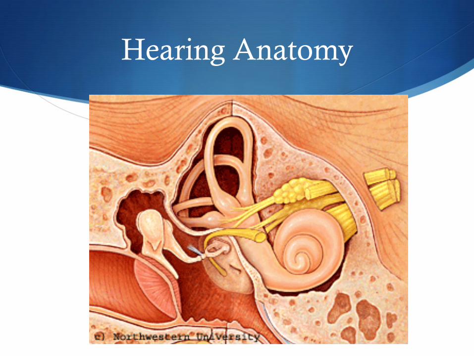
Hearing Anatomy
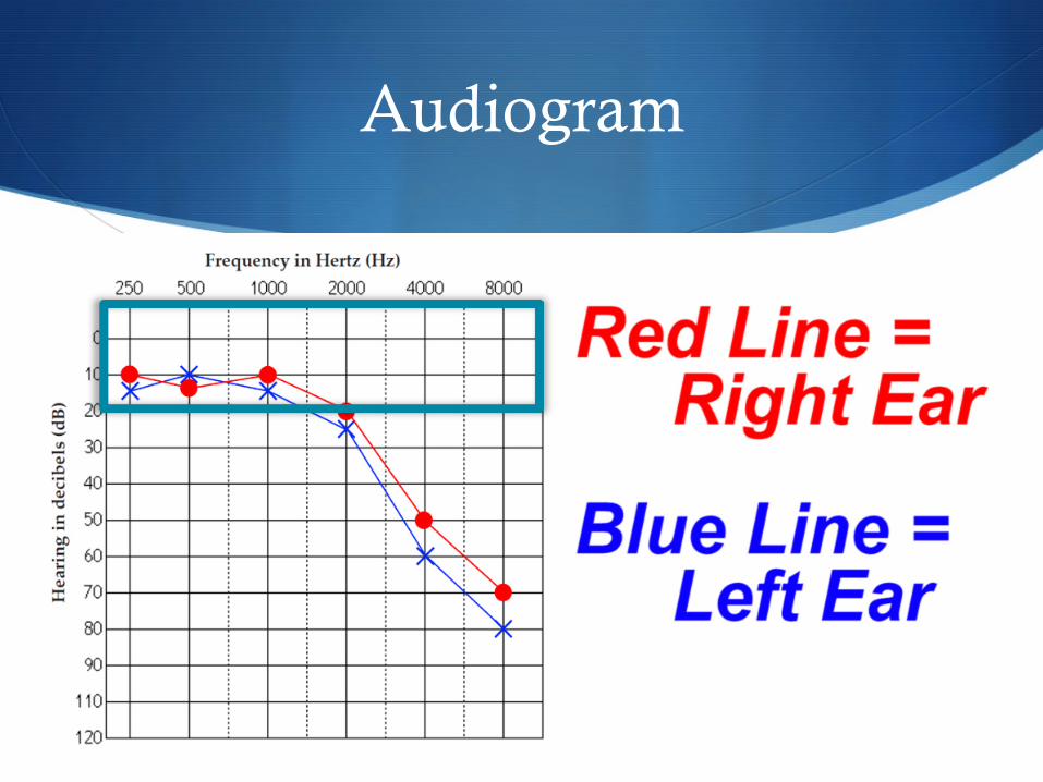
Audiogram
Types of Hearing Loss
S Sensorineural Hearing Loss (nerve loss)
S Conductive Hearing Loss
S Mixed Hearing Loss (both nerve and conductive loss)
Most Common Causes of CHL
1) Cerumen impaction
2) Otitis media with effusion
3) TM perforation
4) Otosclerosis
5) Foreign Body

Conductive Hearing Loss
External Ear Canal Cerumen impaction, foreign body
Tympanic Membrane Perforation, tympanosclerosis, Hematoma
Middle Ear Otitis Media with Effusion, Cholesteatoma
Ossicles Otosclerosis
Sensorineural Hearing Loss Etiologies
S Infectious: S Meningitis, Herpes virus, HIV, Mumps, Rubella, Rubeola, Mycoplasma, Toxoplasmosis, Syphillis, Lyme disease
S Autoimmune S Lupus erythematosus, Cogan’s syndrome, Wegener’s granulomatosis
S Traumatic S Perilymph fistula, T-bone fracture, Acute blast injury
SNHL Etiologies
S Vascular S Vertebrobasilar insufficiency (VBI), Sickle cell disease, Hyperviscosity syndromes, Waldenstrom’s macroglobulinemia,
Polycythemia vera, thrombocythemia
S Neurologic S Multiple sclerosis, Migraine,
S Neoplastic S Acoustic neuroma, Meningioma, Metastasis, Leukemia, Myeloma
SNHL Etiologies
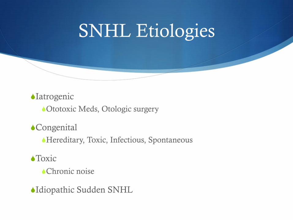
S Iatrogenic S Ototoxic Meds, Otologic surgery
S Congenital S Hereditary, Toxic, Infectious, Spontaneous
S Toxic S Chronic noise
S Idiopathic Sudden SNHL
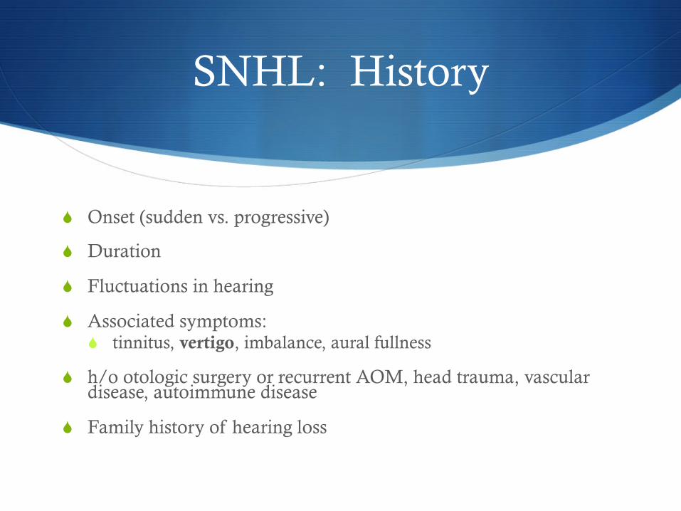
SNHL: History
S Onset (sudden vs. progressive)
S Duration
S Fluctuations in hearing
S Associated symptoms: S tinnitus, vertigo, imbalance, aural fullness
S h/o otologic surgery or recurrent AOM, head trauma, vascular disease, autoimmune disease
S Family history of hearing loss
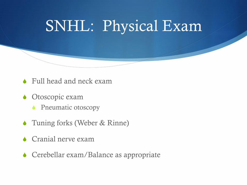
SNHL: Physical Exam
S Full head and neck exam
S Otoscopic exam S Pneumatic otoscopy
S Tuning forks (Weber & Rinne)
S Cranial nerve exam
S Cerebellar exam/Balance as appropriate
SNHL: Physical Exam
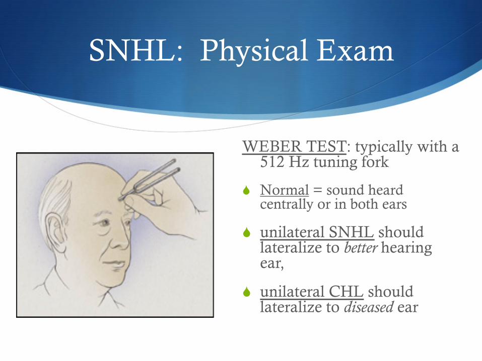
WEBER TEST: typically with a 512 Hz tuning fork
S Normal = sound heard centrally or in both ears
S unilateral SNHL should lateralize to better hearing ear,
S unilateral CHL should lateralize to diseased ear
SNHL: Physical Exam
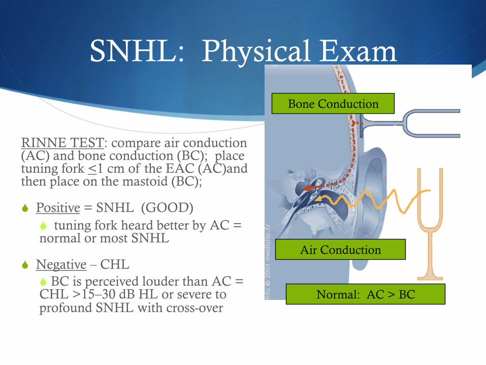
RINNE TEST: compare air conduction (AC) and bone conduction (BC); place tuning fork <1 cm of the EAC (AC)and then place on the mastoid (BC);
S Positive = SNHL (GOOD) S tuning fork heard better by AC = normal or most SNHL
S Negative – CHL S BC is perceived louder than AC = CHL >15–30 dB HL or severe to profound SNHL with cross-over
Bone Conduction
Air Conduction
Normal: AC > BC
SNHL: Workup
S Full audiogram with pure tones, speech recognition, and word recognition
S For sudden sensorineural hearing loss or asymmetric sensorineural hearing loss: MRI + gadolinium
Presbyacusis
S Age-related hearing loss
S 40% U.S. population >75 y/o affected
S Often familial (>50%)
S Bilateral and symmetric
Presbyacusis: Treatment
S Treatment S Hearing aids
S Assistive listening devices
S Cochlear Implantation
Noise Induced Hearing Loss (NIHL)
S Most common cause of preventable SNHL
S Most frequently occurs from exposure through years (> 90dB)
S Can result from single exposure to very loud noise (>120-130 dB)
S Typically bilateral and symmetric
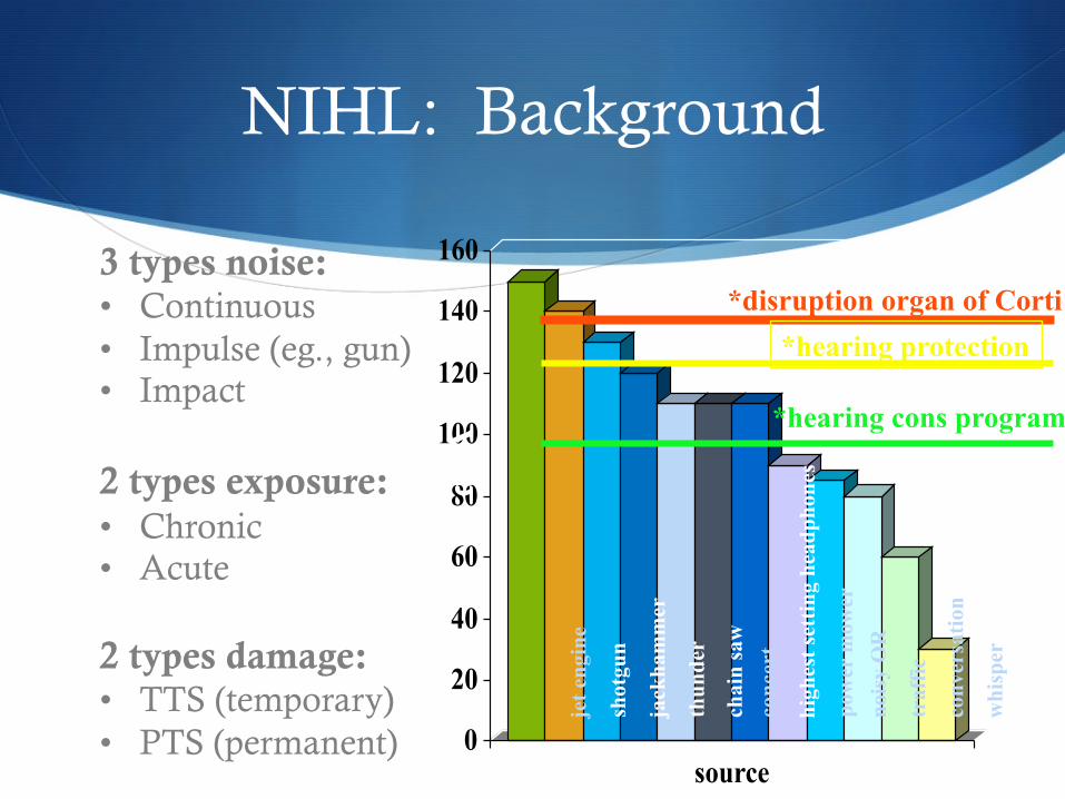
NIHL: Background
0
20
40
60
80
100
120
140
160
source
jet e
ngin
e
shot
gun
jack
ham
mer
thun
der
chai
n sa
w
conc
ert
high
est s
ettin
g he
adph
ones
pow
er m
ower
no
isy
OR
traf
fic
conv
ersa
tion
whi
sper
dBA
3 types noise: • Continuous • Impulse (eg., gun) • Impact 2 types exposure: • Chronic • Acute 2 types damage: • TTS (temporary) • PTS (permanent)
*disruption organ of Corti
*hearing cons program
*hearing protection
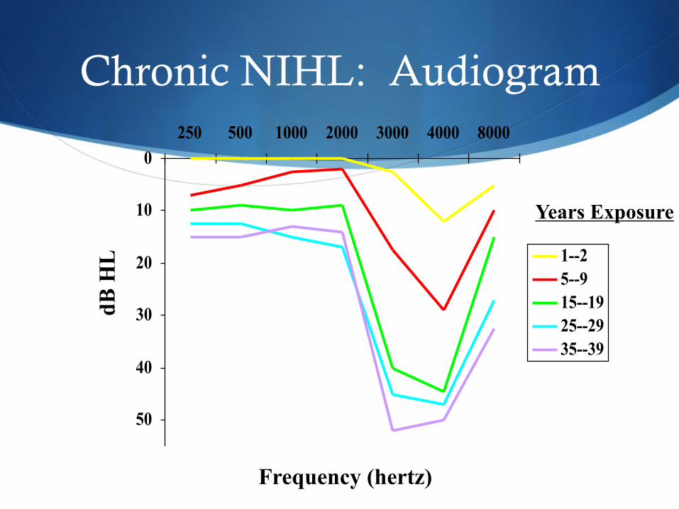
Chronic NIHL: Audiogram
0
10
20
30
40
50
250 500 1000 2000 3000 4000 8000
1--25--915--1925--2935--39
dB H
L
Frequency (hertz)
Years Exposure
Ototoxic medications
S Macrolides S High frequency SNHL, tinnitus, vertigo
S Usually reversible within 2 weeks
S Unknown mechanism
S Vancomycin S High freq SNHL progresses to bilateral profound SNHL
S ASA S Doses > 2700 mg/day
S Affects stria vascularis, reversible
Ototoxic Medications
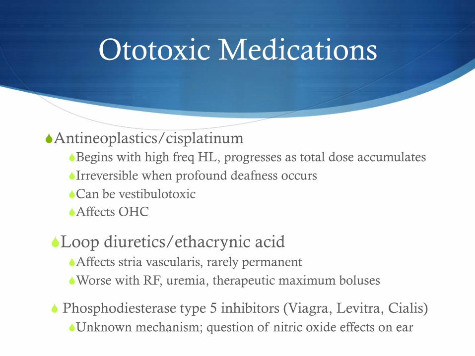
S Antineoplastics/cisplatinum S Begins with high freq HL, progresses as total dose accumulates S Irreversible when profound deafness occurs S Can be vestibulotoxic S Affects OHC
S Loop diuretics/ethacrynic acid S Affects stria vascularis, rarely permanent S Worse with RF, uremia, therapeutic maximum boluses
S Phosphodiesterase type 5 inhibitors (Viagra, Levitra, Cialis) S Unknown mechanism; question of nitric oxide effects on ear
Perilymphatic Fistula (PLF)
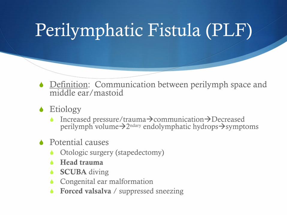
S Definition: Communication between perilymph space and middle ear/mastoid
S Etiology S Increased pressure/traumaàcommunicationàDecreased
perilymph volumeà2ndary endolymphatic hydropsàsymptoms
S Potential causes S Otologic surgery (stapedectomy) S Head trauma S SCUBA diving S Congenital ear malformation S Forced valsalva / suppressed sneezing
Neoplasia
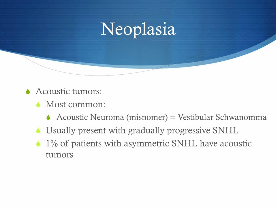
S Acoustic tumors: S Most common:
S Acoustic Neuroma (misnomer) = Vestibular Schwanomma
S Usually present with gradually progressive SNHL S 1% of patients with asymmetric SNHL have acoustic
tumors
Idiopathic Sudden Sensorineural Hearing Loss (ISSNHL)
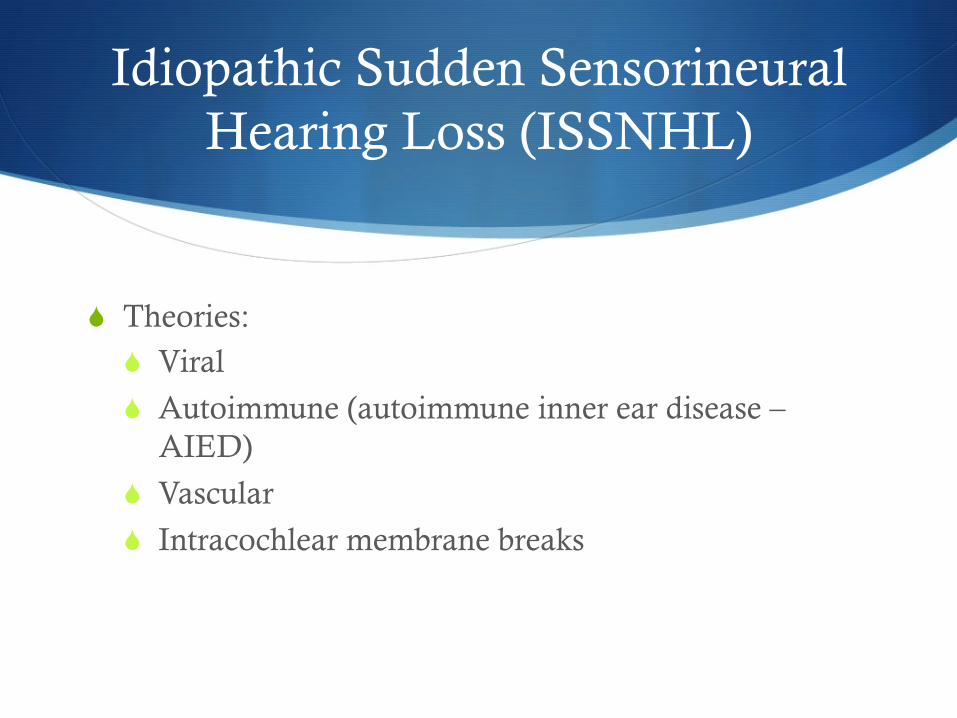
S Theories: S Viral
S Autoimmune (autoimmune inner ear disease – AIED)
S Vascular
S Intracochlear membrane breaks
ISSNHL: Viral
S Current belief – viral cochleitis causes the majority of cases of ISSNHL
S 1983 – Wilson and colleagues S Viral seroconversion rates greater in patients with
ISSNHL (63%) compared to control (40%) S Influenza B S Mumps S Rubeola S VZV
ISSNHL: Viral
S 1981- Veltri et al. S 65% seroconversion
S 1986 – Schuknecht and Donovan S Temporal bone studies (n. 12)
S ISSNHL vs. cases of known viral labyrinthitis
S Similar pathologic findings
S Atrophy of the organ of Corti, tectorial membrane, stria vascularis, cochlear nerve, and vestibular organ

ISSNHL: Treatment
S 90% of cases will be Idiopathic
S Treat known causes by addressing the underlying condition
ISSHNL: Treatment
S Therapy for ISSNHL is controversial
S Difficult to study S High spontaneous recovery rate
S Low incidence
S Makes validation of empiric treatment modalities difficult
ISSNHL: Treatment
S Proposed treatment modalities S Anti-inflammatory – steroids, cytotoxic agents
S Diuretics
S Antiviral agents
S Vasodilators
S Volume expanders/hemodilutors
S Defibrinogenators
Treatment
S Acyclovir S 1999 -Stokroos and Albers
S Showed therapeutic efficacy of combined steroid and acyclovir in experimental HSV-1 viral labyrinthitis S Earlier hearing recovery S Less extensive cochlear destruction
S 1996 – Adour et al. S Combination therapy shown to be beneficial for tx of Bell’s
palsy S Benefit of combined therapy has been shown in patients
with Ramsay Hunt syndrome
Treatment
S 2000 survey of 100 ENTs (43% otologists) in the United Kingdom S 78% - CBC, ESR, Syphilis serology
S 38% - MRI on initial visit
S 98.5% - steroids
S 41% - Carbogen
S 31% - acyclovir
Autoimmune Inner Ear Disease (AIED)
S 1979 – McCabe S Described patients with bilateral rapidly-progressive SNHL
(BRPSNHL)
S Proposed the term – autoimmune inner ear disease (AIED)
S Evidence of autoimmunity S Lymphocyte inhibition test
S Substantial hearing improvement with steroids
AIED
S Clinical characteristics S Middle-aged females S BPRSNHL S Absence of systemic immune disease S 50% with dizziness S Light-headedness and ataxia more common than vertigo S Episodes – multiple, daily S Hearing loss sudden, rapidly progressive, or protracted
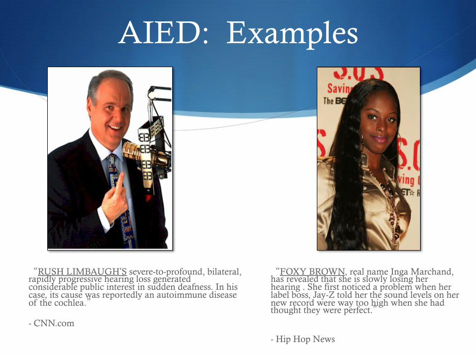
“RUSH LIMBAUGH’S severe-to-profound, bilateral, rapidly progressive hearing loss generated considerable public interest in sudden deafness. In his case, its cause was reportedly an autoimmune disease of the cochlea.”
- CNN.com
“FOXY BROWN, real name Inga Marchand, has revealed that she is slowly losing her hearing . She first noticed a problem when her label boss, Jay-Z told her the sound levels on her new record were way too high when she had thought they were perfect.”
- Hip Hop News
AIED: Examples
AIED
S Diagnosis S Based on Hearing loss and response to treatment S Hughes –
S Lymphocyte transformation test S Sensitivity – 50-80% S Specificity – 93% S Positive predictive value 56-73%
S Western blot S Sensitivity – 88% S Specificity – 80% S Positive predictive value – 92%
AIED
S 1990 – Harris and Colleagues S Used Western blot to discover anti 68KD autoantibody in sera
of patients with ISSNHL
S 22%-58% will have +test
S 94% specificity
S However, current studies are discounting the 68KD test as invalid
AIED
S Further studies S Billings and Harris
S Linkage of 68KD protein to heat shock protein 70 (hsp 70)
S Theories 1. Cross reactivity
2. Over expression leads to autoimmunity
AIED Treatment
1. Prednisone 1mg/Kg/day for 4 weeks
2. Slow taper
3. Relapse during taper – restart
4. Slow taper
5. If relapse during taper – Cytotoxic agent S Methotrexate
S Cyclophosphamide
S Monitor electrolytes, LFTs, blood counts
Vascular
S Embolism, vasospasm, hypercoagulable states/sludging
S Pathophysiology – anoxia to vestibulocochlear apparatus
S Cochlea is intolerant to disruption of blood supply S 1957 Kimura and Perlman
S Clamped the labyrinthine artery in guinea pigs S Demonstrated irreversible loss of cochlear function after 30
minutes of disruption
Vascular
S 1980 – Belal S Examined two temporal bones of patients with SHL
S Histopathology was similar to animal models of vascular occlusion
S Extensive fibrosis and ossification

Vascular-histopathology
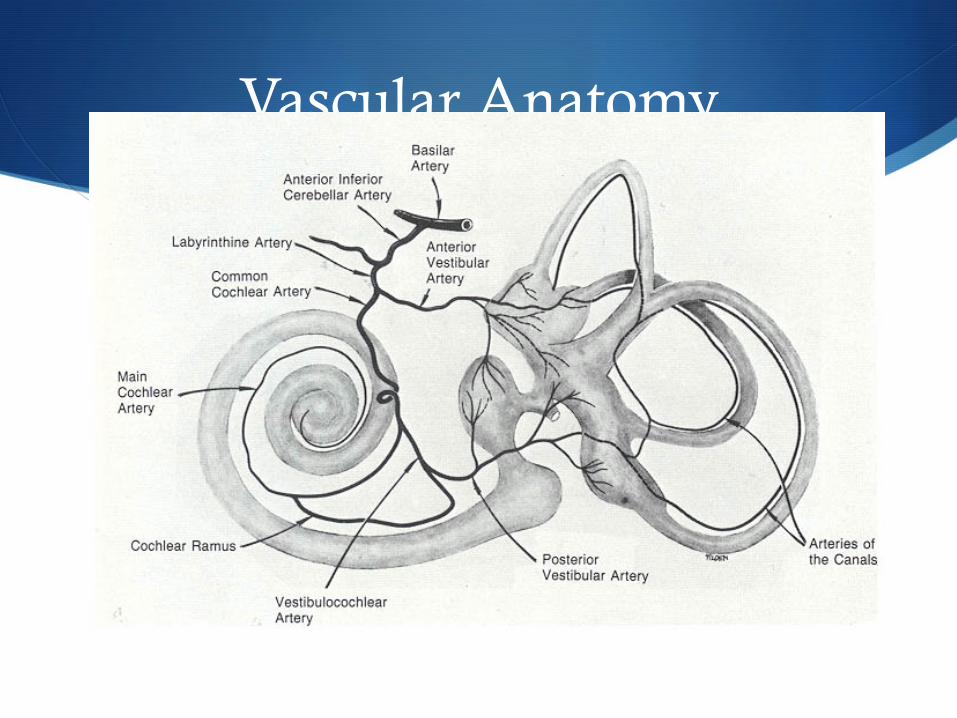
Vascular Anatomy

Vascular
S Abnormal circulatory states S Sickle-cell disease
S Waldenstrom’s macroglobulinemia
S Hearing loss is usually reversible with tx
S AICA strokes
S Cardiopulmonary bypass
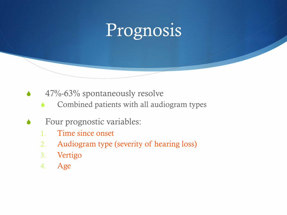
Prognosis
S 47%-63% spontaneously resolve S Combined patients with all audiogram types
S Four prognostic variables: 1. Time since onset 2. Audiogram type (severity of hearing loss) 3. Vertigo 4. Age
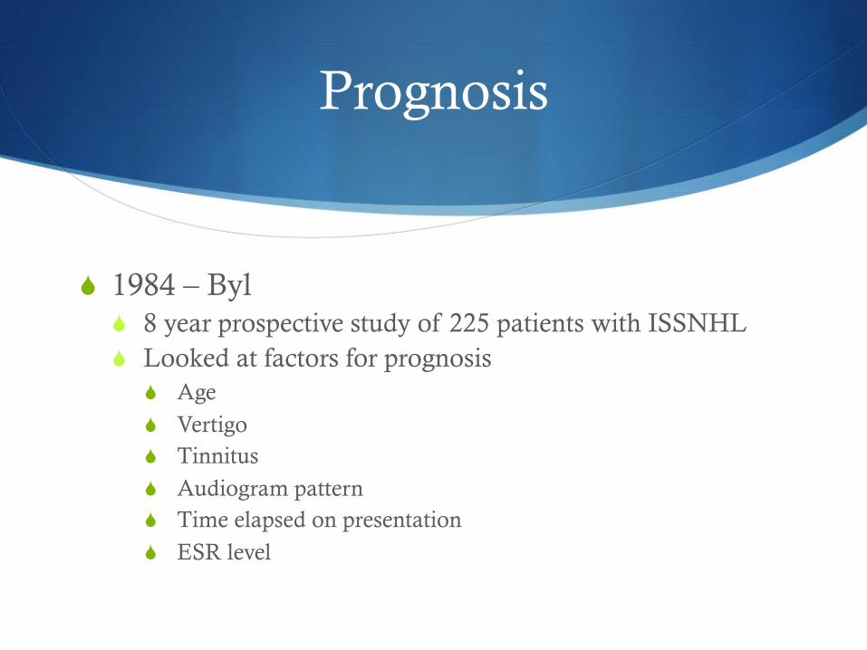
Prognosis
S 1984 – Byl S 8 year prospective study of 225 patients with ISSNHL S Looked at factors for prognosis
S Age
S Vertigo S Tinnitus
S Audiogram pattern S Time elapsed on presentation
S ESR level
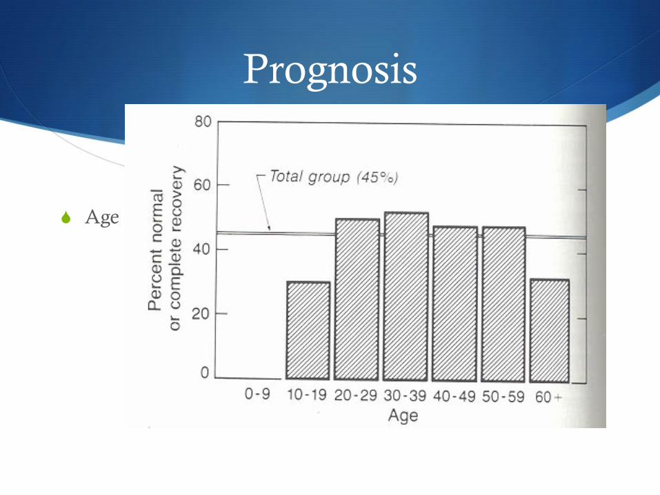
Prognosis
S Age
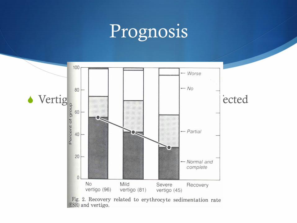
Prognosis
S Vertigo – 29% affected vs. 55% not affected
Prognosis
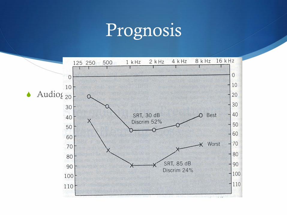
S Audiogram type
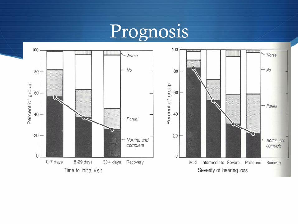
Prognosis
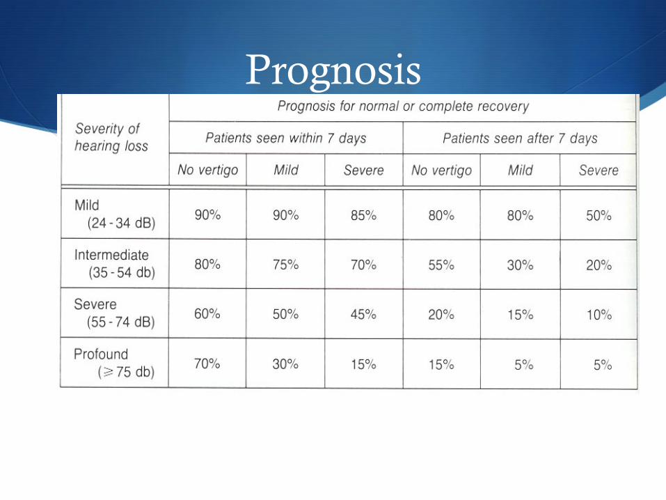
Prognosis

S
Vertigo
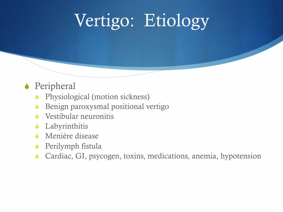
Vertigo: Etiology
S Peripheral S Physiological (motion sickness) S Benign paroxysmal positional vertigo S Vestibular neuronitis S Labyrinthitis S Meniére disease S Perilymph fistula S Cardiac, GI, psycogen, toxins, medications, anemia, hypotension
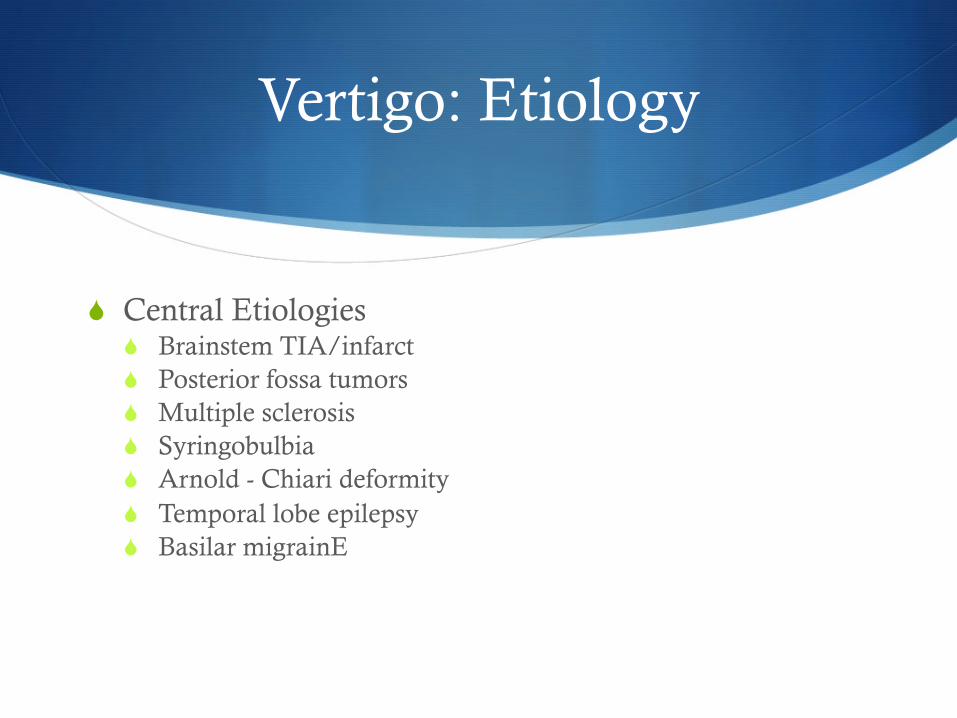
Vertigo: Etiology
S Central Etiologies S Brainstem TIA/infarct S Posterior fossa tumors S Multiple sclerosis S Syringobulbia S Arnold - Chiari deformity S Temporal lobe epilepsy S Basilar migrainE
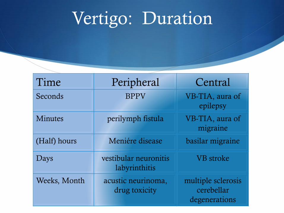
Vertigo: Duration
Time Peripheral Central Seconds BPPV VB-TIA, aura of
epilepsy
Minutes perilymph fistula VB-TIA, aura of migraine
(Half) hours Meniére disease basilar migraine
Days vestibular neuronitis labyrinthitis
VB stroke
Weeks, Month acustic neurinoma, drug toxicity
multiple sclerosis cerebellar
degenerations
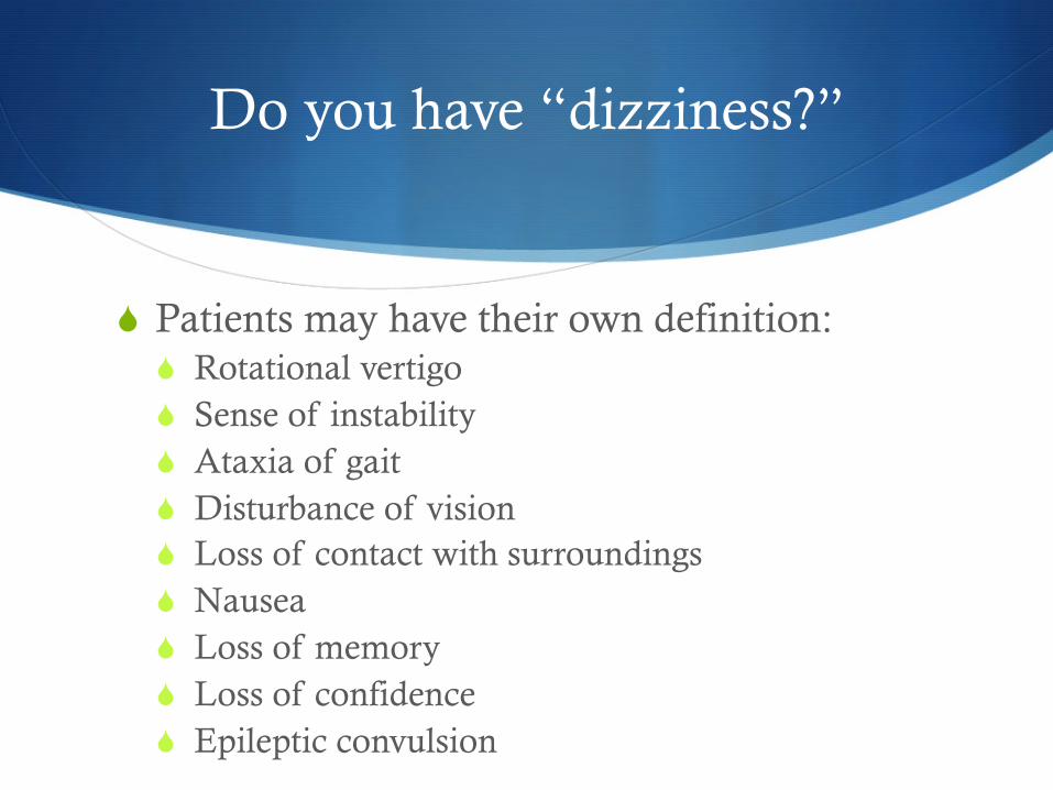
Do you have “dizziness?”
S Patients may have their own definition: S Rotational vertigo S Sense of instability S Ataxia of gait S Disturbance of vision S Loss of contact with surroundings S Nausea S Loss of memory S Loss of confidence S Epileptic convulsion
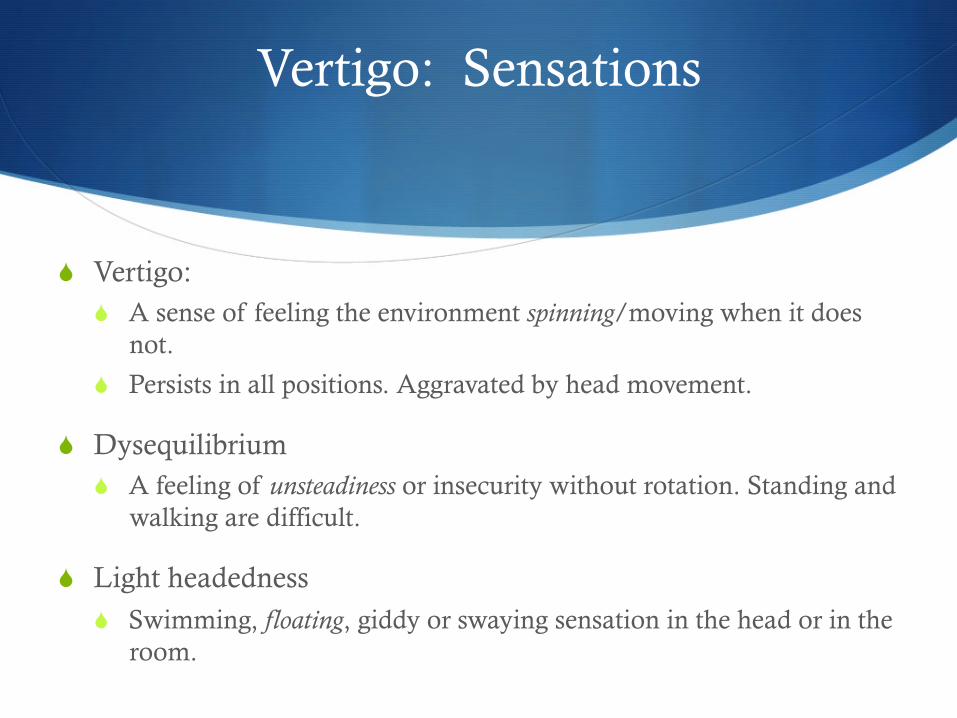
Vertigo: Sensations
S Vertigo: S A sense of feeling the environment spinning/moving when it does
not.
S Persists in all positions. Aggravated by head movement.
S Dysequilibrium S A feeling of unsteadiness or insecurity without rotation. Standing and
walking are difficult.
S Light headedness S Swimming, floating, giddy or swaying sensation in the head or in the
room.
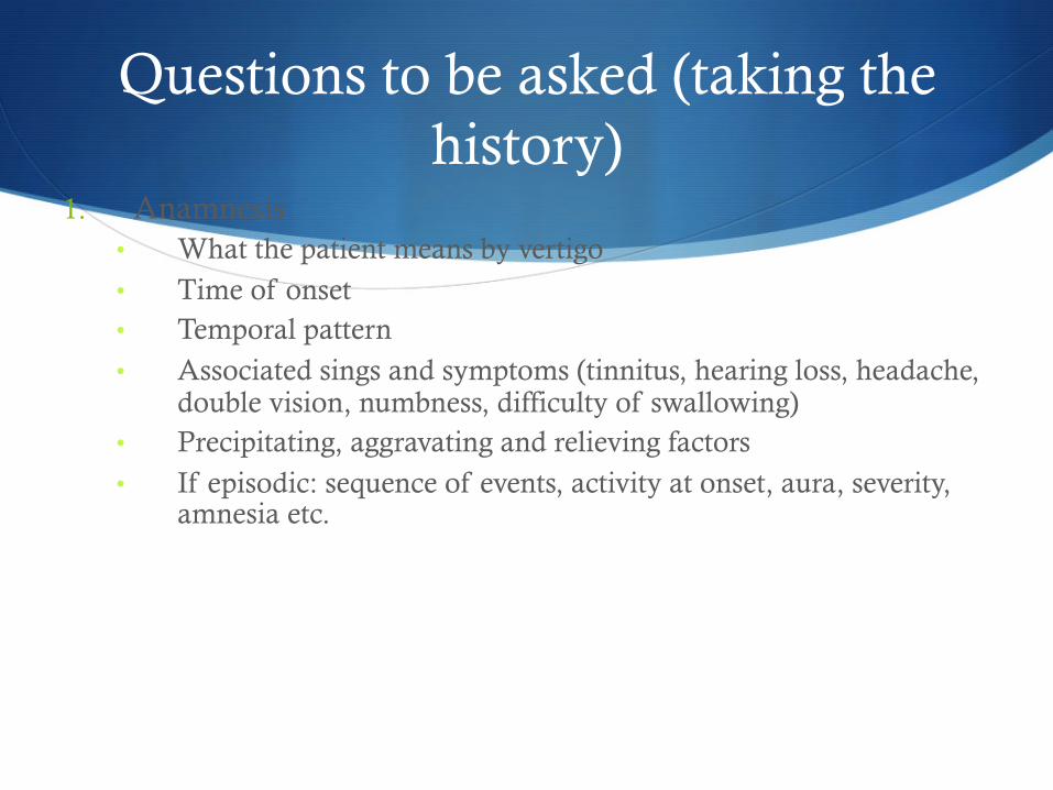
Questions to be asked (taking the history)
1. Anamnesis • What the patient means by vertigo • Time of onset • Temporal pattern • Associated sings and symptoms (tinnitus, hearing loss, headache,
double vision, numbness, difficulty of swallowing) • Precipitating, aggravating and relieving factors • If episodic: sequence of events, activity at onset, aura, severity,
amnesia etc.
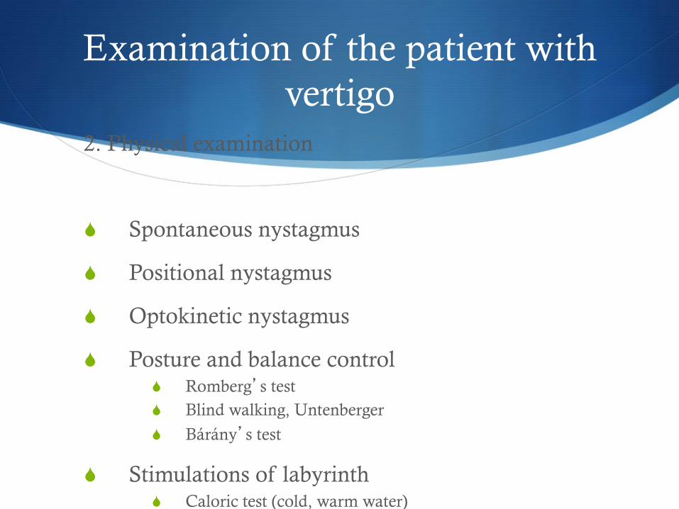
Examination of the patient with vertigo
2. Physical examination
S Spontaneous nystagmus
S Positional nystagmus
S Optokinetic nystagmus
S Posture and balance control S Romberg’s test S Blind walking, Untenberger
S Bárány’s test
S Stimulations of labyrinth S Caloric test (cold, warm water)
S Rotational test
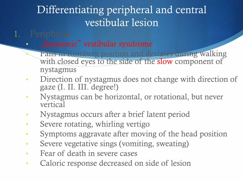
Differentiating peripheral and central vestibular lesion
1. Peripheral • „harmonic” vestibular syndrome • Falls in Romberg position and deviates during walking
with closed eyes to the side of the slow component of nystagmus
• Direction of nystagmus does not change with direction of gaze (I. II. III. degree!)
• Nystagmus can be horizontal, or rotational, but never vertical
• Nystagmus occurs after a brief latent period • Severe rotating, whirling vertigo • Symptoms aggravate after moving of the head position • Severe vegetative sings (vomiting, sweating) • Fear of death in severe cases • Caloric response decreased on side of lesion
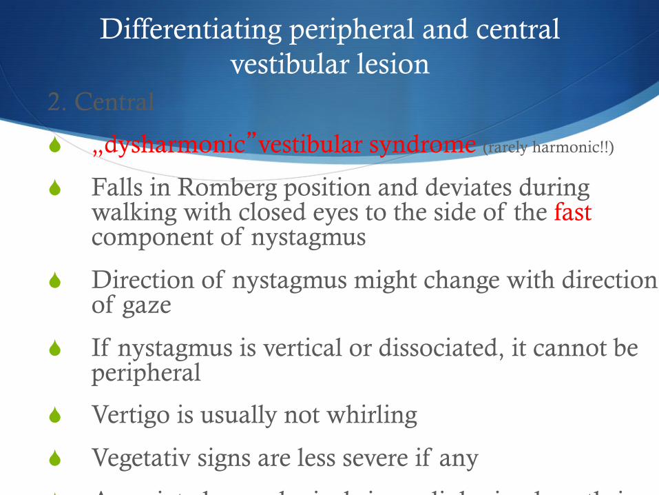
2. Central
S „dysharmonic”vestibular syndrome (rarely harmonic!!)
S Falls in Romberg position and deviates during walking with closed eyes to the side of the fast component of nystagmus
S Direction of nystagmus might change with direction of gaze
S If nystagmus is vertical or dissociated, it cannot be peripheral
S Vertigo is usually not whirling
S Vegetativ signs are less severe if any
S Associated neurological signs: diplopia, dysarthria, dysphagia, numbness, paresis, ataxia.
Differentiating peripheral and central vestibular lesion
Examination of the patient with vertigo
S Laboratory examinations and imaging S Videonystagmography
S Audiometry
S CT
S MRI
Peripheral Vertigo
S Benign paroxysmal positional vertigo S Most often
S Lasts less than 30 seconds
S Occurs only with a change in head position
S Nystagmus is transient, fatigable and its direction is constant
S Reason: otoconia
S Positional vertigo is not always benign and not always vestibular in origin!

Left Right
HC HC
AC AC
PC PC
+
-
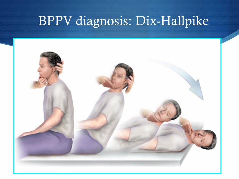
BPPV diagnosis: Dix-Hallpike
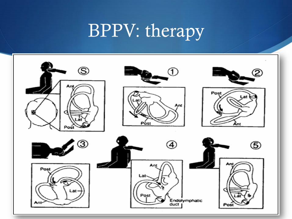
BPPV: therapy
Semont Brandt-Daroff
Vestibular Neuronitis
S Sudden severe vertigo
S No cochlear symptoms (tinnitus, hearing loss)
S Reduced caloric reaction on affected side
S Recurrent attacks
S Lasts for several days
Vestibular Neuronitis
S Etiology: viral infection, vascular or unknown origin
S Therapy: S 1-3. days. bedrest, vestibular suppressants (diazepam,
clonazepam) antiemetics, vitamin B S antiviral agents (?), corticosteriods(?) S position training
S Labyrinthitis S As vestibular neuronitis, but there are also
cochlear symptoms.
Menière’s disease
S Recurrent fluctuating attacks: 1. Tinnitus 2. Progressive hearing loss, unilateral first 3. Vertigo for at least 5 to 30 min 4. Aural Pressure
Menière’s disease
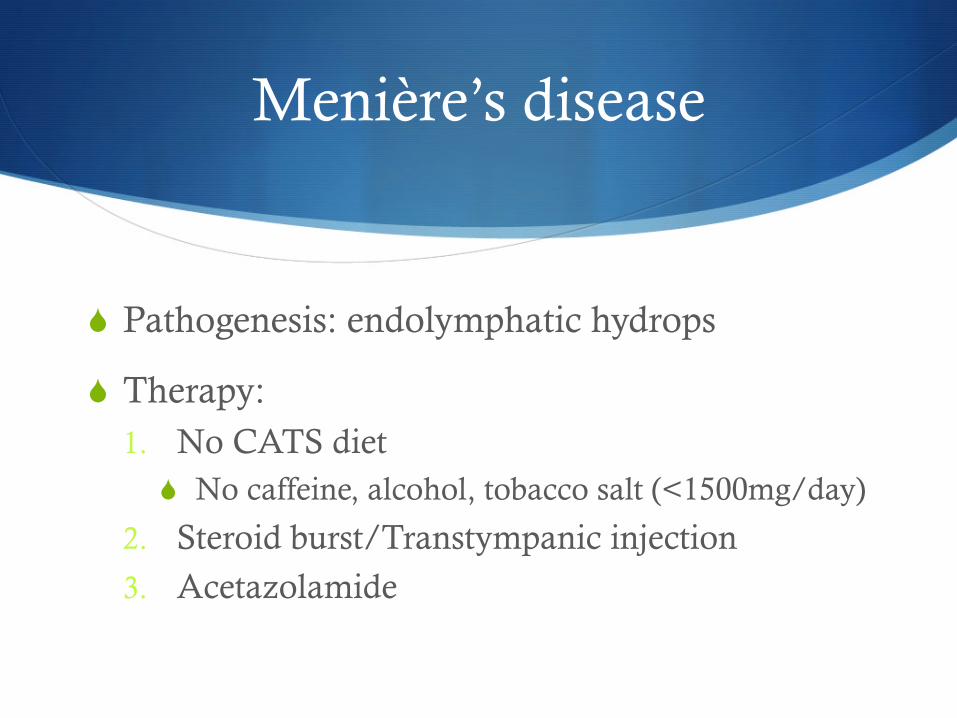
S Pathogenesis: endolymphatic hydrops
S Therapy: 1. No CATS diet
S No caffeine, alcohol, tobacco salt (<1500mg/day)
2. Steroid burst/Transtympanic injection
3. Acetazolamide
5. Perilymphatic fistula
S Fistula of the round window
S Hearing loss with or without vertigo
S Sudden changes of pressure in the middle ear (weight lifting, diving, nose blowing)

Drug toxicity
S Aminoglycoside antibiotics
S Anticonvulsants
S Salycilates
S Alcohol
S Sedatives
S Antihistamines
S Antidepressants
Other causes of vertigo
S Cervical spondylosis
S Sensory deprivation (neuropathy, visual impairment)
S Anemia
S Hypoglycaemia
S Orthostatic hypotension
S Hyperventilation

S
Tinnitus
Introduction
S Tinnitus -“The subjective perception of sound in the absence of external stimuli.”
S Tinnere – “ringing” in Latin
S Characteristically described as: S Buzzing, crickets, static, roaring, clicking, pulsatile, ringing
sounds
Tinnitus: Epidemiology
S 40 million affected in the United States
S 10 million severely affected – quality of life issues
S Most common in 40-70 year-olds
S More common in men than women
Tinnitus: Classification
S Objective tinnitus – sound produced by structures outside the ears, which may be heard by an examiner
S Subjective tinnitus – sound is only perceived by the patient (most common)
Tinnitus
S Pulsatile tinnitus – matches pulse or a rushing sound S Possible vascular etiology
S Either objective or subjective
S Increased or turbulent bloodflow through para-auditory structures
Subjective Tinnitus
S Usually nonpulsatile
S Presbycusis
S Noise exposure
S Meniere’s disease
S Otosclerosis
S Head trauma
S Acoustic neuroma
S Drugs
S Middle ear effusion
S TMJ problems
S Depression
S Hyperlipidemia
S Meningitis
S Syphilis
CNS Mechanisms
S Reorganization of central pathways with hearing loss (similar to phantom limb pain)
S Disinhibition of dorsal cochlear nucleus with increase in spontaneous activity of central auditory system
Neurophysiologic Model
S Proposed by Jastreboff
S Result of interaction of subsystems in the nervous system
S Auditory pathways playing a role in development and appearance of tinnitus
S Limbic system responsible for tinnitus annoyance
S Negative reinforcement enhances perception of tinnitus and increases time it is perceived
Role of Depression
S Depression is more prevalent in patients with chronic tinnitus than in those without tinnitus
S Folmer et al (1999) reported patients with depression rated the severity of their tinnitus higher although loudness scores were the same
S Which comes first, depression or tinnitus?
Pulsatile Tinnitus: Etiologies
S Arteriovenous malformations
S Vascular tumors
S Venous hum
S Atherosclerosis
S Ectopic carotid artery
S Persistent stapedial artery
S Dehiscent jugular bulb
S Vascular loops
S Cardiac murmurs
S Pregnancy
S Anemia
S Thyrotoxicosis
S Paget’s disease
S Benign intracranial hypertension
Arteriovenous malformations
S Congenital lesions
S Occipital artery and transverse sinus, internal carotid and vertebral arteries, middle meningeal and greater superficial petrosal arteries
S Mandible
S Brain parenchyma
S Dura
Arteriovenous malformations
S Pulsatile tinnitus
S Headache
S Papilledema
S Discoloration of skin or mucosa
Vascular Tumors
S Glomus tympanicum S Paraganglioma of middle ear
S Pulsatile tinnitus which may decrease with ipsilateral carotid artery compression
S Reddish mass behind tympanic membrane which blanches with positive pressure
S Conductive hearing loss
Vascular Tumors
S Glomus jugulare S Paraganlioma of jugular fossa
S Pulsatile tinnitus S Conductive hearing loss if into middle ear
S Cranial neuropathies
Venous Hum
S Benign intracranial hypertension
S Dehiscent jugular bulb
S Transverse sinus partial obstruction
S Increased cardiac output from S Pregnancy
S Thyrotoxicosis
S Anemia
Benign Intracranial Hypertension
S Young, obese, female patients
S Hearing loss
S Aural fullness
S Dizziness
S Headaches
S Visual disturbance
S Papilledema, pressure >200mm H20 on LP
Benign Intracranial Hypertension
S Sismanis and Smoker 1994 S 100 patients with pulsatile tinnitus
S 42 found to have BIH syndrome
S 16 glomus tumors
S 15 atherosclerotic carotid artery disease
BIH Syndrome
S Treatment S Weight loss
S Diuretics S Subarachnoid-peritoneal shunt
S Gastric bypass for weight reduction
Muscular Causes of Tinnitus
S Palatal myoclonus S Clicking sound
S Rapid (60-200 beats/min), intermittent
S Contracture of tensor palantini, levator palatini, levator veli palatini, tensor tympani, salpingopharyngeal, superior constrictors
S Muscle spasm seen orally or transnasally
S Rhythmic compliance change on tympanogram
Myoclonus
S Palatal myoclonus associations: S Multiple Sclerosis and other degenerative neurological
disorders
S Small vessel disease
S Tumors
S Treatments: muscle relaxants, botulinum toxin injection
Stapedius Muscle Spasm
S Idiopathic stapedial muscle spasm S Rough, rumbling, crackling sound
S Exacerbated by outside sounds
S Brief and intermittent
S May be able to see tympanic membrane movement
S Treatments: avoidance of stimulants, muscle relaxants, sometimes surgical division of tensor tympani and stapedius muscles
Patulous Eustachian Tube
S Eustachian tube remains open abnormally
S Ocean roar sound S Changes with respiration
S Lying down or head in dependent position provides relief
Patulous Eustachian Tube
S Tympanogram will show changes in compliance with respiration
S Significant weight loss, radiation to the nasopharynx
S Previous treatments: caustics, mucosal irritants, saturated solution of potassium iodide, Teflon or gelfoam injection around torus tubarius
Drugs that cause tinnitus
S Antinflammatories
S Antibiotics (aminoglycosides)
S Antidepressants (heterocyclines)
S Aspirin
S Quinine
S Loop diuretics
S Chemotherapeutic agents (cisplatin, vincristine)
Tinnitus: History
S Careful history
S Quality
S Pitch
S Loudness
S Constant/intermittent
S Onset
S Alleviating/aggravating factors
Tinnitus: History
S Infection
S Trauma
S Noise exposure
S Medication usage
S Medical history
S Hearing loss
S Vertigo
S Pain
S Family history
S Impact on patient
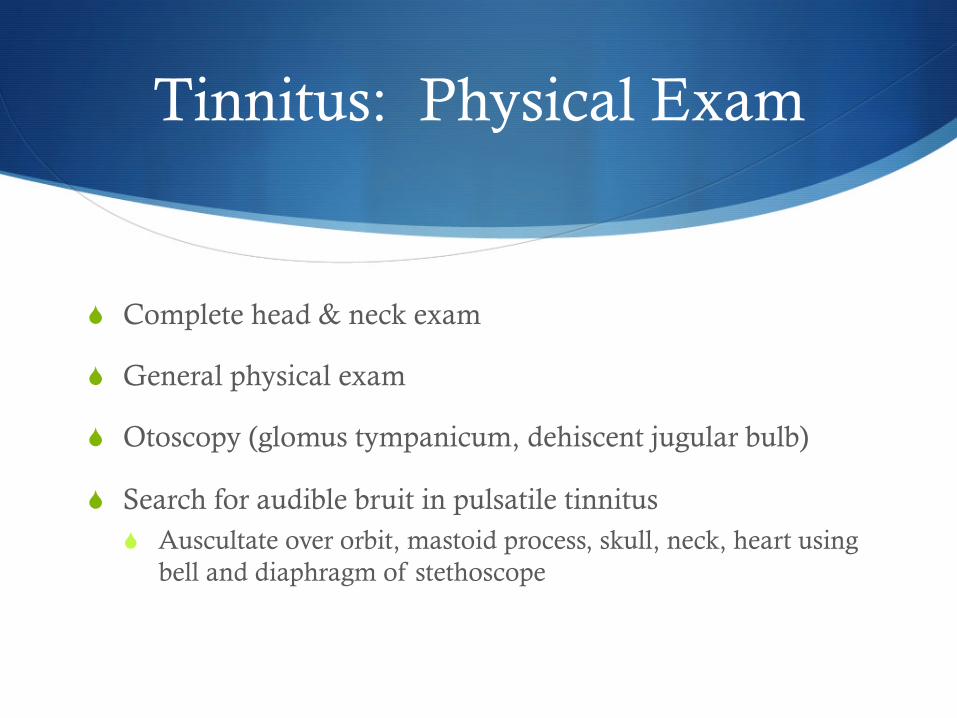
Tinnitus: Physical Exam
S Complete head & neck exam
S General physical exam
S Otoscopy (glomus tympanicum, dehiscent jugular bulb)
S Search for audible bruit in pulsatile tinnitus S Auscultate over orbit, mastoid process, skull, neck, heart using
bell and diaphragm of stethoscope
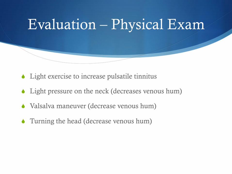
Evaluation – Physical Exam
S Light exercise to increase pulsatile tinnitus
S Light pressure on the neck (decreases venous hum)
S Valsalva maneuver (decrease venous hum)
S Turning the head (decrease venous hum)

Tinnitus: Audiometry
S Pure Tone Averages (PTA), speech discrimination scores, tympanometry, acoustic reflexes
S Pitch matching
S Loudness matching
S Masking level
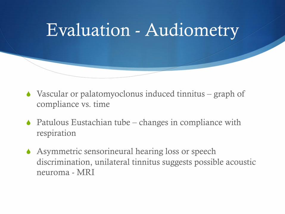
Evaluation - Audiometry
S Vascular or palatomyoclonus induced tinnitus – graph of compliance vs. time
S Patulous Eustachian tube – changes in compliance with respiration
S Asymmetric sensorineural hearing loss or speech discrimination, unilateral tinnitus suggests possible acoustic neuroma - MRI
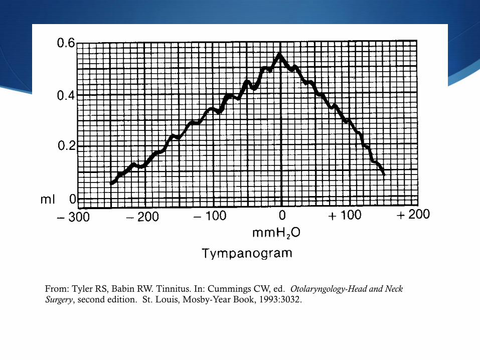
From: Tyler RS, Babin RW. Tinnitus. In: Cummings CW, ed. Otolaryngology-Head and Neck Surgery, second edition. St. Louis, Mosby-Year Book, 1993:3032.
Laboratory studies
S As indicated by history and physical exam
S Possibilities include: S Hematocrit
S FTA absorption test
S Blood chemistries
S Thyroid studies
S Lipid battery
Imaging
S Pulsatile tinnitus
S Reviewed by Weissman and Hirsch (2000)
S Contrast enhanced CT of temporal bones, skull base, brain, calvaria as first-line study
S Sismanis and Smoker (1994) recommended CT for retrotympanic mass, MRI/MRA if normal otoscopy

S Glomus tympanicum – bone algorithm CT scan best shows extent of mass
S May not be able to see enhancement of small tumor
S Tumor enhances on T1-weighted images with gadolinium or on T2-weighted images
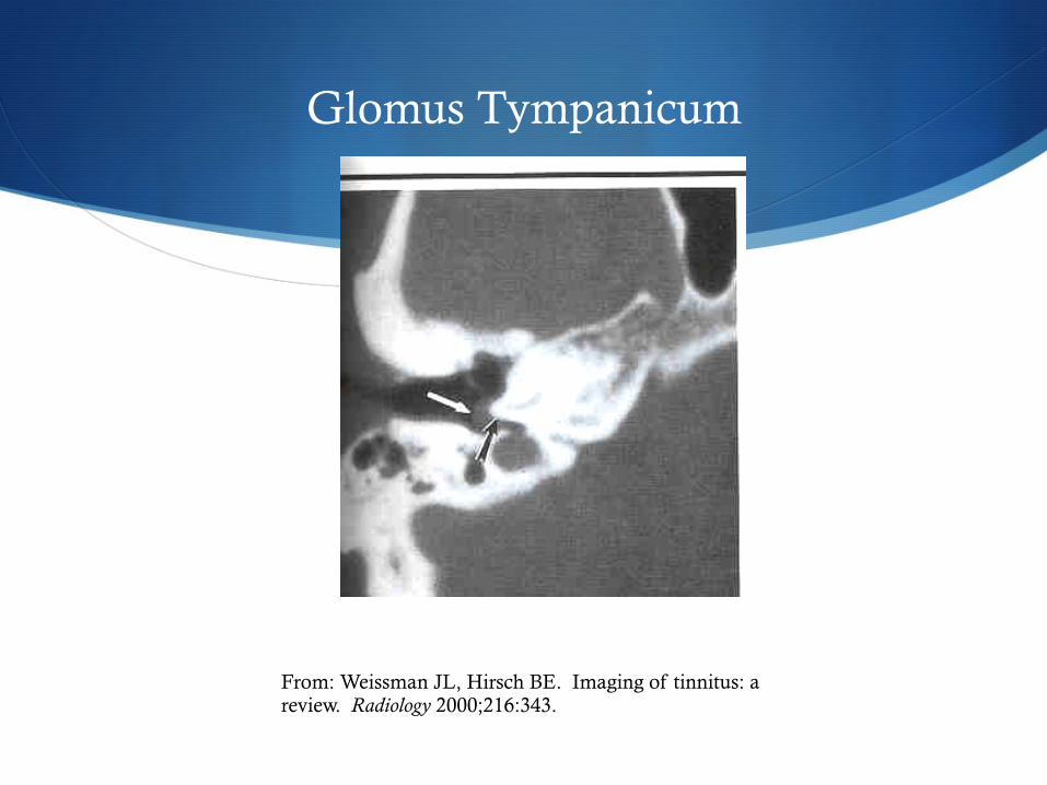
Glomus Tympanicum
From: Weissman JL, Hirsch BE. Imaging of tinnitus: a review. Radiology 2000;216:343.
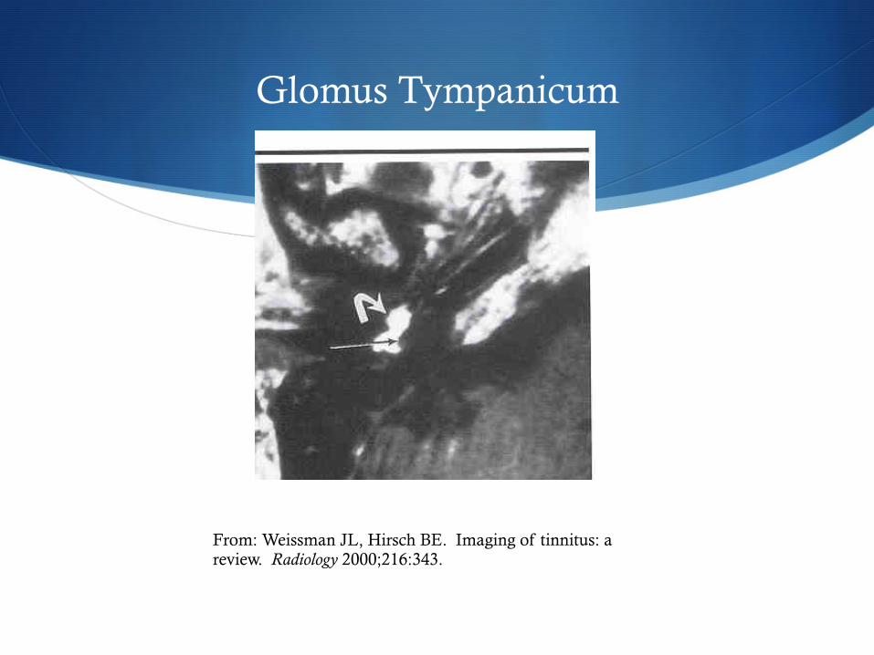
Glomus Tympanicum
From: Weissman JL, Hirsch BE. Imaging of tinnitus: a review. Radiology 2000;216:343.

Imaging
S Glomus jugulare S Erosion of osseous jugular fossa
S Enhance with contrast, may not be able to differentiate jugular vein and tumor
S Enhance with T1-weighted MRI with gadolinium and on T2-weighted images
S Characteristic “salt and pepper” appearance on MRI
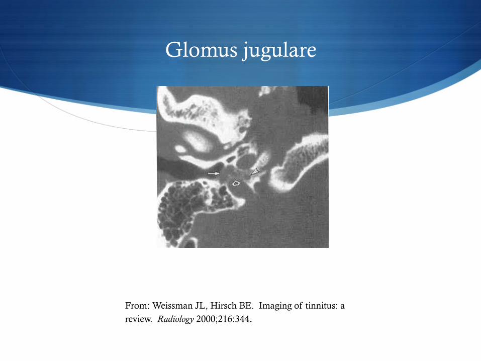
Glomus jugulare
From: Weissman JL, Hirsch BE. Imaging of tinnitus: a
review. Radiology 2000;216:344.
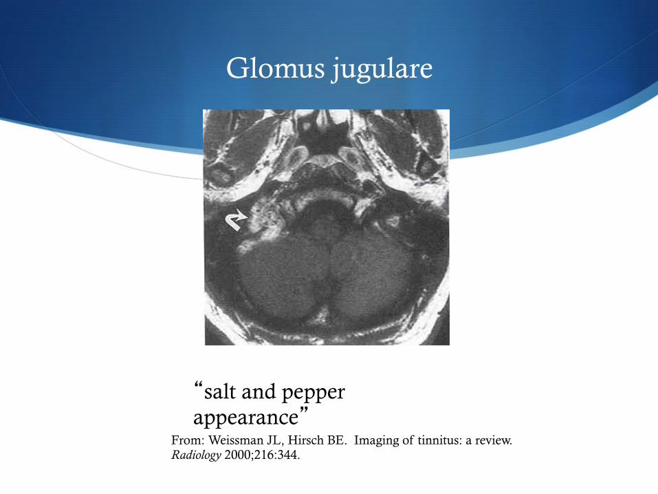
Glomus jugulare
“salt and pepper appearance”
From: Weissman JL, Hirsch BE. Imaging of tinnitus: a review. Radiology 2000;216:344.
Imaging
S Arteriovenous malformations – readily apparent on contrasted CT and MRI
S Normal otoscopic exam and pulsatile tinnitus may be dural arteriovenous fistula S Often invisible on contrasted CT and MRI/MRA
S Angiography may be only diagnostic test
Imagining
S Shin et al (2000) S MRI/MRA initially if subjective pulsatile tinnitus
S Angiography if objective with audible bruit in order to identify dural arteriovenous fistula
Imaging
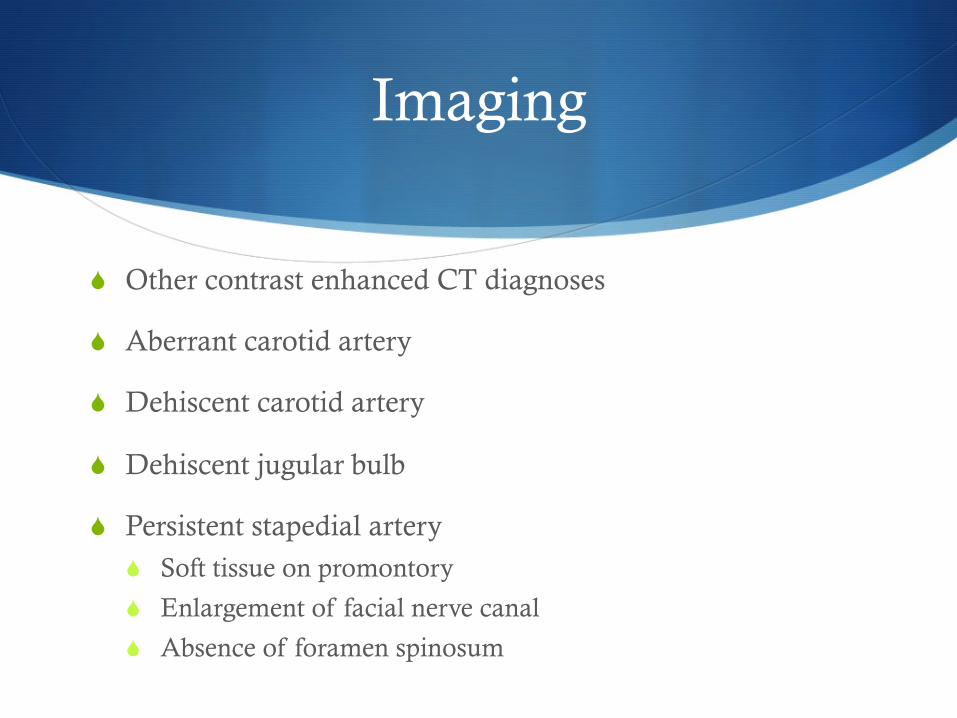
S Other contrast enhanced CT diagnoses
S Aberrant carotid artery
S Dehiscent carotid artery
S Dehiscent jugular bulb
S Persistent stapedial artery S Soft tissue on promontory
S Enlargement of facial nerve canal
S Absence of foramen spinosum
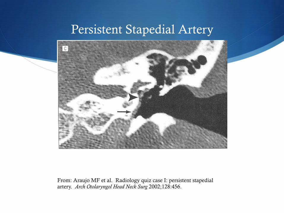
Persistent Stapedial Artery
From: Araujo MF et al. Radiology quiz case I: persistent stapedial artery. Arch Otolaryngol Head Neck Surg 2002;128:456.
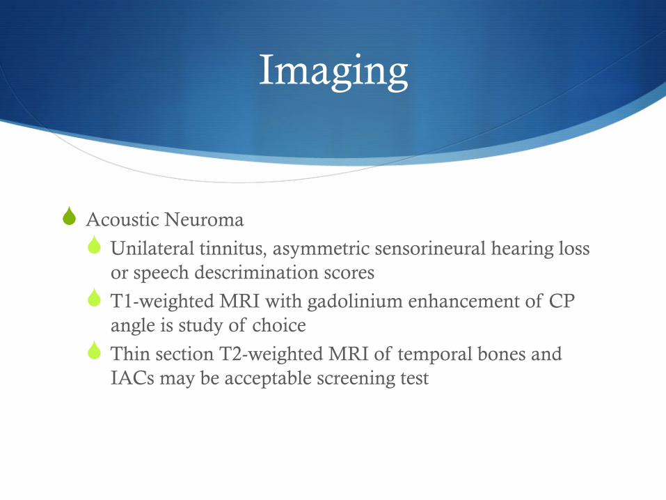
Imaging
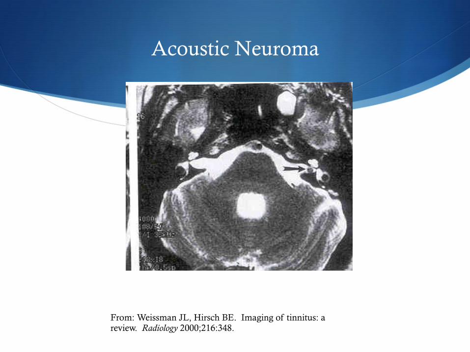
S Acoustic Neuroma S Unilateral tinnitus, asymmetric sensorineural hearing loss
or speech descrimination scores S T1-weighted MRI with gadolinium enhancement of CP
angle is study of choice S Thin section T2-weighted MRI of temporal bones and
IACs may be acceptable screening test
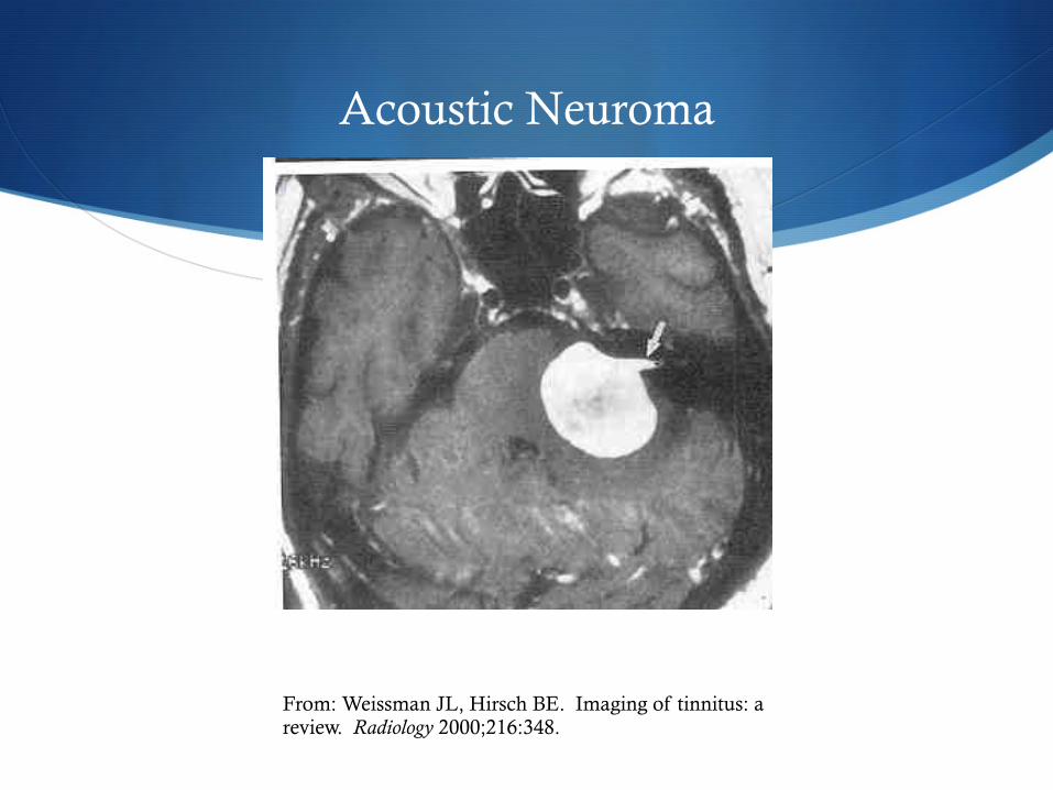
Acoustic Neuroma
From: Weissman JL, Hirsch BE. Imaging of tinnitus: a review. Radiology 2000;216:348.
Acoustic Neuroma
From: Weissman JL, Hirsch BE. Imaging of tinnitus: a review. Radiology 2000;216:348.

Imaging
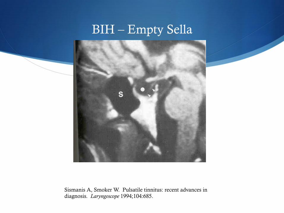
S Benign intracranial hypertension S MRI
S Small ventricles
S Empty sella
BIH – Empty Sella
Sismanis A, Smoker W. Pulsatile tinnitus: recent advances in diagnosis. Laryngoscope 1994;104:685.
Treatments
S Multiple treatments
S Avoidance of dietary stimulants
S No CATS diet
S Smoking cessation
S Avoid medications known to cause tinnitus
S Reassurance
S White noise from radio or home masking machine
Treatments - Medicines
S Many medications have been researched for the treatment of tinnitus: S Intravenous lidocaine suppresses tinnitus but is impractical to
use clinically
S Tocainide is oral analog which is ineffective
S Carbamazepine ineffective and may cause bone marrow suppression
Treatments - Medicines
S Alprazolam (Xanax) S Johnson et al (1993) found 76% of 17 patients had reduction in
the loudness of their tinnitus using both a tinnitus synthesizer and VAS (dose 0.5mg-1.5 mg/day)
S Dependence problem, long-term use is not recommended
Treatments - Medicines
S Nortriptyline and amitriptyline S May have some benefit S Dobie et al reported on 92 patients S 67% nortriptlyine benefit, 40%placebo
S Ginko biloba S Extract at doses of 120-160mg per day S Shown to be effective in some trials and not in others S Needs further study
Treatments
S Hearing aids – amplification of background noise can decrease tinnitus
S Maskers – produce sound to mask tinnitus
S Tinnitus instrument – combination of hearing aid and masker
Treatments
S Tinnitus Retraining Therapy S Based on neurophysiologic model
S Combination of masking with low level broadband noise for several hours per day and counseling to achieve habituation of the reaction to tinnitus and perception of the tinnitus itself
Treatments
S Electrical stimulation of the cochlea S Transcutaneous, round window, promontory stimulation have
all been tried
S Direct current can cause permanent damage
S Steenersen and Cronin have used transcutaneous stimulation of the auricle and tragus decreasing tinnitus in 53% of 500 patients
Treatments
S Cochlear implants S Have shown some promise in relief of tinnitus
S Ito and Sakakihara (1994) reported that in 26 patients implanted who had tinnitus 77% reported either tinnitus was abolished or suppressed, 8% reported worsening
Treatments
S Surgery S Used for treatment of arteriovenous malformations, glomus
tumors, otosclerosis, acoustic neuroma
S Some authors have reported success with cochlear nerve section in patients who have intractable tinnitus and have failed all other treatments, this is not widely accepted
Treatments
S Biofeedback
S Hypnosis
S Magnetic stimulation
S Acupuncture
S Conflicting reports of benefit
Conclusions
S Tinnitus is a common problem with an extensive differential
S Need to identify medical process if involved
S Pulsatile/Nonpulsatile is important distinction
S Will only become more common with aging of our population
S Research into mechanism and treatments is needed to better help our patients
FIN!
S Questions?
S 3172 N. Swan Road, Tucson
S 1521 E. Tangerine Road Ste 225, Oro Valley S (520) 795-8777 S www.carlsonent.com
James R. Carlson, M.D., M.B.A.
Thomas S. Kang, M.D.
Jonathan Lara, D.O.


![Retrospective investigation of the relationship between the … · vision, vertigo, pulsatile tinnitus, and Horner's syndrome) [7,8]. In addition, some investigators reported that](https://img.pdfslide.us/doc/110x75/6030c1c8066012286a6ac1c5/retrospective-investigation-of-the-relationship-between-the-vision-vertigo-pulsatile.jpg)

![Original Article - westminsterresearch.westminster.ac.uk · tuating aural fullness, tinnitus, and hearing levels that are associated with vertigo episodes[1,2]. The reported prevalence](https://img.pdfslide.us/doc/110x75/5dd0b3dad6be591ccb624777/original-article-tuating-aural-fullness-tinnitus-and-hearing-levels-that-are.jpg)