Embed Size (px)
Citation preview

1
David J. Brenner, PhD, DSc
Center for Radiological Research
Columbia University Medical Center
The Radiobiology of Small Fraction Numbers
The Radiobiology of Small Fraction Numbers
1. Single-Fraction Radiotherapy
What determines optimal fractionation for conventional radiotherapy?
The Four R’s
� Repair
� Reoxygenation
� Repopulation
� Redistribution
2
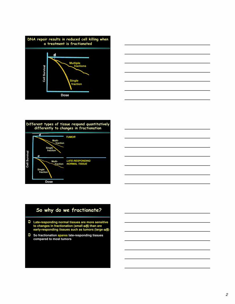
DNA repair results in reduced cell killing when a treatment is fractionated
Singlefraction
Multiplefractions
Dose
� Less curvy� Less sensitive to
changes in protraction
Quantified by large α/β α/β α/β α/β ratio (≥10 Gy)
� More curvy� More sensitive to
changes in protraction
Quantified by small α/βα/βα/βα/β
ratio (≤5 Gy)
Different types of tissue respond quantitatively differently to changes in fractionation
Single fraction
Single fraction
Multi-fraction
Multi-fraction
Dose
•
•
TUMOR
LATE-RESPONDING
NORMAL TISSUE
So why do we fractionate?
� Late-responding normal tissues are more sensitive to changes in fractionation (small α/βα/βα/βα/β) than are early-responding tissues such as tumors (large α/βα/βα/βα/β)
� So fractionation spares late-responding tissues compared to most tumors
3
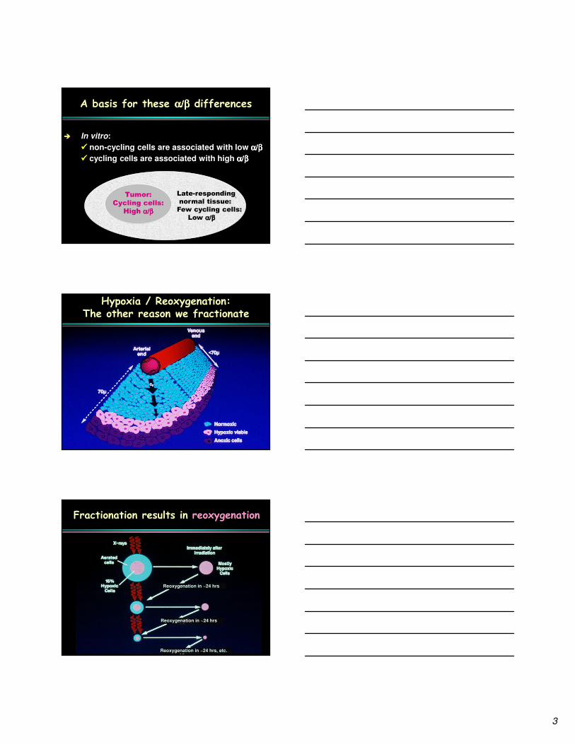
A basis for these α/βα/βα/βα/β differences
� In vitro:
���� non-cycling cells are associated with low α/βα/βα/βα/β
���� cycling cells are associated with high α/βα/βα/βα/β
Tumor:
Cycling cells:
High α/βα/βα/βα/β
Normal tissue:
Non-cycling cells:
Low α/βα/βα/βα/β
Tumor:
Cycling cells:
High α/βα/βα/βα/β
Normal tissue:
Non-cycling cells:
Low α/βα/βα/βα/β
Late respondingLate-responding
normal tissue:
Few cycling cells:
Low α/βα/βα/βα/β
Hypoxia / Reoxygenation: The other reason we fractionate
Fractionation results in reoxygenation
Reoxygenation in ~24 hrs
Reoxygenation in ~24 hrs
Reoxygenation in ~24 hrs, etc.

4
Hypoxia and malignancies
� Almost all malignant tumors, even a few mm in diameter, contain hypoxic cells
� A malignant tumor containing evena small hypoxic fraction would require an exceedingly high single-fraction dose for sterilization
� Therefore fractionation appears highly
advantageous when attempting to cure malignant tumors
Accelerated Repopulation
As a tumor shrinks, surviving clonogens
proliferate at an accelerated rate
So… for external beam RT of tumors…
� Must fractionate treatment
* to overcome hypoxia
* for differential response with late effects
� Must prolong treatment
* to limit early sequellae
� Would like to shorten treatment
* to prevent accelerated repopulation

5
� Must fractionate treatment
* to overcome hypoxia
* for differential response with late effects
� Must prolong treatment
* to limit early sequellae
� Would like to shorten treatment
* to prevent accelerated repopulation
For stereotactic RT of malignant tumors..
“Very high doses per fraction of radiation might trigger an entirely different method of cell kill via an anti-angiogenic pathway involving endothelial apoptosis”
Is the biology different at high doses per fraction?
Is the standard model (tumor control related primarily to
radiation killing of tumor clonogens) inapplicable at high doses?
“The single-fraction dose to inactivate
experimental tumors is well predicted by measured clonogenic fraction and
in-vitro radiation-induced cell survival”
Predicted TCD50 based on in-vitro
cell survival, vs observed TCD50
� Observed
� Predicted
Tumors

6
Other papers showing correlations between in vitro tumor-cell radiosensitivty and in vivo tumor control
at high single doses / fraction
“Ablative” Radiotherapy:A different dominant mechanism for tumor control
at high doses per fraction?
• The LQ model fails to predict high dose / Fx tumor control
… “so some new biology must be going on”
• The LQ model will certainly fail at very high single doses
• But this doesn’t imply that the standard model
(tumor control related primarily to radiation killing
of tumor clonogens) is wrong
• Nor does it contradict the rationale that fractionation will
improve outcome after RT for tumors
Fractionation for malignant tumors
All malignant tumors, small or large,
slow or fast growing, sensitive or resistant,
will be more effectively treated with fractionation
than with a single fraction

7
Stereotactic radiosurgery / ablative RT The Bottom Line
� Stereotactic radiosurgery and SABR have had
impressive success in treating some malignancies, particular in the brain and lung
� But, for any given malignancy, better results should be obtained with fractionation
� There is no need for conventional fractionation, 5-10 fractions is optimal
� For vascular disease and benign tumors, fractionation may only be needed if the lesions are large
The Radiobiology of Small Fraction Numbers
2. Hypofractionated radiotherapy
In most RT scenarios, we fractionate to take advantage
of the differential α/βα/βα/βα/β ratio between
tumor and late responding normal tissue
� non-cycling cells are associated with low α/βα/βα/βα/β
� cycling cells are associated with high α/βα/βα/βα/β
Tumor:
Cycling cells:
High α/βα/βα/βα/β
Normal tissue:
Non-cycling cells:
Low α/βα/βα/βα/β
Tumor:
Cycling cells:
High α/βα/βα/βα/β
Normal tissue:
Non-cycling cells:
Low α/βα/βα/βα/β
Late-responding
normal tissue:
Few cycling cells:
Low α/βα/βα/βα/β

8
Prostate Cancer
� Prostate tumors are not typical of most
tumors, containing far fewer cycling cells
� Do prostate tumors have the typical differential response to fractionation for tumor control relative to late-responding
tissues?
� What is an appropriate α/βα/βα/βα/β value
for prostate tumors?
α/βα/βα/βα/β for prostate cancer
= 1.5 Gy [95% CI: 0.8 - 2.2]
“comparable with typical α/βα/βα/βα/βfor late-responding normal tissues”
Brenner and Hall 1999
Based on clinical data fromimplant and external beam data....
� We lose one of the fundamental
rationales for using many fractions or
using low dose rate
� So hypo-fractionation or HDR rather than
conventional fractionation or LDR
become potential options for prostate RT
If the α/βα/βα/βα/β ratio for prostate cancer is comparable to that for normal tissue….

9
Since the 1999 estimate of α/βα/βα/βα/β, there have been more than 70 further papers in the
literature relating to α/βα/βα/βα/β values for prostate
Two recent large studies of α/βα/βα/βα/β for prostate
5,969 patients
α/βα/βα/βα/β= 1.4 Gy (95% CI 0.9-2.2)5,093 patients
α/βα/βα/βα/β= 1.55 Gy (95% CI 0.46-4.52)
If α/βα/βα/βα/β for prostate canceris about the same as for
surrounding late-responding normal tissue....
� Less rationale for using many fractions or LDR
� Fewer fractions at the right dose would give
1. The same tumor control and
late sequelae as current regimens
2. Patient convenience
3. Financial / resource advantages
10
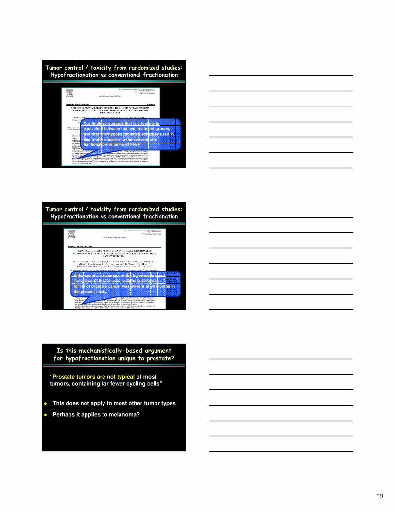
Tumor control / toxicity from randomized studies: Hypofractionation vs conventional fractionation
Our findings suggest that late toxicity is
equivalent between the two treatment groups,
and that the hypofractionated schedule used in
this trial is superior to the conventional
fractionation in terms of FFBF
Tumor control / toxicity from randomized studies: Hypofractionation vs conventional fractionation
A therapeutic advantage of the hypofractionated
compared to the conventional dose schedule
for RT of prostate cancer was evident at 90 months in
the present study
Is this mechanistically-based argument for hypofractionation unique to prostate?
� This does not apply to most other tumor types
� Perhaps it applies to melanoma?
“Prostate tumors are not typical of most tumors, containing far fewer cycling cells”

11
Melanoma is a slow-growing tumor with few dividing cells
α/βα/βα/βα/β= 0.57 (95% CI: 1.07 – 2.5)
Successful hypofractionation for choroidal melanoma
Is this mechanistically-based argument for hypofractionation unique to prostate?
� This does not apply to most other tumor types
� Perhaps it applies to melanoma?
� Perhaps it applies to some other early-stage tumors?
“Prostate tumors are not typical of most tumors, containing far fewer cycling cells”
12
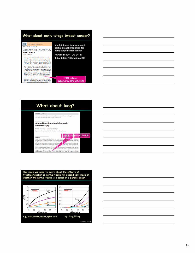
What about early-stage breast cancer?
Much interest in accelerated partial breast irradiation forearly-stage breast cancer
NSABP B-39/RTOG 0413:
3.4 or 3.85 x 10 fractions BID
2,236 patients
α/βα/βα/βα/β= 4.6 Gy (95% CI 1.1-8.1)
What about lung?
α/βα/βα/βα/β= 8.2 Gy (95% CI 7.0-9.4)
SERIAL PARALLEL
e.g., lung, kidneye.g., brain, bladder, rectum, spinal cord
How much you need to worry about the effects of hypofractionation on normal tissue will depend very much on whether the normal tissue is a serial or a parallel organ
Dose
DoseNT
CP
Goitein 2008

13
� When the goal is to optimize the therapeutic ratio between tumor control and late complications, basic radiobiological principles tell us that we should fractionate
� Prostate is a mechanistically understood exception
� Melanoma and possibly early breast cancer may likewise be exceptions
� No such radiobiological rationale appears to exists for lunghypofractionation, but the parallel nature of normal lung tissue may well permit less fractionation than at other sites, when the irradiated volume is small
� This speaker does not think there is persuasive evidence for
“new radiobiology” at high doses per fraction
Meet the new boss – same as the old boss!radiobiology
Xradiobiology
X