Embed Size (px)
Citation preview
OCTOBER 2015 | CATARACT & REFRACTIVE SURGERY TODAY EUROPE 47
COV
ER FOCU
S
To give the Binkhorst Medal Lecture is without doubt one of the greatest honors that can be bestowed on an ophthalmologist. Cornelius D. Binkhorst, MD, for whom the lecture was named, was a giant in our specialty. Having tried the IOL designed by Sir Harold Ridley soon after its development, Binkhorst real-ized that its main shortcomings related to its
weight and its ability to remain stable in the eye. He therefore designed his four-loop iris-fixated IOL for use with intracapsular surgery; this was the first IOL to produce consistent results.
Binkhorst also recognized that, if he could use the lens capsule to support the IOL, this would give even better centration and stability, and thus he advocated a return to extracapsular cataract surgery (ECCE). I was privileged to assist Dr. Binkhorst at surgery during a conference held at Charing Cross Hospital in London in 1980.
The subject of my Binkhorst lecture relates to the capsule—specifically to the methods of opening it to remove the cataractous lens nucleus and how these have changed over the centuries (http://eyetube.net/?v=erejo). The capsular opening has evolved, first from a roughly made tear to allow access to the nucleus for extraction, to the creation of more regular openings to allow support for IOLs, then to a continu-ous circular tear to help contain the IOL, and most recently to precision sizing and location with laser and other technologies. Although this opening is usually called a capsulotomy, the term capsulectomy should more correctly be used because the tissue is normally removed in modern surgery.
THE STORY BEGINSThe story begins with the father of modern cataract surgery,
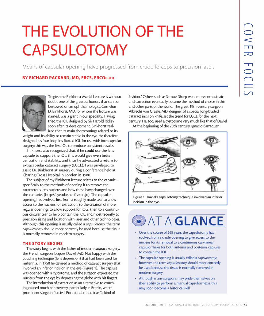
the French surgeon Jacques Daviel, MD. Not happy with the couching technique (lens depression) that had been used for millennia, in 1750 he devised a method of cataract surgery that involved an inferior incision in the eye (Figure 1). The capsule was opened with a cystotome, and the surgeon expressed the nucleus from the eye by depressing the globe with his fingers.
The introduction of extraction as an alternative to couch-ing caused much controversy, particularly in Britain, where prominent surgeon Percival Pott condemned it as “a kind of
fashion.” Others such as Samuel Sharp were more enthusiastic, and extraction eventually became the method of choice in this and other parts of the world. The great 19th-century surgeon Albrecht von Graefe, MD, designer of a special long-bladed cataract incision knife, set the trend for ECCE for the next century. He, too, used a cystotome very much like that of Daviel.
At the beginning of the 20th century, Ignacio Barraquer
Means of capsular opening have progressed from crude forceps to precision laser.
BY RICHARD PACKARD, MD, FRCS, FRCOphth
THE EVOLUTION OF THE CAPSULOTOMY
Figure 1. Daviel’s capsulotomy technique involved an inferior
incision in the eye.
• Over the course of 265 years, the capsulotomy has evolved from a crude opening to give access to the nucleus for its removal to a continuous curvilinear capsulorrhexis for both anterior and posterior capsules to contain the IOL.
• The capsular opening is usually called a capsulotomy; however, the term capsulectomy should more correctly be used because the tissue is normally removed in modern surgery.
• Although many surgeons may pride themselves on their ability to perform a manual capsulorrhexis, this may soon become a historical skill.
AT A GLANCE
48 CATARACT & REFRACTIVE SURGERY TODAY EUROPE | OCTOBER 2015
COV
ER F
OCU
S
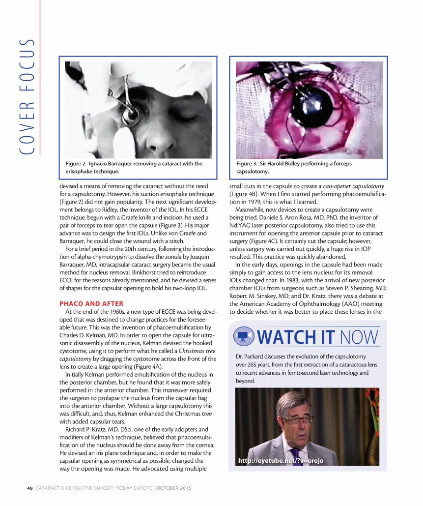
devised a means of removing the cataract without the need for a capsulotomy. However, his suction erisophake technique (Figure 2) did not gain popularity. The next significant develop-ment belongs to Ridley, the inventor of the IOL. In his ECCE technique, begun with a Graefe knife and incision, he used a pair of forceps to tear open the capsule (Figure 3). His major advance was to design the first IOLs. Unlike von Graefe and Barraquer, he could close the wound with a stitch.
For a brief period in the 20th century, following the introduc-tion of alpha-chymotrypsin to dissolve the zonula by Joaquin Barraquer, MD, intracapsular cataract surgery became the usual method for nucleus removal. Binkhorst tried to reintroduce ECCE for the reasons already mentioned, and he devised a series of shapes for the capsular opening to hold his two-loop IOL.
PHACO AND AFTERAt the end of the 1960s, a new type of ECCE was being devel-
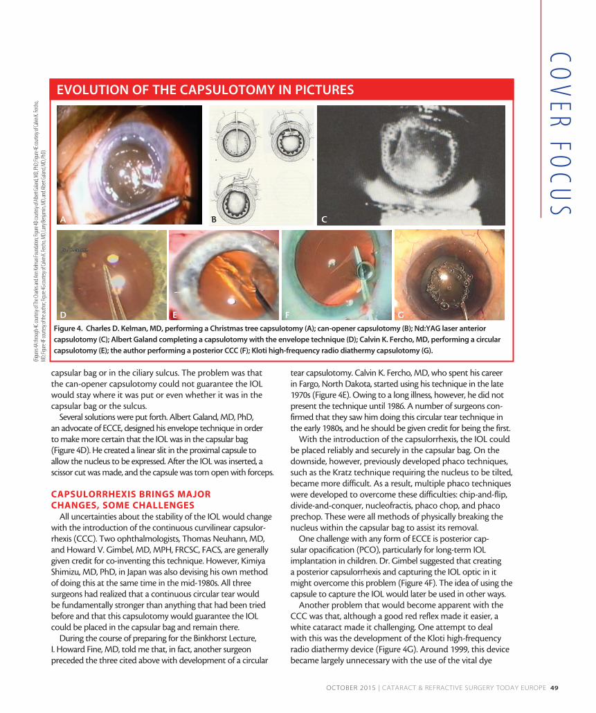
oped that was destined to change practices for the foresee-able future. This was the invention of phacoemulsification by Charles D. Kelman, MD. In order to open the capsule for ultra-sonic disassembly of the nucleus, Kelman devised the hooked cystotome, using it to perform what he called a Christmas tree capsulotomy by dragging the cystotome across the front of the lens to create a large opening (Figure 4A).
Initially Kelman performed emulsification of the nucleus in the posterior chamber, but he found that it was more safely performed in the anterior chamber. This maneuver required the surgeon to prolapse the nucleus from the capsular bag into the anterior chamber. Without a large capsulotomy this was difficult, and, thus, Kelman enhanced the Christmas tree with added capsular tears.
Richard P. Kratz, MD, DSci, one of the early adopters and modifiers of Kelman’s technique, believed that phacoemulsi-fication of the nucleus should be done away from the cornea. He devised an iris plane technique and, in order to make the capsular opening as symmetrical as possible, changed the way the opening was made. He advocated using multiple
small cuts in the capsule to create a can-opener capsulotomy (Figure 4B). When I first started performing phacoemulsifica-tion in 1979, this is what I learned.
Meanwhile, new devices to create a capsulotomy were being tried. Daniele S. Aron Rosa, MD, PhD, the inventor of Nd:YAG laser posterior capsulotomy, also tried to use this instrument for opening the anterior capsule prior to cataract surgery (Figure 4C). It certainly cut the capsule; however, unless surgery was carried out quickly, a huge rise in IOP resulted. This practice was quickly abandoned.
In the early days, openings in the capsule had been made simply to gain access to the lens nucleus for its removal. IOLs changed that. In 1983, with the arrival of new posterior chamber IOLs from surgeons such as Steven P. Shearing, MD; Robert M. Sinskey, MD; and Dr. Kratz, there was a debate at the American Academy of Ophthalmology (AAO) meeting to decide whether it was better to place these lenses in the
Figure 2. Ignacio Barraquer removing a cataract with the
erisophake technique.
Figure 3. Sir Harold Ridley performing a forceps
capsulotomy.
Dr. Packard discusses the evolution of the capsulotomy over 265 years, from the first extraction of a cataractous lens to recent advances in femtosecond laser technology and beyond.
WATCH IT NOW
http://eyetube.net/?v=erejo
OCTOBER 2015 | CATARACT & REFRACTIVE SURGERY TODAY EUROPE 49
COV
ER FOCU
S
capsular bag or in the ciliary sulcus. The problem was that the can-opener capsulotomy could not guarantee the IOL would stay where it was put or even whether it was in the capsular bag or the sulcus.
Several solutions were put forth. Albert Galand, MD, PhD, an advocate of ECCE, designed his envelope technique in order to make more certain that the IOL was in the capsular bag (Figure 4D). He created a linear slit in the proximal capsule to allow the nucleus to be expressed. After the IOL was inserted, a scissor cut was made, and the capsule was torn open with forceps.
CAPSULORRHEXIS BRINGS MAJOR CHANGES, SOME CHALLENGES
All uncertainties about the stability of the IOL would change with the introduction of the continuous curvilinear capsulor-rhexis (CCC). Two ophthalmologists, Thomas Neuhann, MD, and Howard V. Gimbel, MD, MPH, FRCSC, FACS, are generally given credit for co-inventing this technique. However, Kimiya Shimizu, MD, PhD, in Japan was also devising his own method of doing this at the same time in the mid-1980s. All three surgeons had realized that a continuous circular tear would be fundamentally stronger than anything that had been tried before and that this capsulotomy would guarantee the IOL could be placed in the capsular bag and remain there.
During the course of preparing for the Binkhorst Lecture, I. Howard Fine, MD, told me that, in fact, another surgeon preceded the three cited above with development of a circular
tear capsulotomy. Calvin K. Fercho, MD, who spent his career in Fargo, North Dakota, started using his technique in the late 1970s (Figure 4E). Owing to a long illness, however, he did not present the technique until 1986. A number of surgeons con-firmed that they saw him doing this circular tear technique in the early 1980s, and he should be given credit for being the first.
With the introduction of the capsulorrhexis, the IOL could be placed reliably and securely in the capsular bag. On the downside, however, previously developed phaco techniques, such as the Kratz technique requiring the nucleus to be tilted, became more difficult. As a result, multiple phaco techniques were developed to overcome these difficulties: chip-and-flip, divide-and-conquer, nucleofractis, phaco chop, and phaco prechop. These were all methods of physically breaking the nucleus within the capsular bag to assist its removal.
One challenge with any form of ECCE is posterior cap-sular opacification (PCO), particularly for long-term IOL implantation in children. Dr. Gimbel suggested that creating a posterior capsulorrhexis and capturing the IOL optic in it might overcome this problem (Figure 4F). The idea of using the capsule to capture the IOL would later be used in other ways.
Another problem that would become apparent with the CCC was that, although a good red reflex made it easier, a white cataract made it challenging. One attempt to deal with this was the development of the Kloti high-frequency radio diathermy device (Figure 4G). Around 1999, this device became largely unnecessary with the use of the vital dye
EVOLUTION OF THE CAPSULOTOMY IN PICTURES
Figure 4. Charles D. Kelman, MD, performing a Christmas tree capsulotomy (A); can-opener capsulotomy (B); Nd:YAG laser anterior
capsulotomy (C); Albert Galand completing a capsulotomy with the envelope technique (D); Calvin K. Fercho, MD, performing a circular
capsulotomy (E); the author performing a posterior CCC (F); Kloti high-frequency radio diathermy capsulotomy (G).
A
D E F G
B C
(Figu
res 4A
throu
gh 4C
court
esy o
f The
Charl
es an
d Ann
Kelm
an Fo
unda
tion;
Figure
4D co
urtes
y of A
lbert G
aland
, MD,
PhD;
Figure
4E co
urtes
y of C
alvin
K. Fe
rcho,
MD;
Figure
4F co
urtes
y of th
e auth
or; Fi
gure
4G co
urtes
y of C
alvin
K. Fe
rcho,
MD,
Larry
Benja
min,
MD,
and A
lbert G
aland
, MD,
PhD)
50 CATARACT & REFRACTIVE SURGERY TODAY EUROPE | OCTOBER 2015
COV
ER F
OCU
S
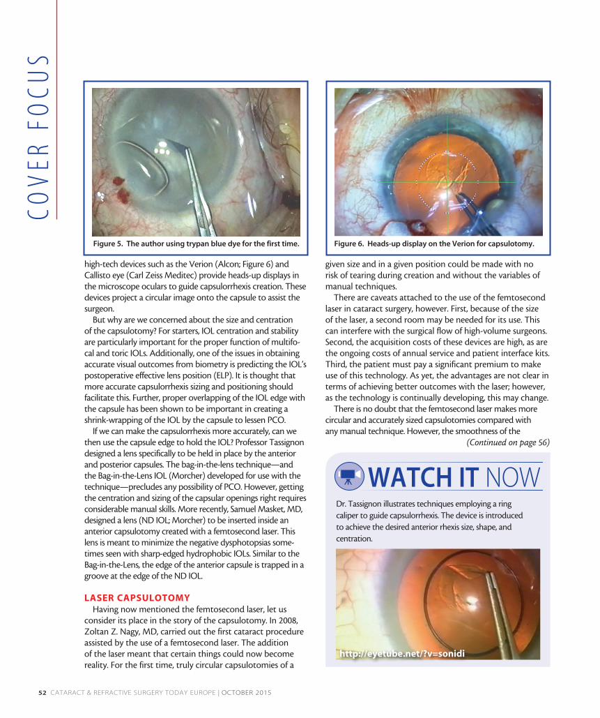
trypan blue to stain the capsule. Although my initial research suggested that Dutch surgeon Gerrit R.J. Melles, MD, PhD, was the first to stain the capsule with trypan blue dye in 1999, it appears that Minas Coroneo, MD, from Australia, had an earlier patent for the same method. Coroneo developed the technique when operating on patients with very mature cata-racts in a remote area of Australia. With use of the dye, every capsule could now be visualized for capsulorrhexis (Figure 5).
GREATER PRECISIONOnce surgeons could make the capsulotomy in most
eyes reliably, attention turned to making the results more precise. Marie-José Tassignon, MD, PhD, FEBO, developed a ring-shaped caliper, intended to be centered on the capsule, to help make the size and centration of capsulorrhexis more accurate (http://eyetube.net/?v=sonidi). Another approach is to indent the cornea with a circular guide. More recently,
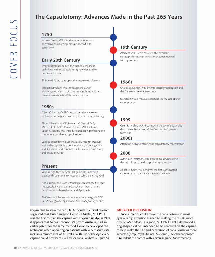
The Capsulotomy: Advances Made in the Past 265 Years
1750Jacques Daviel, MD, introduces extraction as an alternative to couching; capsule opened with cystotome
Early 20th CenturyIgnacio Barraquer debuts the suction erisophake technique with no capsulotomy; however, it never becomes popular
Sir Harold Ridley tears open the capsule with forceps
Joaquim Barraquer, MD, introduces the use of alpha-chymotrypsin to dissolve the zonula; intracapsular cataract extraction briefly becomes popular
19th CenturyAlbrecht von Graefe, MD, sets the trend for extracapsular cataract extraction; capsule opened with cystotome
1960sCharles D. Kelman, MD, invents phacoemulsification and the Christmas tree capsulotomy
Richard P. Kratz, MD, DSci, popularizes the can-opener capsulotomy
1999Gerrit R.J. Melles, MD, PhD, suggests the use of trypan blue dye to stain the capsule; Minas Coroneo, MD, patents technique
2000sAttention turns to making the capsulotomy more precise
2008Marie-José Tassignon, MD, PhD, FEBO, devises a ring-shaped caliper to guide capsulorrhexis creation
Zoltan Z. Nagy, MD performs the first laser-assisted capsulotomy and cataract surgery procedure
PresentVarious high-tech devices that guide capsulorrhexis creation through the microscope oculars are introduced
Nonfemtosecond-laser technologies are designed to open the capsule, including the CapsuLaser (thermal laser), Zepto capsulorrhexis device, and ApetureRx
The Verus ophthalmic caliper is introduced to guide CCC (see A Cost-Effective Approach to Increased Efficiency in CCC)
1980sAlbert Galand, MD, PhD, introduces the envelope technique to make certain the IOL is in the capsular bag
Thomas Neuhann, MD; Howard V. Gimbel, MD, MPH, FRCSC, FACS; Kimya Shimizu, MD, PhD; and Calvin K. Fercho, MD, introduce and begin perfecting the continuous curvilinear capsulorrhexis
Various phaco techniques that allow nuclear breakup within the capsular bag are introduced, including chip-and-flip, divide-and-conquer, nucleofractis, phaco chop, and phaco prechop
52 CATARACT & REFRACTIVE SURGERY TODAY EUROPE | OCTOBER 2015
COV
ER F
OCU
S
high-tech devices such as the Verion (Alcon; Figure 6) and Callisto eye (Carl Zeiss Meditec) provide heads-up displays in the microscope oculars to guide capsulorrhexis creation. These devices project a circular image onto the capsule to assist the surgeon.
But why are we concerned about the size and centration of the capsulotomy? For starters, IOL centration and stability are particularly important for the proper function of multifo-cal and toric IOLs. Additionally, one of the issues in obtaining accurate visual outcomes from biometry is predicting the IOL’s postoperative effective lens position (ELP). It is thought that more accurate capsulorrhexis sizing and positioning should facilitate this. Further, proper overlapping of the IOL edge with the capsule has been shown to be important in creating a shrink-wrapping of the IOL by the capsule to lessen PCO.
If we can make the capsulorrhexis more accurately, can we then use the capsule edge to hold the IOL? Professor Tassignon designed a lens specifically to be held in place by the anterior and posterior capsules. The bag-in-the-lens technique—and the Bag-in-the-Lens IOL (Morcher) developed for use with the technique—precludes any possibility of PCO. However, getting the centration and sizing of the capsular openings right requires considerable manual skills. More recently, Samuel Masket, MD, designed a lens (ND IOL; Morcher) to be inserted inside an anterior capsulotomy created with a femtosecond laser. This lens is meant to minimize the negative dysphotopsias some-times seen with sharp-edged hydrophobic IOLs. Similar to the Bag-in-the-Lens, the edge of the anterior capsule is trapped in a groove at the edge of the ND IOL.
LASER CAPSULOTOMYHaving now mentioned the femtosecond laser, let us
consider its place in the story of the capsulotomy. In 2008, Zoltan Z. Nagy, MD, carried out the first cataract procedure assisted by the use of a femtosecond laser. The addition of the laser meant that certain things could now become reality. For the first time, truly circular capsulotomies of a
given size and in a given position could be made with no risk of tearing during creation and without the variables of manual techniques.
There are caveats attached to the use of the femtosecond laser in cataract surgery, however. First, because of the size of the laser, a second room may be needed for its use. This can interfere with the surgical flow of high-volume surgeons. Second, the acquisition costs of these devices are high, as are the ongoing costs of annual service and patient interface kits. Third, the patient must pay a significant premium to make use of this technology. As yet, the advantages are not clear in terms of achieving better outcomes with the laser; however, as the technology is continually developing, this may change.
There is no doubt that the femtosecond laser makes more circular and accurately sized capsulotomies compared with any manual technique. However, the smoothness of the
Figure 5. The author using trypan blue dye for the first time. Figure 6. Heads-up display on the Verion for capsulotomy.
Dr. Tassignon illustrates techniques employing a ring caliper to guide capsulorrhexis. The device is introduced to achieve the desired anterior rhexis size, shape, and centration.
http://eyetube.net/?v=sonidi
(Continued on page 56)
WATCH IT NOW
54 CATARACT & REFRACTIVE SURGERY TODAY EUROPE | OCTOBER 2015
COV
ER F
OCU
S
A COST-EFFECTIVE APPROACH TO INCREASED EFFICIENCY IN CCCWalking the anterior capsule flap around the internal rim of the Verus device can help novice and experienced surgeons create a well-centered, circular, and accurately sized capsulotomy.
BY MALIK Y. KAHOOK, MD
The continuous curvilinear capsulorrhexis (CCC) has several advantages over previous iterations of capsular openings, including signifi-cant mechanical strength to withstand surgical manipulation and enhanced ability to stabilize the position of the IOL after implantation. One of the downsides of the CCC, however, is that it is one of the hardest aspects of cataract
surgery to learn and perfect. Even the most seasoned cataract surgeons still experience inconsistencies from surgery to surgery in terms of centration, circularity, and sizing of the CCC.
Although femtosecond lasers do a fantastic job of creating a cir-cular and consistently sized capsulotomy, there is a high cost associ-ated with the purchase of these devices and additional costs for the docking system used in each case. The amount of time added to each case is also a deterrent for many busy practices. Consequently, surgeons’ uptake of this technology around the world has been slow.
Recently, devices that use thermal energy to alter tissue such as the ApertureRx (International BioMedical Devices) and the Zepto (Mynosys) have been introduced as lower cost alternatives to the femtosecond laser. These, too, boast efficiency of capsulotomy creation, but, although they are cheaper than the femtosecond laser, significant cost is still associated with their use, and consis-tency of performance is still unclear. Perhaps most important, the capsulotomies created by all of these devices do not posses the mechanical strength that is unique to the CCC.
ANOTHER ALTERNATIVEIn contrast to high-tech femtosecond lasers and thermal
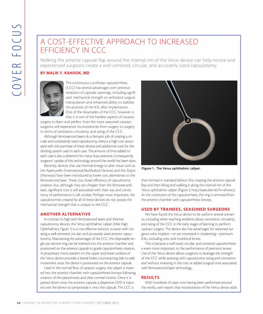
capsulotomy devices, the Verus ophthalmic caliper (Mile High Ophthalmics; Figure 1) is a cost-effective solution to assist with cre-ating a well-centered, circular, and accurately sized anterior capsu-lotomy. Maintaining the advantages of the CCC, this disposable sin-gle-use silicone ring can be inserted into the anterior chamber and positioned on the anterior capsule to guide capsulorrhexis creation. A proprietary micro-pattern on the upper and lower surfaces of the Verus device provides a lateral brake, counteracting side-to-side movement once the device is positioned on the anterior capsule.
Used in the normal flow of cataract surgery, the caliper is insert-ed into the anterior chamber with capsulorrhexis forceps following creation of the paracentesis and clear corneal incision. Once it is patted down onto the anterior capsule, a dispersive OVD is inject-ed over the device to tamponade it onto the capsule. The CCC is
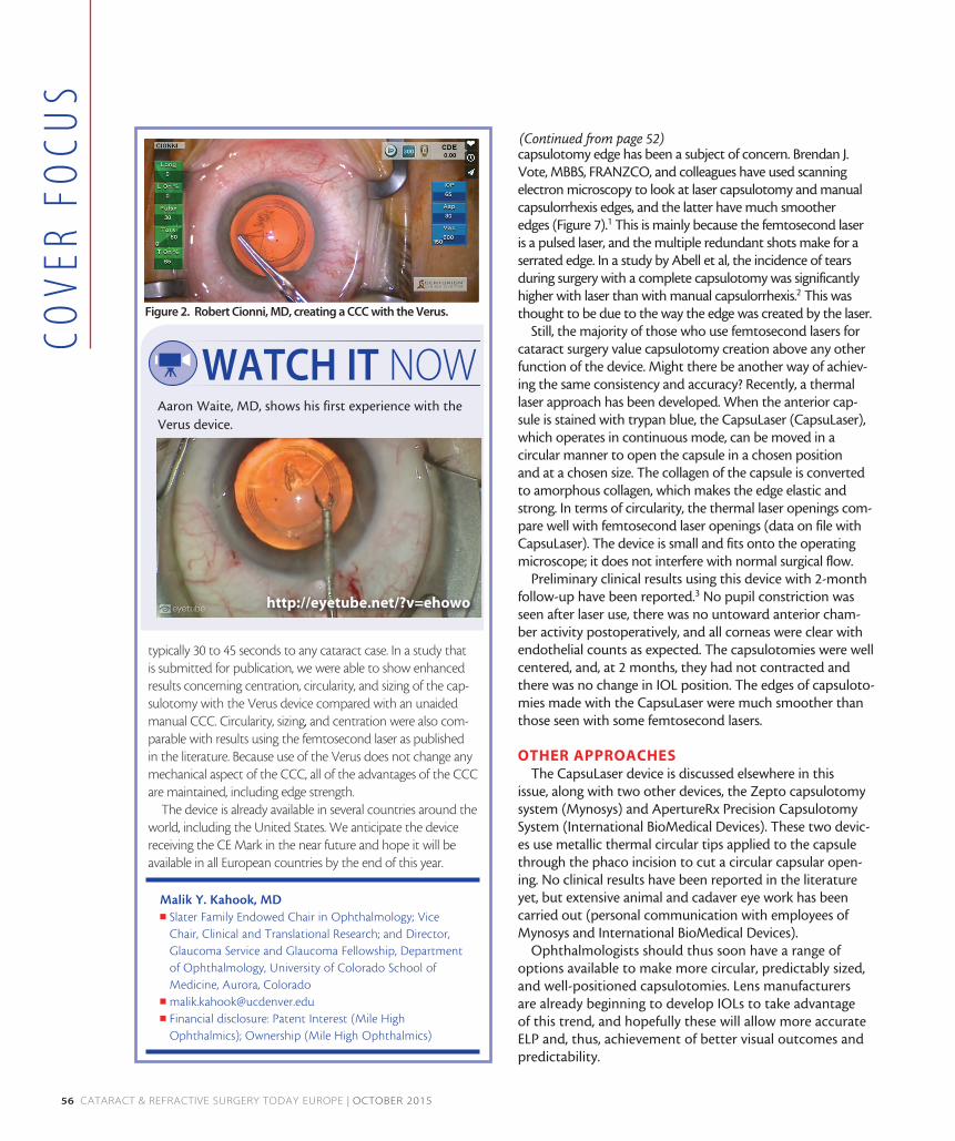
then formed in standard fashion, first creating the anterior capsule flap and then lifting and walking it along the internal rim of the Verus ophthalmic caliper (Figure 2; http://eyetube.net/?v=ehowo). At the conclusion of the capsulorrhexis, the ring is removed from the anterior chamber with capsulorrhexis forceps.
USED BY TRAINEES, SEASONED SURGEONSWe have found the Verus device to be useful in several scenari-
os, including when teaching residents about centration, circularity, and sizing of the CCC in the early stages of learning to perform cataract surgery. The device also has advantages for seasoned sur-geons who implant—or are interested in implanting—premium IOLs, including toric and multifocal lenses.
This is because a well-sized, circular, and centered capsulorrhexis is even more important to the performance of premium lenses. Use of the Verus device allows surgeons to leverage the strength of the CCC while assisting with capsulotomy sizing and centration and without investing in the cost or added surgical time associated with femtosecond laser technology.
RESULTSWith hundreds of cases now having been performed around
the world, users report that incorporation of the Verus device adds
Figure 1. The Verus ophthalmic caliper.
56 CATARACT & REFRACTIVE SURGERY TODAY EUROPE | OCTOBER 2015
COV
ER F
OCU
S
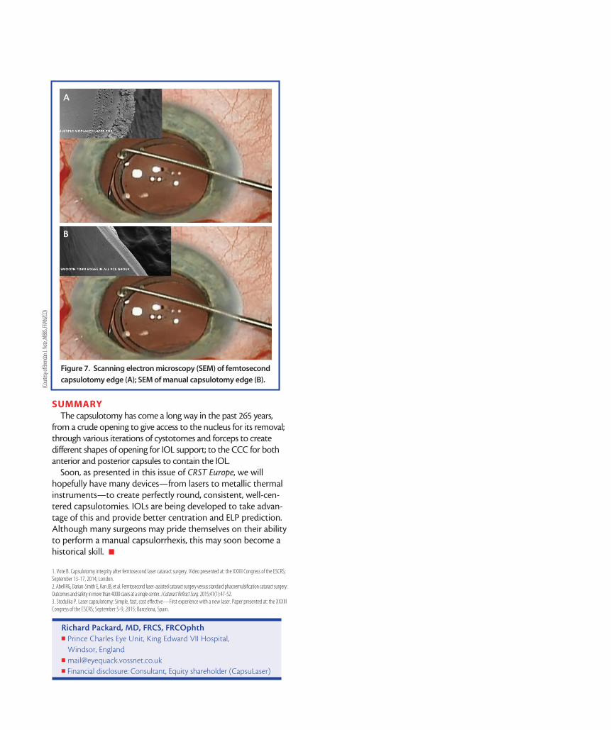
capsulotomy edge has been a subject of concern. Brendan J. Vote, MBBS, FRANZCO, and colleagues have used scanning electron microscopy to look at laser capsulotomy and manual capsulorrhexis edges, and the latter have much smoother edges (Figure 7).1 This is mainly because the femtosecond laser is a pulsed laser, and the multiple redundant shots make for a serrated edge. In a study by Abell et al, the incidence of tears during surgery with a complete capsulotomy was significantly higher with laser than with manual capsulorrhexis.2 This was thought to be due to the way the edge was created by the laser.
Still, the majority of those who use femtosecond lasers for cataract surgery value capsulotomy creation above any other function of the device. Might there be another way of achiev-ing the same consistency and accuracy? Recently, a thermal laser approach has been developed. When the anterior cap-sule is stained with trypan blue, the CapsuLaser (CapsuLaser), which operates in continuous mode, can be moved in a circular manner to open the capsule in a chosen position and at a chosen size. The collagen of the capsule is converted to amorphous collagen, which makes the edge elastic and strong. In terms of circularity, the thermal laser openings com-pare well with femtosecond laser openings (data on file with CapsuLaser). The device is small and fits onto the operating microscope; it does not interfere with normal surgical flow.
Preliminary clinical results using this device with 2-month follow-up have been reported.3 No pupil constriction was seen after laser use, there was no untoward anterior cham-ber activity postoperatively, and all corneas were clear with endothelial counts as expected. The capsulotomies were well centered, and, at 2 months, they had not contracted and there was no change in IOL position. The edges of capsuloto-mies made with the CapsuLaser were much smoother than those seen with some femtosecond lasers.
OTHER APPROACHESThe CapsuLaser device is discussed elsewhere in this
issue, along with two other devices, the Zepto capsulotomy system (Mynosys) and ApertureRx Precision Capsulotomy System (International BioMedical Devices). These two devic-es use metallic thermal circular tips applied to the capsule through the phaco incision to cut a circular capsular open-ing. No clinical results have been reported in the literature yet, but extensive animal and cadaver eye work has been carried out (personal communication with employees of Mynosys and International BioMedical Devices).
Ophthalmologists should thus soon have a range of options available to make more circular, predictably sized, and well-positioned capsulotomies. Lens manufacturers are already beginning to develop IOLs to take advantage of this trend, and hopefully these will allow more accurate ELP and, thus, achievement of better visual outcomes and predictability.
typically 30 to 45 seconds to any cataract case. In a study that is submitted for publication, we were able to show enhanced results concerning centration, circularity, and sizing of the cap-sulotomy with the Verus device compared with an unaided manual CCC. Circularity, sizing, and centration were also com-parable with results using the femtosecond laser as published in the literature. Because use of the Verus does not change any mechanical aspect of the CCC, all of the advantages of the CCC are maintained, including edge strength.
The device is already available in several countries around the world, including the United States. We anticipate the device receiving the CE Mark in the near future and hope it will be available in all European countries by the end of this year.
Figure 2. Robert Cionni, MD, creating a CCC with the Verus.
Aaron Waite, MD, shows his first experience with the Verus device.
http://eyetube.net/?v=ehowo
Malik Y. Kahook, MDn Slater Family Endowed Chair in Ophthalmology; Vice
Chair, Clinical and Translational Research; and Director, Glaucoma Service and Glaucoma Fellowship, Department of Ophthalmology, University of Colorado School of Medicine, Aurora, Colorado
n [email protected] Financial disclosure: Patent Interest (Mile High
Ophthalmics); Ownership (Mile High Ophthalmics)
(Continued from page 52)
WATCH IT NOW
SUMMARYThe capsulotomy has come a long way in the past 265 years,
from a crude opening to give access to the nucleus for its removal; through various iterations of cystotomes and forceps to create different shapes of opening for IOL support; to the CCC for both anterior and posterior capsules to contain the IOL.
Soon, as presented in this issue of CRST Europe, we will hopefully have many devices—from lasers to metallic thermal instruments—to create perfectly round, consistent, well-cen-tered capsulotomies. IOLs are being developed to take advan-tage of this and provide better centration and ELP prediction. Although many surgeons may pride themselves on their ability to perform a manual capsulorrhexis, this may soon become a historical skill. n
1. Vote B. Capsulotomy integrity after femtosecond laser cataract surgery. Video presented at: the XXXII Congress of the ESCRS; September 13-17, 2014; London. 2. Abell RG, Darian-Smith E, Kan JB, et al. Femtosecond laser-assisted cataract surgery versus standard phacoemulsification cataract surgery: Outcomes and safety in more than 4000 cases at a single center. J Cataract Refract Surg. 2015;41(1):47-52. 3. Stodulka P. Laser capsulotomy: Simple, fast, cost effective—First experience with a new laser. Paper presented at: the XXXIII Congress of the ESCRS; September 5-9, 2015; Barcelona, Spain.
Figure 7. Scanning electron microscopy (SEM) of femtosecond
capsulotomy edge (A); SEM of manual capsulotomy edge (B).
A
B
Richard Packard, MD, FRCS, FRCOphthn Prince Charles Eye Unit, King Edward VII Hospital,
Windsor, England n [email protected] Financial disclosure: Consultant, Equity shareholder (CapsuLaser)
(Cou
rtesy
of Bre
ndan
J. Vo
te, M
BBS,
FRAN
ZCO)