Embed Size (px)
Citation preview
Effect of the Capsular Material of Cryptococcus
neoformans on the interplay between Microglial
cells and Neutrophils
by
Dr PRATHNA BHOLA
Submitted in fulfilment of the requirements for the degree of
Doctor of Philosophy (Ph.D.)
In the Discipline of Medical Microbiology,
School of Laboratory Medicine and Medical Sciences,
College of Health Science, University of KwaZulu-Natal,
Durban, South Africa
October 2019

i
SUPERVISOR’S STATEMENT
As the candidate’s supervisor, I agree to the submission of this thesis.
S
P
ii
AUTHOR’S DECLARATION
I, Dr P Bhola, hereby declare that:
This thesis contains my own work except where specifically acknowledged, and all experiments were
carried out in the department of Infection Prevention and Control, School of Laboratory Medicine and
Medical Sciences, University of KwaZulu-Natal, under the supervision of Professor A W Sturm.
This research has not been previously submitted to the University of KwaZulu-Natal or any other
tertiary institution for the purposes of obtaining any other degree or academic qualification.
Student name: Prathna Bhola
Student number: 903482310
Signed:
Date:
Supervisor: Prof A. Willem Sturm
Signed:
Date: 03-10-2019

iii
ETHICS DECLARATION
The Biomedical Research Ethics Committee of the University of KwaZulu-Natal (BE094/12) granted
approval for this PhD project.

iv
ACKNOWLEDGEMENTS
I wish to express my sincere gratitude to everyone who contributed to my project. I could not have
done it without them:
Prof A. Willem Sturm for being my mentor, for believing in me and for his unending expert
guidance and patience throughout this project.
Dr O. Emmanuel Asowata for his wholehearted assistance in optimizing my chemotaxis and
gene expression experiments and for availing himself during weekends to assist me with this.
Dr Ravesh Singh for his support and assistance with the PCR investigations and willingness to
assist with the numerous technical issues that I encountered during this project.
Dr Bronwyn Joubert for training me in tissue culture, Ms Shalona Beeput for sharing her
knowledge and training me in neutrophil separation, and Dr Sobia Parveen for receiving and
storing my study samples during the early stages of my project.
Dr Abraham J. Niehaus for his willingness to share his experiences for submission of my thesis.
Ms Hyanthavanie Jack, Ms Viloshni Pillay, Ms Eden O Goodman and Ms Moganaygie
Govender at Mahatma Gandhi Memorial Hospital for their willingness in receiving the study
samples at the early stages of the project.
Ms Cathy Connolly for her assistance with statistical analysis.
All my laboratory colleagues and fellow students at the University of KwaZulu-Natal and the
National Health Laboratory Services for the innumerable little things they helped me with.
The National Health Laboratory Services for awarding a research grant for this project.
Ms Inga Elson for ensuring that the equipment I used was always available and fully functional.
Ms Zareena Solwa for her wise words, encouragement and continuous help with the many
laboratory things that went wrong.
My mum and my siblings for your constant support and for believing in me.
My children Kiaav and Shiuli Sunderlall for your constant encouragement especially during
this final year, to complete my writing even though it may have meant depriving you of my full
attention.
Navin Sunderlall for being a pillar of strength throughout my life, both emotionally as well as
during my academic career. There are no words to describe my gratitude to you.
God, for giving me the courage to continue despite the many obstacles along the way.
v
LIST OF TABLES
Table 3.1 Neutrophil count after 30, 60 and 90 minutes Incubation for Control Organisms
Table 3.2 Thermal Cycle Programme for Reverse Transcription
Table 4.1.1 Clinical and Laboratory Parameters of 71 patients presenting with meningitis
at the time of admission
Table 4.1.2 Baseline Blood and Cerebrospinal Fluid (CSF) Investigations of 71 patients
presenting with meningitis
Table 4.2.1 Comparison between Blood Neutrophil and CSF Neutrophil Count
Table 4.3.1 Association between Fever and Neutrophil count, Cryptococcal capsule size and
Shed Cryptococcal capsular material in 71 patients presenting with meningitis
Table 4.3.2 Headache in association with Six other Parameters in 71 patients presenting
with meningitis
Table 4.4.1 Shed Capsular Material in Relation with CSF Neutrophils, Opening pressure
and Capsule size
Table 4.5.1 Capsule Size in Relation with Opening pressure, Blood and CSF Neutrophils
and CSF CLAT Titre
Table 4.5.2 Capsule size in Relation with Neutrophil Count
Table 4.6 Chemotaxis Inhibition in Relation with CSF CLAT Titre, Capsule size
and CSF Neutrophils
Table 4.7.1 Expression of TLR2 genes in Relation to Average Capsule size, CSF neutrophils
and CSF CLAT Titre
vi
Table 4.7.2 Expression of TLR2 genes in Relation to Average Capsule size, CSF neutrophils
and CSF CLAT Titre
vii
LIST OF FIGURES
Figure 2.1 Neutrophil function in antifungal immune response
Figure 2.2 Innate vs Adaptive Immunity
Figure 3.1 Transwell Migration Assay Diagram
Figure 3.2 Myeloperoxidase Assay
Figure 3.3 Amplification Plot and Standard Curve of the 18S Gene
Figure 3.4 Amplification Plot and Standard Curve for the TLR2 Gene
Figure 3.5 Amplification Plot and Standard Curve for the TLR4 Gene
Figure 4.1 Shed Cryptococcal Capsular Material: Association with Opening
Pressure
Figure 4.2 Shed Capsular Material: Association with Capsule size
Figure 4.3 Cryptococcus Capsule size in Relation with CSF Neutrophil Count
Figure 5 Summary of Association between Fever and CSF Neutrophils
viii
LIST OF ABBREVIATIONS AND ACRONYMS
°C degrees Celsius
AIDS Acquired Immune Deficiency Syndrome
ART Anti-Retroviral Therapy
ATCC American Type Culture Collection
BBB blood brain barrier
BHI Brain Heart Infusion
cDNA complementary deoxyribonucleic acid
CFU colony forming unit
CLAT cryptococcal latex agglutination test
CLR C-type lectin receptor
CNS central nervous system
CO₂ carbon dioxide
CR complement receptors
CSF cerebrospinal fluid
DC dendritic cell
DMSO dimethylsulphoxide
DNA deoxyribonucleic acid
DNase deoxyribonuclease
EDTA ethylene-diamine-tetra-acetic acid
GalXM galactoxylomannan
GXM glucuronoxylomannan

ix
HIV human immuno-deficiency virus
HRP horseradish peroxidase
IFNγ interferon gamma
IL-1β Interleukin-1 beta
IL-8 Interleukin-8
LP lumbar puncture
MCP-1 monocyte chemoattractant protein 1
MIP-1α macrophage inflammatory protein form 1 alpha
MOI multiplicity of infection
MP mannoprotein
MPO Myeloperoxidase
NET neutrophil extracellular trap
NK natural killer
NOD nucleotide-binding oligomerization domain
OD optical density
OP opening pressure
PAMP pathogen associated molecular pattern
PCR polymerase chain reaction
PMNL polymorphonuclear leucocytes
PRR pattern recognition receptor
RNA ribonucleic acid
RNS reactive nitrogen species
ROS reactive oxygen species
rpm revolutions per minute

x
RPMI Rosewell Park Memorial Institute
RT reverse transcriptase
SA South Africa
SOD superoxide dismutase
Th1 helper T cell type 1
Th2 helper T cell type 2
TLR toll-like receptor
TMB tetramethylbenzidine
TNF-α tumour necrosis factor alpha
μl microliter
μm micrometer
× g g-force

xi
LIST OF APPENDICES
APPENDIX A – PROCEDURES
A1 Cell Count: Neubauer Chamber
A2 Gram Staining Principle and Procedure
A3 Flow Diagram for Storage of Specimens
A4 Giemsa Staining Procedure (Electron Microscopy Sciences, USA)
A5 Reagent Preparation for MPO Human SimpleStep ELISA® Kit (Abcam, Cambridge, UK)
A6 Standard Preparation for MPO Human SimpleStep ELISA® Kit (Abcam, Cambridge, UK)
A7 Complete Culture Medium (for M059K cells)
APPENDIX B – MEDIA AND REAGENTS
B1 Sheep Blood Agar
B2 Pronase (Meridian Bioscience, Inc, Ohio, USA)
B3 Sample Diluent (Meridian Bioscience, Inc, Ohio, USA)
B4 Brain Heart Infusion Agar with Horse Blood
B5 McFarland Turbidity Standard 1
B6 Phosphate Buffered Saline
B7 Foetal Bovine Serum (FBS)
xii
APPENDIX C – RAW DATA RESULTS
C1 CSF CLAT Titre with Opening pressure, CSF Neutrophils and Capsule size
i CSF CLAT Titre 1 - 512
ii CSF CLAT Titre 1024 - 4096
iii CSF CLAT Titre 8192 - >131072
C2 Capsule size with Opening Pressure, CSF Neutrophils, Blood Neutrophils and CSF CLAT Titre
i Capsule size 0.5 – 1.198 μm
ii Capsule size >1.198μm
C3 Neutrophil Chemotaxis Inhibition data with CSF Neutrophil counts, CSF CLAT Titre and Capsule size
i No Chemotaxis Inhibition
ii Positive Chemotaxis Inhibition
C4 TLR2 and TLR4 Gene Expression with Capsule size, CSF Neutrophils and CSF CLAT Titre
i TLR2 Expression with Capsule size, CSF Neutrophils and CSF CLAT Titre
ii TLR4 Expression with Capsule size, CSF Neutrophils and CSF CLAT Titre
1
TABLE OF CONTENTS
SUPERVISOR’S STATEMENT ............................................................................................................. i
AUTHOR’S DECLARATION ............................................................................................................... ii
ETHICS DECLARATION .................................................................................................................... iii
ACKNOWLEDGEMENTS ................................................................................................................... iv
LIST OF TABLES .................................................................................................................................. v
LIST OF FIGURES .............................................................................................................................. vii
LIST OF ABBREVIATIONS AND ACRONYMS ............................................................................. viii
LIST OF APPENDICES ........................................................................................................................ xi
APPENDIX A – PROCEDURES ...................................................................................................... xi
APPENDIX B – MEDIA AND REAGENTS .................................................................................... xi
APPENDIX C – RAW DATA RESULTS ....................................................................................... xii
SUMMARY ............................................................................................................................................ 3
CHAPTER 1 – INTRODUCTION ......................................................................................................... 4
1.1 Background and Research Rationale ...................................................................................... 4
1.2 Aims ........................................................................................................................................ 5
1.3 Objectives ............................................................................................................................... 5
CHAPTER 2 – LITERATURE REVIEW .............................................................................................. 6
2.1 A Brief History of the Organism Cryptococcus neoformans ........................................................ 6
2.1.1 Nomenclature and Classification ........................................................................................... 6
2.1.2 Cryptococcal Disease: Epidemiology and Infection .............................................................. 7
2.1.3 Virulence Factors of Cryptococcus neoformans .................................................................... 8
2.2 Cryptococcal Meningitis ............................................................................................................. 10
2.3 Interactions of Cryptococcus with the Host Immune System ..................................................... 11
2.3.1 Innate Immune Response to Cryptococci ............................................................................ 11
2.3.2 Adaptive Immune Response ................................................................................................ 19
2.4 Paucity of Neutrophils in Cryptococcal Meningitis .................................................................... 21
CHAPTER 3 – METHODS .................................................................................................................. 24
3.1 Patient Recruitment and Clinical Data ........................................................................................ 24
3.2 Quantification of Immune Cells in CSF and Blood .................................................................... 24
3.2.1 Quantification of Immune Cells in CSF............................................................................... 24
3.2.2 Quantification of Immune Cells in Blood ............................................................................ 25
3.3 Microscopy and Culture of CSF Specimens ............................................................................... 25
3.4 Concentration of Capsule Material Components in CSF and Serum .......................................... 25
3.4.1 Principle of the Cryptococcal Antigen Latex Agglutination Test ........................................ 25
3.4.2 Procedure of the Cryptococcal Antigen Latex Agglutination Test ...................................... 26

2
3.5 Storage of CSF and Blood Specimens ........................................................................................ 27
3.6 Retrieval of Stored Cryptococcus Cultures for Determination of Capsule Thickness ................ 27
3.6.1 Determination of Capsule Thickness ................................................................................... 27
3.7 Neutrophil Chemotaxis Inhibition .............................................................................................. 29
3.7.1 Boyden Chamber assay ........................................................................................................ 29
3.7.2 Preparation for the Transwell Migration Assay ................................................................... 30
3.7.3 Transwell Assay ................................................................................................................... 31
3.8 Gene Expression – Quantitation of TLR2 and TLR4 in Human Microglial Cells ..................... 36
3.8.1 Cell Culture .......................................................................................................................... 37
3.8.2 Exposure of Cells to Cryptococcus ...................................................................................... 39
3.8.3 Gene Expression .................................................................................................................. 39
3.9 Statistical Analysis ...................................................................................................................... 43
CHAPTER 4 - RESULTS ..................................................................................................................... 44
4.1 Description of Clinical Data ....................................................................................................... 44
4.2 Association between CSF Neutrophil and Blood Neutrophil Count .......................................... 45
4.3 Association between Clinical parameters and Neutrophil count, Cryptococcal Capsule size and
Shed Cryptococcal capsular material ................................................................................................ 46
4.4 Shedding of Cryptococcal capsular material: Effect on Opening pressure, CSF Neutrophil
count and Capsule Size ..................................................................................................................... 48
4.5 Capsule thickness and its Association with Opening pressure, Blood and CSF Neutrophils
and CSF CLAT titre .......................................................................................................................... 50
4.6 Neutrophil Chemotaxis inhibition ............................................................................................... 52
4.7 Expression of TLR2 and TLR4 genes in Human Microglial Cells ............................................. 52
CHAPTER 5 – DISCUSSION AND CONCLUSION ......................................................................... 54
REFERENCES ..................................................................................................................................... 62
APPENDIX A: PROCEDURES ........................................................................................................... 76
APPENDIX B: MEDIA AND REAGENTS ........................................................................................ 81
APPENDIX C: RAW DATA FOR CSF CLAT TITRES, CAPSULE SIZE, CHEMOTAXIS
INHIBITION AND GENE EXPRESSION .......................................................................................... 85
3
SUMMARY
Cryptococcal meningitis is an important opportunistic infection in immunocompromised patients. It has
been well established that a distinguishing feature of this form of meningitis is a relatively low
neutrophil count in the cerebrospinal fluid (CSF) compared to bacterial meningitis. There has been
speculation and research undertaken previously to understand this phenomenon, however, little
information is available in human studies. Furthermore, there is insufficient information on expression
and function of Toll-like receptors (TLR) in the human central nervous system (CNS). The work
presented here investigated the effect of the capsular material of a series of clinical isolates of
Cryptococcus neoformans on neutrophil recruitment at the site of infection and determined whether
downregulation occurs at the level of TLR expression. This was done in a multiple component study.
Clinical information was collected from patients with cryptococcal meningitis and baseline blood and
CSF investigations were performed, which included the quantification of neutrophils in CSF and blood
specimens. The size of the Cryptococcus capsule was measured in each isolate and shed capsular
material was quantified in individual CSF specimens. The extent of neutrophil chemotaxis inhibition
by individual strains of C. neoformans was determined by using a Transwell migration assay. Toll-like
receptor (TLR)2 and TLR4 gene expression induced by individual C. neoformans isolates in human
microglial cells was quantified. The possible associations among these experiments were subsequently
evaluated.
As anticipated, a paucity of neutrophils in the CSF was observed. The cryptococcal capsule was larger
in isolates of patients with lower CSF neutrophil counts. In addition, patients with lower CSF neutrophil
counts shed more capsular material in the CSF. Chemotaxis inhibition occurred in close to 70% of tested
isolates. The concentration of shed capsular material in this group was higher compared to the group
with no chemotaxis inhibition. Patients presenting with fever had higher CSF neutrophil counts as well
as elevated intracranial pressures. The majority of isolates expressed downregulation for TLR2 and
TLR4 in microglial cells exposed to C. neoformans. CSF neutrophil counts were lower in this group.
These findings imply that the capsular components of C. neoformans downregulated recruitment of
neutrophils into the CSF. Downregulation of neutrophil recruitment was observed at the level of TLR
expression.

4
CHAPTER 1 – INTRODUCTION
1.1 Background and Research Rationale
Cryptococcus neoformans is a facultative intracellular pathogen. This fungal pathogen is known to
cause meningitis in the immunocompromised patient. The capsule of Cryptococcus neoformans is an
established virulence factor. It has been shown previously that the cryptococcal capsule can change in
size in response to environmental conditions [1, 2].
It is well known that a characteristic of cryptococcal meningitis is the relatively low number of
leucocytes in the CSF as compared to bacterial meningitis [3-5]. This is associated with a protracted
versus an acute clinical presentation. The presence of leucocytes is regulated through chemotaxis.
Chemotaxis is dependent on chemokine production at the site of infection and the level of expression
of corresponding chemokine receptors on the migrating cells. Chemokines are produced when TLRs on
cells at the infection site recognize specific microbes. For C. neoformans, TLR2 and TLR4 have been
identified as important TLRs [6-8]. TLR2 and TLR4 are membrane protein receptors that are expressed
on certain cell surfaces and together with CD14 are able to recognize various microbial products. When
these receptors are activated, signaling cascades are triggered which result in a proinflammatory
response [8]. While it is evident that TLR expression is significant, there is not much published
information on TLR expression and function in human CNS glial cells [7].
High levels of the cryptococcal capsular components glucuronoxylomannan (GXM),
galactoxylomannan (GalXM), mannoprotein (MP)-1, and MP-2 in the CSF and blood of affected
patients are a characterististic feature of disseminated cryptococcosis [3]. Some of these components
have been shown to be involved in chemotaxis inhibition.
Endothelial cells, astrocytes, and mononuclear cells are activated by TNF- and IL-1 to produce IL-8
in the brain of patients who have meningitis [3]. IL-8 is a strong chemoattractant for polymorphonuclear
leucocytes (PMNs). However, in cryptococcal meningitis, there is a paucity of PMNs in the CSF.
Previous studies have shown that neutrophil migration does not occur in the presence of GXM because
of the absence of a chemotactic gradient across the blood brain barrier [5].
In this study, I hypothesized that the crytpococcal capsular material interferes with neutrophil
chemotaxis and that the level of chemotaxis inhibition is related to severity of disease. In order to
prevent cryptococcosis, one requires a functional innate immune response followed by an acquired
immune response if this is not successful.

5
1.2 Aims
1.2.1 To investigate the quantitative effect of the cryptococcal capsular material on neutrophil
recruitment at the site of infection.
1.2.2 To investigate whether downregulation of neutrophil chemotaxis occurs at the level of TLR
expression.
1.3 Objectives
1.3.1 Obtain clinical information from patients with cryptococcal meningitis.
1.3.2 Quantify the number of neutrophils in the CSF and blood.
1.3.3 Measure the thickness of the cryptococcal capsule in each isolate.
1.3.4 Determine the concentration of cryptococcal capsular material in individual CSF and serum
samples by using antibodies against capsular components.
1.3.5 Determine the extent of inhibition of neutrophil chemotaxis by individual strains of C.
neoformans.
1.3.6 Quantify the TLR2 and TLR4 expression induced by individual strains of C. neoformans in
human microglial cells.
1.3.7 To investigate possible associations between 1.3.1.and 1.3.2 with 1.3.3 to 1.3.6.
6
CHAPTER 2 – LITERATURE REVIEW
2.1 A Brief History of the Organism Cryptococcus neoformans
2.1.1 Nomenclature and Classification
The genus Cryptococcus belongs to the basidiomycetous fungi and includes two species that are known
to cause human disease, C. neoformans and C. gattii. In mammals, these pathogens can exist as
intracellular yeasts, where they are found freely in tissue and body fluids or they may be enclosed by
phagocytic cells, whilst in the environment, they can grow either freely or in soil amoeba [9, 10]. During
the mid-twentieth century, scientists used rabbit antisera and defined four capsule serotypes (A – D) [9,
11, 12]. This categorization has been refined over the years by various analyses including DNA
sequencing, epidemiology and ecology among a few methods. There are two species in the current
classification: C. neoformans, var. grubii (serotype A) and var. neoformans (serotype D) and C. gattii
(serotypes B and C) [9, 13]. A further eight major molecular types: VNI and VNII (var. grubii), VNIV
(var. neoformans), VNIII (AD hybrids), and VGI – VGIV (C. gattii) occur within the two species [9,
14-17]. The genomes of these species have diverged more than 34 million years ago thus revealing
species that have distinct ecological and pathological differences [9, 18]. The species names of the
genus Cryptococcus still remains controversial. In a recent perspective by Hagen, et al, they discussed
the advantages of recognising seven species within the genus rather than two species complexes, which
would enhance further research into their possible differences [19]. C. neoformans is saprophytic in
nature and is distributed worldwide. It has been associated with bird droppings (especially pigeon
droppings), decaying vegetables and soil [20]. Although C. neoformans changes from yeast to hyphae
during its sexual cycle, some do not consider it to be a dimorphic fungus because of its predominant
yeast form both outside and within the human host [21]. C. neoformans is an important cause of
opportunistic infections in the immunocompromised patient whereas C. gattii mainly affects
immunocompetent individuals [20]. C. gattii has predominantly been found in tropical and subtropical
areas and is associated with certain species of trees, particularly the eucalyptus tree and is also less
responsible for human disease [22].

7
2.1.2 Cryptococcal Disease: Epidemiology and Infection
Prior to the onset of the Acquired immune deficiency syndrome (AIDS) pandemic, cryptococcal
infection was not considered a very common disease entity. There were less than 300 reported cases of
cryptococcosis globally during the 1950s [9, 23]. As Human immuno-deficiency virus (HIV) became
more prevalent during the following years, a distinct rise in cryptococcosis became evident. During the
mid-1980s, the incidence of disease significantly increased with HIV/AIDS accounting for greater than
80% of cases of cryptococcosis worldwide [24, 25]. Subsequent widespread implementation of
antiretroviral therapy (ART) during the mid-1990s significantly reduced the incidence of HIV-
associated cryptococcosis in most developed countries, although the incidence in other at-risk
populations did not change [25, 26]. Furthermore, in settings, where access to health care resources as
well as access to ARTs is limited, for example, in sub-Saharan Africa and certain parts of Asia, the
prevalence of cryptococcal meningitis remains high [25]. Mortality rate had actually peaked at
approximately 600 000 deaths per year at the beginning of the twenty first century [27]. An estimated
957 900 cases of meningoencephalitis was shown to have occurred every year globally, which resulted
in more than 600 000 deaths per year [27]. In 2006, sub-Saharan Africa was found to have the most
estimated cases (720 000 cases) followed by South and Southeast Asia (120 000 cases) [27]. It has been
estimated that at least 222 000 HIV infected patients present with cryptococcal meningitis throughout
the world annually [28]. This in turn results in 181 000 deaths every year, of which the majority of cases
occur in sub-Saharan Africa [28]. A recent study has implied that cryptococcosis remained the second
commonest cause of AIDS-related mortality, and this is only narrowly overtaken by tuberculosis [28].
Cryptococcal meningitis usually occurs in people with impaired cell-mediated immunity and is an
important AIDS-related opportunistic infection. A large majority of cases are found in AIDS patients
whose CD4 counts drop to less than 100 cells per microliter (cells/μl) [25]. The remaining cases are
found in patients with haematological malignancies and transplant patients who are receiving
immunosuppressive therapy [29]. Cryptococcal infection in general can be acute, chronic, or even
asymptomatic. Generally, a pulmonary infection initially occurs which may spread systemically and
ultimately infects the CNS [20]. Most cases of pulmonary infection are asymptomatic. However,
infection in the CNS is usually more serious and can be life threatening and usually present as meningitis
or meningoencephalitis. Other less common infections include skin, lung, prostate and eye infections.
[20, 25].

8
2.1.3 Virulence Factors of Cryptococcus neoformans
The key virulence factors of C. neoformans are its polysaccharide capsule that prevents phagocytosis,
melanin production that protects the organism from environmental stresses and ability of the organism
to grow at human body temperature of 37ºC [30-32]. There have been various other virulence factors
established in C. neoformans such as phospholipase B, urease, and many signalling cascades [33-35].
2.1.3.1 Polysaccharide Capsule
The polysaccharide capsule of C. neoformans is a significant virulence factor and is situated just outside
of the cell wall of the organism. It has been hypothesized that the size of the capsule plays an important
role in virulence and that the capability of phagocytes to clear the organism in vitro is related inversely
to the size of the capsule [36, 37]. There are two major components of the polysaccharide capsule,
which include glucuronoxylomannan (GXM) and galactoxylomannan (GalXM) [38-41]. GXM is the
most abundant component and makes up approximately 90% of the capsular mass whilst GalXM
constitutes approximately seven percent of the total capsule mass [41]. Mannoprotein (MP) is a
nonpolysaccharide component of the capsule, and has been found to represent transient capsular
components that are destined for cellular exit. [39, 40, 42, 43].
With regards to environmental growth of the organism, the role of the capsule is not entirely clear
however there has been some speculation that the capsule acts as a food source and protects the fungus
from dehydration [39, 44]. During infection in mammals, the capsule plays a role in resisting
phagocytosis as well as modulating host immune response [39, 45-49]. A possible explanation for
resisting phagocytosis is that macrophage receptors that bind most of the antigen determinants are at
the cell wall, and the capsule is able to hide them from phagocytic cells [50]. The capsule also functions
to protect the fungus after its ingestion, against free radicals, and protects the cell from oxidative bursts
[39, 51]. There are various studies that show that the capsular components are secreted and this affects
the host immune response in different ways [52, 53]. Much of the data has focused on the capsular
component GXM because it is the most abundant. GXM inhibits neutrophil migration in various ways.
It prevents leucocytes exiting the blood vessels because of its chemoattracting properties [50, 54]. It
also induces shedding of L-selectin and E-cadherin from neutrophils and binds to CD18 thus inhibiting
the binding of leucocytes to endothelium [50, 55-58].

9
The capsule has been shown to produce immunological tolerance in some early studies and this is
manifested by an inhibition of antibody production against the capsule components [50, 59, 60]. Further
studies of the capsule showed that both GXM and GalXM interfere with production of cytokines and
affect dendritic cell maturation and antigen presentation [61-65]. In addition, these components are able
to induce apoptosis of leucocytes [66-69].
2.1.3.2 Melanin Production
The accumulation of melanin is protective to C. neoformans in that it confers resistance to heat and
cold, to free radicals and to ionizing radiation [70-72]. Melanin synthesis is dependent on the enzyme
diphenol oxidase, which is encoded by the genes, LAC1 and LAC2 [50, 73]. In C. neoformans,
production of melanin only occurs when exogenous dihydrophenolic compounds are present, since an
endogenous substrate for this organism is not yet known [39].
This pigment displays other important characteristics in that it also binds and decreases the
susceptibility to antifungal agents [50, 74, 75]. Melanin seems to be an important factor in the
dissemination of disease from lung to brain [76]. It changes host cytokine production and protects
against macrophages [50, 77, 78].
2.1.3.3 Survival at Human Body Temperature
Usually, environmental fungi do not tolerate higher temperatures and this includes human body
temperature [50, 79]. However, most pathogenic fungi appear to have the ability to replicate at 37ºC.
This seems to be an important factor for it becoming a pathogen in the immunocompromised patient
[80]. Various processes are involved in enabling replication at higher temperatures. These include
antioxidant responses, accumulation of trehalose, and stimulation of certain signalling pathways [50,
81-84].
10
2.1.3.4 Extracellular Enzymes
C. neoformans, like many other fungi, secrete various degradative enzymes such as proteases, DNases
and lipases. During the course of infection, these enzymes enhance tissue destruction promoting
survival of the fungus, and interfere with a competent immune response [39].
Urease is considered an important cryptococcal virulence factor [33]. This enzyme contributes to the
organism crossing the blood-brain barrier after its escape from the lung, but once inside the brain, it
appears not to be required for fungal growth [39, 85]. C. neoformans produces extracellular DNase,
which degrades host DNA that neutrophils secrete as part of the innate immune response. It could also
supply the organism with nucleotides [39, 86]. Two superoxide dismutases (SODs) have been reported
in C. neoformans, which promotes growth of the organism within macrophages [39, 87]. Interestingly,
temperature influences the production of SOD, where expression is increased at 37ºC, and this may also
protect the organism against oxidising agents that effector cells of the host produce [39, 88]. The activity
of phospholipase supports attachment of the fungal pathogen to host cells and various phospholipases
have been identified in C. neoformans extracellular supernatants [39, 89, 90]. Phospholipase B has been
shown to promote invasion of host tissue by the fungus and is responsible for hydrolysing phospholipids
in plasma membrane as well as in lung surfactant [91-93]. It also maintains integrity of the cell wall
and provides essential nutrients required by the organism during infection [39]. Another important
enzyme that contributes to virulence is protease. This enzyme contributes to colonisation, tissue
invasion and alters host defence response [39].
2.2 Cryptococcal meningitis
C. neoformans has the ability to cause infection anywhere in the human body. The lung and CNS
however, appear to have a greater predisposition to infection. The lung usually forms the avenue of
entry and the symptoms of disease may vary from asymptomatic to severe infection. The most frequent
manifestation of cryptococcosis is meningitis [94]. Patients present clinically with a variety of
symptoms and signs. These include headache, fever, cranial neuropathies, altered mental state, malaise
and loss of memory [25]. Headache appears to be the predominant symptom and is a common finding
in various studies [29, 95, 96]. Other symptoms are variable and are dependent on the immune status of
the patient. Clinical signs are usually absent, but when present, include meningism, cranial nerve
palsies, papilloedema, certain focal neurological deficits and a decreased level of consciousness [94].

11
Disease severity varies mostly in accordance with degree of immunosuppression and may be acute (a
few days to a week), sub-acute (2-4 weeks duration) or chronic (extending beyond 4 weeks) [29].
Disease of the CNS is life threatening and is a major cause of mortality in patients with HIV especially
in sub-Saharan Africa with an incidence of 15 to 30% [97]. Of these patients 30 to 60% succumb to
their illness within 12 months [20, 97]. In severely immunocompromised patients, there is often a high
fungal burden in the CSF and these patients tend to have a more acute clinical presentation, a higher
CSF polysaccharide antigen titre and weak CSF inflammatory response determined by low white blood
cell count [25, 94].
2.3 Interactions of Cryptococcus with the host immune system
The significant and profoundly more severe course of infection that occurs in the immunocompromised
population highlights the significance of an effective immune response to this organism. The innate as
well as the adaptive immune responses have an essential role in host defence against cryptococcal
infection.
2.3.1 Innate Immune Response to Cryptococci
Various physical barriers are involved in the initial defence against Cryptococcus. Some of these
include the skin, nasal mucosa, saliva as well as anti-cryptococcal activity of human serum [98, 99].
However, together with these, the main components of the host’s innate immune response to
Cryptococcus involve the complement system and the phagocytic effector cells.
2.3.1.1 Complement response
The complement pathway consists of a cascade of serum proteins and plays an important role in
mediating phagocytosis of C. neoformans by the cells of the innate immune system. Studies have shown
that opsonisation by complement improves the ingestion and inevitably, the death of C. neoformans by
12
phagocytes and mediates dendritic cell responses to the organism [100-103]. Complement activation
can take place through the classical, lectin or alternative pathway. All of these pathways converge
eventually to form C3-convertase and cleaves C3 into C3a and C3b [20]. C3b can either facilitate
opsonisation and consequently enhance ingestion of the pathogen by phagocytes or it may facilitate
cleavage of C5 into C5a and C5b [20]. C5a and C3a both act as mediators of the inflammatory response
and thus attract phagocytic effector cells, while C5b plays a role in initiating membrane attack complex
(C5b, C6, C7, C8, C9) formation [20, 104]. During cryptococcal infection, two important functions of
the complement system include stimulation of phagocytic effector cell chemotaxis, and secondly,
enhancing Cryptococcus cell ingestion by phagocytes [20]. As previously discussed, the polysaccharide
capsule of C. neoformans is a well-known and important virulence factor especially important in
inhibiting phagocytosis. The capsule of C. neoformans inhibits complement system activation through
the lectin pathway by inhibiting the binding of mannan-binding lectin [20, 105]. Complement
component 3 (C3) has been shown to bind to the cryptococcal capsule and is then converted into
inactivated C3b (iC3b) [100, 106-108]. This enables phagocytosis to occur via complement receptors
(CR). In vitro studies showed that when these complement receptors (CR1, CR3, CR4) are blocked,
there is decreased interaction between human macrophages and C. neoformans [102]. While
complement-mediated phagocytosis has been facilitated by CR3 in murine macrophages, both CR3 and
CR4 have the ability to mediate complement-independent phagocytosis [100, 109, 110].
The complement system, being the first line of defence in the bloodstream, is significant in preparing
for subsequent host responses. The Cryptococcus capsule likely inhibits complement related host
responses like phagocytosis by suppressing the classical complement pathway and inhibiting C3-
convertase activity by effectively removing C3b which is an important part of the C3-convertase of the
alternative pathway [20].
2.3.1.2 Phagocytic effector cells
Various phagocytic cells are involved in phagocytosis of Cryptococcus (Figure 2.2). Phagocytosis
occurs either by directly recognising the organism or through receptor mediated recognition, which
involves complement or antibodies. Presence of the organism is recognised by cells of the innate
immune system using pattern recognition receptors (PRR) which are present on host cells. These PRRs
recognise certain conserved molecular structures called pathogen-associated molecular patterns
(PAMPs) which can only be produced by pathogens and not host cells. Toll-like receptors (TLRs), non-
TLRs, like intracellular nucleotide-binding oligomerization domain (NOD)-like proteins and the C-type
13
lectin receptors (CLRs) are some of the important PRRs [111-114]. The interaction between PRRs and
PAMPs allows for activation of innate immune cells and eventually production and release of mediators
that destroy pathogens and control the adaptive immune response [111].
i. Dendritic cells
Dendritic cells (DC) function predominantly in antigen processing and presentation to T-cells and
to activate adaptive immunity [100, 115, 116]. They are considered major initiators of protective
cell-mediated immunity during cryptococcal infection [20, 117, 118]. These cells are important in
connecting the innate and the adaptive immune response [119]. During cryptococcal infection, DCs
are responsible for presentation of the major antigens, glycoantigens and mannoproteins which are
required for activation of T-cell responses [120, 121]. Apart from antigen presentation to naïve T-
cells, DCs regulate the adaptive immune response by producing cytokines [122, 123]. Dendritic
cells are able to generate differential helper T-cell responses which depend on the type of co-
stimulatory molecules that are expressed on them [122]. There are three main groups of helper T-
lymphocytes that play a role in fungal infections: (a) helper T-cell type 1 (Th1) , (b) helper T-cell
type 2 (Th2) and (c) helper T-cell type 17 (Th17) [122]. During cryptococcal infection, a Th1 type
response is a protective pro-inflammatory response that kills intracellular pathogens whilst a Th2
response is non-protective and promotes an anti-inflammatory immune response [124-126]. Th17
cells are often seen in relation with autoimmune diseases and mucosal immunity and during fungal
infections have protective as well as non-protective roles [127-131]. Therefore, in the division of
Th-mediated adaptive immune responses, it is important to know the type of antigen-presenting
DCs [122].
ii. Macrophages
Macrophages are phagocytic cells that have for decades been considered the first cell of the innate
immune system in the host defence against C. neoformans [100]. There are two types of
macrophages that occur, which include macrophages that remain fixed in tissues, and migrating
macrophages which are found mainly in interstitial fluid. Macrophages play an important role in
determining disease outcome by either assisting in clearance of the organism or by aiding in its
dissemination depending on their activation status [119]. Some researchers have revealed C.
neoformans to be an intracellular parasite where the organism has found a method to manipulate
host macrophages [20, 132]. The fungus survives phagocytosis and proliferates within infected host
cells, which subsequently leads to lysis of the cell, and this is an important route for escape of
intracellular pathogens [133-135]. Researchers recently have described a non-lytic expulsion

14
mechanism for the organism to be removed from macrophages without destroying the host cell; a
process which depends on living cryptococci and which occurs very rapidly (< 60 seconds) [20,
136, 137]. In addition, cryptococci can be transferred from one macrophage to another laterally and
this does not depend on the route of ingestion or strain of Cryptococcus, but this does depend on
viability of the cryptococci [20, 133]. A well-known observation is that C. neoformans is able to
disseminate to different sites in the human body but shows a predilection for the CNS. It seems that
the intracellular environment within the macrophage is favourable for the organism in that it
protects it from the immune system thus allowing it to proliferate. A “Trojan horse” mechanism of
dissemination has been described by some authors, which implies that the replication intracellularly
within macrophages, the transfer laterally between them and their exit from macrophages could
explain how the organism remains latent and therefore disseminates in the host without activating
immediate immune responses [20, 138, 139]. In fact, it is thought that lateral transfer and expulsion
particularly, might be responsible for the yeast being able to cross the blood-brain barrier by
possibly being transferred by macrophages directly to its endothelial layer and then being released
into the CNS [20]. Two phenotypes appear to be important determinants of whether macrophages
are beneficial or harmful during cryptococcosis: the classically activated macrophages (M1) or
alternatively activated macrophages (M2) [119]. M1 macrophages are involved in eradicating
Cryptococcus by their ability to produce reactive oxygen species (ROS) and reactive nitrogen
species (RNS) while M2 macrophages allows persistence of infection by supporting intracellular
survival and proliferation of cryptococci [119, 140]. The ability to polarize towards either M1 or
M2 macrophages depends on the cytokine response during infection [141]. In pulmonary
cryptococcal infection for example, the cytokine profile needed to activate M1 macrophages
depends on a cytokine environment that is IFN-dominant, whereas for M2 macrophages activation
requires a cytokine profile that is IL-4 or IL-3-dominant [119, 142, 143].
iii. Neutrophils
Neutrophils are phagocytic cells that migrate to the site of infection, release enzymes against
microbes and produce neutrophil extracellular traps (NETs) [100, 144]. They are considered the
“rapid responders” of innate immunity and are considered the most important immune cells that
control the early stages of many fungal infections [145]. These cells are also the most abundant
leucocyte type in human blood accounting for 40 – 75% of all leucocytes [145]. Neutrophils are
derived from bone marrow precursors, are very motile and importantly, have a relatively short life-
span [145]. Although neutrophils have been shown to have clear roles in the innate response to
other fungi such as Aspergillus fumigatus, their role in defence against Cryptococcus species is not
clear [100, 146]. Previous in vitro studies have shown human polymorphonuclear leucocytes
15
(PMNs) to have activity against cryptococci by oxidative and non-oxidative mechanisms like
hydrogen peroxide, hypochlorous acid, calprotectin, and defensins [147, 148]. Complement,
especially C5b acts as a chemoattractant thus causing neutrophils to migrate towards the C5b coated
Cryptococcus [148, 149]. However, despite neutrophils ability to kill this organism, C. neoformans
capsular component GXM has been shown to inhibit migration of neutrophils, neutrophil
extracellular trap (NET) formation, killing and respiratory burst [3, 150-152]. Therefore in view of
data that show only low numbers of neutrophils present in infected tissues during early infection it
might suggest an immune-regulatory rather than an antimicrobial role for neutrophils [20, 135].
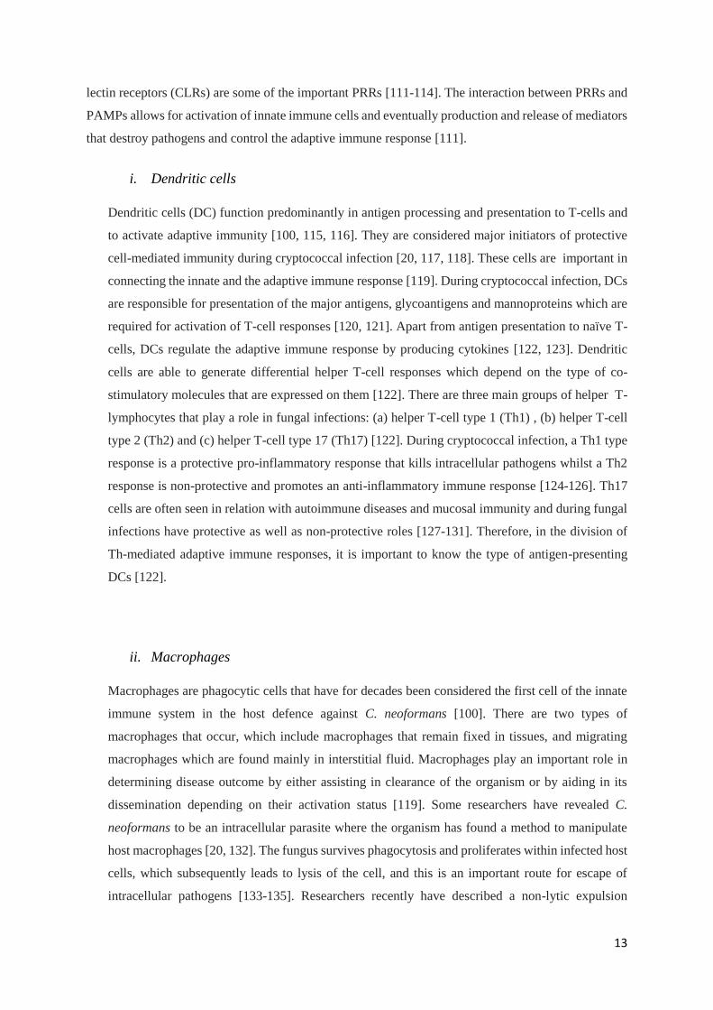
Complement receptor 3 (C3) and Fc-γ R are significant human neutrophil receptors which recognise
C3b and IgG opsonised fungal pathogens (Figure 2.1) [145]. Fungal contact with these receptors
initiates signalling cascades, which stimulates various mechanisms such as phagocytosis, oxidative
burst, NET, and release of antimicrobial peptides from neutrophil granules [145]. All of these
contribute to fungal killing. Neutrophils also secrete cytokines and chemokines during fungal
contact which leads to recruitment of further immune cells [145].
Figure 2.1 Neutrophil function in antifungal immune response
Hunniger K, et al, 2019, 89, 3-15
16
iv. Natural killer cells
Natural killer (NK) cells are cytotoxic cells that form part of the innate immune system. Some
studies have shown that these cells play a role in defence against cryptococci in murine experiments
through direct cytotoxic effect against the organism [100, 153-155]. Other studies in mice suggest
that NK cells enhance the ability of macrophages to kill fungal cells by producing interferon gamma
(IFNγ) [156, 157]. In human studies, NK cells and T-lymphocytes inhibit growth of Cryptococcus
through direct interaction with the organism [158, 159]. NK cells express granulysin and perforin
but perforin only is required for anti-cryptococcal activity by NK cells [148, 160].
v. Eosinophils
Eosinophils are granulocytes that are especially known for their role in allergies and parasitic
infections [100]. In rats, it was established that C. neoformans was phagocytosed by eosinophils
and that eosinophils primed B and T cells to generate protective host Th1 responses [100, 161-
163]. There is however, no clarity on whether eosinophils have any significant role in innate
immunity to C. neoformans or whether their recruitment is just a by-product of an inadequate Th2
response [100].
2.3.1.3 Pattern Recognition Receptors
Usually, pathogens are recognised by PRRs on host cells, which recognise PAMPs that are produced
by the pathogen. This interaction of PRRs and PAMPs induces signal transduction, which allows for
innate immune processes to occur. Components of the cell wall including glucans, mannans and chitin
are common fungal PAMPs. C. neoformans polysaccharide capsule has the ability to mask these
PAMPs and interestingly, PRRs such as C-type lectin receptor (CLR) and Toll-like receptor (TLR)
families, that usually detect other fungi, do not play similar roles in recognising C. neoformans [100].
The process involved in the recognition of C. neoformans by the host therefore, is still not clearly
defined [100].

17
i. Toll-Like Receptors
Of the PRRs , TLRs are the family studied most extensively, with 13 known members coded for in
the human genome [119]. These receptors are able to recognise bacteria, viruses and fungal
pathogens and they play a role in regulating both the pro-inflammatory as well as the anti-
inflammatory immune responses. Most TLRs recognise PAMPs and this initiates signal
transduction cascades that are associated with the adaptor molecule myeloid differentiation primary
response protein 88 (MyD88) [119]. Evidence that MyD88 is involved in anti-cryptococcal
responses in rats supports the possible involvement of TLRs as cryptococcal PRRs [164, 165].
However, there is still insufficient experimental evidence that supports the role of many of the TLRs
in cryptococcal infection [100].
TLR2 and TLR4 are able to recognise several pathogen ligands especially cell wall-associated
ligands and this has earned them much attention among researchers [119]. TLR2 and TLR4 are cell
surface receptors and are expressed on many innate cells such as neutrophils, macrophages,
monocytes, and DCs [166]. β-glucans that are expressed in the fungal cell wall are usually
recognised by TLR2, however the capsule of C. neoformans has the ability to conceal the β-glucan
layer [167]. The function of TLR2 during the immune responses to cryptococci appears to be varied
among studies [119].
Microglial cells, which are the resident phagocytes within the CNS, as well as meningeal
macrophages, make up the first line of defence within the brain during infections of the CNS [168-
170]. These cells may be found throughout the brain parenchyma and constitutes between 10 and
20% of the glial cell population in the CNS [171]. Koutsouras et al found that a comparison between
microglia and macrophages aided in the understanding of microglial physiology. However, unlike
macrophages, microglial cells maintain themselves entirely by self-replication [171]. Perivascular
microglial cells, also named perivascular macrophages, are derived from bone marrow and are
maintained continuously from the periphery while simultaneously being maintained by the self-
replication properties of parenchymal microglia [171, 172]. During infection, these cells release
chemokines which increase recruitment of dendritic cells, neutrophils, and lymphocytes from
peripheral tissue [171]. Microglial cells express TLRs, which identify PAMPs and are therefore
important in regulating the innate immune response [170, 173, 174]. TLR2 as well as TLR4 are
known to recognize cryptococcal GXM [170, 175]. It was determined that GXM had the ability to
bind to TLR2 and TLR4 with the co-receptor CD14 but it was unable to activate the MAPK
(mitogen-activated protein kinase) pathway and produce TNF-α [170, 175]. TLRs also have the
ability to form heterodimers like TLR1/2 and TLR2/6 that recognize GXM in the cryptococcal
capsule [176]. In vitro studies show that O-linked mannans activate TLR4 in C. neoformans [119].
Furthermore, pro-inflammatory responses were increased in C. neoformans infection when

18
microglial cells were stimulated with TLR1/2, TLR3, TLR4, and TLR9 (TLR agonists), however,
the significance is not clear [170].
While TLR function in the lymphatic system has stimulated much interest in recent years, not much
information is available on the expression and role of TLRs in CNS microglial cells [7]. It is
however, well established that these cells do participate in the innate immune response [177].
Emerging data is becoming available on TLR expression in the rodent CNS, however minimum
information is available on expression of TLRs in human microglial cells [7].
ii. C-type Lectin Receptors
The C-type Lectin Receptors (CLRs) are a group of receptors that recognise fungal carbohydrate
ligands such as β-glucans and mannans [100]. CLRs usually initiate signalling pathways through
their own intracellular signalling domain or via signalling adapters that have an immunoreceptor
tyrosine-based motif (ITAM) [100]. It has been established that CLRs play a role in host immune
responses to other fungi, however their role in C. neoformans is not significant [100, 178]. Evidence
exists that β-glucans are accessible on capsules of C. neoformans, however, in vivo, it seems
evident that the capsule interferes with many of these interactions [179, 180].
iii. Nucleotide-Binding Oligomerization Domain (NOD)-Like Receptors
NOD-like receptors (NLRs) are cytoplasmic PRRs that share a role in immunity and are able to
recognise PAMPs and damage-associated molecular patterns (DAMPs) [119]. Some studies
recently, have shown that NLRs were involved in sensing the presence of fungi [119]. C.
neoformans cells stimulated formation of the NLRP3 inflammasome, and studies with mice that are
deficient in the NLRP3 inflammasome components showed that they were more susceptible to
infection [181-183]. However, sufficient information on the role of NLRs in response to C.
neoformans is not available [100].
19
2.3.2 Adaptive Immune Response
2.3.2.1 Cell-mediated Immunity: T-cells
The patient group predominantly at risk for developing cryptococcal disease usually have severe T-cell
function defects [148]. This clearly indicates that T-cell mediated immunity plays a vital role in the
control of cryptococcal infections (Figure 2.2). Both CD4 and CD8 cells inhibit C. neoformans, either
directly or by producing pro-inflammatory cytokines that are required for recruiting and activating other
phagocytic cells to kill the organism [122, 184-186]. A review of in vitro studies indicate that CD4 and
CD8 T cells both produce Th1 cytokines, however the sole source of Th2 cytokines are the CD4 T cells
[122]. Various T cell subsets exist that have a function in cryptococcal infection. Regulatory T-cells are
protective against cryptococcal infection in that they suppress the damaging Th2 immune response
[122]. Other examples of T-cell subsets include NK cells, Natural killer T (NKT) cells and gamma delta
T (γδT) cells which all play a role in in protective immunity against cryptococcal infection [122, 187,
188]. T-cells however, are also able to downregulate the protective Th1 response [189].
The proliferation of naïve T-cells may be induced by complete cryptococcal cells or by cell extracts
such as cell wall and cell membrane protein extracts [190, 191]. To ensure presentation of C.
neoformans antigen to T-lymphocytes, it is necessary for phagocytosis and protein processing to occur
[192]. Cryptococcal cells are then either inhibited or killed by CD4 and CD8 T cells.
2.3.2.2 Antibody-Mediated Immunity
There has been conflict regarding the role of antibody-mediated immunity in cryptococcal infections.
There are however, many reports of cryptococcal infections in patients who have B-cell deficiencies,
as well as antibody or even lymphoproliferative deficiencies [193]. In addition, antibodies against
cryptococcal proteins and capsular components have been found in people without infection [20, 194,
195]. In HIV-patients, there is an association with cryptococcal infections when there is a decrease in
B-cells secreting IgM [196]. Anti-cryptococcal antibodies opsonise the pathogen resulting in Fc
receptor-dependant phagocytosis and activation of the classical complement pathway [20]. Studies in
mice indicate that the protective effect of antibodies may be to an extent, due to interactions with cell-
mediated immunity [20]. Some studies show that antibody responses may amplify cryptococcal disease
20
in humans [122]. In non-HIV patients with normal CD4 counts autoantibodies have been associated
with infection [197].
2.3.2.3 Cytokine response
Knowledge regarding cytokine response has been obtained predominantly in mouse models and
therefore information and evidence regarding their response in cryptococcal infection relates to mouse
models [198-200]. Cytokines consist of proteins, peptides or glycoproteins that are secreted by immune
cells and have a role in mediating and regulating immunity. Various cytokines and chemokines are
involved in cryptococcal infection. These cytokines and chemokines either induce the Th1 response
and/or suppress Th2 immune responses [122]. Both protective and non-protective cytokines are
produced during infections by C. neoformans. Protective cytokines include IFN-γ, IL-12 and IL-2 which
have been shown in humans as well as mouse models [122]. Additionally, other protective cytokines
have been identified which include TNF-α, IL-6, IL-8, IL-18, IL-23, and IP-10 [122]. Non-protective
cytokines include IL-5 and IL-13, which are the Th2 cytokines, and these cytokines stimulate
cryptococcal disease. There are certain cytokines/chemokines that may play a dual role in protection as
well as disease aggravation during cryptococcal infection, and these include IL-4, IL-8, IL-10, IL-1β,
MCP-1 (monocyte chemoattractant protein 1), MIP-1α (macrophage inflammatory protein form 1
alpha) and RANTES (CCL5) [122].

21
Figure 2.2 Innate vs Adaptive Immunity
Nature Reviews Cancer, 4, 11-22
2.4 Paucity of neutrophils in cryptococcal meningitis
Cryptococcal meningitis is known to be characterized by a relative paucity of leucocytes in the CSF
compared to that in bacterial meningitis. [3-5, 56, 201]. The presence of leucocytes is regulated through
chemotaxis. Chemotaxis is dependent on chemokine production at the infection site and the level of
expression of corresponding chemokine receptors on the migrating cells. Chemokines are important in
mediating recruitment of leucocytes into infection sites, including infections with C. neoformans [202].
Microglial cells in the brain are important sources of IL-8, IP-10, MIP-1, MIP-1, RANTES, KC, and
MCP-1[203]. Chemokines are produced when TLRs on cells at the site of infection recognize specific
microbes. For C. neoformans, TLR2 has been identified as a significant TLR [6, 7, 175].
22
TLR2 and TLR4, as described above, are receptors found on the cell surface that enable phagocytic
inflammatory responses to various microbial products. When these receptors are activated, signaling
cascades are triggered and this results in a pro-inflammatory response including production of TNF.
[175]. Whilst it is clear that TLR expression is significant, there is not much published information on
expression and function of TLRs in human CNS microglial cells [7].
High levels of the capsular components GXM, GalXM, MP-1, and MP-2 is characteristic of
disseminated cryptococcal infection, in the CSF and serum of patients [3]. These components induce
peripheral blood monocytes and polymorphonuclear neutrophils (PMN) to produce the cytokines TNF-
and IL-1 [3]. TNF- and IL-1 stimulates endothelial cells, astrocytes and mononuclear cells to
produce IL-8 in the brain of patients with meningitis.[3]. IL-8 is a potent chemo-attractant for PMNs
[3]. However, in cryptococcal meningitis, there is a paucity of PMNs [4, 5, 56, 201].
A study by Lipovsky et al [5] confirmed what others have observed that GXM has chemoattractant
activity. They have suggested that IL-8 production occurs in the brain but neutrophils do not cross the
blood brain barrier (BBB) in response to IL-8. Their explanation is that migration of PMNLs does not
occur because a chemotactic gradient does not exist across the blood brain barrier. Furthermore, a
phenomenon called cross-desensitization of the IL-8 receptor could be another possible explanation for
chemotaxis inhibition by GXM [5, 204].
Glucuronoxylomannan has been observed to inhibit adhesion of PMNs to activated endothelium in a
concentration-dependent manner [56]. Furthermore, another component of the capsule, MP-4, was
studied as a new capsular antigen which activates neutrophils and desensitizes them toward a
chemotactic challenge [3].
Another possibility for the paucity of leucocytes is that even though IL-8 was produced in the CSF of
patients with cryptococcal meningitis, the circulating GXM was downregulating recruitment of
leucocytes, perhaps through shedding of L-selectin, thus impairing migration of leucocytes into the CSF
[205, 206].
A series of steps are involved during the inflammatory response with regards to leucocytes and
endothelium interactions [57]. These progress from rolling to adhesion and ultimately migration into
tissues. Margination and rolling of leucocytes on the cytokine-activated endothelium are the first steps
in adhesion [57, 207]. It was shown that GXM impedes the early rolling phase of neutrophil adhesion
to endothelium and to Chinese Hamster Ovary (CHO) cells that were E-selectin-transfected [57].

23
Various observations as described above suggest the presence of neutrophil chemokines in the brain of
infected patients and that microglial cells are a potent source, yet a paucity of neutrophils still occurs in
cryptococcal infection. Toll-like receptors have been described as receptors involved in initiating
phagocytic inflammatory responses, however little evidence is available on its expression and function
in human microglial cells. This study therefore aims to describe this.
24
CHAPTER 3 – METHODS
3.1 Patient Recruitment and Clinical Data
A prospective study was conducted at a regional hospital in KwaZulu-Natal, South Africa, from 16 July
2013 to 03 February 2016. HIV infected adult patients with a diagnosis of meningitis with Cryptococcus
neoformans as causative agent were invited to participate in the study. The criteria for exclusion were
age <18 years, HIV uninfected, and concomitant infections other than HIV. The diagnosis was made
on clinical grounds and confirmed by laboratory examination of CSF. Only patients with isolates
identified as C. neoformans were included (3.3). Patients who provided written informed consent were
enrolled in the study within 24 hours of presentation. Clinical details were recorded and a second lumbar
puncture was performed 48 hours following presentation as part of the routine patient work-up and CSF
opening pressure was recorded. Two tubes with CSF as well as two with venous blood collected at this
time point were transported to the laboratory. A total of 71 patients who fulfilled all the criteria were
enrolled in the study. None of the patients had received prior antifungal treatment. Antifungal therapy
was commenced after the first CSF specimen was collected.
3.2 Quantification of Immune Cells in CSF and Blood
3.2.1 Quantification of Immune Cells in CSF
Cells in the CSF were quantified using a haemocytometer (Thermo Fischer Scientific, Massachusetts,
USA). One drop of trypan blue was added to a clean tube using a sterile glass pipette. Nine drops of
CSF was then added to the same tube using an equivalent glass pipette. This made a 1:10 dilution. This
was left to stand for five minutes to allow the cells to absorb the stain. The solution was mixed by gently
tapping the bottom of the tube. A coverslip was placed over the counting chamber and using a capillary
tube the counting chamber was filled with the diluted specimen. It must be noted that the counting
chamber can only accommodate a predetermined fixed volume of 20 μl. The slide was then viewed
under a light microscope using a 10× objective and the number of cells were counted using a cell
counter. A standard formula was used to calculate number of cells (Appendix A1).
25
3.2.2 Quantification of Immune Cells in Blood
A full blood count was performed using an automated system (Advia 2120i, Siemens, Munich,
Germany). A differential count was performed using the same instrument. Enumeration of neutrophils
was done by establishing the percentage of these cells from the total white blood cell count using the
same automated instrument.
3.3 Microscopy and Culture of CSF Specimens
The first CSF received from the patient was centrifuged at 1109 × g for 10 minutes. The supernatant
was aseptically decanted into a sterile capped tube labelled with the study number. The deposit was
resuspended in the remaining supernatant by gently tapping the bottom of the tube. A smear was
prepared by placing one drop of the resuspended deposit onto a sterile glass slide using a sterile pipette.
The slide was allowed to air dry followed by Gram staining (Appendix A2). Fifty microliters of the
deposit were inoculated onto sheep blood agar (Appendix B1), and incubated at 37°C for 48 hours in a
regular incubator. Each isolate was identified to species level using the automated Vitek® 2 system
(bioMérieux, France). The CSF supernatant was used to perform a Cryptococcus latex agglutination
test (3.4).
3.4 Concentration of Capsule Material Components in CSF and Serum
3.4.1 Principle of the Cryptococcal Antigen Latex Agglutination Test
The Cryptococcal Antigen Latex Agglutination System (CALAS® - Cryptococcal Antigen Latex
Agglutination System, Meridian Bioscience Inc., Ohio, USA) was used to measure the titre of
cryptococcal antigen in serum and in CSF. This was used as a proxy for the amount of shed capsular
material. The procedural instructions by the manufacturer were followed to complete the tests. CALAS
utilizes latex particles coated with rabbit anti-cryptococcal IgG in glycine buffered saline (pH 8.4 ± 0.1)
that contains a preservative thimerosal (< 0.01%). This is referred to as detection latex. Visible
agglutination is observed when the antibodies on the latex particles and the cryptococcal polysaccharide
antigen bind together. Latex particles that were coated with immune-globulins not containing
26
cryptococcal binding sites were used as negative control. The positive control consisted of purified C.
neoformans polysaccharide antigen, which contained the preservative thimerosal (0.01%). Goat anti-
rabbit serum with thimerosal (0.01%) was the antibody control. Macroglobulins such as rheumatoid
factor may be present that could cause nonspecific agglutination. If this does occur specimens are
treated with pronase (Appendix B2) to remove these macroglobulins.
3.4.2 Procedure of the Cryptococcal Antigen Latex Agglutination Test
One free falling drop of positive control was added to the two designated rings on the reaction card
provided with the kit. Twenty-five microliters each of the antibody and negative controls were pipetted
to the relevant rings. Twenty-five microliters of patient CSF supernatant was pipetted into each of the
two rings on the test-card. Thereafter, one free falling drop of the detection latex was added into each
ring. Similarly, one drop of negative control latex (3.4.1) was added into the additional rings. The
contents of the different rings were then mixed using separate applicator sticks. Subsequently, the card
was placed on a horizontal rotator and rotated at 0.4 × g for 5 minutes. Results were read immediately.
3.4.2.1 Titrations
Two-fold serial dilutions of each specimen were done until the agglutination end point was reached.
Two hundred and fifty microliters of sample diluent (Appendix B3) was added into five labelled test
tubes and placed in a rack. Using a clean pipette, 250 l of CSF or serum was added into tube labelled
1 and mixed well. Two hundred and fifty microliters from tube 1 was then transferred to tube 2 and
mixed well. Following this pattern, the dilution series was continued through to tube five. Two hundred
and fifty microliters from tube 5 was transferred into a “holding” tube to allow for further dilutions that
may be necessary. Thereafter, the same antigen detection procedure as described in 3.4.2 was followed.

27
3.5 Storage of CSF and Blood specimens
The second tube with CSF from each patient was centrifuged at 1109 × g for five minutes. The
supernatant was aseptically decanted into at least two sterile cryovials labelled with the study number
and stored at -80°C. The deposit was resuspended by gently tapping the bottom of the tube and
inoculated onto sheep blood agar (Appendix B1). After 48 hours of incubation at 37°C, the growth was
harvested using a sterile plastic loop and suspended in 1 mL of sterile distilled water. These suspensions
were stored at room temperature for further work (Appendix A3).
Blood specimens were centrifuged at 1109 × g for 5 minutes. The serum was then aseptically decanted
into at least two sterile cryovials with labelled with the study number and stored at -20°C.
3.6 Retrieval of Stored Cryptococcus Cultures for Determination of Capsule Thickness
Stored Cryptococcus suspensions were plated out on Brain Heart Infusion Agar plates (Oxoid Ltd,
Cheshire, England) supplemented with 10% Horse blood (Appendix B4). These were incubated for 48
hours at 37°C aerobically.
3.6.1 Determination of capsule thickness
A microscopy slide was made on the first subculture of organisms grown from the 48-hour specimen
obtained by lumbar puncture (3.6). Anthony’s capsule stain was applied to visualize the polysaccharide
capsule [208].
3.6.1.1 Principle of Anthony’s Stain
This procedure for staining capsules contains crystal violet as the primary stain, which is taken up by
all parts of the cell. A 20% copper sulphate solution is used as a decolorizing agent as well as a counter
stain. When the copper sulphate washes out the crystal violet, the capsule becomes decolorized but not

28
the cell. As the copper sulphate decolorizes the capsule, it simultaneously counter stains the capsule.
The capsule therefore can be visualised as a faint blue halo surrounding a purple cell.
3.6.1.2 Preparation of the inoculum
i. Determining Yeast cell concentration of 1-McFarland Standard for Cryptococcus
A suspension of the culture was made in phosphate buffered saline, pH = 7.2 (Appendix B6) and
adjusted to a 1-McFarland suspension (Appendix B5) which is equivalent to a suspension of 3.0 × 10⁸
CFU/mL for bacteria of average size (0.5 – 2.0 μm). Considering that C. neoformans is larger than an
average bacterial cell (5 – 10 μm vs 0.5 – 2.0 μm), the concentration of the yeast cell equivalent to a 1-
McFarland standard was determined. A series of 1:10 dilutions was made in RPMI with L-Glutamine
(Lonza Group Ltd, Basel, Switzerland) starting with a density of 3.0 × 10⁸ CFU/mL and ending at 1.0
× 10² CFU/mL. Ten microliters of each suspension was inoculated on individual sheep blood agar plates
and incubated aerobically for 48 hours at 37°C. This was performed in duplicate. Thereafter, colonies
were counted on the plates that contained on viewing 20 – 200 colonies. The average number of
colonies was then adjusted for volume and inoculum size and thus it was determined that the bacterial
1-McFarland standard was equivalent to 1.0 × 10⁶ CFU/mL for C. neoformans.
3.6.1.3 Anthony’s staining procedure
Fifty microliters of the cryptococcal suspension was placed on a glass slide and a thin film was made
using a second slide under an approximately 45o angle. After air drying, the slide was flooded with 1%
crystal violet for two minutes [208]. It was then rinsed gently with a 20% copper sulphate solution and
air-dried. It is not recommended to blot the slide because blotting removes the un-fixed microbes from
the slide and cause disruption of the capsule [208]. The slide was viewed under an oil immersion lens
(Leica DM 3000; Leica microsystems, Wetzlar, Germany). Bacterial cells and the proteinaceous
background appeared purplish while the capsules appeared transparent. Photographs were taken of the
organisms on the slide and viewed at a magnification of 100×. A camera (Image Pro-Plus of Media
Cybernetics, Inc., Maryland, USA), was used to capture these images. The number of images taken
depended on the density of cryptococcal cells on the slide. The images were used to measure the capsule
thickness. This was done by first measuring the diameter of the entire cell including the capsule
29
followed by measuring the smaller diameter of the cell excluding the capsule. The difference between
the two diameters was recorded as the capsule thickness in micrometers. The average capsule size of
each isolate was determined using three different cryptococcus capsule measurements per isolate. Three
cells per isolate were randomly selected on three microscopic fields and capsule measurements were
performed as described above.
3.7 Neutrophil Chemotaxis Inhibition
Neutrophil chemotaxis inhibition experiments were performed to compare the number of migrating
neutrophils toward the chemoattractant (E. coli), with the number of neutrophils that migrate toward
the chemoattractant mixed with the cryptococcal isolates. The amount of inhibition was determined by
calculating the difference between these two values (no. of migrated neutrophils towards E. coli – no.
of migrated neutrophils towards E. coli/Cryptococcus combination = Chemotaxis inhibition).
A qualitative approach using the Boyden Chamber method was used. A gradient separation method was
used to separate neutrophils from the rest of the human blood cells (3.7.2.2).
3.7.1 Boyden Chamber assay
The Boyden Chamber method [209, 210] was used to determine the rate of neutrophil chemotaxis in
the presence and absence of C. neoformans yeast cells. Escherichia coli ATCC 25922 was used as the
chemoattractant. This assay is based on a chamber that consists of two compartments that are separated
by a microporous membrane. The assay was developed originally by Boyden to analyse leucocyte
chemotaxis [211]. Cells added into the upper compartment migrate through the membrane pores into
the lower compartment, which potentially contains the chemotactic agents. The chamber is incubated
for an appropriate/predetermined time, following which the membrane between the two compartments
is fixed and stained and the number of migrated cells in the lower compartment is determined using a
cell counting method. This assay has therefore also been called “filter membrane migration assay”,
“trans-well migration assay”, or “chemotaxis assay” [211]. Various different Boyden chamber devices
are available commercially. Here, Transwell® inserts (Corning Life Sciences, New York, USA) in 24-
well tissue culture plates were used.

30
3.7.2 Preparation for the Transwell Migration Assay
3.7.2.1 Acquisition of neutrophils from volunteer donors
Healthy adult volunteers were recruited with informed consent to donate blood on the day of an
experiment. Approximately 25 mL of blood was drawn from one donor. Blood samples were collected
in EDTA tubes (BD Life Sciences, New Jersey, USA) and transported immediately to the laboratory
for neutrophil separation.
3.7.2.2 Neutrophil separation
A gradient separation method using Histopaque®-1119 (Sigma-Aldrich, Missouri, USA) and
Histopaque®-1077 (Sigma-Aldrich, Missouri, USA) was used in the laboratory to separate neutrophils
from the rest of the human blood cells. The procedure was followed according to specifications by the
manufacturer. Before commencing on the separation procedure, cells and plasma were mixed evenly
by gentle tilting of vials. Twelve millilitres Histopaque®-1119 was added to a 50 mL conical centrifuge
tube using a sterile needle and syringe. Twelve milliliters of Histopaque®-1077 was then carefully
layered onto the Histopaque®-1119 using a sterile needle and syringe. This was followed by carefully
layering 24 mL of whole blood onto the upper gradient of the tube using a sterile needle and syringe.
Without disturbing the gradients, the tube was centrifuged at 700 × g (centrifugal force) for thirty
minutes at room temperature (25°C). Two distinct opaque layers were visible, one containing
mononuclear cells and platelets and the other containing granulocytes including the neutrophils. The
granulocyte layer was gently aspirated using a sterile Pasteur pipette (Sigma-Aldrich, Missouri, USA)
and cells were transferred to a sterile 15 mL conical centrifuge tube. The cells were washed by adding
10 mL of isotonic phosphate buffered saline (PBS), pH = 7.2 (Appendix B6). This was centrifuged at
200 × g for 10 minutes at room temperature (~25°C). The supernatant was removed using a sterile
Pasteur pipette and discarded. The cells were resuspended by gentle aspiration three times with a sterile
Pasteur pipette. The wash step was repeated for a further two times. Cells were then resuspended in
isotonic PBS, pH = 7.2 to achieve a cell count of 1 × 10⁶ cells/mL [212]. To determine the dilution
factor to obtain this neutrophil concentration, a cell count was done using a haemocytometer as
described previously (Appendix A1). Further dilutions were made as required. The purity of the
neutrophil suspension was tested by means of the Giemsa staining method (Appendix A4)

31
3.7.2.3 Preparation of the inoculum of C. neoformans isolates and E. coli ATCC 25922
For C. neoformans, in a series of preliminary experiments (3.6.1.2), the appropriate density was
determined to be 1.0 × 10⁶ CFU/mL and for E. coli, it was determined to be 3.0 × 10⁸ CFU/mL.
Therefore, a 1-McFarland suspension of each organism suspension was used in the chemotaxis assay.
For the E. coli/Cryptococcus mixture, equal volumes of the E. coli ATCC 25922 (3.0 × 10⁸ CFU/mL)
and C. neoformans (1.0 × 10⁶ CFU/mL) suspensions were mixed in a sterile tube at a 1:2 dilution for
each organism. Thus, a suspension of each culture was made in PBS, pH = 7.2 containing 0.5 × 10⁶
CFU/mL Cryptococcus and 1.5 × 10⁸ CFU/mL E. coli. The mixed suspension contained the same
amount of microbes of each species as above. For the Transwell assay the cryptococcal suspension, the
E. coli suspension as well as the mixed suspension were used.
3.7.3 Transwell Assay
Twenty-four well tissue culture plates that have inserts in the plate with a collagen-coated membrane
of 3-μm pore size were used (Figure 3.1). Transwell® inserts (Corning Life Sciences, New York, USA)
were used. A membrane with a 3 μm pore size was used to ensure that neutrophils actively migrate
through them to the lower chamber (baso-lateral compartment) and move through freely [213]. Six
hundred microliters of organism suspension was added to the bottom of a well of a 24-well plate. The
Transwell membrane filter was then placed carefully in the same well. Typically 1.0 × 10⁶ cells/mL is
used for nearly all types of cells when using the 24-well Transwell insert [212]. One hundred microliters
of neutrophil cell suspension was added onto the top (upper compartment) of the filter membrane. These
volumes were determined to ensure that the suspension medium in the bottom of the well makes contact
with the membrane to form a chemotactic gradient [212]. The assay was incubated at 37°C for 30
minutes. The number of migrated cells was determined using the Myeloperoxidase (MPO) Human
SimpleStep ELISA® Kit (Abcam®, Cambridge, UK)). As described in 3.7, the chemotaxis assay was
performed with each cryptococcal isolate alone and a combination of the same cryptococcus plus the
chemoattractant (E. coli ATCC 25922). E. coli (ATCC 25922) alone was used as the control. The
amount of inhibition was determined by calculating the difference between two values ( no. of migrated
neutrophils towards E. coli – no. of migrated neutrophils towards E. coli/Cryptococcus combination =
Chemotaxis inhibition). All experiments were performed in duplicate.
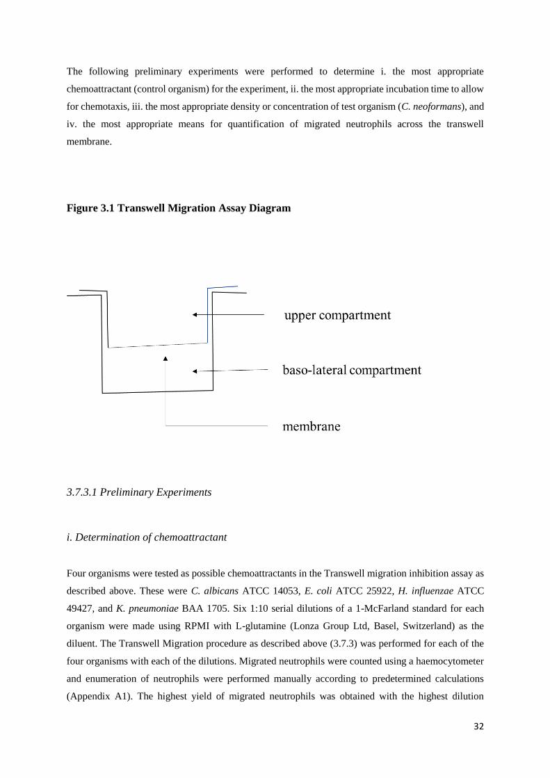
32
The following preliminary experiments were performed to determine i. the most appropriate
chemoattractant (control organism) for the experiment, ii. the most appropriate incubation time to allow
for chemotaxis, iii. the most appropriate density or concentration of test organism (C. neoformans), and
iv. the most appropriate means for quantification of migrated neutrophils across the transwell
membrane.
Figure 3.1 Transwell Migration Assay Diagram
3.7.3.1 Preliminary Experiments
i. Determination of chemoattractant
Four organisms were tested as possible chemoattractants in the Transwell migration inhibition assay as
described above. These were C. albicans ATCC 14053, E. coli ATCC 25922, H. influenzae ATCC
49427, and K. pneumoniae BAA 1705. Six 1:10 serial dilutions of a 1-McFarland standard for each
organism were made using RPMI with L-glutamine (Lonza Group Ltd, Basel, Switzerland) as the
diluent. The Transwell Migration procedure as described above (3.7.3) was performed for each of the
four organisms with each of the dilutions. Migrated neutrophils were counted using a haemocytometer
and enumeration of neutrophils were performed manually according to predetermined calculations
(Appendix A1). The highest yield of migrated neutrophils was obtained with the highest dilution
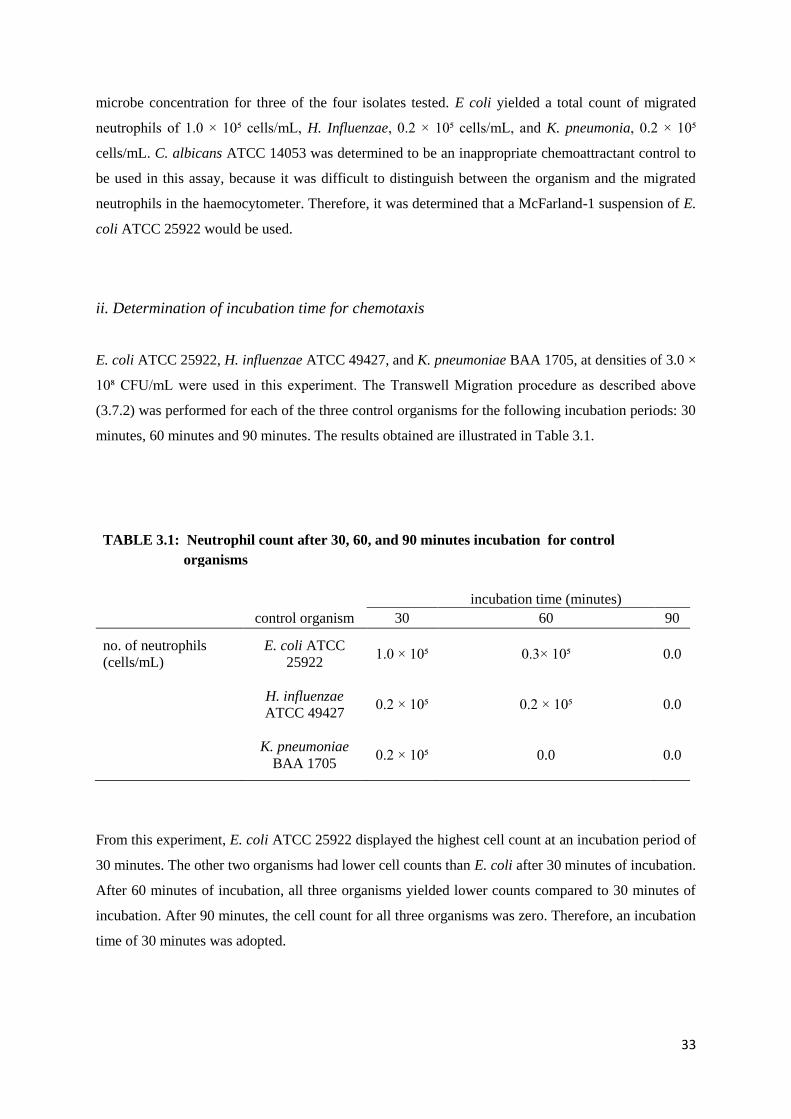
33
microbe concentration for three of the four isolates tested. E coli yielded a total count of migrated
neutrophils of 1.0 × 10⁵ cells/mL, H. Influenzae, 0.2 × 10⁵ cells/mL, and K. pneumonia, 0.2 × 10⁵
cells/mL. C. albicans ATCC 14053 was determined to be an inappropriate chemoattractant control to
be used in this assay, because it was difficult to distinguish between the organism and the migrated
neutrophils in the haemocytometer. Therefore, it was determined that a McFarland-1 suspension of E.
coli ATCC 25922 would be used.
ii. Determination of incubation time for chemotaxis
E. coli ATCC 25922, H. influenzae ATCC 49427, and K. pneumoniae BAA 1705, at densities of 3.0 ×
10⁸ CFU/mL were used in this experiment. The Transwell Migration procedure as described above
(3.7.2) was performed for each of the three control organisms for the following incubation periods: 30
minutes, 60 minutes and 90 minutes. The results obtained are illustrated in Table 3.1.
TABLE 3.1: Neutrophil count after 30, 60, and 90 minutes incubation for control
organisms
incubation time (minutes) control organism 30 60 90
no. of neutrophils
(cells/mL)
E. coli ATCC
25922 1.0 × 10⁵ 0.3× 10⁵ 0.0
H. influenzae
ATCC 49427 0.2 × 10⁵ 0.2 × 10⁵ 0.0
K. pneumoniae
BAA 1705 0.2 × 10⁵ 0.0 0.0
From this experiment, E. coli ATCC 25922 displayed the highest cell count at an incubation period of
30 minutes. The other two organisms had lower cell counts than E. coli after 30 minutes of incubation.
After 60 minutes of incubation, all three organisms yielded lower counts compared to 30 minutes of
incubation. After 90 minutes, the cell count for all three organisms was zero. Therefore, an incubation
time of 30 minutes was adopted.

34
iii. Inoculum size for C. neoformans
Inoculum size for E. coli ATCC 25922 was determined to be 3.0 × 10⁸ CFU/mL following preliminary
experiments to determine chemoattractant (3.7.3.1.i). For C. neoformans, a series of 1:10 dilutions was
made in RPMI with L-Glutamine (Lonza Group Ltd, Basel, Switzerland) starting with a density of 3.0
× 10⁸ CFU/mL and ending at 1.0 × 10² CFU/mL (3.6.1.2.i). The Transwell Migration procedure as
described above (3.7.3) was performed on each of the prepared dilutions. Migrated neutrophils were
counted using a haemocytometer and enumeration of neutrophils was performed manually according to
predetermined calculations (Appendix A1). At this point, due to similar size, it was difficult to
distinguish microscopically between the organism and the migrated neutrophil in the lower chamber of
the assay using the haemocytometer. Therefore, an alternative method for quantification of migrated
neutrophils was determined. Using this method (described below), it was determined that the lowest
concentration at which Cryptococcus caused neutrophil migration was at a density of 1.0 × 10⁶
CFU/mL.
iv. Determination of quantification method for migrated neutrophils – The Myeloperoxidase
(MPO) Human Simple Step ELISA® (Abcam)
Myeloperoxidase (MPO) is an enzyme belonging to the subfamily of peroxidases. A variety of cells
including neutrophils, lymphocytes [214-216] monocytes, and macrophages [214, 217] are able to
express this enzyme. The azurophilic granules of neutrophils contain stored myeloperoxidase. When
the neutrophil is stimulated, exocytosis or degranulation occurs, and the content of the granules is
released out to the extracellular space [214, 218, 219]. Although various other antimicrobial enzymes
and proteins are present in these granules, MPO is most abundant and accounts for 5% of neutrophils’
dry weight and 25% of azurophilic granular proteins [214, 220]. A modification to the Boyden
Chamber method was made by Kristina Somersalo, et al, where the response to chemotaxis was
determined by measuring myeloperoxidase (MPO) specific to neutrophils and monocytes [213]. They
found that the MPO Boyden chamber method had the advantages of being faster, was easier to carry
out and was more objective than the traditional cell count method [213].
Considering the difficulty encountered when counting cells using the conventional method described
above (haemocytometer) especially when using Cryptococcus in the preliminary experiments, it was
decided to measure chemotactic response in this study, by measuring myeloperoxidase in the cell
extracts after the 30 minute incubation step in the Transwell assay procedure (3.7.3).

35
3.7.3.2 Myeloperoxidase ELISA
i. Principle of the test
In this study, a commercially available Myeloperoxidase (MPO) in vitro SimpleStep ELISA®
(Enzyme-Linked Immunosorbent Assay) kit (Abcam®, Cambridge, UK) was used. Abcam’s
Myeloperoxidase (MPO) in vitro SimpleStep ELISA® kit is designed to quantitatively measure human
myeloperoxidase in various cell culture supernatants, plasma, human serum, urine, milk, saliva, as well
as cell and tissue extracts. Two antibodies are used in this ELISA, which capture the analyte (MPO) in
the solution, and these include an affinity tag labelled capture antibody and a reporter conjugated
detector antibody (Figure 3.2). Immobilization of the capture antibody/analyte/detector antibody-
complex occurs next through immunoaffinity of an anti-tag antibody that coats the well. When
performing the assay, standards and samples are added first to the designated wells. The antibody mix
is added next. Following incubation, the wells are washed to ensure removal of any unbound material.
TMB substrate (3.3′,5.5′-tetramethylbenzidine) is then added and the reaction is catalysed by
horseradish peroxidase (HRP) during an incubation step. A blue colour change is observed. The reaction
is stopped by adding Stop Solution, which causes a change in colour from blue to yellow. The colour
intensity is proportional to the quantity of bound MPO and is measured on a suitable spectrophotometer
at 450 nm. This is a sensitive assay (the lowest detection limit is 0.026 ng/mL) [221].
Figure 3.2 Myeloperoxidase Assay
The reaction follows the steps described in 3.7.3.2. The detection enzyme changes the colour of the
substrate
36
ii. Preparation of reagents, standards and samples
A 96 well plate was used that was provided in the SimpleStep ELISA® (Enzyme-Linked
Immunosorbent Assay) kit (Abcam, Cambridge, UK). All reagents were equilibrated to room
temperature (18-25°C) prior to use. The kit contained sufficient reagents for 96 wells (Appendix A5).
Sample volumes were adjusted as required for the number of wells in each experiment. Serially diluted
standards were prepared immediately before use. This was constituted in accordance with
manufacturer’s instructions (Appendix A6).
The samples on which the assay was performed were obtained for each cryptococcal isolate following
the Transwell procedure described above (3.7.3) i.e. the solution containing migrated neutrophils
retrieved in individual wells for each cryptococcal isolate. This was centrifuged at 18 000 × g at 4°C
for 20 minutes. Supernatants were transferred to clean tubes and pellets were discarded. A standard
1:200 dilution was made for all samples that were assayed.
iii. Assay procedure
Fifty microliters each of sample or standard was added to appropriately labelled wells. Fifty microliters
of prepared antibody cocktail consisting of capture and detector antibodies in antibody diluent
(Appendix A5) was then added to each well. A plate sealer was used to seal the plate and this was
incubated for one hour on a plate shaker set at 3.6 × g, at room temperature. Each well was then washed
with 3 × 350 l of 1 × Wash buffer PT (Appendix A5). Care was taken to ensure that all excess liquid
was removed following the last wash step. One hundred microliters of TMB substrate was added to
individual wells and incubated for 10 minutes in the dark on a plate shaker set at 3.6 × g. Following
this, 100 l of Stop Solution was immediately added to each well and the plate was again placed for 1
minute on a plate shaker to mix the solution. The OD was recorded at 450 nm using a microplate reader
software (Thermo Fischer Scientific, Massachusetts, USA).
3.8 Gene Expression – Quantitation of TLR2 and TLR4 in Human Microglial Cells
A human microglial cell line (M059K, ATCC® - CRL-2365ᵀᴹ) was purchased from American Type
Culture Collection (ATCC), Virginia (USA). Cells were exposed to cryptococcal isolates for a period
of two hours.

37
3.8.1 Cell Culture
3.8.1.1 Cell propogation
M059K cells (ATCC® CRL-2365ᵀᴹ) as described above (3.8), were regrown from frozen stock as
advised by ATCC. Using strict aseptic conditions, the cell suspension was rapidly thawed (not more
than two minutes) by agitating gently in a water bath at 37°C. The outside of the vial was then
disinfected with 70% ethanol. Within a laminar flow cabinet the contents of the vial was transferred to
a 15 mL centrifuge tube, which contained nine millilitres of complete culture medium (Appendix A7).
This was centrifuged (Heraeus multifuge 3S-R, 2006, Thermo Electron Corporation) at 125 × g for five
minutes and the supernatant was discarded. This process is recommended to remove cytotoxic
preservatives such as dimethylsulphoxide (DMSO) that may be present in the freezing fluid. The cell
pellet was then resuspended in 10 mL of pre-warmed complete culture medium (Appendix A7) and
transferred into 75 cm³ tissue culture flasks. Pre-warming of culture medium for 15 minutes at 37°C
was done to ensure that the medium reached the normal pH (7.0 – 7.6) and to prevent cold-shock of the
cells. Cells were then incubated at 37°C in 5% CO₂ until approximately 90% confluent monolayer was
obtained. Duration of incubation was determined to be between four to six days to achieve 90%
confluence. Cells were maintained by refreshing the culture medium every 48 hours. Cell growth was
observed at 40 × magnification using an inverted light microscope (Nikon Diaphot, Carl Zeis (PTY)
Limited, Germany).
3.8.1.2 Cell counts
Cells were enumerated using the “trypan blue exclusion method”. This staining method is one of the
traditional methods used to do cell viability analyses. Various academic research laboratories and
industrial biotechnology units use this technique as their standard procedure for this purpose [222]. The
purpose of this technique is to determine and quantify viable cells in a cell suspension. The test is based
on the concept that live cells possess cell membranes that are intact and exclude certain dyes, such as
trypan blue and eosin while this is not the case in dead cells [223]. In this test, 10 μl of cell suspension
was diluted in 10 μl of phosphate buffered saline (PBS). An equal volume (20 μl) of 0.4 % trypan blue
was added to this suspension. Ten microliters of the cell-dye suspension was then filled into a Neubauer
Haemocytometer slide (Appendix A1) with a cover slip and cells were counted using a Coulter counter
within 3 minutes. Viable cells appeared clear microscopically whilst nonviable cells appeared blue.

38
The number of cells per millilitre was calculated using the following formula:
Number of Viable Cells/mL = total number of viable cells counted × dilution factor (= 4) × 10⁴
number of squares counted (4)
The factor 4 corrects for the dilution with PBS and trypan blue and 104 for the volume.
3.8.1.3 Passaging of cells
Once a sufficiently confluent monolayer was attained, the monolayer was trypsinised with 0.25%
Trypsin-0.53 mM EDTA solution (Lonza Group Ltd, Basel, Switzerland). Trypsin is a proteolytic
enzyme that degrades the extracellular tissue protein matrix, which causes cells to adhere to each other.
The EDTA is a chelating agent, and removes calcium and magnesium ions, which allow attachment of
the cells to the flask wall. To achieve this, old medium was removed from the flask and discarded. Cells
remaining were washed three times with PBS, pH = 7.2 (Appendix B6). One millilitre of trypsin-EDTA
was added and spread evenly by gently rocking the flask. The trypsin-covered monolayer was then
incubated for 30 seconds at 37°C to facilitate dispersal. Cells were observed at 40 × magnification under
an inverted microscope (Nikon Diaphot, Carl Zeis (PTY) Limited, Germany) until the cell layer was
completely dispersed. Care was taken to expose cells to trypsin for the shortest possible duration (not
more than five minutes) because trypsin may damage living cells. One millilitre of foetal bovine serum
(FBS) (Appendix B7) was added to the flask and gently dispersed to counteract the effect of trypsin.
Cells were gently aspirated and collected into sterile tubes. The cell concentration was adjusted
according to a predetermined ratio of 1:6. Complete growth medium was added to this suspension and
seeded to new culture flasks, which were incubated at 37°C in 5% CO₂ until a confluent monolayer was
obtained.
3.8.1.4 Cryopreservation of cells
Cryopreservation medium containing 95% complete culture medium and 5% DMSO was added to the
cell suspension, slowly to prevent cell destruction. A stepwise process was followed to ensure slow
freezing of cells. This entailed first placing cells at 4°C for one hour followed by a 24-hour storage at
-20°C. Thereafter, cells were stored at -70°C until required for use.

39
3.8.2 Exposure of cells to Cryptococcus
Prepared M059K cells were exposed to cryptococcal isolates for a period of two hours at a ratio of 1000
cryptococcal cells to 1.0 × 10⁵ M059K cells per well. The Cryptococcus suspension was prepared in a
series of 1:10 dilutions as previously described (3.7.2.2) to achieve this concentration.
3.8.2.1 Confirmation of attachment of C. neoformans to M059K cells
M059K cells were regrown in a 75cm³ flask using complete culture medium as described previously
(3.8.1.1) until confluent. The cells were trypsinized (3.8.1.3) and 100 μl of a 1.0 x 10⁵ cells/mL was
seeded into the wells of a 96 well flat bottom tissue culture plate and incubated at 37oC in 5% CO2 for
24 hours. Thereafter, the media was carefully removed from the wells by aspiration and the adherent
cells were gently washed with PBS, pH = 7.2 three times. Cells were next exposed to 1.0 x 10³ CFU/mL
of C. neoformans and incubated at 37oC in 5% CO2 for 2 hours. The wells with organism-exposed cells
were subsequently washed with PBS, pH = 7.2 without disturbing the attached infected cells. These
cells were fixed with 70% ethanol for 10 minutes in the wells and stained thereafter with 1% crystal
violet for 5 minutes. The stained cells were visualized under the inverted microscope (Nikon Diaphot,
Carl Zeis (PTY) Limited) at 100 x magnification. Cells infected with C. neoformans were visualized to
confirm infection before gene expression assay was carried out.
3.8.3 Gene Expression
3.8.3.1.RNA extraction
The TaqMan® Gene Expression Cells-to-CT™ Kit (Thermo Fischer Scientific, Massachusetts, USA
Part No. 4399002) was used to produce DNase I digested cell lysates in accordance with manufacturer’s
instructions. Reagents were provided in the kit. Cells were exposed to C. neoformans and incubated at
37oC in 5% CO2 for 2 hours as described previously (3.8.2.1). The culture medium was then aspirated
and discarded from the wells. One wash of each well with 50 l PBS, pH = 7.2 was done. Without
40
disturbing the cells, the PBS was aspirated and removed from each well. To remove genomic DNA that
may be present, DNase 1 was diluted into Lysis Solution at a ratio of 1:100. Fifty microliters of Lysis
Solution with DNase 1 was added to each well and mixed well by gentle pipetting. The lysis reactions
were incubated for 5 minutes at room temperature following which 5 l of Stop Solution was added to
each well. This was incubated at room temperature for 2 minutes. RNA lysates were stored at -20oC
until cDNA synthesis was performed. Six RNA extractions were performed for each sample and these
were tested separately.
3.8.3.2 cDNA synthesis
We used the TaqMan® Gene Expression Cells-to-CT™ Kit (Thermo Fischer Scientific, Massachusetts,
USA, Part No. 4399002) to generate cDNA from RNA lysates. The cDNA synthesis was carried out as
per manufactures instruction. Under aseptic conditions, 22.5 μl of RNA lysate was added to 27.5 μl
master mix consisting of 2X RT Buffer, 20X RT Enzyme Mix in nuclease free water and mixed well.
All steps were performed on ice. The cDNA synthesis was performed on a SimpliAmp™ Thermal
Cycler (Thermo Fischer Scientific, Massachusetts, USA, Cat no. A24811) which was programmed as
shown in Table 3.2.
TABLE 3.2: Thermal cycle programme for reverse transcription
sequence no. of cycles temperature time (minutes)
reverse transcription (RT) 1 37°C 120
RT inactivation 1 95°C 5
hold 1 4°C indefinite
The total cDNA concentration was quantified using a Nanodrop 1000 spectrophotometer (Thermo
Fischer Scientific, Massachusetts, USA) and samples were only used if the optical density at 260 nm
(OD260)/OD280 ratio was 1.8 or higher.

41
3.8.3.3 Real Time Polymerase Chain Reaction
Quantitative real time Polymerase chain reaction (PCR) was performed on the following genes, 18S
(Thermo Fischer Scientific, Massachusetts, USA, Cat no: Hs99999901_s1 18S), TLR2 (Thermo Fischer
Scientific, Massachusetts, USA, Cat no: Hs02621280_ s1 TLR2) and TLR4 (Thermo Fischer Scientific,
Massachusetts, USA, Cat no: Hs00152939_ m1 TLR4). TaqMan probes were specifically designed
from highly conserved regions of the genes. Each cDNA sample was diluted 1:10 for real time PCR
analysis. Each PCR reaction comprised of 0.5 l FAMᵀᴹ labelled probe primer mix for individual
targets, 5.0 l TaqMan® Gene Expression Master Mix (2X) (Thermo Fischer Scientific, Massachusetts,
USA, Part No. 4399002), 1.0 μl cDNA and 3.5 l water resulting in an end volume of 10 l. Reactions
were run on a Quant Studio 5 real time PCR machine (Thermo Fischer Scientific, Massachusetts, USA,
Cat no: A28140) with one cycle at 95 ˚C for 10 minutes, followed by 45 cycles consisting of
denaturation at 95 C for 15 seconds, and annealing for one minute at 60 C. Detection of the fluorescent
products was carried out at the end of the 60 ˚C annealing period. Standard curves were made for each
gene from a pool of cDNA from every sample (Figures 3.3, 3.4, 3.5). Serial dilutions of cDNA from
RNA lysates were made for each target gene. Standard curves were made from these and used for
quantitative analysis. All samples where run in duplicate. Data is represented as normalized gene
expression to 18S.
Figure 3.3 Amplification Plot and Standard Curve of the 18S Gene
Amplification Plot of 18S Standard Curve for 18S
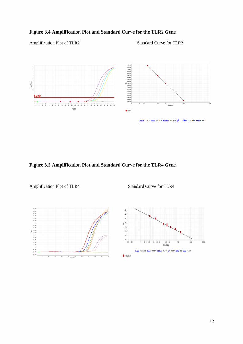
42
Figure 3.4 Amplification Plot and Standard Curve for the TLR2 Gene
Amplification Plot of TLR2 Standard Curve for TLR2
Figure 3.5 Amplification Plot and Standard Curve for the TLR4 Gene
Amplification Plot of TLR4 Standard Curve for TLR4

43
3.9 Statistical Analysis
All experiments were done in triplicate. The distribution of cell count and neutrophil data were
summarized using medians and interquartile ranges. Mann-Whitney non-parametric tests were used for
two group comparisons and Kruskal-Wallis for more than two groups. Numeric variables such as CSF,
capsule size and opening pressure were also categorized and chi square tests or Fisher’s exact were used
to compare sub-groups. The association between two number variables was examined using scatter
plots and Spearman’s correlation coefficient. Stata v13 statistical software was used in the analysis.
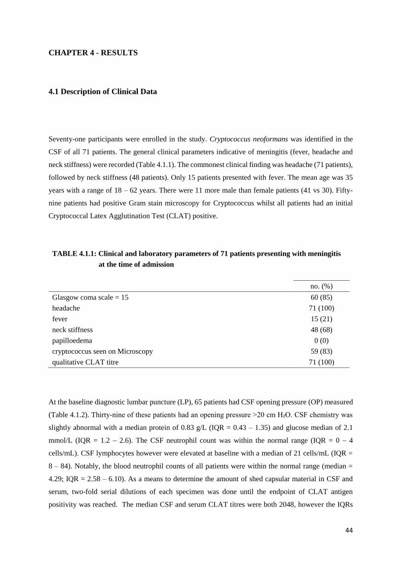
44
CHAPTER 4 - RESULTS
4.1 Description of Clinical Data
Seventy-one participants were enrolled in the study. Cryptococcus neoformans was identified in the
CSF of all 71 patients. The general clinical parameters indicative of meningitis (fever, headache and
neck stiffness) were recorded (Table 4.1.1). The commonest clinical finding was headache (71 patients),
followed by neck stiffness (48 patients). Only 15 patients presented with fever. The mean age was 35
years with a range of 18 – 62 years. There were 11 more male than female patients (41 vs 30). Fifty-
nine patients had positive Gram stain microscopy for Cryptococcus whilst all patients had an initial
Cryptococcal Latex Agglutination Test (CLAT) positive.
TABLE 4.1.1: Clinical and laboratory parameters of 71 patients presenting with meningitis
at the time of admission
no. (%)
Glasgow coma scale = 15 60 (85)
headache 71 (100)
fever 15 (21)
neck stiffness 48 (68)
papilloedema 0 (0)
cryptococcus seen on Microscopy 59 (83)
qualitative CLAT titre 71 (100)
At the baseline diagnostic lumbar puncture (LP), 65 patients had CSF opening pressure (OP) measured
(Table 4.1.2). Thirty-nine of these patients had an opening pressure >20 cm H₂O. CSF chemistry was
slightly abnormal with a median protein of 0.83 g/L (IQR = 0.43 – 1.35) and glucose median of 2.1
mmol/L (IQR = 1.2 – 2.6). The CSF neutrophil count was within the normal range (IQR = 0 – 4
cells/mL). CSF lymphocytes however were elevated at baseline with a median of 21 cells/mL (IQR =
8 – 84). Notably, the blood neutrophil counts of all patients were within the normal range (median =
4.29; IQR = 2.58 – 6.10). As a means to determine the amount of shed capsular material in CSF and
serum, two-fold serial dilutions of each specimen was done until the endpoint of CLAT antigen
positivity was reached. The median CSF and serum CLAT titres were both 2048, however the IQRs
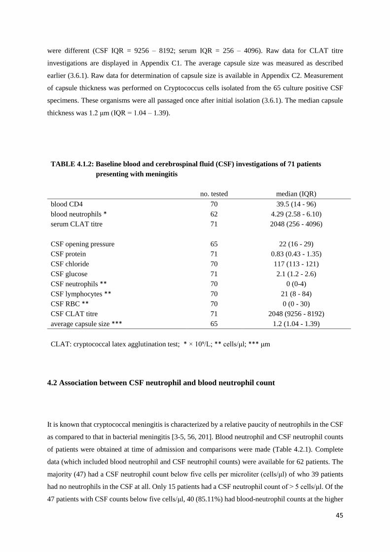
45
were different (CSF IQR = 9256 – 8192; serum IQR = 256 – 4096). Raw data for CLAT titre
investigations are displayed in Appendix C1. The average capsule size was measured as described
earlier (3.6.1). Raw data for determination of capsule size is available in Appendix C2. Measurement
of capsule thickness was performed on Cryptococcus cells isolated from the 65 culture positive CSF
specimens. These organisms were all passaged once after initial isolation (3.6.1). The median capsule
thickness was 1.2 μm (IQR = 1.04 – 1.39).
TABLE 4.1.2: Baseline blood and cerebrospinal fluid (CSF) investigations of 71 patients
presenting with meningitis
no. tested median (IQR)
blood CD4 70 39.5 (14 - 96)
blood neutrophils * 62 4.29 (2.58 - 6.10)
serum CLAT titre 71 2048 (256 - 4096)
CSF opening pressure 65 22 (16 - 29)
CSF protein 71 0.83 (0.43 - 1.35)
CSF chloride 70 117 (113 - 121)
CSF glucose 71 2.1 (1.2 - 2.6)
CSF neutrophils ** 70 0 (0-4)
CSF lymphocytes ** 70 21 (8 - 84)
CSF RBC ** 70 0 (0 - 30)
CSF CLAT titre 71 2048 (9256 - 8192)
average capsule size *** 65 1.2 (1.04 - 1.39)
CLAT: cryptococcal latex agglutination test; * × 10⁹/L; ** cells/μl; *** μm
4.2 Association between CSF neutrophil and blood neutrophil count
It is known that cryptococcal meningitis is characterized by a relative paucity of neutrophils in the CSF
as compared to that in bacterial meningitis [3-5, 56, 201]. Blood neutrophil and CSF neutrophil counts
of patients were obtained at time of admission and comparisons were made (Table 4.2.1). Complete
data (which included blood neutrophil and CSF neutrophil counts) were available for 62 patients. The
majority (47) had a CSF neutrophil count below five cells per microliter (cells/μl) of who 39 patients
had no neutrophils in the CSF at all. Only 15 patients had a CSF neutrophil count of > 5 cells/μl. Of the
47 patients with CSF counts below five cells/μl, 40 (85.11%) had blood-neutrophil counts at the higher

46
end of the normal range (2 - 7 ×10⁹ cells/L). Nine patients had an elevated blood neutrophil count. Eight
of these had CSF neutrophil counts of < 5 cells/μl. An association was noted between CSF neutrophil
and blood neutrophil count where a lower CSF neutrophil count appeared to be associated with higher
blood neutrophil count. However, this was not statistically significant (p = 0.37).
Table 4.2.1: Comparison between blood neutrophil and CSF neutrophil count
CSF neutrophils (cells/μl) no. (%) of patients with blood neutrophils *: Total
<2 2 to 7 >7
<5 7 (15) 32 (68) 8 (17) 47
>5 1 (7) 13 (87) 1 (7) 15
Total 8 (13) 45 (73) 9 (62) 62
* x 10⁹ cells/L
4.3 Association between clinical parameters and neutrophil count, cryptococcal capsule
size and shed cryptococcal capsular material
In keeping with meningitis, all of the study patients presented with headache as the predominant clinical
symptom. There were however only 15 patients who presented with fever. The association between
fever and total CSF and blood neutrophil counts, cryptococcal capsule size and cryptococcal shed
antigen (CSF CLAT titre) were further investigated (Table 4.3.1). Nine of the 15 patients with fever
had a CSF neutrophil count of < 5 cells/μl (p=0.05). Forty-six patients presented without fever. Of
statistical significance was that patients with fever had a higher CSF neutrophil count (p = 0.003). In
contrast with the CSF neutrophil-count, those patients who presented with fever had a lower blood-
neutrophil count. This displayed a borderline significance (p = 0.08). However, the blood neutrophil
count remained within the normal range in all patients with or without fever. The capsule size of
cryptococcal cells isolated from patients with fever tended to be smaller than in patients without fever,
however, there was no statistical significance (p = 0.18). Similarly, the comparison between shed
capsular material in the CSF of patients with fever and those without fever showed no significant
difference (p = 0.86).
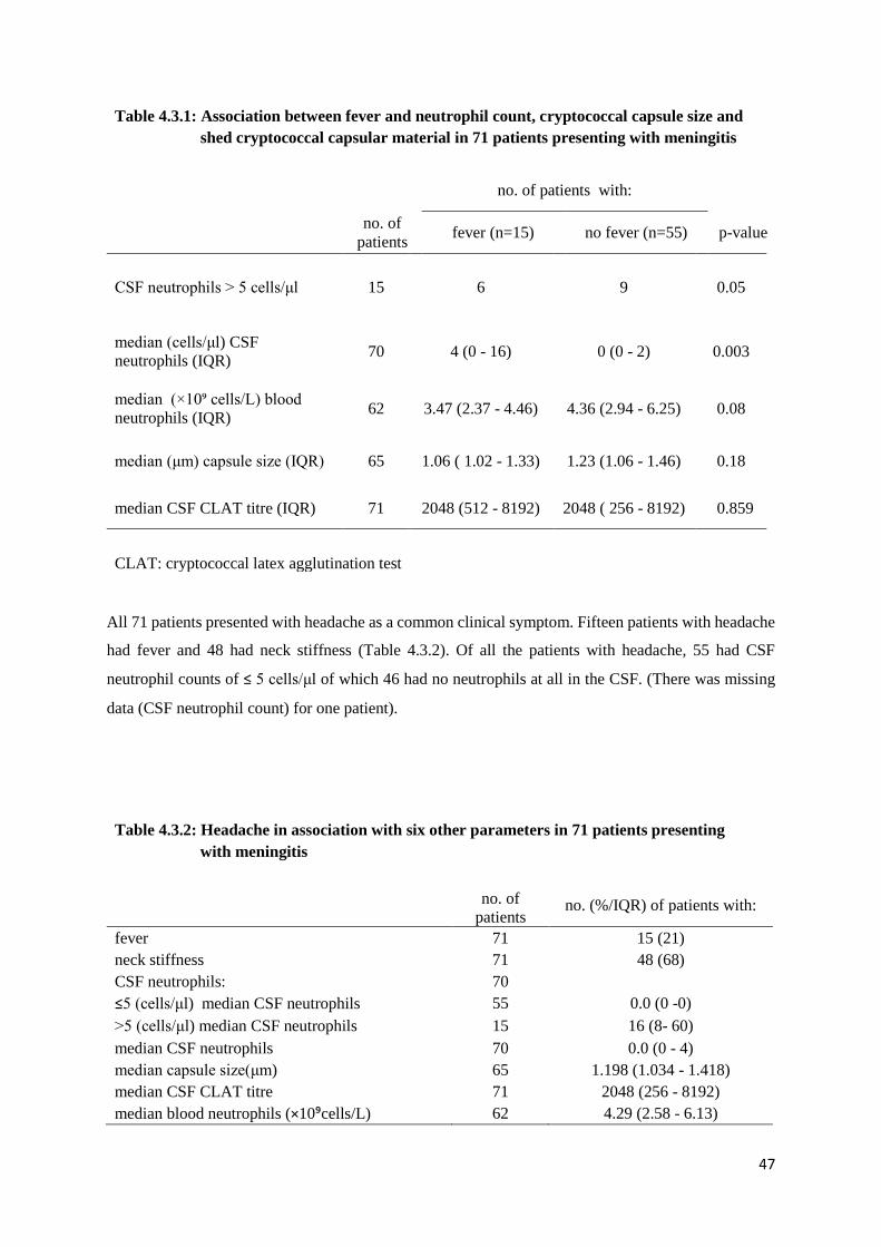
47
Table 4.3.1: Association between fever and neutrophil count, cryptococcal capsule size and
shed cryptococcal capsular material in 71 patients presenting with meningitis
no. of patients with:
no. of
patients fever (n=15) no fever (n=55) p-value
CSF neutrophils > 5 cells/μl 15 6 9 0.05
median (cells/μl) CSF
neutrophils (IQR) 70 4 (0 - 16) 0 (0 - 2) 0.003
median (×10⁹ cells/L) blood
neutrophils (IQR) 62 3.47 (2.37 - 4.46) 4.36 (2.94 - 6.25) 0.08
median (μm) capsule size (IQR) 65 1.06 ( 1.02 - 1.33) 1.23 (1.06 - 1.46) 0.18
median CSF CLAT titre (IQR) 71 2048 (512 - 8192) 2048 ( 256 - 8192) 0.859
CLAT: cryptococcal latex agglutination test
All 71 patients presented with headache as a common clinical symptom. Fifteen patients with headache
had fever and 48 had neck stiffness (Table 4.3.2). Of all the patients with headache, 55 had CSF
neutrophil counts of ≤ 5 cells/μl of which 46 had no neutrophils at all in the CSF. (There was missing
data (CSF neutrophil count) for one patient).
Table 4.3.2: Headache in association with six other parameters in 71 patients presenting
with meningitis
no. of
patients no. (%/IQR) of patients with:
fever 71 15 (21)
neck stiffness 71 48 (68)
CSF neutrophils: 70 ≤5 (cells/μl) median CSF neutrophils 55 0.0 (0 -0)
>5 (cells/μl) median CSF neutrophils 15 16 (8- 60)
median CSF neutrophils 70 0.0 (0 - 4)
median capsule size(μm) 65 1.198 (1.034 - 1.418)
median CSF CLAT titre 71 2048 (256 - 8192)
median blood neutrophils (×10⁹cells/L) 62 4.29 (2.58 - 6.13)
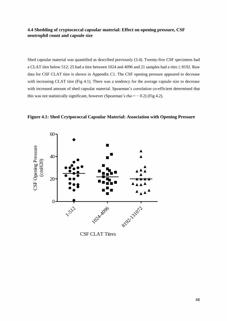
48
4.4 Shedding of cryptococcal capsular material: Effect on opening pressure, CSF
neutrophil count and capsule size
Shed capsular material was quantified as described previously (3.4). Twenty-five CSF specimens had
a CLAT titre below 512; 25 had a titre between 1024 and 4096 and 21 samples had a titre ≥ 8192. Raw
data for CSF CLAT titre is shown in Appendix C1. The CSF opening pressure appeared to decrease
with increasing CLAT titre (Fig 4.1). There was a tendency for the average capsule size to decrease
with increased amount of shed capsular material. Spearman’s correlation co-efficient determined that
this was not statistically significant, however (Spearman’s rho = − 0.2) (Fig 4.2).
Figure 4.1: Shed Crytpococcal Capsular Material: Association with Opening Pressure
1-51
2
1024
-409
6
8192
-131
072
0
20
40
60
CSF CLAT Titres
CS
F O
penin
g P
ress
ure
(cm
H20)
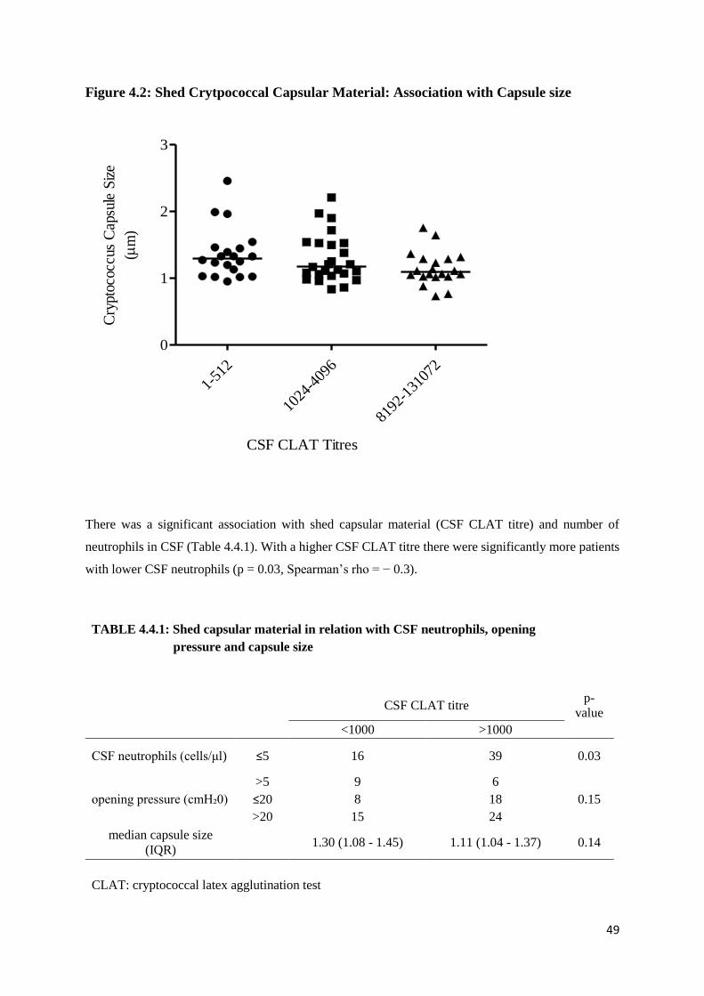
49
Figure 4.2: Shed Crytpococcal Capsular Material: Association with Capsule size
1-51
2
1024
-409
6
8192
-131
072
0
1
2
3
CSF CLAT Titres
Cry
ptoc
occu
s C
apsu
le S
ize
(m
)
There was a significant association with shed capsular material (CSF CLAT titre) and number of
neutrophils in CSF (Table 4.4.1). With a higher CSF CLAT titre there were significantly more patients
with lower CSF neutrophils (p = 0.03, Spearman’s rho = − 0.3).
TABLE 4.4.1: Shed capsular material in relation with CSF neutrophils, opening
pressure and capsule size
CSF CLAT titre
p-
value
<1000 >1000
CSF neutrophils (cells/μl) ≤5 16 39 0.03
>5 9 6 opening pressure (cmH₂0) ≤20 8 18 0.15
>20 15 24 median capsule size
(IQR) 1.30 (1.08 - 1.45) 1.11 (1.04 - 1.37) 0.14
CLAT: cryptococcal latex agglutination test
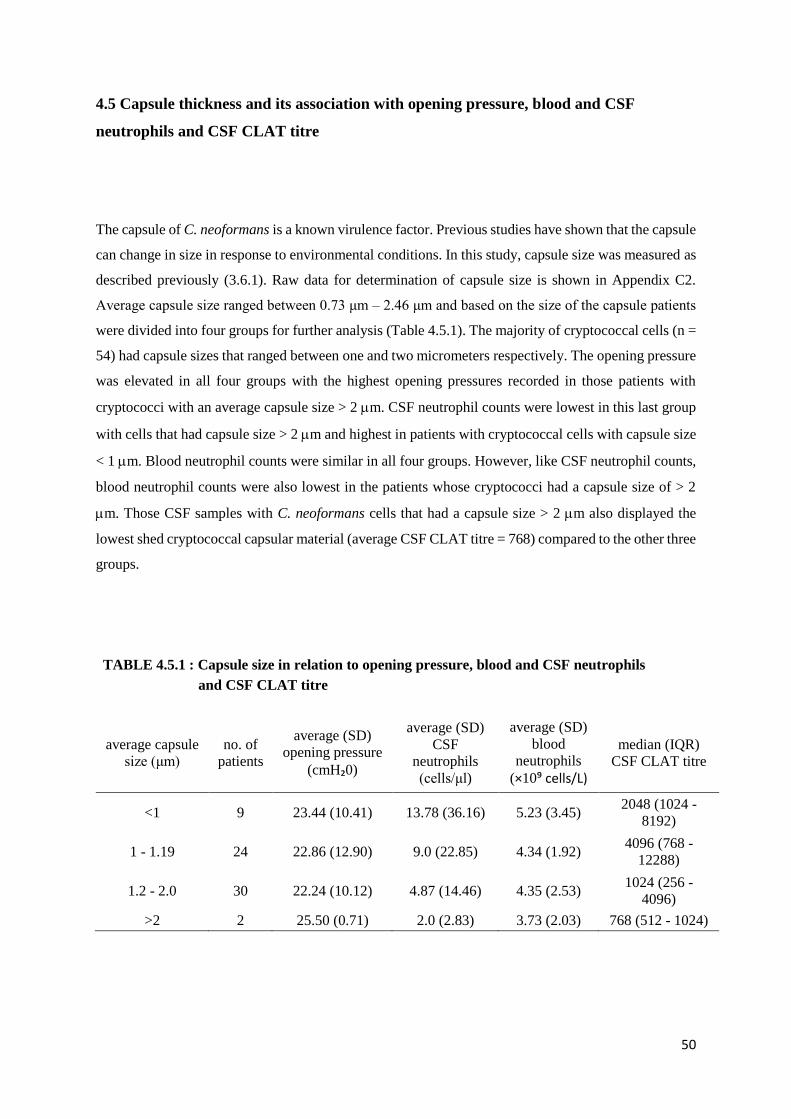
50
4.5 Capsule thickness and its association with opening pressure, blood and CSF
neutrophils and CSF CLAT titre
The capsule of C. neoformans is a known virulence factor. Previous studies have shown that the capsule
can change in size in response to environmental conditions. In this study, capsule size was measured as
described previously (3.6.1). Raw data for determination of capsule size is shown in Appendix C2.
Average capsule size ranged between 0.73 μm – 2.46 μm and based on the size of the capsule patients
were divided into four groups for further analysis (Table 4.5.1). The majority of cryptococcal cells (n =
54) had capsule sizes that ranged between one and two micrometers respectively. The opening pressure
was elevated in all four groups with the highest opening pressures recorded in those patients with
cryptococci with an average capsule size > 2 m. CSF neutrophil counts were lowest in this last group
with cells that had capsule size > 2 m and highest in patients with cryptococcal cells with capsule size
< 1 m. Blood neutrophil counts were similar in all four groups. However, like CSF neutrophil counts,
blood neutrophil counts were also lowest in the patients whose cryptococci had a capsule size of > 2
m. Those CSF samples with C. neoformans cells that had a capsule size > 2 m also displayed the
lowest shed cryptococcal capsular material (average CSF CLAT titre = 768) compared to the other three
groups.
TABLE 4.5.1 : Capsule size in relation to opening pressure, blood and CSF neutrophils
and CSF CLAT titre
average capsule
size (μm)
no. of
patients
average (SD)
opening pressure
(cmH₂0)
average (SD)
CSF
neutrophils
(cells/μl)
average (SD)
blood
neutrophils
(×10⁹ cells/L)
median (IQR)
CSF CLAT titre
<1 9 23.44 (10.41) 13.78 (36.16) 5.23 (3.45) 2048 (1024 -
8192)
1 - 1.19 24 22.86 (12.90) 9.0 (22.85) 4.34 (1.92) 4096 (768 -
12288)
1.2 - 2.0 30 22.24 (10.12) 4.87 (14.46) 4.35 (2.53) 1024 (256 -
4096)
>2 2 25.50 (0.71) 2.0 (2.83) 3.73 (2.03) 768 (512 - 1024)

51
Figure 4.3: Cryptococcus Capsule size in Relation to CSF Neutrophil Count
<1
1-1.
191.
2-2 >2
0
50
100
150
Cryptococcus Capsule Size (m)
CS
F N
eutr
ophil
Count
(cells
/mL
)
In this analysis, the capsule size is larger in those patients with lower CSF neutrophil counts (Table
4.5.2). However, this is not statistically significant (p = 0.28). There was no relationship between
capsule size and blood neutrophils. Blood neutrophils were essentially within the normal range in the
study group.
TABLE 4.5.2: Capsule size in relation to neutrophil count
no. of
patients
median (IQR)
capsule size (μm) p-value
CSF neutrophils *: ≤5 52 1.21 (1.06 - 1.41) 0.28
>5 13 1.06 (1.02 - 1.39) Blood neutrophils **: <2 8 1.51 (1.12 - 1.81) 0.22
2 to 7 41 1.10 (1.03 -1.33) >7 8 1.26 (1.03 - 1.45)
* cells/μl ; ** x 10⁹ cells/L

52
4.6 Neutrophil chemotaxis inhibition
Various explanations are presented in the literature regarding the possibility of inhibition of neutrophil
chemotaxis in the CSF. In this study, chemotaxis experiments were conducted as described earlier (3.7)
and chemotaxis inhibition was calculated. Raw data is displayed in Appendix C3. There was no
inhibition by 21 cryptococcal isolates while 44 displayed chemotaxis inhibition (Table 4.6). The amount
of shed cryptococcal capsular material (CLAT titre) was higher in the group that displayed chemotaxis
inhibition (but not statistically significant). Capsule size was similar in both groups. The number of
neutrophils in the CSF was significantly higher in the group that displayed chemotaxis inhibition (10.82
vs 0.67 cells/mL respectively; p = 0.04).
TABLE 4.6: Chemotaxis inhibition in relation to CSF CLAT titre, capsule size
and CSF neutrophils
average (SD) of patients with:
chemotaxis no. of
patients:
median (IQR) CSF
CLAT titre capsule size (μm)
CSF neutrophils
(cells/mL)
activation 21 1024 (512 - 4096) 1.27 (0.30) 0.67 (1.59)
inhibition 44 4096 (384 - 8192) 1.26 (0.37) 10.82 (25.38)
CLAT: cryptococcal latex agglutination test
4.7 Expression of TLR2 and TLR4 genes in human microglial cells
In this study, we attempted to show TLR2 and TLR4 gene expression induced by the individual strains
of Cryptococcus in human microglial cells. This was performed on 64 C. neoformans isolates. Gene
expression was performed as described earlier (3.8.3). Forty-five (70 %) of these isolates displayed
downregulation for TLR2 and 19 showed upregulation (Table 4.7.1). Capsule size did not vary
remarkably in both these groups. Neutrophil counts were higher in those CSF specimens that grew
cryptococci that upregulated TLR2 expression more than 10 times, and was lower in those specimens
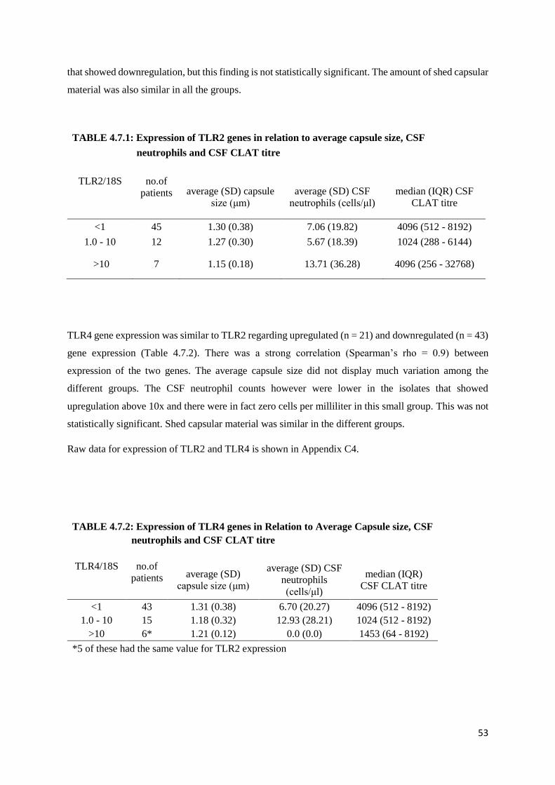
53
that showed downregulation, but this finding is not statistically significant. The amount of shed capsular
material was also similar in all the groups.
TABLE 4.7.1: Expression of TLR2 genes in relation to average capsule size, CSF
neutrophils and CSF CLAT titre
TLR2/18S no.of
patients average (SD) capsule
size (μm)
average (SD) CSF
neutrophils (cells/μl)
median (IQR) CSF
CLAT titre
<1 45 1.30 (0.38) 7.06 (19.82) 4096 (512 - 8192)
1.0 - 10 12 1.27 (0.30) 5.67 (18.39) 1024 (288 - 6144)
>10 7 1.15 (0.18) 13.71 (36.28) 4096 (256 - 32768)
TLR4 gene expression was similar to TLR2 regarding upregulated (n = 21) and downregulated (n = 43)
gene expression (Table 4.7.2). There was a strong correlation (Spearman’s rho = 0.9) between
expression of the two genes. The average capsule size did not display much variation among the
different groups. The CSF neutrophil counts however were lower in the isolates that showed
upregulation above 10x and there were in fact zero cells per milliliter in this small group. This was not
statistically significant. Shed capsular material was similar in the different groups.
Raw data for expression of TLR2 and TLR4 is shown in Appendix C4.
TABLE 4.7.2: Expression of TLR4 genes in Relation to Average Capsule size, CSF
neutrophils and CSF CLAT titre
TLR4/18S no.of
patients average (SD)
capsule size (μm)
average (SD) CSF
neutrophils
(cells/μl)
median (IQR)
CSF CLAT titre
<1 43 1.31 (0.38) 6.70 (20.27) 4096 (512 - 8192) 1.0 - 10 15 1.18 (0.32) 12.93 (28.21) 1024 (512 - 8192)
>10 6* 1.21 (0.12) 0.0 (0.0) 1453 (64 - 8192) *5 of these had the same value for TLR2 expression
54
CHAPTER 5 – DISCUSSION AND CONCLUSION
Cryptococcal meningitis is an important opportunistic infection especially in the immunocompromised
patient. It is well known that cryptococcal meningitis characteristically has a relative paucity of
polymorphonuclear neutrophils (PMN) in the CSF when compared to bacterial meningitis [3-5]. This
is despite evidence that the capsular material induces production of the early proinflammatory cytokines
IL-1β and TNF-α by peripheral blood PMNs and monocytes [3, 36, 224]. Mouse model studies
previously showed that the inhibition of PMN infiltration was mainly due to major capsule component
GXM [225]. Several studies have attempted to determine the mechanisms involved in regulating
passage of leucocytes through endothelial cells yet the mechanism by which signaling in brain
inflammation occur, is still unclear [3]. In this study, we hypothesized that the capsular material
interfered with neutrophil chemotaxis and that this had an inverse impact on severity of disease. Few
studies have been done on human infection and therefore this study focused on human infection and
effect on host inflammatory response. Similar to previous studies, our study shows typically few
mononuclear cells and virtually no PMNs in patients’ CSF and interestingly, all patients’ blood PMNs
were within the normal range that is, neither elevated nor decreased. Furthermore, we observed that a
low CSF neutrophil count was associated with a higher blood neutrophil count although this was not
statistically significant.
It is important to note that the sample size in this study had to be considerably reduced (by 70%) due
to a general decrease in patients presenting with crytpococcal meningitis to hospital. The recruitment
of patients for the study was therefore significantly compromised. A possible explanation for this drastic
decrease could be the fact that highly active antiretroviral therapy (HAART) was introduced to patients
infected with HIV, and studies show that there is a positive impact of HAART to patients with
crytpococcal infection especially in Western countries [226, 227].
Although there is no severity of disease score for patients with cryptococcal meningitis, a trend in
clinical parameters was evident in these patients in keeping with the literature. Of the clinical parameters
measured, headache was the predominant clinical symptom in all patients followed by neck stiffness.
Fever occurred in only a fraction of the patients. These findings are consistent with other studies that
describe the clinical course of cryptococcal meningitis as subacute or chronic with headache being a
predominant finding [228, 229]. In fact, it is known that patients with AIDS may even remain
asymptomatic early in the disease process and that the classic symptoms of meningitis (headache,
photophobia and meningism) may be absent [230, 231]. Furthermore, it has been observed that
cryptococcal encephalitis may remain undiagnosed due to poor inflammation at the site of infection
[139, 232]. Vaidhya et al identified fever, headache and neck stiffness respectively as the predominant
symptoms in their cohort, which differs from the findings in this study where fever occurred in only a

55
small fraction (21%) of patients [96]. Clinical signs such as papilloedema, hydrocephalus and focal
seizures during cryptococcal meningitis is a common occurrence in immunocompetent patients [228,
233]. This study population included only immunocompromised patients and consistent with this
observation, none of the patients showed signs of papilloedema. More than 50% of patients also showed
evidence of mildly raised intracranial pressure (ICP) with a median opening pressure of 22 cmH₂0. This
finding is noteworthy considering that in this study the opening pressure was recorded only at the second
lumbar puncture where it is performed routinely to reduce intracranial pressure. Raised intracranial
pressure is a common complication of cryptococcal meningitis and contributes to morbidity and
mortality [234, 235]. It is not clearly understood what the causes of raised intracranial pressure in
cryptococcal meningitis are, however, some hypotheses are that it may be related to impaired CSF
reabsorption caused by the organism or perhaps its shed polysaccharide capsule [234, 236]. It was also
noted in this study that the CSF chemistry (protein and glucose) was only slightly abnormal. This relates
to previous studies that describe clear differences in CSF chemistry between cryptococcal meningitis
(moderate elevation in protein and moderate decrease to normal glucose) and bacterial meningitis (high
protein and low glucose) [228, 237].
Significantly, patients who presented with fever had a higher CSF PMN count compared to patients
with no fever. Patients with fever also had a lower blood PMN count. Cellular components of the innate
immune system (including neutrophils) form part of the first line of defense in cryptococcosis [119].
These cells recognize and phagocytose the yeast resulting ultimately in their degradation. Fever is often
a manifestation of an aggressive inflammatory response [238]. This was confirmed in our finding that
patients who mounted a neutrophil response presented clinically with fever. The concept of the
association of fever with leucocyte infiltration dates as far back as the 19th century [239]. The
hypothalamus in the brain regulates and maintains normal body temperature. The febrile response is a
prominent component of various inflammatory and immunologically mediated diseases [239]. These
infections, including bacterial, viral and fungal infections, may result in the production of pyrogens that
alter the “thermostat setting” of the hypothalamus to raise body temperature leading to fever. Pyrogens
may be both exogenous as well as endogenous. Cytokines that are known to be intrinsically pyrogenic
are IL-1α, IL-1β, TNF-α, TNF-β, IFN-γ, and IL-6 [239]. Following CNS invasion by C. neoformans,
the microglial response is critical. Microglial cells together with astrocytes recognize fungal PAMPS.
During fungal antigen exposure, cytokines, which include the pyrogenic cytokines (IL-1β, TNF-α, IFN-
γ, and IL-6) recruit peripheral CD4 and CD8 T-cells, peripheral macrophages, and neutrophils, which
are then able to seed into the CNS [171]. In addition, the literature demonstrates that neutrophils also
produce and express various cytokines either constitutively or upon activation by micro-environmental
stimuli [240]. The pyrogenic cytokines are known examples of cytokines that neutrophils can
potentially express and/or produce [240]. The relationship between fever and PMN counts in CSF or
blood in cryptococcal meningitis has not been explored previously. Fernandes et al noted that there was

56
a positive correlation between fever and increased capsule thickness [238]. We found no significant
correlation between fever and phenotypic characteristics of Cryptococcus such as the capsule thickness
or the amount of shed capsule.
The polysaccharide capsule of C. neoformans is one of its most important virulence factors and plays a
role in evading phagocytosis as well as regulating the immune response [39, 45-49]. Some studies
suggest that the ability of phagocytes to clear the organism inversely relates to the capsule size [36, 37].
The phenomenon of capsular enlargement during infection is important as this has an effect on the size
of the cell thus creating a problem for phagocytosis [101, 109, 241]. Various studies suggest an
association between increased capsule size and resistance to phagocytosis [241-243]. In particular, one
compartment in which induction of capsule increase took place, although less efficiently than in the
lung, was found to be the brain [241, 244]. Littman demonstrated that the capsule size was quite variable
and depended on the organ affected [241, 245]. Charlier et al reported that tissue invasion was
associated with changes in size and composition of the organism’s capsule in the brain of AIDS patients
with both acute and chronic meningoencephalitis [1]. In our study, the capsule size ranged between 0.7
μm and 2.5 μm. The normal size of the cryptococcal capsule ranges between 2.0 – 4.0 μm, which
indicates that the capsule size was not enlarged in the study group. There were however, a large number
(97%) that expressed a capsule size below the normal range. A possible explanation for this is that the
organisms were isolated from CSF on sheep blood agar (3.3) and passaged once in vitro on Brain Heart
Infusion Agar plates supplemented with 10% horse blood (3.6) before the capsule measurements took
place. In an in vitro murine study, Dykstra et al observed that cryptococcal cells in diseased tissue had
large capsules, but when grown on ordinary media such as Sabouraud glucose agar, there was notable
variation in capsule size [246]. They postulated that capsule size decreased in high osmolarity media
and enlarged in media of low osmolarity. Furthermore, they suggested that although cells may produce
the same amount of capsular material despite osmolarity, the volume occupied in high osmolarity media
could possibly be less due to shrinkage [246]. In our study, the capsule size was larger in patients with
lower CSF neutrophil counts, although this was not statistically significant. This is in keeping with
previous findings that the capsule suppresses immune cell infiltration into the CNS [247, 248]. Our
study also suggests that patients’ whose isolates had larger capsules also had higher CSF opening
pressures. Robertson et al demonstrated that patients who had C. neoformans infection had a greater
chance of having raised intracranial pressures and diminished CSF inflammatory response when
infecting strains produced larger capsules [248]. This finding corroborates an earlier study in a rat model
that showed that capsular phenotype influences raised intracranial pressure [248, 249].
A major polysaccharide component of the Cryptococcus capsule is glucuronoxylomannan (GXM),
which is shed freely from the capsule and accumulates in body fluids and tissue of patients during
infection [2, 151]. In disseminated cryptococcal infection, there are high serum and CSF titres of GXM
in affected patients and this has been associated with disease progression as well as a diminished

57
inflammatory response [3, 151, 250]. In this study, we quantified shed capsular material in individual
patient CSF samples and analyzed the effect of shed capsular material on CSF neutrophil count as well
as on CSF opening pressure and capsule size. There was a significant association with shed capsular
material and number of neutrophils in CSF. With a higher CSF CLAT titre there were significantly
more patients with lower CSF neutrophils (p = 0.03). Various explanations have been described in the
literature regarding the possibility of inhibition of chemotaxis in the CSF. One such explanation is a
phenomenon called cross desensitization of the IL-8 receptor which describes the relationship of FMLP
and C5a (also neutrophil chemoattractants) to IL-8 [5, 204]. In that study, rapid desensitization for IL-
8 was shown to have occurred when PMNLs were previously exposed to the chemoattractants IL-8
itself, C5a or FMLP. Receptor phosphorylation and IL-8 receptor internalization was believed to be the
mechanism involved during this process [204]. In another study, GXM was observed to inhibit
polymorphonuclear leucocyte (PMNL) adhesion to activated endothelial cells in a concentration
dependent manner [56]. They used a static adhesion assay and found that GXM exerts this action by
affecting both PMNs and endothelial cells [56]. Another possibility for the paucity of leucocytes
described in the literature was that although production of interleukin-8 (IL-8) occurred in the CSF of
patients with cryptococcal meningitis, the circulating GXM was downregulating recruitment of
leucocytes, thus leading to impaired migration of leucocytes into the CSF [205, 206].
In this study, chemotaxis experiments were performed on individual isolates of Cryptococcus obtained
from patients CSF to determine the extent of inhibition of neutrophil chemotaxis. Close to 70% of
isolates displayed chemotaxis inhibition. Although this was not statistically significant, the amount of
shed cryptococcal capsular material was higher in the group that displayed chemotaxis inhibition. This
is in keeping with several previous studies that showed evidence of capsular material especially GXM
inhibiting leucocyte migration [3-5, 56, 57]. Lipovsky et al, further determined that the different GXM
serotypes varied in their ability to induce production of IL-8 [5]. They also found that most clinically
important isolates of C. neoformans were of the serotype A. They concluded in their study that the
cryptococcal polysaccharide capsule inhibited neutrophil migration toward IL-8 in both GXM serotypes
A and D [5]. The number of neutrophils in the CSF was significantly higher in the group that displayed
chemotaxis inhibition (p=0.04). This finding is interesting because it was expected that with chemotaxis
inhibition, there would be a lower number of neutrophils in the CSF. This is also contrary to the finding
that fewer neutrophils were found in the CSF of patients whose cryptococcal isolates shed more capsular
material in the CSF. A possible explanation for this could be that there was a small percentage of
patients (7%) in this group that had very high neutrophil counts (above 60 cells/mL) in their CSF, which
may have influenced the average neutrophil count in this group. When this group (7%) was excluded
and the average neutrophil count in the CSF was recalculated, this observation turned out not to be true.
The presence of leucocytes is regulated through chemotaxis. Chemotaxis depends on chemokine
production at the site of infection and the level of expression of corresponding chemokine receptors on
58
the migrating cells. Chemokines are major mediators of leucocyte recruitment into infection sites,
including infections with C. neoformans [202]. In the brain, microglial cells are known sources of not
only IL-8, but also other chemokines such as IP-10, MIP-1, MIP-1, RANTES, KC, and MCP-1
[203]. Therefore, a further explanation for this discrepant finding could be the varying effects of
chemokines at the site of infection. Chemokines were not measured in this study and this finding
stimulates further research.
Capsule size was similar in both groups (chemotaxis inhibition and no inhibition) although interestingly,
there was a tendency for the average capsule size to decrease with an increased amount of shed capsular
material. This is in accordance with studies that showed how the capsule of C. neoformans responded
to various environmental factors [251-253]. Larger capsules were related to greater production of
polysaccharides, a greater and more extensive assembly of polysaccharide, and also less shedding of
capsule [251]. This could imply that with greater shedding of capsular material, the capsule size would
also be smaller, as was observed in our study. In a recent study by Fernandes et al, cryptococcal capsule
size was found to differ between individual isolates, genotypes, as well as species [238]. They have also
stated that it is not clear whether a large capsular phenotype enhances the overall virulence of
Cryptococcus. However, a finding in their study was that capsule size correlated strongly with yeast
cell size, and as such, the possibility existed that capsule alone has less significance than overall size of
the cell [238].
Microglial cells, which are phagocytic cells within the central nervous system, along with meningeal
and perivascular macrophages, make up the first line of defence in the brain during CNS infections
[168-170]. These cells are major regulators of the innate immune response because they express Toll-
like receptors (TLRs) which can identify pathogen associated molecular patterns (PAMPs) [170, 173,
174]. Chemokines are produced when TLRs recognize specific microbes. For C. neoformans, TLR2
has been identified as the most important TLR [6, 7, 175]. It has been shown that TLR2 and TLR4
recognize cryptococcal capsular component GXM [170, 175]. GXM bound to both TLR2 and TLR4
with the co-receptor CD14 but it was unable to activate the MAPK (mitogen-activated protein kinase)
pathway and produce TNF-α [170, 175]. Although increasing interest in expression of TLRs is
becoming evident, little information is available on expression and function of TLRs in the CNS
microglial cells [7]. Some data is becoming available on TLR expression in rodent CNS, however little
information exists on TLR expression in human microglial cells [7].
In this study, we measured TLR2 and TLR4 gene expression induced by the individual strains of
Cryptococcus in human microglial cells. Both TLR2 as well as TLR4 were expressed similarly in
microglial cells by individual cryptococcal isolates. This concurs with earlier studies that associated
both TLRs with cryptococcal infection [170, 175]. Biondo et al, however, concluded in their study, that
TLR2 but not TLR4 contributed to anti-cryptococcal defenses [164]. We showed that exposure to the

59
majority (nearly 70%) of cryptococcal isolates induced downregulation for both TLRs in the glial cells
and only a small proportion resulted in upregulation. This supports one of our research questions that
downregulation of neutrophil chemotaxis occurs at the level of TLR expression.
In keeping with this finding was that neutrophils were lower in the CSF specimens where there was
downregulation (TLR4/18s <1) of the TLR4 genes in human microglial cells. This was however not
true for TLR2 gene expression. Netea et al reported the effect of TLRs in another fungal pathogen
(Candida albicans) [254, 255]. Their experiments in mice which express defective TLR4 genes showed
impaired neutrophil recruitment to the site of infection [254]. Further studies on the fungal pathogen C.
albicans showed conflicting results with TLR2, which displayed either protective or harmful effects on
the pathogen [167, 255, 256]. Furthermore, various research on Aspergillus fumigatus described the
function of TLR2, TLR4 and MyD88 in neutrophil activation and cytokine secretion [255, 257-259].
Redlich et al stimulated microglial cells with various TLR agonists and found that this enhanced
phagocytosis of fungi as well as bacteria [170, 260, 261]. They have also shown that a role of TLRs in
the phagocytosis of C. neoformans requires the MyD88 signaling cascade, which is used by all TLRs
except TLR3. Although there is some evidence of the role of TLRs in the early immune response to
fungal infection, none of these studies shows clarity on the role of TLRs and their effect on neutrophil
activation in cryptococcal infection in humans. In our study there is a possible association between
neutrophil recruitment and TLR expression in human microglial cells, although further studies are
needed to confirm this.
There was no significant association between the amount of shed capsular material and expression of
TLRs in our study. Several studies have reported the ability of capsular GXM to activate a TLR-
mediated immune response [165, 175, 176, 262]. GXM has been shown to interact with both TLR2
and TLR4, which is believed to influence response to cryptococcal infection [164, 165, 175, 263]. In
our study, a possible explanation for the lack of an association could be the small sample size. Further
studies would be beneficial to confirm this observation.
In conclusion, there is increasing interest and research focusing on understanding the pathogenesis of
cryptococcosis. Most of this research has thus far, focused on animal models. This study is among the
first to examine the host-pathogen relationship in human microglial cells. It is also the first study to
show a possible association between neutrophil recruitment and TLR expression in human microglial
cells. Our hypothesis that the cryptococcal capsular material interfered with neutrophil chemotaxis and
that the level of chemotaxis inhibition was related to severity of disease was pursued in a
multicomponent approach.
A relative paucity of CSF neutrophils in the study cohort was observed, which is consistent with the
literature. Although the capsule of C. neoformans was not enlarged, we noted an association between
larger capsule size and lower CSF neutrophil counts in these patients. In addition, a significant

60
association occurred between shed capsular material and CSF neutrophil counts. Lower CSF neutrophil
counts occurred in patients whose CSF had higher amounts of shed capsular material. These findings
imply that the capsular components (mainly GXM) do in fact downregulate recruitment of neutrophils
into the CSF. Chemotaxis inhibition occurred in almost 70% of the isolates tested. This group also had
a higher amount of shed capsular material in the CSF, which further supports the hypothesis that the
capsular material interfered with neutrophil chemotaxis.
There is no known severity of disease score for cryptococcal meningitis, however this study shows that
chemotaxis inhibition influences clinical parameters. Headache was a predominant finding in all
patients. Significantly, patients who presented with fever had higher CSF neutrophil counts, which
suggests that these patients are able to mount an immune response, a finding that has not been explored
previously. The association between fever and raised CSF neutrophil counts is demonstrated in Figure
5. These patients also had raised intracranial pressures.
Figure 5: Summary of Association between Fever and CSF Neutrophils
Contact between Cryptococcus and Microglial cells
TLR2 upregulation [n = 19 (30%)] TLR4 upregulation [n = 21(33%)]
Low capsule shedding [ n = 9 (13%)]
No chemotaxis inhibition [ n = 21 ( 32%)]
Increased CSF neutrophils [ n = 15 (21%)]
Fever [ n = 15 (21%)]
61
In support of one of our research questions, we showed that downregulation for both TLR2 and TLR4
occurred in microglial cells exposed to C. neoformans in the majority of isolates. A strong correlation
between TLR2 and TLR4 gene expression was demonstrated. To further support this, CSF neutrophil
counts were lower in the group where downregulation of TLR4 occurred.
This study contributes to understanding the host-immune response to cryptococcal meningitis in
humans. Our findings address the possible reasons for the paucity of neutrophils in the CSF of
individuals with cryptococcal meningitis. Further research on a larger study population would be
valuable to support these findings.
SUMMARY OF THE STUDY LIMITATIONS
The study sample size was smaller than planned initially. During the course of the study, there was a
general decrease in the number of patients presenting with cryptococcal meningitis, which significantly
affected patient recruitment. As discussed above, this could possibly be explained by the roll-out of
HAART in the HIV-infected population.
A second limitation was that cryptococci were isolated from CSF on sheep blood agar and passaged
once on BHI plates supplemented with 10% horse blood before capsule measurements were performed.
Some studies have suggested variations in capsule size when grown in different media with rich media
resulting in smaller capsules. In this study, all cryptococci were passaged only once on the same media,
therefore results obtained for each isolate could be compared.

62
REFERENCES
1. Charlier, C., et al., Capsule structure changes associated with Cryptococcus neoformans crossing of the blood-brain barrier. The American journal of pathology, 2005. 166(2): p. 421-32.
2. McFadden, D., O. Zaragoza, and A. Casadevall, The capsular dynamics of Cryptococcus neoformans. Trends in microbiology, 2006. 14(11): p. 497-505.
3. Coenjaerts, F.E., et al., Potent inhibition of neutrophil migration by cryptococcal mannoprotein-4-induced desensitization. J Immunol, 2001. 167(7): p. 3988-95.
4. Lipovsky, M.M., et al., Cryptococcal glucuronoxylomannan delays translocation of leukocytes across the blood-brain barrier in an animal model of acute bacterial meningitis. J Neuroimmunol, 2000. 111(1-2): p. 10-4.
5. Lipovsky, M.M., et al., Cryptococcal glucuronoxylomannan induces interleukin (IL)-8 production by human microglia but inhibits neutrophil migration toward IL-8. J Infect Dis, 1998. 177(1): p. 260-3.
6. Levitz, S.M., Interactions of Toll-like receptors with fungi. Microbes Infect, 2004. 6(15): p. 1351-5.
7. Bsibsi, M., et al., Broad expression of Toll-like receptors in the human central nervous system. J Neuropathol Exp Neurol, 2002. 61(11): p. 1013-21.
8. Shoham, S., et al., Toll-like receptor 4 mediates intracellular signaling without TNF-alpha release in response to Cryptococcus neoformans polysaccharide capsule. Journal of immunology (Baltimore, Md. : 1950), 2001. 166(7): p. 4620-6.
9. Srikanta, D., F.H. Santiago-Tirado, and T.L. Doering, Cryptococcus neoformans: historical curiosity to modern pathogen. Yeast (Chichester, England), 2014. 31(2): p. 47-60.
10. Steenbergen, J.N., H.A. Shuman, and A. Casadevall, Cryptococcus neoformans Interactions with Amoebae Suggest an Explanation for Its Virulence and Intracellular Pathogenic Strategy in Macrophages. Proceedings of the National Academy of Sciences of the United States of America, 2001. 98(26): p. 15245-15250.
11. Evans, E.E., The antigenic composition of Cryptococcus neoformans. I. A serologic classification by means of the capsular and agglutination reactions. Journal of immunology (Baltimore, Md. : 1950), 1950. 64(5): p. 423-30.
12. Wilson, D.E., J.E. Bennett, and J.W. Bailey, Serologic grouping of Cryptococcus neoformans. Proceedings of the Society for Experimental Biology and Medicine. Society for Experimental Biology and Medicine (New York, N.Y.), 1968. 127(3): p. 820-3.
13. Kwon-Chung, K.J. and A. Varma, MINIREVIEW: Do major species concepts support one, two or more species within Cryptococcus neoformans? FEMS Yeast Research, 2006. 6(4): p. 574-587.
14. Igreja, R.P., et al., Molecular epidemiology of Cryptococcus neoformans isolates from AIDS patients of the Brazilian city, Rio de Janeiro. Medical mycology, 2004. 42(3): p. 229-38.
15. Kidd, S.E., et al., A rare genotype of Cryptococcus gattii caused the cryptococcosis outbreak on Vancouver Island (British Columbia, Canada). Proceedings of the National Academy of Sciences of the United States of America, 2004. 101(49): p. 17258.
16. Meyer, W., et al., Consensus multi-locus sequence typing scheme for Cryptococcus neoformans and Cryptococcus gattii. Medical mycology, 2009. 47(6): p. 561-70.
17. Litvintseva, A.P., et al., Multilocus sequence typing reveals three genetic subpopulations of Cryptococcus neoformans var. grubii (serotype A), including a unique population in Botswana. Genetics, 2006. 172(4): p. 2223-38.
63
18. D’Souza, C.A., et al., Genome Variation in Cryptococcus gattii, an Emerging Pathogen of Immunocompetent Hosts. mBio, 2011. 2(1).
19. Hagen, F., et al., Importance of Resolving Fungal Nomenclature: the Case of Multiple Pathogenic Species in the Cryptococcus Genus. mSphere, 2017. 2(4): p. 00238-17.
20. Voelz, K. and R.C. May, Cryptococcal Interactions with the Host Immune System. Eukaryotic Cell, 2010. 9(6): p. 835-846.
21. Kozubowski, L. and J. Heitman, Profiling a killer, the development of <i>Cryptococcus neoformans</i>. FEMS Microbiology Reviews, 2012. 36(1): p. 78-94.
22. Ellis, D.H. and T.J. Pfeiffer, Natural habitat of Cryptococcus neoformans var. gattii. Journal of Clinical Microbiology, 1990. 28(7): p. 1642-1644.
23. Littman, M.L. and L.E. Zimmerman, Cryptococcosis, torulosis, or European blastomycosis. 1956, New York: Grune & Stratton.
24. Rana, A.H., et al. Cryptococcosis: Population-Based Multistate Active Surveillance and Risk Factors in Human Immunodeficiency Virus—Infected Persons. Journal of Infectious Diseases, 1999. 179, 449-454 DOI: 10.1086/314606.
25. Maziarz, E.K. and J.R. Perfect, Cryptococcosis. Infectious disease clinics of North America, 2016. 30(1): p. 179-206.
26. Bratton, E.W., et al., Comparison and temporal trends of three groups with cryptococcosis: HIV-infected, solid organ transplant, and HIV-negative/non-transplant. PloS one, 2012. 7(8): p. e43582.
27. Park, B.J., et al., Estimation of the current global burden of cryptococcal meningitis among persons living with HIV/AIDS. AIDS (London, England), 2009. 23(4): p. 525-30.
28. Rajasingham, R., et al., Global burden of disease of HIV-associated cryptococcal meningitis: an updated analysis. The Lancet. Infectious diseases, 2017. 17(8): p. 873-881.
29. Baradkar, V., et al., Prevalence and clinical presentation of Cryptococcal meningitis among HIV seropositive patients. Indian journal of sexually transmitted diseases and AIDS, 2009. 30(1): p. 19-22.
30. Desjardins, C.A., et al., Population genomics and the evolution of virulence in the fungal pathogen Cryptococcus neoformans. Genome research, 2017. 27(7): p. 1207-1219.
31. Bulmer, G.S., M.D. Sans, and C.M. Gunn, Cryptococcus neoformans I. Nonencapsulated Mutants. Journal of Bacteriology, 1967. 94(5): p. 1475-1479.
32. Kwon-Chung, K.J. and J.C. Rhodes, Encapsulation and melanin formation as indicators of virulence in Cryptococcus neoformans. Infection and Immunity, 1986. 51(1): p. 218-223.
33. Cox, G.M., et al., Urease as a virulence factor in experimental cryptococcosis. Infection and immunity, 2000. 68(2): p. 443-8.
34. Cox, G.M., et al., Extracellular phospholipase activity is a virulence factor for Cryptococcus neoformans. Molecular microbiology, 2001. 39(1): p. 166-75.
35. Kozubowski, L., S.C. Lee, and J. Heitman, Signalling pathways in the pathogenesis of Cryptococcus. Cellular microbiology, 2009. 11(3): p. 370-80.
36. Retini, C., et al., Capsular polysaccharide of Cryptococcus neoformans induces proinflammatory cytokine release by human neutrophils. Infection and immunity, 1996. 64(8): p. 2897-903.
37. Miller, M.F. and T.G. Mitchell, Killing of Cryptococcus neoformans strains by human neutrophils and monocytes. Infection and immunity, 1991. 59(1): p. 24-8.
38. Cherniak, R., E. Reiss, and S.H. Turner, A galactoxylomannan antigen of Cryptococcus neoformans serotype A. Carbohydrate Research, 1982. 103(2): p. 239-250.
39. Almeida, F., J.M. Wolf, and A. Casadevall, Virulence-Associated Enzymes of Cryptococcus neoformans. Eukaryotic Cell, 2015. 14(12): p. 1173-1185.
40. Cherniak, R. and J.B. Sundstrom, Polysaccharide antigens of the capsule of Cryptococcus neoformans. Infection and Immunity, 1994. 62(5): p. 1507-1512.

64
41. Bose, I., et al., A yeast under cover: the capsule of Cryptococcus neoformans. Eukaryotic cell, 2003. 2(4): p. 655-63.
42. Jesus, M.D., et al., Glucuronoxylomannan, galactoxylomannan, and mannoprotein occupy spatially separate and discrete regions in the capsule of Cryptococcus neoformans. Virulence, 2010. 1(6): p. 500-8.
43. Rodrigues, M.L. and L. Nimrichter, In good company: association between fungal glycans generates molecular complexes with unique functions. Frontiers in Microbiology, 2012. 3.
44. O'Meara, T.R. and J.A. Alspaugh, The Cryptococcus neoformans Capsule: a Sword and a Shield. Clinical Microbiology Reviews, 2012. 25(3): p. 387-408.
45. Chang, Y.C. and K.J. Kwon-Chung, Complementation of a capsule-deficient mutation of Cryptococcus neoformans restores its virulence. Molecular and Cellular Biology, 1994. 14(7): p. 4912-4919.
46. Rodrigues, M.L., C.S. Alviano, and L.R. Travassos, Pathogenicity of Cryptococcus neoformans: virulence factors and immunological mechanisms. Microbes and infection, 1999. 1(4): p. 293-301.
47. Kozel, T.R., et al., Role of the capsule in phagocytosis of Cryptococcus neoformans. Reviews of infectious diseases, 1988. 10(Suppl 2): p. 436-9.
48. Pericolini, E., et al., Cryptococcus neoformans capsular polysaccharide component galactoxylomannan induces apoptosis of human T-cells through activation of caspase-8. Cellular microbiology, 2006. 8(2): p. 267-75.
49. Vecchiarelli, A. and C. Monari, Capsular Material of Cryptococcus neoformans: Virulence and Much More. Mycopathologia, 2012.
50. Zaragoza, O., Basic principles of the virulence of Cryptococcus. Virulence, 2019. 10(1): p. 490-501.
51. Zaragoza, O., et al., The capsule of the fungal pathogen Cryptococcus neoformans. Advances in applied microbiology, 2009. 68: p. 133-216.
52. Vecchiarelli, A., et al., Elucidating the immunological function of the Cryptococcus neoformans capsule. Future microbiology, 2013. 8(9): p. 1107-16.
53. Vecchiarelli, A., Immunoregulation by capsular components of Cryptococcus neoformans. Medical mycology, 2000. 38(6): p. 407-17.
54. Dong, Z.M. and J.W. Murphy, Mobility of human neutrophils in response to Cryptococcus neoformans cells, culture filtrate antigen, and individual components of the antigen. Infection and Immunity, 1993. 61(12): p. 5067-5077.
55. Dong, Z.M. and J.W. Murphy, Cryptococcal polysaccharides induce L-selectin shedding and tumor necrosis factor receptor loss from the surface of human neutrophils. The Journal of clinical investigation, 1996. 97(3): p. 689-98.
56. Ellerbroek, P.M., et al., Cryptococcal glucuronoxylomannan inhibits adhesion of neutrophils to stimulated endothelium in vitro by affecting both neutrophils and endothelial cells. Infection and immunity, 2002. 70(9): p. 4762-71.
57. Ellerbroek, P.M., et al., Cryptococcal glucuronoxylomannan interferes with neutrophil rolling on the endothelium. Cell Microbiol, 2004. 6(6): p. 581-92.
58. Dong, Z.M. and J.W. Murphy, Cryptococcal polysaccharides bind to CD18 on human neutrophils. Infection and immunity, 1997. 65(2): p. 557-563.
59. Blackstock, R. and N.K. Hall, Non-specific immunosuppression by <i>Cryptococcus neoformans</i> infection. Mycopathologia, 1984. 86(1): p. 35-43.
60. Murphy, J.W. and G.C. Cozad, Immunological Unresponsiveness Induced by Cryptococcal Capsular Polysaccharide Assayed by the Hemolytic Plaque Technique. Infection and Immunity, 1972. 5(6): p. 896-901.
61. Vecchiarelli, A., et al., Cryptococcus neoformans galactoxylomannan is a potent negative immunomodulator, inspiring new approaches in anti-inflammatory immunotherapy. Immunotherapy, 2011. 3(8): p. 997-1005.

65
62. Blackstock, R., Cryptococcal capsular polysaccharide utilizes an antigen-presenting cell to induce a T-suppressor cell to secrete TsF. Journal of medical and veterinary mycology : bi-monthly publication of the International Society for Human and Animal Mycology, 1996. 34(1): p. 19-30.
63. Blackstock, R. and A. Casadevall, Presentation of cryptococcal capsular polysaccharide (GXM) on activated antigen-presenting cells inhibits the T-suppressor response and enhances delayed-type hypersensitivity and survival. Immunology, 1997. 92(3): p. 334-9.
64. Vecchiarelli, A. The Cellular Responses Induced by the Capsular Polysaccharide of Cryptococcus neoformans Differ Depending on the Presence or Absence of Specific Protective Antibodies. Current Molecular Medicine, 2005. 5, 413-420 DOI: 10.2174/1566524054022585.
65. Chiapello, L.S., et al., Immunosuppression, interleukin-10 synthesis and apoptosis are induced in rats inoculated with Cryptococcus neoformans glucuronoxylomannan. Immunology, 2004. 113(3): p. 392-400.
66. De Jesus, M., et al., Galactoxylomannan-mediated immunological paralysis results from specific B cell depletion in the context of widespread immune system damage. Journal of immunology (Baltimore, Md. : 1950), 2009. 183(6): p. 3885-94.
67. Chiapello, L.S., et al., Cryptococcus neoformans glucuronoxylomannan induces macrophage apoptosis mediated by nitric oxide in a caspase-independent pathway. International immunology, 2008. 20(12): p. 1527-41.
68. Monari, C., et al., Cryptococcus neoformans capsular glucuronoxylomannan induces expression of fas ligand in macrophages. Journal of immunology (Baltimore, Md. : 1950), 2005. 174(6): p. 3461-8.
69. Villena, S.N., et al., Capsular polysaccharides galactoxylomannan and glucuronoxylomannan from Cryptococcus neoformans induce macrophage apoptosis mediated by Fas ligand. Cellular microbiology, 2008. 10(6): p. 1274-85.
70. Rosas, A.L. and A. Casadevall, Melanization affects susceptibility of Cryptococcus neoformans to heat and cold. FEMS microbiology letters, 1997. 153(2): p. 265-72.
71. Wang, Y. and A. Casadevall, Decreased susceptibility of melanized Cryptococcus neoformans to UV light. Applied and Environmental Microbiology, 1994. 60(10): p. 3864-3866.
72. Nosanchuk, J.D. and A. Casadevall, Impact of Melanin on Microbial Virulence and Clinical Resistance to Antimicrobial Compounds. Antimicrobial Agents and Chemotherapy, 2006. 50(11): p. 3519-3528.
73. Pukkila-Worley, R., et al., Transcriptional Network of Multiple Capsule and Melanin Genes Governed by the Cryptococcus neoformans Cyclic AMP Cascade. Eukaryotic Cell, 2005. 4(1): p. 190-201.
74. Wang, Y. and A. Casadevall, Growth of Cryptococcus neoformans in presence of L-dopa decreases its susceptibility to amphotericin B. Antimicrobial Agents and Chemotherapy, 1994. 38(11): p. 2648-2650.
75. van Duin, D., A. Casadevall, and J.D. Nosanchuk, Melanization of Cryptococcus neoformans and Histoplasma capsulatum reduces their susceptibilities to amphotericin B and caspofungin. Antimicrobial agents and chemotherapy, 2002. 46(11): p. 3394-400.
76. Noverr, M.C., et al., CNLAC1 Is Required for Extrapulmonary Dissemination of Cryptococcus neoformans but Not Pulmonary Persistence. Infection and Immunity, 2004. 72(3): p. 1693-1699.
77. Mednick, A.J., J.D. Nosanchuk, and A. Casadevall, Melanization of Cryptococcus neoformans affects lung inflammatory responses during cryptococcal infection. Infection and immunity, 2005. 73(4): p. 2012-9.
78. Liu, L., R.P. Tewari, and P.R. Williamson, Laccase protects Cryptococcus neoformans from antifungal activity of alveolar macrophages. Infection and immunity, 1999. 67(11): p. 6034-9.

66
79. Robert, V.A. and A. Casadevall, Vertebrate Endothermy Restricts Most Fungi as Potential Pathogens. The Journal of Infectious Diseases, 2009. 200(10): p. 1623-1626.
80. Bergman, A., A. Casadevall, and F.o. Dromer, Mammalian Endothermy Optimally Restricts Fungi and Metabolic Costs. mBio, 2010. 1(5).
81. Petzold, E.W., et al., Characterization and regulation of the trehalose synthesis pathway and its importance in the pathogenicity of Cryptococcus neoformans. Infection and immunity, 2006. 74(10): p. 5877-87.
82. Giles, S.S., et al., Cryptococcus neoformans mitochondrial superoxide dismutase: an essential link between antioxidant function and high-temperature growth. Eukaryotic cell, 2005. 4(1): p. 46-54.
83. Kraus, P.R., et al., The Cryptococcus neoformans MAP kinase Mpk1 regulates cell integrity in response to antifungal drugs and loss of calcineurin function. Molecular microbiology, 2003. 48(5): p. 1377-87.
84. Alspaugh, J.A., et al., RAS1 regulates filamentation, mating and growth at high temperature of Cryptococcus neoformans. Molecular microbiology, 2000. 36(2): p. 352-65.
85. Olszewski, M.A., et al., Urease expression by Cryptococcus neoformans promotes microvascular sequestration, thereby enhancing central nervous system invasion. The American journal of pathology, 2004. 164(5): p. 1761-71.
86. Rocha, J.D.B., et al., Capsular polysaccharides from Cryptococcus neoformans modulate production of neutrophil extracellular traps (NETs) by human neutrophils. Scientific Reports, 2015. 5(1).
87. Cox, G.M., et al., Superoxide Dismutase Influences the Virulence of Cryptococcus neoformans by Affecting Growth within Macrophages. Infection and Immunity, 2003. 71(1): p. 173-180.
88. Jacobson, E.S., N.D. Jenkins, and J.M. Todd, Relationship between superoxide dismutase and melanin in a pathogenic fungus. Infection and Immunity, 1994. 62(9): p. 4085-4086.
89. Chen, S.C.A., et al., Phospholipase Activity in Cryptococcus neoformans: A New Virulence Factor? The Journal of Infectious Diseases, 1997. 175(2): p. 414-420.
90. Barrett-Bee, K., et al., A comparison of phospholipase activity, cellular adherence and pathogenicity of yeasts. Journal of general microbiology, 1985. 131(5): p. 1217-21.
91. Chen, S.C., et al., Identification of extracellular phospholipase B, lysophospholipase, and acyltransferase produced by Cryptococcus neoformans. Infection and immunity, 1997. 65(2): p. 405-411.
92. Santangelo, R., et al., Role of extracellular phospholipases and mononuclear phagocytes in dissemination of cryptococcosis in a murine model. Infection and immunity, 2004. 72(4): p. 2229-39.
93. Chen, S.C.A., et al., Purification and characterization of secretory phospholipase B, lysophospholipase and lysophospholipase/transacylase from a virulent strain of the pathogenic fungus Cryptococcus neoformans. Biochemical Journal, 2000. 347(2): p. 431.
94. Bicanic, T. and T.S. Harrison, Cryptococcal meningitis. British Medical Bulletin, 2004. 72(1): p. 99-118.
95. Olufunso Oladipupo, S., J. Melanie-Anne Amanda, and L. Umesh Management of cryptococcal meningitis in adults at Mthatha Hospital Complex, Eastern Cape, South Africa. Southern African Journal of HIV Medicine, 2014. 15, 104-107 DOI: 10.4102/sajhivmed.v15i3.10.
96. Vaidhya, S.A., et al., Combination Versus Monotherapy for the Treatment of HIV Associated Cryptococcal Meningitis. Journal of clinical and diagnostic research : JCDR, 2015. 9(2): p. 14-6.
97. Mitchell, T.G. and J.R. Perfect, Cryptococcosis in the era of AIDS--100 years after the discovery of Cryptococcus neoformans. Clinical microbiology reviews, 1995. 8(4): p. 515-548.
98. Baum, G.L. and D. Artis, FUNGISTATIC EFFECTS OF CELL FREE HUMAN SERUM. The American Journal of the Medical Sciences, 1961. 242(6): p. 761-770.
67
99. Baum, G.L. and D. Artis, Growth inhibition of Cryptococcus neoformans by cell free human serum. The American journal of the medical sciences, 1961. 241: p. 613-6.
100. Heung, L.J., Innate Immune Responses to Cryptococcus. Journal of fungi (Basel, Switzerland), 2017. 3(3).
101. Kozel, T.R. and E.C. Gotschlich, The capsule of cryptococcus neoformans passively inhibits phagocytosis of the yeast by macrophages. Journal of immunology (Baltimore, Md. : 1950), 1982. 129(4): p. 1675-80.
102. Levitz, S.M. and A. Tabuni, Binding of Cryptococcus neoformans by human cultured macrophages. Requirements for multiple complement receptors and actin. The Journal of clinical investigation, 1991. 87(2): p. 528-35.
103. Kelly, R.M., et al., Opsonic requirements for dendritic cell-mediated responses to Cryptococcus neoformans. Infection and immunity, 2005. 73(1): p. 592-8.
104. Janeway, C. and C.A. Janeway, Immunobiology : the immune system in health and disease. 6th ed. ed. 2005, New York: Garland Science.
105. Panepinto, J.C., et al., Binding of Serum Mannan Binding Lectin to a Cell Integrity-Defective Cryptococcus neoformans ccr4Δ Mutant. Infection and Immunity, 2007. 75(10): p. 4769-4779.
106. Kozel, T.R., et al., Activation and binding of opsonic fragments of C3 on encapsulated Cryptococcus neoformans by using an alternative complement pathway reconstituted from six isolated proteins. Infection and Immunity, 1989. 57(7): p. 1922-1927.
107. Kozel, T.R. and G.S. Pfrommer, Activation of the complement system by Cryptococcus neoformans leads to binding of iC3b to the yeast. Infection and Immunity, 1986. 52(1): p. 1-5.
108. Pfrommer, G.S., et al., Accelerated decay of C3b to iC3b when C3b is bound to the Cryptococcus neoformans capsule. Infection and Immunity, 1993. 61(10): p. 4360-4366.
109. Zaragoza, O., C.P. Taborda, and A. Casadevall, The efficacy of complement-mediated phagocytosis of Cryptococcus neoformans is dependent on the location of C3 in the polysaccharide capsule and involves both direct and indirect C3-mediated interactions. European journal of immunology, 2003. 33(7): p. 1957-67.
110. Taborda, C.P. and A. Casadevall, CR3 (CD11b/CD18) and CR4 (CD11c/CD18) are involved in complement-independent antibody-mediated phagocytosis of Cryptococcus neoformans. Immunity, 2002. 16(6): p. 791-802.
111. Calich, V.L.G., et al., MINIREVIEW: Toll-like receptors and fungal infections: the role of TLR2, TLR4 and MyD88 in paracoccidioidomycosis. FEMS Immunology & Medical Microbiology, 2008. 53(1): p. 1-7.
112. Gordon, S., Pattern recognition receptors: doubling up for the innate immune response. Cell, 2002. 111(7): p. 927-30.
113. Brown, G.D. and S. Gordon, Fungal beta-glucans and mammalian immunity. Immunity, 2003. 19(3): p. 311-5.
114. Akira, S., S. Uematsu, and O. Takeuchi, Pathogen recognition and innate immunity. Cell, 2006. 124(4): p. 783-801.
115. Ramirez-Ortiz, Z.G. and T.K. Means, The role of dendritic cells in the innate recognition of pathogenic fungi (A. fumigatus, C. neoformans and C. albicans). Virulence, 2012. 3(7): p. 635-46.
116. Eastman, A.J., J.J. Osterholzer, and M.A. Olszewski, Role of dendritic cell-pathogen interactions in the immune response to pulmonary cryptococcal infection. Future microbiology, 2015. 10(11): p. 1837-57.
117. Bauman, S.K., K.L. Nichols, and J.W. Murphy, Dendritic cells in the induction of protective and nonprotective anticryptococcal cell-mediated immune responses. Journal of immunology (Baltimore, Md. : 1950), 2000. 165(1): p. 158-67.

68
118. Osterholzer, J.J., et al., Role of Dendritic Cells and Alveolar Macrophages in Regulating Early Host Defense against Pulmonary Infection with Cryptococcus neoformans. Infection and Immunity, 2009. 77(9): p. 3749-3758.
119. Campuzano, A. and F.L. Wormley, Innate Immunity against Cryptococcus, from Recognition to Elimination. Journal of fungi (Basel, Switzerland), 2018. 4(1).
120. Levitz, S.M. and C.A. Specht, MINIREVIEW: The molecular basis for the immunogenicity of Cryptococcus neoformans mannoproteins. FEMS Yeast Research, 2006. 6(4): p. 513-524.
121. Mansour, M.K., E. Latz, and S.M. Levitz, Cryptococcus neoformans glycoantigens are captured by multiple lectin receptors and presented by dendritic cells. Journal of immunology (Baltimore, Md. : 1950), 2006. 176(5): p. 3053-61.
122. Liliane, M. and N. Kirsten Adaptive Immunity to Cryptococcus neoformans Infections. Journal of Fungi, 2017. 3, DOI: 10.3390/jof3040064.
123. Upham, J.o.W., The role of dendritic cells in immune regulation and allergic airway inflammation. Respirology, 2003. 8(2): p. 140-148.
124. Berger, A. Th1 and Th2 responses: what are they? BMJ, 2000. 321, 424 DOI: 10.1136/bmj.321.7258.424.
125. Arora, S., et al., Role of IFN-gamma in regulating T2 immunity and the development of alternatively activated macrophages during allergic bronchopulmonary mycosis. Journal of immunology (Baltimore, Md. : 1950), 2005. 174(10): p. 6346-56.
126. Jain, A.V., et al., Th2 but not Th1 immune bias results in altered lung functions in a murine model of pulmonary Cryptococcus neoformans infection. Infection and immunity, 2009. 77(12): p. 5389-99.
127. Guglani, L. and S.A. Khader, Th17 cytokines in mucosal immunity and inflammation. Current opinion in HIV and AIDS, 2010. 5(2): p. 120-7.
128. Langrish, C.L., et al., IL-23 drives a pathogenic T cell population that induces autoimmune inflammation. The Journal of experimental medicine., 2005. 201(3): p. 233.
129. Zelante, T., et al., IL-17/Th17 in anti-fungal immunity: what's new? European journal of immunology, 2009. 39(3): p. 645-8.
130. van de Veerdonk, F.L., et al., Differential effects of IL-17 pathway in disseminated candidiasis and zymosan-induced multiple organ failure. Shock (Augusta, Ga.), 2010. 34(4): p. 407-11.
131. Wiesner, D.L., et al., Different Lymphocyte Populations Direct Dichotomous Eosinophil or Neutrophil Responses to Pulmonary Cryptococcus Infection. Journal of immunology (Baltimore, Md. : 1950), 2017. 198(4): p. 1627-1637.
132. Feldmesser, M., S. Tucker, and A. Casadevall, Intracellular parasitism of macrophages by Cryptococcus neoformans. Trends in microbiology, 2001. 9(6): p. 273-8.
133. Alvarez, M. and A. Casadevall Cell-to-cell spread and massive vacuole formation after Cryptococcus neoformans infection of murine macrophages. BMC Immunology, 2007. 8, 1-7 DOI: 10.1186/1471-2172-8-16.
134. Del Poeta, M., Role of Phagocytosis in the Virulence of Cryptococcus neoformans. Eukaryotic Cell, 2004. 3(5): p. 1067-1075.
135. Feldmesser, M., et al., Cryptococcus neoformans is a facultative intracellular pathogen in murine pulmonary infection. Infection and immunity, 2000. 68(7): p. 4225-37.
136. Alvarez, M. and A. Casadevall, Phagosome Extrusion and Host-Cell Survival after <b>Cryptococcus neoformans</b> Phagocytosis by Macrophages. Current Biology, 2006. 16(21): p. 2161-2165.
137. Ma, H., et al., Expulsion of Live Pathogenic Yeast by Macrophages. Current Biology, 2006. 16(21): p. 2156-2160.
138. Charlier, C., et al., Evidence of a role for monocytes in dissemination and brain invasion by Cryptococcus neoformans. Infection and immunity, 2009. 77(1): p. 120-7.
139. Chretien, F., et al., Pathogenesis of Cerebral Cryptococcus neoformans Infection after Fungemia. The Journal of Infectious Diseases, 2002. 186(4): p. 522-530.

69
140. Wager, C.M.L. and F.L. Wormley, Classical versus alternative macrophage activation: The Ying and the Yang in host defense against pulmonary fungal infections. Mucosal Immunology, 2014. 7(5): p. 1023-1035.
141. Davis, M.J., et al., Macrophage M1/M2 Polarization Dynamically Adapts to Changes in Cytokine Microenvironments in Cryptococcus neoformans Infection. mBio, 2013. 4(3).
142. Arora, S., et al., Effect of cytokine interplay on macrophage polarization during chronic pulmonary infection with Cryptococcus neoformans. Infection and immunity, 2011. 79(5): p. 1915-26.
143. Hardison, S.E., et al., Pulmonary infection with an interferon-gamma-producing Cryptococcus neoformans strain results in classical macrophage activation and protection. The American journal of pathology, 2010. 176(2): p. 774-85.
144. Drummond, R.A., et al., Innate Defense against Fungal Pathogens. Cold Spring Harbor perspectives in medicine, 2014. 5(6).
145. Hünniger, K. and O. Kurzai, Phagocytes as central players in the defence against invasive fungal infection. Seminars in Cell and Developmental Biology, 2019. 89: p. 3-15.
146. Espinosa, V. and A. Rivera, First Line of Defense: Innate Cell-Mediated Control of Pulmonary Aspergillosis. Frontiers in Microbiology, 2016. 7.
147. Mambula, S.S., et al., Human neutrophil-mediated nonoxidative antifungal activity against Cryptococcus neoformans. Infection and immunity, 2000. 68(11): p. 6257-64.
148. Hole, C. and F.L. Wormley, Innate host defenses against <i>Cryptococcus neoformans</i>. Journal of Microbiology, 2016. 54(3): p. 202-211.
149. Sun, D., et al., Real-Time Imaging of Interactions of Neutrophils with Cryptococcus neoformans Demonstrates a Crucial Role of Complement C5a-C5aR Signaling. Infection and Immunity, 2015. 84(1): p. 216-229.
150. Chaturvedi, V., et al., Stress tolerance and pathogenic potential of a mannitol mutant of Cryptococcus neoformans. Microbiology (Reading, England), 1996. 142(( Pt 4)): p. 937-43.
151. Ellerbroek, P.M., et al., O-acetylation of cryptococcal capsular glucuronoxylomannan is essential for interference with neutrophil migration. J Immunol, 2004. 173(12): p. 7513-20.
152. Qureshi, A., et al., Cryptococcus neoformans modulates extracellular killing by neutrophils. Frontiers in microbiology, 2011. 2: p. 193.
153. Salkowski, C.A. and E. Balish, Role of Natural Killer Cells in Resistance to Systemic Cryptococcosis. Journal of Leukocyte Biology, 1991. 50(2): p. 151-159.
154. Hidore, M.R. and J.W. Murphy, Murine natural killer cell interactions with a fungal target, Cryptococcus neoformans. Infection and Immunity, 1989. 57(7): p. 1990-1997.
155. Nabavi, N. and J.W. Murphy, In vitro binding of natural killer cells to Cryptococcus neoformans targets. Infection and Immunity, 1985. 50(1): p. 50-57.
156. Kawakami, K., et al., NK cells eliminate Cryptococcus neoformans by potentiating the fungicidal activity of macrophages rather than by directly killing them upon stimulation with IL-12 and IL-18. Microbiology and immunology, 2000. 44(12): p. 1043-50.
157. Zhang, T., et al., Interleukin-12 (IL-12) and IL-18 synergistically induce the fungicidal activity of murine peritoneal exudate cells against Cryptococcus neoformans through production of gamma interferon by natural killer cells. Infection and immunity, 1997. 65(9): p. 3594-3599.
158. Levitz, S.M., M.P. Dupont, and E.H. Smail, Direct activity of human T lymphocytes and natural killer cells against Cryptococcus neoformans. Infection and Immunity, 1994. 62(1): p. 194-202.
159. Murphy, J.W., M.R. Hidore, and S.C. Wong, Direct interactions of human lymphocytes with the yeast-like organism, Cryptococcus neoformans. The Journal of clinical investigation, 1993. 91(4): p. 1553-66.
160. Ma, L.L., et al., NK cells use perforin rather than granulysin for anticryptococcal activity. Journal of immunology (Baltimore, Md. : 1950), 2004. 173(5): p. 3357-65.
70
161. Feldmesser, M., et al., Eosinophil-Cryptococcus neoformans interactions in vivo and in vitro. Infection and immunity, 1997. 65(5): p. 1899-1907.
162. Garro, A.P., et al., Eosinophils elicit proliferation of naive and fungal-specific cells in vivo so enhancing a T helper type 1 cytokine profile in favour of a protective immune response against Cryptococcus neoformans infection. Immunology, 2011. 134(2): p. 198-213.
163. Garro, A.P., et al., Rat eosinophils stimulate the expansion of Cryptococcus neoformans-specific CD4(+) and CD8(+) T cells with a T-helper 1 profile. Immunology, 2011. 132(2): p. 174-87.
164. Biondo, C., et al., MyD88 and TLR2, but not TLR4, are required for host defense against <i>Cryptococcus neoformans</i>. European Journal of Immunology, 2005. 35(3): p. 870-878.
165. Yauch, L.E., et al., Involvement of CD14, toll-like receptors 2 and 4, and MyD88 in the host response to the fungal pathogen Cryptococcus neoformans in vivo. Infection and immunity, 2004. 72(9): p. 5373-82.
166. D. Shane O'Mahony, U.P.R.I.T.R.H.W.C.L. Differential Constitutive and Cytokine-Modulated Expression of Human Toll-like Receptors in Primary Neutrophils, Monocytes, and Macrophages. International Journal of Medical Sciences, 2008. 5, 1-8.
167. Netea, M.G., et al., Recognition of fungal pathogens by Toll-like receptors. European Journal of Clinical Microbiology and Infectious Diseases, 2004. 23(9): p. 672-676.
168. Hanisch, U.K. and H. Kettenmann, Microglia: active sensor and versatile effector cells in the normal and pathologic brain. Nature neuroscience, 2007. 10(11): p. 1387-94.
169. Nau, R. and W. Bruck, Neuronal injury in bacterial meningitis: mechanisms and implications for therapy. Trends in Neurosciences, 2002. 25(1): p. 38-45.
170. Redlich, S., et al. Toll-like receptor stimulation increases phagocytosis of Cryptococcus neoformans by microglial cells. Journal of Neuroinflammation, 2013. 10, 1-7 DOI: 10.1186/1742-2094-10-71.
171. Koutsouras, G.W., R.L. Ramos, and L.R. Martinez, Role of microglia in fungal infections of the central nervous system. Virulence, 2017. 8(6): p. 705-718.
172. Yang, I., et al., The role of microglia in central nervous system immunity and glioma immunology. Journal of Clinical Neuroscience, 2010. 17(1): p. 6-10.
173. Takeda, K. and S. Akira, Toll receptors and pathogen resistance. Cellular microbiology, 2003. 5(3): p. 143-53.
174. Takeda, K., T. Kaisho, and S. Akira, Toll-like receptors. Annual review of immunology, 2003. 21: p. 335-76.
175. Shoham, S., et al., Toll-like receptor 4 mediates intracellular signaling without TNF-alpha release in response to Cryptococcus neoformans polysaccharide capsule. J Immunol, 2001. 166(7): p. 4620-6.
176. Fonseca, F.L., et al., Immunomodulatory Effects of Serotype B Glucuronoxylomannan from Cryptococcus gattii Correlate with Polysaccharide Diameter. Infection and Immunity, 2010. 78(9): p. 3861-3870.
177. Aloisi, F., Immune function of microglia. Glia, 2001. 36(2): p. 165-179. 178. Vautier, S., D.M. MacCallum, and G.D. Brown, C-type lectin receptors and cytokines in fungal
immunity. Cytokine, 2012. 58(1): p. 89-99. 179. Cross, C.E. and G.J. Bancroft, Ingestion of acapsular Cryptococcus neoformans occurs via
mannose and beta-glucan receptors, resulting in cytokine production and increased phagocytosis of the encapsulated form. Infection and immunity, 1995. 63(7): p. 2604-2611.
180. Fromtling, R.A., H.J. Shadomy, and E.S. Jacobson, Decreased virulence in stable, acapsular mutants of<i>Cryptococcus neoformans</i>. Mycopathologia, 1982. 79(1): p. 23-29.
181. Lei, G., et al., Biofilm from a clinical strain of Cryptococcus neoformans activates the NLRP3 inflammasome. Cell research, 2013. 23(7): p. 965-8.
182. Guo, C., et al., Acapsular <b>Cryptococcus neoformans</b> activates the NLRP3 inflammasome. Microbes and Infection, 2014. 16(10): p. 845-854.

71
183. Chen, M., et al., Internalized Cryptococcus neoformans Activates the Canonical Caspase-1 and the Noncanonical Caspase-8 Inflammasomes. Journal of immunology (Baltimore, Md. : 1950), 2015. 195(10): p. 4962-72.
184. Ma, L.L., et al., CD8 T cell-mediated killing of Cryptococcus neoformans requires granulysin and is dependent on CD4 T cells and IL-15. Journal of immunology (Baltimore, Md. : 1950), 2002. 169(10): p. 5787-95.
185. Syme, R.M., et al., Both CD4+ and CD8+ human lymphocytes are activated and proliferate in response to Cryptococcus neoformans. Immunology, 1997. 92(2): p. 194-200.
186. Huffnagle, G.B., et al., The role of CD4⁺ and CD8⁺ T cells in the protective inflammatory response to a pulmonary cryptococcal infection. Journal of Leukocyte Biology, 1994. 55(1): p. 35-42.
187. Kawakami, K., et al., Monocyte chemoattractant protein-1-dependent increase of V alpha 14 NKT cells in lungs and their roles in Th1 response and host defense in cryptococcal infection. Journal of immunology (Baltimore, Md. : 1950), 2001. 167(11): p. 6525-32.
188. Wozniak, K.L., J.K. Kolls, and F.L. Wormley, Depletion of neutrophils in a protective model of pulmonary cryptococcosis results in increased IL-17A production by gamma/delta T cells. BMC Immunology, 2012. 13(1): p. 65.
189. Uezu, K., et al., Accumulation of gammadelta T cells in the lungs and their regulatory roles in Th1 response and host defense against pulmonary infection with Cryptococcus neoformans. Journal of immunology (Baltimore, Md. : 1950), 2004. 172(12): p. 7629-34.
190. Mody, C.H., et al., Proteins in the cell wall and membrane of Cryptococcus neoformans stimulate lymphocytes from both adults and fetal cord blood to proliferate. Infection and immunity, 1996. 64(11): p. 4811-4819.
191. Mody, C.H., et al., The cell wall and membrane of Cryptococcus neoformans possess a mitogen for human T lymphocytes. Infection and immunity, 1999. 67(2): p. 936-41.
192. Syme, R.M., et al., Phagocytosis and protein processing are required for presentation of Cryptococcus neoformans mitogen to T lymphocytes. Infection and immunity, 2000. 68(11): p. 6147-53.
193. Casadevall, A. and L. Pirofski Insights into Mechanisms of Antibody-Mediated Immunity from Studies with Cryptococcus neoformans. Current Molecular Medicine, 2005. 5, 421-433 DOI: 10.2174/1566524054022567.
194. Abadi, J. and L.-a. Pirofski, Antibodies Reactive with the Cryptococcal Capsular Polysaccharide Glucuronoxylomannan Are Present in Sera from Children with and without Human Immunodeficiency Virus Infection. The Journal of Infectious Diseases, 1999. 180(3): p. 915-919.
195. Houpt, D.C., et al., Occurrences, immunoglobulin classes, and biological activities of antibodies in normal human serum that are reactive with Cryptococcus neoformans glucuronoxylomannan. Infection and Immunity, 1994. 62(7): p. 2857-2864.
196. Subramaniam, K., et al., IgM⁺ Memory B Cell Expression Predicts HIV-Associated Cryptococcosis Status. The Journal of Infectious Diseases, 2009. 200(2): p. 244-251.
197. Browne, S.K., et al., Adult-onset immunodeficiency in Thailand and Taiwan. The New England journal of medicine, 2012. 367(8): p. 725-34.
198. Hoag, K.A., et al., Early cytokine production in pulmonary Cryptococcus neoformans infections distinguishes susceptible and resistant mice. American journal of respiratory cell and molecular biology, 1995. 13(4): p. 487-95.
199. Wiesner, D.L., et al., Chitin recognition via chitotriosidase promotes pathologic type-2 helper T cell responses to cryptococcal infection. PLoS pathogens, 2015. 11(3): p. e1004701.
200. Muller, U., et al., IL-13 induces disease-promoting type 2 cytokines, alternatively activated macrophages and allergic inflammation during pulmonary infection of mice with Cryptococcus neoformans. Journal of immunology (Baltimore, Md. : 1950), 2007. 179(8): p. 5367-77.
72
201. Diamond, R.D. and N.F. Erickson, 3rd, Chemotaxis of human neutrophils and monocytes induced by Cryptococcus neoformans. Infection and immunity, 1982. 38(1): p. 380-2.
202. Huffnagle, G.B. and L.K. McNeil, Dissemination of C. neoformans to the central nervous system: role of chemokines, Th1 immunity and leukocyte recruitment. Journal of neurovirology, 1999. 5(1): p. 76-81.
203. Kunkel SL, S.R., Lindley IJ, Westwick J, Chemokines: new ligands, receptors and activities. Immunol today, 1995: p. 559 - 561.
204. Sabroe, I., et al., Chemoattractant cross-desensitization of the human neutrophil IL-8 receptor involves receptor internalization and differential receptor subtype regulation. J Immunol, 1997. 158(3): p. 1361-9.
205. Chaka, W., et al., Cytokine profiles in cerebrospinal fluid of human immunodeficiency virus-infected patients with cryptococcal meningitis: no leukocytosis despite high interleukin-8 levels. University of Zimbabwe Meningitis Group. J Infect Dis, 1997. 176(6): p. 1633-6.
206. Dong, Z.M. and J.W. Murphy, Intravascular cryptococcal culture filtrate (CneF) and its major component, glucuronoxylomannan, are potent inhibitors of leukocyte accumulation. Infection and immunity, 1995. 63(3): p. 770-8.
207. Smith, C.W., Leukocyte-endothelial cell interactions. Semin Hematol, 1993. 30: p. 45-53. 208. Anthony, E.E., Jr., A NOTE ON CAPSULE STAINING. Science (New York, N.Y.), 1931. 73(1890):
p. 319-20.
209. Sato, T., et al., Induction of Human Neutrophil Chemotaxis by Candida albicans-derived -1,6 - long glycoside side-chain-branched beta-glucan. Journal of Leucocyte Biology, 2006. 80(1): p. 204 - 211.
210. Seino, K., et al., Chemotactic activity of Soluble Fas ligand against phagocytes. J. immunol, 1998. 161: p. 4484 - 4488.
211. Chen, H.C., Boyden Chamber Assay. Methods Mol Biol, 2005. 294: p. 15 - 22. 212. Justus, C.R., et al., In vitro Cell Migration and Invasion Assays. Journal of Visualized
Experiments, 2014(88). 213. K, S.K.S.O.P.B.F.M.K., A Simplified Boyden Chamber assay for Neutrophil Chemotaxis based
on Quantitation of Myeloperoxidase. Analytical Biochemistry, 1990. 185(2): p. 238 - 242. 214. Khan, A.A., M.A. Alsahli, and A.H. Rahmani, Myeloperoxidase as an Active Disease Biomarker:
Recent Biochemical and Pathological Perspectives. Medical sciences (Basel, Switzerland), 2018. 6(2).
215. Khan, A.A., et al., Biochemical and pathological studies on peroxidases -an updated review. Global journal of health science, 2014. 6(5): p. 87-98.
216. Liu, W.-Q., et al., Myeloperoxidase-derived hypochlorous acid promotes ox-LDL-induced senescence of endothelial cells through a mechanism involving β-catenin signaling in hyperlipidemia. Biochemical and Biophysical Research Communications, 2015. 467(4): p. 859-865.
217. Nicholls, S.J. and S.L. Hazen, Myeloperoxidase and cardiovascular disease. Arteriosclerosis, thrombosis, and vascular biology, 2005. 25(6): p. 1102-11.
218. Epstein, F.H. and S.J. Weiss, Tissue Destruction by Neutrophils. New England Journal of Medicine, 1989. 320(6): p. 365-376.
219. Chen, Y., et al., Hypertonic saline enhances neutrophil elastase release through activation of P2 and A3 receptors. American journal of physiology. Cell physiology., 2006. 59(4): p. C1051.
220. Segal, A.W., HOW NEUTROPHILS KILL MICROBES. Annual Review of Immunology, 2005. 23: p. 197-223.
221. Mayyas, et al., Level and significance of plasma myeloperoxidase and the neutrophil to lymphocyte ratio in patients with coronary artery disease. Experimental and Therapeutic Medicine, 2014. 8(6): p. 1951-1957.
222. Louis, K.S. and A.C. Siegel, Cell viability analysis using trypan blue: manual and automated methods. Methods in molecular biology (Clifton, N.J.), 2011. 740: p. 7-12.
73
223. Strober, W., Trypan Blue Exclusion Test of Cell Viability Trypan Blue Exclusion Test of Cell Viability. 2015.
224. Levitz, S.M., et al., Production of tumor necrosis factor alpha in human leukocytes stimulated by Cryptococcus neoformans. Infection and Immunity, 1994. 62(5): p. 1975-1981.
225. Dong, Z.M. and J.W. Murphy, Intravascular cryptococcal culture filtrate (CneF) and its major component, glucuronoxylomannan, are potent inhibitors of leukocyte accumulation. Infection and immunity, 1995. 63(3): p. 770-778.
226. Antinori, S., New Insights into HIV/AIDS-Associated Cryptococcosis %J ISRN AIDS. 2013. 2013: p. 22.
227. Sara, A.M., et al., The Changing Epidemiology of Cryptococcosis: An Update from Population-Based Active Surveillance in 2 Large Metropolitan Areas, 1992-2000. Clinical Infectious Diseases, 2003. 36(6): p. 789-794.
228. Satishchandra, P., et al., Cryptococcal meningitis: clinical, diagnostic and therapeutic overviews. Neurology India, 2007. 55(3): p. 226-32.
229. Chuck, S.L. and M.A. Sande, Infections with Cryptococcus neoformans in the acquired immunodeficiency syndrome. The New England journal of medicine, 1989. 321(12): p. 794-9.
230. Wang, W. and A.R. Carm, Clinical manifestations of AIDS with cryptococcal meningitis. Chinese medical journal, 2001. 114(8): p. 841-3.
231. Kanji, S.S., R. Kakai, and R.O. Onyango, Cryptococcal meningitis among human immunodeficiency virus patients attending major hospitals in Kisumu, Western Kenya. Archives of Clinical Microbiology, 2011. 2(1).
232. Lee, S.C., et al., Pathology of cryptococcal meningoencephalitis: Analysis of 27 patients with pathogenetic implications. Human Pathology, 1996. 27(8): p. 839-847.
233. Kiertiburanakul, S., et al., Cryptococcosis in human immunodeficiency virus-negative patients. International Journal of Infectious Diseases, 2006. 10(1): p. 72-78.
234. Macsween, K.F., et al., Lumbar drainage for control of raised cerebrospinal fluid pressure in cryptococcal meningitis: case report and review. Journal of Infection, 2005. 51: p. e221.
235. R, S., et al., Managing intracranial pressure in HIV-associated cryptococcal meningitis saves Life: Case
report of two patients admitted to a Tanzanian hospital. SM J Case Rep, 2018. 4(3). 236. Denning, D.W., et al., Elevated cerebrospinal fluid pressures in patients with cryptococcal
meningitis and acquired immunodeficiency syndrome. The American journal of medicine, 1991. 91(3): p. 267-72.
237. David, R.B. and C.B. Nathan, Methods of rapid diagnosis for the etiology of meningitis in adults. Biomarkers in Medicine, 2014. 8(9): p. 1085-1103.
238. Fernandes, K.E., et al., Phenotypic Variability Correlates with Clinical Outcome in Cryptococcus Isolates Obtained from Botswanan HIV/AIDS Patients. mBio, 2018. 9(5): p. 02016-18.
239. Charles, A.D. Cytokines as Endogenous Pyrogens. Journal of Infectious Diseases, 1999. 179, S294-S304 DOI: 10.1086/513856.
240. Tecchio, C., A. Micheletti, and M.A. Cassatella, Neutrophil-derived cytokines: facts beyond expression. Frontiers in immunology, 2014. 5: p. 508.
241. Zaragoza, O., B.C. Fries, and A. Casadevall, Induction of capsule growth in Cryptococcus neoformans by mammalian serum and CO(2). Infection and immunity, 2003. 71(11): p. 6155-64.
242. Kozel, T.R., et al., Strain variation in phagocytosis of Cryptococcus neoformans: dissociation of susceptibility to phagocytosis from activation and binding of opsonic fragments of C3. Infection and Immunity, 1988. 56(11): p. 2794-2800.
243. Mitchell, T.G. and L. Friedman, In Vitro Phagocytosis and Intracellular Fate of Variously Encapsulated Strains of Cryptococcus neoformans. Infection and Immunity, 1972. 5(4): p. 491-498.
74
244. Rivera, J., et al., Organ-dependent variation of capsule thickness in Cryptococcus neoformans during experimental murine infection. Infection and immunity, 1998. 66(10): p. 5027-30.
245. Littman, M.L., Capsule synthesis by Cryptococcus neoformans. Transactions of the New York Academy of Sciences,
1958. 20(7): p. 623-48. 246. Dykstra, M.A., L. Friedman, and J.W. Murphy, Capsule size of Cryptococcus neoformans:
control and relationship to virulence. Infection and Immunity, 1977. 16(1): p. 129-135. 247. Denham, S.T., et al., Regulated Release of Cryptococcal Polysaccharide Drives Virulence and
Suppresses Immune Cell Infiltration into the Central Nervous System. Infection and immunity, 2018. 86(3).
248. Robertson, E.J., et al., Cryptococcus neoformans ex vivo capsule size is associated with intracranial pressure and host immune response in HIV-associated cryptococcal meningitis. The Journal of infectious diseases, 2014. 209(1): p. 74-82.
249. Fries, B.C., et al., Phenotypic Switching of Cryptococcus neoformans Can Produce Variants That Elicit Increased Intracranial Pressure in a Rat Model of Cryptococcal Meningoencephalitis. Infection and Immunity, 2005. 73(3): p. 1779-1787.
250. Diamond, R.D. and J.E. Bennett, Prognostic factors in cryptococcal meningitis. A study in 111 cases. Annals of internal medicine, 1974. 80(2): p. 176-81.
251. Yoneda, A. and T.L. Doering, Regulation of Cryptococcus neoformans Capsule Size Is Mediated at the Polymer Level. Eukaryotic Cell, 2008. 7(3): p. 546-549.
252. Granger, D.L., J.R. Perfect, and D.T. Durack, Virulence of Cryptococcus neoformans. Regulation of capsule synthesis by carbon dioxide. The Journal of clinical investigation, 1985. 76(2): p. 508-16.
253. Won Hee, J., et al. Iron regulation of the major virulence factors in the AIDS-associated pathogen Cryptococcus neoformans. PLoS Biology, 2006. 4, DOI: 10.1371/journal.pbio.0040410.
254. Netea, M.G., et al., The Role of Toll-like Receptor (TLR) 2 and TLR4 in the Host Defense against Disseminated Candidiasis. The Journal of Infectious Diseases, 2002. 185(10): p. 1483-1489.
255. Calich, V.L.G., et al., Toll-like receptors and fungal infections: the role of TLR2, TLR4 and MyD88 in paracoccidioidomycosis. FEMS Immunology & Medical Microbiology, 2008. 53(1): p. 1-7.
256. Villamon, E., et al., Toll-like receptor-2 is essential in murine defenses against Candida albicans infections. Microbes and Infection, 2004. 6(1): p. 1-7.
257. Wang, J.E., et al., Involvement of CD14 and Toll-Like Receptors in Activation of Human Monocytes by Aspergillus fumigatus Hyphae. Infection and Immunity, 2001. 69(4): p. 2402-2406.
258. Meier, A., et al., Toll-like receptor (TLR) 2 and TLR4 are essential for Aspergillus-induced activation of murine macrophages. Cellular microbiology, 2003. 5(8): p. 561-70.
259. Braedel, S., et al., <i>Aspergillus fumigatus</i> antigens activate innate immune cells via toll-like receptors 2 and 4. British Journal of Haematology, 2004. 125(3): p. 392-399.
260. Ribes, S., et al., Toll-Like Receptor Stimulation Enhances Phagocytosis and Intracellular Killing of Nonencapsulated and Encapsulated Streptococcus pneumoniae by Murine Microglia. Infection and Immunity, 2010. 78(2): p. 865-871.
261. Ribes, S., et al., Toll-like receptor prestimulation increases phagocytosis of Escherichia coli DH5alpha and Escherichia coli K1 strains by murine microglial cells. Infection and immunity, 2009. 77(1): p. 557-64.
262. Huang, H.-R., et al., Dectin-3 Recognizes Glucuronoxylomannan of Cryptococcus neoformans Serotype AD and Cryptococcus gattii Serotype B to Initiate Host Defense Against Cryptococcosis. Frontiers in Immunology, 2018. 9.

75
263. Nakamura, K., et al., Limited contribution of Toll-like receptor 2 and 4 to the host response to a fungal infectious pathogen, <i>Cryptococcus neoformans</i>. FEMS Immunology & Medical Microbiology, 2006. 47(1): p. 148-154.

76
APPENDIX A: PROCEDURES
A1. Cell Count: Neubauer Chamber
Neubauer’s chamber is a thick glass plate with the size of a glass slide (30 x 70 x 4 mm). The
counting region consists of two square shaped ruled areas. There are depressions on either side
on which the squares are marked, giving it an “H” shape. The ruled area is 3 mm² and it is
divided into nine large squares each with an area of 1 mm2. The large central square is divided
into 25 medium squares with double or triple lines. Each of these 25 squares is divided into 16
small squares with single lines, so that each of the smallest squares has an area of 1/400 mm2.
The coverslip is a square glass 22 mm in width . The glass cover is placed on the top of the
Neubauer chamber and must cover the central area. The ruled area is 0.1 mm lower than the
rest of the chamber to allow for a gap of 0.1 mm (1/10 mm) between the cover slip and the
ruled area.
RBCs are counted in the central large square. Polymorphs and lymphocytes are each counted
in the four corner squares.
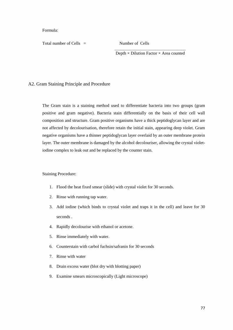
77
Formula:
Total number of Cells = Number of Cells
_________________________________
Depth × Dilution Factor × Area counted
A2. Gram Staining Principle and Procedure
The Gram stain is a staining method used to differentiate bacteria into two groups (gram
positive and gram negative). Bacteria stain differentially on the basis of their cell wall
composition and structure. Gram positive organisms have a thick peptidoglycan layer and are
not affected by decolourisation, therefore retain the initial stain, appearing deep violet. Gram
negative organisms have a thinner peptidoglycan layer overlaid by an outer membrane protein
layer. The outer membrane is damaged by the alcohol decolouriser, allowing the crystal violet-
iodine complex to leak out and be replaced by the counter stain.
Staining Procedure:
1. Flood the heat fixed smear (slide) with crystal violet for 30 seconds.
2. Rinse with running tap water.
3. Add iodine (which binds to crystal violet and traps it in the cell) and leave for 30
seconds .
4. Rapidly decolourise with ethanol or acetone.
5. Rinse immediately with water.
6. Counterstain with carbol fuchsin/safranin for 30 seconds
7. Rinse with water
8. Drain excess water (blot dry with blotting paper)
9. Examine smears microscopically (Light microscope)

78
A3. Flow Diagram for Storage of Specimens
Clotted Blood CSF
Centrifuge at 3000rpm for 5 minutes Remove supernatant and store at -80°C Culture sediment on Sheep Blood agar
Incubate plate for 48 hours at 37°C
Centrifuge at 3000rpm for 5 minutes Extract serum Store at -20°C
No Growth: Reincubate overnight
Growth: Store in distilled water at room temperature
79
A4. Giemsa Staining Procedure (Electron Microscopy Sciences, USA)
1. Place 1.0 mL of the Wright-Giemsa Stain upon the smear, in sufficient quantity to cover the
entire surface, for 3-4 minutes.
2. Add 2.0 mL distilled water or Phosphate Buffer, and let stand twice as long as in step 1.
3. Rinse stained smear with water or the Phosphate Buffer, until the edges show faintly pinkish
red.
4. The film is allowed to air dry. The preparation may be blotted gently to hasten drying.
5. Permanent mounts can be made with mounting medium
A5. Reagent Preparation for MPO Human SimpleStep ELISA® Kit (Abcam, Cambridge, UK)
1. 1× Cell Extraction Buffer PTR
Prepare 1× Cell Extraction Buffer PTR by diluting 5× Cell Extraction Buffer PTR and 50× Cell
Extraction Enhancer Solution to 1× with deionized water. To make 10 mL 1× Cell Extraction
Buffer PTR combine 7.8 mL deionized water, 2 mL 5× Cell Extraction Buffer PTR and 200 μl
50× Cell Extraction Enhancer Solution. Mix thoroughly and gently. If required protease
inhibitors can be added.
2. 1× Wash Buffer PT
Prepare 1× Wash Buffer PT by diluting 10× Wash Buffer PT with deionized water. To make
50 mL 1× Wash Buffer PT combine 5 mL 10× Wash Buffer PT with 45 mL deionized water.
Mix thoroughly and gently.
80
3. Antibody Cocktail
Prepare Antibody Cocktail by diluting the capture and detector antibodies in Antibody
Diluent 4BI. To make 3 mL of the Antibody Cocktail combine 300 μl 10× Capture
Antibody and 300 μl 10× Detector Antibody with 2.4 mL antibody Diluent 4BI. Mix
thoroughly and gently.
A6. Standard Preparation for MPO Human SimpleStep ELISA® Kit (Abcam, Cambridge, UK)
The Myeloperoxidase lyophilized standard sample is reconstituted by adding 1mL 1× Cell
Extraction Buffer PTR by pipette. Mix thoroughly and gently. Keep this at room temperature
for 10 minutes and mix gently. This is the 5,000 pg/mL Stock Standard Solution. Label eight
tubes, Standards 1– 8. Add 180 μl 1× Cell Extraction Buffer PTR into tube number 1 and 150
μl of 1× Cell Extraction Buffer PTR into numbers 2-8. Use the Stock Standard to prepare the
dilution series. Standard #8 contains no protein and is the Blank control.
A7. Complete Culture Medium (for M059K cells)
Cells are grown in a medium containing a 1:1 mixture of Dulbecco’s Modified Eagle’s Medium
(DMEM) and Ham’s F12 medium with 2.5 mM L-glutamine adjusted to contain 15 mM
HEPES, 0.5 mM sodium pyruvate, and 1.2 g/L sodium bicarbonate (Lonza Group Ltd, Basel,
Switzerland) supplemented with 0.05 mM non-essential amino acids and 10% foetal bovine
serum.
81
APPENDIX B: MEDIA AND REAGENTS
B1. Sheep Blood Agar
Requirements for 1 litre media:
1. 40 grams blood agar base (Oxoid Ltd, Cheshire, England)
2. 1 litre distilled water
3. 70 mL horse blood (7%)
4. Petri dishes
Method:
1. Suspend 40 g blood agar base (Oxoid Ltd, Cheshire, England) in 1 litre distilled water.
2. Bring to boil to dissolve completely.
3. Sterilise by autoclaving at 121°C for 15 minutes.
4. Remove from autoclave. Place in waterbath at 56.5°C to cool.
5. Once cooled, remove from waterbath, allow to cool to at least 50°C.
6. Add 70 mL to 930 mL agar solution and mix gently, without creating bubbles.
7. Pour immediately into petri dishes at a volume of approximately 18 – 20 mL per petri dish.
8. Keep at room temperature overnight in sterile environment.
9. Store prepared plates at 2-8°C until use.
B2. Pronase (Meridian Bioscience, Inc, Ohio, USA)
Non-specific protease. It’s activity extends to both denatured and native proteins leading to complete
or nearly complete digestion into individual amino acids.

82
B3. Sample Diluent (Meridian Bioscience, Inc, Ohio, USA)
Glycine buffered saline (pH 8.4 ± 0.1) containing bovine serum albumin and 0.01% thimerosal as
preservative
B4. Brain Heart Infusion Agar with Horse Blood
Requirements for 1 litre media ( Approximately 50 plates):
1. 47 grams BHI agar powder (Oxoid Ltd, Cheshire, England)
2. 1 litre distilled water
3. 100 mL horse blood (10%)
4. Petri dishes
Method:
1. Add 47 grams BHI Agar powder (Oxoid, England) in 1 litre distilled water
2. Autoclave at 121°C for 15 minutes
3. Remove from autoclave. Place in waterbath (56.5°C) to cool.
4. Once cooled, remove from water bath, allow to cool to at least 50°C.
5. Add 100 mL blood to 900 mL agar solution and mix gently, without creating bubbles.
6. Pour immediately into petri dishes at a volume of approximately 18 – 20 mL per petri dish.
7. Keep at room temperature overnight in sterile environment.
8. Store prepared plates at 2-8°C until use.
83
B5. McFarland Turbidity Standard 1
1. Add 0.5 mL of concentrated H₂SO₄ (Merck, Kenilworth, USA) to 49.5 mL of sterilized
distilled water to make a 1% H₂SO₄ solution
2. Add 0.175 gram of BaCl₂.2H₂O (Sigma-Aldrich, Missouri, USA) to 10 mL of sterilized
distilled water to make a 1% BaCl₂.2H₂O solution
For a McFarland Standard of 1.0, mix 0.1 mL of the 1% BaCl₂.2H₂O solution
with 9.9 mL of the 1% H₂SO₄ solution
3. Cover tube with tin foil and store away from direct sunlight
B6. Phosphate Buffered Saline
1. Add 10 PBS tablets (Oxoid, England) to 1000 mL distilled water in a 3-litre Erlenmeyer flask
2. If it is required that bacterial clumping be minimized, add (1%) 10 mL Tween-80 (Merck, SA)
3. Slowly bring to boil and swirl flask and to mix contents
4. Sterilize by autoclaving at 121°C for 15 minutes
5. Cool down at room temperature
6. Dispense into the required volumes
7. Store at 4 to 8°C until use

84
B7. Foetal Bovine Serum (FBS)
1. Heat inactivate by de-complementing at 56°C for 1 hour.
2. Filter sterilize, aliquote into 10 mL tubes and store at -20°C.
3. Quality control by incubating 5 mL serum at 37°C for one week. Observe for no growth.
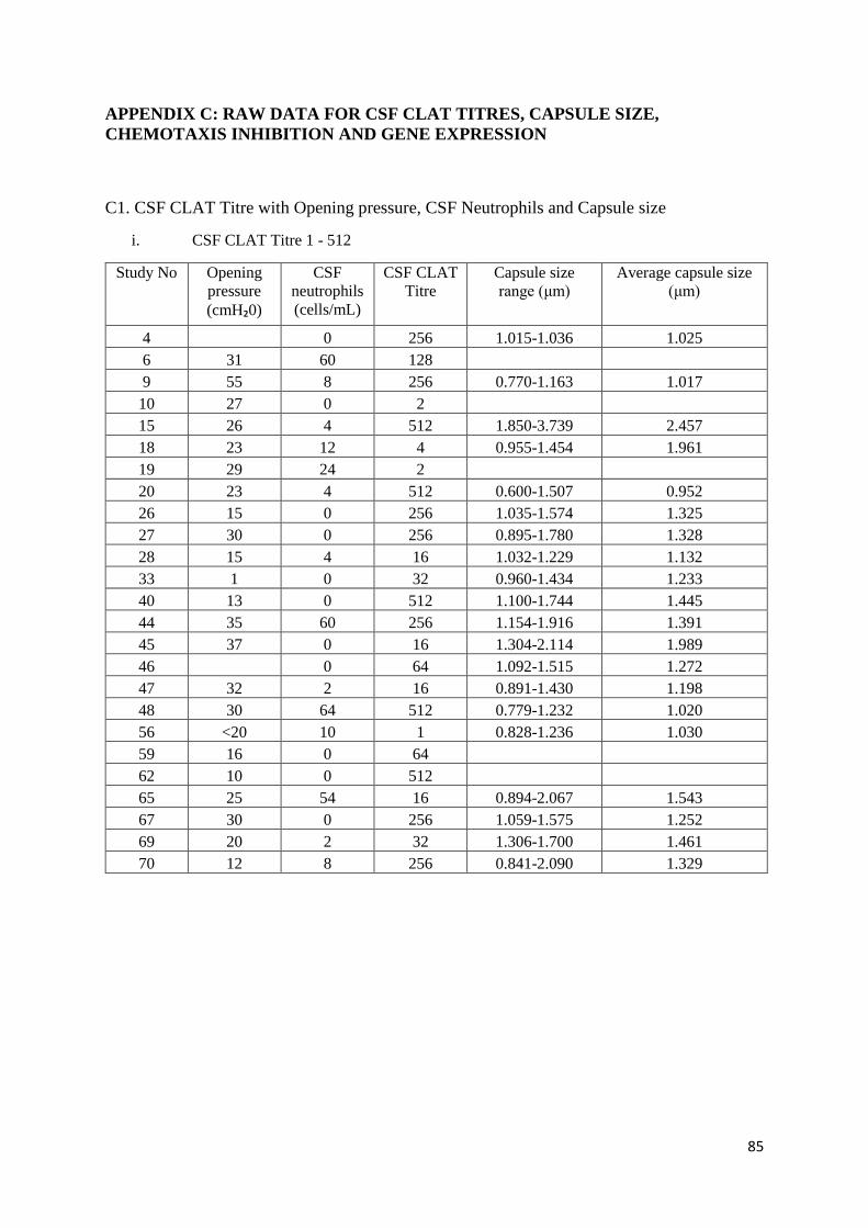
85
APPENDIX C: RAW DATA FOR CSF CLAT TITRES, CAPSULE SIZE,
CHEMOTAXIS INHIBITION AND GENE EXPRESSION
C1. CSF CLAT Titre with Opening pressure, CSF Neutrophils and Capsule size
i. CSF CLAT Titre 1 - 512
Study No Opening
pressure
(cmH₂0)
CSF
neutrophils
(cells/mL)
CSF CLAT
Titre
Capsule size
range (μm)
Average capsule size
(μm)
4 0 256 1.015-1.036 1.025
6 31 60 128
9 55 8 256 0.770-1.163 1.017
10 27 0 2
15 26 4 512 1.850-3.739 2.457
18 23 12 4 0.955-1.454 1.961
19 29 24 2
20 23 4 512 0.600-1.507 0.952
26 15 0 256 1.035-1.574 1.325
27 30 0 256 0.895-1.780 1.328
28 15 4 16 1.032-1.229 1.132
33 1 0 32 0.960-1.434 1.233
40 13 0 512 1.100-1.744 1.445
44 35 60 256 1.154-1.916 1.391
45 37 0 16 1.304-2.114 1.989
46 0 64 1.092-1.515 1.272
47 32 2 16 0.891-1.430 1.198
48 30 64 512 0.779-1.232 1.020
56 <20 10 1 0.828-1.236 1.030
59 16 0 64
62 10 0 512
65 25 54 16 0.894-2.067 1.543
67 30 0 256 1.059-1.575 1.252
69 20 2 32 1.306-1.700 1.461
70 12 8 256 0.841-2.090 1.329
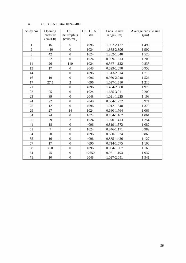
86
ii. CSF CLAT Titre 1024 - 4096
Study No Opening
pressure
(cmH₂0)
CSF
neutrophils
(cells/mL)
CSF CLAT
Titre
Capsule size
range (μm)
Average capsule size
(μm)
1 16 6 4096 1.052-2.127 1.495
2 <10 0 1024 1.368-2.396 1.902
3 42 0 1024 1.282-1.848 1.526
5 32 0 1024 0.959-1.613 1.208
11 26 110 1024 0.567-1.122 0.835
13 17 0 2048 0.823-1.098 0.958
14 0 4096 1.313-2.014 1.719
16 19 0 4096 0.960-2.048 1.526
17 27,5 2 4096 1.027-1.610 1.210
21 0 4096 1.464-2.808 1.970
22 25 0 1024 1.635-3.011 2.209
23 39 0 2048 1.021-1.225 1.108
24 22 0 2048 0.684-1.232 0.971
25 12 0 4096 1.012-1.848 1.379
29 27 14 1024 0.680-1.764 1.068
34 24 0 1024 0.764-1.162 1.061
35 29 2 1024 1.070-1.413 1.254
41 18 0 4096 0.819-1.572 1.082
51 7 0 1024 0.846-1.171 0.982
54 20 0 4096 0.680-1.024 0.860
55 16 0 4096 0.835-1.426 1.127
57 17 0 4096 0.714-1.575 1.103
58 >50 0 4096 0.894-1.307 1.169
64 25 0 >2650 0.951-1.193 1.037
71 10 0 2048 1.027-2.051 1.541
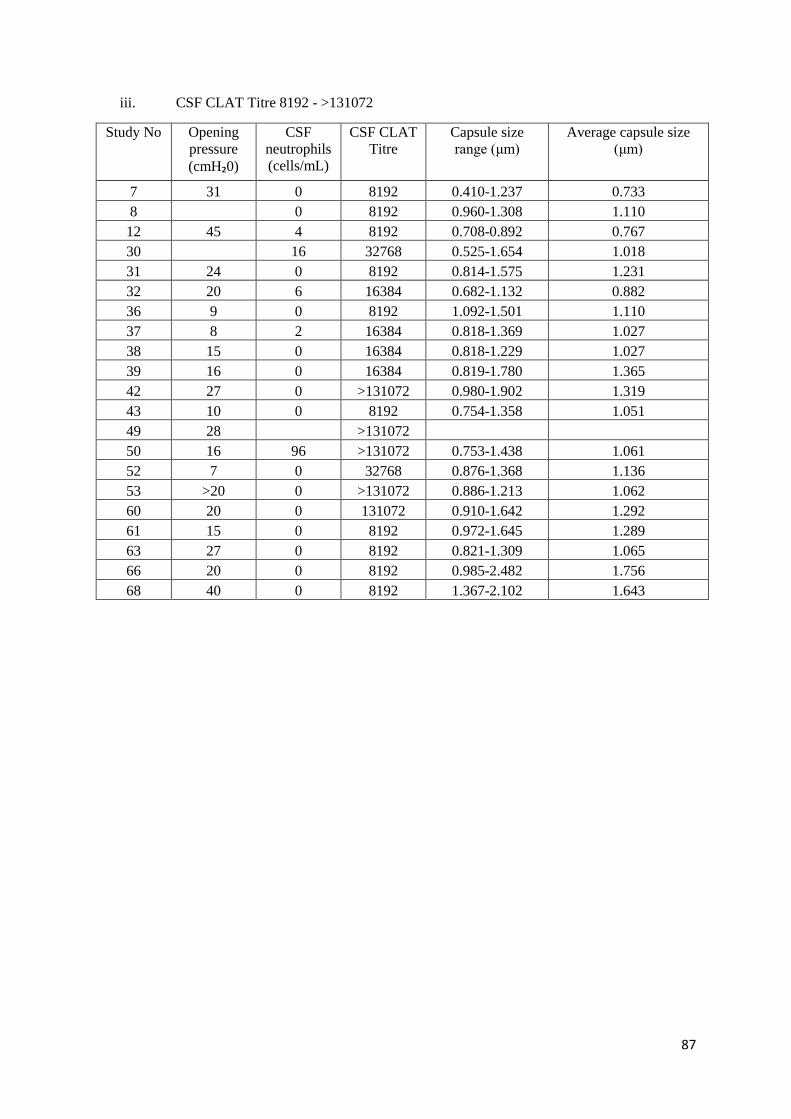
87
iii. CSF CLAT Titre 8192 - >131072
Study No Opening
pressure
(cmH₂0)
CSF
neutrophils
(cells/mL)
CSF CLAT
Titre
Capsule size
range (μm)
Average capsule size
(μm)
7 31 0 8192 0.410-1.237 0.733
8 0 8192 0.960-1.308 1.110
12 45 4 8192 0.708-0.892 0.767
30 16 32768 0.525-1.654 1.018
31 24 0 8192 0.814-1.575 1.231
32 20 6 16384 0.682-1.132 0.882
36 9 0 8192 1.092-1.501 1.110
37 8 2 16384 0.818-1.369 1.027
38 15 0 16384 0.818-1.229 1.027
39 16 0 16384 0.819-1.780 1.365
42 27 0 >131072 0.980-1.902 1.319
43 10 0 8192 0.754-1.358 1.051
49 28 >131072
50 16 96 >131072 0.753-1.438 1.061
52 7 0 32768 0.876-1.368 1.136
53 >20 0 >131072 0.886-1.213 1.062
60 20 0 131072 0.910-1.642 1.292
61 15 0 8192 0.972-1.645 1.289
63 27 0 8192 0.821-1.309 1.065
66 20 0 8192 0.985-2.482 1.756
68 40 0 8192 1.367-2.102 1.643
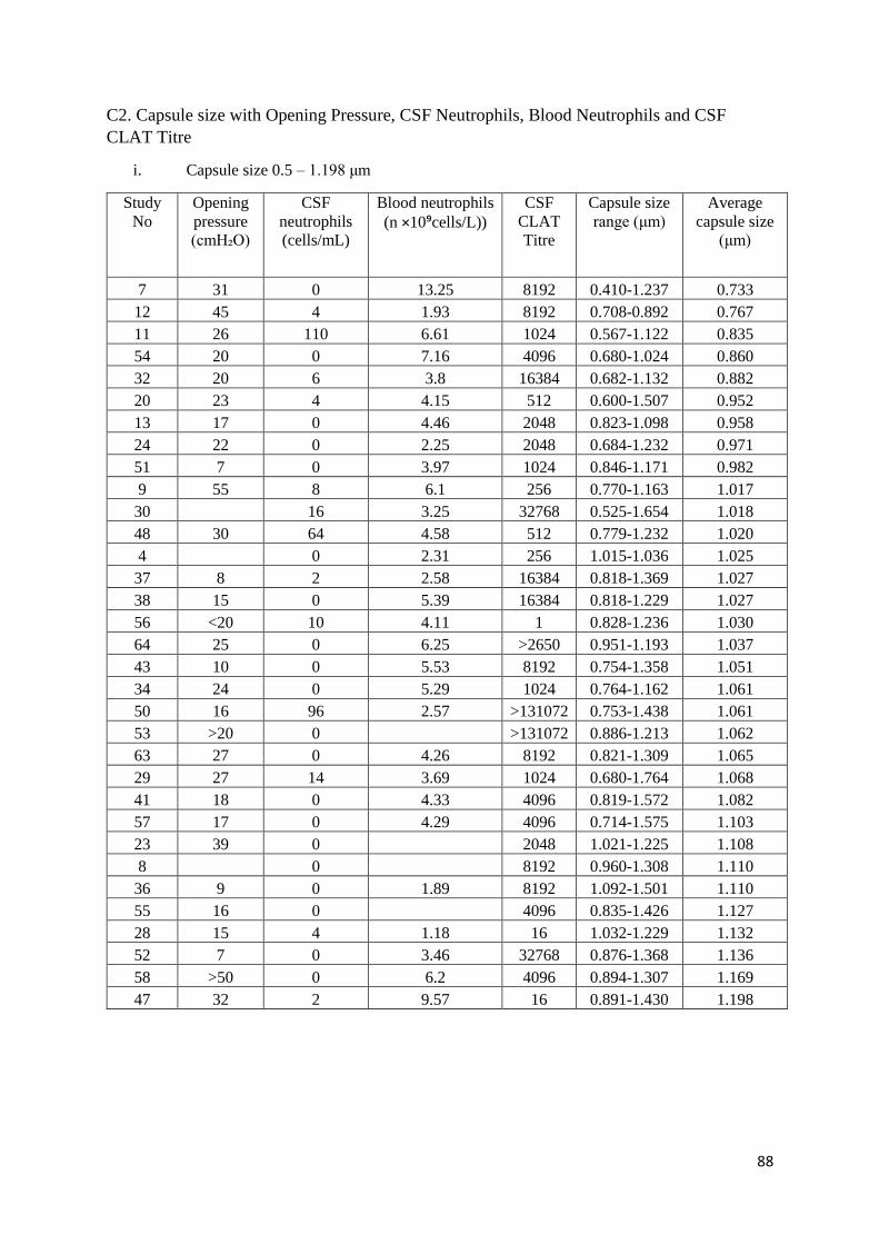
88
C2. Capsule size with Opening Pressure, CSF Neutrophils, Blood Neutrophils and CSF
CLAT Titre
i. Capsule size 0.5 – 1.198 μm
Study
No
Opening
pressure
(cmH₂O)
CSF
neutrophils
(cells/mL)
Blood neutrophils
(n ×10⁹cells/L))
CSF
CLAT
Titre
Capsule size
range (μm)
Average
capsule size
(μm)
7 31 0 13.25 8192 0.410-1.237 0.733
12 45 4 1.93 8192 0.708-0.892 0.767
11 26 110 6.61 1024 0.567-1.122 0.835
54 20 0 7.16 4096 0.680-1.024 0.860
32 20 6 3.8 16384 0.682-1.132 0.882
20 23 4 4.15 512 0.600-1.507 0.952
13 17 0 4.46 2048 0.823-1.098 0.958
24 22 0 2.25 2048 0.684-1.232 0.971
51 7 0 3.97 1024 0.846-1.171 0.982
9 55 8 6.1 256 0.770-1.163 1.017
30 16 3.25 32768 0.525-1.654 1.018
48 30 64 4.58 512 0.779-1.232 1.020
4 0 2.31 256 1.015-1.036 1.025
37 8 2 2.58 16384 0.818-1.369 1.027
38 15 0 5.39 16384 0.818-1.229 1.027
56 <20 10 4.11 1 0.828-1.236 1.030
64 25 0 6.25 >2650 0.951-1.193 1.037
43 10 0 5.53 8192 0.754-1.358 1.051
34 24 0 5.29 1024 0.764-1.162 1.061
50 16 96 2.57 >131072 0.753-1.438 1.061
53 >20 0 >131072 0.886-1.213 1.062
63 27 0 4.26 8192 0.821-1.309 1.065
29 27 14 3.69 1024 0.680-1.764 1.068
41 18 0 4.33 4096 0.819-1.572 1.082
57 17 0 4.29 4096 0.714-1.575 1.103
23 39 0 2048 1.021-1.225 1.108
8 0 8192 0.960-1.308 1.110
36 9 0 1.89 8192 1.092-1.501 1.110
55 16 0 4096 0.835-1.426 1.127
28 15 4 1.18 16 1.032-1.229 1.132
52 7 0 3.46 32768 0.876-1.368 1.136
58 >50 0 6.2 4096 0.894-1.307 1.169
47 32 2 9.57 16 0.891-1.430 1.198

89
ii. Capsule size >1.198μm
Study
No
Opening
pressure
(cmH₂O)
CSF
neutrophils
(cells/mL)
Blood neutrophils
(n ×10⁹cells/L)
CSF
CLAT
Titre
Casule size
range (μm)
Average
capsule size
(μm)
5 32 0 4.02 1024 0.959-1.613 1.208
17 27,5 2 2.37 4096 1.027-1.610 1.210
31 24 0 6.72 8192 0.814-1.575 1.231
33 1 0 7.88 32 0.960-1.434 1.233
67 30 0 2.75 256 1.059-1.575 1.252
35 29 2 4.4 1024 1.070-1.413 1.254
46 0 64 1.092-1.515 1.272
61 15 0 7.71 8192 0.972-1.645 1.289
60 20 0 4.34 131072 0.910-1.642 1.292
42 27 0 2.15 >131072 0.980-1.902 1.319
26 15 0 3.54 256 1.035-1.574 1.325
27 30 0 256 0.895-1.780 1.328
70 12 8 6.34 256 0.841-2.090 1.329
39 16 0 7.03 16384 0.819-1.780 1.365
25 12 0 4.14 4096 1.012-1.848 1.379
44 35 60 2.46 256 1.154-1.916 1.391
40 13 0 4.37 512 1.100-1.744 1.445
69 20 2 32 1.306-1.700 1.461
1 16 6 0.82 4096 1.052-2.127 1.495
3 42 0 5.08 1024 1.282-1.848 1.526
16 19 0 1.31 4096 0.960-2.048 1.526
71 10 0 4.28 2048 1.027-2.051 1.541
65 25 54 8.97 16 0.894-2.067 1.543
68 40 0 1.43 8192 1.367-2.102 1.643
14 0 4096 1.313-2.014 1.719
66 20 0 9.95 8192 0.985-2.482 1.756
2 <10 0 5.05 1024 1.368-2.396 1.902
18 23 12 3.12 4 0.955-1.454 1.961
21 0 0.84 4096 1.464-2.808 1.970
45 37 0 1.97 16 1.304-2.114 1.989
22 25 0 2.29 1024 1.635-3.011 2.209
15 26 4 5.16 512 1.850-3.739 2.457
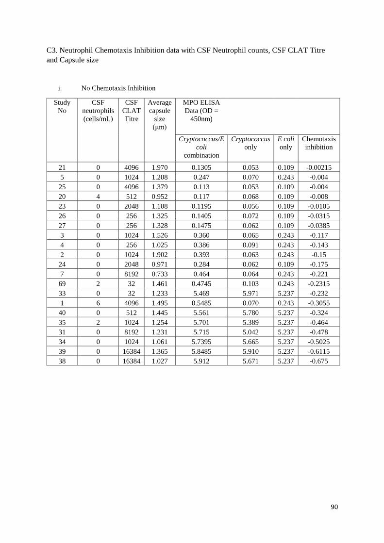
90
C3. Neutrophil Chemotaxis Inhibition data with CSF Neutrophil counts, CSF CLAT Titre
and Capsule size
i. No Chemotaxis Inhibition
Study
No
CSF
neutrophils
(cells/mL)
CSF
CLAT
Titre
Average
capsule
size
(μm)
MPO ELISA
Data (OD =
450nm)
Cryptococcus/E
coli
combination
Cryptococcus
only
E coli
only
Chemotaxis
inhibition
21 0 4096 1.970 0.1305 0.053 0.109 -0.00215
5 0 1024 1.208 0.247 0.070 0.243 -0.004
25 0 4096 1.379 0.113 0.053 0.109 -0.004
20 4 512 0.952 0.117 0.068 0.109 -0.008
23 0 2048 1.108 0.1195 0.056 0.109 -0.0105
26 0 256 1.325 0.1405 0.072 0.109 -0.0315
27 0 256 1.328 0.1475 0.062 0.109 -0.0385
3 0 1024 1.526 0.360 0.065 0.243 -0.117
4 0 256 1.025 0.386 0.091 0.243 -0.143
2 0 1024 1.902 0.393 0.063 0.243 -0.15
24 0 2048 0.971 0.284 0.062 0.109 -0.175
7 0 8192 0.733 0.464 0.064 0.243 -0.221
69 2 32 1.461 0.4745 0.103 0.243 -0.2315
33 0 32 1.233 5.469 5.971 5.237 -0.232
1 6 4096 1.495 0.5485 0.070 0.243 -0.3055
40 0 512 1.445 5.561 5.780 5.237 -0.324
35 2 1024 1.254 5.701 5.389 5.237 -0.464
31 0 8192 1.231 5.715 5.042 5.237 -0.478
34 0 1024 1.061 5.7395 5.665 5.237 -0.5025
39 0 16384 1.365 5.8485 5.910 5.237 -0.6115
38 0 16384 1.027 5.912 5.671 5.237 -0.675

91
ii. Positive Chemotaxis Inhibition Study
No
CSF
neutrophils
(cells/mL)
CSF
CLAT
Titre
Average
capsule
size (μm)
MPO ELISA Data (OD = 450nm)
Cryptococcus/E
coli combination
Cryptococcus
only
E coli
only
Chemotaxis
inhibition
22 0 1024 2.209 0.091 0.055 0.109 0.018
30 16 32768 1.018 0.0715 0.056 0.109 0.0375
28 4 16 1.132 0.068 0.059 0.109 0.041
29 14 1024 1.068 0.0645 0.054 0.109 0.0445
68 0 8192 1.643 1.627 0.122 1.710 0.083
36 0 8192 1.110 5.1275 6.000 5.237 0.1095
71 0 2048 1.541 0.0755 0.053 0.243 0.1675
37 2 16384 1.027 5.061 6.000 5.237 0.176
48 64 512 1.020 0.066 0.065 0.243 0.177
58 0 4096 1.169 0.066 0.056 0.243 0.177
70 8 256 1.329 0.0635 0.053 0.243 0.1795
9 8 256 1.017 0.0625 0.053 0.243 0.1805
18 12 4 1.961 0.062 0.058 0.243 0.181
47 2 16 1.198 0.0605 0.057 0.243 0.1825
50 96 >131072 1.061 0.0605 0.053 0.243 0.1825
51 0 1024 0.982 0.0565 0.054 0.243 0.1865
32 6 16384 0.882 5.0395 5.141 5.237 0.1975
53 0 >131072 1.062 0.0401 0.072 0.243 0.2029
46 0 64 1.272 5.7375 5.477 6.000 0.2625
44 60 256 1.391 5.668 5.031 6.000 0.332
8 0 8192 1.110 5.6485 4.252 6.000 0.3515
41 0 4096 1.082 5.5325 4.845 6.000 0.4675
17 2 4096 1.210 5.4475 4.819 6.000 0.5525
66 0 8192 1.756 1.0875 0.077 1.710 0.6225
43 0 8192 1.051 5.3305 5.360 6.000 0.6695
45 0 16 1.989 5.249 6.000 6.000 0.751
11 110 1024 0.835 5.1795 4.800 6.000 0.8205
42 0 >131072 1.319 5.1385 5.132 6.000 0.8615
14 0 4096 1.719 4.942 4.695 6.000 1.058
67 0 256 1.252 0.645 0.075 1.710 1.065
61 0 8192 1.289 0.5755 0.081 1.710 1.1345
63 0 8192 1.065 0.539 0.092 1.710 1.171
15 4 512 2.457 4.8085 4.716 6.000 1.1915
12 4 8192 0.767 4.7935 4.785 6.000 1.2065
65 54 16 1.543 0.4915 0.099 1.710 1.2185
54 0 4096 0.860 0.434 0.062 1.710 1.276
64 0 >2650 1.037 0.4165 0.102 1.710 1.2935
60 0 131072 1.292 0.352 0.102 1.710 1.358
55 0 4096 1.127 0.3505 0.083 1.710 1.3595
13 0 2048 0.958 4.5875 5.091 6.000 1.4125
57 0 4096 1.103 0.2485 0.072 1.710 1.4615
56 10 1 1.030 0.190 0.071 1.710 1.52
52 0 32768 1.136 0.1185 0.065 1.710 1.5915
16 0 4096 1.526 4.4035 5.059 6.000 1.5965

92
C4. TLR2 and TLR4 Gene Expression with Capsule size, CSF Neutrophils and CSF CLAT Titre
i. TLR2 Expression with Capsule size, CSF Neutrophils and CSF CLAT Titre
Study
No
18S TLR2 TLR2/18S Average
Capsule size
(μm)
CSF
neutrophils
(cells/mL)
CSF CLAT
Titre
17 2 294,964 60,661 0,026432272 1.210 2 4096
18 2 265,975 93,697 0,041349735 1.961 12 4
14 1 981,369 92,927 0,046900188 1.719 0 4096
9 6 154,244 312,673 0,050806008 1.017 8 256
22 2 100,092 124,734 0,059394704 2.209 0 1024
25 455,093 27,135 0,059625389 1.379 0 4096
15 650,646 39,298 0,060397783 2.457 4 512
7
16
713,160 1 016,068 0,060794479 0.733 0 8192
36 420,346 27,712 0,065925527 1.110 0 8192
24 2 626,968 175,981 0,066990088 0.971 0 2048
37 685,151 48,447 0,070710671 1.027 2 16384
8 2 818,096 205,638 0,072970383 1.110 0 8192
4
19
745,727 1 509,424 0,076443082 1.025 0 256
16 536,981 47,055 0,087629236 1.526 0 4096
28 1 948,331 202,018 0,103687608 1.132 4 16
1 3 690,052 388,610 0,105312901 1.495 6 4096
11 1 382,593 151,777 0,10977693 0.835 110 1024
71 546,041 65,906 0,120698172 1.541 0 2048
27 537,477 71,055 0,132200652 1.328 0 256
20 1 637,611 219,187 0,133845604 0.952 4 512
3 946,743 131,188 0,138567397 1.526 0 1024
12
24
245,568 3 371,908 0,139073186 0.767 4 8192
38 218,612 36,861 0,168614471 1.027 0 16384
34 124,800 23,025 0,184497434 1.061 0 1024
2 652,840 127,875 0,195874664 1.902 0 1024
60 4 316,575 858,120 0,198796512 1.292 0 131072
21 1 815,927 368,559 0,202958964 1.970 0 4096
58 99,963 26,062 0,260712545 1.169 0 4096
39 238,221 62,182 0,261026419 1.365 0 16384
57 1 710,332 450,786 0,263566623 1.103 0 4096
42 5 777,510 1 555,493 0,269232438 1.319 0 >131072
13 2 252,085 642,026 0,285080576 0.958 0 2048
40 322,352 99,226 0,307819171 1.445 0 512
30 2 831,050 1 048,441 0,370336553 1.018 16 32768
32 159,672 65,942 0,412982795 0.882 6 16384
63 498,928 222,402 0,445760166 1.065 0 8192
69 146,038 74,622 0,510978429 1.461 2 32
65 380,572 211,396 0,555467891 1.543 54 16
41 4 804,269 2 728,569 0,567946778 1.082 0 4096
68 191,610 150,010 0,782889092 1.643 0 8192

93
44 2 368,476 1 874,138 0,791284363 1.391 60 256
29 30,195 25,219 0,835215402 1.068 14 1024
53 272,440 252,851 0,928099114 1.062 0 >131072
56 41,524 40,743 0,981180815 1.030 10 1
67 587,515 577,637 0,983185793 1.252 0 256
51 52,161 68,345 1,310271498 0.982 0 1024
55 5 059,433 6 946,552 1,372990271 1.127 0 4096
31 43,820 68,113 1,554383304 1.231 0 8192
48 52,265 119,380 2,28414244 1.020 64 512
43 501,151 1 154,218 2,303133241 1.051 0 8192
66 280,022 702,605 2,509107253 1.756 0 8192
23 236,879 879,060 3,711001352 1.108 0 2048
45 40,914 153,671 3,755951138 1.989 0 16
5 5,841 27,565 4,719394713 1.208 0 1024
35 983,777 4 929,847 5,01114318 1.254 2 1024
47 88,278 492,042 5,573766105 1.198 2 16
46 3,315 32,414 9,778819152 1.272 0 64
50 36,034 474,061 13,15577383 1.061 96 >131072
52 3,965 52,571 13,25874626 1.136 0 32768
54 26,767 397,587 14,85380324 0.860 0 4096
61 77,637 2 661,790 34,28529245 1.289 0 8192
33 473,438 21 647,914 45,72492868 1.233 0 32
64 45,667 2 136,321 46,7801742 1.037 0 >2650
26 251,322 39 695,664 157,9473017 1.325 0 256

94
ii. TLR4 Expression with Capsule size, CSF Neutrophils and CSF CLAT Titre
Study
No
18S TLR4 TLR4/18S Average
Capsule size
(μm)
CSF
neutrophils
(cells/mL)
CSF CLAT
Titre
17 2 294,964 106,345 0,046338 1.210 2 4096
14 1 981,369 122,941 0,062049 1.719 0 4096
9 6 154,244 437,227 0,071045 1.017 8 256
4 19 745,727 1 499,273 0,075929 1.025 0 256
13 2 252,085 178,796 0,079391 0.958 0 2048
12 24 245,568 2 214,537 0,091338 0.767 4 8192
7 16 713,160 1 555,925 0,093096 0.733 0 8192
22 2 100,092 206,800 0,098472 2.209 0 1024
8 2 818,096 283,355 0,100549 1.110 0 8192
24 2 626,968 277,665 0,105698 0.971 0 2048
1 3 690,052 395,961 0,107305 1.495 6 4096
15 650,646 70,163 0,107836 2.457 4 512
28 1 948,331 212,416 0,109025 1.132 4 16
16 536,981 66,460 0,123767 1.526 0 4096
3 946,743 122,351 0,129234 1.526 0 1024
37 685,151 96,414 0,14072 1.027 2 16384
20 1 637,611 273,631 0,167091 0.952 4 512
60 4 316,575 770,819 0,178572 1.292 0 131072
25 455,093 92,539 0,20334 1.379 0 4096
11 1 382,593 284,593 0,20584 0.835 110 1024
57 1 710,332 369,721 0,216169 1.103 0 4096
21 1 815,927 454,140 0,250087 1.970 0 4096
36 420,346 109,359 0,260164 1.110 0 8192
18 2 265,975 615,022 0,271416 1.961 12 4
38 218,612 59,645 0,272834 1.027 0 16384
27 537,477 149,452 0,278063 1.328 0 256
30 2 831,050 800,647 0,282809 1.018 16 32768
42 5 777,510 1 779,079 0,307932 1.319 0 >131072
65 380,572 118,138 0,310423 1.543 54 16
41 4 804,269 1 661,625 0,345864 1.082 0 4096
2 652,840 272,093 0,416784 1.902 0 1024
40 322,352 142,002 0,44052 1.445 0 512
39 238,221 110,497 0,463843 1.365 0 16384
67 587,515 277,476 0,472287 1.252 0 256
34 124,800 68,884 0,551955 1.061 0 1024
63 498,928 283,300 0,567817 1.065 0 8192
44 2 368,476 1 486,484 0,627612 1.391 60 256
69 146,038 95,009 0,650582 1.461 2 32
71 546,041 355,259 0,650608 1.541 0 2048
55 5 059,433 3 793,110 0,749711 1.127 0 4096
53 272,440 233,399 0,856697 1.062 0 >131072
68 191,610 177,153 0,924549 1.643 0 8192
58 99,963 98,001 0,980367 1.169 0 4096

95
31 43,820 50,523 1,152969 1.231 0 8192
32 159,672 206,113 1,290857 0.882 6 16384
56 41,524 56,142 1,352031 1.030 10 1
48 52,265 80,358 1,537522 1.020 64 512
43 501,151 804,667 1,605639 1.051 0 8192
51 52,161 129,441 2,481561 0.982 0 1024
23 236,879 656,257 2,770427 1.108 0 2048
66 280,022 798,036 2,849905 1.756 0 8192
29 30,195 92,281 3,056175 1.068 14 1024
45 40,914 134,885 3,296804 1.989 0 16
35 983,777 3 457,118 3,514128 1.254 2 1024
47 88,278 424,330 4,806735 1.198 2 16
50 36,034 194,437 5,395878 1.061 96 >131072
5 5,841 34,795 5,957274 1.208 0 1024
54 26,767 183,914 6,87099 0.860 0 4096
33 473,438 7 274,291 15,36483 1.233 0 32
52 3,965 84,838 21,3965 1.136 0 32768
61 77,637 1 987,636 25,60182 1.289 0 8192
64 45,667 1 242,781 27,21384 1.037 0 >2650
46 3,315 127,953 38,60136 1.272 0 64
26 251,322 12 193,188 48,51616 1.325 0 256