Embed Size (px)
Citation preview

The evolution of the aortic neck
after EVAR
Prof. Carlo Setacci
Chief of Vascular Surgery
University of Siena - Italy

Proximal Necks
Straight Tapered Reversed tapered Angulated* Bulge Short+

- Makaroun MS, et al. Is proximal aortic neck dilatation after endovascular aneurysm exclusion a cause for concern? J Vasc Surg 2001;33:S39-45.- Diehm N, et al. Aortic neck dilatation after endovascular abdominal aortic aneurysm repair: a word of caution. J VascSurg. 2008;47:886–892
A continuous aortic enlargement at the level of infrarenal aortic neck has been reported after endovascular repair (EVAR)
Background

- Oberhuber A, et al. Influence of different self-expanding stent-graft types on remodeling of the aortic neck after endovascular aneurysm repair. J Endovasc Ther. 2010;17:677-84. - Sternbergh WC et al. Zenith Investigators Influence of endograft oversizing on device migration, endoleak, aneurysm shrinkage, and aortic neck dilation: results from the Zenith Multicenter Trial. J Vasc Surg. 2004 ;39:20-6.
The amount of proximal device oversizing with self-expanding stent grafts (SESG) influences neck progression
Background

- Cao PG et al. Predictive factors and clinical consequences of proximal aortic neck dilatation in 230 patients undergoing abdominal aorta aneurysm repair with self-expandable stent-grafts. J Vasc Surg 2003;37:1200-5.- Dillavou ED, et al. Is neck dilatation after endovascular aneurysm repair graft dependent? Results of 4 US Phase II trials. Vasc Endovasc Surg 2005;39:47-54.
When aortic neck dilatation
occurs, it is related to adverse mid-term outcomes
Background

Aortic neck dilatation with balloon exp. stent graft
* Parodi JC, Ferreira LM. Ten-year experience with endovascular therapy in aortic aneurysms. J Am Coll Surg.
2002;194:S58-66
** Malas MB, Ohki T, Veith FJ, et al. Absence of proximal neck dilatation and graft migration after endovascular
aneurysm repair with balloon-expandable stent based endografts. J Vasc Surg. 2005;42:639-44.
Home made Parodi stent graft Montefiore Endograft System
0% * 0% **
Background

EndoAnchors Mimic Open Surgically
Sutured Anastomosis
BUILDING ON HISTORY
1st Vascular Anastomosis
by Alexis Carrel
1902
1st Open AAA Repair
by Charles Dubost
1951
1st Endovascular AAA Repair by
Juan Parodi
1991
1st Endovascular Anastomosis via
EndoAnchoring in EVAR by David
Deaton and Takao Ohki
2005

EndoAnchoringSurgical Anastomosis
Case images from John Aruny MD, Bart Edward Muhs, MD, PhD.
TAILORED SEAL AND FIXATION OF
ENDOANCHORS
0
50
100
150
Talent Endurant Excluder Zenith Mean Hand Sewn
No EndoAnchors With EndoAnchors
Create the stability of a surgical anastomosis in EVAR and
TEVAR
Dis
pla
ce
me
nt
forc
e in
Ne
wto
ns
Melas et al. JVS 2012;55(6):1726-33

Do EndoAnchors have value in preventing proximal neck complications in patients with
challenging neck anatomy?

Baseline anatomy in propensity-matched cohortsAnatomic Measures for Propensity Matching
ControlsN = 103
EndoAnchorsN = 103
P Value
Max AAA Diameter 56 ± 13 mm 56 ± 10 mm .674
Suprarenal Diameter 27 ± 4 mm 27 ± 3 mm .999
Diameter at Lowest Renal 25 ± 4 mm 26 ± 4 mm .458
Proximal Neck Length 23 ± 14 mm 20 ± 13 mm .093
Suprarenal Angulation 16 ± 11◦ 17 ± 13◦ .664
Infrarenal Angulation 37 ± 16◦ 37 ± 18◦ .885
Neck Thrombus 23± 54◦ 38 ± 71◦ .107
Neck Calcium 20± 29◦ 19 ± 30◦ .845
Necks <10mm Length 18.4% 26.5% .097
The EVAR Durability Question and a Potential Solution
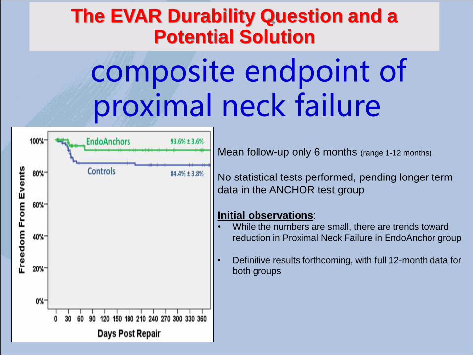
Initial resultscomposite endpoint of proximal neck failure
Mean follow-up only 6 months (range 1-12 months)
No statistical tests performed, pending longer term
data in the ANCHOR test group
Initial observations:• While the numbers are small, there are trends toward
reduction in Proximal Neck Failure in EndoAnchor group
• Definitive results forthcoming, with full 12-month data for
both groups
The EVAR Durability Question and a Potential Solution

- New concept of sealing by non-expansive circumferential apposition of polymer-filled ring to the aortic wall
The Ovation stent graft (TriVascular, Santa Rosa – CA)

METHODS
- Retrospective, multicenter registry (Nov 2014)
- 13 Italian Centers of Vascular Surgery
- Only patients who had undergone implantation of a Trivascular Ovation at least 24 months previously (before Nov 2012)
- CT scans available at a minimum 2-year follow-up were collected and sent for blind reading to a centralized core laboratory.
Trivascular Ovation Italian Registry (TOIS)

Central database for the core lab review of morphological changes
- OsiriX MD (v.6.5.1 64-bit)
- 1 VS as single observer
(intra-observer agreement 0.91)
- All vessel measurements after center
line lumen (CLL) reconstructions
(manual segmentation)
Trivascular Ovation Italian Registry (TOIS)

Proximal aortic neck segmentation
- Zone A (fixation area)
- Zone B (infrarenal aorta)
- Zone C (sealing zone)
Trivascular Ovation Italian Registry (TOIS)

RESULTS
161 patientsmean age 74 ± 5 92% male
Median 32 months (range 24-50)
CT @ ≥ 24 moths 89 pts
Trivascular Ovation Italian Registry (TOIS)

FOLLOW-UP
- 17 pts died (no AAA-related death)
- 15 pts lost atfollow-up
Trivascular Ovation Italian Registry (TOIS)

Trivascular Ovation Italian Registry (TOIS)

Reinterventions (total n=8) - 3 type IA endoleak
1 aortic cuff, 1 balloon-expandable stent 1 coil and glue emboliz
- 4 iliac limb occlusion 1 bypass,3 surgical thrombectomy
- 1 type II endoleakcoil embolization
Trivascular Ovation Italian Registry (TOIS)

Morphological changes @ 2 yearsCore lab 89 CT scans
Zone A
Patency of visceral arteries in Zone A was 100%
Morphological changes

Zone A at 2 years
The mean change was 0.18 ±0.22 mm (SE 0.02)
Morphological changes

Zone B_deployment
The mean stent-graft landingdistance to the lowest renal arterywas 3.13 ± 4.25 mm (SE 0.45)
* * *
* Type I endoleak in less precise stent graft deployment
Morphological changes
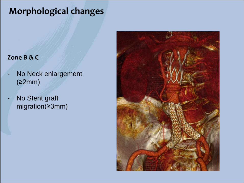
Zone B & C
- No Neck enlargement
(≥2mm)
- No Stent graft
migration(≥3mm)
Morphological changes

Zone B
The mean change was -0.32±0.87 mm (SE 0.09)
Morphological changes

Zone C
The mean change in diameters-0,06 ± 0.97 mm (SE 0,1)
Morphological changes

correlation coefficient 0.000 P=1.0
Spearman correlation test (Zone B vs Zona A)
Correlation Zone B vs Zone A

Spearman correlation test (Zone B vs Zone C)
correlation coefficient 0.183 P=.05
Correlation Zone B vs Zone C

Neck evolution
- Zone A +0.18 ± 0.22 mm
- Zone B -0.32±0.87 mm
- Zone C -0,06 ± 0.97 mm
No correlation
Correlation
Morphological changes_Summary

Zone B -0.32±0.87 mm
- nitinol stent touches the aortic wall but no self expanding force is applied
- graft material is slightly free to move independently of the stent and may conform to the aortic wall (“mainsail effect”), preserving blood stream between the fabric and the wall.
Neck evolution
zone B is protected from arterial pressure

- No aortic neck dilatation occurred in patients treated with TriVascular Ovation stent graft at CT scan after a minimum 24-month follow-up.
- This may suggest that aortic neck evolution is not associated to EVAR at mid-term follow-up when an endograft with no chronic outward radial force is implanted.
Considerations 1

- No aortic neck dilatation occurred in patients treated with TriVascularOvation stent graft at CT scan after a minimum 24-month follow-up.
- This may suggest that aortic neck evolution is not associated to EVAR at mid-term follow-up when an endograft with no chronic outward radial force is implanted.
Considerations 2
Even in very short neck <7mm

Conclusions
• Proximal aortic neck complications represent a
critical issue after EVAR
• New technologies seems to provide better results
• Follow up is mandatory after EVAR

Piazza del Campo, Siena – Italy
ThANK YOU

The evolution of the aortic neck
after EVAR
Prof. Carlo Setacci
Chief of Vascular Surgery
University of Siena - Italy


![Endovascular treatment of complex aortic aneurysms · 2019-07-12 · aortic aneurysm repair (EVAR) by Volodos’ and Parodi almost 20 years ago [1,2], the tech-nique has matured into](https://img.pdfslide.us/doc/110x75/5e7ddabd23ab9f279a2ef315/endovascular-treatment-of-complex-aortic-aneurysms-2019-07-12-aortic-aneurysm.jpg)