Embed Size (px)
Citation preview

5
The Evolution of Biocompatibility: From Microinflammation to Microvesiscles
Ciro Tetta1,2, Stefano Maffei3, Barbara Cisterna4, Valentina Fonsato4, Giorgio Triolo3, Giuseppe Paolo Segoloni5, Giovanni Camussi4,5,
Maria Chiara Deregibus4 and Emanuele Gatti1,6*
1. Introduction
Haemodialysis (HD) is a life-saving treatment for patients with chronic kidney disease
(CKD) stage 5. CKD persists as a chronic worldwide epidemic and HD is the more
frequently (70%) adopted treatment modality. Exponential growth trend continues on a
global scale. The HD population becomes every year increasingly older (average age: 75 yrs)
and sicker due to the associated co-morbidities such as cardiovascular disease (heart failure,
coronary heart disease, and peripheral vascular disease), diabetes, hypertension, and
peripheral vascular disease. Most of the complications associated with HD involve the
cardiovascular system (Go et al., 2004; Culleton et al., 1999, Goodkin et al., 2003, Foley 2004;
Barret, 2002). The evolution in the history of HD technology has greatly helped to make the
HD procedure a safe and more biocompatible extracorporeal therapy. However, it must be
admitted that despite significant improvements in HD technology and in the management
of patients due to a better understanding of uremia toxicity, improvements in dialysis
technology, better correction of anaemia and metabolic abnormalities, implementation of
best practice guidelines, no significant improvement has been achieved in patient survival
over the last decade (Rayner et al., 2004). The extracorporeal circuit offers a large surface of
contact of the blood with foreign materials, namely the dialysis membrane, the tubings and
the large volumes of the dialysate. The concept of biocompatibility has greatly evolved in
the last two decades. Initially, numerous studies focused on the blood-dialyzer membrane
interaction, leading to the activation of plasma systems (complement, coagulation,
fibrinolysis). These studies helped in the understanding of some unknown effects occurring
in the early stages of the HD session leading to pulmonary sequestration of leukocytes
(mainly neutrophils) that explained the profound neutropenia associated with the
cuproammonium membranes. The availability of reliable testing of complement-activated
*1Biologics Research, Intl Research and Development, Fresenius Medical Care, Bad Homburg, Germany. 2Doctoral School of Biotechnology, University of Torino, Torino, Italy. 3Department of Medicine, Nephrology and Dialysis Unit, CTO Hospital, Torino, Italy. 4Department of Internal Medicine, Centre for Molecular Biotechnology and Centre for Research in Experimental Medicine (CeRMS), Torino, Italy. 5Chair of Nephrology and Department of Nephrology, Dialysis and Transplantation, University of Torino, Italy 6Danube University, Center for Biomedical Technology, Krems, Austria.
www.intechopen.com

Progress in Hemodialysis – From Emergent Biotechnology to Clinical Practice
94
products (C3a and C5a and their desarginated products) guided the development of less
neutropenia-inducing membranes and ultimately to the final development of fully synthetic
membranes which have very low if at all capacity to induce complement activation. At that
time, coagulation was an important reason for frequent interruptions and delays in the HD
sessions. Due to the complex interplay known to occur between the activation of the
complement and coagulation systems, it became of great interest to try to reduce the
propensity for intravascular coagulation. The development of high-flux membranes and
growing awareness of the benefits of convective and convective/diffusive under several
contexts (intradialytic cardiovascular stability, better control or the uremic status and fluid
control) gave impetus to a large number of enlightening studies on another mechanism of
HD bioINcompatibility. The contamination by bacterial products, particularly with the
widespread use of bicarbonate-based dialysates opened a new era in the field of
biocompatibility. The formulation of the “interleukin hypothesis” was a posteriori not only
the basis for further studies on the monocyte stimulation during HD, but also provided a
link between biocompatibility and chronic inflammation. Basically, the evolution of
biocompatibility has led us to consider two sides of the same coin: on one side, the biological
responses at the blood-membrane interface; on the other hand, the consequences derived
from the contact on the membrane performances (e.g. hydraulic permeability and sieving
coefficients).
In this review, we will summarize the most important steps in the evolution from the
concept of the blood-dialyzer membrane interaction to that of the whole HD system
compatibility. In face of very recent developments of cell-to-cell communication and signal
transduction, we will also discuss the new hypothesis for a role of microvesicles (MVs) in
cell activation, as well as in tissue and vascular repair. We will not deal with other important
aspects of biocompatibility such as the oxidant stress, the relevant role of additives in
dialyzer manufacturing, and of leachables and the effects of different sterilization modes.
2. Blood-membrane interaction: the role of complement, coagulation, kinin-kallikrein systems and soluble mediators
2.1 Activation of the complement alternative pathway
Early studies on biocompatibility focused on acute hypersensitivity-like reactions which in some cases were fatal. Various mechanisms were elucidated. Activation of complement was shown by Craddock et al in 1977 (Craddock et al, 1977). Hydroxyl radicals, present on the surface of cellulosic membranes, bind with the C3b in the blood and activate the alternative pathway leading to the release of potent anaphylatoxins, C3a and C5a. Both C3a and C5a and their relative desarginated products induce prompt activation and aggregation of polymorphonuclear neutrophils (PMNs) and leukopenia. This is a very rapid process reaching a nadir from 15 to 30 min after initiation of dialysis. Aggregates of PMNs are sequestered particularly in the lung capillaries. Although the extent of the anaphylatoxin generation and of the neutropenia is also patient-dependent, these studies failed to find a relationship with chronic clinical trade-offs despite the hypothesis that recurrent pulmonary sequestration could induce pulmonary fibrosis. Reduction of the hydroxyl groups on the membrane surface or new synthetic polymers reduced the activation of the alternative pathway of the complement cascade. Temperature could also reduce complement activation (Maggiore Q, personal communication, 1988). Testing
www.intechopen.com

The Evolution of Biocompatibility: From Microinflammation to Microvesiscles
95
complement activation (C3a or C5a plasma levels) by highly sensitive ELISA tests has become a standard requirement for the evaluation of biocompatibility ever since along with the precise characterization of the polymer structure (Krieter et al, 2008). It also became clear that synthetic polymers had a very low neutropenia-inducing effect. In some cases such as the polyacrylonitrile membrane, this was also due to the capacity of the membrane to adsorb C3b and the anaphylytoxins thus masking in fact complement activation (Pascual et al 1993) (Figure 1).
Fig. 1. Pathways involved in blood-membrane interactions. LTB4 denotes leukotriene B4,
PAF, platelet-activating factor, IL-1, interleukin-1, TNF-, tumor necrosis factor.
2.2 Activation of the coagulation system
Numerous acquired hemostatic abnormalities have been identified in chronic renal failure.
HD adds to these disturbances as it repetitively implies turbulent blood flow, high shear
stress, and contact of blood to artificial surfaces. Anticoagulation in HD is targeted to
prevent activation of coagulation during the procedure. Most anticoagulant agents inhibit
the plasmatic coagulation cascade. Still commonly used is unfractionated heparin, followed
by low-molecular-weight heparin preparations with distinct advantages. Immune-mediated
heparin-induced thrombocytopenia constitutes a potentially life-threatening complication of
www.intechopen.com

Progress in Hemodialysis – From Emergent Biotechnology to Clinical Practice
96
heparin therapy requiring immediate switch to nonheparin alternative anticoagulants.
Danaparoid, lepirudin, and argatroban are currently being used for alternative
anticoagulation, all of which possess both advantages and limitations. Recently citrate has
been proposed as anticoagulant in maintenance HD (Wright et al, 2010). In the past,
empirical strategies reducing or avoiding heparin were applied for patients at bleeding risk,
whereas nowadays regional citrate anticoagulation is increasingly used to prevent bleeding
by allowing procedures without any systemic anticoagulation. Avoidance of clotting within
the whole hemodialyzer circuit is not granted. Specific knowledge of the mechanisms of
coagulation, the targets of the anticoagulants in use, and their respective characteristics
constitutes the basis for individualized anticoagulation aimed at achieving full patency of
the circuit throughout the procedure. Patency of the circuit is an important prerequisite for
optimal HD quality. Intrinsic coagulation Hageman factor XII as well as other coagulation
factors are also activated (Fischer, 2007). However, the activation of the coagulation is a very
complex phenomenon that may be enhanced by different independent factors other than the
membrane surface per se such as: the dynamics at the dialyzer heads, defects in the hollow
fibre cutting of the polyurethane, any condition that predisposes for blood to be stagnant.
The activation of coagulation by a membrane in a dialyzer is difficult to assess given the
above-mentioned factors and the host's response to the anticoagulation regime put in place
(Figure 1).
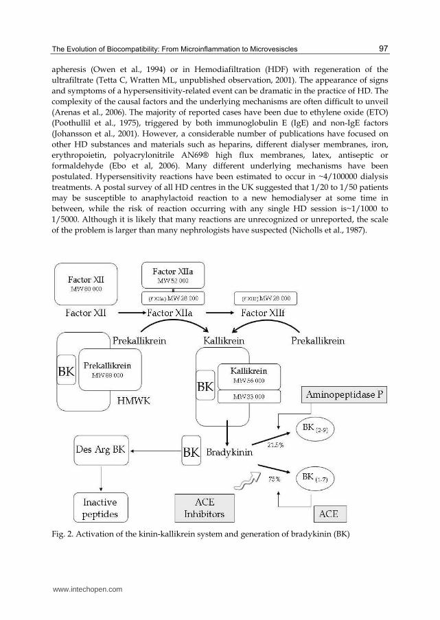
2.3 Activation of the kinin-kallikrein system
Surface activation of Factor XII induces the kinin-kallikrein that ensues in the generation of
bradykinin (Figure 2).
Bradykinin is physiologically under the tight control of very potent kinases that are able to
promptly lyse the molecule and inactivate its potent vasodilator activity. In certain
conditions, however, the lytic effect of this kinase is deficient. This occurs in patients under
therapy with angiotensin converting enzyme enzyme (ACE) inhibitors. However, there are
patients who experience hypersensitivity-like phenomena, that can be reconducted to
bradykinin generation, even in the absence of concomitant therapy with ACE inhibitors. The
explanation of this phenomenon came from pioneering studies on angio-edema, a rare but
potentially fatal condition (Adam et al., 2002). These reactions are mainly associated to
defects in the enzymatic activity of the aminopeptidase P (Figure 2). Bradykinin acts
through two types of tissue receptors: R1 are mostly located in the skin and respiratory
tissues (lungs and bronchi), while R2 are mostly found in the gastrointestinal tract. The
overproduction of bradykinin may lead to two different clinical presentations: the first is
mainly characterized by a rapid developing skin flushing, hypotension, and dyspnoea.
These reactions may be mild but very severe, fatal episodes of shock have been described. In
the second instance, these reactions, which were for some time unexplained, occur after 1 h-
2 hr of HD treatment, may but may be not associated with the use of ACE inhibitors. The
patient has severe diarrhoea which requires immediate interruption of the extracorporeal
treatment. This manifestation may unpredictably recur and disappears upon disconnection.
Bradykinin-induced reaction, may in principle occur following the contact with any foreign
surface. Their potential, unpredictable severity should call for immediate action even in
patients with mild forms. The commonest causes have been the use of strongly negative
surfaces such as AN-69 membranes (Tielemans et al., 1990), or adsorbents used in LDL
www.intechopen.com
The Evolution of Biocompatibility: From Microinflammation to Microvesiscles
97
apheresis (Owen et al., 1994) or in Hemodiafiltration (HDF) with regeneration of the
ultrafiltrate (Tetta C, Wratten ML, unpublished observation, 2001). The appearance of signs
and symptoms of a hypersensitivity-related event can be dramatic in the practice of HD. The
complexity of the causal factors and the underlying mechanisms are often difficult to unveil
(Arenas et al., 2006). The majority of reported cases have been due to ethylene oxide (ETO)
(Poothullil et al., 1975), triggered by both immunoglobulin E (IgE) and non-IgE factors
(Johansson et al., 2001). However, a considerable number of publications have focused on
other HD substances and materials such as heparins, different dialyser membranes, iron,
erythropoietin, polyacrylonitrile AN69® high flux membranes, latex, antiseptic or
formaldehyde (Ebo et al, 2006). Many different underlying mechanisms have been
postulated. Hypersensitivity reactions have been estimated to occur in ~4/100000 dialysis
treatments. A postal survey of all HD centres in the UK suggested that 1/20 to 1/50 patients
may be susceptible to anaphylactoid reaction to a new hemodialyser at some time in
between, while the risk of reaction occurring with any single HD session is~1/1000 to
1/5000. Although it is likely that many reactions are unrecognized or unreported, the scale
of the problem is larger than many nephrologists have suspected (Nicholls et al., 1987).
Fig. 2. Activation of the kinin-kallikrein system and generation of bradykinin (BK)
www.intechopen.com

Progress in Hemodialysis – From Emergent Biotechnology to Clinical Practice
98
2.3.1 Soluble mediators
Many soluble mediators are produced and released following the blood-membrane interaction. Products of the phospholipase A2 such as platelet-activating factor (PAF) and leukotrienes are released by the direct interaction of PMNs and platelets with complement-activating membranes. Although in the presence of blood, the mechanisms of production of PAF and leukotrienes can not be readily differentiated from the activation, as they follow the same kinetics, we could show that for PAF for example, its production and release could be observed in complement-independent conditions such as in the absence of plasma by purified cells incubated with flat HD membranes (Tetta et al., 1996). A large number of studies have also suggested the occurrence in the plasma of lytic enzymes normally present in the vacuoles of inflammatory cells such as elastases, and metalloenzymes. The release of these lytic enzymes is caused by a phenomenon named by cell physiologists as "frustrated phagocytosis".
3. The effect of blood on dialyzer performances
When blood enters the HD system via the arterial line, a complex interplay of factors alters membrane performances e.g. clearances, ultrafiltration rates and sieving coefficients. These factors are patient- and system- dependent.
3.1 Patient-dependent factors
3.1.1 Albumin: Relevant amount of albumin fragments are detectable in the serum of patients undergoing HD. Uremia appears to facilitate the fragmentation of albumin and/or the retention of albumin fragments in blood (Donadio et al., 2009). Depending on their molecular weight, albumin fragments may be either lost in the dialysate or remain trapped in the wall of the hollow fibre. More in general, plasma proteins may cause a phenomenon names as “protein fouling”.
3.1.2 Plasma viscosity which is related (but not exclusively) to albumin, fibrinogen and lipids. 3.1.3 Free hemoglobin: In vitro data have shown that blood circulation produces an increase
of up to 280% in free hemoglobin levels and an increase of 320% in electronegative LDL
(LDL(-) subfraction, a highly atherogenic form of oxidized LDL. The significant
correlation between LDL(-) and free hemoglobin levels shows the oxidative activity of
free hemoglobin (Ziouzenkova et al., 1999) (Figure 3). 3.1.4 System-dependent factors 3.1.4.1 Several factors are here involved such as the vascular access flow rate, and the pump
rate and the response of the dialyzer depending on the membrane resistance and
geometry. As seen from a kinetic perspective, the blood flow, and pressures are on-off
events which are reflected in a “push-pull” effect on the dialyzer hollow fibre. Although
these effects are still not completely known, they seem to be relevant on the shear rates, the
erythrocyte orientation, leading in the worst conditions to predispose to their agglutination
and clogging of the hollow fibre. Calculating clearances, ultrafiltration rates and sieving
coefficient using aqueous solution can lead to an overestimate of 30%and is therefore
hardly informative of the dialyzer behaviour in vivo. Finally, it was shown that sieving
coefficients may change over the time of treatment rendering the calculation of clearances
on the basis of the quantization of urea on the ultrafiltrate may also lead to an
overestimation of the dialyzer performances (Claure-Del Granado et al., 2010).
www.intechopen.com

The Evolution of Biocompatibility: From Microinflammation to Microvesiscles
99
Fig. 3. Microhemolysis is the release of small quantities of hemoglobin (micro- or nanomolar) from erythrocytes. The tyrosine of a hemoglobin molecule can undergo a transition to a reactive free radical. This can react with other protein tyrosine residues to form a dityrosine molecule. Microhemolysis occurs during the HD procedure in which the erythrocytes are slightly damaged and tend to „leak“ very small quantities of hemoglobin. This is a very common phenomena in HD and should not be confused with gross hemolysis.
4. The evolution of treatment biocompatibility
4.1 From system biocompatibility to systemic chronic inflammation
The concept that inflammation underlines many diseases once considered to be linked to degenerative processes has revolutionized the approach to the research into the pathogenesis and new therapeutics alike. In the field of cardiovascular disease, the process of endothelial dysfunction, vascular damage and atherosclerosis is now seen as a continuum (Libby et al., 2002). Cardiovascular disease is among the leading cause of morbidity and mortality in CKD patients on maintenance HD (US Renal Data System, 1997; Parfey & Foley, 1999). Even before reaching the state of chronic kidney disease Stage 5, patients with chronic renal failure present signs of chronic inflammation. Once patients are on HD, the risk of cardiovascular death is approximately 30 times higher than in the general population, and still remains 10 to 20 times higher after stratification for age, gender, and presence of diabetes. Traditional risk factors seem inadequate to explain the remarkable prevalence of cardiovascular disease observed in the uremic population (Foley et al 1998).
4.1.1.1 Systemic Chronic Inflammation
Inflammatory mechanisms play a relevant role in the development and progression of atherosclerosis (Ross, 1999) and heart failure (Vasan et al., 2003). Epidemiological studies in the general population have shown that even minor elevations of C-reactive protein (CRP), an acute phase reactant that markedly increases during an inflammatory response (Ridker PM, et al., 1997) predict the development of coronary heart disease and cardiac failure
www.intechopen.com

Progress in Hemodialysis – From Emergent Biotechnology to Clinical Practice
100
(Liuzzo et al 1994, Lagrand et al., 1999, Badht et al, 2002). C-reactive protein may directly promote the development of atherosclerosis, through complement activation, tissue damage and activation of endothelial cells. Recent studies performed in CKD patients have shown that CRP is a strong predictor of cardiovascular death (Stenvinkel, 2001, Kaysen, 2005). The link between CRP and cardiovascular risk was initially thought to be indirect, reflecting circulating CRP only to the extent of the acute phase reaction in response to nonspecific stimuli such as confounding risk factors, atherosclerosis, vascular injury, ischemia and necrosis. (Figure 4).
Fig. 4. Acute phase response is a defence response which occurs as a consequence of an inflammatory stimulus occurring in the blood or at tissue level. The enhanced production of interleukin-6 (IL-6), the most potent inducer of this reaction at the level of the liver, triggers the synthesis of newly synthesized proteins, e.g., C-reactive protein (which plasma levels may increase up to 50-to 100-fold the normal levels) as well as to the shut-down of the translation of genes coding for proteins, e.g., albumin.
Stenvinkel et al (1999) first convincingly showed that the malnutrition-inflammation complex syndrome described as MIA syndrome is associated with the highest mortality rates in ESRD. Their results were confirmed and extended (Panichi et al. 2008). As reviewed by Stenvinkel & Barany (2002), there is consensus on a link between CKD and inflammation. A number of studies have highlighted the association between increased inflammatory indexes and a reduced response to Erythropoietin-stimulating agents (ESAs), in particular, high CRP levels were found in HD patients requiring higher ESAs doses (Singh et al., 2007; Bradbury et al. 2009). However, the association between ESAs resistance and increased CRP levels (Barany et al. 1997; Gunnell et al. 1999) is unclear. Plasma IL-6 rather than CRP seem to better predict outcomes in CKD patients (Panichi et al., 2004). Various possible explanations may underline the advantage of IL-6 over CRP as a predictor of ESAs resistance. One possibility is that IL-6, being located upstream in the cascade of events
www.intechopen.com
The Evolution of Biocompatibility: From Microinflammation to Microvesiscles
101
which lead to the synthesis of many acute-phase reactants, is a better marker for the inflammatory burden affecting the development of CVD (Panichi et al., 2011). A frequently asked question is what is the contribution of HD bioincompatibility to the chronic inflammatory state. In this context, the evolution of HD technology has moved the focus from membrane bioincompatibility only to a more complex and integrated view of the HD system. The possibility that HD may be shift to a “cardioprotective’’ therapy is inherent to new technologies in machines, water treatment, dialysis fluids and blood tubings.
4.1.1.2 The Interleukin Hypothesis
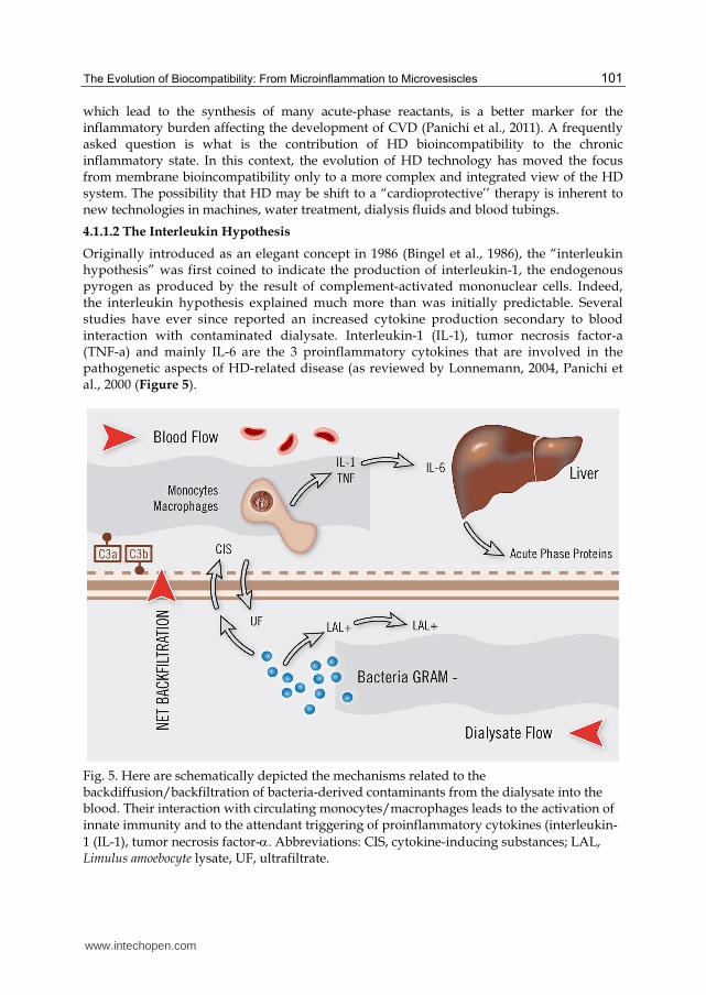
Originally introduced as an elegant concept in 1986 (Bingel et al., 1986), the “interleukin hypothesis” was first coined to indicate the production of interleukin-1, the endogenous pyrogen as produced by the result of complement-activated mononuclear cells. Indeed, the interleukin hypothesis explained much more than was initially predictable. Several studies have ever since reported an increased cytokine production secondary to blood interaction with contaminated dialysate. Interleukin-1 (IL-1), tumor necrosis factor-a (TNF-a) and mainly IL-6 are the 3 proinflammatory cytokines that are involved in the pathogenetic aspects of HD-related disease (as reviewed by Lonnemann, 2004, Panichi et al., 2000 (Figure 5).
Fig. 5. Here are schematically depicted the mechanisms related to the backdiffusion/backfiltration of bacteria-derived contaminants from the dialysate into the blood. Their interaction with circulating monocytes/macrophages leads to the activation of innate immunity and to the attendant triggering of proinflammatory cytokines (interleukin-
1 (IL-1), tumor necrosis factor-. Abbreviations: CIS, cytokine-inducing substances; LAL, Limulus amoebocyte lysate, UF, ultrafiltrate.
www.intechopen.com
Progress in Hemodialysis – From Emergent Biotechnology to Clinical Practice
102
The proposed mechanisms include blood interaction with endotoxins from the
contaminated dialysate through HD membranes. A large number of studies have greatly
contributed to increasing our knowledge in the mechanisms of endotoxin transfer across the
membrane In fact, when using high permeability membranes, backfiltration and
backdiffusion occur and have ebeen extensively described (Fiore & Ronco, 2007, Ronco,
2007). Thus, the transmembrane passage of endotoxins or other cytokine stimulating
substances (CIS) occurs during HD (Schindler et al., 2004, Tetta et al., 2006). The reduction of
backfiltration of standard dialysate may reduce the plasma concentration of IL-1ra, a
sensitive indicator of inflammation in HD patients (Dinarello personal communication, 2004),
and IL-1 (Panichi et al., 1998). Studies on large groups of patients have shown that high-
volume exchange HDF, a treatment in which dialysate backfiltration is minimal, if any, is
associated with significantly lower CRP plasma values (Panichi et. 1998). Comparing in a
double cross-over study patients treated with high-flux and on line HDF using ultrapure
dialysate and infusate, it was shown that a significant reduction of pro-inflammatory
CD14+/CD16+ mononuclear subset (Carracedo et al., 2006) occurs in on line HDF. These
studies emphasize that the convective component has an additional anti-inflammatory
effects (Ramirez et al., 2007).
The new technology of pyrogen-adsorbing, non-complement activating, high-permeability
synthetic membrane and dedicated machines (Tetta et al., 2011), as well as the awareness of
the deleterious effects derived from contamination of dialysis fluids has reduced the clinical
impact to a periodic microinflammatory stimulus. Undoubtedly, the availability of monitors
for on-line HDF and its increased popularity have spurred more restrictive measures on
safety issues and monitoring. Water quality is a mandatory issue. The safety of online HDF
has been shown repeatedly in several monocenter (Canaud et al., 1998, Pizzarelli et al., 1998
and multicenter studies (Canaud et al., 2001,Vaslaki et al., 2000).
Nowadays, the philosophy of ‘‘ultrapure dialysate’’is in common practice (Kessler et
al., 2002). The clinical, consolidated experience on line HDF warrants well-defined
procedures and leaves no space for ‘‘experiments’’ in what is now routine (Canaud et al,
2011). The ‘‘hemocompatibility network’’ should eventually prevent the periodic
microinflammation induction through the implementation of rigid protocols of disinfection
and maintenance of water-treatment systems and HD monitors (Cappelli et al., 2006; Kessler
et al. 2002).
5. Microvesicles: their nature, release and pathophysiological relevance
A chronic inflammatory state has been widely documented since the early stages of CKD
and becomes more pronounced in those with CKD stage V undergoing HD. Oxidant stress
(Wratten et al., 2000, Morena et al. 2011), endothelial dysfunction (Recio-Mayoral et al.,
2011), high circulating cytokine-producing monocyte subpopulation (Ramirez et al., 2006),
reduced number and/or impaired function of endothelial progenitor cells (Krenning et al.,
2009), are today considered as hallmarks of vascular damage and defective repair. Uremia
also causes telomere shortening and premature cellular senescence of immunocompetent
cells (Jimenez et al, 2005). In recent years, increasing attention has been drawn by the
awareness of the pathophysiologic role of small, circular membrane fragments named as
Microvesiscles (MVs) (Ratajczak et al., 2006) (Figure 6).
www.intechopen.com

The Evolution of Biocompatibility: From Microinflammation to Microvesiscles
103
Fig. 6.
For long time MVs were considered to be inert cellular debris. The frequently observed vesicles by electron microscopy in the interstitial space of tissues or in blood were considered as the consequence of cell damage or the result of dynamic plasma membrane turnover (Siekevitz et al., 1972). As the vesicle population detectable both in vitro and in vivo is a mixed population of exosomes and shedding vesicles, we will refer to them collectively as MVs. Released MVs may remain in the extracellular space in proximity of the place of origin or may enter into the biological fluids reaching distant sites. This may explain the presence of MVs in plasma, urine, milk and cerebrospinal fluid. The bulk of MVs present in the circulation is derived from platelets (George, 1982), and in less extent from other blood cells and endothelial cells (Martinez et al., 2005). The MVs derived from platelets are also designed as microparticles while those derived from polymorphonuclear leukocytes are also named ectosomes (Hess et al., 1999). Finally, MVs released during morphogenesis of multicellular organisms are indicated as argosomes (Greco et al., 2001). Besides normal cells, also tumor cells may release MVs and in patients suffering for neoplastic diseases tumor-derived MVs may be detected within the biological fluids (Kim et al, 2003, Iero et al., 2008). Therefore, MVs are an assorted population, differing in cellular origin, number, size and antigenic composition (Diamant et al., 2004) shed by various cell types in physiological and pathological conditions. The release of MVs may be constitutive or consequent to cell activation by soluble agonists, by physical or chemical stress such as the oxidative stress and hypoxia, and by shear stress (Ratajczak et al., 2006). Exosomes have an endosome origin and are a rather homogenous population with a size ranging from 30 to 120nm (7). They are stored as intraluminal vesicles within multivesicular bodies of the late-endosome and are released when these multivesicular bodies fuse with the cell membrane. Our knowledge on
www.intechopen.com

Progress in Hemodialysis – From Emergent Biotechnology to Clinical Practice
104
the mechanism of assembly and sorting of the exosomes is only partial, due to the fact that a common sorting signal for all cell types has not so far been identified (Johnstone et al., 2006).
Shedding vesicles are usually larger than exosomes with size ranging from 100nm to 1m. Formation of shedding vesicles takes place from the budding of small cytoplasmic protrusions followed by their detachment from the cell surface. This process is dependent on calcium influx, calpain and cytoskeleton reorganization.
5.1 MV biological activities
It is now recognized that MVs are an integral part of the intercellular microenvironment and
may act as regulators of cell-to-cell communication. This concept is based on the observation
that MVs released from a given cell type may interact through specific receptor-ligands with
other cells leading to target cell stimulation directly or by transferring surface receptors
(Janowska-Wieczorek et al., 2001, Morel et al., 2004). This interaction may either be limited
to a receptor-mediated binding to the surface of target cells forming a platform for assembly
of multimolecular complexes or leading to cell signaling, either be followed by
internalization as result of direct fusion or endocytic uptake by target cells (Cocucci et al.,
2008). Once internalized, MVs can fuse their membranes with those of endosomes, thus
leading to a horizontal transfer of their content in the cytosol of target cells. Alternatively,
they may remain segregated within endosomes and be transferred to lysosomes or
dismissed by the cells following the fusion with the plasmamembrane, thus leading to a
process of transcytosis. It was proposed that MV-mediated cell-to-cell communication
emerged very early during evolution as a template for the development of further more
refined mechanisms of cell communication (Ratajczak et al., 2006). MVs may influence the
behavior of target cells in multiple ways.
5.1.1 MVs may act as signaling complexes by direct stimulation of target cells (Ratajczak et al., 2006, Cocucci et al., 2008). MVs derived from platelets, for instance, play an important role in coagulation as their phosphatidylserine-enriched membranes provide a surface for assembly of clotting factors (Zwaal et al., 2004). After activation, platelets shed MVs coated with tissue factor which may interact with macrophages, neutrophils and other platelets by ligation with molecules expressed on the surface of these cells such as P-selectin (Polgar et al., 2005). On the other hand, MVs released from neutrophils express activated Mac-1 able to induce platelet activation (Andrews & Berndt, 2004). Moreover, platelet-derived MVs, besides coagulation, trigger various cell responses as they activate endothelial cells (Barry et al., 1997), polymorphonuclear neutrophils (Miyhamoto et al., 1988) and monocytes (Barry et al., 1999). .
5.1.2 MVs may act by transferring receptors between cells. The transferring of receptors between cells is supported by the observation that bystander B cells rapidly acquire antigen receptors from activated B cells by a membrane transfer (Quah et al., 2008).
5.1.3 MVs may deliver proteins within the target cells. An example of this mechanism is the recently reported MV-mediated transfer of a cell death message via encapsulated caspase-1 (Sarkar et al., 2009). It has been found that endotoxin stimulated monocytes induce the cell death of vascular smooth muscle cells by releasing MVs containing caspase-1. This trans-cellular apoptosis induction pathway depends on the function of the delivered caspase-1 within the target cells. It has been also suggested that MVs may contribute to dissemination of certain infective agents, such as HIV or prions (Facler & Peterlin, 2000, Fevrier et al., 2004).
www.intechopen.com
The Evolution of Biocompatibility: From Microinflammation to Microvesiscles
105
5.1.4 MVs may mediate a horizontal transfer of genetic information. The occurrence of epigenetic changes has been frequently reported in co-culture conditions. An explanation of this phenomenon is the transfer of genetic information between cells. We demonstrated that MVs derived from human endothelial progenitors (EPC) can also act as a vehicle for mRNA transport among cells (Deregibus et al., 2007). MVs generated
from EPC were incorporated in normal endothelial cells by interaction with 4- and 1-integrins expressed on their surface and activated an angiogenic program. Besides mRNA, MVs may transfer microRNAs (miRNA) to target cells (Yuan et al., 2009). Since miRNAs are naturally occurring regulators of protein translation, this observation opens the possibility that stem cells can alter the expression of genes in neighbouring cells by transferring microRNAs contained in MVs. We recently characterized miRNA shuttled by MVs released by human adult mesenchymal stem cells (MSCs) (Collino et al., 2010). Hierarchical clustering and similarity analysis of microRNAs showed that microRNA compartmentalization and secretion by MVs are both highly regulated processes.
5.2 Microvesicles in CKD
The biologic role of MVs and their implication in pathophysiology depends on the several
factors namely the cell of origin, their phenotype, the genetic material (mRNA and
microRNA) and the target cells. In CKD, several studies have accrued evidence that MVs or
MPs could participate to the vascular damage and the evolution of the atherosclerotic lesion.
5.2.1 Circulating platelet-derived microparticles (PMPs) with procoagulant activity are
considered a potential cause of thrombosis in uremic patients undergoing HD (Ando et
al., 2002). Elevated counts of circulating PMPs have been reported in association with
thrombotic disorders, such as cerebrovascular accidents (Katopodis et al., 1997),
unstable angina (Katopodis et al., 1997), and acute myocardial infarction (Gawaz et al.,
1996). In addition, PMPs that adhered to vascular endothelium and leukocytes activate
such cells and transport their chemical mediators to those cells, potentially leading to
the development of thrombosis and atherosclerosis (Mallat et al., 1999, Barry et al.,
1997).
5.2.2 Endothelial MVs (EMVs) - Treatment modalities that reduce the inflammatory
potential of the cells originating MVs have interestingly been correlated with a
decreased number of endothelial microparticles (Carracedo et al., 2005, Ramirez et al.,
2005). Circulating EMPs have recently been reported to correlate with impaired
vascular function in HD patients (Faure et al., 2006). A recent study showed an increase
in the percentage of CD14+CD16+ monocytes in CKD-NonD and HD patients. In PD
patients, regardless of RRF, the percentage of CD14+CD16+ was similar to controls
(Merino et al., 2010). It is interesting to note that HD patients displayed significantly
higher apoptotic EMPs and VEGF levels than the two PD and CKD-non dialyszed
groups. In contrast, there were no differences between CKD-NonD and PD groups. In
CKD-non dialyszed and HD patients, the percentage of CD14+CD16+ was correlated
with endothelial damage. It appears that PD, compared with HD, reduces but does not
fully prevent the endothelial damage induced by uremia, in spite of presenting a
microinflammatory status similar to that of the controls. The role of EMVs is still to be
elucidated in the complex unbalance observed in CKD patients between circulating
endothelial cells and endothelial progenitor cells.
www.intechopen.com

Progress in Hemodialysis – From Emergent Biotechnology to Clinical Practice
106
5.2.3 MVs in treatment modalities Preliminary studies in our laboratory have shown an interesting trend in the reduction of total MVs in a cross-over clinical study when patients shifted from high-flux HD to on-line HDF (Figure 7).
Fig. 7. Total MVs count in patients on maintenance HD. In a cross-over design, 8 patients were started on bicarbonate HD (black columns) and 8 patients on on-line HDF (grey columns). MVs were counted by cytofluorimetry. MVs were also characterized (data not shown) by the following specific markers: CD62P, CD41, CD42, CD31, for platelets; CD45, for leukocytes; CD31, CD146, CD144 for the endothelium: CD235 and CD242 (ICAM 4), for erythrocytes.
More studies are needed to better assess the relevance of these observations and to better
characterize the type and biological effects of the MVs. It is still to be fully elucidated
whether MVs are a consequence or a cause of disease. Increasing evidence for their
pathophysiologic role in other human diseases such as sepsis and tumors (Camussi et al.,
2011) is rapidly accruing. Many points require further investigation. i. The stimuli and the
molecular pathways that regulate the assembly within MVs of the biological active
molecules that they shuttle. ii. The stimuli that trigger their release. iii. The surface receptors
that may confer selective specificity. iv. The full diagnostic potential of MVs in different
pathological conditions. v. The strategy to inhibit formation or to remove from circulation
potentially harmful MVs. The recognition of MVs has opened a new era and new
perspectives of investigation also in biocompatibility of extracorporeal treatments.
6. Conclusions
The outlook of more biocompatible and physiological dialysis is today confronted with a
older and sicker population in need of maintenance HD. The knowledge of biological
mechanisms operating at the system level will be approached with the help of improved
technologies hopefully able to reduce the deleterious effect of the repetitive contact with a
foreign surface and to insure optimal performances for the elimination of small and middle
molecule solutes. Advances in dialyzer membranes and geometries, as well as blood tubings
www.intechopen.com
The Evolution of Biocompatibility: From Microinflammation to Microvesiscles
107
together with new concepts in machine technology have already shown their great potential
to improve survival and cardiovascular stability.
7. Conflict of interest
Ciro Tetta and Emanuele Gatti are full-time employees of Fresenius Medical Care.
8. Acknowledgement
The authors thank Dr Sudhir Bowry for critically reviewing the manuscript.
9. References
Adam A, Cugno M, Molinaro G, Perez M, Lepage Y, & Agostoni A. (2002). Aminopeptidase P in individuals with a history of angio-oedema on ACE inhibitors. Lancet. 359(9323):2088-9.
Ando M, Iwata A, Ozeki Y, Tsuchiya K, Akiba T, & Nihei H (2002). Circulating platelet-derived microparticles with procoagulant activity may be a potential cause of thrombosis in uremic patients Kidney Int, 62:1757–1763
Arenas MD, Niveiro E, Moledous A, Gil MT, Albiach B, & Carretón MA (2006) .Fatal acute systemic hypersensitivity reaction during haemodialysis. Nephrol Dial Transplant. 21(10):2966-70
Barret BJ. (2002). Reducing the burden of cardiovascular disease in patients on dialysis. Dial Transplant 31: 155–163
Barry OP, Pratico D, Lawson JA, & FitzGerald GA (1997). Transcellular activation of platelets and endothelial cells by bioactive lipids in platelet microparticles. J Clin Invest 99: 2118-2127.
Barry OP, Kazanietz MG, Praticò D, & Fitzgerald GA(1999). Arachidonic acid in platelet microparticles up-regulates cyclooxygenase-2-dependent prostaglandin formation via a protein kinase C/mitogen-activated protein kinase-dependent pathway. J Biol Chem 274: 7545-7556.
Bhatt DL, & Topol EJ. (2002). Need to test the arterial Inflammation hypothesis. Circulation 106:136–140.
Bingel M, Lonnemann G, Shaldon S, Koch KM, & Dinarello CA. (1986). Human interleukin-1 production during hemodialysis. Nephron. 1986;43(3):161-3
Bradbury BD, Critchlow CV, Weir MR Stewart R, Krishnan M, & Hakim RH (2009). Impact of elevated C-reactive protein levels on Erythropoiesis-stimulating agent (ESA) dose and responsiveness in naemodialysis patients. Nephrol Dial Transplant 24: 919-925.
Camussi G, Deregibus MC, Bruno S, Grange C, Fonsato V, & Tetta C (2011). Exosome / microvesicle - mediated epigenetic reprogramming of cells Am J Cancer Res 1(1):98-110
Canaud B, Bosc JY, Leray H, Stec F, Argiles A, Leblanc M, & Mion C.(1998). On line hemodiafiltration: State of the art. Nephrol Dial Transplant. 5:3–11
Canaud B, Wizemann V, Pizzarelli F, Greenwood R, Schultze G, Weber C, & Falkenhagen D (2001). Cellular interleukin- 1 receptor antagonist production in patients receiving on-line haemodiafiltration therapy. Nephrol Dial Transplant. 16:2181–2187.
Canaud B, Chenine L, Renaud S, & Leray H. (2011). Optimal therapeutic conditions for online hemodiafiltration. Contrib Nephrol. 168:28-38.
www.intechopen.com

Progress in Hemodialysis – From Emergent Biotechnology to Clinical Practice
108
Cappelli G, Riccardi M, Perrone S, Bondi M, Ligabue G, & Albertazzi A (2006). Water treatment and monitor disinfection. Hemodialysis Int. 10, Suppl 1: S13–S18.
Cocucci E, Racchetti G, & Meldolesi J (2008). Shedding microvesicles: artefacts no more. Trends Cell Biol 19: 43-51.
Carracedo J, Ramirez R, Soriano S, Alvarez de Lara MA, Rodriguez M, Martin-Malo A, & Aljama P. (2005). Monocytes from dialysis patients exhibit characteristics of senescent cells: does it really mean inflammation? Contrib Nephrol. 149:208-18.
Carracedo J, Merino A, Nogueras S, Carretero D, Berdud I, Ramírez R, Tetta C, Rodríguez M, Martín-Malo A, & Aljama P (2006). On-line hemodiafiltration reduces the proinflammatory CD14+CD16+ monocyte-derived dendritic cells: A prospective, crossover study. J Am Soc Nephrol. 17(8):2315-21.
Claure-Del Granado R, Macedo E, Chertow GM, Soroko S, Himmelfarb J, Ikizler TA, Paganini EP, & Metha RL (2010).. Effulent volume in continuous renal replacement therapy overestimates the delived dose of dialysis. Clin J Am Soc Nephrol epub november 29,
Collino F, Deregibus MC, Bruno S, Sterpone L, Aghemo G, Viltono L, Tetta C, & Camussi G (2010): Microvesicles derived from adult human bone marrow and tissue specific mesenchymal stem cells shuttle selected pattern of miRNAs. PLoS One 5: e11803.
Craddock PR, Fehr J, Dalmasso AP, Brighan KL, & Jacob HS. (1977) Hemodialysis leukopenia. Pulmonary vascular leukostasis resulting from complement activation by dialyzer cellophane membranes. J Clin Invest. 59(5):879-88.
Culleton BF, Larson MG, Wilson PW, Evans JC, Parfrey PS, & Levy D. (1999). Cardiovascular disease and mortality in a community- based cohort with mild renal insufficiency. Kidney Int 56: 2214–2219
Deregibus MC, Cantaluppi V, Calogero R, Lo Iacono M, Tetta C, Biancone L, & Bruno S (2007). Endothelial progenitor cell derived microvesicles activate an angiogenic program in endothelial cells by a horizontal transfer of mRNA. Blood. 110: 2440-2448.
Diamant M,, Tushuizen ME, Sturk A, & Nieuwland R (2004). Cellular microparticles: new players in the field of vascular disease? Eur. J. Clin. Invest. 34: 392-401.
Donadio E, Piccolomini F, Dimuccio V, Felicioli A, Balestreri E, Cianti R, Armini A, Bini L, Felicioli R, & Donadio C (2009). Serum albumin fragmentation in end-stage renal disease patients: a pilot study. Clin Chem Lab Med. 47(11):1373-9.
Kaysen GA (2002). Role of inflammation and its treatment in ESRD patients. Blood Purif. 20:70–80
Ebo DG, Bosmans JL, Counttenye MM, & Stevens WJ. (2006) Haemodialysis–associated anaphylactic and anaphylactoid reactions. Allergy 61:211–220.
Facler OT, & Peterlin BM. Endocytic entry of HIV-1 (2000). Curr Biol 10: 1005-1008. Faure V, Dou L, Sabatier F, Cerini C, Sampol J, Berland Y, Brunet P, & Dignat-George F
(2006). Elevation of circulating endothelial microparticles in patients with chronic renal failure. J Thromb Haemost. 4(3):566-73. Epub 2005 Dec 23.
Fevrier B, Vilette D, Archer F, Loew D, Faigle W, Vidal M, Laude H, & Raposo G. (2004). Cells release prions in association with exosomes. Proc. Natl Acad Sci USA 101: 9683-9688.
Fiore GB, & Ronco C. (2007) Principles and practice of internal hemodiafiltration. Contrib Nephrol. 158:177-84.
Fischer KG. (2007). Essentials of anticoagulation in hemodialysis. Hemodial Int. 11(2):178-89. Foley RN, Parfrey PS, & Sarnak MJ (1998). Clinical epidemiology of cardiovascular disease
in chronic renal failure. Am J Kidney Dis. 32(Suppl 5):S112–S119.
www.intechopen.com
The Evolution of Biocompatibility: From Microinflammation to Microvesiscles
109
Foley RN (2004). Cardiac disease in chronic uremia: can it explain the reverse epidemiology of hypertension and survival in dialysis patients? Semin Dial 17: 275–278
Hess C, Sadallah S, Hefti A, Landmann R, & Schifferli JA (1999). Ectosomes released by human neutrophils are specialized functional units. J Immunol 163: 4564-4573.
Gawaz M, Neumann JF, Ott I, Schliessler A, & Schoemig A (1996). Platelet function in acute myocardial infarction treated with direct angioplasty. Circulation.3493:229–237.
George JN, Thoi LL, McManus LM, & Reinmann TA (1982). Isolation of human platelet membrane microparticles from plasma and serum. Blood 60:834-840.
Go AS, Chertow GM, Fan D, McCulloch CE, & Hsu CY (2004). Chronic kidney disease and the risks of death, cardiovascular events, and hospitalization. N Engl J Med 351: 1296–1305
Greco V, Hannus M, & Eaton S (2001). Argosomes: a potential vehicle for the spread of morphogens through epithelia. Cell 106: 633-645.
Katopodis JN, Kolodny L, Jy W, Horstman LL, Dearchena EJ, Tao JG, Haynes DH, & Ahn Ys (1997): Platelet microparticles and calcium hemostasis in acute coronary ischemias. Am J Hematol 33;54:95–101,
Kessler M on behalf of the EBPG Working Group (2002). Section IV. Dialysis fluid purity. European Best Practice Guidelines for Haemodialysis (Part 1). European Dialysis and Transplant Association. Nephrol Dial Transplant 17 Suppl 7:45.-54
Kim HK, Song KS, Park YS, Kang YH, Lee YJ, Lee KR, Kim HK, Ryu KW, Bae JM, & Kim S. (2003). Elevated levels of circulating platelet microparticles, VEGF, IL-6 and RANTES in patients with gastric cancer: possible role of a metastasis predictor. Eur J Cancer 39: 184-91.
Krenning G, Dankers PY, Drouven JW, Waanders F, Franssen CF, van Luyn MJ, Harmsen MC, & Popa ER. (2009). Endothelial progenitor cell dysfunction in patients with progressive chronic kidney disease. Am J Physiol Renal Physiol. 296(6):F1314-22.
Krieter DH, Lemke HD, & Wanner C (2008). A new synthetic dialyzer with advanced permselectivity for enhanced low-molecular weight protein removal. Artif Organs. 32(7):547-54.
Iero M, Valenti R, Huber V, Filipazi P, Parmiani G, Fais S, & Rivoltini L. (2008). Tumour-released exosomes and their implications in cancer immunity. Cell Death Differ 15: 80-88.
Goodkin DA, Bragg-Gresham JL, Koenig KG, Woolfe RA, Akiba T, Andreucci VE, Saito A, Rayner HC, Kurokawa K, Port FK, Held PJ, & Young EW. (2003). Association of comorbid conditions and mortality in hemodialysis patients in Europe, Japan, and the United States; the Dialysis Outcomes and Practice Patterns Study (DOPPS). J Am Soc Nephrol 14:3270–3277
Janowska-Wieczorek A, Majka M, Kijowski J, Baj-Krzyworzeka M, Reca R, Turner AR, Ratajczak J, Emerson SG, Kowalska MA, & Ratajczak MZ. (2001) Platelet-derived microparticles bind to hematopoietic progenitor cells and enhance their engraftment. Blood 98: 3143-3149
Jimenez R, Carracedo J, Santamaría R, Soriano S, Madueño JA, Ramírez R, Rodríguez M, Martín-Malo A, & Aljama P. (2005). Replicative senescence in patients with chronic kidney failure. Kidney Int .Suppl. 99:S11-5.
Johansson SG, Hourihane JO, Bousquet J, Bruijnzeel-Koomen C, Dreborg S, Haahtela T, Kowalski ML, Mygind N, Ring J, van Cauwenberge P, van Hage-Hamsten M, & Wüthrich B. (2001) EAACI (the European Academy of Allergology and Cinical Immunology) nomenclature task force. A revised nomenclature for allergy. An EAACI position statement from the EAACI nomenclature task force. Allergy 56:813–824.
www.intechopen.com

Progress in Hemodialysis – From Emergent Biotechnology to Clinical Practice
110
Johnstone RM. (2006). Exosomes biological significance: A concise review. Blood Cells Mol Dis. 36: 315-321.
Lagrand WK, Visser CA, Hermens WT, Niesssen HW, Verheught FW, Wolbink GJ, & Hack CE. (1999). C-reactive protein as a cardiovascular risk: More than an epiphenomenon? Circulation 100:96–102.
Libby P, Ridker PM, & Maseri A. (2002). Inflammation and atherosclerosis. Circulation. 3:187–197.
Liuzzo G, Biasucci LM, Gallimore JR, Grillo RL, Rebuzzi AG, Pepys MB, & Maseri A. (1994) . The prognostic value of C-reactive protein and serum amyloid a protein in severe unstable angina. New Engl J Med. 331:417–424.
Lonnemann G. (2004) When good water goes bad: How it happens, clinical consequences and possibile solutions. Blood Purif. 22:124–129.
Morel O, Toti F, Hugel B, & Freyssinet JM (2004) Cellular microparticles: a disseminated storage pool of bioactive vascular effectors. Curr Opin Hematol. 11: 156-164.
Mallat Z, Hugel B, Ohan J, Leseche G, Freyssinet JM, & Tedqui A. (1999) Shed membrane microparticles with procoagulant potential in human atherosclerotic plaques. A role for apoptosis in plaque thrombogenicity. Circulation 99:348–353.
Martinez MC, Tesse A, Zobairi F, & Andriantsihohaina R. (2005). Shed membrane microparticles from circulating and vascular cells in regulating vascular function. Am J Physiol Hearth Circ Physiol 288: H1004-H1009.
Merino A, Portolés J, Selgas R, Ojeda R, Buendia P, Ocaña J, Bajo MA, del Peso G, Carracedo J, Ramírez R, Martín-Malo A, & Aljama P. (2010) Effect of different dialysis modalities on microinflammatory status and endothelial damage. Clin J Am Soc Nephrol. 5(2):227-34.
Meziani F, Delabranche X, Asfar P, & Toti F. (2010). Bench-to-bedside review: Circulating microparticles - a new player in sepsis? Critical Care 14:236-244.
Miyamoto S, Kowalska MA, Marcinkiewicz C, Marcinkiewicz MM, Mosser D, Edmunds LH, & Niewiaroswski S. (1998) Interaction of leukocytes with platelet microparticles derived from outdated platelet concentrates. Thromb Haemost 80: 982-988.
Morena M, Patrier L, Jaussent I, Bargnoux AS, Dupuy AM, Badiou S, Leray-Moragues H, Klouche K, Canaud B, & Cristol JP (2011). Reduced glomerular filtration rate, inflammation and HDL cholesterol as main determinants of superoxide production in non-dialysis chronic kidney disease patients. Free Radic Res. 45(6):735-45.
Nicholls AJ. (1987) Hypersensitivity to haemodialysis: the United Kingdom experience. Artif Organs 11:87–89.
Owen HG, Brecher ME (1994). Atypical reactions associated with use of angiotensin-converting enzyme inhibitors and apheresis. Transfusion 94; 891–894,
Panichi V, De Pietro S, Andreini B, Migliori M, Tessore V, Taccola D, Rindi P, Palla R, & Tetta C. (1998). Cytokine production in hemodiafiltration: A multicentre study. Nephrol Dial Transplant. 13:1452–1459.
Panichi V, Migliori M, De Pietro S, Taccola D, Andreini B, Metelli MR, Giovannini L, & Palla R. (2000). The link of biocompatibility to cytokine production. Kidney Int. 59, Suppl 76: 96–103.
Panichi V, Tetta C, Rindi P, Palla R, & Lonnemann G. Plasma C-reactive protein is linked to ackfiltration associated interleukin 6 production. ASAIO J. 1998; 744: M415–M417.
Panichi V, Maggiore U, Taccola D, Migliori M, Rizza GM, Consani C, Bertini A, Sposini-Garcia R, Rindi P, Palla R, & Tetta C. (2004). Interleukin-6 is a stronger predictor of total and cardiovascular mortality than C-reactive protein in hemodialysis patients. Nephrol Dial Transplant 19:1154-60.
www.intechopen.com

The Evolution of Biocompatibility: From Microinflammation to Microvesiscles
111
Panichi V, Rizza GM, Paoletti S, Bigazzi R, Aloisi M, Barsotti G, Rindi P, Donati G, Antonelli A, Panicucci E, Tripepi G, Tetta C, & Palla R; RISCAVID Study Group (2008) Chronic inflammation and mortality in haemodialysis: effect of different renal replacement therapies. Results from the RISCAVID study. Nephrol Dial Transplant. 23(7):2337-43.
Panichi V, Rosati A, Bigazzi R, Paoletti S, Mantuano E, Beati S, Marchetti V, Bernabini G, Grazi G, Rizza GM, Migliori M, Giusti R, Lippi A, Casani A, Barsotti G, & Tetta C; on behalf of the RISCAVID Study. (2011). Group.Anaemia and resistance to erythropoiesis-stimulating agents as prognostic factors in haemodialysis patients: results from the RISCAVID study. Nephrol Dial Transplant. Feb 16. [Epub ahead of print]
Pascual M, Schifferli JA. (1993). Adsorption of complement factor D by polyacrylonitrile dialysis membranes. Kidney Int. 43(4):903-11.
* Parfey PS, & Foley RN. (1999) The clinical epidemiology of cardiac disease in chronic renal failure. J Am Soc Nephrol. 10:1606–1615.
Pizzarelli F, Cerrai T, Dattolo P, Tetta C, & Maggiore Q. (1998) Convective treatments with on line production of replacement fluid: A clinical experience lasting 6 years. Nephrol Dial Transplant. 13:363–369.
Polgar J, Matuskova J, & Wagner DD. (2005) The P-selectin, tissue factor, coagulation triad. J Thromb Haemost; 3: 1590-1596.
* Poothullil J, Shimizu A, Day RP, & Dolovich J. (1975) Anaphylaxis from the product(s) of ethylene oxide gas. Ann Intern Med 82:58–60.
Quah BJ, Barlow VP, McPhun V, Matthaei KI, Hulett MD, & Parish CR. (2008). Bystander B cells rapidly acquire antigen receptors from activated B cells by membrane transfer. Proc Natl Acad Sci USA 105: 4259-4264.
Ramirez R, Carracedo J, Berdud I, Carretero D, Merino A, Rodríguez M, Tetta C, Martín-Malo A, & Aljama P. (2006). Microinflammation in hemodialysis is related to a preactivated subset of monocytes. Hemodial Int., Suppl 1:S24-7.
Ramirez R, Carracedo J, Merino A, Nogueras S, Alvarez-Lara MA, Rodríguez M, Martin-Malo A, Tetta C, & Aljama P. (2007) Microinflammation induces endothelial damage in hemodialysis patients: the role of convective transport. Kidney Int. 72(1):108-13.
Ratajczak J, Wysoczynski M, Hayek F, Janowska-Wieczorek A, & Ratajczak MZ. (2006) Membrane-derived microvesicles: important and underappreciated mediators of cell-to-cell communication. Leukemia; 20: 1487-1495.
* Rayner HC, Pisoni RL, Bommer J, Canaud B, Hecking E, Locatelli F, Piera L, Bragg-Gresham JL, Feldman HI, Goodkin DA, Gillespie B, Wolfe RA, Held PJ, & Port FK. (2004). Mortality and hospitalization in haemodialysis patients in five European countries: results from the Dialysis Outcomes and Practice Patterns Study (DOPPS). Nephrol Dial Transplant. 19(1):108-20.
Ramírez R, Carracedo J, Soriano S, Jiménez R, Martín-Malo A, Rodríguez M, Blasco M, & Aljama P. (2005). Stress-induced premature senescence in mononuclear cells from patients on long-term hemodialysis. Am J Kidney Dis. 45(2):353-9.
Recio-Mayoral A, Beneriee D, Streather C, & Kaski JC. (2011). Endothelial dysfunction, inflammation and atherosclerosis in chronic kidney disease - a cross-sectional study of predialysis, dialysis and kidney-transplantation patients. Atherosclerosis. Feb. 18 [Epub ahead of print].
Ridker PM, Cushman M, Stampfer MJ, Tracy RP, & Hennekens CH.(1997). Inflammation, aspirin and the risk of cardiovascular disease in apparently healthy men. N Engl J Med. 336: 973–979.
Ronco C. (2007) Fluid mechanics and crossfiltration in hollow-fiber hemodialyzers. Contrib Nephrol. 158:34-49.
www.intechopen.com

Progress in Hemodialysis – From Emergent Biotechnology to Clinical Practice
112
Ross R. (1999) Atherosclerosis: An inflammatory disease. N Engl J Med. 340:115–126. Sarkar A, Mitra S, Mehta S, Raices R, & Wewers MD. (2009). Monocyte derived microvesicles
deliver a cell death message via encapsulated caspase-1. PLoS One 4: e7140. Schindler R, Beck W, Deppisch R, Aussieker M, Wilde A, Goehl H, & Frei U. (2004). Short
bacterial DNA fragments: Detection in dialysate and induction of cytokines. J Am Soc Nephrol. 15:3207–3214.
Siekevitz P. (1972) Biological membranes: the dynamics of their organization. Annu Rev Physiol 34: 117-140.
Singh AK, Coyne DW, Shapiro W, Rizkala AR. (2007). Predictors of the response to treatment in anemic haemodialysis patients with high serum ferritin and low transferring saturation. Kidney Int 71: 1163-1171.
Stenvinkel P, Heinburger O, Paultre F, Diczfalusy U, Wang T, Berglund L, & Jogestrand T.. (1999). Strong associations between malnutrition, inflammation and atherosclerosis in chronic renal failure. Kidney Int 55: 1899-1911,
Stenvinkel P. (2001). Malnutrition and chronic inflammation as risk factors for cardiovascular disease in chronic renal failure. Blood Purif. 19:143–151.
Tetta C, Haeffner-Cavaillon N, Navino C, David S, Franceschi C, Mariano F, & Camussi G (1996). The role of platelet-activating actor in the biocompatibility of hemodialysis membranes. Adv Exp Med Biol. 416:243-8.
* Tetta C, David S, Marcelli D, Cogliati P, Formica M, Inguaggiato P, & Panichi V. (2006). Clinical effects of online dialysate and infusion fluids. Hemodial Int 10: S60–S66
Tetta C, Roy T, Gatti E, & Cerutti S. (2011). The rise of hemodialysis machines: new technologies in minimizing cardiovascular complications. Expert Rev. Cardiovasc. Ther. 9(2), 155–164.
Tielemans C, Madhoun P, Lenaers M, Schandene L, Goldman M, Vanherweghem JL. (1990). Anaphylactoid reactions during hemodialysis on AN69 membranes in patients receiving ACE inhibitors. Kidney Int. 38(5):982-4.
US Renal Data System. Excerpts from the USRDS 1997 annual data report. (1997) Am J Kidney Dis. 30:S1–S195.
Vasan RS, Sullivan LM, Roubenoff R, Dinarello CA, Harris T, Benjamin EJ, Sawyer DB, Wilson PW, D’Agostino RB: Framingham Heart Study (2003). Inflammatory markers and risk of heart failure in erderly subjects without prior myocardial infarction: The Framingham Heart Study. Circulation 107:1486–1491.
Vaslaki L, Karatzon A, Voros P, Maior L, Pethoe F, Ladanyi E, Weber C, Mitteregger R, & Falkengagen D. (2000). Can sterile and pyrogen-free on-line substitution fluid be rotuineley delivered? A multicentre study on the microbiological safety of on-line hemodiafiltration. Nephrol Dial Transplant. 15, Suppl 1: 74–78.
Wratten ML, Tetta C, Ursini F, & Sevanian A. (2000). Oxidant stress in hemodialysis: prevention and treatment strategies. Kidney Int. Suppl. 2000, 76: S126-32.
Wright S, Steinwandel U, & Ferrari P. (2010) Citrate anticoagulation during long-term haemodialysis. Nephrology (Carlton). Nov 3. [Epub ahead of print]
Yuan A, Farber EL, Rapoport AL, Tejada D, Deniskin R, Akhmedov NB, & Farber DB. (2009). Transfer of microRNAs by embryonic stem cell microvesicles. PLoS One 4: e4722
Ziouzenkova O, Asatryan L, Akmal M, Tetta C, Wratten ML, Loseto-Wich G, Jürgens G, Heinecke J, & Sevanian A. (1999). Oxidative cross-linking of ApoB100 and hemoglobin results in low density lipoprotein modification in blood. Relevance to atherogenesis caused by hemodialysis. J Biol Chem. 274(27):18916-24.
Zwaal RF, Comfurius P, & Bevers EM, Scott syndrome, a bleeding disorder caused by defective scrambling of membrane phospholipids. Biochim Biophys Act 1636: 119-128.
www.intechopen.com
Progress in Hemodialysis - From Emergent Biotechnology toClinical PracticeEdited by Prof. Angelo Carpi
ISBN 978-953-307-377-4Hard cover, 444 pagesPublisher InTechPublished online 07, November, 2011Published in print edition November, 2011
InTech EuropeUniversity Campus STeP Ri Slavka Krautzeka 83/A 51000 Rijeka, Croatia Phone: +385 (51) 770 447 Fax: +385 (51) 686 166www.intechopen.com
InTech ChinaUnit 405, Office Block, Hotel Equatorial Shanghai No.65, Yan An Road (West), Shanghai, 200040, China
Phone: +86-21-62489820 Fax: +86-21-62489821
Hemodialysis (HD) represents the first successful long-term substitutive therapy with an artificial organ forsevere failure of a vital organ. Because HD was started many decades ago, a book on HD may not appear tobe up-to-date. Indeed, HD covers many basic and clinical aspects and this book reflects the rapid expansion ofnew and controversial aspects either in the biotechnological or in the clinical field. This book revises newtechnologies and therapeutic options to improve dialysis treatment of uremic patients. This book consists ofthree parts: modeling, methods and technique, prognosis and complications.
How to referenceIn order to correctly reference this scholarly work, feel free to copy and paste the following:
Ciro Tetta, Stefano Maffei, Barbara Cisterna, Valentina Fonsato, Giorgio Triolo, Giuseppe Paolo Segoloni,Giovanni Camussi, Maria Chiara Deregibus and Emanuele Gatti (2011). The Evolution of Biocompatibility:From Microinflammation to Microvesiscles, Progress in Hemodialysis - From Emergent Biotechnology toClinical Practice, Prof. Angelo Carpi (Ed.), ISBN: 978-953-307-377-4, InTech, Available from:http://www.intechopen.com/books/progress-in-hemodialysis-from-emergent-biotechnology-to-clinical-practice/the-evolution-of-biocompatibility-from-microinflammation-to-microvesiscles

© 2011 The Author(s). Licensee IntechOpen. This is an open access articledistributed under the terms of the Creative Commons Attribution 3.0License, which permits unrestricted use, distribution, and reproduction inany medium, provided the original work is properly cited.